


Abstract
Background/Objectives: Facial discoid dermatosis is a rare inflammatory dermatosis presenting with round, superficial erythematous lesions located on the face. Diagnosis may be challenging and often requires careful clinicopathological correlation due to overlapping clinical and histopathological features. Skin lesions are typically resistant to a wide range of topical and systemic treatments. From the perspective of personalized medicine, improved phenotyping of rare inflammatory dermatoses may support more precise diagnosis, individualized therapeutic decisions, and non-invasive disease monitoring. This study aimed to characterize facial discoid dermatosis using line-field confocal optical coherence tomography and reflectance confocal microscopy and to discuss its differential diagnosis and therapeutic implications. Methods: We report a case of facial discoid dermatosis in a 35-year-old patient examined with line-field confocal optical coherence tomography and reflectance confocal microscopy. The imaging findings were interpreted in correlation with clinical and histopathological features. A literature review was performed to summarize differential diagnoses, therapeutic perspectives, and the proposed relationship between facial discoid dermatosis and pityriasis rubra pilaris. Results: Non-invasive imaging revealed morphological features consistent with a psoriasiform inflammatory dermatosis and provided additional in vivo information supporting the diagnosis. The literature review showed limited evidence for a direct association between facial discoid dermatosis and pityriasis rubra pilaris, with only isolated reports suggesting possible overlap or progression. Conclusions: Facial discoid dermatosis appears to represent a distinct psoriasiform dermatosis. Line-field confocal optical coherence tomography and reflectance confocal microscopy may contribute to a personalized diagnostic approach by supporting differential diagnosis and potentially guiding individualized monitoring in rare inflammatory facial dermatoses.
1. Introduction
Facial discoid dermatosis (FDD) is a relatively recently described entity, first reported by Ko et al. in 2010 [1]. To date, FDD remains a rarely recognized condition [2,3]. The available literature on FDD is limited to case reports and small case series. Therefore, its true incidence, prevalence, and geographic distribution remain unknown. The pathogenesis has not been established, although the histopathological findings suggest a chronic inflammatory papulosquamous process affecting the face.
Clinically, FDD presents as discrete, discoid, well-demarcated, pink-to-orange papulosquamous facial lesions, which show resistance to topical therapies and usually remain stable over time [1,4]. The lesions measure from a few millimeters to 3.5 cm [1] and favor the cheeks (93%), chin (69%), and forehead (38%) [5]. Itching and annular scale may be present [6]. The age of onset is usually between the second and fourth decades of life [1]. FDD shows a strong female predominance (5:1) [2,5,7,8]. Histopathological features may include hyperkeratosis, focal parakeratosis, acanthosis with psoriasiform hyperplasia, spongiosis, follicular plugging, and involuted sebaceous lobules [1,6,9,10].
Non-invasive skin imaging with line-field confocal optical coherence tomography (LC-OCT) and reflectance confocal microscopy (RCM) enable real-time microscopic assessment of skin lesions without biopsy. This is particularly useful in rare, treatment-refractory facial dermatoses such as FDD, where repeated biopsies of cosmetically sensitive areas may lead to scarring and patient discomfort. By providing in vivo morphological information at the level of individual lesions, these techniques support a personalized approach to diagnosis and longitudinal monitoring in such challenging cases.
2. Methods
2.1. Case
The case described in this report was managed at the Department of Dermatology, Medical University of Warsaw. Dermoscopy was performed with a DermLite DL4 dermatoscope, LC-OCT with the deepLive™ device (DAMAE Medical, Paris, France) in both vertical and horizontal acquisition modes, and RCM with the VivaScope® 1500/3000 (Caliber I.D., Rochester, NY, USA). Histopathological sections were stained with hematoxylin and eosin and reviewed by a board-certified dermatopathologist (J.C.).
2.2. Literature Search and Selection
A narrative literature review was conducted to summarize the current evidence on facial discoid dermatosis, with particular focus on (i) its clinical, dermoscopic, histopathological and in vivo imaging features, (ii) its differential diagnosis, (iii) reported therapeutic strategies, and (iv) the proposed relationship between FDD and pityriasis rubra pilaris (PRP). The following electronic databases were searched from inception to March 2026: PubMed/MEDLINE, Embase, Scopus and Web of Science using the term: “facial discoid dermatosis”. Peer-reviewed publications in English were included. Records were screened by 4 authors (J.Z., L.B., J.N. and K.M.) in two stages: titles and abstracts were first reviewed to identify thematically relevant publications, and full-text assessment was subsequently performed only for those records judged relevant to the scope of the review. Owing to the rarity of FDD, the predominance of single-patient case reports and small case series in the literature, a formal quality appraisal of individual studies was not performed, and evidence was synthesized narratively, consistent with the descriptive aim of this review.
3. Case Presentation
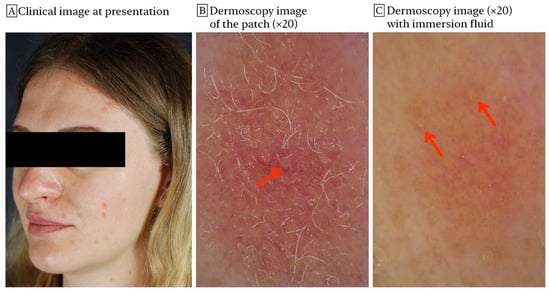
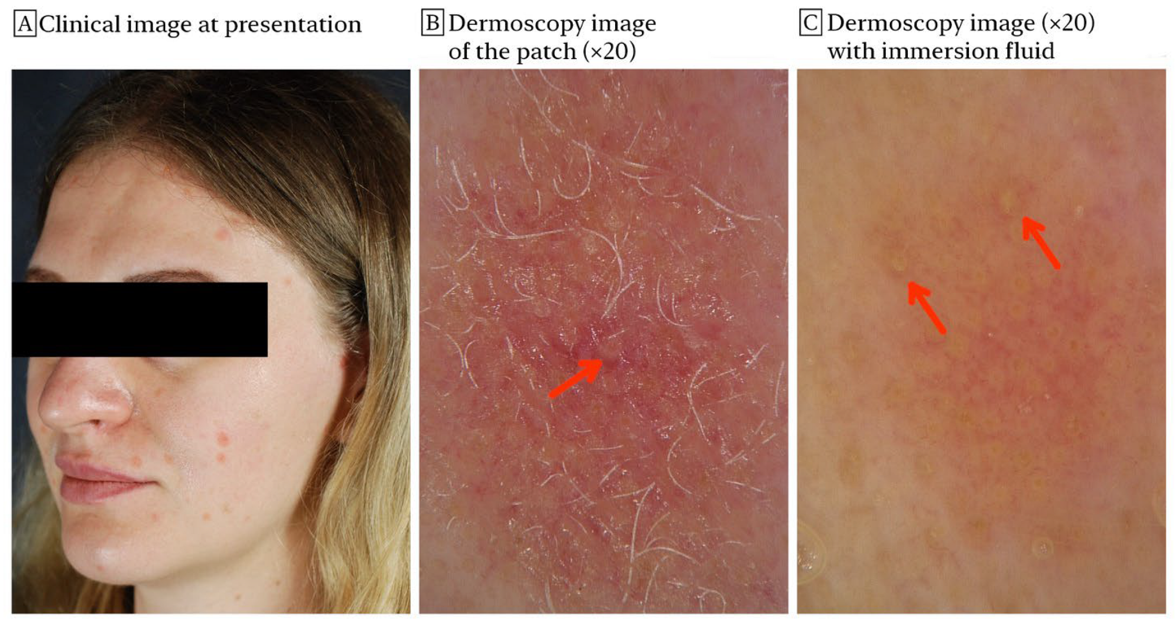
A 35-year-old woman presented with round, erythematous, scaly patches located on the cheeks, forehead, and chin (Figure 1A). The lesions measured up to 1.5 cm in diameter and had slightly progressed over two years. History revealed no inciting factors. At the time of onset, histopathological examination performed outside of our institution demonstrated hyperkeratosis, focal parakeratosis, and mild spongiosis. No conclusive diagnosis was made. Prior to presenting at our center, the patient had received treatment with systemic doxycycline and numerous topical agents including tacrolimus ointment, imiquimod cream, diflucortolone/isoconazole cream and adapalene gel with no improvement. She had no concomitant diseases, allergies and was not receiving any chronic medication.
Figure 1.
(A) Clinical photograph of a 35-year-old patient with facial discoid dermatosis. (B,C) Dermoscopy of skin lesions showed large yellow dots corresponding to enlarged follicular ostia (red arrows).
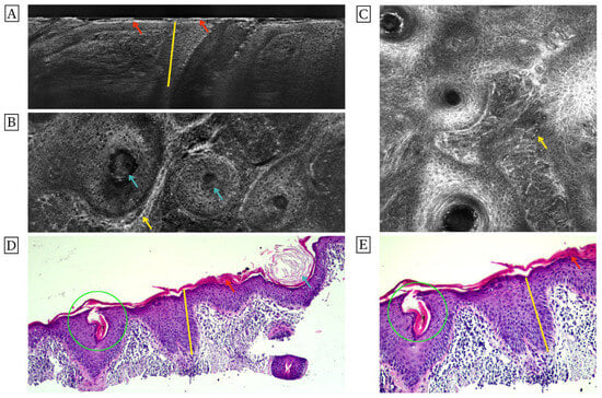
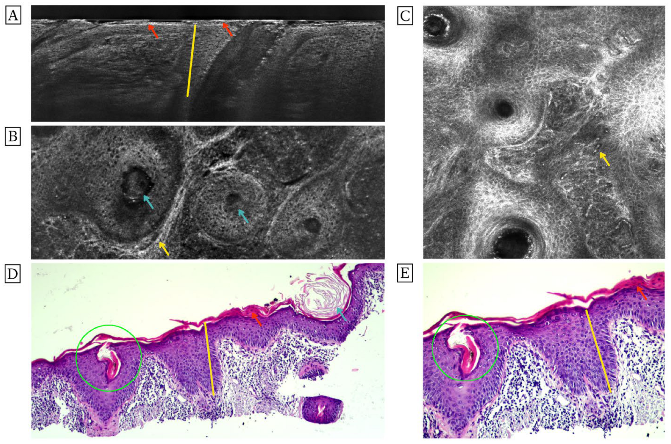
Dermoscopy showed fine scale overlying dilated blood vessels and large yellow dots corresponding to enlarged follicular ostia (Figure 1B), which were better seen after the application of immersion fluid (Figure 1C). We performed line-field confocal optical coherence tomography in two modes of acquisition: vertical and horizontal (Figure 2A,B), and reflectance confocal microscopy (Figure 2C). Both LC-OCT and RCM revealed acanthosis, focal parakeratosis (hyperreflective particles in the keratinocytes in the stratum corneum), areas of fine scale overlying the epidermis (hyperreflective superficial streaks), and plugs in dilated follicular openings (hyperreflective signal in the follicular ostia). In the papillary dermis, hyporeflective linear areas corresponding to dilated blood vessels were seen. They were surrounded by round hyperreflective cells consistent with an inflammatory infiltrate. To confirm the non-invasive imaging findings in histopathology, a shave biopsy was performed (Figure 2D,E). The histopathological features were analogous to those observed in LC-OCT and RCM. The epidermis showed parakeratosis occasionally alternating with orthokeratosis. Histopathology confirmed a predominantly lymphohistiocytic infiltrate. Additionally, Demodex mites were observed within hair follicles and were interpreted as a concomitant finding. Direct and indirect immunofluorescence tests were negative. Laboratory results showed no abnormalities.
Figure 2.
(A,B) Images from line-field confocal optical coherence tomography in vertical and horizontal modes of acquisition and (C), reflectance confocal microscopy. The primary findings involved epidermal acanthosis (yellow vertical lines), focal parakeratosis (red arrows), follicular plugging (blue arrows), and dilated blood vessels in the papillary dermis surrounded by a dense inflammatory infiltrate (yellow arrow). (D,E) Histopathology confirmed those findings and additionally demonstrated features of demodicosis (green circle) (H&E; original magnification ×40 in (D) and ×100 in (E)).
Based on the clinical features, recalcitrant disease course and histological findings, the diagnosis of facial discoid dermatosis was made. The patient was prescribed oral hydroxychloroquine 200 mg twice daily, topical ivermectin cream and tacrolimus ointment. After 3 months, she showed only minor improvement.
4. Discussion
To the best of our knowledge, this is the first report presenting LC-OCT and RCM features of FDD. Both methods enable precise, real-time non-invasive imaging of the epidermis and superficial dermis. RCM is superior to LC-OCT in terms of resolution, but inferior with respect to the depth of analysis [11,12]. Additionally, LC-OCT enables both vertical and horizontal imaging, which yields the possibility of creating a three-dimensional model of the analyzed skin sample. Taken together, these methods can be regarded as complementary tools in lesions occupying not only the epidermis, but also the superficial dermis. Indeed, in FDD, which is a superficial inflammatory process, we showed a close correlation between histopathology and both LC-OCT and RCM findings. This underscores the applicability of these tools in differentiating between FDD and other possible diagnoses, such as psoriasis, pityriasis rubra pilaris (PRP), seborrheic dermatitis, discoid lupus erythematosus, Senear–Usher syndrome, and tinea faciei (Table 1).
Table 1.
Differential diagnoses of facial discoid dermatosis.
4.1. Facial Discoid Dermatosis—A Distinct Psoriasiform Dermatosis
The clinical picture and histology of FDD favor its classification as a psoriasiform dermatosis sharing many features with PRP. Gan et al. [24] reported the development of type II PRP years after the initial diagnosis of FDD and proposed classifying it as a novel type VII PRP, following the types I–V originally distinguished by Griffiths [25] and type VI proposed by Miralles et al. [26] in HIV-infected individuals. However, only one out of eight patients in the described case series showed disease progression to type II PRP [24], and no similar case reports have been published to date. Furthermore, Allegue et al. [9] described a case of unilateral ectropion in the course of FDD involving the lower eyelid, which also prompted them to associate this disease with PRP. This assumption was based on the well-established risk of ectropion occurrence in a subset of patients with facial PRP lesions [27]. The treatment of FDD led to the resolution of ocular symptoms. However, in our opinion, this remote association is insufficient to justify the identification of FDD as a subtype of PRP. Moreover, FDD does not typically display the hallmark features of PRP such as palmoplantar keratoderma or classic “islands of sparing” [28]. The genetic background of FDD is still unknown. Mutations in CARD14, a gene linked to PRP and related papulosquamous eruptions [29], have not been reported in FDD cases; thus, a shared molecular basis cannot be ruled out, and dedicated genetic studies are warranted.
Therefore, we propose that the chronic, localized, treatment-refractory nature of FDD and its distinct demographic and clinical pattern argue for its recognition as an independent entity within the spectrum of psoriasiform dermatoses rather than as a PRP variant.
4.2. Treatment
FDD is resistant to a wide range of conventional treatments, which constitutes one of its most distinctive hallmarks [1]. Phototherapy may be considered, in theory, due to the psoriasiform inflammatory pattern of FDD. However, the available evidence does not support its efficacy. Rahmatulla et al. and Bohdanowicz and DeKoven reported the use of narrowband UVB phototherapy in a total of three patients with FDD, without clinical improvement [4,7]. Topical agents such as corticosteroids, retinoids, antifungals, calcipotriol, calcineurin inhibitors, and imiquimod, as well as other treatment modalities, including oral acitretin, doxycycline, prednisone, methotrexate, hydroxychloroquine, and pulsed-dye laser, have been reported in the literature [1,9,30]. One study [30] reported complete response with topical rapamycin (sirolimus) 0.2% after one-month therapy in a patient recalcitrant to topical treatments (corticosteroids, tacrolimus, pimecrolimus, crisaborole) and systemic therapy with secukinumab and guselkumab.
Bohdanowicz and DeKoven [7] published a paper demonstrating partial resolution of skin lesions after a two-month therapy with calcipotriol/betamethasone ointment combined with low-dose acitretin. Amarnani et al. [31] reported an almost complete remission of FDD with topical clobetasol propionate and calcipotriol for three weeks followed by topical calcipotriol monotherapy for another three weeks. Favorable response to treatment prompted the authors to advocate the use of topical calcipotriol instead of systemic medications and long-term topical steroid therapy to avoid treatment-related adverse events. This is reinforced by the already mentioned case of Allegue et al. [9], who also achieved a remission of FDD lesions following a two-month therapy with daily application of calcipotriol/betamethasone ointment. Nevertheless, maintenance with topical 0.1% tacrolimus twice weekly was necessary to preserve disease remission. Finally, Rypka et al. [32] and Fera et al. [8] reported significant improvement of FDD following treatment with ustekinumab. A similar response to ustekinumab has been reported in CARD14-associated papulosquamous eruptions of the face, which share features of both psoriasis and PRP [29]. Whether ustekinumab works in FDD through the same IL-12/23/Th17 pathway or through a still-unrecognized role of CARD14 needs further genetic studies.
Our patient received numerous topical and systemic treatments with little effect. Partial improvement achieved at our center most probably resulted from the suppression of Demodex spp.-induced inflammation by topical ivermectin. The patient refused to apply the calcipotriol/betamethasone preparation for fear of potential side effects following its application on the face.
From a personalized medicine perspective, LC-OCT and RCM may support a more individualized assessment of rare facial dermatoses such as FDD. These methods provide real-time microscopic information without repeated biopsies, which is particularly important for lesions located on the face. They may help refine the differential diagnosis, guide treatment decisions, and monitor the response to therapy in a non-invasive way.
5. Conclusions
FDD is a rare skin condition typically affecting adult females. It is a disfiguring disease characterized by a chronic course and frequent resistance to conventional treatments. FDD seems to belong to the spectrum of psoriasiform dermatoses, which is supported by its clinical and histological picture. Although progression to PRP has been reported in one case described to date, the classification of FDD as a subtype of PRP is questionable. As a superficial inflammatory process, it can be diagnosed using LC-OCT, RCM, or a combination of both. Based on the available data, it seems that patients could benefit most from the use of topicals combining calcipotriol and betamethasone, as well as from ustekinumab and other biologics targeting the IL-12/23 and IL-17 pathways. This case illustrates how non-invasive imaging may contribute to personalized dermatology by supporting diagnosis and treatment selection in rare, recalcitrant inflammatory dermatoses of the face. Future multicenter, prospective, and molecular studies are needed to validate LC-OCT and RCM findings, standardize diagnostic criteria, and better define targeted therapeutic strategies for patients with FDD.
Author Contributions
Conceptualization, J.Z., L.B. and J.C.; methodology, J.Z. and L.B.; formal analysis, J.Z., L.B. and J.C.; investigation, J.Z., L.B., J.N. and K.M.; writing—original draft preparation, J.Z., L.B. and J.C.; writing—review and editing, J.N., K.M., M.O. and L.R.; visualization, J.Z. and L.B.; supervision, M.O. and L.R. All authors have read and agreed to the published version of the manuscript.
Funding
This research received no external funding.
Institutional Review Board Statement
Our manuscript presents a retrospective case report. This study does not constitute a medical experiment. Ethical review and approval are not applicable.
Informed Consent Statement
Written informed consent has been obtained from the patient for the publication of this paper.
Data Availability Statement
All further inquiries should be directed to the corresponding author.
Conflicts of Interest
The authors declare no conflicts of interest.
Abbreviations
The following abbreviations are used in this manuscript:
| FDD | Facial discoid dermatosis |
| LC-OCT | Line-field confocal optical coherence tomography |
| RCM | Reflectance confocal microscopy |
| PRP | Pityriasis rubra pilaris |
| SLE | Systemic lupus erythematosus |
| PAS | Periodic acid–Schiff |
| H&E | Hematoxylin and eosin |
| IL-17 | Interleukin-17 |
| IL-23 | Interleukin-23 |
| CARD14 | Caspase recruitment domain family member 14 |
| HIV | Human immunodeficiency virus |
| IgG | immunoglobulin G |
| IgM | immunoglobulin M |
| C3 | Complement component 3 |
References
- Ko, C.J.; Heald, P.; Antaya, R.J.; Bolognia, J.L. Facial discoid dermatosis. Int. J. Dermatol. 2010, 49, 189–192. [Google Scholar] [CrossRef] [Scilit] [PubMed]
- Butrón-Bris, B.; Buján, C.; Berenguer-Ruiz, S.; Martos-Cabrera, L.; Rodríguez-Jiménez, P.; Fraga, J.; Eguren, C.; Serrano-Pardo, R.; Iranzo, P.; Mascaró, J.M., Jr.; et al. [Translated article] Facial Discoid Dermatosis. A 13-Case Series. Actas Dermo-Sifiliográficas 2025, 116, T633–T637. [Google Scholar] [CrossRef] [Scilit] [PubMed]
- Rodríguez-Troncoso, M.; De la Torre-Gomar, F.J.; Pegalajar-García, M.D.; Martín-Castro, A.; Ruiz-Villaverde, R. Oral Isotretinoin for Facial Discoid Dermatosis: Effectiveness, Safety, and Clinical Suitability—Case Report of Complete Response and Literature Review. Case Rep. Dermatol. 2025, 17, 362–369. [Google Scholar] [CrossRef] [Scilit] [PubMed]
- Rahmatulla, S.; Batta, K.; Tatnall, F.; Sandhu, D.; Brown, V. Facial discoid dermatosis: A cosmetically disfiguring and challenging condition to treat. Skin Health Dis. 2021, 1, e56. [Google Scholar] [CrossRef] [Scilit] [PubMed]
- Condal, L.; Quer, A.; Ferrándiz, C.; Bielsa, I. Facial Discoid Dermatosis: An Enigmatic Disease. Actas Dermosifiliogr. 2021, 112, 934–935. [Google Scholar] [CrossRef] [Scilit]
- Salman, A.; Tekin, B.; Berenjian, A.; Cinel, L.; Demirkesen, C. Facial discoid dermatosis: A further case of a novel entity. J. Dermatol. 2015, 42, 1132–1133. [Google Scholar] [CrossRef] [Scilit] [PubMed]
- Bohdanowicz, M.; DeKoven, J.G. Improvement in facial discoid dermatosis with calcipotriol/betamethasone ointment and low-dose acitretin. Clin. Exp. Dermatol. 2018, 43, 820–821. [Google Scholar] [CrossRef] [Scilit] [PubMed]
- Fera, C.; Chamaillard, M.; Beylot-Barry, M.; Levavasseur, M. Facial Discoid Dermatosis, a Still Unknown Entity. Acta Derm. Venereol. 2025, 105, adv40911. [Google Scholar] [CrossRef] [Scilit] [PubMed]
- Allegue, F.; Fachal, C.; Iglesias, B.; Zulaica, A. Facial Discoid Dermatosis: A New Variant of Pityriasis Rubra Pilaris? Actas Dermosifiliogr. 2022, 113, 728–731. [Google Scholar] [CrossRef] [Scilit] [PubMed]
- Welborn, M.; Fletcher, D.; Motaparthi, K. Atrophy of sebaceous lobules in facial discoid dermatosis: A link to psoriasis and seborrheic dermatitis? J. Cutan. Pathol. 2022, 49, 320–323. [Google Scholar] [CrossRef] [Scilit]
- Welzel, J.; Schuh, S. Noninvasive diagnosis in dermatology. J. Dtsch. Dermatol. Ges. 2017, 15, 999–1016. [Google Scholar] [CrossRef] [Scilit] [PubMed]
- Ogien, J.; Tavernier, C.; Fischman, S.; Dubois, A. Line-field confocal optical coherence tomography (LC-OCT): Principles and practical use. Ital. J. Dermatol. Venerol. 2023, 158, 171–179. [Google Scholar] [CrossRef] [Scilit] [PubMed]
- Errichetti, E.; Stinco, G. Dermoscopy in General Dermatology: A Practical Overview. Dermatol. Ther. 2016, 6, 471–507. [Google Scholar] [CrossRef] [Scilit]
- Wang, D.; Chong, V.C.; Chong, W.S.; Oon, H.H. A Review on Pityriasis Rubra Pilaris. Am. J. Clin. Dermatol. 2018, 19, 377–390. [Google Scholar] [CrossRef] [Scilit] [PubMed]
- Pietroleonardo, L.; Di Stefani, A.; Campione, E.; Chimenti, S.; Orlandi, A.; Bianchi, L. Confocal reflectance microscopy in pityriasis rubra pilaris. J. Am. Acad. Dermatol. 2013, 68, 689–691. [Google Scholar] [CrossRef] [Scilit] [PubMed]
- Verzì, A.E.; Broggi, G.; Micali, G.; Sorci, F.; Caltabiano, R.; Lacarrubba, F. Line-field confocal optical coherence tomography of psoriasis, eczema and lichen planus: A case series with histopathological correlation. J. Eur. Acad. Dermatol. Venereol. 2022, 36, 1884–1889. [Google Scholar] [PubMed]
- Hoogedoorn, L.; Peppelman, M.; van de Kerkhof, P.C.; van Erp, P.E.; Gerritsen, M.J. The value of in vivo reflectance confocal microscopy in the diagnosis and monitoring of inflammatory and infectious skin diseases: A systematic review. Br. J. Dermatol. 2015, 172, 1222–1248. [Google Scholar] [PubMed]
- Agozzino, M.; Berardesca, E.; Donadio, C.; Franceschini, C.; de Felice, C.M.; Cavallotti, C.; Sperduti, I.; Ardigò, M. Reflectance confocal microscopy features of seborrheic dermatitis for plaque psoriasis differentiation. Dermatology 2014, 229, 215–221. [Google Scholar] [CrossRef] [Scilit] [PubMed]
- Gambichler, T.; Hyun, J.; Moussa, G.; Tomi, N.S.; Boms, S.; Altmeyer, P.; Hoffmann, K.; Kreuter, A. Optical coherence tomography of cutaneous lupus erythematosus correlates with histopathology. Lupus 2007, 16, 35–38. [Google Scholar] [CrossRef] [Scilit] [PubMed]
- Hobbs, L.K.; Noland, M.B.; Raghavan, S.S.; Gru, A.A. Pemphigus erythematosus: A case series from a tertiary academic center and literature review. J. Cutan. Pathol. 2021, 48, 1038–1050. [Google Scholar] [CrossRef] [Scilit] [PubMed]
- Hui, D.; Sun, X.; Xu, A. Evaluation of reflectance confocal microscopy in dermatophytosis. Mycoses 2013, 56, 130–133. [Google Scholar] [PubMed]
- Thamm, J.R.; Welzel, J.; Schuh, S. Line-field confocal optical coherence tomography, optical coherence tomography and reflectance confocal microscopy in a case of cutaneous sarcoidosis. J. Eur. Acad. Dermatol. Venereol. 2023, 37, e739–e741. [Google Scholar] [CrossRef] [Scilit] [PubMed]
- Segurado-Miravalles, G.; Fernández-Nieto, D.; Suárez-Valle, A.; Guevara, B.; Katherine, E.; Navarro, A.; González, S. Reflectance confocal microscopy of cutaneous sarcoidosis. Skin. Res. Technol. 2021, 27, 980–981. [Google Scholar] [CrossRef] [Scilit] [PubMed]
- Gan, E.Y.; Ng, S.K.; Goh, C.L.; Lee, S.S.J. Recalcitrant psoriasiform dermatosis of the face: Is it related to pityriasis rubra pilaris? J. Cutan. Pathol. 2018, 45, 491–497. [Google Scholar] [CrossRef] [Scilit] [PubMed]
- Griffiths, W.A. Pityriasis rubra pilaris. Clin. Exp. Dermatol. 1980, 5, 105–112. [Google Scholar] [CrossRef] [Scilit] [PubMed]
- Miralles, E.S.; Núñez, M.; De Las Heras, M.E.; Pérez, B.; Moreno, R.; Ledo, A. Pityriasis rubra pilaris and human immunodeficiency virus infection. Br. J. Dermatol. 1995, 133, 990–993. [Google Scholar] [CrossRef] [Scilit] [PubMed]
- Haridas, A.S.; Sullivan, T.J. Surgical Management of Cicatricial Ectropion in Pityriasis Rubra Pilaris. Ophthalmic Plast. Reconstr. Surg. 2016, 32, e12–e15. [Google Scholar] [CrossRef] [Scilit] [PubMed]
- Joshi, T.P.; Duvic, M. Pityriasis Rubra Pilaris: An Updated Review of Clinical Presentation, Etiopathogenesis, and Treatment Options. Am. J. Clin. Dermatol. 2024, 25, 243–259. [Google Scholar] [CrossRef] [Scilit] [PubMed]
- Craiglow, B.G.; Boyden, L.M.; Hu, R.; Virtanen, M.; Su, J.; Rodriguez, G.; McCarthy, C.; Luna, P.; Larralde, M.; Humphrey, S.; et al. CARD14-associated papulosquamous eruption: A spectrum including features of psoriasis and pityriasis rubra pilaris. J. Am. Acad. Dermatol. 2018, 79, 487–494. [Google Scholar] [CrossRef] [Scilit] [PubMed]
- Huang, Y.W.; Tsai, T.F. Topical rapamycin (sirolimus) for the treatment of facial discoid dermatosis. Eur. J. Dermatol. 2024, 34, 201–203. [Google Scholar] [CrossRef] [Scilit] [PubMed]
- Amarnani, R.; Hughes, S.; Morris-Jones, R.; Kanwar, A.J.; Bunker, C.B. Persistent facial discoid dermatosis successfully treated with topical calcipotriol. Clin. Exp. Dermatol. 2022, 47, 229–231. [Google Scholar] [PubMed]
- Rypka, K.J.; Fulk, T.S.; Afsaneh, A.; Miller, D.D.; Goldfarb, N.I. Improvement of Facial Discoid Dermatosis with Ustekinumab Treatment. JAMA Dermatol. 2022, 158, 1079–1080. [Google Scholar] [CrossRef] [Scilit] [PubMed]

Disclaimer/Publisher’s Note: The statements, opinions and data contained in all publications are solely those of the individual author(s) and contributor(s) and not of MDPI and/or the editor(s). MDPI and/or the editor(s) disclaim responsibility for any injury to people or property resulting from any ideas, methods, instructions or products referred to in the content. |
© 2026 by the authors. Licensee MDPI, Basel, Switzerland. This article is an open access article distributed under the terms and conditions of the Creative Commons Attribution (CC BY) license.