


Abstract
Cancer therapies have significantly improved survival but are frequently limited by cancer therapy-related cardiovascular toxicity (CTR-CVT). Cardiovascular imaging plays a central role in baseline risk stratification, surveillance during therapy and long-term follow-up. Transthoracic echocardiography (TTE) remains the first-line imaging modality; however, conventional parameters such as left ventricular ejection fraction (LVEF) often fail to detect early myocardial injury. Myocardial deformation imaging, particularly global longitudinal strain (GLS), has emerged as a sensitive marker of subclinical dysfunction across multiple cardiotoxic phenotypes. Cardiac magnetic resonance (CMR) further enhances diagnostic accuracy through tissue characterization techniques, enabling the detection of myocardial edema, inflammation, and fibrosis before overt functional decline. Different anticancer therapies induce distinct pathophysiological mechanisms of injury, each associated with characteristic imaging patterns. Emerging imaging biomarkers and multimodality approaches may improve early detection, the spatial characterization of myocardial injury and individualized surveillance strategies. Pediatric patients represent a uniquely vulnerable population due to their myocardial immaturity, altered pharmacokinetics and prolonged post-treatment life expectancy, resulting in a higher cumulative lifetime cardiovascular risk. In conclusion, a mechanism-based multimodality imaging approach integrating echocardiography, CMR and emerging data-driven technologies is essential to optimize early detection, risk stratification and long-term cardiovascular outcomes in both adult and pediatric cardio-oncology populations.
1. Introduction
1.1. The State of the Art of Cardio-Oncology
Antineoplastic agents have significantly improved the life expectancy of many patients affected by cancer. However, it has been noticed that many of these drugs can lead to cardiovascular toxicity (CTR-CVT) consistent with a broad spectrum of cardiovascular complications that includes five major categories: cardiac dysfunction (cardiomyopathy and heart failure (HF)), myo-pericarditis, vascular toxicity, hypertension, and arrhythmias with or without QTc prolongation [1,2,3,4].
Traditionally, these adverse events have been associated with anthracyclines, tyrosine kinase inhibitors (TKIs), anti-human epidermal growth factor receptor 2 (HER2) agents and inhibitors of the vascular endothelial growth factor (VEGF); however, newer therapeutic classes, such as immune checkpoint inhibitors (ICIs), have been shown not to be without these risks either.
Indeed, CTR-CVT represents one of the most serious adverse effects of anticancer therapy, leading to increased morbidity and mortality, making cardiac surveillance as essential as monitoring for cancer recurrence [5]. Cardiac injury can appear early (during therapy), late (within the first year after treatment), or very late (beyond one year post-therapy) [6].
Therefore, early risk identification, structured surveillance, and informed therapeutic decisions are essential to balance oncologic efficacy with cardiovascular safety.
The 2022 European Society of Cardiology (ESC) cardio-oncology guideline identifies baseline cardiovascular risk stratification as the critical first step in preventing CTR-CVT and recommends the use of the Heart Failure Association–International Cardio-Oncology Society (HFA-ICOS) risk assessment tool [7]. Based on these parameters, patients are stratified into low-, moderate-, high-, or very-high-risk groups, according to the estimated risk of CTR-CVT [8].
Although some real-world studies support this method’s utility in predicting moderate to severe or symptomatic cancer therapy-related cardiac dysfunction (CTRCD), it may underestimate risk when mild or subclinical forms of cardiotoxicity are present [9,10,11]. Nevertheless, this integrated approach is a good base for tailoring surveillance and guiding the early implementation of cardioprotective therapies, such as ACE inhibitors (ACEis), angiotensin receptor blockers (ARBs), β-blockers, angiotensin receptor–neprilysin inhibitors (ARNIs), and SGLT2 inhibitors [2,12,13].
1.2. Why Is Imaging Paramount in Detecting Subtle Changes?
Cardiovascular imaging is pivotal for risk assessment, surveillance, and evaluation of new cardiac symptoms [8,14]. Transthoracic echocardiography (TTE) remains the first-line modality [14], with the integration of left ventricular ejection fraction (LVEF) and global longitudinal strain (GLS) enabling the classification of CTRCD into three categories [7]. However, LVEF alone is limited by its dependence on loading conditions and its delayed reduction, typically occurring 3–12 months after treatment, when myocardial damage may already be advanced. For this reason, GLS should be systematically incorporated at baseline and during follow-up to detect early subclinical dysfunction [9,15,16,17,18]; in addition, atrial strain is emerging as a complementary parameter that may further improve the early detection of CTRCD [19,20,21]. Cardiac magnetic resonance (CMR) is a key complementary modality and should be used when TTE is inconclusive or when specific conditions such as myocarditis, coronary artery disease, Takotsubo syndrome, pericardial disease or valvular abnormalities are suspected. Notably, tissue abnormalities may be identified before changes in ventricular volumes or LVEF become evident [20,21,22,23].
2. Mechanisms of Cancer Therapy-Related Cardiotoxicity and Imaging Correlates
The comprehension of the mechanisms underlying cardiotoxic effects is essential to enhance early detection and timely intervention. Despite significant progress, elucidating the pathways underlying chemotherapy-related cardiotoxicity remains challenging, as cardiac injury results from multiple interacting mechanisms and may emerge years after treatment completion [24,25,26]. Traditionally, chemotherapeutic agents have been categorized as type I or type II, reflecting irreversible or reversible damage, respectively [27]. However, this dichotomous classification does not adequately capture the cardiotoxic profiles of many antineoplastic agents, which often exhibit mixed or atypical patterns of myocardial damage. Recent paradigms emphasize that pre-existing systemic pathologies, such as chronic inflammation, act as a primary catalyst that worsens drug-induced cardiac stress in CTRCD [28]. Systemic chronic inflammation modulates crucial signaling cascades, releasing elevated levels of pro-inflammatory cytokines such as interleukin-6 (IL-6), tumor necrosis factor-alpha (TNF-alpha), and interleukin-1beta (IL-1beta), which crosstalk with metabolic and electrophysiological pathways within cardiomyocytes, driving pathological changes and precipitating downstream functional decline and heart failure development [28].
2.1. Anthracyclines
Anthracyclines, particularly doxorubicin, remain a cornerstone of anticancer therapy but are associated with well-established dose-dependent cardiotoxicity. From a pathophysiological perspective, anthracycline-induced cardiac injury is primarily mediated by DNA damage and mitochondrial dysfunction, leading to excessive reactive oxygen species (ROS) production, impaired ATP synthesis, and the activation of apoptotic pathways [29,30,31,32,33] (Table 1, Figure 1). The activation of inflammatory cascades, including the NLRP3 inflammasome, further amplifies myocardial injury, promoting cardiomyocyte loss, microvascular damage, and progressive adverse remodeling [33,34,35]. Furthermore, the mechanistic target of rapamycin (mTOR) signaling network serves as a critical upstream hub in doxorubicin (DOX)-induced myocardial injury. Systematic in vivo evidence underscores that DOX administration fundamentally disrupts cardiac homeostatic signaling, predominantly through the suppression of the survival-promoting PI3K/Akt/mTOR pathway, thereby depriving cardiomyocytes of vital growth signals and accelerating apoptotic signaling cascades [36].
Table 1.
Mechanisms, cardiotoxic effects, and multimodality imaging findings associated with major classes of antineoplastic therapies.
Figure 1.
Mechanisms of cancer therapy-related cardiovascular toxicity (CTR-CVT) and corresponding multimodality imaging findings. Different anticancer therapies induce distinct patterns of myocardial injury, including oxidative stress (ROS), mitochondrial dysfunction, immune-mediated inflammation, endothelial injury, and fibrosis. These mechanisms are associated with characteristic imaging abnormalities detectable through echocardiography, cardiac magnetic resonance (CMR), and nuclear imaging techniques. Global longitudinal strain (GLS) reduction may identify early subclinical dysfunction, while CMR tissue characterization techniques, including native T1/T2 mapping, extracellular volume (ECV), and late gadolinium enhancement (LGE), provide assessment of myocardial edema, inflammation, and fibrosis.
Clinically, this translates into a spectrum of cardiotoxic effects ranging from asymptomatic LV dysfunction to overt heart failure, dilated cardiomyopathy, arrhythmias, and, in severe cases, sudden cardiac death [37,38,39] (Figure 1). Unlike other forms of cancer therapy-related cardiotoxicity, anthracycline-induced damage is typically progressive and often irreversible, particularly at higher cumulative doses.
In this context, cardiac imaging plays a pivotal role in both early detection and longitudinal monitoring. While LVEF remains the standard, pooled data confirm its insensitivity to early myocardial insult, as it often remains within normal limits (>50–55%) for up to 6 months post-chemotherapy [40]. Conversely, speckle tracking echocardiography-derived GLS demonstrates robust diagnostic accuracy (AUC = 0.818), identifying functional reduction as early as 1 to 3 months after treatment initiation [40] (Figure 1). In a prospective study of 114 HER2-negative breast cancer patients treated with anthracycline-based chemotherapy, it was shown that, despite a preserved LVEF, up to 38% of patients exhibited a ≥10% reduction in GLS, and 47% had a reduced absolute GLS to <16% [41] at 12 months. Notably, a baseline GLS ≥ 20.5% had a sensitivity and specificity of 79% and 87% of maintaining a normal GLS at follow-up, even in the presence of a significant relative decline [41]. Beyond mechanical deformation, a large meta-analysis indicated that anthracycline exposure is associated with an immediate, significant elevation in native myocardial T1 relaxation times compared to both baseline and healthy cohorts [42]. Several studies confirmed that anthracycline therapy significantly increases myocardial extracellular volume (ECV) in cancer survivors with preserved LVEF. Specifically, significant post-treatment ECV elevations have been reported (29.5 ± 4.5% vs. 27.4 ± 2.3%, p = 0.006) [43] (Table 1, Figure 1). Prospective CMR feature-tracking studies reveal that significant impairment in RV GLS and biatrial reservoir strain occurs more frequently than conventionally defined left ventricular cardiotoxicity [44]. Taken together, these findings support a paradigm shift toward integrating both speckle tracking GLS and CMR parametric mapping into routine cardio-oncology surveillance to exploit the early “window of vulnerability” and guide timely cardioprotective interventions in anthracycline-treated patients.
2.2. Immune Checkpoint Inhibitors
ICIs have drastically changed the treatment of some malignant tumors by blocking key immune checkpoints such as cytotoxic T-lymphocyte antigen 4 (CTLA-4) and anti-PD/PD-L1 (programmed death receptor and receptor ligand-1) [45,46]. This blockage prompts the immune activation of T-cells against cancer but can, in turn, cause immune-related adverse events in off-target organs [47] (Table 1, Figure 1). The therapeutic blockade of co-inhibitory pathways (e.g., PD-1/PD-L1, CTLA-4) triggers a metabolic conflict in the cardiovascular microenvironment. While activated T-cells shift to glycolysis to drive proliferation and cytokine release, neighboring cardiomyocytes experience impaired fatty acid beta-oxidation. This metabolic crosstalk initiates a damaging intracellular cascade via the AMPK/mTOR axis, resulting in mitochondrial dysfunction, ROS accumulation, and NLRP3 inflammasome activation, which ultimately leads to cardiomyocyte pyroptosis and myocarditis [47]. From a pathophysiological perspective, ICI-related cardiotoxicity is primarily driven by immune-mediated myocardial inflammation resulting from the loss of immune tolerance and T-cell cross-reactivity, leading to lymphocytic infiltration, myocardial edema, and cardiomyocyte injury [48,49,50,51].
A recent study found an overall prevalence of 4.2% of cardiac ICI-related adverse events, which is almost doubled when dual ICI therapies are compared to monotherapy (5.8 vs. 3.1%, respectively) [50]. Myocarditis represents the most severe clinical manifestation, with reported mortality rates up to 50% [52] and clinical presentations ranging from asymptomatic troponin elevation to chest pain, dyspnea, arrhythmias, and cardiogenic shock [53].
In this context, imaging findings reflect the underlying inflammatory myocardial injury. TTE represents the first-line modality in suspected ICI-related cardiotoxicity, potentially demonstrating segmental wall motion abnormalities, increased LV wall thickness due to edema, global hypokinesia, or pericardial effusion [54]. However, LVEF is frequently preserved (up to 50% of cases), and this does not confer a better prognosis [54] (Figure 1).
A large retrospective study enrolling 101 patients with ICI myocarditis demonstrated that, despite similar baseline GLS values, only patients who developed myocarditis exhibited a significant GLS reduction (from 20.3 ± 2.6% to 14.1 ± 2.8%, p < 0.001), irrespective of LVEF. Importantly, GLS reduction was strongly associated with MACE, with a 4.4-fold increased risk even in patients with preserved EF [55].
The overall superiority of strain parameters over conventional echocardiography in detecting ICI-related cardiotoxicity has been confirmed in a systematic review and meta-analysis pooling together 12 studies and 554 patients [56]. The increase in myocardial stiffness and filling pressures induced by immune-mediated edema is also reflected in the deterioration of atrial reservoir strain, with average reductions of 9.0% for the left atrium (LASr) and 13.0% for the right atrium (RASr) [56]. Another study conducted in a Chinese cohort of 55 patients found a significant reduction in both TAPSE and RV GLS post-ICI treatment, revealing the impact of this therapy also on the RV [57]. The RV involvement was also confirmed by Pohl J et al. [58], demonstrating changes in both RV and RA strain, while no alteration could be found using conventional RV parameters such as TAPSE, FAC and PAPs.
CMR represents the non-invasive gold standard for myocarditis diagnosis and directly reflects the inflammatory substrate through tissue characterization. According to the updated Lake Louise criteria, diagnosis requires at least one T2-based marker of edema and one T1-based marker of injury or fibrosis [59,60] (Table 1). LGE, typically with a non-ischemic distribution, is associated with an increased risk of MACE, particularly when localized in mid-myocardial or subepicardial layers [61,62] (Figure 1). Native T1 and ECV values are often elevated, reflecting interstitial expansion, whereas T2 may be less predictive of long-term outcomes [61,62]. The expansion of the extracellular volume (ECV) reflects interstitial infiltration, with average values of 34.3% compared to 25.3% in healthy controls [63].
Despite its central role, CMR may be negative in a significant proportion of cases, highlighting the patchy nature of immune-mediated inflammation. Indeed, less than 50% of patients demonstrate LGE, and up to 40% may have completely normal scans, even in biopsy-proven myocarditis [64]. In such cases, endomyocardial biopsy remains essential [64,65]. FDG-PET may provide additional information by detecting metabolic inflammatory activity, although its routine use is not yet established [66,67].
2.3. Chimeric Antigen Receptor T-Cell Therapy
Chimeric antigen receptor T-cell (CAR-T) therapy is an advanced form of adoptive immunotherapy in which autologous T lymphocytes are genetically engineered to express receptors that specifically target tumor-associated antigens, enabling the direct recognition and destruction of malignant cells [68].
Cardiotoxicity associated with CAR-T therapy is primarily mediated by cytokine release syndrome (CRS), which induces a systemic inflammatory response, leading to myocardial stunning, increased vascular permeability, and hemodynamic instability [68]. This inflammatory cascade results in transient myocardial dysfunction rather than direct structural damage (Figure 1).
Clinically, cardiotoxicity manifests as tachycardia, hypotension, arrhythmias, acute heart failure, cardiogenic shock, and myocarditis-like syndromes, with an incidence of cardiovascular events exceeding 19% in patients treated for hematological malignancies [68,69].
Imaging findings reflect this dynamic and often reversible myocardial impairment. TTE is the cornerstone modality for both baseline and follow-up assessment. The most common findings include a transient reduction in LVEF, segmental wall motion abnormalities, and diastolic dysfunction with increased filling pressures, particularly in patients with severe CRS [70] (Figure 1).
Strain imaging plays a crucial role in detecting early myocardial involvement, as reductions in GLS often precede LVEF decline and are strongly associated with adverse outcomes [70,71]. Prospective data show early deterioration in GLS and left atrial reservoir strain within the first week after infusion, accompanied by an increase in the E/e′ ratio, reflecting combined systolic and diastolic dysfunction [72]. Importantly, these changes tend to be reversible within 30 days, supporting the concept of transient cytokine-mediated myocardial injury [72].
A TTE examination is recommended at baseline, during CRS with an ASTCT grade ≥ 2 (defined by the presence of fever ≥ 38.0 °C, hypotension not requiring vasopressor support and/or hypoxia requiring supplemental oxygen), and at follow-up in the case of altered symptoms or biomarkers [71,73]. Even though the exact role of CMR in this context has yet to be defined and robust data are currently lacking, some experienced centers recommend a CMR examination for LV function assessment in patients with clinical suspicion of cardiotoxicity within one month of CAR-T-cell infusion [74]. The metabolic response evaluated at FDG-PET/CT may have a prognostic role in this context; patients with relapsed or refractory (r/r) aggressive B-cell lymphomas but complete metabolic response at FDG-PET/CT after CAR T-cell treatment were more likely to still be in remission at 1-year follow-up [75].
2.4. Tyrosine Kinase Inhibitors
TKIs are targeted anticancer agents designed to inhibit specific intracellular signaling pathways that regulate tumor cell proliferation, angiogenesis, and survival. By blocking these kinase-driven pathways, they effectively limit tumor growth; however, their lack of complete selectivity results in off-target effects on signaling cascades that are essential for cardiovascular function [76,77].
TKIs exert cardiotoxic effects through complex off-target mechanisms, including endothelial dysfunction, impaired angiogenesis, oxidative stress, and reduced nitric oxide bioavailability [76,77] (Table 1, Figure 1). These alterations disrupt vascular homeostasis, increase afterload, and promote both myocardial dysfunction and vascular complications.
Tyrosine kinase inhibitors (TKIs) like imatinib and sorafenib induce cardiotoxicity by disrupting mitochondrial function through distinct pathways [78]. Evidence from rat heart fibers and H9c2 cells reveals that both TKIs caused mitochondrial superoxide accumulation and decreased the cellular GSH pool, inducing caspase 3/7 activation, suggesting apoptosis as a mechanism of cell death. Imatinib and sorafenib impaired the function of cardiac mitochondria in isolated rat cardiac fibers and in H9c2 cells at plasma concentrations reached in humans. Both TKIs impaired the function of the enzyme complexes of the electron transfer system (ETS), which was associated with mitochondrial ROS accumulation and cell death by apoptosis [78].
Third-generation EGFR inhibitors (e.g., osimertinib, lazertinib) carry a higher risk of cardiac events than first-generation agents, including arrhythmias, QTc prolongation, heart failure, and vascular toxicity [76].
Clinically, TKI-related cardiotoxicity encompasses a broad spectrum, including hypertension, LV dysfunction, heart failure, pulmonary hypertension, arrhythmias, Takotsubo syndrome and arterial occlusive events [77,79] (Figure 1).
Imaging findings primarily reflect vascular and hemodynamic alterations. TTE is the main modality for evaluation, allowing for the detection of LV systolic dysfunction, increased left atrial pressure, and diastolic abnormalities [80] (Table 1, Figure 1). A case of rapid aortic stenosis progression after nilotinib treatment, likely related to its in vitro capability to increase calcification and induce osteogenic activation, was described by Carracedo et al. [81]. In addition, TTE plays a key role in identifying pulmonary hypertension through the estimation of right ventricular systolic pressure [82].
Specific TKIs, such as dasatinib, can significantly elevate left atrial pressure compared to alternative agents like nilotinib and imatinib, directly correlating with the development of pulmonary effusion [80]. Furthermore, in chronic myeloid leukemia patients receiving second-generation TKIs, 4D speckle tracking echocardiography (STE) Vivid E9 ultrasound system (GE Vingmed Ultrasound) successfully identifies early subclinical myocardial dysfunction, revealing profound impairments in GLS, GCS and GAS [83]. This pattern of acute, subclinical cardiovascular alteration is highly prevalent even in pediatric and adolescent cohorts undergoing multi-targeted TKI therapy, where over 30% of patients develop new imaging evidence of cardiac dysfunction—predominantly abnormal global longitudinal strain (GLS)—alongside a high incidence of new-onset hypertension [84]. Despite these frequent functional shifts, major treatment disruptions or dose reductions remain notably rare (3.6%), suggesting that these early-stage changes are clinically manageable if caught in a timely manner [84]. Complementing these echocardiographic insights, CMR imaging in patients suspected of ibrutinib-related cardiotoxicity has revealed that nearly two-thirds exhibit signs of myocardial injury, characterized by significant post-treatment elevations in native T1 and maximum T2 mapping, as well as an increased prevalence of myocardial fibrosis identified through LGE [85]. Taken together, these findings strongly advocate for the routine integration of advanced strain imaging and parametric CMR mapping to ensure the early detection and comprehensive long-term monitoring of TKI-induced subclinical cardiotoxicity.
2.5. Epidermal Growth Factor Receptor-2 Inhibitors
HER2 inhibitors are targeted anticancer therapies designed to block HER2-mediated signaling pathways that drive tumor cell proliferation, survival, and resistance to apoptosis. HER2 is a transmembrane tyrosine kinase receptor belonging to the ErbB family, and its overexpression or amplification—particularly in breast cancer—is associated with aggressive tumor behavior and poor prognosis [86]. Monoclonal antibodies such as trastuzumab and pertuzumab inhibit HER2 signaling by preventing receptor dimerization and the downstream activation of key proliferative pathways, including PI3K/AKT and MAPK cascades, thereby reducing tumor growth and promoting cancer cell death [86].
However, HER2 signaling also plays a critical role in maintaining cardiomyocyte homeostasis. In the heart, the activation of the neuregulin-1 (NRG-1)/HER2 pathway is essential for cell survival, stress adaptation, and repair processes (Figure 1). The inhibition of this pathway impairs cardiomyocyte resilience, reduces the ability to respond to physiological stress, and increases susceptibility to injury [87].
As a result, cardiotoxicity associated with HER2 inhibitors is primarily mediated by the disruption of the NRG-1/HER2 signaling axis, leading to impaired cellular survival, increased oxidative stress, and mitochondrial dysfunction, ultimately resulting in contractile impairment [88] (Table 1, Figure 1).
Clinically, this translates into predominantly reversible LV systolic dysfunction (Figure 1), with an incidence of asymptomatic LVEF reduction ranging from 5% to 19% and symptomatic heart failure in up to 4% of cases, with a higher risk in the presence of previous anthracycline therapy or cardiovascular risk factors [1,89,90].
Anti-HER2 antibody–drug conjugates (ADCs) (T-DM1, trastuzumab deruxtecan) have a lower risk of cardiotoxicity than trastuzumab, with an incidence of LVEF reduction between 1% and 12% [90]. Pertuzumab, approved by the FDA for several oncological indications, shows a favorable cardiac safety profile, with heart failure rates <1% in randomized trials [90].
In this context, speckle tracking echocardiography provides incremental value, as early deterioration in global longitudinal strain (GLS ≥ 10% from baseline) predicts subsequent LVEF reduction [91] (Table 1, Figure 1). In a retrospective single-center study in 67 patients with HER2-positive breast or salivary gland cancer undergoing serial echocardiographic surveillance before and after the first, third, and sixth cycles of therapy, a relative GLS reduction > 15% from baseline was used as the trigger for the initiation or intensification of cardioprotective therapy when appropriate [92]. During follow-up, 28.4% of patients developed GLS-defined subclinical dysfunction, most cases occurring by the sixth cycle. Compared with standard management, the GLS-guided approach was associated with a significantly lower rate of trastuzumab discontinuation for cardiogenic reasons (2.4% vs. 24.0%) and a lower incidence of LVEF-based CTRCD (4.8% vs. 24.0%) while also favoring the subsequent recovery of LV function [92]. In addition to LV involvement, HER2-targeted therapies may affect other cardiac chambers, with evidence of impaired right ventricular mechanics and reduced left atrial strain in patients developing CTRCD [93,94].
CMR may offer additional tissue characterization, with small studies demonstrating subepicardial LGE in trastuzumab-induced cardiomyopathy [95] (Figure 1). Subclinical cardiotoxicity is frequent even in HER2-positive breast cancer patients at low-to-moderate cardiovascular risk, with CTRCD occurring in 25.7% of cases [96]. Early after therapy initiation, CMR detected reductions in LVEF and myocardial strain together with transient increases in native T1 and T2. Patients with baseline risk factors or anthracycline exposure showed higher T1 values, supporting T1 mapping as a sensitive marker of early myocardial injury during anti-HER2 therapy [96].
3. Clinical Interpretation of Imaging Findings and Surveillance Thresholds
CTRCD is diagnosed and graded primarily by changes in LVEF, GLS, and cardiac biomarkers: asymptomatic CTRCD is classified as severe when LVEF newly falls below 40%, moderate when LVEF decreases by ≥10 percentage points to 40–49% or when LVEF is 40–49% with either a relative GLS reduction >15% from baseline or new biomarker elevation, and mild when LVEF remains ≥50% but GLS falls by >15% and/or biomarkers increase [7]. In clinical practice, GLS values < 16% indicate high risk, 16–18% are borderline, and ≥18% are generally normal; a relative GLS decline ≥15% suggests early subclinical myocardial dysfunction, whereas a decline <8% makes cardiotoxicity unlikely [97,98] (Table 2). Systematic assessments of diastolic function and filling pressure remain a recommended part of routine examinations according to current guidelines [99]. Baseline echocardiography should be performed in virtually all patients before initiating cardiotoxic therapy, preferably via 3D LVEF evaluation [7]. Echocardiographic follow-up should be tailored to baseline risk and treatment type: for anthracyclines, imaging is recommended at baseline and according to risk, with closer monitoring at the second, fourth, and sixth cycles; 3 months; and 1 year in high-risk patients and additional evaluation once the cumulative doxorubicin-equivalent dose exceeds 250 mg/m2; after treatment, follow-up is advised at completion and at 6 and 12 months [98,99,100] (Table 2). For anti-HER2 therapy, surveillance is generally performed at baseline and every 3 months during treatment, while for VEGF/angiogenesis inhibitors, it is risk-adapted, usually every 4 months in low/moderate-risk patients and every 3 months in high-risk patients, with earlier reassessment in very-high-risk cases [98] (Table 2). For immune checkpoint inhibitors, no echo-specific cut-off is established; myocarditis should be suspected in the presence of troponin elevation with compatible clinical, ECG, CMR, or echocardiographic findings, and early GLS reduction may support detection, particularly within the first 3 months of therapy [7,101] (Table 2). After the completion of systemic therapy, patients exposed to anthracyclines should undergo echocardiography at the end of treatment and at 6 and 12 months, whereas follow-up after non-anthracycline regimens may stop if post-treatment imaging is normal [98]. Radiotherapy-induced heart disease (RIHD) is considered a distinct entity, with no dedicated LVEF/GLS diagnostic threshold; risk stratification is based mainly on mean heart dose, cumulative anthracycline exposure, left/anterior chest irradiation, total dose > 30 Gy, fraction dose > 2 Gy/day, young age, pre-existing cardiovascular disease, and conventional risk factors [16] (Table 2). Because RIHD may emerge 5–10 years or even decades after exposure, periodic long-term echocardiographic surveillance is recommended in moderate-, high-, and very-high-risk survivors, with earlier assessment if symptoms or suspected structural, ischemic, pericardial, valvular, or pulmonary pressure abnormalities occur [7,102] (Table 2). Multimodality approaches to diagnosing CTRCD involve CMR-based tissue characterization, which establishes a validated subclinical diagnostic threshold through quantitative mapping algorithms: early myocardial involvement is defined by a progressive elongation in both native T1 and T2 relaxation times exceeding ≥2 standard deviations (SDs) from normal reference baselines, whereas chronic subclinical changes shift to permanently elevated native T1 values ≥ 2SD alongside normalized T2 times, either with or without a concomitant impairment in global longitudinal strain (GLS < 17%) [54,103] (Table 2). Furthermore, subclinical deteriorations in CMR global circumferential strain (GCS) have emerged as robust, geometry-independent predictors of early-stage toxicity, which can be further supported by an absolute extracellular volume (ECV) expansion threshold of ≥29% reflecting diffuse interstitial fibrosis [104]. Although CCTA’s role remains primarily focused on ruling out coronary artery disease in low-to-intermediate-risk patients presenting with a newly reduced LVEF < 50%, modern non-contrast CT applications utilize an Agatston Calcium Score baseline assessment to stratify long-term major adverse cardiovascular event (MACE) risk [105] (Table 2). Concurrently, emerging dual-energy CT protocols are being investigated to quantify CCTA-derived myocardial ECV, demonstrating strong diagnostic correlation with CMR and histological fibrosis to comprehensively integrate standard echocardiographic workflows [106].
Table 2.
Imaging thresholds and surveillance strategies for cancer therapy-related cardiovascular toxicity across major anticancer therapies. This table summarizes the main imaging-based thresholds and surveillance strategies currently used for the early detection and follow-up of cancer therapy-related cardiovascular toxicity (CTR-CVT). It integrates echocardiographic criteria for asymptomatic CTRCD grading, GLS-based risk stratification, the complementary role of CMR tissue characterization, and the selective use of cardiac CT/CCTA, together with treatment-specific surveillance recommendations for anthracyclines, anti-HER2 agents, VEGF/angiogenesis inhibitors, tyrosine kinase inhibitors, immune checkpoint inhibitors, and radiotherapy-associated cardiotoxicity.
4. Cardiac Effects of Radiotherapy: Mechanisms and Imaging Findings
Radiotherapy (RT) can also induce cardiotoxicity, with clinical manifestations occurring both acutely and decades after treatment [107,108] (Figure 2). The risk is strongly influenced by total dose, fractionation, irradiation techniques, age at exposure, and the presence of cardiovascular comorbidities or concomitant cardiotoxic therapies [109,110,111]. Epidemiological studies have demonstrated a linear dose–response relationship, with a 7.4% increase in major cardiac events for each Gray of mean cardiac dose in breast cancer patients [111,112].
Figure 2.
Regional myocardial injury patterns and advanced imaging biomarkers in radiotherapy-induced cardiotoxicity. Radiation exposure may induce heterogeneous and region-specific myocardial injury through endothelial dysfunction, chronic inflammation, oxidative stress, and fibrosis. Advanced deformation imaging techniques, including regional and principal strain analysis, may identify localized subclinical dysfunction corresponding to irradiated myocardial regions before global ventricular impairment develops.
RT-related cardiotoxicity encompasses a broad spectrum of conditions, including pericardial disease, coronary artery disease, valvular heart disease, myocardial dysfunction, and arrhythmias, often presenting years after exposure with non-specific symptoms such as dyspnea, chest pain, or arrhythmias [111].
4.1. Molecular and Inflammatory Mechanisms
Radiotherapy-induced cardiotoxicity is driven by endothelial dysfunction, oxidative stress, chronic inflammation, and progressive microvascular injury with the NLRP3 inflammasome emerging as a central molecular hub [113] (Figure 2). Ionizing radiation induces direct DNA damage and ROS generation, activating ATM/NF-kB signaling and promoting pro-inflammatory mediators, including NLRP3, pro-IL-1beta, and pro-IL-18 [113,114]. Mitochondrial dysfunction further amplifies redox stress and inflammasome activation, while impaired mitochondrial quality control and cell death pathways sustain DAMP release and chronic innate immune signaling. Additional upstream signals include K+ efflux, Ca2+ flux, lysosomal destabilization, cholesterol crystal formation, and ceramide accumulation, all of which further contribute to NLRP3 activation [115,116]. Impaired mitochondrial quality control, particularly altered mitophagy and fusion–fission dynamics, sustains a feed-forward cycle of oxidative stress, while apoptosis, necroptosis, ferroptosis, and pyroptosis release DAMPs that reinforce chronic innate immune activation [113]. Overall, persistent inflammasome activity links radiation exposure to fibrosis, vascular damage, and electrophysiological remodeling.
4.2. RT Protocols and Dose–Response Relationships
Modern RT techniques have reduced overall cardiac exposure, but the risk of cardiotoxicity remains strongly influenced by spatial dose distribution (a). Compared with 3DCRT, intensity-modulated radiotherapy (IMRT) and volumetric modulated arc therapy (VMAT) improve dose conformity and reduce high-dose exposure to cardiac substructures such as the LAD and LV, although often at the expense of greater low-dose spread to surrounding tissues. Conversely, intensity-modulated radiotherapy (IMRT) and volumetric modulated arc therapy (VMAT) improve dose conformity and reduce high-dose volumes to the LAD and LV but may increase very-low-dose volumes delivered to the lungs and the contralateral breast, highlighting a distinct trade-off between conformity and the “low-dose bath” [117,118]. In the context of whole-breast RT with deep-inspiration breath-hold (DIBH), tangential VMAT (tVMAT) offers superior homogeneity and conformity compared to field-in-field 3DCRT. It achieves this while maintaining comparable mean doses to the heart, LAD, and LV and reducing contralateral “spillover” relative to continuous-arc VMAT, all with moderate monitor unit requirements [119]. More broadly, incorporating angular complexity (such as short-arc VMAT or multi-beam IMRT) allows for a reduction in high doses to the LAD and LV (e.g., LAD V40/V30, LV V23) compared to traditional tangential fields. Meanwhile, 3DCRT often remains more favorable for very-low-dose volumes, such as LV V5, reinforcing the inherent balance between high-dose sparing and low-dose spreading [118]. When DIBH is not feasible, 4DCT-based robust IMRT/VMAT planning represents a valid alternative, mainly by limiting LAD hotspots. In pediatric and long-term survivor settings, proton therapy provides the lowest mean doses to the heart and other organs at risk, supporting individualized modality selection to minimize late cardiovascular effects [120]. Finally, hybrid approaches that integrate field-in-field techniques with IMRT or VMAT, including post-mastectomy regimens, improve conformity and limit low-dose contralateral spreading compared to “pure” IMRT or VMAT while maintaining adequate planning efficiency [117].
4.3. RT-Related Cardiotoxicity
Despite technological improvements in treatment planning and delivery conformality, radiation-induced heart disease (RIHD)—encompassing arrhythmia, heart failure, and ischemic events—affects approximately 20% of patients [121]. The clinical relevance of this toxicity has been highlighted by landmark trials such as RTOG-0617 and LungART, where higher cardiac radiation doses were significantly correlated with worse overall survival, establishing heart dose as a critical therapeutic limiting factor [121]. The long-term results of the phase III randomized NRG Oncology RTOG 0617 trial evaluated standard-dose (SD; 60 Gy) versus high-dose (HD; 74 Gy) radiation therapy (RT) with concurrent chemotherapy, with or without cetuximab, demonstrating that SD RT provided a significantly superior median overall survival (OS) compared to HD RT (28.7 versus 20.3 months, p = 0.0072), alongside a significantly higher 5-year OS rate (32.1% versus 23.0%) [122]. Multivariable analysis confirmed that standard radiation dose, smaller planning target volumes, tumor location, higher institution accrual volume, and reduced heart radiation doses (heart V5/V30) were significantly associated with improved OS [122]. Similarly, the LungART trial demonstrated that despite improving local disease control, postoperative radiation exposure carried severe cardiopulmonary toxicity that directly negated overall survival benefits [123]. When radiation fields disproportionately impact specific sub-regions, the downstream functional consequences diverge systematically: excess doses to the atria and pulmonary veins trigger acute electrical remodeling and arrhythmias like atrial fibrillation, whereas targeted exposure to the coronary arteries and left ventricle provokes accelerated atherosclerosis, ischemic myocardial infarction, and congestive heart failure [121].
4.4. Translational Implications: Preclinical vs. Clinical Outcomes
Preclinical and clinical studies converge in demonstrating that myocardial strain imaging detects radiation-induced cardiotoxicity at an early stage, when the left ventricular ejection fraction (LVEF) is still preserved. In murine models undergoing partial or total heart irradiation, early, regional, and dose-dependent CMR alterations in strain manifest as early as 4 to 10 weeks as long-term predictors of systolic–diastolic dysfunction, remodeling, and diffuse fibrosis [124,125,126,127]. Strain maps reveal a heterogeneous decline that is more pronounced in segments receiving higher doses, which correlates histologically with vacuolization, inflammatory infiltrates, necrosis, and, notably, capillary rarefaction and reduced microvascular density [128]. Clinically, prospective studies in breast cancer patients undergoing RT document a subclinical reduction in GLS in approximately 12–14% of cases within 6 months, even while conventional echocardiographic parameters and LVEF remain substantially stable (Figure 2). This risk increases with higher mean heart doses and larger low-dose left ventricular volumes (e.g., LV V5, LVDmean > 3 Gy), confirming a clear dose–response relationship [129,130]. The consistency between animal and human data—including the initial preservation of LVEF and the chronological precedence of regional strain alterations over global markers—strongly supports the use of speckle tracking and advanced cardiovascular magnetic resonance (CMR) as sensitive tools for the early identification of radiation-induced myocardial injury.
CMR parametric mapping techniques enable the detection of diffuse interstitial fibrosis, with increased native T1 and ECV observed in irradiated patients compared to controls, even in the absence of focal LGE [131,132] (Figure 2). However, these alterations appear attenuated with modern low-dose radiotherapy techniques, suggesting a reduced structural impact compared with historical cohorts [133].
Cardiac CT is particularly sensitive for detecting radiation-induced coronary calcifications, although its use in surveillance is limited by cumulative radiation exposure, especially in younger patients [70]. Current guidelines recommend baseline echocardiographic assessment in high-risk patients prior to RT, with CMR reserved for cases with inconclusive findings or suspected structural abnormalities [8,70].
5. Age-Related Differences in Cardiotoxicity
Cardiotoxicity is not limited to adults. Childhood cancer survivors experience a markedly increased lifetime cardiovascular burden, with up to 21.6% developing cardiomyopathy and 83.5% valvular disease by the age of 50, making cardiovascular disease the leading noncancer cause of death in this population [134,135]. Unlike adults, in whom cardiotoxicity often manifests during or shortly after treatment, pediatric patients are exposed to a substantially higher cumulative lifetime risk, with cardiac complications that may emerge years or decades after therapy and continue to increase over time without reaching a clear plateau [136,137,138,139] (Table 3).
This increased vulnerability reflects fundamental developmental differences. The pediatric myocardium is structurally immature and more susceptible to apoptosis, with a reduced capacity to compensate for cellular injury [140,141] (Table 3). Exposure to cardiotoxic therapies during critical phases of growth interferes with myocardial maturation, limiting long-term adaptive reserve and, in some cases, resulting in reduced cardiac mass and impaired functional capacity, a phenotype described as “Grinch syndrome” [142]. In addition, pharmacokinetic differences further contribute to susceptibility, particularly in infants and young children, who exhibit reduced clearance of anthracyclines and therefore higher effective exposure even at lower administered doses [143,144,145] (Table 3). Consistently, structural abnormalities such as reduced LV wall thickness, decreased LV mass, and adverse remodeling have been documented even at relatively low cumulative doses, supporting the concept that no truly safe dose exists for the developing heart [141].
A key feature of pediatric cardiotoxicity is the high prevalence of subclinical myocardial dysfunction. As in adults, early cardiac injury may remain undetected if surveillance relies solely on conventional parameters such as LVEF. For this reason, multimodal approaches integrating advanced echocardiographic indices—particularly myocardial strain and diastolic parameters—with electrocardiographic findings and circulating biomarkers are increasingly being investigated to improve early detection and long-term risk stratification [146]. In pediatric patients, this approach is particularly relevant, as even subtle abnormalities detected during childhood may have long-term clinical implications when combined with future physiological stressors such as pregnancy, hypertension, or metabolic disease [70,142,147].
These pathophysiological and temporal differences translate into distinct clinical priorities for cardiovascular imaging and surveillance. In adults, imaging strategies are primarily focused on baseline risk assessment and early detection of treatment-related dysfunction during active therapy, with particular attention to acute complications. In contrast, pediatric imaging must provide a lifelong reference framework, as most patients begin treatment without pre-existing cardiovascular disease. Baseline imaging therefore acquires particular importance, serving as a comparator for longitudinal evaluation over decades [140,141,148].
Given the progressive and often delayed nature of cardiotoxicity in this population, surveillance must be structured, risk-adapted, and lifelong. Current recommendations suggest echocardiographic follow-up every two years in high-risk survivors and every five years in moderate-risk individuals, with adjustments based on clinical status and treatment exposure [97] (Table 3). Importantly, the consistency of imaging techniques and reproducibility of measurements are essential, particularly during the transition from pediatric to adult care, to ensure accurate longitudinal comparisons [140,146,149].
Imaging in pediatric cardio-oncology also presents specific technical challenges. The continuous changes in cardiac size, geometry, and physiology during growth require the use of age- and size-adjusted reference values, while longitudinal assessment may be complicated by limited reproducibility across different stages of development [110]. In this context, myocardial strain analysis has emerged as a particularly valuable tool, offering greater sensitivity for detecting subtle myocardial dysfunction and improved reproducibility compared with conventional measures such as LVEF [70,141,150] (Table 3).
CMR is widely used to characterize myocardial tissue subclinical alterations in children; however, its use in younger children is often limited by the need for sedation or general anesthesia, longer acquisition times, and challenges related to patient cooperation [151,152,153]. As a result, CMR is generally reserved for selected cases, such as suboptimal echocardiographic windows or borderline findings, in accordance with current ESC recommendations [1].
Although cardiac CT and positron emission tomography (PET) can provide valuable information in specific clinical scenarios, their use in pediatric patients is limited by concerns related to radiation exposure and contrast administration. In children, thoracic RT is a known risk factor for late CVD, but doses < 10 Gy do not significantly increase the risk of late heart disease, although the pediatric population is exposed to a higher cumulative risk due to their long post-treatment life expectancy (Table 3). The PENTEC review highlights that for every 10 Gy of mean cardiac dose, the risk of coronary artery disease, heart failure, and valvular heart disease doubles in pediatric survivors [112]. Consequently, echocardiography and CMR remain the cornerstone imaging modalities in pediatric cardio-oncology [141]. In contrast, these techniques may become more relevant later in adulthood, particularly in survivors exposed to mediastinal radiotherapy, in whom coronary artery calcium scoring and CT angiography can aid in the detection of radiation-induced coronary artery disease [105]. Similarly, PET imaging, rarely used during childhood follow-up, may gain a role in adulthood for the evaluation of myocardial ischemia and microvascular dysfunction [154].
Long-term management extends beyond imaging and includes the need for a structured transition from pediatric to adult care. This process should be initiated early in survivorship and supported by patient education, clear communication, and the coordinated transfer of clinical information. Individualized transition plans based on treatment exposure, cumulative doses, and current cardiac status are essential to ensure continuity of care and adherence to surveillance strategies [155,156].
Finally, long-term care should also incorporate active strategies to reduce cardiovascular risk. Lifestyle interventions, including dietary counseling, blood pressure control, and weight management, are particularly important in this high-risk population [142,156,157]. Exercise prescription represents a key component of preventive care, with recommendations including regular moderate-intensity aerobic activity combined with strength training, tailored to individual functional capacity and treatment-related limitations [158,159]. Supervised programs led by clinicians or exercise specialists with expertise in cardio-oncology are preferred, as they have demonstrated greater effectiveness in improving cardiopulmonary fitness and long-term adherence [160,161,162].
Table 3.
A comparison of cardiotoxicity mechanisms, structure–function relationships, clinical outcomes and imaging surveillance implications between adults and children/AYA cancer survivors. This table summarizes the distinct differences in cancer therapy-related cardiovascular toxicity between adult patients and pediatric or adolescent and young adult (AYA) cancer survivors. The discrepancies are categorized across four core domains: pathophysiological and pharmacokinetic mechanisms (e.g., myocardial substrate and anthracycline clearance), structure–function phenotypes characterized by advanced imaging (CMR and GLS), long-term clinical burden and radiation dose–response relationships, and the subsequent divergent strategies required for long-term imaging and surveillance.
6. Future Directions and AI
In recent years, artificial intelligence (AI) and machine learning (ML) have been driving personalized risk stratification for CTR-CVT [163,164,165,166]. The methodological foundation for this transition stems from computational advances in general cardiology and surgery, where models overcome the limits of traditional linear risk calculators (EuroSCORE II/STS) [167]. A key example is multi-fidelity learning applied to myocardial infarction, which combines rodent-based in silico simulations with limited human data [168]. This framework allows UNet neural networks to quantify infarcted tissue using only cardiac strain maps, offering a contrast-free alternative to LGE-CMR [168]. Parallelly, these algorithms are transitioning into real-world clinical implementation, translating perioperative frameworks and surgical computer vision directly into cardio-oncology surveillance [167]. In echocardiography, AI enables the automated and reproducible quantification of LVEF and GLS during cardiotoxic regimens (e.g., trastuzumab) [169]. In CMR workflows, deep learning optimizes volumetric segmentation, strain feature tracking, and parametric T1/T2 mapping, intercepting latent edema from immune checkpoint inhibitors (ICIs) or diffuse fibrosis [164]. Furthermore, machine learning applied to pretreatment cardiovascular CT scans extracts advanced texture features from cardiac substructures and the thoracic aorta to predict radiation-induced toxicity, guiding preventive dosimetry [170]. Artificial intelligence (AI) is emerging as a promising tool for enhancing follow-up care in childhood cancer survivors by supporting cardiovascular surveillance across the lifespan [166,171]. Early studies show that AI models have demonstrated the ability to integrate complex data, including clinical history, treatment exposures, risk factors, and genetic data, to predict a range of cardiovascular outcomes [163,164,165,166]. However, it should be noted that so far, AI models have undergone only internal validation, resulting in a high risk of bias and therefore limiting their clinical applicability. Consequently, the future of the discipline lies in the rigorous consolidation of hybrid and multimodal models capable of combining biophysical simulations with real-world clinical datasets while ensuring standardized, prospective, and multicenter clinical validation.
7. Conclusions
Cancer therapy-related cardiotoxicity is an increasingly important clinical challenge in both adult and pediatric populations, reflecting the growing number of long-term cancer survivors. In this context, multimodality imaging plays a central role in early detection, risk stratification, and longitudinal monitoring, enabling the identification of subclinical dysfunction before overt disease develops. Advanced imaging techniques have improved the detection of early myocardial injury beyond conventional parameters.
Pediatric patients represent a uniquely vulnerable population due to their myocardial immaturity and long life expectancy. This highlights the need for dedicated lifelong surveillance strategies and a structured transition to adult care.
In conclusion, integrating multimodality imaging with mechanistic understanding and emerging technologies is essential to improve early detection and long-term cardiovascular outcomes, particularly in pediatric cancer survivors.
Author Contributions
Conceptualization: C.C. and S.M.; methodology: C.C. and S.M.; writing—original draft preparation: I.L., J.I., V.D., L.L., G.L.R. and G.G.; writing—review and editing: C.C. and S.M.; supervision: C.C. and S.M. All authors have read and agreed to the published version of the manuscript.
Funding
This research received no external funding.
Institutional Review Board Statement
Not applicable.
Informed Consent Statement
Not applicable.
Data Availability Statement
No new data were created or analyzed in this study.
Acknowledgments
The authors would like to thank all collaborators involved in the screening and data collection process. During the preparation of this manuscript/study, the authors used ChatGPT (GPT-4o version; OpenAI, San Francisco, CA, USA), for the purposes of generating, structuring, and conceptualizing the layout and visual elements of the medical illustrations (Figure 1 and Figure 2). The authors have reviewed and edited the output and take full responsibility for the content of this publication.
Conflicts of Interest
The authors declare no conflicts of interest.
Abbreviations
The following abbreviations are used in this manuscript:
| Ach | acetylcholine |
| ATP | adenosine triphosphate |
| CAD | coronary artery disease |
| CAR-T | chimeric antigen receptor T-cell |
| CMR | cardiac magnetic resonance |
| CT | computed tomography |
| CTLA-4 | cytotoxic T-lymphocyte antigen 4 |
| CTR-CVT | cancer therapy-related cardiovascular toxicity |
| CTRCD | cancer therapy-related cardiac dysfunction |
| ECG | electrocardiogram |
| ECV | extracellular volume |
| EGFR | epidermal growth factor receptor |
| ERK | extracellular signal-regulated kinase |
| ESC | European Society of Cardiology |
| FDG | fluorodeoxyglucose |
| GLS | global longitudinal strain |
| HER2 | human epidermal growth factor receptor-2 |
| HF | heart failure |
| HFA-ICOS | Heart Failure Association–International Cardio-Oncology Society |
| ICI | immune checkpoint inhibitor |
| IL | interleukin |
| LAG-3 | lymphocyte activation gene 3 |
| LGE | late gadolinium enhancement |
| LV | left ventricle |
| LVEF | left ventricular ejection fraction |
| NO | nitric oxide |
| NRG-1 | neuregulin-1 |
| NLRP3 | NLR family pyrin domain-containing 3 |
| PD-1 | programmed cell death protein 1 |
| PD-L1 | programmed death ligand 1 |
| PET | positron emission tomography |
| QTc | corrected QT interval |
| ROS | reactive oxygen species |
| RT | radiotherapy |
| RV | right ventricle |
| SCD | sudden cardiac death |
| TKI | tyrosine kinase inhibitor |
| TTE | transthoracic echocardiography |
| TTS | Takotsubo syndrome |
| VEGF | vascular endothelial growth factor |
References
- Herrmann, J.; Lenihan, D.; Armenian, S.; Barac, A.; Blaes, A.; Cardinale, D.; Carver, J.; Dent, S.; Ky, B.; Lyon, A.R.; et al. Defining Cardiovascular Toxicities of Cancer Therapies: An International Cardio-Oncology Society (IC-OS) Consensus Statement. Eur. Heart J. 2022, 43, 280–299. [Google Scholar] [CrossRef] [Scilit]
- Quagliariello, V.; Berretta, M.; Maurea, F.; Barbato, M.; Paccone, A.; Iovine, M.; Mauriello, A.; Fonderico, C.; Gabrielli, D.; Camerini, A.; et al. Healthcare Management in Cardio-Oncology, Clinical Strategies and Future Perspectives: A Narrative Review. Healthcare 2025, 13, 2599. [Google Scholar] [CrossRef] [Scilit]
- Vallerio, P.; Orenti, A.; Tosi, F.; Maistrello, M.; Palazzini, M.; Cingarlini, S.; Colombo, P.; Bertuzzi, M.; Spina, F.; Amatu, A.; et al. Major Adverse Cardiovascular Events Associated with VEGF-Targeted Anticancer Tyrosine Kinase Inhibitors: A Real-Life Study and Proposed Algorithm for Proactive Management. ESMO Open 2022, 7, 100338. [Google Scholar] [CrossRef] [Scilit]
- Izquierdo-Condoy, J.S.; Arias-Intriago, M.; Cardona, D.A.B.; García-Cañarte, S.; Vinueza-Moreano, P. Anticancer Chemotherapy-Induced Atherosclerotic Cardiovascular Disease: A Comprehensive Review. Life 2025, 15, 245. [Google Scholar] [CrossRef] [Scilit]
- Lee, C.S. Mechanisms of Cardiotoxicity and the Development of Heart Failure. Crit. Care Nurs. Clin. N. Am. 2015, 27, 469–481. [Google Scholar] [CrossRef] [Scilit]
- Curigliano, G.; Cardinale, D.; Suter, T.; Plataniotis, G.; De Azambuja, E.; Sandri, M.T.; Criscitiello, C.; Goldhirsch, A.; Cipolla, C.; Roila, F. Cardiovascular Toxicity Induced by Chemotherapy, Targeted Agents and Radiotherapy: ESMO Clinical Practice Guidelines. Ann. Oncol. 2012, 23, vii155–vii166. [Google Scholar] [CrossRef] [Scilit]
- Lyon, A.R.; López-Fernández, T.; Couch, L.S.; Asteggiano, R.; Aznar, M.C.; Bergler-Klein, J.; Boriani, G.; Cardinale, D.; Cordoba, R.; Cosyns, B.; et al. 2022 ESC Guidelines on Cardio-Oncology Developed in Collaboration with the European Hematology Association (EHA), the European Society for Therapeutic Radiology and Oncology (ESTRO) and the International Cardio-Oncology Society (IC-OS). Eur. Heart J. 2022, 43, 4229–4361. [Google Scholar] [CrossRef] [Scilit] [PubMed]
- Lyon, A.R.; Dent, S.; Stanway, S.; Earl, H.; Brezden-Masley, C.; Cohen-Solal, A.; Tocchetti, C.G.; Moslehi, J.J.; Groarke, J.D.; Bergler-Klein, J.; et al. Baseline Cardiovascular Risk Assessment in Cancer Patients Scheduled to Receive Cardiotoxic Cancer Therapies: A Position Statement and New Risk Assessment Tools from the Cardio-Oncology Study Group of the Heart Failure Association of the European Society of Cardiology in Collaboration with the International Cardio-Oncology Society. Eur. J. Heart Fail. 2020, 22, 1945–1960. [Google Scholar] [PubMed]
- Di Lisi, D.; Madaudo, C.; Faro, D.C.; Rossetto, L.; Triolo, O.F.; Losi, V.; Galassi, A.R.; Monte, I.P.; Novo, G. The Added Value of the HFA/ICOS Score in the Prediction of Chemotherapy-Related Cardiac Dysfunction in Breast Cancer. J. Cardiovasc. Med. 2024, 25, 218–224. [Google Scholar] [CrossRef] [Scilit] [PubMed]
- Gomes, C.; Geels, J.; Debray, T.P.A.; Malekzadeh, A.; Asselbergs, F.W.; Linschoten, M. Risk Prediction Models for Cancer Therapy Related Cardiac Dysfunction in Patients with Cancer and Cancer Survivors: Systematic Review and Meta-Analysis. BMJ 2025, 390, 084062. [Google Scholar] [CrossRef] [Scilit]
- Rivero-Santana, B.; Saldaña-García, J.; Caro-Codón, J.; Zamora, P.; Moliner, P.; Martínez Monzonis, A.; Zatarain, E.; Álvarez-Ortega, C.; Gómez-Prieto, P.; Pernas, S.; et al. Anthracycline-Induced Cardiovascular Toxicity: Validation of the Heart Failure Association and International Cardio-Oncology Society Risk Score. Eur. Heart J. 2025, 46, 273–284. [Google Scholar] [CrossRef] [Scilit]
- Avagimyan, A.A.; Pogosova, N.; Fogacci, F.; Urazova, O.; Djndoyan, Z.; Mirzoyan, L.; Avetisyan, G.; Bernardi, M.; Spadafora, L.; Shafie, D.; et al. Pharmacological Prevention in Cardio-Oncology: From Bench-to-Bedside. Heart Fail. Rev. 2025, 31, 7. [Google Scholar] [CrossRef] [Scilit] [PubMed]
- Kourek, C.; Touloupaki, M.; Rempakos, A.; Loritis, K.; Tsougkos, E.; Paraskevaidis, I.; Briasoulis, A. Cardioprotective Strategies from Cardiotoxicity in Cancer Patients: A Comprehensive Review. J. Cardiovasc. Dev. Dis. 2022, 9, 259. [Google Scholar] [CrossRef] [Scilit]
- Sperlongano, S.; Verde, G.; Guarnaccia, N.; Gragnano, F.; Benfari, G.; De Sio, V.; Ilardi, F.; Lisi, M.; Malagoli, A.; Mandoli, G.E.; et al. Echocardiographic Monitoring in Cancer Therapy: Clinical Guidance for Cardiologists and Oncologists. Heart Fail. Rev. 2025, 30, 1591–1602. [Google Scholar] [CrossRef] [Scilit] [PubMed]
- Zhang, K.W.; Finkelman, B.S.; Gulati, G.; Narayan, H.K.; Upshaw, J.; Narayan, V.; Plappert, T.; Englefield, V.; Smith, A.M.; Zhang, C.; et al. Abnormalities in 3-Dimensional Left Ventricular Mechanics With Anthracycline Chemotherapy Are Associated With Systolic and Diastolic Dysfunction. JACC Cardiovasc. Imaging 2018, 11, 1059–1068. [Google Scholar] [CrossRef] [Scilit] [PubMed]
- Lancellotti, P.; Nkomo, V.T.; Badano, L.P.; Bergler-Klein, J.; Bogaert, J.; Davin, L.; Cosyns, B.; Coucke, P.; Dulgheru, R.; Edvardsen, T.; et al. Expert Consensus for Multi-Modality Imaging Evaluation of Cardiovascular Complications of Radiotherapy in Adults: A Report from the European Association of Cardiovascular Imaging and the American Society of Echocardiography. Eur. Heart J. Cardiovasc. Imaging 2013, 14, 721–740. [Google Scholar] [CrossRef] [Scilit]
- Thavendiranathan, P.; Poulin, F.; Lim, K.-D.; Plana, J.C.; Woo, A.; Marwick, T.H. Use of Myocardial Strain Imaging by Echocardiography for the Early Detection of Cardiotoxicity in Patients During and After Cancer Chemotherapy. J. Am. Coll. Cardiol. 2014, 63, 2751–2768. [Google Scholar] [CrossRef] [Scilit]
- Cascino, G.J.; Voss, W.B.; Canaani, J.; Furiasse, N.; Rademaker, A.; Ky, B.; Luger, S.; Altman, J.K.; Foran, J.M.; Litzow, M.R.; et al. Two-dimensional Speckle-tracking Strain Detects Subclinical Cardiotoxicity in Older Patients Treated for Acute Myeloid Leukemia. Echocardiography 2019, 36, 2033–2040. [Google Scholar] [CrossRef] [Scilit]
- Di Lisi, D.; Moreo, A.; Casavecchia, G.; Cadeddu Dessalvi, C.; Bergamini, C.; Zito, C.; Madaudo, C.; Madonna, R.; Cameli, M.; Novo, G. Atrial Strain Assessment for the Early Detection of Cancer Therapy-Related Cardiac Dysfunction in Breast Cancer Women (The STRANO STUDY: Atrial Strain in Cardio-Oncology). J. Clin. Med. 2023, 12, 7127. [Google Scholar] [CrossRef] [Scilit]
- Lenihan, D.; Whayne, J.; Osman, F.; Rivero, R.; Montenbruck, M.; Schwarz, A.K.; Kelle, S.; Wülfing, P.; Dent, S.; Andre, F.; et al. Myocardial Strain Measurements Obtained with Fast-Strain-Encoded Cardiac Magnetic Resonance for the Risk Prediction and Early Detection of Chemotherapy-Related Cardiotoxicity Compared to Left Ventricular Ejection Fraction. Diagnostics 2025, 15, 1948. [Google Scholar] [CrossRef] [Scilit]
- Makavos, G.; Ikonomidis, I.; Palios, J.; Rigopoulos, A.; Katogiannis, K.; Parissis, J.; Paraskevaidis, I.; Noutsias, M. Cardiac Imaging in Cardiotoxicity: A Focus on Clinical Practice. Heart Fail. Rev. 2021, 26, 1175–1187. [Google Scholar] [CrossRef] [Scilit]
- Zheng, Y.; Liu, H.; Zhao, L.; Guan, S.; Huo, H.; Li, H.; Guo, J.; Peng, X.; Hao, Y.; Jin, S.; et al. Serial Cardiac MRI for Quantification of the Dynamics of Anthracycline-Induced Subclinical Myocardial Injury. J. Magn. Reson. Imaging 2023, 58, 1533–1541. [Google Scholar] [CrossRef] [Scilit]
- Jafari, F.; Safaei, A.M.; Hosseini, L.; Asadian, S.; Kamangar, T.M.; Zadehbagheri, F.; Rezaeian, N. The Role of Cardiac Magnetic Resonance Imaging in the Detection and Monitoring of Cardiotoxicity in Patients with Breast Cancer after Treatment: A Comprehensive Review. Heart Fail. Rev. 2021, 26, 679–697. [Google Scholar] [CrossRef] [Scilit]
- Gao, F.; Xu, T.; Zang, F.; Luo, Y.; Pan, D. Cardiotoxicity of Anticancer Drugs: Molecular Mechanisms, Clinical Management and Innovative Treatment. Drug Des. Dev. Ther. 2024, 18, 4089–4116. [Google Scholar] [CrossRef] [Scilit]
- Shil, S.; Kumar, P.; Mumbrekar, K.D. Cancer Therapy-Induced Cardiotoxicity: Mechanisms and Mitigations. Heart Fail. Rev. 2025, 30, 1075–1092. [Google Scholar] [CrossRef] [Scilit]
- Vera, C.D.; López, A.R.; Ewaneewane, A.S.; Lewis, K.; Parmisano, S.; Mondejar-Parreño, G.; Upadhyaya, C.; Mullen, M. Disparities in Cardio-Oncology: Implication of Angiogenesis, Inflammation, and Chemotherapy. Life Sci. 2023, 332, 122106. [Google Scholar] [CrossRef] [Scilit]
- Moudgil, R.; Haddad, H. Chemotherapy-Related Cardiac Dysfunction: Grey Area in Type I and Type II Classification. Curr. Opin. Cardiol. 2017, 32, 181–188. [Google Scholar] [CrossRef] [Scilit]
- Campana, C.; Dariolli, R.; Boutjdir, M.; Sobie, E.A. Inflammation as a Risk Factor in Cardiotoxicity: An Important Consideration for Screening During Drug Development. Front. Pharmacol. 2021, 12, 598549. [Google Scholar] [CrossRef] [Scilit]
- Hutchins, E.; Yang, E.H.; Stein-Merlob, A.F. Inflammation in Chemotherapy-Induced Cardiotoxicity. Curr. Cardiol. Rep. 2024, 26, 1329–1340. [Google Scholar] [CrossRef] [Scilit]
- Montalvo, R.N.; Doerr, V.; Min, K.; Szeto, H.H.; Smuder, A.J. Doxorubicin-Induced Oxidative Stress Differentially Regulates Proteolytic Signaling in Cardiac and Skeletal Muscle. Am. J. Physiol.-Regul. Integr. Comp. Physiol. 2020, 318, 227–233. [Google Scholar] [CrossRef] [Scilit]
- Bartlett, J.J.; Trivedi, P.C.; Yeung, P.; Kienesberger, P.C.; Pulinilkunnil, T. Doxorubicin impairs cardiomyocyte viability by suppressing transcription factor EB expression and disrupting autophagy. Biochem. J. 2016, 473, 3769–3789. [Google Scholar] [CrossRef] [Scilit]
- Tokarska-Schlattner, M.; Wallimann, T.; Schlattner, U. Multiple Interference of Anthracyclines with Mitochondrial Crea-Tine Kinases: Preferential Damage of the Cardiac Isoenzyme and Its Implications for Drug Cardiotoxicity. Mol. Pharmacol. 2002, 61, 516–523. [Google Scholar] [CrossRef] [Scilit]
- Dean, M.; Kim, M.J.; Dimauro, S.; Tannenbaum, S.; Graham, G.; Liang, B.T.; Kim, A.S. Cardiac and Noncardiac Biomarkers in Patients Undergoing Anthracycline Chemotherapy—A Prospective Analysis. Cardio-Oncology 2023, 9, 23. [Google Scholar] [CrossRef] [Scilit]
- Dessì, M.; Madeddu, C.; Piras, A.; Cadeddu, C.; Antoni, G.; Mercuro, G.; Mantovani, G. Long-Term, up to 18 Months, Protective Effects of the Angiotensin II Receptor Blocker Telmisartan on Epirubin-Induced Inflammation and Oxidative Stress Assessed by Serial Strain Rate. SpringerPlus 2013, 2, 198. [Google Scholar] [CrossRef] [Scilit]
- Grover, S.; Leong, D.P.; Chakrabarty, A.; Joerg, L.; Kotasek, D.; Cheong, K.; Joshi, R.; Joseph, M.X.; DePasquale, C.; Koczwara, B.; et al. Left and Right Ventricular Effects of Anthracycline and Trastuzumab Chemotherapy: A Prospective Study Using Novel Cardiac Imaging and Biochemical Markers. Int. J. Cardiol. 2013, 168, 5465–5467. [Google Scholar] [CrossRef] [Scilit]
- Shackebaei, D.; Hesari, M.; Gorgani, S.; Vafaeipour, Z.; Salaramoli, S.; Yarmohammadi, F. The Role of mTOR in the Doxorubicin-Induced Cardiotoxicity: A Systematic Review. Cell Biochem. Biophys. 2025, 83, 43–52. [Google Scholar] [CrossRef] [Scilit]
- Thadani, S.R.; Go, A.S.; Liu, J.Y.; Parikh, R.V.; Garcia, E.A.; Cespedes Feliciano, E.M.; Bhatt, A.S.; Adatya, S.; Kwan, M.L.; Lin, A.Y.; et al. Real-World Incidence of Cancer Therapy-Related Cardiac Dysfunction in a Large, Diverse, and Contemporary Cohort. ESC Heart Fail. 2026, 13, xvag113. [Google Scholar] [CrossRef] [Scilit]
- Ran, T.; Chen, J.; She, Q.; Mu, Y.; Zhang, M.; Mao, M.; Zuo, Z.; Li, J. Anthracycline-Induced Arrhythmias in Breast Cancer Therapy: A Meta-Analysis of Single-Arm Trials. PLoS ONE 2024, 19, e0303208. [Google Scholar] [CrossRef] [Scilit]
- Porta-Sánchez, A.; Gilbert, C.; Spears, D.; Amir, E.; Chan, J.; Nanthakumar, K.; Thavendiranathan, P. Incidence, Diagnosis, and Management of QT Prolongation Induced by Cancer Therapies: A Systematic Review. J. Am. Heart Assoc. 2017, 6, e007724. [Google Scholar] [CrossRef] [Scilit]
- Linhares, B.G.; Linhares, D.G.; de Souza Vale, R.G.; Gonçalves, D.M. Time to Adopt a New Standard Method for Assessing Cardiac Function in Chemotherapy-Induced Cardiotoxicity in Breast Cancer? A Systematic Review and Meta-Analysis. Curr. Probl. Cardiol. 2026, 51, 103289. [Google Scholar] [CrossRef] [Scilit]
- Terluk, A.; Stefani, L.; Boyd, A.; Vo, K.; Byth, K.; Hui, R.; Richards, D.; Thomas, L. Redefining Anthracycline-Related Subclinical Cardiotoxicity: “Absolute” and “Relative” Change in Longitudinal Strain. ESC Heart Fail. 2024, 11, 3210–3221. [Google Scholar] [CrossRef] [Scilit]
- Mohamed, A.A.; Elmancy, L.Y.; Abulola, S.M.; Al-Qattan, S.A.; Mohamed Ibrahim, M.I.; Maayah, Z.H. Assessment of Native Myocardial T1 Mapping for Early Detection of Anthracycline-Induced Cardiotoxicity in Patients with Cancer: A Systematic Review and Meta-Analysis. Cardiovasc. Toxicol. 2024, 24, 563–575. [Google Scholar] [CrossRef] [Scilit]
- Musella, F.; Librera, M.; Sibilio, G.; Boccalatte, M.; Tagliamonte, G.; Cavaglià, E.; Ferrara, I.; Puglia, M.; Dell’Aversana, S.; Ducci, C.B.; et al. Cardiovascular Magnetic Resonance Parametric Techniques to Characterize Myocardial Effects of Anthracycline Therapy in Adults with Normal Left Ventricular Ejection Fraction: A Systematic Review and Meta-Analysis. Curr. Probl. Cardiol. 2024, 49, 102609. [Google Scholar] [CrossRef] [Scilit]
- Aydemir, D.G.; Molwitz, I.; Beitzen-Heineke, A.; Chen, H.; Meyer, M.; Müller, V.; Adam, G.; Cavus, E.; Tahir, E.; Erley, J. Right Ventricular and Biatrial CMR Strain Analysis Detects Myocardial Functional Impairment after Breast Cancer Therapy. Eur. Radiol. 2026, 36, 4037–4048. [Google Scholar] [CrossRef] [Scilit]
- Hodi, F.S.; O’Day, S.J.; McDermott, D.F.; Weber, R.W.; Sosman, J.A.; Haanen, J.B.; Gonzalez, R.; Robert, C.; Schadendorf, D.; Hassel, J.C.; et al. Improved Survival with Ipilimumab in Patients with Metastatic Melanoma. N. Engl. J. Med. 2010, 363, 711–723. [Google Scholar] [CrossRef] [Scilit]
- Postow, M.A.; Sidlow, R.; Hellmann, M.D. Immune-Related Adverse Events Associated with Immune Checkpoint Block-Ade. N. Engl. J. Med. 2018, 378, 158–168. [Google Scholar] [CrossRef] [Scilit]
- Li, Z.; Yang, K.; Jiang, F.; Mei, J.; Zeng, T.; Zhao, X.; Liang, Y.; Huang, X.; Yin, Y. Metabolic Pathways Regulate Cardiovascular Toxicity in Cancer Immunotherapy. Cancer Lett. 2026, 652, 218531. [Google Scholar] [CrossRef] [Scilit]
- Waliany, S.; Lee, D.; Witteles, R.M.; Neal, J.W.; Nguyen, P.; Davis, M.M.; Salem, J.-E.; Wu, S.M.; Moslehi, J.J.; Zhu, H. Immune Checkpoint Inhibitor Cardiotoxicity: Understanding Basic Mechanisms and Clinical Characteristics and Finding a Cure. Annu. Rev. Pharmacol. Toxicol. 2021, 61, 113–134. [Google Scholar] [CrossRef] [Scilit]
- Salloum, F.N.; Tocchetti, C.G.; Ameri, P.; Ardehali, H.; Asnani, A.; De Boer, R.A.; Burridge, P.; Cabrera, J.-Á.; De Castro, J.; Córdoba, R.; et al. Priorities in Cardio-Oncology Basic and Translational Science. JACC CardioOncol. 2023, 5, 715–731. [Google Scholar] [CrossRef] [Scilit]
- Rubio-Infante, N.; Ramírez-Flores, Y.A.; Castillo, E.C.; Lozano, O.; García-Rivas, G.; Torre-Amione, G. A Systematic Re-View of the Mechanisms Involved in Immune Checkpoint Inhibitors Cardiotoxicity and Challenges to Improve Clinical Safe-Ty. Front. Cell Dev. Biol. 2022, 10, 851032. [Google Scholar] [CrossRef] [Scilit]
- Becker, R.C. Immune Checkpoint Inhibitors and Cardiovascular Toxicity: Immunology, Pathophysiology, Diagnosis, and Management. J. Thromb. Thrombolysis 2025, 58, 1021–1044. [Google Scholar] [CrossRef] [Scilit]
- Mahmood, S.S.; Fradley, M.G.; Cohen, J.V.; Nohria, A.; Reynolds, K.L.; Heinzerling, L.M.; Sullivan, R.J.; Damrongwatanasuk, R.; Chen, C.L.; Gupta, D.; et al. Myocarditis in Patients Treated With Immune Checkpoint Inhibitors. J. Am. Coll. Cardiol. 2018, 71, 1755–1764. [Google Scholar] [CrossRef] [Scilit]
- Frascaro, F.; Bianchi, N.; Sanguettoli, F.; Marchini, F.; Meossi, S.; Zanarelli, L.; Tonet, E.; Serenelli, M.; Guardigli, G.; Campo, G.; et al. Immune Checkpoint Inhibitors-Associated Myocarditis: Diagnosis, Treatment and Current Status on Rechallenge. J. Clin. Med. 2023, 12, 7737. [Google Scholar] [CrossRef] [Scilit]
- Lisi, C.; Catapano, F.; Rondi, P.; Figliozzi, S.; Lo Monaco, M.; Brilli, F.; Monti, L.; Francone, M. Multimodality Imaging in Cardio-Oncology: The Added Value of CMR and CCTA. Br. J. Radiol. 2023, 96, 20220999. [Google Scholar] [CrossRef] [Scilit]
- Awadalla, M.; Mahmood, S.S.; Groarke, J.D.; Hassan, M.Z.O.; Nohria, A.; Rokicki, A.; Murphy, S.P.; Mercaldo, N.D.; Zhang, L.; Zlotoff, D.A.; et al. Global Longitudinal Strain and Cardiac Events in Patients With Immune Checkpoint Inhibitor-Related Myocarditis. J. Am. Coll. Cardiol. 2020, 75, 467–478. [Google Scholar] [CrossRef] [Scilit]
- Sonaglioni, A.; Bruno, A.; Nicolosi, G.L.; Fossile, E.; Rubuano, C.; Ricotta, R.; Gemelli, M.; Lombardo, M.; Muti, P.; Bassani, B. Effect of Immune Checkpoint Inhibitor Therapy on Biventricular Mechanics in Cancer Patients over a Short-Term Follow-up: A Systematic Review. Front. Immunol. 2025, 16, 1576175. [Google Scholar] [CrossRef] [Scilit]
- Xu, A.; Yuan, M.; Zhan, X.; Zhao, G.; Mu, G.; Wang, T.; Hu, H.; Fu, H. Early Detection of Immune Checkpoint Inhibitor-Related Subclinical Cardiotoxicity: A Pilot Study by Using Speckle Tracking Imaging and Three-Dimensional Echocardiography. Front. Cardiovasc. Med. 2022, 9, 1087287. [Google Scholar] [CrossRef] [Scilit]
- Pohl, J.; Totzeck, M.; Mincu, R.-I.; Margraf, S.M.; Scheipers, L.; Michel, L.; Mahabadi, A.A.; Zimmer, L.; Rassaf, T.; Hendgen-Cotta, U.B. Right Ventricular and Atrial Strain in Patients with Advanced Melanoma Undergoing Immune Checkpoint Inhibitor Therapy. ESC Heart Fail. 2022, 9, 3533–3542. [Google Scholar] [CrossRef] [Scilit]
- Schulz-Menger, J.; Collini, V.; Gröschel, J.; Adler, Y.; Brucato, A.; Christian, V.; Ferreira, V.M.; Gandjbakhch, E.; Heidecker, B.; Kerneis, M.; et al. 2025 ESC Guidelines for the Management of Myocarditis and Pericarditis. Eur. Heart J. 2025, 46, 3952–4041. [Google Scholar] [CrossRef] [Scilit]
- Giri, L.; Singh, R.; Marey, A.; Li, Y.; Venkatesh, B.A.; Abdulla, J.; Zimmerman, S.; Umair, M. Diagnostic Performance of Cardiovascular Magnetic Resonance Parametric Mapping as per Modified Lake Louise Criteria in Acute Myocarditis: An Updated Systematic Review and Meta-Analysis. J. Cardiovasc. Imaging 2025, 33, 5. [Google Scholar] [CrossRef] [Scilit]
- Ederhy, S.; Salem, J.-E.; Dercle, L.; Hasan, A.S.; Chauvet-Droit, M.; Nhan, P.; Ammari, S.; Pinna, B.; Redheuil, A.; Boussouar, S.; et al. Role of Cardiac Imaging in the Diagnosis of Immune Checkpoints Inhibitors Related Myocarditis. Front. Oncol. 2021, 11, 640985. [Google Scholar] [CrossRef] [Scilit]
- Song, W.; Zhang, N.; Lv, T.; Zhao, Y.; Li, G.; Tse, G.; Liu, T. Prognostic Value of Cardiovascular Magnetic Resonance in Immune Checkpoint Inhibitor-associated Myocarditis: A Systematic Review and Meta-analysis. Cancer Innov. 2024, 3, e109. [Google Scholar] [CrossRef] [Scilit]
- Li, Y.; Liu, P.-J.; Zhang, Z.-L.; Wang, Y.-N. Cardiac Imaging Techniques for the Assessment of Immune Checkpoint Inhibitor-Induced Cardiotoxicity and Their Potential Clinical Applications. Am. J. Cancer Res. 2022, 12, 3548–3560. [Google Scholar]
- Caforio, A.L.P.; Pankuweit, S.; Arbustini, E.; Basso, C.; Gimeno-Blanes, J.; Felix, S.B.; Fu, M.; Helio, T.; Heymans, S.; Jahns, R.; et al. Current State of Knowledge on Aetiology, Diagnosis, Management, and Therapy of Myocarditis: A Position Statement of the European Society of Cardiology Working Group on Myocardial and Pericardial Diseases. Eur. Heart J. 2013, 34, 2636–2648. [Google Scholar] [CrossRef] [Scilit]
- Friedrich, M.G.; Sechtem, U.; Schulz-Menger, J.; Holmvang, G.; Alakija, P.; Cooper, L.T.; White, J.A.; Abdel-Aty, H.; Gutberlet, M.; Prasad, S.; et al. Cardiovascular Magnetic Resonance in Myocarditis: A JACC White Paper. J. Am. Coll. Cardiol. 2009, 53, 1475–1487. [Google Scholar] [CrossRef] [Scilit]
- Boughdad, S.; Latifyan, S.; Fenwick, C.; Bouchaab, H.; Suffiotti, M.; Moslehi, J.J.; Salem, J.-E.; Schaefer, N.; Nicod-Lalonde, M.; Costes, J.; et al. 68Ga-DOTATOC PET/CT to Detect Immune Checkpoint Inhibitor-Related Myocarditis. J. Immunother. Cancer 2021, 9, e003594. [Google Scholar]
- Finke, D.; Heckmann, M.B.; Herpel, E.; Katus, H.A.; Haberkorn, U.; Leuschner, F.; Lehmann, L.H. Early Detection of Checkpoint Inhibitor-Associated Myocarditis Using 68Ga-FAPI PET/CT. Front. Cardiovasc. Med. 2021, 8, 614997. [Google Scholar]
- Totzeck, M.; Michel, L.; Lin, Y.; Herrmann, J.; Rassaf, T. Cardiotoxicity from Chimeric Antigen Receptor-T Cell Therapy for Advanced Malignancies. Eur. Heart J. 2022, 43, 1928–1940. [Google Scholar]
- Mazetto, R.A.S.V.; Monteiro, S.O.N.; Bulhões, E.; Defante, M.L.R.; Antunes, V.L.J.; Balieiro, C.C.A.; Feitoza, L.; Ferreira, A.L.C.; Carvalho, A.M.; Guida, C. The Cardiotoxic Effects of CAR-T Cell Therapy: An Updated Systematic Review and Meta-analysis. Eur. J. Haematol. 2024, 113, 798–809. [Google Scholar]
- Addison, D.; Neilan, T.G.; Barac, A.; Scherrer-Crosbie, M.; Okwuosa, T.M.; Plana, J.C.; Reding, K.W.; Taqueti, V.R.; Yang, E.H.; Zaha, V.G.; et al. Cardiovascular Imaging in Contemporary Cardio-Oncology: A Scientific Statement From the American Heart Association. Circulation 2023, 148, 1271–1286. [Google Scholar] [CrossRef] [Scilit]
- Nenna, A.; Carpenito, M.; Chello, C.; Nappi, P.; Annibali, O.; Vincenzi, B.; Grigioni, F.; Chello, M.; Nappi, F. Cardiotoxicity of Chimeric Antigen Receptor T-Cell (CAR-T) Therapy: Pathophysiology, Clinical Implications, and Echocardiographic Assessment. Int. J. Mol. Sci. 2022, 23, 8242. [Google Scholar] [CrossRef] [Scilit]
- Camilli, M.; Lombardo, A.; Crea, F.; Minotti, G. Temporal Patterns of Left Ventricular Systolic and Diastolic Metrics Changes in Adult Patients with Haematological Malignancies Treated with Chimeric Antigen Receptor (CAR)-T Cells: Results from the CARdio-Tox Prospective Study. Eur. Heart J. Cardiovasc. Imaging 2024, 25, 101–103. [Google Scholar] [CrossRef] [Scilit]
- Meyersohn, N.M.; Parakh, A.; Ghoshhajra, B.B.; Agarwal, P.P.; Bourque, J.M.; Chamarthy, M.R.K.; De Cecco, C.N.; Ehrhardt, M.; Fuss, C.; Kallianos, K.; et al. ACR Appropriateness Criteria® Assessment of Cardiac Function and Baseline Cardiac Risk Stratification in Oncology Patients. J. Am. Coll. Radiol. 2025, 22, S67–S78. [Google Scholar] [CrossRef] [Scilit]
- Ghosh, A.K.; Chen, D.H.; Guha, A.; Mackenzie, S.; Walker, J.M.; Roddie, C. CAR T Cell Therapy-Related Cardiovascular Outcomes and Management: Systemic Disease or Direct Cardiotoxicity? JACC CardioOncol. 2020, 2, 97–109. [Google Scholar] [CrossRef] [Scilit]
- Georgi, T.W.; Kurch, L.; Franke, G.-N.; Jentzsch, M.; Schwind, S.; Perez-Fernandez, C.; Petermann, N.; Merz, M.; Metzeler, K.; Borte, G.; et al. Prognostic Value of Baseline and Early Response FDG-PET/CT in Patients with Refractory and Relapsed Aggressive B-Cell Lymphoma Undergoing CAR-T Cell Therapy. J. Cancer Res. Clin. Oncol. 2023, 149, 6131–6138. [Google Scholar] [CrossRef] [Scilit]
- Ma, Z.; Cao, F.; Liao, M.; Min, R.; Zheng, R.; Sun, X.; Chen, X.; Gong, Y.; Ai, S.; Kang, X. Cardiovascular Adverse Events Associated with Epidermal Growth Factor Receptor Tyrosine Kinase Inhibitors in EGFR- Mutated Non-Small Cell Lung Cancer: Systematic Review and Network Meta-Analysis. BMJ 2025, 390, e082834. [Google Scholar] [CrossRef] [Scilit]
- Ekram, J.; Rathore, A.; Avila, C.; Hussein, R.; Alomar, M. Unveiling the Cardiotoxicity Conundrum: Navigating the Seas of Tyrosine Kinase Inhibitor Therapies. Cancer Control 2024, 31, 10732748241285755. [Google Scholar] [CrossRef] [Scilit]
- Bouitbir, J.; Panajatovic, M.V.; Krähenbühl, S. Mitochondrial Toxicity Associated with Imatinib and Sorafenib in Isolated Rat Heart Fibers and the Cardiomyoblast H9c2 Cell Line. Int. J. Mol. Sci. 2022, 23, 2282. [Google Scholar] [CrossRef] [Scilit]
- Dobbin, S.J.H.; Cameron, A.C.; Petrie, M.C.; Jones, R.J.; Touyz, R.M.; Lang, N.N. Toxicity of Cancer Therapy: What the Car-Diologist Needs to Know about Angiogenesis Inhibitors. Heart 2018, 104, 1995–2002. [Google Scholar] [CrossRef] [Scilit]
- Tan, S.; Baggio, D.; Porch, K.; Nerlekar, N.; Shortt, J.; Ko, B. An Association between Dasatinib, Elevated Left Atrial Pressure and Pleural Effusion. Int. J. Cardiol. 2024, 410, 132216. [Google Scholar] [CrossRef] [Scilit]
- Carracedo, M.; Stenke, L.; Franco-Cereceda, A.; Bäck, M. Aortic Stenosis and the Tyrosine Kinase Inhibitor Nilotinib in Chronic Myeloid Leukemia. JACC CardioOncol. 2020, 2, 123–126. [Google Scholar] [CrossRef] [Scilit]
- Lee, S.; Hyun Kong, J.; Kim, S.; Jang, E.; Chung, N.; Cho, B.; Joong Oh, S.; Jung, H.; Youn, H.; Chung, W.; et al. Change of Right Ventricular Systolic Pressure Can Indicate Dasatinib-induced Pulmonary Arterial Hypertension in Chronic Myeloid Leukemia. Cancer Med. 2021, 10, 1515–1524. [Google Scholar] [CrossRef] [Scilit]
- Karakulak, U.N.; Aladag, E.; Hekimsoy, V.; Sahiner, M.L.; Kaya, E.B.; Ozer, N.; Aksu, S.; Demiroglu, H.; Goker, H.; Buyukasik, Y.; et al. Four-Dimensional Echocardiographic Evaluation of Left Ventricular Systolic Functions in Patients with Chronic Myeloid Leukaemia Receiving Tyrosine Kinase Inhibitors. Cardiovasc. Toxicol. 2021, 21, 216–223. [Google Scholar] [CrossRef] [Scilit]
- Gartrell, J.; Mulrooney, D.A.; Hsu, C.-W.; Pan, H.; Federico, S.M.; Pappo, A.S.; Helmig, S.; Meredith, C.J.; Pease, P.; Christensen, A.M.; et al. Acute Cardiotoxicity in Pediatric and Adolescent Patients with Solid Tumors Treated with Tyrosine Kinase Inhibitors. Cancer 2025, 131, e35940. [Google Scholar] [CrossRef] [Scilit]
- Buck, B.; Chum, A.P.; Patel, M.; Carter, R.; Nawaz, H.; Yildiz, V.; Ruz, P.; Wiczer, T.; Rogers, K.A.; Awan, F.T.; et al. Cardiovascular Magnetic Resonance Imaging in Patients With Ibrutinib-Associated Cardiotoxicity. JAMA Oncol. 2023, 9, 552–555. [Google Scholar] [CrossRef] [Scilit]
- Zhu, K.; Yang, X.; Tai, H.; Zhong, X.; Luo, T.; Zheng, H. HER2-Targeted Therapies in Cancer: A Systematic Review. Biomark. Res. 2024, 12, 16. [Google Scholar] [CrossRef] [Scilit]
- Uy, N.F.; Merkhofer, C.M.; Baik, C.S. HER2 in Non-Small Cell Lung Cancer: A Review of Emerging Therapies. Cancers 2022, 14, 4155. [Google Scholar] [CrossRef] [Scilit]
- Cheng, X. A Comprehensive Review of HER2 in Cancer Biology and Therapeutics. Genes 2024, 15, 903. [Google Scholar] [CrossRef] [Scilit]
- Taha, A.M.; Odat, R.M.; Moawad, W.A.E.-T.; Saed, S.A.A.; Amer, B.E.; Nguyen, D.; Salem, D.S.; Tran, L.; Kassem, L. Safety of Permissive Cardiotoxicity of Trastuzumab in Patients with Breast Cancer: A Systematic Review and Meta-Analysis. Cardiovasc. Toxicol. 2025, 25, 1369–1380. [Google Scholar] [CrossRef] [Scilit]
- Contaldi, C.; D’Aniello, C.; Panico, D.; Zito, A.; Calabrò, P.; Di Lorenzo, E.; Golino, P.; Montesarchio, V. Cancer-Therapy-Related Cardiac Dysfunction: Latest Advances in Prevention and Treatment. Life 2025, 15, 471. [Google Scholar] [CrossRef] [Scilit]
- Banke, A.; Schou, M.; Ewertz, M.; Dahl, J.; Frederiksen, P.H.; Videbæk, L.; Cold, S.; Møller, J.E. Immediate Evaluation of Global Longitudinal Strain at Initiation of Trastuzumab Treatment in Breast Cancer Patients. Echocardiography 2021, 38, 1702–1710. [Google Scholar] [CrossRef] [Scilit]
- Yamada, K.; Tamura, Y.; Taniguchi, H.; Furukawa, A.; Iwasawa, J.; Yada, H.; Kawamura, A.; Tamura, Y. Usefulness of Global Longitudinal Strain-Guided Management in Preventing Human Epidermal Growth Factor Receptor 2 (HER2) Inhibitor-Induced Myocardial Damage. Circ. Rep. 2022, 4, 526–532. [Google Scholar] [CrossRef] [Scilit]
- Jain, H.; Ahmed, M.; Tariq, M.D.; Jain, J.; Goyal, A.; Odat, R.M.; Jha, M.; Shashikumar, S.; Scott, I.; Ahmed, R. Right Ventricular Strain as a Predictor of Trastuzumab-Induced Chemotherapy-Related Cardiac Dysfunction: A Meta-Analysis. Curr. Probl. Cardiol. 2025, 50, 102919. [Google Scholar] [CrossRef] [Scilit]
- Lassen, M.C.H.; Arya, F.; Biering-Sørensen, T.; Reeh, J.L.T.; Melisko, M.E.; Sarwary, S.; Baik, A.H.; Aras, M.A.; Qasim, A. Left Atrial Strain Is Reduced Following Trastuzumab in Breast Cancer Patients. Echocardiography 2024, 41, e15751. [Google Scholar] [CrossRef] [Scilit]
- Fallah-Rad, N.; Lytwyn, M.; Fang, T.; Kirkpatrick, I.; Jassal, D.S. Delayed Contrast Enhancement Cardiac Magnetic Reso-Nance Imaging in Trastuzumab Induced Cardiomyopathy. J. Cardiovasc. Magn. Reson. 2008, 10, 5. [Google Scholar] [CrossRef] [Scilit]
- Cheng, S.; Deng, M.; Qi, L.; Li, F.; Chen, J.; Cui, S.; Wang, Y.; Liu, J.; Fan, Y.; Xie, L.; et al. Cardiotoxicity in Low-to-Moderate Cardiovascular Risk Patients Undergoing Anti-HER2 Therapy: A Prospective Cardiac Magnetic Resonance Study. Radiol. Oncol. 2025, 59, 510–521. [Google Scholar] [CrossRef] [Scilit]
- Plana, J.C.; Galderisi, M.; Barac, A.; Ewer, M.S.; Ky, B.; Scherrer-Crosbie, M.; Ganame, J.; Sebag, I.A.; Agler, D.A.; Badano, L.P.; et al. Expert Consensus for Multimodality Imaging Evaluation of Adult Patients during and after Cancer Therapy: A Report from the American Society of Echocardiography and the European Association of Cardiovascular Imaging. J. Am. Soc. Echocardiogr. 2014, 27, 911–939. [Google Scholar] [CrossRef] [Scilit]
- Tanaka, H.; Fukuda, Y.; Yamada, H.; Miyazaki, S.; Sakamoto, J.; Daimon, M. Guideline Committee of the Japanese Society of Echocardiography Practical Guidance for Echocardiography for Cancer Therapy-Related Cardiac Dysfunction: 2026 Focused Update. J. Echocardiogr. 2026, 24, 123–140. [Google Scholar] [CrossRef] [Scilit]
- Nagueh, S.F.; Sanborn, D.Y.; Oh, J.K.; Anderson, B.; Billick, K.; Derumeaux, G.; Klein, A.; Koulogiannis, K.; Mitchell, C.; Shah, A.; et al. Recommendations for the Evaluation of Left Ventricular Diastolic Function by Echocardiography and for Heart Failure With Preserved Ejection Fraction Diagnosis: An Update From the American Society of Echocardiography. J. Am. Soc. Echocardiogr. 2025, 38, 537–569. [Google Scholar] [CrossRef] [Scilit]
- Cardinale, D.; Colombo, A.; Bacchiani, G.; Tedeschi, I.; Meroni, C.A.; Veglia, F.; Civelli, M.; Lamantia, G.; Colombo, N.; Curigliano, G.; et al. Early Detection of Anthracycline Cardiotoxicity and Improvement with Heart Failure Therapy. Circulation 2015, 131, 1981–1988. [Google Scholar] [CrossRef] [Scilit]
- Tamura, Y.; Tamura, Y.; Takemura, R.; Yamada, K.; Taniguchi, H.; Iwasawa, J.; Yada, H.; Kawamura, A. Longitudinal Strain and Troponin I Elevation in Patients Undergoing Immune Checkpoint Inhibitor Therapy. JACC CardioOncol. 2022, 4, 673–685. [Google Scholar] [CrossRef] [Scilit]
- DeVine, A.; Landier, W.; Hudson, M.M.; Constine, L.S.; Bhatia, S.; Armenian, S.H.; Gramatges, M.M.; Chow, E.J.; Friedman, D.N.; Ehrhardt, M.J. The Children’s Oncology Group Long-Term Follow-Up Guidelines for Survivors of Childhood, Adolescent, and Young Adult Cancers: A Review. JAMA Oncol. 2025, 11, 544–553. [Google Scholar] [CrossRef] [Scilit]
- Parashar, A.; Hundley, W.G. The Role of Cardiovascular Magnetic Resonance for Surveillance of Cardiac Performance upon Receipt of Potentially Cardiotoxic Cancer Therapeutics. Curr. Cardiol. Rep. 2018, 20, 142. [Google Scholar] [CrossRef] [Scilit]
- Thavendiranathan, P.; Zhang, L.; Zafar, A.; Drobni, Z.D.; Mahmood, S.S.; Cabral, M.; Awadalla, M.; Nohria, A.; Zlotoff, D.A.; Thuny, F.; et al. Myocardial T1 and T2 Mapping by Magnetic Resonance in Patients With Immune Checkpoint Inhibitor-Associated Myocarditis. J. Am. Coll. Cardiol. 2021, 77, 1503–1516. [Google Scholar] [CrossRef] [Scilit]
- Lopez-Mattei, J.C.; Yang, E.H.; Ferencik, M.; Baldassarre, L.A.; Dent, S.; Budoff, M.J. Cardiac Computed Tomography in Cardio-Oncology: JACC: CardioOncology Primer. JACC CardioOncol. 2021, 3, 635–649. [Google Scholar] [CrossRef] [Scilit] [PubMed]
- Monti, C.B.; Zanardo, M.; Bosetti, T.; Alì, M.; De Benedictis, E.; Luporini, A.; Secchi, F.; Sardanelli, F. Assessment of Myocardial Extracellular Volume on Body Computed Tomography in Breast Cancer Patients Treated with Anthracyclines. Quant. Imaging Med. Surg. 2020, 10, 934–944. [Google Scholar] [CrossRef] [Scilit]
- Bergom, C.; Bradley, J.A.; Ng, A.K.; Samson, P.; Robinson, C.; Lopez-Mattei, J.; Mitchell, J.D. Past, Present, and Future of Radiation-Induced Cardiotoxicity: Refinements in Targeting, Surveillance, and Risk Stratification. JACC CardioOncol. 2021, 3, 343–359. [Google Scholar] [CrossRef] [Scilit]
- Wilson, J.; Hua, C.J.; Aziminia, N.; Manisty, C. Imaging of the Acute and Chronic Cardiovascular Complications of Ra-Diation Therapy. Circ. Cardiovasc. Imaging 2025, 18, e017454. [Google Scholar] [CrossRef] [Scilit]
- Daher, J.; Rizza, A.; Tonacci, A.; Borghini, A. A Comprehensive Review of Radiotherapy-Induced Coronary Artery Dis-Ease—Epidemiology, Biological Mechanisms, and Preventive Strategies. Int. J. Mol. Sci. 2025, 26, 5401. [Google Scholar] [CrossRef] [Scilit]
- Siaravas, K.C.; Katsouras, C.S.; Sioka, C. Radiation Treatment Mechanisms of Cardiotoxicity: A Systematic Review. Int. J. Mol. Sci. 2023, 24, 6272. [Google Scholar] [CrossRef] [Scilit]
- Ell, P.; Martin, J.M.; Cehic, D.A.; Ngo, D.T.M.; Sverdlov, A.L. Cardiotoxicity of Radiation Therapy: Mechanisms, Man-Agement, and Mitigation. Curr. Treat. Options Oncol. 2021, 22, 70. [Google Scholar] [CrossRef] [Scilit]
- Bates, J.E.; Rancati, T.; Keshavarz, H.; Gagliardi, G.; Aznar, M.C.; Howell, R.M.; Shrestha, S.; Moiseenko, V.; Yorke, E.; Armenian, S.; et al. Cardiac Disease in Childhood Cancer Survivors Treated With Radiation Therapy: A PENTEC Comprehensive Review. Int. J. Radiat. Oncol. Biol. Phys. 2024, 119, 522–532. [Google Scholar] [CrossRef] [Scilit]
- Boncompagni, C.; Giacovazzi, S.; Perrone, M.; Missiroli, S.; Righes, G.; Pinton, P.; Giorgi, C. Radiation Meets Inflammation: NLRP3 Inflammasome at the Core of Radiation-Induced Cardiac Injury. J. Transl. Med. 2025, 23, 1330. [Google Scholar] [CrossRef] [Scilit]
- Baselet, B.; Sonveaux, P.; Baatout, S.; Aerts, A. Pathological Effects of Ionizing Radiation: Endothelial Activation and Dysfunction. Cell. Mol. Life Sci. 2019, 76, 699–728. [Google Scholar] [CrossRef] [Scilit]
- Wei, J.; Wang, H.; Wang, H.; Wang, B.; Meng, L.; Xin, Y.; Jiang, X. The Role of NLRP3 Inflammasome Activation in Radiation Damage. Biomed. Pharmacother. 2019, 118, 109217. [Google Scholar] [CrossRef] [Scilit]
- Yaron, J.R.; Gangaraju, S.; Rao, M.Y.; Kong, X.; Zhang, L.; Su, F.; Tian, Y.; Glenn, H.L.; Meldrum, D.R. K(+) Regulates Ca(2+) to Drive Inflammasome Signaling: Dynamic Visualization of Ion Flux in Live Cells. Cell Death Dis. 2015, 6, e1954. [Google Scholar] [CrossRef] [Scilit]
- Alsaihaty, Z.; Abdul Manan, H.; Sabarudin, A.; Yahya, N. Hybrid Treatment Planning for Chest Wall Irradiation Utilizing Three-Dimensional Conformal Radiotherapy (3DCRT), Intensity-Modulated Radiation Therapy (IMRT), and Volumetric Modulated Arc Therapy (VMAT): A Systematic Review. Cureus 2024, 16, e59583. [Google Scholar] [CrossRef] [Scilit]
- Konstantinou, E.; Varveris, A.; Solomou, G.; Antoniadis, C.; Tolia, M.; Mazonakis, M. Radiation Dose to Critical Cardiac Structures from Three-Dimensional Conformal Radiation Therapy (3D-CRT), Intensity-Modulated Radiation Therapy (IMRT) and Volumetric Modulated Arc Therapy (VMAT) Techniques for Left-Sided Breast Cancer. J. Pers. Med. 2024, 14, 63. [Google Scholar] [CrossRef] [Scilit]
- Burchardt, K.; Fischer, M.; Bremer, M.; Merten, R.; Knöchelmann, A.C. Comparison of Heart-Sparing Radiation Techniques for Left-Sided Breast Cancer: DIBH Combined with Tangential 3D Radiation vs. Conventional and Tangential VMAT Techniques. Radiat. Oncol. 2026, 21, 30. [Google Scholar] [CrossRef] [Scilit]
- Hoppe, B.S.; Mailhot-Vega, R.B.; Renfro, L.A.; Pei, Q.; Charpentier, A.-M.; Parikh, R.R.; Roberts, K.B.; Keller, F.G.; Punnett, A.; Parsons, S.; et al. Comparison of Contemporary Radiation Therapy Approaches in Combined Modality Treatment on Pediatric High-Risk Classic Hodgkin Lymphoma Study: AHOD 1331. Int. J. Radiat. Oncol. Biol. Phys. 2025, 123, 972–979. [Google Scholar] [CrossRef] [Scilit]
- Walls, G.M.; Bergom, C.; Mitchell, J.D.; Rentschler, S.L.; Hugo, G.D.; Samson, P.P.; Robinson, C.G. Cardiotoxicity Following Thoracic Radiotherapy for Lung Cancer. Br. J. Cancer 2025, 132, 311–325. [Google Scholar] [CrossRef] [Scilit]
- Bradley, J.D.; Hu, C.; Komaki, R.R.; Masters, G.A.; Blumenschein, G.R.; Schild, S.E.; Bogart, J.A.; Forster, K.M.; Magliocco, A.M.; Kavadi, V.S.; et al. Long-Term Results of NRG Oncology RTOG 0617: Standard- Versus High-Dose Chemoradiotherapy With or Without Cetuximab for Unresectable Stage III Non-Small-Cell Lung Cancer. J. Clin. Oncol. 2020, 38, 706–714. [Google Scholar] [CrossRef] [Scilit]
- Le Pechoux, C.; Pourel, N.; Barlesi, F.; Lerouge, D.; Antoni, D.; Lamezec, B.; Nestle, U.; Boisselier, P.; Dansin, E.; Paumier, A.; et al. Postoperative Radiotherapy versus No Postoperative Radiotherapy in Patients with Completely Resected Non-Small-Cell Lung Cancer and Proven Mediastinal N2 Involvement (Lung ART): An Open-Label, Randomised, Phase 3 Trial. Lancet Oncol. 2022, 23, 104–114. [Google Scholar] [CrossRef] [Scilit]
- Lee, C.-L.; Lee, J.W.; Daniel, A.R.; Holbrook, M.; Hasapis, S.; Wright, A.O.; Brownstein, J.; Da Silva Campos, L.; Ma, Y.; Mao, L.; et al. Characterization of Cardiovascular Injury in Mice Following Partial-Heart Irradiation with Clinically Relevant Dose and Fractionation. Radiother. Oncol. 2021, 157, 155–162. [Google Scholar] [CrossRef] [Scilit]
- Ghita-Pettigrew, M.; Edgar, K.S.; Kuburas, R.; Brown, K.H.; Walls, G.M.; Facchi, C.; Grieve, D.J.; Watson, C.J.; McWilliam, A.; van Herk, M.; et al. Dose-Dependent Changes in Cardiac Function, Strain and Remodelling in a Preclinical Model of Heart Base Irradiation. Radiother. Oncol. 2024, 193, 110113. [Google Scholar] [CrossRef] [Scilit]
- Mukherjee, T.; Elliott, S.; Manikandan, N.; Higgins, T.-J.; Zhong, Y.; Montalvo, S.K.; Saha, D.; Wansapura, J.; Avazmohammadi, R.; Alluri, P.G. Principal Strain Analysis for Early Detection of Radiation-Induced Cardiotoxicity in a Mouse Model. Int. J. Radiat. Oncol. Biol. Phys. 2025, 123, 306–315. [Google Scholar] [CrossRef] [Scilit]
- Mukherjee, T.; Elliott, S.; Higgins, T.-J.; Wansapura, J.; Alluri, P.; Avazmohammadi, R. Abstract Mo024: Integrated Analysis of Ex Vivo Biomechanical Ventricular Remodeling and Imaging-Based in Vivo Strain Changes in a Pre-Clinical Murine Model of Radiation-Induced Cardiotoxicity. Circ. Res. 2024, 135, AMo024. [Google Scholar] [CrossRef] [Scilit]
- Ibrahim, E.-S.H.; Baruah, D.; Croisille, P.; Stojanovska, J.; Rubenstein, J.C.; Frei, A.; Schlaak, R.A.; Lin, C.-Y.; Pipke, J.L.; Lemke, A.; et al. Cardiac Magnetic Resonance for Early Detection of Radiation Therapy-Induced Cardiotoxicity in a Small Animal Model. JACC CardioOncol. 2021, 3, 113–130. [Google Scholar] [CrossRef] [Scilit]
- Locquet, M.; Spoor, D.; Crijns, A.; van der Harst, P.; Eraso, A.; Guedea, F.; Fiuza, M.; Santos, S.C.R.; Combs, S.; Borm, K.; et al. Subclinical Left Ventricular Dysfunction Detected by Speckle-Tracking Echocardiography in Breast Cancer Patients Treated With Radiation Therapy: A Six-Month Follow-Up Analysis (MEDIRAD EARLY-HEART Study). Front. Oncol. 2022, 12, 883679. [Google Scholar] [CrossRef] [Scilit]
- Walker, V.; Lairez, O.; Fondard, O.; Pathak, A.; Pinel, B.; Chevelle, C.; Franck, D.; Jimenez, G.; Camilleri, J.; Panh, L.; et al. Early Detection of Subclinical Left Ventricular Dysfunction after Breast Cancer Radiation Therapy Using Speckle-Tracking Echocardiography: Association between Cardiac Exposure and Longitudinal Strain Reduction (BACCARAT Study). Radiat. Oncol. 2019, 14, 204. [Google Scholar] [CrossRef] [Scilit]
- Folco, G.; Monti, C.B.; Zanardo, M.; Silletta, F.; Capra, D.; Secchi, F.; Sardanelli, F. MRI-Derived Extracellular Volume as a Biomarker of Cancer Therapy Cardiotoxicity: Systematic Review and Meta-Analysis. Eur. Radiol. 2023, 34, 2699–2710. [Google Scholar] [CrossRef] [Scilit]
- Podlesnikar, T.; Berlot, B.; Dolenc, J.; Goričar, K.; Marinko, T. Radiotherapy-Induced Cardiotoxicity: The Role of Multimo-Dality Cardiovascular Imaging. Front. Cardiovasc. Med. 2022, 9, 887705. [Google Scholar] [CrossRef] [Scilit]
- Krug, P.; Geets, X.; Berlière, M.; Duhoux, F.; Beauloye, C.; Pasquet, A.; Vancraeynest, D.; Pouleur, A.-C.; Gerber, B.L. Cardiac Structure, Function, and Coronary Anatomy 10 Years after Isolated Contemporary Adjuvant Radiotherapy in Breast Cancer Patients with Low Cardiovascular Baseline Risk. Eur. Heart J. Cardiovasc. Imaging 2024, 25, 645–656. [Google Scholar] [CrossRef] [Scilit]
- Brickler, M.; Raskin, A.; Ryan, T.D. Current State of Pediatric Cardio-Oncology: A Review. Children 2022, 9, 127. [Google Scholar] [CrossRef] [Scilit]
- Hudson, M.M.; Ness, K.K.; Gurney, J.G.; Mulrooney, D.A.; Chemaitilly, W.; Krull, K.R.; Green, D.M.; Armstrong, G.T.; Nottage, K.A.; Jones, K.E.; et al. Clinical Ascertainment of Health Outcomes Among Adults Treated for Childhood Cancer. JAMA 2013, 309, 2371. [Google Scholar] [CrossRef] [Scilit]
- Ehrhardt, M.J.; Leerink, J.M.; Mulder, R.L.; Mavinkurve-Groothuis, A.; Kok, W.; Nohria, A.; Nathan, P.C.; Merkx, R.; De Baat, E.; Asogwa, O.A.; et al. Systematic Review and Updated Recommendations for Cardiomyopathy Surveillance for Survivors of Childhood, Adolescent, and Young Adult Cancer from the International Late Effects of Childhood Cancer Guideline Harmonization Group. Lancet Oncol. 2023, 24, e108–e120. [Google Scholar] [CrossRef] [Scilit]
- Ryan, T.D.; Bates, J.E.; Kinahan, K.E.; Leger, K.J.; Mulrooney, D.A.; Narayan, H.K.; Ness, K.; Okwuosa, T.M.; Rainusso, N.C.; Steinberger, J.; et al. Cardiovascular Toxicity in Patients Treated for Childhood Cancer: A Scientific Statement From the American Heart Association. Circulation 2025, 151, e926–e943. [Google Scholar] [CrossRef] [Scilit]
- Mulrooney, D.A.; Armstrong, G.T.; Huang, S.; Ness, K.K.; Ehrhardt, M.J.; Joshi, V.M.; Plana, J.C.; Soliman, E.Z.; Green, D.M.; Srivastava, D.; et al. Cardiac Outcomes in Adult Survivors of Childhood Cancer Exposed to Cardiotoxic Therapy: A Cross-Sectional Study. Ann. Intern. Med. 2016, 164, 93–101. [Google Scholar]
- Tukenova, M.; Guibout, C.; Oberlin, O.; Doyon, F.; Mousannif, A.; Haddy, N.; Guérin, S.; Pacquement, H.; Aouba, A.; Hawkins, M.; et al. Role of Cancer Treatment in Long-Term Overall and Cardiovascular Mortality After Childhood Cancer. J. Clin. Oncol. 2010, 28, 1308–1315. [Google Scholar] [CrossRef] [Scilit]
- Bennati, E.; Girolami, F.; Spaziani, G.; Calabri, G.B.; Favre, C.; Parrini, I.; Lucà, F.; Tamburini, A.; Favilli, S. Cardio-Oncology in Childhood: State of the Art. Curr. Oncol. Rep. 2022, 24, 1765–1777. [Google Scholar] [CrossRef] [Scilit]
- Mertens, L.; Singh, G.; Armenian, S.; Chen, M.-H.; Dorfman, A.L.; Garg, R.; Husain, N.; Joshi, V.; Leger, K.J.; Lipshultz, S.E.; et al. Multimodality Imaging for Cardiac Surveillance of Cancer Treatment in Children: Recommendations From the American Society of Echocardiography. J. Am. Soc. Echocardiogr. 2023, 36, 1227–1253. [Google Scholar] [CrossRef] [Scilit]
- Lipshultz, E.R.; Chow, E.J.; Doody, D.R.; Armenian, S.H.; Asselin, B.L.; Baker, K.S.; Bhatia, S.; Constine, L.S.; Freyer, D.R.; Kopp, L.M.; et al. Cardiometabolic Risk in Childhood Cancer Survivors: A Report from the Children’s Oncology Group. Cancer Epidemiol. Biomark. Prev. 2022, 31, 536–542. [Google Scholar] [CrossRef] [Scilit]
- Leger, K.; Slone, T.; Lemler, M.; Leonard, D.; Cochran, C.; Bowman, W.P.; Bashore, L.; Winick, N. Subclinical Cardiotoxicity in Childhood Cancer Survivors Exposed to Very Low Dose Anthracycline Therapy. Pediatr. Blood Cancer 2015, 62, 123–127. [Google Scholar] [CrossRef] [Scilit]
- Johnson, P.; Whitney, E.; Evans, C.; Beam, D. A Systematic Review of Long-Term Cardiotoxic Effects of Treatment in Survivors of Childhood Acute Lymphoblastic Leukemia. Mol. Cell. Pediatr. 2025, 12, 9. [Google Scholar] [CrossRef] [Scilit]
- Sieswerda, E.; Dalen, E.C.; Postma, A.; Cheuk, D.K.; Caron, H.N.; Kremer, L.C. Medical Interventions for Treating An-Thracycline-Induced Symptomatic and Asymptomatic Cardiotoxicity during and after Treatment for Childhood Cancer. Cochrane Database Syst. Rev. 2011, 2, CD008011. [Google Scholar]
- Leerink, J.M.; De Baat, E.C.; Feijen, E.A.M.; Bellersen, L.; Van Dalen, E.C.; Grotenhuis, H.B.; Kapusta, L.; Kok, W.E.M.; Loonen, J.; Van Der Pal, H.J.H.; et al. Cardiac Disease in Childhood Cancer Survivors. JACC CardioOncol. 2020, 2, 363–378. [Google Scholar] [CrossRef] [Scilit]
- Hammoud, R.A. The Burden of Cardiovascular Disease and Risk for Subsequent Major Adverse Cardiovascular Events in Survivors of Childhood Cancer: A Prospective, Longitudinal Analysis from the St Jude Lifetime Cohort Study. Lancet Oncol. 2024, 25, 811–822. [Google Scholar] [CrossRef] [Scilit]
- Chow, E.J.; Leger, K.J.; Bhatt, N.S.; Mulrooney, D.A.; Ross, C.J.; Aggarwal, S.; Bansal, N.; Ehrhardt, M.J.; Armenian, S.H.; Scott, J.M.; et al. Paediatric Cardio-Oncology: Epidemiology, Screening, Prevention, and Treatment. Cardiovasc. Res. 2019, 115, 922–934. [Google Scholar] [CrossRef] [Scilit]
- Niemelä, J.; Ylänen, K.; Suominen, A.; Pushparajah, K.; Mathur, S.; Sarkola, T.; Jahnukainen, K.; Eerola, A.; Poutanen, T.; Vettenranta, K.; et al. Cardiac Function After Cardiotoxic Treatments for Childhood Cancer—Left Ventricular Longitudinal Strain in Screening. Front. Cardiovasc. Med. 2021, 8, 715953. [Google Scholar] [CrossRef] [Scilit]
- Mihos, C.G.; Liu, J.E.; Anderson, K.M.; Pernetz, M.A.; O’Driscoll, J.M.; Aurigemma, G.P.; Ujueta, F.; Wessly, P.; on behalf of the American Heart Association Council on Peripheral Vascular Disease; Council on Cardiovascular and Stroke Nursing; et al. Speckle-Tracking Strain Echocardiography for the Assessment of Left Ventricular Structure and Function: A Scientific Statement From the American Heart Association. Circulation 2025, 152, e96–e109. [Google Scholar]
- Scialò, V.; Brilli, F.; Lisi, C.; Figliozzi, S.; Catapano, F.; Francone, M. Advanced Cardiovascular Imaging for Cardiotoxicity Detection in Pediatric Oncological Patients. Q. J. Nucl. Med. Mol. Imaging 2026, 69, 265–271. [Google Scholar] [CrossRef] [Scilit]
- Mitchell, J.D.; Cehic, D.A.; Morgia, M.; Bergom, C.; Toohey, J.; Guerrero, P.A.; Ferencik, M.; Kikuchi, R.; Carver, J.R.; Zaha, V.G.; et al. Cardiovascular Manifestations From Therapeutic Radiation. JACC CardioOncol. 2021, 3, 360–380. [Google Scholar]
- Meléndez, G.C.; Jordan, J.H.; D’Agostino, R.B.; Vasu, S.; Hamilton, C.A.; Hundley, W.G. Progressive 3-Month Increase in LV Myocardial ECV After Anthracycline-Based Chemotherapy. JACC Cardiovasc. Imaging 2017, 10, 708–709. [Google Scholar]
- Milanesi, O.; Stellin, G.; Zucchetta, P. Nuclear Medicine in Pediatric Cardiology. Semin. Nucl. Med. 2017, 47, 158–169. [Google Scholar] [CrossRef] [Scilit]
- Wams, J.; Van Dalen, E.C.; Den Hartogh, J.G.; Otth, M.; Costa, T.; Gorter, J.W.; Van Der Aa-van Delden, A.M.; Panasiuk, A.; Wörmann, B.; Berger, C.; et al. Health-Care Transitions for Young People Living beyond Childhood and Adolescent Cancer: Recommendations from the EU–CAYAS–NET Consortium. Lancet Oncol. 2025, 26, e525–e535. [Google Scholar] [CrossRef] [Scilit]
- Tonorezos, E.S.; Cohn, R.J.; Glaser, A.W.; Lewin, J.; Poon, E.; Wakefield, C.E.; Oeffinger, K.C. Long-Term Care for People Treated for Cancer during Childhood and Adolescence. Lancet 2022, 399, 1561–1572. [Google Scholar] [CrossRef] [Scilit]
- Armenian, S.H.; Hudson, M.M.; Mulder, R.L.; Chen, M.H.; Constine, L.S.; Dwyer, M.; Nathan, P.C.; Tissing, W.J.E.; Shankar, S.; Sieswerda, E.; et al. Recommendations for Cardiomyopathy Surveillance for Survivors of Childhood Cancer: A Report from the International Late Effects of Childhood Cancer Guideline Harmonization Group. Lancet Oncol. 2015, 16, e123–e136. [Google Scholar] [CrossRef] [Scilit]
- Campana, N.; Fazzini, L.; Donisi, C.; Nava, A.; Migliari, M.; Deidda, M.; Pretta, A.; Scartozzi, M.; Cadeddu Dessalvi, C. Exercise Prescription in Cardio-Oncology. J. Clin. Med. 2025, 14, 3724. [Google Scholar] [CrossRef] [Scilit]
- Viamonte, S.G.; Joaquim, A.V.; Alves, A.J.; Vilela, E.; Capela, A.; Ferreira, C.; Duarte, B.F.; Rato, N.D.; Teixeira, M.P.; Tavares, A.; et al. Cardio-Oncology Rehabilitation for Cancer Survivors With High Cardiovascular Risk: A Randomized Clinical Trial. JAMA Cardiol. 2023, 8, 1119. [Google Scholar]
- Campbell, K.L.; Winters-Stone, K.M.; Wiskemann, J.; May, A.M.; Schwartz, A.L.; Courneya, K.S.; Zucker, D.S.; Matthews, C.E.; Ligibel, J.A.; Gerber, L.H.; et al. Exercise Guidelines for Cancer Survivors: Consensus Statement from International Multidisciplinary Roundtable. Med. Sci. Sports Exerc. 2019, 51, 2375–2390. [Google Scholar] [CrossRef] [Scilit]
- Hartman, A.; Te Winkel, M.L.; Van Beek, R.D.; De Muinck Keizer-Schrama, S.M.P.F.; Kemper, H.C.G.; Hop, W.C.J.; Van Den Heuvel-Eibrink, M.M.; Pieters, R. A Randomized Trial Investigating an Exercise Program to Prevent Reduction of Bone Mineral Density and Impairment of Motor Performance during Treatment for Childhood Acute Lymphoblastic Leukemia. Pediatr. Blood Cancer 2009, 53, 64–71. [Google Scholar] [CrossRef] [Scilit]
- Reverte-Pagola, G.; Sánchez-Trigo, H.; Saxton, J.; Sañudo, B. Supervised and Non-Supervised Exercise Programs for the Management of Cancer-Related Fatigue in Women with Breast Cancer: A Systematic Review and Meta-Analysis. Cancers 2022, 14, 3428. [Google Scholar] [CrossRef] [Scilit]
- Guha, A.; Shah, V.; Nahle, T.; Singh, S.; Kunhiraman, H.H.; Shehnaz, F.; Nain, P.; Makram, O.M.; Mahmoudi, M.; Al-Kindi, S.; et al. Artificial Intelligence Applications in Cardio-Oncology: A Comprehensive Review. Curr. Cardiol. Rep. 2025, 27, 56. [Google Scholar] [CrossRef] [Scilit]
- Madan, N.; Lucas, J.; Akhter, N.; Collier, P.; Cheng, F.; Guha, A.; Zhang, L.; Sharma, A.; Hamid, A.; Ndiokho, I.; et al. Artificial Intelligence and Imaging: Opportunities in Cardio-Oncology. Am. Heart J. Plus 2022, 15, 100126. [Google Scholar]
- Al-Droubi, S.S.; Jahangir, E.; Kochendorfer, K.M.; Krive, M.; Laufer-Perl, M.; Gilon, D.; Okwuosa, T.M.; Gans, C.P.; Arnold, J.H.; Bhaskar, S.T.; et al. Artificial Intelligence Modelling to Assess the Risk of Cardiovascular Disease in Oncology Patients. Eur. Heart J. Digit. Health 2023, 4, 302–315. [Google Scholar]
- Khera, R.; Asnani, A.H.; Krive, J.; Addison, D.; Zhu, H.; Vasbinder, A.; Fleming, M.R.; Arnaout, R.; Razavi, P.; Okwuosa, T.M.; et al. Artificial Intelligence to Enhance Precision Medicine in Cardio-Oncology: A Scientific Statement From the American Heart Association. Circ. Genom. Precis. Med. 2025, 18, e000097. [Google Scholar] [CrossRef] [Scilit]
- Magouliotis, D.E.; Sicouri, N.; Ramlawi, L.; Baudo, M.; Androutsopoulou, V.; Sicouri, S. Artificial Intelligence in Adult Cardiovascular Medicine and Surgery: Real-World Deployments and Outcomes. J. Pers. Med. 2026, 16, 69. [Google Scholar] [CrossRef] [Scilit]
- Mehdi, R.R.; Kadivar, N.; Mukherjee, T.; Mendiola, E.A.; Bersali, A.; Shah, D.J.; Karniadakis, G.; Avazmohammadi, R. Non-Invasive Diagnosis of Chronic Myocardial Infarction via Composite In-Silico-Human Data Learning. Adv. Sci. 2025, 12, e06933. [Google Scholar]
- Jiang, J.; Liu, B.; Li, Y.W.; Hothi, S.S. Clinical Service Evaluation of the Feasibility and Reproducibility of Novel Artificial Intelligence Based-Echocardiographic Quantification of Global Longitudinal Strain and Left Ventricular Ejection Fraction in Trastuzumab-Treated Patients. Front. Cardiovasc. Med. 2023, 10, 1250311. [Google Scholar] [CrossRef] [Scilit]
- Long, M.; Alnoury, M.; Udupa, J.K.; Tong, Y.; Wu, C.; Poole, N.; Mannikeri, S.; Ky, B.; Feigenberg, S.J.; Zou, J.W.; et al. Prediction of Radiation Therapy Induced Cardiovascular Toxicity from Pretreatment CT Images in Patients with Thoracic Malignancy via an Optimal Biomarker Approach. Acad. Radiol. 2025, 32, 1895–1905. [Google Scholar] [CrossRef] [Scilit]
- Chaix, M.-A.; Parmar, N.; Kinnear, C.; Lafreniere-Roula, M.; Akinrinade, O.; Yao, R.; Miron, A.; Lam, E.; Meng, G.; Christie, A.; et al. Machine Learning Identifies Clinical and Genetic Factors Associated With Anthracycline Cardiotoxicity in Pediatric Cancer Survivors. JACC CardioOncol. 2020, 2, 690–706. [Google Scholar] [CrossRef] [Scilit]
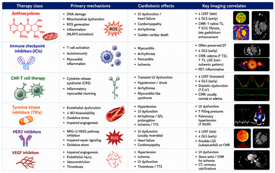
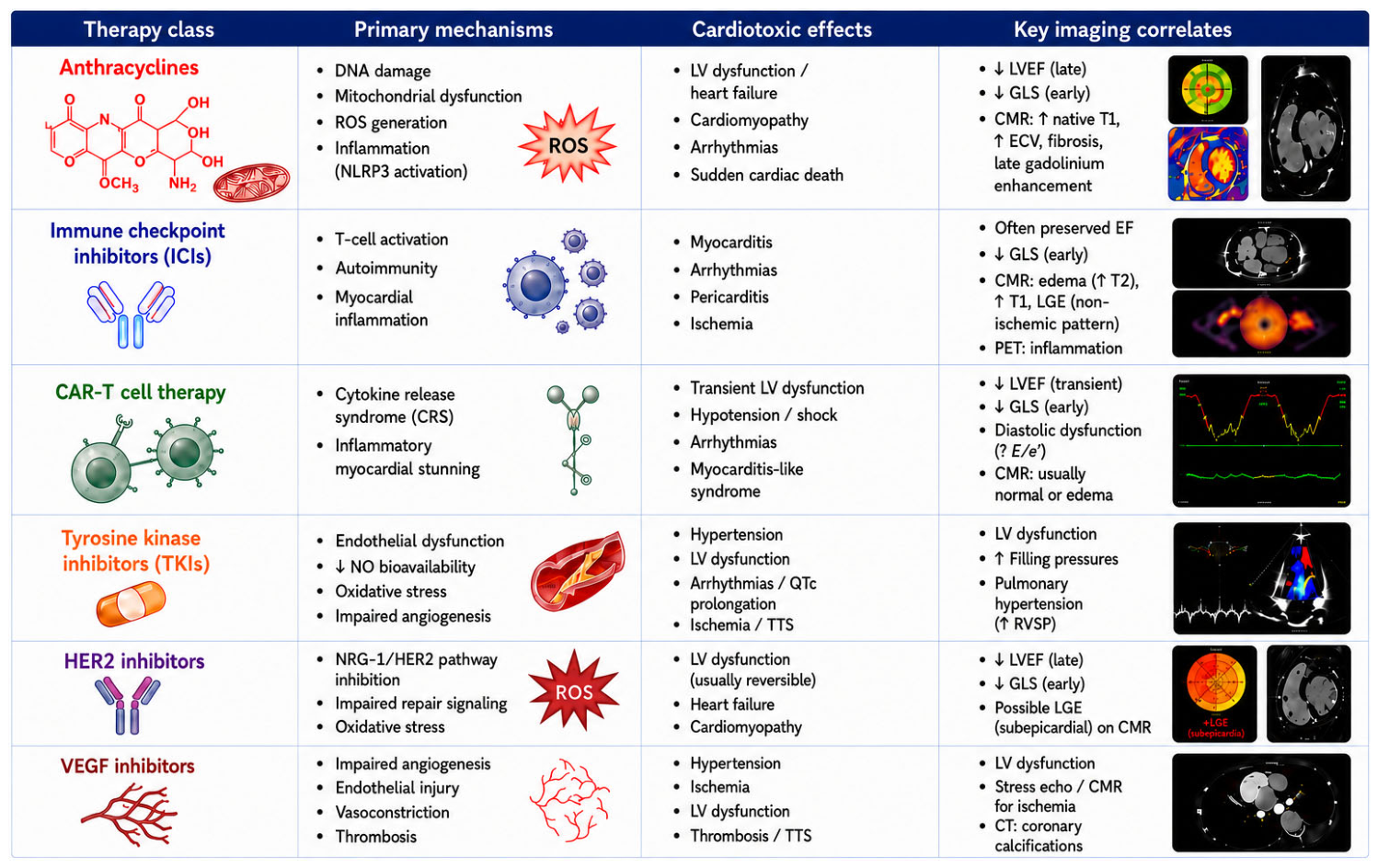
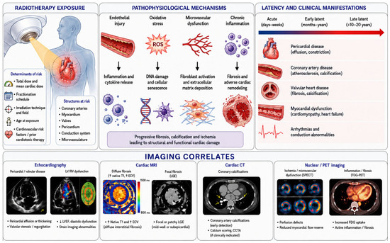
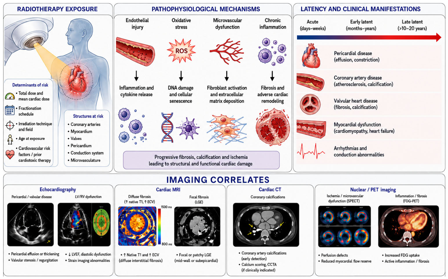




Disclaimer/Publisher’s Note: The statements, opinions and data contained in all publications are solely those of the individual author(s) and contributor(s) and not of MDPI and/or the editor(s). MDPI and/or the editor(s) disclaim responsibility for any injury to people or property resulting from any ideas, methods, instructions or products referred to in the content. |
© 2026 by the authors. Licensee MDPI, Basel, Switzerland. This article is an open access article distributed under the terms and conditions of the Creative Commons Attribution (CC BY) license.