


Abstract
Background/Objectives: Primary airway epithelia grown at air–liquid interface (ALI) are widely used to model respiratory endophenotypes and to enable individualized functional testing. However, culture conditions used to establish ALI vary considerably and may introduce selection biases. We aimed to adapt our lung explant-based ALI protocol to nasal basal cells obtained by minimally invasive brushing and to identify expansion conditions that optimize the number of primary epithelia that can be generated from nasal brushing while preserving donor-specific features. Methods: Primary nasal basal cells from nine healthy adult donors were expanded using different protocol variations in a paired design. We systematically varied two factors during the expansion phase: (a) supplementation with Rho-associated kinase (ROCK) and SMAD pathway inhibitors and (b) a single passage prior to ALI differentiation. Outcomes included the likelihood of obtaining a well-formed, differentiated ALI epithelium and functional readouts relevant to respiratory phenotyping. Results: The probability of establishing a well-differentiated ALI epithelium in a single attempt was highest when expansion was performed without ROCK/SMAD inhibitors and without passaging. Among successfully established ALI cultures, replicate inserts from the same donor showed comparable CFTR expression and ciliary beat frequency; however, functional chloride secretion could only be robustly detected in a subset of epithelia and was associated with low epithelial apoptosis. Conclusions: Avoiding strong selection pressures during the expansion phase—by omitting ROCK/SMAD inhibitors and minimizing passaging—improves the success rate of obtaining well-differentiating primary nasal ALI cultures and likely preserves cellular diversity among founder cells of the epithelium. These findings support the use of patient-derived nasal ALI cultures as robust, reproducible platforms for individualized respiratory phenotyping and functional assessment in personalized medicine.
1. Introduction
Air–liquid-interface (ALI) cultures can be established from basal cells derived from airways of the lung [1,2,3] or the nasal inferior turbinate [4,5,6]. These primary respiratory epithelial cultures can be differentiated to display the typical cell types found in native epithelia, such as goblet, ciliated, and basal cells [3]. Single-cell analyses of ALI cultures have shown that rare epithelial cell types, such as ionocytes [7,8], can also be present in such primary cultures. To generate a fully differentiated ALI epithelium, basal cells are harvested from a donor, expanded in submerged adherent culture, seeded onto permeable inserts at a density sufficient to form a confluent, tight monolayer, and then differentiated after apical medium removal to establish ALI epithelia [3].
Primary human nasal basal cells, which must be expanded to reconstruct primary epithelia, are heterogeneous and comprise subpopulations with distinct proliferative capacities and lineage-primed states [9]. During in vitro expansion, this heterogeneity is vulnerable to clonal drift: each enzymatic dissociation and replating step imposes a population bottleneck, and subsequent outgrowth tends to be dominated by the fastest-cycling, stress-resistant clones, highlighting cell detachment and replating as a critical bottleneck during ALI generation [1]. Consequently, repeated passaging can reduce clonal richness and skew the culture toward a narrow subset of cells with high replicative fitness [10,11], at the expense of more quiescent or lineage-biased progenitors among the basal cells that may be critical for obtaining an epithelial composition comparable to the in vivo situation as well as preserving donor-specific phenotypes. Pharmacologic pathway modulation during expansion can amplify these selective effects: Rho kinase (ROCK) inhibition, effectively used in the cultivation of human pluripotent stem cells [12], enhances survival after dissociation, suppresses dissociation-induced apoptosis, and accelerates colony outgrowth, properties that underpin “conditional reprogramming” [12]. Similarly, inhibition of SMAD-dependent signaling (e.g., TGF-β/Activin and BMP pathways) removes physiological brakes on cell cycle progression and differentiation, enabling long-term expansion of basal cells [13] but biasing the culture toward a subset with enhanced growth and altered fate propensity [14,15].
We previously used a modified keratinocyte medium to expand basal cells from explanted lungs or donor tracheae [16]. This strategy generated ALI epithelia that formed a well-differentiated epithelium and retained donor-specific characteristics [16] and enabled a focused genotype–endophenotype analysis investigating how transcript plasticity of a cytokine receptor influences disease outcomes in cystic fibrosis [17]. Acknowledging the limited amount of biomaterial that can be obtained by nasal brushing, we sought to increase the number of basal cells that can be expanded from a single brushing. Because ROCK and SMAD inhibitors are widely used to increase expansion and maximize the number of cells available for downstream applications, we asked whether their use during the expansion phase, or the introduction of a single passage before ALI differentiation, would improve the yield of nasal-derived ALI cultures without compromising downstream phenotyping. To test the applicability of each expansion strategy, we used paired expansions from nine healthy volunteers. Unexpectedly, we could not see the benefit of further modifications aimed at basal cell expansion to our lung explant-derived, basic ALI protocol.
2. Materials and Methods
2.1. Generation of Primary Epithelia
All study participants were healthy volunteers who provided nasal brushings after written informed consent (ethical approval no. 10989_2-_BO_K_2023). The generation of primary epithelia from nasal brushings was adapted from our previous ALI protocols for lung transplant materials [16] and from the ECFS-CTN standard operating procedure for the isolation, cultivation, and application of primary nasal respiratory epithelial cells obtained by nasal brushing (ECFS Working Group on Airway Epithelial Cell Models for Theranostics, version 2, 28 September 2024) [18]. Primary ALI epithelia, as well as all live-cell readouts (Ussing chamber recordings, cil-ia beat frequency), were generated between April and June 2024. Analysis of whole-cell lysates and histology, as well as data analysis across readouts, were done from January to July 2025.
Expansion cultures were established in T25 flasks. Briefly, cells from the nasal turbinates were collected using one brush per nostril. Based on our previous experience with basal cells expanded from explanted lungs or donor tracheae [16,17,19], each brush was expected to yield sufficient basal cells for expansion to confluency in a T25 flask, and each confluent T25 flask was expected to provide sufficient cells to generate two ALI inserts.
For expansion, cells harvested from one brush of each volunteer were cultured in modified keratinocyte medium consisting of keratinocyte serum-free medium (Life Technologies, Carlsbad, CA, USA) supplemented with epidermal growth factor (0.2 ng/mL; Life Technologies, Carlsbad, CA, USA), bovine pituitary extract (25 µg/mL; Life Technologies, Carlsbad, CA, USA), and isoproterenol (1 µM; Sigma-Aldrich, St. Louis, MO, USA) [2].
Cells from the contralateral brush of the same volunteer were expanded in the medium recommended by the ECFS-CTN SOP, i.e., PneumaCult-Ex Plus medium (STEMCELL Technologies, Vancouver, BC, Canada) supplemented with hydrocortisone (0.2 µM; STEMCELL Technologies, Vancouver, BC, Canada), Y-27632 (10 µM), DMH-1 (1 µM), and A-83-01 (1 µM) (all Selleck Chemicals, Houston, TX, USA).
Once expansion cultures reached confluency, half of the cells were seeded onto ALI inserts as passage 0 (P0). The remaining cells were expanded further and, if successful, seeded onto two additional inserts as passage 1 (P1). All ALI epithelia were generated on 12-well inserts (0.4 μm pore polyethylene terephthalate Transwell inserts; Corning, Corning, NY, USA).
For expansion cultures that had grown in the modified keratinocyte medium, after seeding onto ALI inserts, cells were cultured on both the apical and basolateral sides for 1 to a maximum of 4 days. After confluency was reached, medium was changed to bronchial epithelial growth medium prepared by first mixing Bronchial Epithelial Cell Growth Medium BulletKit (BEGM; Lonza, Basel, Switzerland) and bronchial/tracheal epithelial cell growth medium (Cell Applications, San Diego, CA, USA; distributed by PELOBiotech, Planegg, Germany) in equal proportions, followed by a further 1:1 dilution of this mixture with high-glucose Dulbecco’s Modified Eagle Medium (DMEM; Life Technologies, Carlsbad, CA, USA) [2].
Expansion cultures that were generated in the PneumaCult-Ex Plus medium (STEMCELL Technologies, Vancouver, BC, Canada) supplemented with ROCK/SMAD inhibitors were seeded and cultured on both the apical and basolateral sides overnight using PneumaCult-Ex Plus medium as recommended by the manufacturer.
Following this step, cell culture protocols were kept identical for all inserts irrespective of the expansion protocol used before: the apical medium was removed, and the basolateral medium was replaced with PneumaCult-ALI maintenance medium (STEMCELL Technologies, Vancouver, BC, Canada) according to the manufacturer’s instructions.
Analyses were performed after full epithelial differentiation had been confirmed by the presence of ciliary beating on light microscopy.
2.2. Analysis of Primary Epithelia
The primary readout parameters for this study were: 1. Capability to generate a primary epithelium at ALI; and if a primary epithelium could be obtained, 2. Analysis of ciliary beat frequency of the entire insert [20]; 3. Assessment of ion channel function in a micro-Ussing chamber with two recordings from two subparts of the insert [21]; 4. Morphology score for epithelial cell composition and differentiation from another subpart of the insert [16]; 5. CFTR protein expression from SDS-rich lysates [22] provided from another subpart of the insert as well as from the insert subparts that were subjected to Ussing chamber measurements.
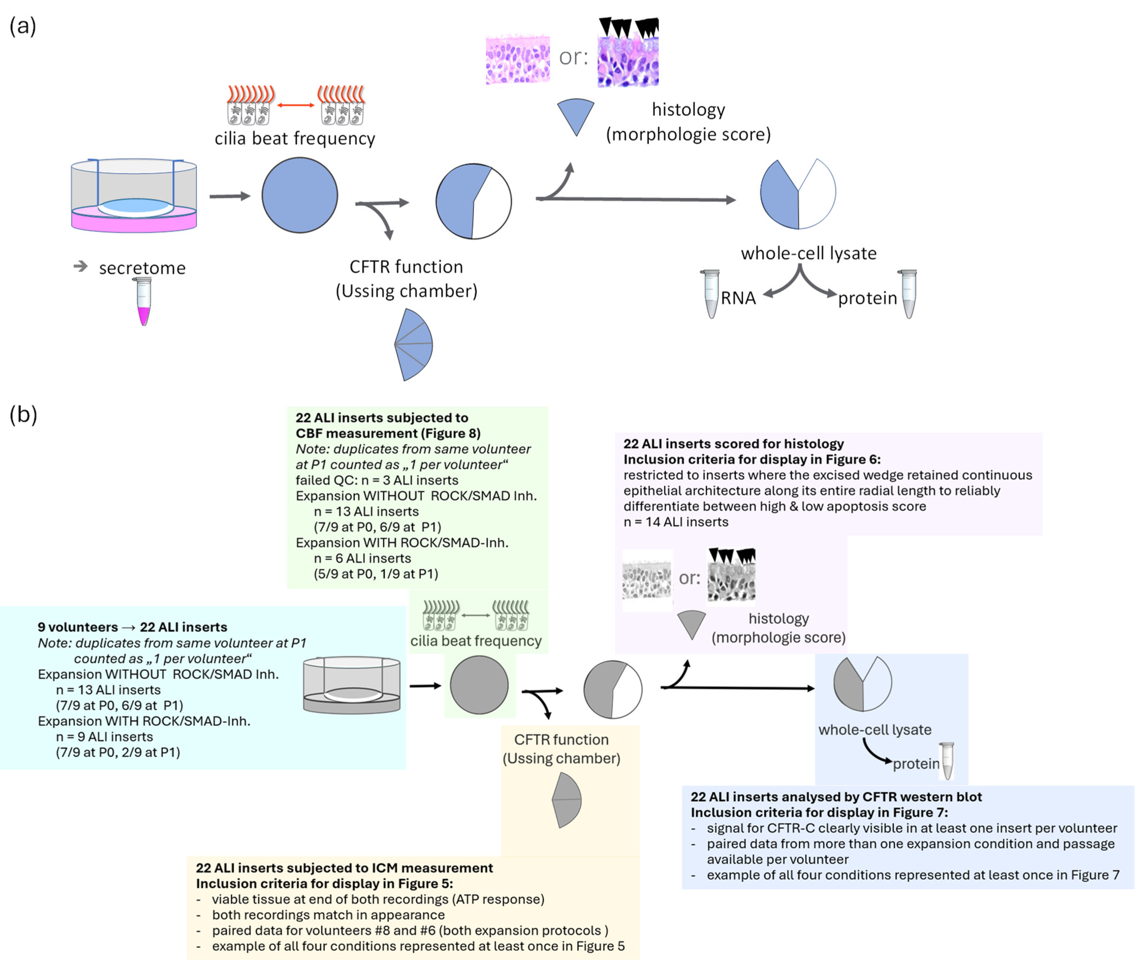
An overview of the data and biomaterial that can be generated from an ALI insert is shown in Figure 1. For this study, neither the secretome nor the mRNA was analyzed. RNA data obtained with this strategy from primary epithelia grown on ALI are shown in [17].
Figure 1.
(a): Overview of workflow for serial generation of multiple readout parameters from a single ALI insert. The insert surface devoted to each technique is shown in blue. From these 12-well inserts (area: 1.12 cm2), the following readouts and biomaterials can be obtained: culture media for secretome analysis; assessment of ciliary beat frequency [20]; ion channel measurement in a micro Ussing chamber including CFTR functional assessment (Figure 2) [6]; histology using a morphology score [16]; whole-cell lysates including material for RNA analysis (see [17] for RNA data from ALI from another study); and SDS-rich lysate for protein analysis by Western blot including CFTR expression analysis [22]. (b): Data presented in this study. In this study, a total of 22 viable ALI inserts were successfully generated across nine healthy donors and processed identically for all downstream analyses. The cohort comprised 13 inserts from the modified keratinocyte medium (seven passage 0 and six passage 1) and nine inserts from the PneumaCult™-Ex Plus condition supplemented with ROCK/SMAD inhibitors (seven passage 0 and two passage 1). Growth of expansion cultures is shown in Figure 3. This exact donor- and condition-specific allocation is visually detailed in Figure 4. For each of the 22 epithelia, the insert surface was partitioned as follows: the entire surface was first used for high-speed video microscopy to assess ciliary beat frequency and active ciliated area, followed by subfractionation. Approximately two-fifths were mounted in a micro-Ussing chamber for duplicate electrophysiological recordings; a further one-fifth to one-sixth was harvested for formalin fixation and histological morphology scoring; and the residual biomaterial, including subfractions from Ussing chamber measurements, was processed into SDS-rich lysates for CFTR protein analysis by Western blot. This insert-based allocation strategy maximized data yield from limited nasal brushing material while ensuring that all functional, morphological, and molecular readouts were derived from the same differentiated epithelial layer.
2.2.1. Cilia Beat Frequency
Ciliary beat frequency (CBF) [20] was measured in primary ALI cultures by high-speed video microscopy using a Zeiss Axiovert A.1 microscope (Carl Zeiss AG, Oberkochen, Germany) equipped with a Basler SCA640-120FM camera (Basler AG, Ahrensburg, Germany) in a humidified environmental chamber maintained at 37 °C and 5% CO2. Videos were recorded in phase contrast with a 40× objective at 100 frames/s. For each insert, at least 15 fields of view were acquired in a meandering pattern across the entire membrane while avoiding edge regions affected by the meniscus. Prior to measurement, the apical surface was washed with PBS 24 h in advance. CBF and ciliary coverage were analyzed by whole-field evaluation using Sisson–Ammons Video Analysis (SAVA) software, (version 1.0; Sisson & Ammons, University of Iowa, Iowa City, IA, USA). which quantified both the mean CBF and the area of moving pixels as a measure of active ciliary motion. Inserts with fewer than 10,000 moving pixels (<0.01% of the total imaged area) were classified as non-motile. For each insert, results were expressed as the mean of all recorded fields.
2.2.2. Ion Channel Measurement
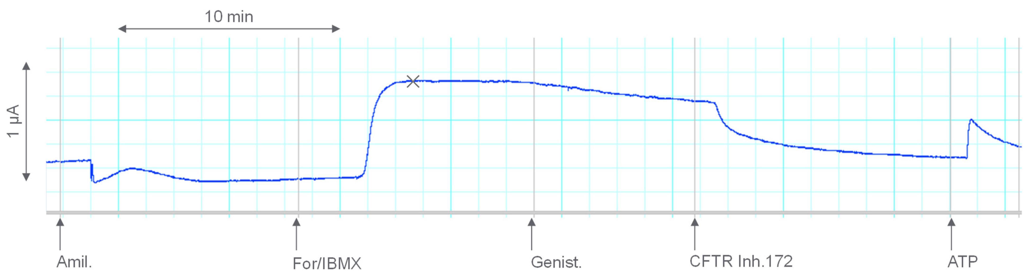
The protocol for ion channel assessment in ALI epithelia as described by Noel et al. [6] is briefly shown in Figure 2. Briefly, inserts were mounted in a low-volume EasyMount chamber system with P2400 EasyMount diffusion chambers (Physiologic Instruments, San Diego, CA, USA), connected to Ag/AgCl voltage-sensing and current-passing electrodes. Chambers were maintained at 37 °C using a circulating temperature-controlled water bath and continuously gassed with carbogen (95% O2/5% CO2). Electrophysiological recordings were obtained under short-circuit conditions using a multichannel voltage clamp amplifier with compatible preamplifiers. Signals were digitized via a PowerLab acquisition unit (ADInstruments, Oxford, UK) and recorded using LabChart software (version 7 or later, ADInstruments). Before each experiment, electrode offsets were adjusted to 0 mV, and fluid resistance compensation was performed according to the manufacturer’s instructions. Short-circuit current was recorded continuously. Recordings were accepted as valid if two insert segments cut from the same transwell ALI culture showed a highly similar pattern and a final response to ATP could be observed, indicating viability throughout the recording.
Figure 2.
Analysis of ion transport in a micro Ussing chamber in ALI epithelia. Approximately one-tenth of a 1.12 cm2 ALI insert was mounted between half-chambers, and short-circuit current (Isc) was recorded at 0 mV as described by Noël et al. [6]. Secretagogues indicated at arrows below the recording are: amiloride (Amil) to block the epithelial sodium channel ENaC, forskolin/IBMX (For/IBMX) and genistein (Genist.) to activate transport via cAMP-responsive chloride channels, the inhibitor 172 (CFTR Inh.172) to block specifically chloride transport mediated by the cystic fibrosis transmembrane conductance regulator CFTR, and ATP to activate purinergic receptors and evoke a transient Ca2+-dependent Cl− secretion as a viability check. Under these conditions, upward deflections of Isc reflect net Cl− flow from basolateral to apical solution through open chloride channels such as CFTR.
2.2.3. Histology and ALI Morphology Score
Histological assessment of ALI cultures was performed on a small subfraction of each 12-well insert (approximately one-fifth to one-sixth). The harvested material was fixed mainly in 4% buffered formaldehyde for 24 h, paraffin-embedded, re-oriented by 90° to obtain cross-sectional cuts through the epithelial layer, sectioned at 2 µm, and stained with hematoxylin and eosin. Slides were examined along the entire epithelial length by light microscopy, and morphology was scored semi-quantitatively using predefined visual analog reference scales [16]. Four parameters were rated from 0 to 3: epithelial layer score (0, 1–2 cell rows; 1, 3–5 rows; 2, 5–7 rows; 3, >7 rows), apoptosis score (0, none or one solitary apoptotic cell; 1, few apoptotic cells; 2, increased apoptosis; 3, numerous/grouped apoptoses), air inclusion score (0, none; 1, one void; 2, few voids; 3, several voids), and goblet cell score (0, absent; 1, few goblet cells; 2, approximately 50% surface coverage; 3, >50% surface coverage). This rapid scoring approach provides a standardized, insert-specific overview of epithelial morphology and differentiation status in ALI cultures.
2.2.4. CFTR Protein Expression
CFTR protein expression was analyzed by Western blot as described in detail elsewhere [22] using whole-cell lysates prepared from the epithelial biomaterial in SDS-rich lysis buffer containing 50 mM Tris-HCl (pH 6.8), 2% (w/v) SDS, 10% (v/v) glycerol, 100 mM dithiothreitol, protease inhibitor cocktail (1:50), 0.2 mM phenylmethylsulfonyl fluoride, and Omnicleave endonuclease (1:10,000), followed by mechanical homogenization, incubation at 37 °C, repeat homogenization, and centrifugation to obtain cleared supernatants. Protein content was estimated semi-quantitatively by Coomassie spot assay, and equal protein amounts were separated on 6% polyacrylamide gels with a narrow 4% stacking gel in a Mini-PROTEAN Tetra Cell system (Bio-Rad Laboratories GmbH, Munich, Germany). Sample volumes were adjusted with buffer containing 50 mM Tris-HCl (pH 6.8), 2% (w/v) SDS, and 50% (w/v) glycerol, with fresh addition of 100 mM dithiothreitol and protease inhibitor cocktail (1:50) prior to a mild denaturation step. Proteins were transferred by tank blotting onto 0.45 µm supported nitrocellulose membranes (Amersham Protran; Cytiva, Marlborough, MA, USA) using a Mini Trans-Blot cell (Bio-Rad Laboratories GmbH, Munich, Germany). Membranes were blocked in StartingBlock containing 0.1% Tween-20 and probed with an equimolar mixture of four monoclonal anti-CFTR antibodies 596, 570, 217, and 660 (Cystic Fibrosis Foundation Antibody Distribution Program) diluted 1:400, followed by horseradish peroxidase-conjugated goat anti-mouse IgG diluted 1:7500. Chemiluminescent signals were developed sequentially with SuperSignal West Pico and West Femto substrates (Thermo Fisher Scientific, Waltham, MA, USA) and recorded on a DNR-MF-ChemiBIS 3.2 imaging system (Berthold Technologies, Bad Wildbad, Germany).
3. Results
We monitored growth of the cell cultures for two time intervals: (i) the time required for expansion cultures to reach confluency and (ii) the time required for cells seeded onto inserts to form a differentiated epithelium with beating cilia.
Cells were counted after expansion and before seeding onto inserts to assess post-expansion cell yield under both culture conditions. We observed comparable cell numbers available for seeding onto a single insert in P0 in both media (keratinocyte medium without ROCK/SMAD inhibitors: mean 3.0 × 10⁵, range 1.2 × 10⁵ to 8 × 10⁵; PneumaCult-Ex Plus medium with ROCK/SMAD inhibitors: mean 3.6 × 10⁵, range 2.0 × 10⁵ to 4.7 × 10⁵) and slightly reduced cell numbers in P1 in keratinocyte medium (mean 1.6 × 10⁵, range 0.6 × 10⁵ to 3.0 × 10⁵).
For P0 cultures, and independent of the expansion medium used, expansion cultures reached confluency within 5–8 days in 12 of 14 cases that subsequently showed cell growth on inserts. In contrast, expansion cultures that required longer than 8 days to reach confluency failed to generate a cell layer on inserts in four out of six cases. Among cultures that successfully differentiated on inserts and developed cilia beating, the mean time to epithelial differentiation was 10 days for P0 cultures (range 5–17 days) and 13 days for P1 cultures (range 7–32 days), again independent of the expansion medium.
At least one ALI insert with an epithelium showing cilia beating under the microscope could be obtained for each of the nine volunteers. As shown in Figure 3, we observed cases where only the modified keratinocyte medium yielded an insert as well as examples for which only the expansion in the presence of ROCK/SMAD inhibitors yielded an insert with cilia beating at P0. Further passaging at P1 gave rise to ALI epithelia in six cases using the modified keratinocyte medium, but only in one case using ROCK/SMAD inhibitors (Figure 4). For each of the two expansion media tested in this study, for seven out of nine donors, direct seeding onto an insert was successful (Figure 4).
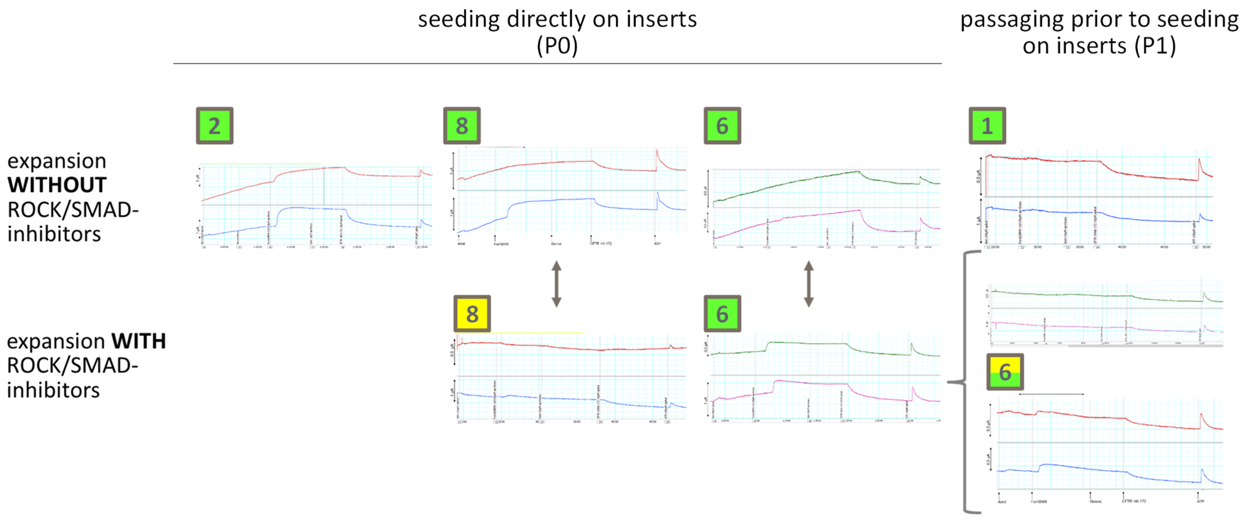
Next, we analyzed the ion channel profile using a micro Ussing chamber [4] with duplicate recordings per insert (Figure 4 and Figure 5). We noticed that while cilia beating was seen under the microscope in all 22 cases subjected to Ussing chamber analysis (Figure 4), 12 recordings showed little or no chloride channel activity while an ATP response was seen, demonstrating viability of the epithelium (Figure 4, marked in yellow). Only 10 recordings showed the expected ion channel profile for healthy volunteers (Figure 4, marked in green). Inserts derived from modified keratinocyte medium showed the expected “healthy” profile for five volunteers at P0 and for two volunteers at P1, while expansion using ROCK/SMAD inhibitors provided such an expected “healthy” profile only in two cases at P0 and for one out of two inserts obtained from one volunteer at P1 (Figure 4). As shown in Figure 5, duplicate measurements from the same insert were highly consistent, indicating that the observed transport profiles reflect intrinsic, donor-specific epithelial properties rather than technical variability. A typical example of this observation is found in #6 (Figure 5), where both at P0 and at P1 only culture with ROCK/SMAD inhibitors permitted generation of an epithelium with sufficient CFTR expression that could be measured in the Ussing chambers. Furthermore, we observed “healthy” and “no ion channel” tracings from biomaterial derived from the same volunteer in three cases at P0 (Figure 4, see recordings for #8 in Figure 5) and in one case at P1 (Figure 4, see recordings for #6 P1 in Figure 5).
Figure 3.
Expansion cultures in the presence or absence of ROCK/SMAD inhibitors. Data are shown from expansion cultures in keratinocyte medium and from expansion culture in keratinocyte medium with ROCK and SMAD inhibitors present. Representative examples showing: A—volunteer with chronic rhinosinusitis, poor yield of basal cells, culture unsuccessful in both media, not further incorporated into the study. #2—healthy volunteer, culture successful without ROCK/SMAD inhibitors during expansion. #3—healthy volunteer, culture successful with ROCK/SMAD inhibitors during expansion. LuTx—basal cells from explanted lung. Kindly note the difference in growth patterns between expansion cultures in keratinocyte medium (A, #2, #3, LuTx), showing isolated cells that grow towards confluency, and expansion in the presence of ROCK/SMAD inhibitors (A, #3, LuTx), which show strong clonal proliferation.
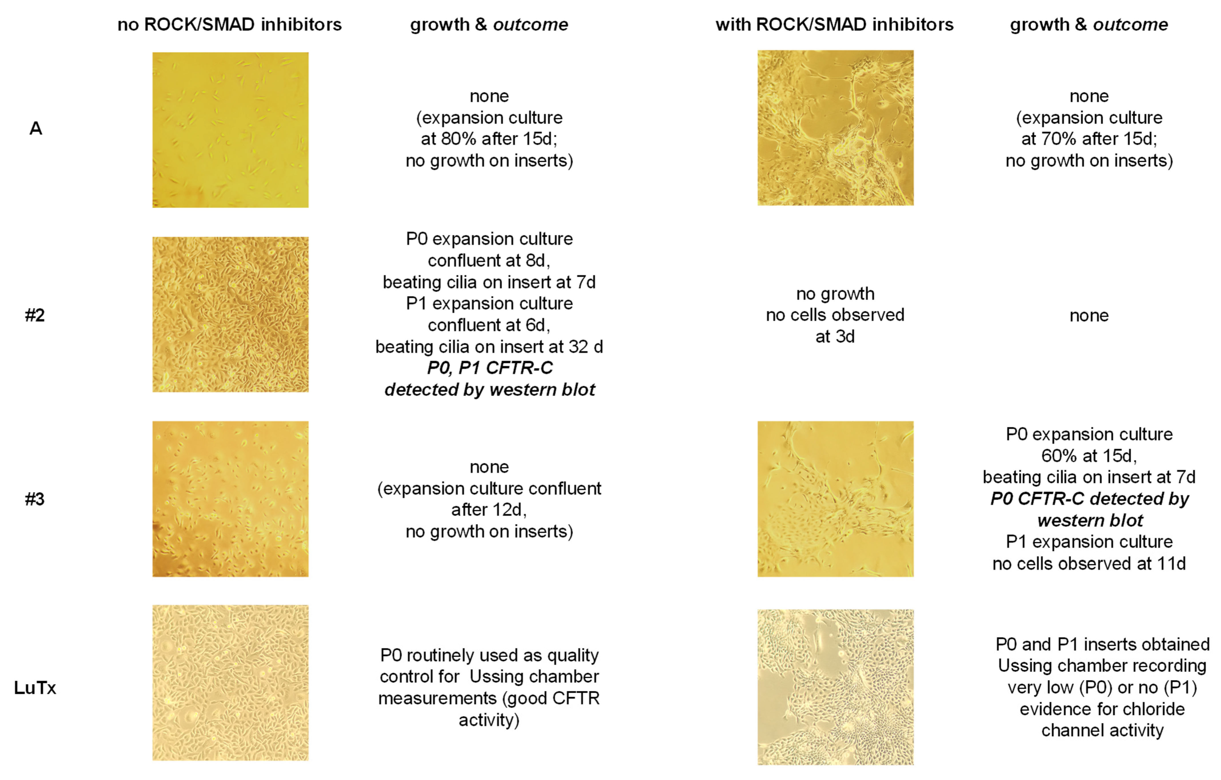
Figure 4.
Outcome of ALI culture of nine healthy volunteers. Expansion culture was done twice from one out of two brushes each obtained from both nostrils of a healthy volunteer: cells from one brush were seeded in our regularly used modified keratinocyte medium (top, WITHOUT ROCK/SMAD inhibitors) [2] and the paired nasal brush taken from the same volunteer was seeded in PneumaCult ExPlus medium with ROCK/SMAD inhibitors (bottom, WITH ROCK/SMAD inhibitors). For each expansion culture that reached confluency, one insert was seeded with half of the expanded cells while the other half was reseeded in a fresh flask to attempt one passaging step. P0: insert was seeded directly after expansion culture; P1: insert was seeded after one passage. Volunteers 1 to 9 are denoted by numbers in squares. White boxes indicate that either the expansion culture failed or that the insert did not reach confluency and did not yield an epithelium visible under the microscope. Yellow boxes indicate that the epithelium on the insert has reached confluency, but that the Ussing chamber recording showed mostly a passive diffusion curve or little to no signs of ion channel activity untypical for healthy controls. Green boxes denote epithelia with clear signs of ion channel activity expected for healthy epithelia.
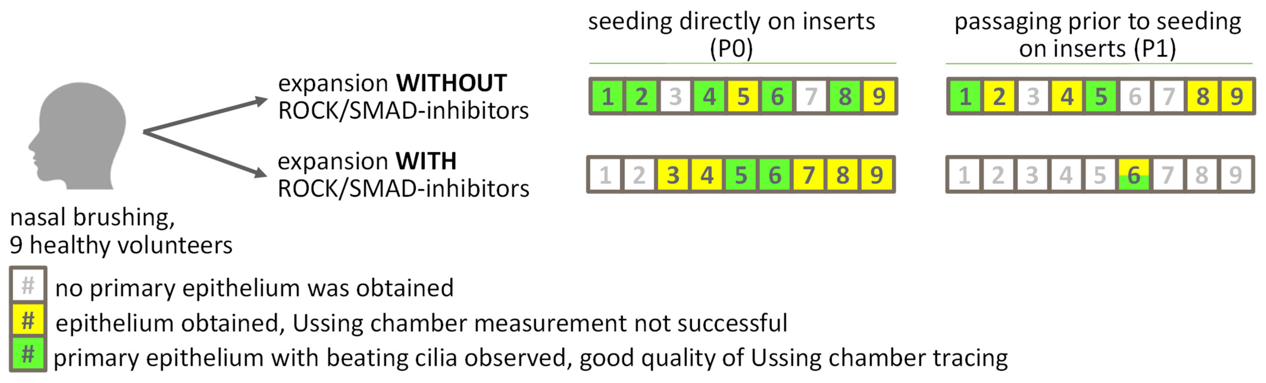
Figure 5.
Ion transport measurements in a micro-Ussing chamber. Selected examples of raw Ussing chamber recordings (see Figure 4 for readout overview from each of the nine healthy volunteers). Tracings are shown in duplicate for each insert. Recordings are grouped by expansion protocol (top: no ROCK/SMAD inhibitors; bottom: with ROCK/SMAD inhibitors) and passage (left five duplicate recordings: P0, right three duplicate recordings: P1). Data from volunteer 8 are shown for inserts derived from both expansion strategies at P0. Recordings for volunteer 6 are shown for inserts derived from both expansion protocols and for P0 and both inserts derived at P1 using ROCK/SMAD inhibitors. Measurements were performed as described by Noel et al. [6], and a detailed interpretation of Ussing chamber recordings is given in Figure 2. In the recordings displayed in this figure, kindly note that (i) both insert segments cut from the same transwell ALI culture show a highly similar pattern; (ii) chloride transport, recognizable by responses to forskolin/IBMX and/or Inh172, is clearly visible and marked in green for ALI without ROCK/SMAD inhibitors during expansion at P0 for volunteers #2, #6, and #8, and at P1 for volunteer #1. For ALI with ROCK/SMAD inhibitors during expansion, chloride transport is seen for volunteer #6 at P0 and for one out of two inserts obtained at P1; (iii) dual recordings marked in yellow show a final response to ATP, indicating viability throughout the recording, but fail to demonstrate a typical chloride secretory pattern expected for healthy controls. Large-scale figures for each of the double recordings are provided in Supplementary Figures S2–S5.
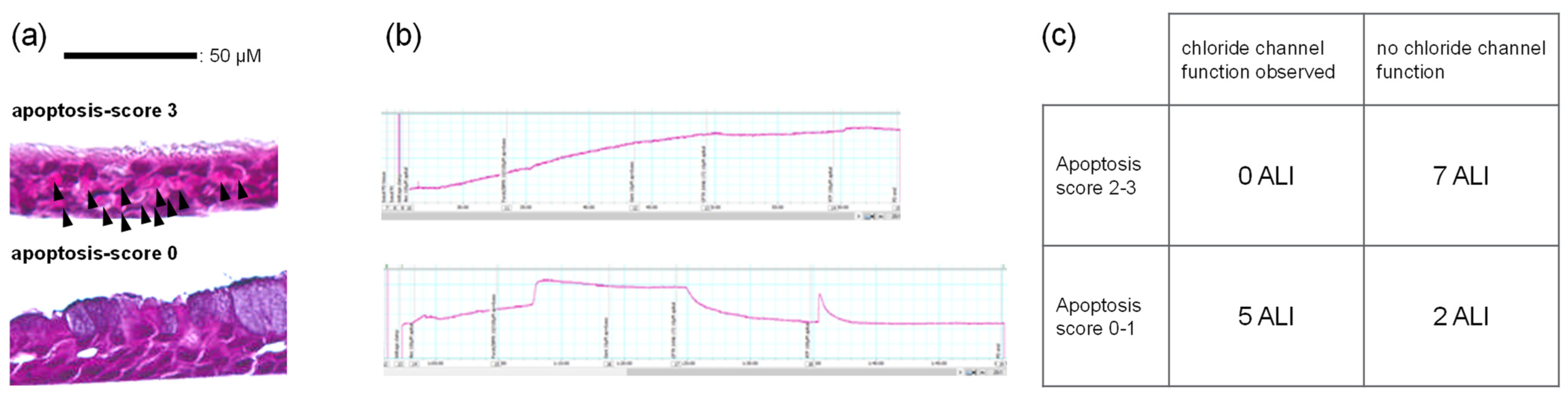
We wanted to understand why several epithelia failed to provide an ion channel signature expected for healthy individuals. Thus, we compared epithelial cell morphology assessed by the ALI histology score of hematoxylin and eosin-stained cross-sectional tissue sections, which is evaluated in a blinded fashion by one observer to ensure unbiased, reproducible results [16]. When comparing inserts with little or no chloride channel activity (marked in yellow in Figure 4) to those with clear chloride channel function expected for healthy epithelia (marked in green in Figure 4), histology score distributions for epithelial layer height and goblet cell density were similar between the two groups. However, as outlined in Figure 6, we observed an increased apoptosis score among seven out of nine ALI inserts that lacked detectable chloride channel activity. In contrast, low apoptosis score values were seen in all five ALI inserts with a “healthy” ion channel signature.
Figure 6.
Correlation of apoptosis score and Ussing chamber tracing quality. Morphology of epithelia was judged using visual analog scales. (a) Examples of high (top) and low (bottom) apoptosis scores among nasal-derived primary ALI epithelia. Arrows show apoptoses, which can be recognized by their disintegrating or fragmented cell nuclei in the initial stage or by the bright eosinophilic discoloration of the underlying cell in the later stages. An apoptosis score of 2 indicated increased apoptoses, some of which were grouped together. An apoptosis score of 3 indicated many observable apoptoses that were conspicuously arranged in larger groups. Scale bar 50 µm. (b) Mostly passive diffusion curve (top) as opposed to ion channel function expected for healthy epithelia (bottom) measured in an Ussing chamber. (c) Passive diffusion curve and leaky epithelium, little or no chloride channel function: nine ALI epithelia whereby high apoptosis score (2–3) was seen in seven thereof; and in contrast: tight epithelium and CFTR function visible in five ALI epithelia, all of which had a low apoptosis score (0–1).
To further characterize the phenotype of the primary epithelia, we analyzed CFTR protein expression by Western blot using whole-insert lysates as well as biomaterial recovered from those insert subsections used for Ussing chamber measurements (Figure 1 and Figure 7). Biomaterial from 22 inserts that were viable based on an ATP response in the Ussing chamber (Figure 4, green and yellow) was analyzed, and lysates yielding less than 5 µg of total protein were excluded. CFTR protein could be detected in either the whole-insert lysate, the post-Ussing chamber lysate, or both in all but two of these inserts. The fully glycosylated CFTR-C band was present in 17 of the 20 analyzable inserts (Figure 7), indicating that most, if not all, successfully differentiated epithelia expressed mature CFTR. This was independent of the expansion medium used and of whether cells were analyzed at P0 or P1, suggesting that primary epithelia generated in this study retain the intrinsic capacity for CFTR-mediated chloride transport once a differentiated epithelial layer is established, as expected for healthy epithelia.
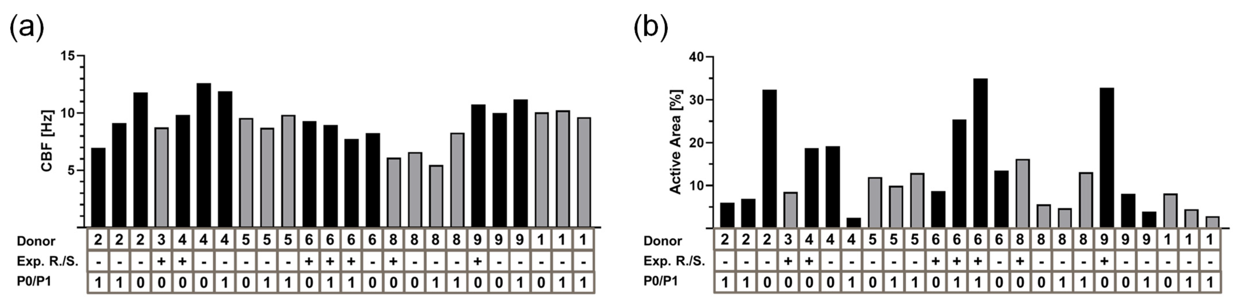
Finally, we assessed ciliary activity by quantifying both the active ciliated area and ciliary beat frequency (Figure 8). The active ciliated area exhibited considerable variability, whereas CBF proved to be a more stable parameter (Klassen & Olmer, personal communication). This variability in active area likely reflects both biological and technical factors. Biologically, primary nasal epithelia exhibit donor-dependent regional differences in ciliary differentiation, and focal apoptosis or uneven cell layering can locally reduce ciliary coverage. Technically, the SAVA software quantifies moving pixels via phase-contrast microscopy, which is sensitive to edge effects, focal depth variations, and differences in ciliary beat amplitude; regions with low ciliary density or asynchronous beating may fall below the detection threshold, leading to underestimation. Despite this variability, CBF remained consistent among inserts from the same donor, and neither metric showed a correlation with expansion medium or passage (Figure 8b).
Figure 7.
CFTR protein expression in ALI epithelia. (A) Detection of CFTR glycoisoforms on control cell lines T84 and 16HBE14o- (HBE) and in three primary epithelia derived from bronchial epithelial samples from explanted lung tissue from three different donors (BEC1, BEC2, BEC3). The polydisperse complex glycosylated isoform CFTR-C varies in size between T84 < HBE < BEC1 < BEC2 and BEC3. 50 µg protein from these control lysates was used. (B) Data are shown for inserts derived from nasal brushes of volunteers 1, 5, 6, and 8 from whole-insert lysates (L) as well as biomaterial recovered from those insert subsections used for Ussing chamber measurements (U) for all fractions where more than 5 µg of total protein content could be retrieved. Loaded protein amount is given on top of the respective lanes (µg). For #6, P1 after expansion with ROCK/SMAD inhibitors, lanes for both inserts obtained in P1 are shown from the material rescued after Ussing chamber measurement. Fully glycosylated CFTR-C and core glycosylated CFTR-B are shown. n.a.1: growth was observed in expansion stage, but no epithelium formed on the insert. n.a.2: no growth in expansion stage. The examples shown have been selected from different blots. From a series of 15 or more exposures taken for each CFTR detection, one for each gel was chosen for this composite. The selection was driven by selecting an exposure time that gave a comparable signal-to-noise ratio for the other CFTR visualizations. No further editing of images for contrast was done.
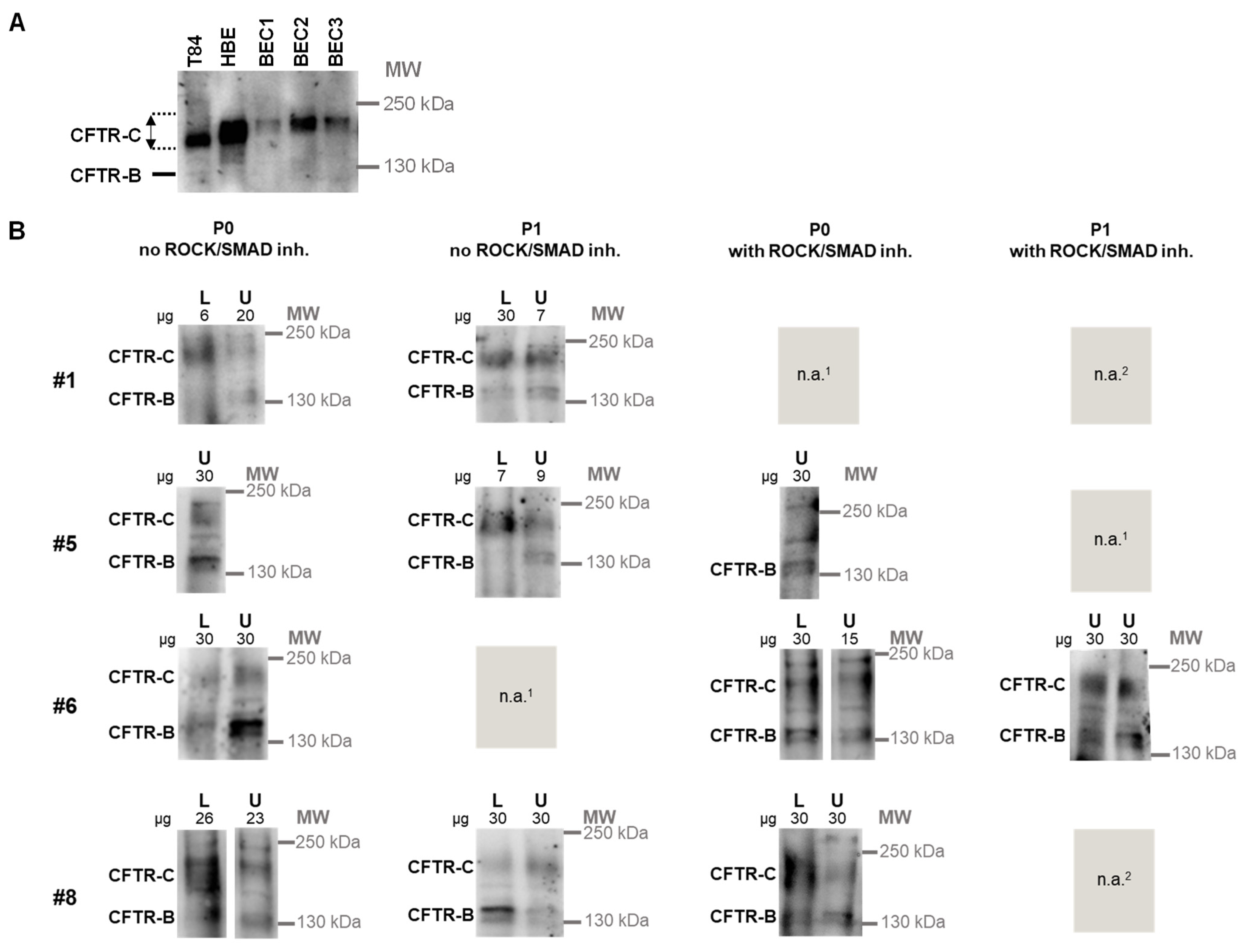
Figure 8.
Cilia beat frequency of ALI epithelia. Ciliary beat frequencies [Hz] (a) and active area (b) are given for viable ALI inserts from this study. Donor # corresponds to participant. Expansion media are listed as Exp. R/S with + indicating presence of ROCK and SMAD inhibitors. Passage P0 refers to direct seeding of expanded basal cells to inserts; passage P1 refers to seeding on inserts after an additional expansion step.
4. Discussion
Systematic evaluation of culture protocols on ALI outcome has been done before: Awatade and colleagues [23] compared conditionally reprogrammed and feeder-free dual SMAD-inhibited expanded human nasal epithelial cells to show that CFTR function is attenuated in ALI epithelia derived from dual SMAD-inhibited expansion cultures. Saint-Criq and colleagues have used ROCK inhibitor and feeder cells to expand basal cells, but then compared the differentiation into ALI epithelia using the medium UNC (University of Chapel Hill) to the commercially available medium SC (StemCell, Cambridge, UK), finding that ion transport as well as responses to CFTR modulators differed markedly [24]. Ruiz García and colleagues compared primary epithelia differentiated in either StemCell (Cambridge, UK) or Lonza (Basel, Switzerland) media and showed that the former yielded more multiciliated and goblet cells while the latter favored the multiciliated cells [25]. Finally, Awatade and colleagues have compared conditionally reprogrammed expansion cultures after differentiation using StemCell or Lonza media for expansion, finding that StemCell-differentiated epithelia had lower cilia beat frequency, more FOXJ1 expression characteristic of ciliated cells, more ciliated cells, a larger active area with beating cilia, and, finally, different ion transport properties than Lonza-differentiated epithelia [26]. In contrast, Lee and colleagues have expanded cells using ROCK inhibitor and feeder cells, then tested four different differentiation media to obtain similar phenotypes in all of them, concluding that expansion is the major factor determining ALI phenotype [27].
Taken together, these findings suggest that the path to culturing primary airway epithelia strongly determines outcome and might explain why laboratories, keen to preserve a reproducible outcome at their site, adhere to their own method, thus reducing comparability of results between sites. Moreover, as the expansion of basal cells to a sufficient amount is a delicate procedure, translational laboratories working with minute patient samples often are reluctant to change, adjust, or compare procedures. In a similar position, we also had a preoptimized protocol to grow primary epithelia [16], avoiding ROCK/SMAD inhibitors and feeder cells [2,28] and now wanted to know whether we could increase the number of successful cultures from minute cell material by modifying our expansion procedure.
To assess the success of different culture media and passaging, our approach uses “ALI epithelium generated” as a primary readout parameter: if too few basal cells are seeded on an insert, or if the viability of the seeded basal cells is too low, both of which are recognizable by the time it takes the expansion culture to reach confluency, the restoration of an epithelium will fail [1,2]. For our study, we have assumed that each brush can yield a cell amount to generate epithelia on two inserts after expansion to confluency, and thus we have used this estimate to normalize the amount of our input material into both protocols that we have compared during expansion. Consistent with this assumption, we observed similar post-expansion cell yields and similar times to confluency in expansion flasks under both culture conditions, despite donor-to-donor and brush-to-brush variability.
Our rationale was that, if ROCK/SMAD inhibitors enhanced expansion sufficiently to increase the number of basal cells available for downstream use, more ALI epithelia might be generated from cultures expanded under these conditions. Likewise, if an additional passage could be performed without substantial loss of expansion potential, further inserts should be recoverable at P1 after renewed expansion to confluency. However, our observation suggests that neither the addition of ROCK/SMAD inhibitors nor the passaging of cells improves the success in converting cells brushed from the nasal epithelium to regenerated primary epithelia (Figure 4). In contrast, most primary epithelia were generated in the absence of ROCK/SMAD inhibitors and at P0.
Failure to generate a fully differentiated epithelium may reflect low initial cell recovery during brushing, limited expansion potential of the harvested basal cells, or systematic effects of the expansion conditions themselves. In this real-world comparison of healthy volunteers, the most successful outcome was obtained when cells were expanded without ROCK/SMAD inhibitors and seeded directly at P0, suggesting that this approach better preserves the subset of basal cells capable of regenerating a differentiated ALI epithelium. Consistent with other studies [24,27], in the subset of cultures where both expansion protocols successfully generated functional epithelia, CFTR expression (Figure 7), ion channel function (Figure 5), and ciliary beating (Figure 8) were comparable. However, this consistency applies only to the minority of ROCK/SMAD-inhibited cultures that overcame early differentiation failure and should not be generalized to the protocol as a whole.
While mature, fully glycosylated CFTR-C protein could be observed as expected from most ALI biomaterials derived from healthy volunteers (Figure 7), not all Ussing chamber recordings could clearly prove chloride channel activity (Figure 4 and Figure 5). In our opinion, the confounder that might overshadow the detection of chloride channel activity in the Ussing chamber recordings is a partially damaged, leaky epithelium due to apoptosis processes, as suggested by our results from the ALI scoring (Figure 6). The driving force for respiratory epithelia to transport chloride is typically generated by a chloride gradient. The basolateral side of the epithelium contains chloride, while chloride on the apical side is replaced with gluconate [21]. Consequently, the intended chloride transport via CFTR may be partially or completely masked by passive diffusion through very thin and leaky epithelia, or cell layers damaged by apoptosis [29]. A lower chloride gradient between the apical and basolateral sides of the epithelium, which we evaluated for use as it would reduce the passive diffusion through leaky epithelia, failed to yield Ussing chamber tracings that enhance ion transport detection in our hands, as lower gradients yielded lower signals (Supplemental Figure S1). As demonstrated in Figure 6, the histological apoptosis score based on hematoxylin and eosin-stained cross-sectional tissue sections strongly correlated with functional barrier status: epithelia with high apoptosis scores consistently exhibited leaky barriers and absent chloride transport, while those with low scores maintained tight junctions and robust CFTR-mediated ion channel activity.
The increased histological apoptosis observed in ROCK/SMAD-inhibited cultures likely reflects a delayed stress response following rapid clonal expansion. While acute ROCK inhibition prevents dissociation-induced anoikis by stabilizing actomyosin tension [30], continuous exposure disrupts cytoskeletal architecture, alters E-cadherin-mediated adhesion, and promotes cytoplasmic spreading that can ultimately trigger apoptosis [30]. This might explain why prolonged ROCK inhibitor treatment has been shown to alter morphology of growing cell colonies [30]. Concurrent SMAD inhibition removes TGF-β-mediated homeostatic signaling that normally regulates epithelial turnover and controlled apoptosis [31]. Together, these interventions create a strong selective pressure that enriches for highly proliferative, stress-resistant clones while suspending physiological differentiation cues. When these cells are subsequently forced into ALI differentiation, the accumulated structural and metabolic stress manifests as delayed apoptosis, compromising barrier integrity and masking underlying ion transport capacity. This aligns with evidence that transient, rather than continuous, ROCK inhibition is required to preserve differentiation competence in expanded progenitor populations [30].
Our within-donor paired comparison suggests that deep phenotyping of primary epithelia is feasible and a powerful method to recognize confounders introduced by cell culture procedures. Standardization across different laboratories is likely an unachievable goal, as culturing primary epithelia is a tedious process and laboratories will be reluctant to adjust their established processes to a novel standard operating procedure that might fail in their hands. As a suggestion, to achieve more standardization for primary cell culture in the future, inter-site comparisons might become feasible if readouts are normalized at each site, for instance, by reporting fold-change values rather than absolute values for transcript and protein expression as well as functional values. Moreover, including basic characteristics of primary epithelia such as histology might help to understand if cross-site comparisons are incompatible.
Our study has several limitations. First, the cohort size (n = 9) restricts statistical power and limits correlation analyses. Second, the ROCK/SMAD inhibitor protocol failed to generate functionally viable epithelia in the majority of cultures at P0, preventing systematic comparison of CFTR function between protocols. Consequently, claims of phenotypic consistency are restricted to the small subset of successfully differentiated cultures and should be interpreted with caution.
In conclusion, our data demonstrate that minimizing selection pressure during expansion—by avoiding ROCK/SMAD inhibition and limiting passaging—improves the likelihood of generating differentiated ALI epithelia suitable for downstream phenotyping. While a small subset of ROCK/SMAD-inhibited cultures retained functional capacity, the protocol’s high failure rate and associated apoptosis underscore the importance of conservative expansion strategies when donor-specific physiological phenotyping is the experimental goal. From a translational perspective, these findings have direct implications for the development of standardized, patient-derived airway models used in personalized medicine and functional theranostics. By demonstrating that conservative expansion protocols preserve donor-specific phenotypes and improve the generation of functionally viable epithelia, our work provides a practical framework for optimizing primary cell culture workflows in clinical research settings. This is particularly relevant for CFTR modulator screening, where reproducible electrophysiological readouts are essential for accurate genotype–phenotype correlation and drug response prediction. Furthermore, our data suggest that minimizing artificial selection pressures during in vitro expansion may reduce inter-laboratory variability, facilitating more reliable cross-site comparisons and accelerating the translation of patient-derived models into routine clinical decision-making.
Supplementary Materials
The following supporting information can be downloaded at: https://www.mdpi.com/article/10.3390/jpm16110540/s1, Figure S1: Recordings using different chloride gradients for Ussing chamber measurements; Figure S2: Large-scale presentation of Tracings in Figure 5 (Ion transport measurements in a micro Ussing-chamber), part 1 of 4; Figure S3: Large-scale presentation of Tracings in Figure 5 (Ion transport measurements in a micro Ussing-chamber), part 2 of 4; Figure S4: Large-scale presentation of Tracings in Figure 5 (Ion transport measurements in a micro Ussing-chamber), part 3 of 4; Figure S5: Large-scale presentation of Tracings in Figure 5 (Ion transport measurements in a micro Ussing-chamber), part 4 of 4; Table S1: Comparison of basal cell expansion media.
Author Contributions
Conceptualization, A.-M.D. and F.S.; methodology, S.T.; investigation, S.T., L.F., M.-C.K., S.H., L.A., I.F., B.H., C.L., R.D. and R.E.; formal analysis, L.D., C.V., A.M., L.A., B.H. and P.B.; data curation, S.H., L.A., I.F. and C.L.; resources, R.D., R.E., P.B. and L.N.; supervision, S.T., L.F., R.O., G.H. and F.S.; writing—original draft preparation, F.S. and L.F.; writing—review and editing, A.-M.D.; visualization, F.S., M.-C.K. and P.B.; funding acquisition, L.N., G.H., R.O., A.-M.D. and F.S. All authors have read and agreed to the published version of the manuscript.
Funding
This research was funded by the German Center for Lung Research (Deutsches Zentrum für Lungenforschung, DZL), grant number 82DZL002A1. L.F. was financed by a stipend provided by the China Scholarship Council (CSC No. 202308500039) and supported by the Hannover Biomedical Research School (HBRS) and the Center for Infection Biology (ZIB).
Institutional Review Board Statement
The study was conducted in accordance with the Declaration of Helsinki, and approved by the Ethics Committee of the Hannover Medical School (Nr. 10989_2-_BO_K_2023, date of approval 4 April 2024).
Informed Consent Statement
Informed consent was obtained from all volunteers involved in the study.
Data Availability Statement
All relevant data supporting the conclusions of this study are contained in the article. No additional datasets were generated or analyzed. No public repository is used. Additional anonymized data may be available from the corresponding author upon reasonable request, subject to privacy, ethical, and legal restrictions and any required approvals.
Acknowledgments
During the preparation of this manuscript, the author(s) used the service ChatAI provided via the Academic Cloud (https://academiccloud.de/) using a local model of Qwen 3.6 35B A3B (17 December 2025–5 October 2026) for the purposes of editing the manuscript text for clarity. The authors have reviewed and edited the output and take full responsibility for the content of this publication.
Conflicts of Interest
The authors declare no conflicts of interest.
References
- Fulcher, M.; Gabriel, S.E.; Burns, K.; Yankaskas, J.R.; Randell, S.H. Well-Differentiated Human Airway Epithelial Cell Cultures. In Human Cell Culture Protocols; Picot, J., Ed.; Methods in Molecular Medicine; Humana Press: Totowa, NJ, USA, 2005. [Google Scholar]
- Zarcone, M.C.; Duistermaat, E.; van Schadewijk, A.; Jedynska, A.; Hiemstra, P.S.; Kooter, I.M. Cellular Response of Mucociliary Differentiated Primary Bronchial Epithelial Cells to Diesel Exhaust. Am. J. Physiol. Lung Cell. Mol. Physiol. 2016, 311, L111–L123. [Google Scholar] [CrossRef] [Scilit] [PubMed]
- Cao, X.; Coyle, J.P.; Xiong, R.; Wang, Y.; Heflich, R.H.; Ren, B.; Gwinn, W.M.; Hayden, P.; Rojanasakul, L. Invited Review: Human Air-Liquid-Interface Organotypic Airway Tissue Models Derived from Primary Tracheobronchial Epithelial Cells—Overview and Perspectives. In Vitro Cell. Dev. Biol.-Anim. 2021, 57, 104–132. [Google Scholar] [CrossRef] [Scilit] [PubMed]
- Liu, J.; Ong, H.H.; Qu, K.; Wang, D.Y. Organotypic Airway Models Derived from Primary Human Nasal Epithelial Cells. Curr. Allergy Asthma Rep. 2025, 25, 47. [Google Scholar] [CrossRef] [Scilit] [PubMed]
- Müller, L.; Brighton, L.E.; Carson, J.L.; Fischer, W.A.; Jaspers, I. Culturing of Human Nasal Epithelial Cells at the Air Liquid Interface. J. Vis. Exp. 2013, 80, 50646. [Google Scholar] [CrossRef] [Scilit] [PubMed]
- Noel, S.; Servel, N.; Hatton, A.; Golec, A.; Rodrat, M.; Ng, D.R.S.; Li, H.; Pranke, I.; Hinzpeter, A.; Edelman, A.; et al. Correlating Genotype with Phenotype Using CFTR-Mediated Whole-Cell Cl- Currents in Human Nasal Epithelial Cells. J. Physiol. 2022, 600, 1515–1531. [Google Scholar] [CrossRef] [Scilit] [PubMed]
- Montoro, D.T.; Haber, A.L.; Biton, M.; Vinarsky, V.; Lin, B.; Birket, S.E.; Yuan, F.; Chen, S.; Leung, H.M.; Villoria, J.; et al. A Revised Airway Epithelial Hierarchy Includes CFTR-Expressing Ionocytes. Nature 2018, 560, 319–324. [Google Scholar] [CrossRef] [Scilit] [PubMed]
- Plasschaert, L.W.; Žilionis, R.; Choo-Wing, R.; Savova, V.; Knehr, J.; Roma, G.; Klein, A.M.; Jaffe, A.B. A Single-Cell Atlas of the Airway Epithelium Reveals the CFTR-Rich Pulmonary Ionocyte. Nature 2018, 560, 377–381. [Google Scholar] [CrossRef] [Scilit] [PubMed]
- Carraro, G.; Mulay, A.; Yao, C.; Mizuno, T.; Konda, B.; Petrov, M.; Lafkas, D.; Arron, J.R.; Hogaboam, C.M.; Chen, P.; et al. Single-Cell Reconstruction of Human Basal Cell Diversity in Normal and Idiopathic Pulmonary Fibrosis Lungs. Am. J. Respir. Crit. Care Med. 2020, 202, 1540–1550. [Google Scholar] [CrossRef] [Scilit] [PubMed]
- O’Sullivan, F.; Meleady, P.; McBride, S.; Clynes, M. Primary Culture. In Animal Cell Culture Techniques; Clynes, M., Ed.; Springer: Berlin/Heidelberg, Germany, 1998; pp. 115–128. ISBN 978-3-642-80412-0. [Google Scholar]
- Freshney, R.I. Culture of Animal Cells: A Manual of Basic Technique and Specialized Applications; John Wiley & Sons: Hoboken, NJ, USA, 2015; ISBN 978-1-118-87337-3. [Google Scholar]
- Kurosawa, H. Application of Rho-Associated Protein Kinase (ROCK) Inhibitor to Human Pluripotent Stem Cells. J. Biosci. Bioeng. 2012, 114, 577–581. [Google Scholar] [CrossRef] [Scilit] [PubMed]
- Tadokoro, T.; Gao, X.; Hong, C.C.; Hotten, D.; Hogan, B.L.M. BMP Signaling and Cellular Dynamics during Regeneration of Airway Epithelium from Basal Progenitors. Development 2016, 143, 764–773. [Google Scholar] [CrossRef] [Scilit] [PubMed]
- Feldman, M.B.; Wood, M.; Lapey, A.; Mou, H. SMAD Signaling Restricts Mucous Cell Differentiation in Human Airway Epithelium. Am. J. Respir. Cell Mol. Biol. 2019, 61, 322–331. [Google Scholar] [CrossRef] [Scilit] [PubMed]
- Kosanke, M.; Osetek, K.; Haase, A.; Wiehlmann, L.; Davenport, C.; Schwarzer, A.; Adams, F.; Kleppa, M.-J.; Schambach, A.; Merkert, S.; et al. Reprogramming Enriches for Somatic Cell Clones with Small-Scale Mutations in Cancer-Associated Genes. Mol. Ther. 2021, 29, 2535–2553. [Google Scholar] [CrossRef] [Scilit] [PubMed]
- Lutsch, C.T.; Feng, L.; Gómez Hohn, A.; Brandt, L.; Tamm, S.; Janciauskiene, S.; Stanke, F.; Jonigk, D.; Dittrich, A.-M.; Braubach, P. A Fast Scoring of Human Primary Respiratory Epithelia Grown at Air-Liquid Interface (ALI) to Assess Epithelial Morphology in Research and Personalized Medicine Settings. J. Pers. Med. 2024, 14, 109. [Google Scholar] [CrossRef] [Scilit] [PubMed]
- Uden, A.; Dunsche, I.; Janciauskiene, S.; Gräber, S.; Feng, L.; Tamm, S.; Hedtfeld, S.; Stege, G.; Jahn, K.; Kontsendorn, J.; et al. Genotype and Transcript Processing of the Tumour Necrosis Factor Receptor TNFRSF1A in Epithelial Cells: Implications for Survival in Cystic Fibrosis. Ebiomedicine 2025, 118, 105848. [Google Scholar] [CrossRef] [Scilit] [PubMed]
- ECFS Working Group on Airway Epithelial Cell Models for Theranostics Isolation, Cultivation, and Application of Primary Nasal Respiratory Epithelial Cells Obtained by Nasal Brushing. Version 2, 28 September 2024 Available from the ECFS on Request. Available online: https://www.ecfs.eu/AECMTWG (accessed on 1 October 2026).
- Stanke, F.; Janciauskiene, S.; Tamm, S.; Wrenger, S.; Raddatz, E.L.; Jonigk, D.; Braubach, P. Effect of Alpha-1 Antitrypsin on CFTR Levels in Primary Human Airway Epithelial Cells Grown at the Air-Liquid-Interface. Molecules 2021, 26, 2639. [Google Scholar] [CrossRef] [Scilit] [PubMed]
- Klassen, M.-C.; Balázs, A.; Zöllner, J.; Cleve, N.; Czichon, L.; von Schledorn, L.; Hegermann, J.; Nawroth, J.C.; Roth, D.; Mielenz, M.; et al. Human Induced Pluripotent Stem Cells for in Vitro Modeling of Impaired Mucociliary Clearance in Cystic Fibrosis Lung Disease. Stem Cell Res. Ther. 2025, 16, 573. [Google Scholar] [CrossRef] [Scilit] [PubMed]
- Noel, S.; Schmidt, B.Z.; Haaf, J.; Leal, T. Cystic Fibrosis Transmembrane Conductance Regulator Modulators in Cystic Fibrosis: Current Perspectives. Clin. Pharmacol. Adv. Appl. 2016, 8, 127–140. [Google Scholar] [CrossRef] [Scilit] [PubMed]
- Stanke, F.; Pallenberg, S.T.; Tamm, S.; Hedtfeld, S.; Eichhorn, E.M.; Minso, R.; Hansen, G.; Welte, T.; Sauer-Heilborn, A.; Ringshausen, F.C.; et al. Changes in Cystic Fibrosis Transmembrane Conductance Regulator Protein Expression Prior to and during Elexacaftor-Tezacaftor-Ivacaftor Therapy. Front. Pharmacol. 2023, 14, 1114584. [Google Scholar] [CrossRef] [Scilit] [PubMed]
- Awatade, N.T.; Wong, S.L.; Capraro, A.; Pandzic, E.; Slapetova, I.; Zhong, L.; Turgutoglu, N.; Fawcett, L.K.; Whan, R.M.; Jaffe, A.; et al. Significant Functional Differences in Differentiated Conditionally Reprogrammed (CRC)- and Feeder-Free Dual SMAD Inhibited-Expanded Human Nasal Epithelial Cells. J. Cyst. Fibros. 2021, 20, 364–371. [Google Scholar] [CrossRef] [Scilit] [PubMed]
- Saint-Criq, V.; Delpiano, L.; Casement, J.; Onuora, J.C.; Lin, J.; Gray, M.A. Choice of Differentiation Media Significantly Impacts Cell Lineage and Response to CFTR Modulators in Fully Differentiated Primary Cultures of Cystic Fibrosis Human Airway Epithelial Cells. Cells 2020, 9, 2137. [Google Scholar] [CrossRef] [Scilit] [PubMed]
- Ruiz García, S.; Deprez, M.; Lebrigand, K.; Cavard, A.; Paquet, A.; Arguel, M.-J.; Magnone, V.; Truchi, M.; Caballero, I.; Leroy, S.; et al. Novel Dynamics of Human Mucociliary Differentiation Revealed by Single-Cell RNA Sequencing of Nasal Epithelial Cultures. Development 2019, 146, dev177428. [Google Scholar] [CrossRef] [Scilit] [PubMed]
- Awatade, N.T.; Reid, A.T.; Nichol, K.S.; Budden, K.F.; Veerati, P.C.; Pathinayake, P.S.; Grainge, C.L.; Hansbro, P.M.; Wark, P.A.B. Comparison of Commercially Available Differentiation Media on Cell Morphology, Function, and Anti-Viral Responses in Conditionally Reprogrammed Human Bronchial Epithelial Cells. Sci. Rep. 2023, 13, 11200. [Google Scholar] [CrossRef] [Scilit] [PubMed]
- Lee, D.D.H.; Petris, A.; Hynds, R.E.; O’Callaghan, C. Ciliated Epithelial Cell Differentiation at Air-Liquid Interface Using Commercially Available Culture Media. Methods Mol. Biol. 2020, 2109, 275–291. [Google Scholar] [CrossRef] [Scilit] [PubMed]
- van Wetering, S.; Zuyderduyn, S.; Ninaber, D.K.; van Sterkenburg, M.A.J.A.; Rabe, K.F.; Hiemstra, P.S. Epithelial Differentiation Is a Determinant in the Production of Eotaxin-2 and -3 by Bronchial Epithelial Cells in Response to IL-4 and IL-13. Mol. Immunol. 2007, 44, 803–811. [Google Scholar] [CrossRef] [Scilit] [PubMed]
- White, S.R. Apoptosis and the Airway Epithelium. J. Allergy (Cairo) 2011, 2011, 948406. [Google Scholar] [CrossRef] [Scilit] [PubMed]
- Gao, L.; Nath, S.C.; Jiao, X.; Zhou, R.; Nishikawa, S.; Krawetz, R.; Li, X.; Rancourt, D.E. Post-Passage Rock Inhibition Induces Cytoskeletal Aberrations and Apoptosis in Human Embryonic Stem Cells. Stem Cell Res. 2019, 41, 101641. [Google Scholar] [CrossRef] [Scilit] [PubMed]
- Zhang, Y.; Tang, H.; Zhou, Z.; Liao, J.; Zhou, X.; Chu, X.; Shao, M.; Zhang, C.; Jiang, L. Comprehensive Analysis of the TGF-β Signaling Pathway: Molecular Mechanisms, Disease Drivers, and Frontiers in Clinical Translation. Front. Immunol. 2026, 17, 1831213. [Google Scholar] [CrossRef] [Scilit] [PubMed]
Disclaimer/Publisher’s Note: The statements, opinions and data contained in all publications are solely those of the individual author(s) and contributor(s) and not of MDPI and/or the editor(s). MDPI and/or the editor(s) disclaim responsibility for any injury to people or property resulting from any ideas, methods, instructions or products referred to in the content. |
© 2026 by the authors. Licensee MDPI, Basel, Switzerland. This article is an open access article distributed under the terms and conditions of the Creative Commons Attribution (CC BY) license.