


Abstract
Primary hyperparathyroidism (PHPT) is a common endocrine disorder and an important cause of secondary osteoporosis, characterized by excessive or inappropriate parathyroid hormone secretion relative to serum calcium concentrations. Although its clinical presentation has shifted toward asymptomatic and mild biochemical phenotypes, skeletal and renal complications remain clinically relevant and may be overlooked. This narrative review discusses the diagnosis and management of PHPT, emphasizing diagnostic pitfalls, hereditary forms, and skeletal involvement. Biochemical assessment requires careful interpretation of calcium and parathyroid hormone measurements, recognition of preanalytical and immunoassay interference, and differentiation from familial hypocalciuric hypercalcemia and secondary causes of elevated parathyroid hormone. Normocalcemic PHPT requires rigorous exclusion of secondary hyperparathyroidism, and evidence guiding its treatment remains limited. Skeletal manifestations range from reduced bone mineral density and impaired cortical and trabecular microarchitecture to osteitis fibrosa cystica, brown tumors, and pathological fractures. Dual-energy X-ray absorptiometry, vertebral fracture assessment, and complementary tools such as trabecular bone score help characterize skeletal involvement beyond bone mass alone. Genetic evaluation in selected patients informs surgical planning, surveillance, and family screening. Parathyroidectomy remains the only curative treatment, whereas localization imaging serves surgical planning rather than diagnostic confirmation. For patients managed without surgery, antiresorptive agents improve bone mineral density, while cinacalcet controls hypercalcemia. Integrating accurate biochemical diagnosis, skeletal and renal assessment, and recognition of hereditary disease supports individualized management and helps avoid unnecessary investigations and inappropriate interventions.
1. Introduction
Primary hyperparathyroidism (PHPT) is a hypercalcemic disorder characterized by excessive or inappropriate production of parathyroid hormone (PTH) in relation to the degree of hypercalcemia, by one or more parathyroid glands. The majority of PHPT cases are caused by a single benign adenoma (80–85% of cases), while multiglandular disease accounts for approximately 15–20% of patients. Parathyroid carcinoma is a rare condition, occurring in less than 1% of cases [1,2,3].
The clinical spectrum of PHPT ranges from asymptomatic presentations to disease with bone and renal involvement and symptoms associated with hypercalcemia [4,5]. Cardiovascular and metabolic changes [6], as well as neuropsychiatric manifestations [4,5], have also been described.
Over the past 50 years, the clinical spectrum of PHPT has shifted and is no longer predominantly highly symptomatic. Instead, oligosymptomatic or asymptomatic cases have become more frequent, both in developed and developing countries, due to greater availability of diagnostic methods and increased awareness of the disease among the medical community [2,4,7].
This change in the clinical presentation profile, while reflecting progress in medical practice through earlier diagnosis, can also generate greater diagnostic challenges due to the less clear signs and symptoms associated with this condition. This manuscript aims to address the diagnosis of PHPT, elucidating the main and potential pitfalls related to this process and thus contributing to the definition of more appropriate clinical approaches.
2. Epidemiology and Clinical Phenotypes
PHPT has an estimated prevalence of 23 cases per 10,000 women and 8.5 per 10,000 men, with an incidence of 66 cases per 100,000 person-years in women and 25 per 100,000 person-years in men in the United States [8,9]. It is a common disorder, ranking as the third most frequent endocrine disorder after diabetes mellitus and hypothyroidism, with a higher incidence mainly among postmenopausal women [10,11,12].
PHPT presents a heterogeneous clinical spectrum and can be classified into symptomatic, asymptomatic, and normocalcemic phenotypes [13,14]. Among symptomatic patients, the clinical presentation encompasses a variety of manifestations that can be categorized as classic or non-classic symptoms. Classic manifestations include bone disease (such as bone pain, pathological fractures, cysts, and brown tumors) and renal involvement (characterized by nephrolithiasis, nephrocalcinosis, and renal dysfunction), as well as symptoms related to hypercalcemia [4,5]. These are more common when serum calcium levels exceed 12 mg/dL or rise rapidly, and may include polyuria, polydipsia, constipation, anorexia, vomiting, dehydration, arrhythmias, and changes in mental status [14].
Non-classic symptoms are characterized by cardiovascular manifestations (atherosclerosis, arrhythmias, systemic arterial hypertension, and left ventricular hypertrophy), metabolic findings (insulin resistance and dyslipidemias) [6]; neurological manifestations may also occur, such as depression, anxiety, irritability, psychosis, delirium, cognitive impairment, and dementia [4,5].
In contrast, asymptomatic PHPT is characterized by the absence of clinically evident signs or symptoms, and is often identified incidentally through biochemical abnormalities on routine laboratory tests. In this context, evaluation of target organs helps distinguish patients with or without skeletal and/or renal involvement. Normocalcemic PHPT, in turn, is a specific biochemical phenotype in which bone and renal manifestations or complications may be present or absent [13,15].
3. Sporadic and Hereditary Forms
3.1. PHPT: Sporadic Form
PHPT can occur in either sporadic or hereditary forms. Sporadic PHPT is characterized by excess circulating PTH relative to serum calcium levels, in the absence of a family history of parathyroid tumors. It may result from either a parathyroid adenoma or carcinoma, and is caused by the accumulation of somatic mutations in parathyroid cells over time [8]. The molecular basis of sporadic PHPT mainly involves genetic and epigenetic alterations, with somatic mutations in the MEN1 gene (30–40% of sporadic adenomas) and cyclin D1 overexpression (20–40% [12] and 10–40% [16]) being the main identified mechanisms.
3.2. PHPT: Hereditary Forms
Hereditary forms of PHPT account for 10% to 15% of cases, with variable penetrance, earlier onset, and a higher frequency of multiglandular disease compared to the sporadic form [8,17]. They are traditionally classified as syndromic, when PHPT is part of a set of endocrine or systemic manifestations, and non-syndromic, when the predominant involvement is restricted to the parathyroid glands [17,18]. The main genetic, clinical, and therapeutic features of selected hereditary forms of PHPT and familial hypocalciuric hypercalcemia are summarized in Table 1.
Table 1.
Hereditary forms of primary hyperparathyroidism: genetic, clinical and therapeutic aspects.
Among the syndromic forms, multiple endocrine neoplasia type 1 (MEN1) stands out, caused by inactivating variants in the MEN1 gene; multiple endocrine neoplasia type 2A (MEN2A), associated with activating variants in the RET gene; multiple endocrine neoplasia type 4 (MEN4), resulting from inactivating variants in CDKN1B; and hyperparathyroidism-jaw tumor syndrome (HPT-JT), related to inactivating variants in CDC73 [17,19].
MEN1 is the most common syndromic cause and is characterized by early onset PHPT, high penetrance, and multiglandular involvement, mainly associated with gastroenteropancreatic neuroendocrine tumors, pituitary tumors, adrenocortical tumors, bronchial/thymic carcinoids, lipomas, and other skin lesions such as angiofibromas and collagenomas [8,20,21]. PHPT associated with MEN1 is a multiglandular disease, with a higher chance of recurrence even after subtotal parathyroidectomy [22,23]. Compared to sporadic PHPT, MEN1-related PHPT has an earlier age of onset, is often the first manifestation of the disease, affects almost 100% of patients by age 50, and frequently appears in adolescence [17,23,24].
In MEN2A, PHPT has lower penetrance, is usually milder, and occurs in association with medullary thyroid carcinoma and pheochromocytoma. Some patients may also present with cutaneous lichen amyloidosis and Hirschsprung’s disease [8,17]. MEN4 has a phenotype similar to MEN1, though it is rarer, usually milder, and has a later onset. PHPT is the most frequent manifestation, generally due to a single adenoma, with low rates of multiglandular disease and recurrence [25]. In HPT-JT, parathyroid involvement is often uniglandular and asymmetric, but there is a substantial risk of parathyroid carcinoma (20–30%), in addition to the occurrence of ossifying fibromas of the jaws and renal and uterine tumors [8,17,18].
Other syndromic associations have been described more recently. Germline variants in MAX, classically related to hereditary pheochromocytoma and paraganglioma, have been identified in families with PHPT and other endocrine tumors, constituting a phenotype still under characterization, sometimes referred to as multiple endocrine neoplasia type 5 (MEN5) [26]. Parathyroid adenomas and carcinomas have also been reported in patients with Birt–Hogg–Dubé syndrome, associated with germline and somatic inactivating variants in the FLCN gene, and PHPT in individuals with X-linked intellectual disability syndrome caused by variants in the ZFX gene. However, these associations are based on a limited number of cases and do not yet have the same level of evidence as classic hereditary syndromes [17].
Non-syndromic forms include familial isolated hyperparathyroidism (FIHP), characterized by familial occurrence of PHPT without associated extraparathyroid manifestations. This is a genetically heterogeneous group, which may represent incomplete expression of variants in the MEN1, CDC73, or CASR genes, be associated with germline activating variants in GCM2, or remain without an identified genetic cause [8,27]. Familial hypocalciuric hypercalcemia (FHH) is an important hereditary differential diagnosis of PTH-dependent hypercalcemia. Its three recognized forms—FHH1, FHH2, and FHH3—are caused, respectively, by inactivating variants in the CASR, GNA11, and AP2S1 genes. In general, they are characterized by persistent hypercalcemia from early ages, hypocalciuria, and normal or slightly elevated PTH concentrations, with FHH3 usually associated with more pronounced biochemical changes and clinical manifestations. Recognizing these conditions is essential to avoid unnecessary and, in most cases, ineffective parathyroidectomy [8,17].
Finally, neonatal severe hyperparathyroidism (NSHPT) is a potentially fatal condition, usually caused by biallelic inactivating variants in CASR, in homozygous or compound heterozygous form, inherited from parents with FHH1 [8,28]. It manifests within the first days to weeks of life with severe hypercalcemia, markedly elevated PTH concentrations, respiratory difficulty, chest deformities, hypotonia, and intense bone demineralization [8]. Most patients require urgent total parathyroidectomy [29].
3.3. Parathyroid Carcinoma
Parathyroid carcinoma (PC) is a rare condition, accounting for less than 1% of cases of PHPT. It is often classified as sporadic, although it may be part of familial syndromes. Unlike PHPT, PC does not show a predominance in females over males, and diagnosis usually occurs, on average, 10 years earlier than PHPT caused by benign disease [30].
The etiology is not fully understood and is generally sporadic, although specific syndromes may be associated with its development. Mutation in the CDC73 gene is one of the most frequently found, occurring in up to 80% of sporadic cases. This explains the possible association between PC and HPT-JT, which also shows a strong correlation with mutation in this gene. Although MEN1 mutations are more commonly associated with benign tumors, there are reports of patients with PC and this genetic mutation as well [31].
The clinical symptoms of PC are primarily determined by the effects of severe hypercalcemia and high PTH concentrations, rather than by local compression resulting from tumor growth. Hypercalcemia may present with serum concentrations higher than those observed in PHPT, often exceeding 14 mg/dL, which leads to clinical manifestations such as muscle weakness, polyuria, polydipsia, confusion, anorexia, and vomiting [32]. PTH concentrations can be 3 to 5 times the upper limit of normal, reaching values above 500–1000 pg/mL. In addition, laboratory tests may show elevated alkaline phosphatase levels, indicating the severity of skeletal involvement [32,33].
Skeletal manifestations are also expected in patients with PC and range from osteopenia/osteoporosis to pathological fracture [34]. There is a notable predominance of cortical involvement, which explains a significant reduction in bone mineral density (BMD) in the distal radius [35]. Other clinical findings may include nephrolithiasis, asthenia, the presence of a palpable mass in the cervical region, and neuropsychiatric manifestations. Although less common, symptoms related to the compressive effect of the tumor on cervical structures may also be present, such as hoarseness secondary to recurrent laryngeal nerve compression or dysphagia [32,33].
4. Diagnostic Evaluation
The diagnosis of hypercalcemic PHPT requires elevated albumin-adjusted serum calcium concentrations in the presence of elevated or inappropriately normal intact PTH concentrations on two occasions at least two weeks apart [13]. In PHPT, this increase in PTH, measured using second- or third-generation “sandwich” immunometric assays, occurs concurrently with hypercalcemia, which is preferably identified by measuring total plasma calcium. It should be noted that in about 10–20% of cases, this measurement may be at the upper limit of normal, requiring additional testing of ionized calcium, which is the biologically active form. Ionized calcium accounts for more than 50% of circulating levels, while the remainder is bound to albumin and other globulins [36]. Calcium measurements can be influenced by sample collection factors. Hemolysis and lack of fasting can falsely elevate total calcium levels, while clinical conditions with increased acidemia and venous stasis can cause lactic acid buildup, resulting in a falsely elevated ionized calcium level [37].
Immunoassays for PTH measurement are commonly used in diagnostic routines and are classified as second- or third-generation based on the affinity of the monoclonal antibody in the assay for recognizing amino acid residues of the PTH molecule. Second-generation assays use two distinct antibodies—one against the amino-terminal segment and another against the carboxy-terminal portion of PTH [38]. Third-generation immunometric assays, which measure biointact PTH, on the other hand, were developed to exclusively identify the biologically active form of PTH (1–84). In these assays, a specific antibody for the amino-terminal region—targeting the first four amino acids—is used to restrict measurement to the active molecular forms (1–84), thus not recognizing long fragments of the carboxy-terminal region [39].
These methods show excellent correlation with each other, and both are equally effective for diagnosing PHPT [40]. However, immunometric assays may be subject to pre-analytical factors such as improper sample storage, biotin contamination, or the presence of heterophilic antibodies. Interference from these antibodies can lead to falsely elevated PTH measurements, potentially resulting in diagnostic errors [41].
Serum phosphorus concentrations are usually reduced or near the lower limit of reference (low normal). This finding is a consequence of the phosphaturic action of excess PTH, which inhibits tubular phosphate reabsorption in the kidneys. Although phosphorus measurement is not mandatory for diagnosing the condition, its assessment can provide relevant laboratory support in the diagnostic investigation of the disease. The evaluation of calciuria is a standard component of the workup and should preferably be performed by collecting 24 h urine, due to the circadian variability of calcium excretion. The mechanism of hypercalciuria in PHPT involves PTH stimulation of the renal 1-alpha-hydroxylase enzyme, which increases the production of 1,25-dihydroxyvitamin D [1,25(OH)2D]. This active form of vitamin D increases intestinal calcium absorption, resulting in a greater renal calcium load that exceeds the tubular reabsorption capacity, despite the direct anticalciuric effect of PTH in the renal tubules [13].
The effects of PHPT on increased bone remodeling can be observed through measurement of bone biomarkers, where both procollagen type I N-terminal propeptide (P1NP), a highly sensitive biomarker for bone formation reflecting osteoblastic activity, and C-terminal telopeptide of type I collagen (CTX), the gold standard for assessing resorption reflecting collagen matrix breakdown by osteoclasts, are elevated. Given the pronounced acceleration of bone turnover caused by excess PTH in PHPT, evaluating these markers before surgical intervention is of interest. Baseline analysis of CTX and P1NP not only demonstrates the intensity of the metabolic disorder but also serves as a relevant prognostic predictor, since significantly elevated preoperative levels directly correlate with marked recovery of BMD in the femur and lumbar spine one year after successful parathyroidectomy [42].
5. Radiological Findings in PHPT
Chronic hypersecretion of PTH promotes a state of high bone turnover. In this condition, resorption exceeds formation, resulting in reduced BMD, compromised bone microarchitecture, and, consequently, greater vulnerability to fractures [43,44,45]. Thus, the spectrum of manifestations ranges from osteopenia/osteoporosis to more severe conditions, such as osteitis fibrosa cystica and brown tumors [1,44,46].
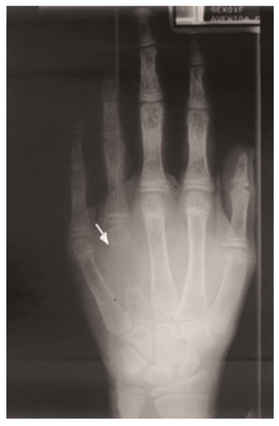
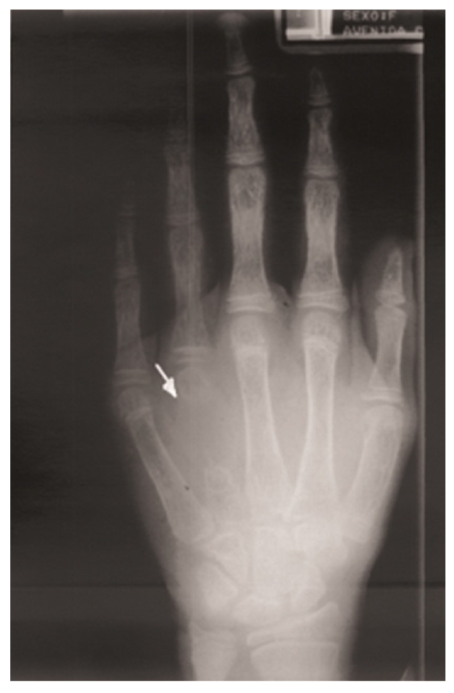
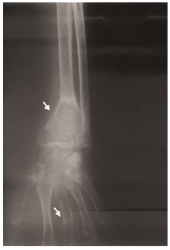
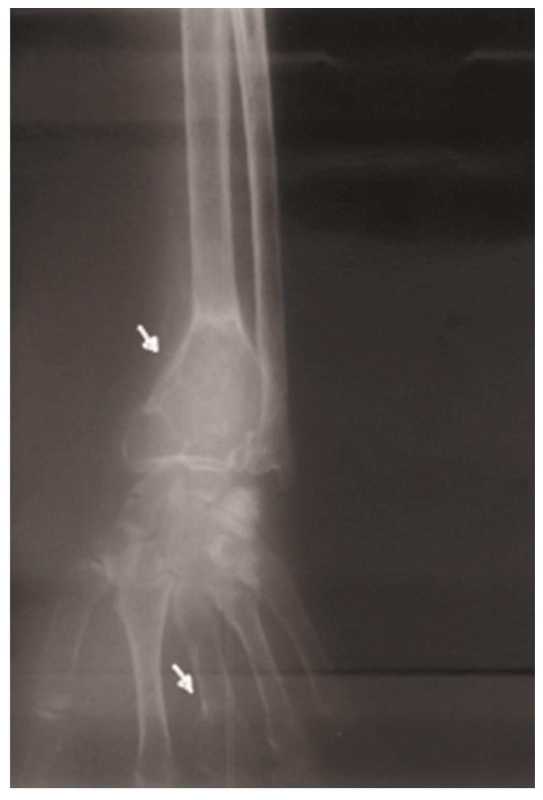
The term “osteitis fibrosa cystica” refers to the set of classic skeletal manifestations of severe PHPT. These manifestations are generally seen on radiological imaging and include: “salt and pepper” appearance of the skull, subperiosteal resorption of the distal phalanges, bone cysts, so-called “brown tumors,” “rugger-jersey” spine, and pathological fractures with skeletal deformities [1]. Currently, the occurrence of osteitis fibrosa cystica has become rare in places that use systematic biochemical calcium screening, with a prevalence of less than 5% in patients from Western Europe and North America. Due to this epidemiological shift, routine radiological investigation is no longer recommended in most reference centers [47] (Figure 1 and Figure 2).
Figure 1.
Radiograph of the left hand showing marked bone rarefaction, associated with subperiosteal resorption and the presence of a resorptive lesion at the level of the fourth metacarpal. Source: Reproduced from Morimitsu et al. [48].
Figure 2.
Plain radiograph of the right wrist showing cystic lesions in the third metacarpal and distal third of the radius. Source: Reproduced from Morimitsu et al. [48].
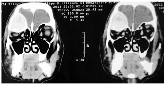
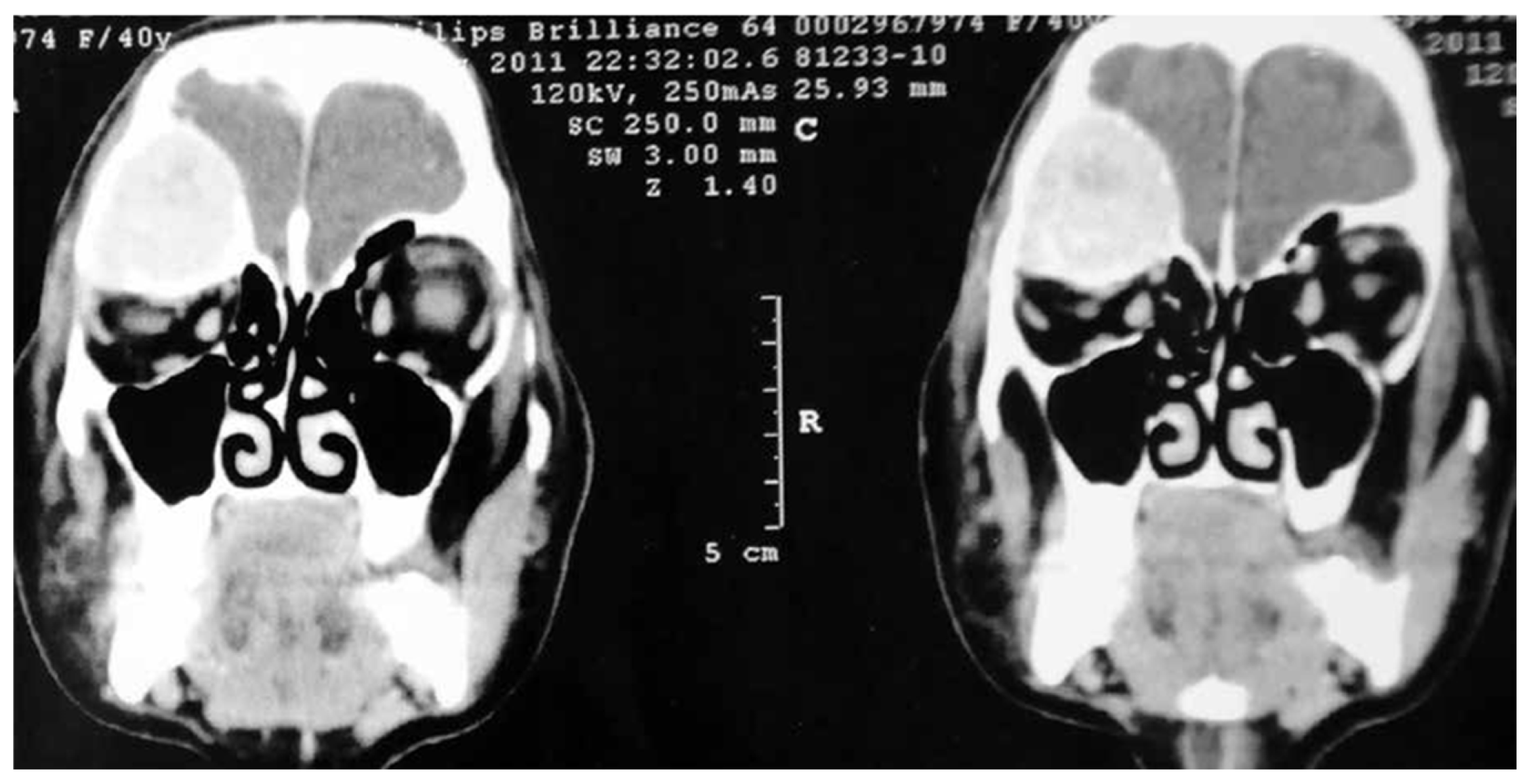
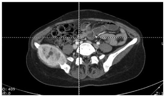
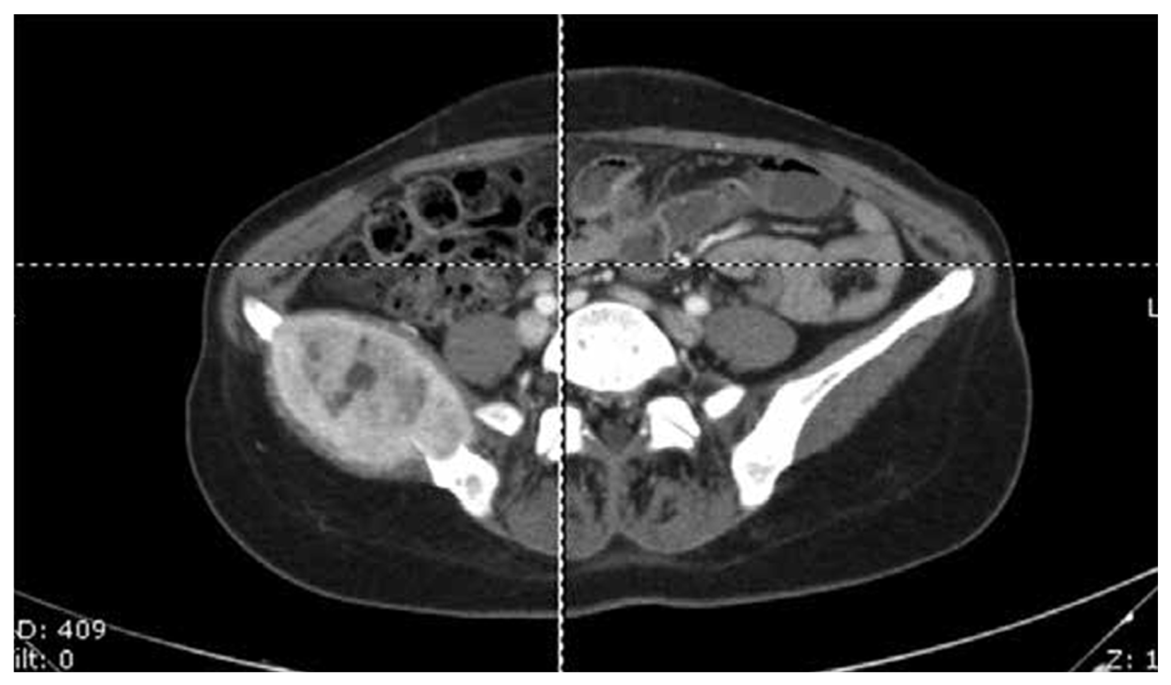
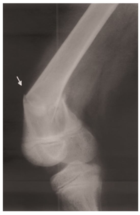
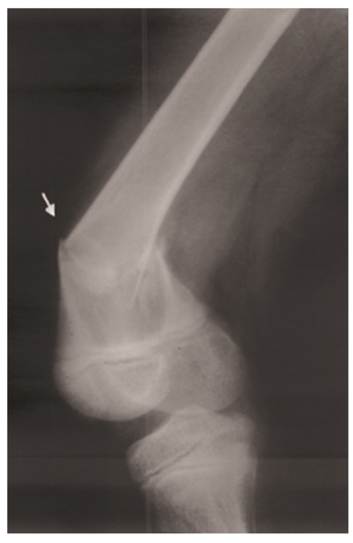
The brown tumor constitutes a non-neoplastic skeletal manifestation associated with severe and chronic states of hyperparathyroidism. Its development occurs through the exacerbation of osteoclastic activity mediated by high PTH levels, leading to severe bone resorption and the consequent replacement of mineral component by vascularized fibrous stroma. From a histological perspective, these lesions exhibit clusters of multinucleated giant cells among spindle-shaped cells, with the presence of microfractures and deposits of hemosiderin, the pigment responsible for the macroscopic brown coloration [49,50]. Despite its historical significance, the incidence of this condition in PHPT has decreased to levels between 0.5% and 4.5% in developed countries, as a result of early detection of the disease in subclinical stages. Clinically, these lesions may present as single or disseminated, with a predilection for sites such as the pelvis, femur, costal arches, mandible, and distal radius. Symptoms such as localized or generalized bone pain occur in half of patients, and diagnosis may be elucidated after the occurrence of a pathological fracture [49,50] (Figure 3, Figure 4 and Figure 5).
Figure 3.
Computed tomography image showing an expansile lesion in the supraorbital region with features consistent with a brown tumor. Source: Reproduced from Ohe et al. [51].
Figure 4.
Computed tomography image showing a heterogeneous, expansile lesion in the ilium with features consistent with a brown tumor. Source: Reproduced from Ohe et al. [51].
Figure 5.
Radiograph of the left femur showing a pathological fracture at the site of a preexisting bone cyst. Source: Reproduced from Morimitsu et al. [48].
6. Bone Density, Microarchitecture, and Fracture Risk
PHPT is a condition characterized by accelerated skeletal turnover, leading to progressive reduction in BMD, with a preference for cortical bone regions, and predisposing to a high risk of fractures at various anatomical sites [52]. The impact of PHPT on the skeleton is multifactorial and deleterious, resulting in decreased BMD—clinically manifested as osteoporosis or osteopenia—and a considerable increase in fracture risk. Although assessment tools such as the Fracture Risk Assessment Tool (FRAX) assume this risk is primarily mediated by reduced BMD, consistent epidemiological data demonstrate that PHPT establishes a state of bone fragility that goes beyond quantitative bone mass loss. Population studies show that individuals with PHPT face an increased risk for both hip fractures and major osteoporotic fractures, even among those under conservative management without immediate surgical indication. Furthermore, fracture risk may be underestimated in this population due to higher mortality risk from general causes, reducing life expectancy and thus attenuating the likelihood of experiencing a future fracture [53].
PHPT is associated with greater susceptibility to fractures in sites such as the spine, wrist, and pelvis. This increased risk is independent of BMD or clinical severity, indicating that fractures are influenced not only by reduced BMD but also by intense bone remodeling, as well as impaired bone quality and microarchitecture [54].
Given the limitations of traditional bone densitometry, dual-energy X-ray absorptiometry (DXA), in fully capturing fracture risk in PHPT, trabecular bone score (TBS) has emerged as a valuable complementary tool. TBS, through analysis of grayscale variation in the texture of the two-dimensional lumbar spine DXA image, serves as an indirect estimator of three-dimensional trabecular microarchitecture, reflecting structural quality and bone connectivity. This assessment allows for the identification of trabecular microarchitecture degradation even in patients whose lumbar spine BMD values are in the osteopenic or normal range. In practical terms, TBS provides an additional advantage over conventional DXA parameters for predicting morphometric vertebral fractures in PHPT [55].
Structural deterioration in PHPT, as evidenced by high-resolution peripheral quantitative computed tomography (HR-pQCT) analysis, occurs in a diffuse manner, extending beyond the cortex to profoundly affect the trabecular compartment. Under the influence of excess PTH, the destruction of plate-like trabeculae is observed, diminishing their connections and compromising skeletal rigidity, which is most prominent in the distal radius. Conversely, in the distal tibia, the hormone’s catabolic impact on trabecular structure is attenuated, as the mechanical stress from weight-bearing plays a partially protective role. This generalized microarchitectural decline forms the biomechanical basis for the overall increased fracture risk, reinforcing that conventional two-dimensional DXA is limited in estimating actual bone fragility and quality in this condition [56].
7. Molecular Diagnosis
According to recommendations from the Fifth International Workshop on the Evaluation and Management of Primary Hyperparathyroidism, genetic testing should be considered in patients diagnosed with PHPT before the age of 30, those with multiglandular disease identified by clinical history, imaging studies, or during surgery, as well as individuals with a family history of hypercalcemia or hereditary syndromes associated with PHPT, such as MEN1, MEN2A, MEN4, and HPT-JT. Testing is also indicated in cases of atypical tumor or parathyroid carcinoma [13]. The possibility of a hereditary condition should also be considered in cases of PTH-dependent hypercalcemia with findings suggestive of FHH, such as mild, non-progressive, early-onset hypercalcemia, normal or mildly elevated PTH concentrations, and hypocalciuria without another apparent cause, particularly when the calcium-to-creatinine clearance ratio (CCCR) is less than 0.02, especially below 0.01 [17].
The choice of genes to be analyzed should be guided by the clinical criteria that prompted the investigation. In patients with suspected hereditary predisposition to PHPT, it is recommended to evaluate the MEN1, CDC73, RET, CDKN1B, GCM2, and CASR genes; in cases suggestive of FHH, CASR, GNA11, and AP2S1 should be included. Since some patients may simultaneously meet criteria for both conditions, multigene panels that include all these genes can be used, allowing their simultaneous analysis by next-generation sequencing [57].
The identification of a germline variant can substantially change clinical and surgical management. In syndromes with a predisposition to multiglandular disease, for example, the result may indicate bilateral neck exploration and subtotal or total parathyroidectomy instead of a focal approach limited to the initially identified gland. In other situations, it may support earlier intervention due to a higher risk of parathyroid carcinoma, guide surveillance of extraparathyroid manifestations, or, in the case of FHH, avoid unnecessary and generally ineffective surgery. Moreover, molecular diagnosis enables cascade screening of family members, distinguishing those who need specific follow-up from those without increased genetic risk, and provides relevant information for reproductive planning [13,57].
8. Differential Diagnoses
The evaluation of hypercalcemia should aim to differentiate between PTH-dependent and PTH-independent causes (Table 2). In the first group, PHPT, tertiary hyperparathyroidism (associated with advanced chronic kidney disease), chronic lithium use, and FHH stand out. In contrast, among the PTH-independent causes, it is essential to rule out so-called “hypercalcemia of malignancy,” and therefore, neoplasms must be excluded. In these cases, three different mechanisms predominate: production of parathyroid hormone-related peptide (PTHrP) (as occurs in breast, kidney, or lung cancer), overproduction of 1,25(OH)2D (e.g., lymphomas), and lytic bone lesions secondary to metastases. In addition to neoplasms, other PTH-independent causes of hypercalcemia include granulomatous diseases, thyrotoxicosis, prolonged immobilization, or intoxication from vitamin A, cholecalciferol, or calcitriol [58].
Table 2.
Differential diagnosis of hypercalcemia.
The assessment of 24 h urinary calcium excretion is crucial to rule out an important differential diagnosis in clinical practice, FHH, in which the calcium/creatinine clearance ratio is typically less than 1%. On the other hand, patients with PHPT may have values >300 mg/24 h for men and >250 mg/24 h for women [60].
Lithium, an essential medication in the management of bipolar disorder, can influence serum calcium homeostasis. The drug can interfere with the calcium-sensing receptor (CaSR) present in the parathyroid glands, increasing the “set point” by which calcium inhibits PTH secretion. This calcilytic effect, antagonism at the CaSR, results in the need for higher serum calcium levels for the gland to reduce PTH synthesis and secretion. Consequently, patients on chronic lithium therapy may present with hypercalcemia associated with elevated PTH levels—a biochemical profile characteristic of PHPT. However, unlike classic PHPT, which is more often related to a parathyroid adenoma, prolonged lithium use can trigger hyperplasia of all four parathyroid glands [61,62].
Hypercalcemia of malignancy occurs in patients with a known cancer diagnosis or in those newly diagnosed after initial investigation prompted by elevated calcium levels. In this context, several pathophysiological mechanisms can explain the hypercalcemia, such as PTHrP production (common in solid tumors like breast, lung, and kidney), overproduction of 1,25(OH)2D (lymphomas), and lytic bone lesions secondary to bone metastases [63].
In addition to malignancies, other conditions can cause hypercalcemia independent of PTH action, such as granulomatous diseases—for example, sarcoidosis, in which there is hyperactivity of 1-alpha-hydroxylase, the enzyme responsible for converting 25-hydroxyvitamin D [25(OH)D] into 1,25(OH)2D, the active form of vitamin D. Hyperthyroidism should also be considered, as thyroid hormone is responsible for increased bone resorption. Intoxication from cholecalciferol, calcitriol, or vitamin A, as well as the side effect of prolonged thiazide diuretic use, should also be considered. Vitamins A and D are fat-soluble and have a prolonged half-life, increasing the risk of persistent hypercalcemia as they are stored in adipose tissue [64].
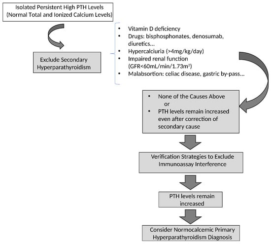
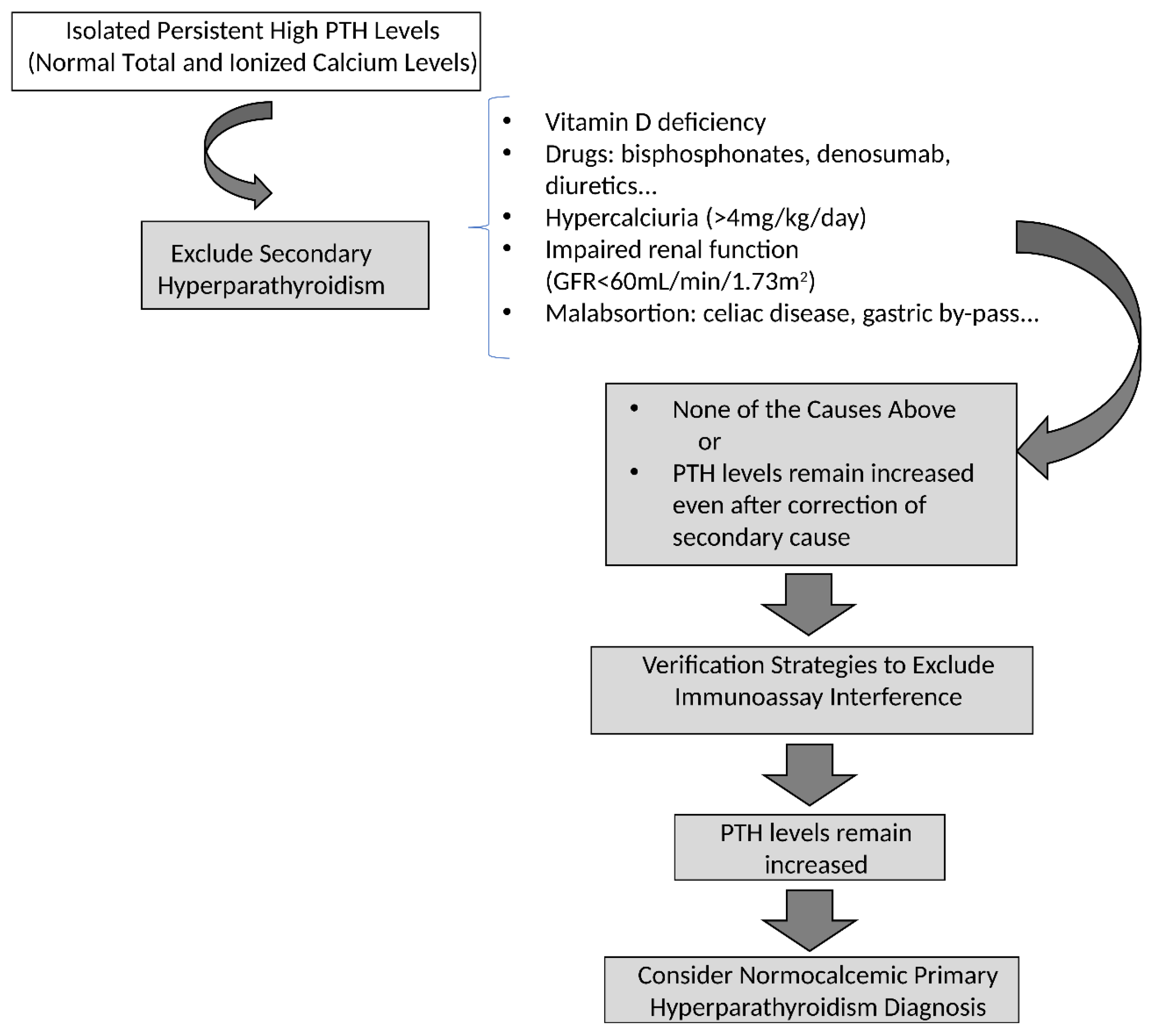
A condition that deserves attention in biochemical investigation is normocalcemic hyperparathyroidism, which, as the name suggests, presents with elevated serum PTH levels but with repeatedly normal serum calcium values (both total and ionized) [65]. Normocalcemic hyperparathyroidism is characterized by elevated PTH levels in the presence of persistently normal serum calcium concentrations, both total and ionized. The diagnosis of this condition is fundamentally based on the exclusion of secondary causes of elevated PTH, requiring a careful investigation to rule out all possible causes of secondary hyperparathyroidism [66] (Figure 6).
Figure 6.
Differential diagnosis of high isolated parathyroid hormone (PTH) levels. Abbreviations: GFR, glomerular filtration rate; PTH, parathyroid hormone. Source: Reproduced from Ohe et al. [66].
Thus, it is essential to exclude factors such as vitamin D deficiency, low intake or poor absorption of calcium, renal insufficiency, and idiopathic hypercalciuria. In addition, the use of medications such as diuretics, bisphosphonates, and denosumab should be investigated, as well as the presence of gastrointestinal disorders, including celiac disease, inflammatory bowel diseases, and sequelae of bariatric surgery. There are also reports of interfering factors in the PTH assay, ranging from pre-analytical issues such as inadequate sample storage, to contamination by biotin, or the presence of heterophile antibodies. The interference of these antibodies may lead to a falsely elevated PTH measurement, which could induce diagnostic error.
Another often overlooked cause of isolated PTH elevation is primary aldosteronism (PA). PA is associated with significantly increased urinary calcium excretion and secondary hyperparathyroidism, which has been reported in approximately 30–55% of patients [67]. This phenomenon may result, at least in part, from reduced proximal tubular calcium reabsorption secondary to decreased sodium reabsorption, leading to increased urinary calcium losses and a compensatory rise in PTH levels. In addition, autonomous cortisol secretion, which may coexist with PA, has been proposed as an additional contributor to disturbances in calcium and bone metabolism [68,69]. These and other abnormalities in mineral and bone metabolism may contribute to the increased risk of both nephrolithiasis [70,71] and fractures [72,73] observed in patients with PA. Importantly, specific treatment of PA can normalize PTH levels and improve mineral and bone metabolism, while reducing the risk of diagnostic confusion with conditions such as normocalcemic PHPT and thereby avoiding unnecessary investigations or interventions. Proper attention and exclusion of all these factors that can cause isolated elevation of PTH should be undertaken before diagnosing normocalcemic hyperparathyroidism, thus avoiding diagnostic errors and unnecessary therapeutic interventions [13,66].
9. Management
9.1. Surgical Treatment of Primary Hyperparathyroidism (PHPT)
The definitive treatment for PHPT is parathyroidectomy, with correct identification and removal of the affected parathyroid gland(s). Surgical indication is well established for symptomatic patients and those with renal and/or skeletal involvement, as these are the main candidates for surgery [74]. Thus, surgery is mandatory for symptomatic patients or for asymptomatic patients who meet specific criteria such as: serum calcium > 1.0 mg/dL above the upper limit of normal; skeletal involvement (vertebral fracture or T score ≤ −2.5 at any site); renal involvement (glomerular filtration rate < 60 mL/min, nephrolithiasis/nephrocalcinosis, or increased calciuria —>250 mg/day in women and >300 mg/day in men); or age < 50 years (Table 3) [74]. Interestingly, the technique involved in surgical treatment of PHPT has also changed over the years [75]. Driven by changes in clinical presentation, with a greater number of oligo/asymptomatic cases, there has been a need to adapt the employed technique, involving a more focused approach, with shorter surgical time and lower risk of perioperative morbidity.
Table 3.
Indications for surgery in asymptomatic PHPT.
Thus, the treatment of PHPT has evolved since the first successful parathyroidectomy performed by Felix Mandl in 1925 [76], which was initially described as a bilateral cervical exploration, and over the years has developed into a more focused approach [75]. It is important to note that 85% to 90% of PHPT cases are attributable to a single adenoma [77], and preoperative identification of abnormal glands allows for minimally invasive unilateral neck exploration, reducing operative time and surgical morbidity without compromising cure rates [78]. Recent parathyroidectomy techniques feature advances in preoperative localization exams to identify abnormal parathyroid glands as well as intraoperative PTH monitoring.
In cases of multiglandular disease, such as MEN1, the most commonly performed surgical technique—considered the “gold standard” today—is subtotal parathyroidectomy [79], in which part of the parathyroid tissue is preserved in situ. Other described techniques include total parathyroidectomy, total parathyroidectomy with autotransplantation of parathyroid tissue into the forearm or presternal muscle [80], and unilateral clearance, where the parathyroids on only one side are identified and removed [75,79]. These various surgical techniques are intended to minimize the risk of disease recurrence while also reducing the risk of postoperative hypoparathyroidism [75,79]. Each technique has its advantages and disadvantages, and discussion is needed to determine the best approach for individualized treatment of patients with multiglandular disease.
Regarding the management of normocalcemic PHPT, it should first be emphasized that this condition is relatively uncommon [65]. Moreover, studies addressing its management remain limited, and evidence-based criteria to guide either surgical or medical intervention are currently lacking. In the absence of robust data, current expert recommendations do not support the establishment of specific criteria for parathyroid surgery in this phenotype of PHPT [74].
Parathyroidectomy enables biochemical cure in more than 95% of patients, that is, it allows normalization of serum calcium, phosphorus, and PTH levels. The literature describes improvements in BMD in both the lumbar spine and femur, with benefits including reduced femur fracture rates in elderly patients [10,81,82].
A recent Cochrane review compared minimally invasive parathyroidectomy to bilateral exploration, showing that both approaches have excellent cure rates (96–99%) with no statistically significant difference. However, the literature also indicates that unilateral exploration is associated with a lower incidence of complications, particularly transient hypocalcemia and recurrent laryngeal nerve injury [83,84].
Cure following parathyroidectomy is defined as normalization of serum calcium levels. This is expected to occur in more than 95% of cases when surgery is performed at a reference center. Recurrence can be classified as early, occurring within 5 years after surgery, or late, occurring after 5 years. Early recurrence is uncommon and is usually related to failure to identify multiglandular disease during surgery. An attenuated drop in intraoperative PTH or persistent elevation of serum PTH and calcium levels during follow-up may be indicative. Late recurrence, which is more prevalent, usually denotes a new pathological process in a previously healthy gland [10,85].
9.2. Adjunct Methods in Surgical Treatment
9.2.1. Parathyroid Localization
Preoperative localization studies include a combination of noninvasive and invasive imaging methods that identify abnormal parathyroid gland(s) and/or structures [75]. These tests neither confirm nor exclude the diagnosis of PHPT and should not be used to influence the surgical indication [77,82]. Localization imaging should be performed only after the surgical indication for parathyroidectomy has been established and is intended solely for surgical planning [82,86].
Cervical ultrasound is the most frequently requested localization test. It has the advantages of being noninvasive, not involving ionizing radiation, being less costly to health services, and having an accuracy of around 76% [86,87]. It also allows for the investigation of concomitant thyroid pathology [82,88].
Parathyroid scintigraphy with technetium-99m sestamibi (99mTc-MIBI), combined with single-photon emission computed tomography/computed tomography (SPECT/CT), is a functional imaging modality and is currently the gold standard for detecting hyperfunctioning parathyroid glands [75]. For example, a meta-analysis including 23 studies with 1236 patients showed a detection rate of 88% [87]. Cervical ultrasound is often used in combination with 99mTc-MIBI, and this association is considered the most cost-effective strategy for localizing hyperfunctioning parathyroid(s) both preoperatively and in cases of recurrence [82,86,88].
Four-dimensional computed tomography (4D-CT) consists of standard CT images with three vascular phases (unenhanced, arterial, and venous) and a fourth dimension that allows assessment of enhancement over time [89,90]. This technique can also provide functional data on enlarged parathyroid glands, where rapid uptake and washout of contrast is considered a typical feature of parathyroid adenoma [91], and it has localization rates comparable to those observed with 99mTc-MIBI [92].
Other tracers used in positron emission tomography/computed tomography (PET/CT), such as methionine [87] and choline, have emerged as better options than 99mTc-MIBI, with choline being the best among them, showing a sensitivity of 95% and a positive predictive value of 97% [93]. This technique can detect small adenomas less than 1.0 cm, which are often not localized by 99mTc-MIBI, but unfortunately it is expensive and still not available in our setting.
The accuracy of localization studies is higher in single-gland disease, while negative localization imaging is suggestive of the presence of multiglandular disease in PHPT [88,94].
Among the invasive methods, intraoperative PTH sampling from the internal jugular veins guided by ultrasound stands out, mainly indicated in cases of negative or discordant localization studies and is especially useful in reoperations [95]. This technique can assist in surgical decision-making by recognizing a PTH gradient, indicating the laterality of the hyperfunctioning gland [95]. There are few studies in the literature involving intraoperative PTH sampling through bilateral central venous access. Initial reports date back to 1996, when Taylor et al. described the use of this technique in 21 patients with PHPT, achieving correct lateralization in 76% of cases [96]. However, this initial study did not yet define a sufficient cutoff value to categorize this lateralization. Only in 2007, when Ito et al. evaluated 216 patients with PHPT operated on with central intraoperative PTH measurement, did these authors manage to define a cutoff value. They considered a 5% difference between right and left jugular measurements for the PTH gradient sufficient to indicate lateralization and thus assist in locating the hypersecreting parathyroid, with a sensitivity of 78.9% [97]. The same authors demonstrated the safety of the method both intraoperatively and postoperatively, and its utility and safety have been observed in studies conducted at other medical centers [97,98].
The ideal sequence of preoperative imaging studies to be requested should be adapted according to the patient’s needs, surgeon’s preference, and availability of the various options. Repeating negative tests adds little information and further delays surgical resolution [86,99].
9.2.2. Intraoperative PTH
In 1991, Irvin et al. developed the rapid intraoperative PTH assay (IO-PTH) [100], and since 1996, IO-PTH has become commercially available and widely used by parathyroid surgeons worldwide [101,102,103,104,105,106], having been introduced in our setting in 2002 [103]. A significant percentage drop in IO-PTH, with a decrease of more than 50% from the pre-resection or pre-excision value of the supposedly hypersecreting parathyroid tissue, is indicative of surgical resolution [75]. On the other hand, absence of this decline indicates persistence of hypersecreting parathyroid tissue, requiring additional surgical exploration [75].
It was the combination of advances in the accuracy of preoperative localization imaging and intraoperative PTH measurement that made minimally invasive surgical techniques possible [75]. With this approach, surgery begins with resection of the previously localized gland and ends after confirmation of appropriate IO-PTH decline, without the need for bilateral exploration and identification of all other parathyroids [104]. This offers the advantage of lower perioperative morbidity and mortality while maintaining the same cure rates as bilateral exploration.
9.3. Clinical Treatment of PHPT
For patients who do not meet the criteria for parathyroidectomy or for those with contraindications to the procedure, clinical management is recommended, although it does not have a curative role like surgery. Clinical treatment consists of prescribing cinacalcet to reduce serum calcium levels, bisphosphonates or denosumab to improve BMD, together with adequate calcium intake and vitamin D repletion. Calcium restriction should be avoided because insufficient intake may further stimulate PTH secretion. Recommended total calcium intake is 800 mg/day for women younger than 50 years and men younger than 70 years, and 1000 mg/day for women older than 50 years and men older than 70 years, preferably from dietary sources, with supplements used when needed to meet these requirements. Vitamin D supplementation should be cautious to avoid worsening hypercalcemia, aiming to maintain serum 25(OH)D above 30 ng/mL and below the laboratory upper limit of normal (e.g., 50 ng/mL) [13,74].
The use of cinacalcet, a calcimimetic agent, quickly normalizes calcium levels in most patients and is associated with a reduction in PTH, with sustained long-term action, although the medication has no effect on BMD. On the other hand, bisphosphonates, such as alendronate, do not reduce serum calcium but do increase BMD in patients with PHPT. Therefore, the combination of cinacalcet and bisphosphonates can be considered in clinical management [1,107].
Denosumab, a monoclonal antibody targeting receptor activator of nuclear factor-κB ligand, is a medication with proven benefits in increasing BMD in the lumbar spine and femoral neck. One study demonstrated a transient increase in PTH with the use of this medication; however, it offers potential control of calcemia when used in combination with cinacalcet [108].
PHPT is a well-established cause of secondary osteoporosis, with parathyroidectomy as its main therapeutic approach, providing increases of 3% to 4% in BMD in the first postoperative year. Nevertheless, many patients continue to have clinically significant skeletal fragility, and the efficacy of adjunctive pharmacotherapy in the postoperative period remains uncertain. Recently, Takebe and colleagues reported the first cases of postoperative use of romosozumab in patients with osteoporosis associated with PHPT, documenting significant gains of 25% to 30% in BMD, concentrated in the lumbar spine. Although preliminary, these findings suggest that sclerostin inhibition may be a potential anabolic strategy to optimize bone mass gain after surgical cure [109].
10. Conclusions
PHPT is a hypercalcemic disorder characterized by excessive or inappropriately elevated production of PTH, relative to the degree of hypercalcemia, by one or more parathyroid glands. It is a common condition, and its clinical presentation profile has changed over the last decades. Previously recognized as a predominantly symptomatic disease, it now more frequently presents as oligosymptomatic or asymptomatic forms, both in developed and developing countries.
The differential diagnosis involves investigating hypercalcemia to differentiate between PTH-dependent and PTH-independent causes, as well as investigating causes of isolated PTH elevation. The possibility of assay interference in PTH measurement must also be ruled out.
Localization imaging should be performed only after diagnostic confirmation and the indication for parathyroidectomy have been established, and its role is thus limited to strategic surgical planning, not to confirm or refute the diagnosis.
There are specific considerations regarding the management and surgical treatment of patients with genetic or syndromic forms of PHPT, a condition that should be especially suspected in young individuals diagnosed with PHPT. A hereditary etiology should be particularly suspected in young individuals diagnosed with PHPT. In hereditary syndromes associated with multiglandular disease, particularly MEN1, bilateral neck exploration is often appropriate; however, the surgical strategy should be individualized according to the syndrome, gland involvement, and risk of malignancy. Recognition of FHH is essential to avoid unnecessary parathyroidectomy.
Curative treatment consists of parathyroidectomy. When performed by experienced surgeons and combined with the use of adjuncts such as preoperative localization studies and intraoperative PTH measurement, surgery achieves cure rates of 96% to 99%, both through bilateral neck exploration and minimally invasive parathyroidectomy.
Author Contributions
Writing—original draft preparation, L.T.S.F., C.A.C.B. and M.N.O.; writing—review and editing, L.T.S.F. and M.N.O. All authors have read and agreed to the published version of the manuscript.
Funding
This research received no external funding.
Institutional Review Board Statement
Not applicable.
Informed Consent Statement
Not applicable.
Data Availability Statement
No new data were created or analyzed in this study. Data sharing is not applicable to this article.
Acknowledgments
During the preparation of this manuscript, the authors used ChatGPT (OpenAI, GPT-6) to assist with translation into English, language and grammar editing, and reference organization. The authors reviewed and edited the output and take full responsibility for the content of this publication.
Conflicts of Interest
The authors declare no conflicts of interest.
References
- Bilezikian, J.P.; Bandeira, L.; Khan, A.; Cusano, N.E. Hyperparathyroidism. Lancet 2018, 391, 168–178. [Google Scholar] [CrossRef] [Scilit]
- Kurtom, S.; Carty, S.E. Primary Hyperparathyroidism: Part One: Evaluation. Surg. Clin. N. Am. 2024, 104, 791–798. [Google Scholar] [CrossRef] [Scilit]
- Shah, U.S.; McCoy, K.L.; Kelley, M.L.; Carty, S.E.; Yip, L. How and When Is Multiglandular Disease Diagnosed in Sporadic Primary Hyperparathyroidism? Surgery 2022, 171, 35–39. [Google Scholar] [CrossRef] [Scilit]
- El-Hajj Fuleihan, G.; Chakhtoura, M.; Cipriani, C.; Eastell, R.; Karonova, T.; Liu, J.; Minisola, S.; Mithal, A.; Moreira, C.A.; Peacock, M.; et al. Classical and Nonclassical Manifestations of Primary Hyperparathyroidism. J. Bone Miner. Res. 2022, 37, 2330–2350. [Google Scholar] [CrossRef] [Scilit]
- Palermo, A.; Tabacco, G.; Makras, P.; Zavatta, G.; Trimboli, P.; Castellano, E.; Yavropoulou, M.P.; Naciu, A.M.; Anastasilakis, A.D. Primary Hyperparathyroidism: From Guidelines to Outpatient Clinic. Rev. Endocr. Metab. Disord. 2024, 25, 875–896. [Google Scholar] [CrossRef] [Scilit]
- Pepe, J.; Minisola, S.; Ettorre, E.; Desideri, G.; Cipriani, C. Cardiovascular Involvement in Primary Hyperparathyroidism. J. Clin. Endocrinol. Metab. 2026, 111, 603–614. [Google Scholar] [CrossRef] [Scilit]
- Ohe, M.N.; Santos, R.O.; Barros, E.R.; Lage, A.; Kunii, I.S.; Abrahão, M.; Cervantes, O.; Hauache, O.M.; Lazaretti-Castro, M.; Vieira, J.G.H. Changes in Clinical and Laboratory Findings at the Time of Diagnosis of Primary Hyperparathyroidism in a University Hospital in São Paulo from 1985 to 2002. Braz. J. Med. Biol. Res. 2005, 38, 1383–1387. [Google Scholar] [CrossRef] [Scilit]
- Jha, S.; Simonds, W.F. Molecular and Clinical Spectrum of Primary Hyperparathyroidism. Endocr. Rev. 2023, 44, 779–818. [Google Scholar] [CrossRef] [Scilit]
- Yeh, M.W.; Ituarte, P.H.G.; Zhou, H.C.; Nishimoto, S.; Liu, I.-L.A.; Harari, A.; Haigh, P.I.; Adams, A.L. Incidence and Prevalence of Primary Hyperparathyroidism in a Racially Mixed Population. J. Clin. Endocrinol. Metab. 2013, 98, 1122–1129. [Google Scholar] [CrossRef] [Scilit]
- Pappachan, J.M.; Lahart, I.M.; Viswanath, A.K.; Borumandi, F.; Sodi, R.; Metzendorf, M.-I.; Bongaerts, B. Parathyroidectomy for Adults with Primary Hyperparathyroidism. Cochrane Database Syst. Rev. 2023, 3, CD013035. [Google Scholar] [CrossRef] [Scilit]
- Best, C.A.E.; Krishnan, R.; Malvankar-Mehta, M.S.; MacNeil, S.D. Echocardiogram changes following parathyroidectomy for primary hyperparathyroidism: A systematic review and meta-analysis. Medicine 2017, 96, e7255. [Google Scholar] [CrossRef] [Scilit]
- Arya, A.K.; Kumari, P.; Singh, P.; Bhadada, S.K. Molecular Basis of Symptomatic Sporadic Primary Hyperparathyroidism: New Frontiers in Pathogenesis. Best. Pract. Res. Clin. Endocrinol. Metab. 2025, 39, 101985. [Google Scholar] [CrossRef] [Scilit]
- Bilezikian, J.P.; Khan, A.A.; Silverberg, S.J.; El-Hajj Fuleihan, G.; Marcocci, C.; Minisola, S.; Perrier, N.; Sitges-Serra, A.; Thakker, R.V.; Guyatt, G.; et al. Evaluation and Management of Primary Hyperparathyroidism: Summary Statement and Guidelines from the Fifth International Workshop. J. Bone Miner. Res. 2022, 37, 2293–2314. [Google Scholar] [CrossRef] [Scilit]
- Silva, B.C.; Cusano, N.E.; Bilezikian, J.P. Primary Hyperparathyroidism. Best. Pract. Res. Clin. Endocrinol. Metab. 2024, 38, 101247. [Google Scholar] [CrossRef] [Scilit]
- Bollerslev, J.; Cetani, F.; Gittoes, N.; Rolighed, L. Individualized Management of Primary Hyperparathyroidism. J. Intern. Med. 2026, 299, 650–669. [Google Scholar] [CrossRef] [Scilit]
- De Leo, A.; Ruscelli, M.; Maloberti, T.; Coluccelli, S.; Repaci, A.; de Biase, D.; Tallini, G. Molecular Pathology of Endocrine Gland Tumors: Genetic Alterations and Clinicopathologic Relevance. Virchows Arch. 2024, 484, 289–319, Correction in Virchows Arch. 2024, 484, 321. https://doi.org/10.1007/s00428-024-03738-3. [Google Scholar] [CrossRef] [Scilit]
- Grover, A.; Jha, S. Heritable Hyperparathyroidism: Genetic Insights and Clinical Implications. Best. Pract. Res. Clin. Endocrinol. Metab. 2025, 39, 101984. [Google Scholar] [CrossRef] [Scilit]
- Minisola, S.; Arnold, A.; Belaya, Z.; Brandi, M.L.; Clarke, B.L.; Hannan, F.M.; Hofbauer, L.C.; Insogna, K.L.; Lacroix, A.; Liberman, U.; et al. Epidemiology, Pathophysiology, and Genetics of Primary Hyperparathyroidism. J. Bone Miner. Res. 2022, 37, 2315–2329. [Google Scholar] [CrossRef] [Scilit]
- Cetani, F.; Dinoi, E.; Pierotti, L.; Pardi, E. Familial States of Primary Hyperparathyroidism: An Update. J. Endocrinol. Investig. 2024, 47, 2157–2176. [Google Scholar] [CrossRef] [Scilit]
- Brandi, M.L.; Pieterman, C.R.C.; English, K.A.; Lines, K.E.; Shariq, O.A.; Marini, F.; Cuny, T.; Lewis, M.A.; Stratakis, C.A.; Perrier, N.D.; et al. Multiple Endocrine Neoplasia Type 1 (MEN1): Recommendations and Guidelines for Best Practice. Lancet Diabetes Endocrinol. 2025, 13, 699–721, Correction in Lancet Diabetes Endocrinol. 2025, 13, e14. https://doi.org/10.1016/S2213-8587(25)00293-1. [Google Scholar] [CrossRef] [Scilit]
- Pieterman, C.R.C.; Valk, G.D. Update on the Clinical Management of Multiple Endocrine Neoplasia Type 1. Clin. Endocrinol. 2022, 97, 409–423. [Google Scholar] [CrossRef] [Scilit]
- Santucci, N.; Ksiazek, E.; Pattou, F.; Baud, G.; Mirallié, E.; Frey, S.; Trésallet, C.; Sébag, F.; Guérin, C.; Mathonnet, M.; et al. Recurrence After Surgery for Primary Hyperparathyroidism in 517 Patients with Multiple Endocrine Neoplasia Type 1. Ann. Surg. 2024, 279, 340–345. [Google Scholar] [CrossRef] [Scilit]
- Al-Salameh, A.; Cadiot, G.; Calender, A.; Goudet, P.; Chanson, P. Clinical Aspects of Multiple Endocrine Neoplasia Type 1. Nat. Rev. Endocrinol. 2021, 17, 207–224. [Google Scholar] [CrossRef] [Scilit]
- Song, J.; Xie, J.; Zhou, J.; Zhang, Y.; Han, R.; Ye, L.; Yang, Y.; Tao, B.; Sun, L.; Chen, X.; et al. Approach to the Patient with Primary Hyperparathyroidism in Multiple Endocrine Neoplasia Type 1. J. Clin. Endocrinol. Metab. 2025, 110, 3546–3558. [Google Scholar] [CrossRef] [Scilit]
- Ruggeri, R.M.; Benevento, E.; De Cicco, F.; Grossrubatscher, E.M.; Hasballa, I.; Tarsitano, M.G.; Centello, R.; Isidori, A.M.; Colao, A.; Pellegata, N.S.; et al. Multiple Endocrine Neoplasia Type 4 (MEN4): A Thorough Update on the Latest and Least Known MEN Syndrome. Endocrine 2023, 82, 480–490. [Google Scholar] [CrossRef] [Scilit]
- Charoenngam, N.; Mannstadt, M. Multiple Endocrine Neoplasia Type 5: Emerging Evidence and Clinical Perspectives. Endocr. Relat. Cancer 2026, 33, e250219. [Google Scholar] [CrossRef] [Scilit]
- Marx, S.J. New Concepts About Familial Isolated Hyperparathyroidism. J. Clin. Endocrinol. Metab. 2019, 104, 4058–4066. [Google Scholar] [CrossRef] [Scilit]
- Marx, S.J.; Sinaii, N. Neonatal Severe Hyperparathyroidism: Novel Insights From Calcium, PTH, and the CASR Gene. J. Clin. Endocrinol. Metab. 2020, 105, 1061–1078. [Google Scholar] [CrossRef] [Scilit]
- Al-Shanafey, S.; Al-Hosaini, R.; Al-Ashwal, A.; Al-Rabeeah, A. Surgical Management of Severe Neonatal Hyperparathyroidism: One Center’s Experience. J. Pediatr. Surg. 2010, 45, 714–717. [Google Scholar] [CrossRef] [Scilit]
- Marcocci, C.; Cetani, F.; Rubin, M.R.; Silverberg, S.J.; Pinchera, A.; Bilezikian, J.P. Parathyroid Carcinoma. J. Bone Miner. Res. 2008, 23, 1869–1880. [Google Scholar] [CrossRef] [Scilit]
- Shattuck, T.M.; Välimäki, S.; Obara, T.; Gaz, R.D.; Clark, O.H.; Shoback, D.; Wierman, M.E.; Tojo, K.; Robbins, C.M.; Carpten, J.D.; et al. Somatic and Germ-Line Mutations of the HRPT2 Gene in Sporadic Parathyroid Carcinoma. N. Engl. J. Med. 2003, 349, 1722–1729. [Google Scholar] [CrossRef] [Scilit]
- Shane, E. Parathyroid Carcinoma. J. Clin. Endocrinol. Metab. 2001, 86, 485–493. [Google Scholar] [CrossRef]
- Roser, P.; Leca, B.M.; Coelho, C.; Schulte, K.-M.; Gilbert, J.; Drakou, E.E.; Kosmas, C.; Chuah, L.L.; Wassati, H.; Miras, A.D.; et al. Diagnosis and Management of Parathyroid Carcinoma: A State-of-the-Art Review. Endocr. Relat. Cancer 2023, 30, e220287. [Google Scholar] [CrossRef] [Scilit]
- Marini, F.; Giusti, F.; Palmini, G.; Aurilia, C.; Donati, S.; Brandi, M.L. Parathyroid Carcinoma: Molecular Therapeutic Targets. Endocrine 2023, 81, 409–418. [Google Scholar] [CrossRef] [Scilit]
- Chen, Q.; Kaji, H.; Nomura, R.; Sowa, H.; Yamauchi, M.; Tsukamoto, T.; Yamaguchi, T.; Kobayashi, A.; Sugimoto, T.; Chihara, K. Trial to Predict Malignancy of Affected Parathyroid Glands in Primary Hyperparathyroidism. Endocr. J. 2003, 50, 527–534. [Google Scholar] [CrossRef] [Scilit]
- Glendenning, P.; Stuckey, B.G.A.; Vasikaran, S.D. Hypercalcemia Differential Diagnosis and Investigation. Clin. Rev. Bone Miner. Metab. 2002, 1, 11–24. [Google Scholar] [CrossRef] [Scilit]
- Ladenson, J.H. Calcium Determination in Primary Hyperparathyroidism. J. Bone Miner. Res. 1991, 6, S33–S41. [Google Scholar] [CrossRef] [Scilit]
- Vieira, J.G.H.; Nishida, S.K.; Camargo, M.T.; Obara, L.H.; Kunii, I.S.; Ohe, M.N.; Hauache, O.M. Valores de Paratormônio Obtidos Com Ensaios Imunométricos Dependem Da Especificidade Do Anticorpo Amino Terminal Empregado. Arq. Bras. Endocrinol. Metabol. 2004, 48, 518–524. [Google Scholar] [CrossRef] [Scilit]
- Vieira, J.G.H.; Kunii, I.S.; Ohe, M.N.; Carvalho, A.B. Heterogeneity of Carboxyl-Terminal Parathyroid Hormone Circulating Forms in Patients with Hyperparathyroidism Due to End Stage Renal Disease. Arq. Bras. Endocrinol. Metabol. 2009, 53, 1074–1078. [Google Scholar] [CrossRef] [Scilit]
- Bonanséa, T.C.P.; Ohe, M.N.; Brandão, C.; Ferrer, C.D.F.; Santos, L.M.; Lazaretti-Castro, M.; Vieira, J.G.H. Experience with a Third-Generation Parathyroid Hormone Assay (BIO-PTH) in the Diagnosis of Primary Hyperparathyroidism in a Brazilian Population. Arch. Endocrinol. Metab. 2016, 60, 420–425. [Google Scholar] [CrossRef] [Scilit]
- Cheng, J.; Mu, D.; Wang, D.; Qiu, L.; Cheng, X. Preanalytical Considerations in Parathyroid Hormone Measurement. Clin. Chim. Acta 2023, 539, 259–265. [Google Scholar] [CrossRef] [Scilit]
- Ohe, M.N.; Bonanséa, T.C.P.; Santos, R.O.; das Neves, M.C.; Santos, L.M.; Rosano, M.; Kunii, I.S.; Castro, M.L.; Vieira, J.G.H. Prediction of Bone Mass Changes after Successful Parathyroidectomy Using Biochemical Markers of Bone Metabolism in Primary Hyperparathyroidism: Is It Clinically Useful? Arch. Endocrinol. Metab. 2019, 63, 394–401. [Google Scholar] [CrossRef] [Scilit]
- Iwanowska, M.; Kochman, M.; Szatko, A.; Zgliczyński, W.; Glinicki, P. Bone Disease in Primary Hyperparathyroidism—Changes Occurring in Bone Metabolism and New Potential Treatment Strategies. Int. J. Mol. Sci. 2024, 25, 11639. [Google Scholar] [CrossRef] [Scilit]
- Rejnmark, L.; Ejlsmark-Svensson, H. Effects of PTH and PTH Hypersecretion on Bone: A Clinical Perspective. Curr. Osteoporos. Rep. 2020, 18, 103–114. [Google Scholar] [CrossRef] [Scilit]
- Roumpou, A.; Palermo, A.; Tournis, S.; Hasenmajer, V.; Pasieka, J.L.; Kaltsas, G.; Isidori, A.; Kassi, E. Bone in Parathyroid Diseases Revisited: Evidence From Epidemiological, Surgical and New Drug Outcomes. Endocr. Rev. 2025, 46, 576–620. [Google Scholar] [CrossRef] [Scilit]
- Silva, B.C.; Bilezikian, J.P. Skeletal Abnormalities in Hypoparathyroidism and in Primary Hyperparathyroidism. Rev. Endocr. Metab. Disord. 2021, 22, 789–802. [Google Scholar] [CrossRef] [Scilit]
- Misiorowski, W.; Czajka-Oraniec, I.; Kochman, M.; Zgliczyński, W.; Bilezikian, J.P. Osteitis Fibrosa Cystica—A Forgotten Radiological Feature of Primary Hyperparathyroidism. Endocrine 2017, 58, 380–385. [Google Scholar] [CrossRef] [Scilit]
- Morimitsu, L.K.; Uyeno, M.N.O.; Goulart, M.L.; Hauache, O.M.; Vieira, J.G.H.; Alberti, V.N.; Abrahão, M.; Cervantes, O.; Lazaretti-Castro, M. Carcinoma de Paratiróide: Características Clínicas e Anátomo-Patológicas de Cinco Casos. Arq. Bras. Endocrinol. Metabol. 2001, 45, 148–156. [Google Scholar] [CrossRef] [Scilit]
- Carsote, M.; Ciobica, M.-L.; Sima, O.-C.; Valea, A.; Bondor, C.I.; Geleriu, A.; Ticolea, M.; Nistor, C.; Rusu, C.C. Brown Tumors: The Hidden Face of Primary and Renal Hyperparathyroidism Amid Real-Life Settings. J. Clin. Med. 2024, 13, 3847. [Google Scholar] [CrossRef] [Scilit]
- Karaca, M.O.; Özyıldıran, M.; Savran, M.D.; Başarır, K.; Yıldız, H.Y. Brown Tumors: Retrospective Analysis of 26 Cases. Arch. Orthop. Trauma. Surg. 2024, 144, 2927–2934. [Google Scholar] [CrossRef] [Scilit]
- Ohe, M.N.; Santos, R.O.; Hojaij, F.; Neves, M.C.; Kunii, I.S.; Orlandi, D.; Valle, L.; Martins, C.; Janovsky, C.; Ferreira, R.; et al. Parathyroid Carcinoma and Hungry Bone Syndrome. Arq. Bras. Endocrinol. Metabol. 2013, 57, 79–86. [Google Scholar] [CrossRef] [Scilit]
- Makras, P.; Anastasilakis, A.D. Bone Disease in Primary Hyperparathyroidism. Metabolism 2018, 80, 57–65. [Google Scholar] [CrossRef] [Scilit]
- Kanis, J.A.; Harvey, N.C.; Liu, E.; Vandenput, L.; Lorentzon, M.; McCloskey, E.V.; Bouillon, R.; Abrahamsen, B.; Rejnmark, L.; Johansson, H.; et al. Primary Hyperparathyroidism and Fracture Probability. Osteoporos. Int. 2023, 34, 489–499. [Google Scholar] [CrossRef] [Scilit]
- Htoo, S.T.Y.; Cusano, N.E. Management of Primary Hyperparathyroidism: Historical and Contemporary Perspectives. Endocr. Pract. 2025, 31, 1488–1494. [Google Scholar] [CrossRef] [Scilit]
- Tuzun Ersan, N.; Cindil, E.; Sodan, H.N.; Cerit, E.T.; Yalcin, M.M.; Altinova, A.E.; Toruner, F.B.; Karakoc, A.; Akturk, M. Assessment of Bone Microarchitecture in Postmenopausal Women with Primary Hyperparathyroidism Using Trabecular Bone Score. J. Clin. Densitom. 2026, 29, 101716. [Google Scholar] [CrossRef] [Scilit]
- Arboiro-Pinel, R.; Mahíllo-Fernández, I.; Díaz-Curiel, M. Bone Analysis Using Trabecular Bone Score and Dual-Energy X-Ray Absorptiometry-Based 3-Dimensional Modeling in Postmenopausal Women with Primary Hyperparathyroidism. Endocr. Pract. 2022, 28, 83–89. [Google Scholar] [CrossRef] [Scilit]
- De Sousa, S.M.C.; Carroll, R.W.; Henderson, A.; Burgess, J.; Clifton-Bligh, R.J. A Contemporary Clinical Approach to Genetic Testing for Heritable Hyperparathyroidism Syndromes. Endocrine 2022, 75, 23–32. [Google Scholar] [CrossRef] [Scilit]
- Walker, M.D.; Shane, E. Hypercalcemia: A Review. JAMA 2022, 328, 1624–1636. [Google Scholar] [CrossRef] [Scilit]
- Maier, J.D.; Levine, S.N. Hypercalcemia in the Intensive Care Unit. J. Intensive Care Med. 2015, 30, 235–252. [Google Scholar] [CrossRef] [Scilit]
- Bargagli, M.; Scoglio, M.; Howles, S.A.; Fuster, D.G. Kidney Stone Disease: Risk Factors, Pathophysiology and Management. Nat. Rev. Nephrol. 2025, 21, 794–808. [Google Scholar] [CrossRef] [Scilit]
- Lerena, V.S.; León, N.S.; Sosa, S.; Deligiannis, N.G.; Danilowicz, K.; Rizzo, L.F.L. Lithium and Endocrine Dysfunction. Medicina 2022, 82, 130–137. [Google Scholar]
- Vandermeulen, L.; Van Melkebeke, L.; Sienaert, P. Lithium-Associated Hypercalcemia and Hyperparathyroidism: A Systematic Review and Meta-Analysis. World J. Biol. Psychiatry 2024, 25, 417–429. [Google Scholar] [CrossRef] [Scilit]
- Almuradova, E.; Cicin, I. Cancer-Related Hypercalcemia and Potential Treatments. Front. Endocrinol. 2023, 14, 1039490. [Google Scholar] [CrossRef] [Scilit]
- Tonon, C.R.; Silva, T.A.A.L.; Pereira, F.W.L.; Queiroz, D.A.R.; Junior, E.L.F.; Martins, D.; Azevedo, P.S.; Okoshi, M.P.; Zornoff, L.A.M.; de Paiva, S.A.R.; et al. A Review of Current Clinical Concepts in the Pathophysiology, Etiology, Diagnosis, and Management of Hypercalcemia. Med. Sci. Monit. 2022, 28, e935821. [Google Scholar] [CrossRef] [Scilit]
- Schini, M.; Jacques, R.M.; Oakes, E.; Peel, N.F.A.; Walsh, J.S.; Eastell, R. Normocalcemic Hyperparathyroidism: Study of Its Prevalence and Natural History. J. Clin. Endocrinol. Metab. 2020, 105, e1171–e1186. [Google Scholar] [CrossRef] [Scilit]
- Ohe, M.N.; Takimoto, R.M.; Ferrer, C.M.A.d.F.; Viana Lima, J.; Biscolla, R.P.; Vieira, J.G.H.; Chiamolera, M.I. PTH Immunoassay Interference: Differential Diagnosis with Normocalcemic Primary Hyperparathyroidism? Arch. Endocrinol. Metab. 2024, 68, e230315. [Google Scholar] [CrossRef] [Scilit]
- Asbach, E.; Bekeran, M.; König, A.; Lang, K.; Hanslik, G.; Treitl, M.; Ladurner, R.; Bidlingmaier, M.; Beuschlein, F.; Quinkler, M.; et al. Primary and Secondary Hyperparathyroidism in Patients with Primary Aldosteronism—Findings From the German Conn’s Registry. Exp. Clin. Endocrinol. Diabetes 2020, 128, 246–254. [Google Scholar] [CrossRef] [Scilit]
- Araujo-Castro, M.; Paja Fano, M.; Pla Peris, B.; González Boillos, M.; Pascual-Corrales, E.; García-Cano, A.M.; Parra Ramírez, P.; Rojas-Marcos, P.M.; Ruiz-Sanchez, J.G.; Vicente, A.; et al. Autonomous Cortisol Secretion in Patients with Primary Aldosteronism: Prevalence and Implications on Cardiometabolic Profile and on Surgical Outcomes. Endocr. Connect. 2023, 12, e230043. [Google Scholar] [CrossRef] [Scilit]
- Huang, C.; Chang, L.-Y.; Sheu, J.-Y.; Huang, Y.-T.; Chen, J.-Y.; Lai, C.-F.; Wu, V.-C. Exploring the High Prevalence, Comorbidities, and Indicators of Mild Autonomous Cortisol Secretion in Primary Aldosteronism: A Cohort Study and Systematic Review. Hypertens. Res. 2025, 48, 1716–1729. [Google Scholar] [CrossRef] [Scilit]
- Asayama, I.; Notsu, M.; Ota, M.; Yamamoto, M.; Kanasaki, K. Primary Aldosteronism Increases the Risk of Urinary Stones. Endocr. J. 2025, 72, 1299–1307. [Google Scholar] [CrossRef] [Scilit]
- Kang, V.J.-W.; Lee, B.-C.; Huang, J.-Z.; Wu, V.-C.; Lin, Y.-H.; Chang, C.-C. Aldosterone-Producing Adenoma Is Associated with Urolithiasis in Primary Aldosteronism. Endocr. Connect. 2023, 12, e230056. [Google Scholar] [CrossRef] [Scilit]
- Wang, A.; Wang, Y.; Liu, H.; Hu, X.; Li, J.; Xu, H.; Nie, Z.; Zhang, L.; Lyu, Z. Bone and Mineral Metabolism in Patients with Primary Aldosteronism: A Systematic Review and Meta-Analysis. Front. Endocrinol. 2022, 13, 1027841. [Google Scholar] [CrossRef] [Scilit]
- Gkaniatsa, E.; Sandström, T.Z.; Rosengren, A.; Trimpou, P.; Muth, A.; Johannsson, G.; Ragnarsson, O. Hip Fractures in Patients with Primary Aldosteronism—A Swedish Nationwide Study. Osteoporos. Int. 2024, 35, 1585–1593. [Google Scholar] [CrossRef] [Scilit]
- Bilezikian, J.P.; Silverberg, S.J.; Bandeira, F.; Cetani, F.; Chandran, M.; Cusano, N.E.; Ebeling, P.R.; Formenti, A.M.; Frost, M.; Gosnell, J.; et al. Management of Primary Hyperparathyroidism. J. Bone Miner. Res. 2022, 37, 2391–2403. [Google Scholar] [CrossRef] [Scilit]
- das Neves, M.C.; Santos, R.O.; Ohe, M.N. Surgery for Primary Hyperparathyroidism. Arch. Endocrinol. Metab. 2022, 66, 678–688. [Google Scholar] [CrossRef] [Scilit]
- Prescott, J.D.; Udelsman, R. Remedial Operation for Primary Hyperparathyroidism. World J. Surg. 2009, 33, 2324–2334. [Google Scholar] [CrossRef] [Scilit]
- Weber, T.; Dotzenrath, C.; Dralle, H.; Niederle, B.; Riss, P.; Holzer, K.; Kußmann, J.; Trupka, A.; Negele, T.; Kaderli, R.; et al. Management of Primary and Renal Hyperparathyroidism: Guidelines from the German Association of Endocrine Surgeons (CAEK). Langenbeck’s Arch. Surg. 2021, 406, 571–585. [Google Scholar] [CrossRef] [Scilit]
- Kim, W.W.; Lee, Y.-M.; Sung, T.-Y.; Chung, K.-W.; Hong, S.J. Selection of Parathyroidectomy Methods for Primary Hyperparathyroidism According to Concordance between Ultrasonography and MIBI Scan Results. Gland Surg. 2021, 10, 298–306. [Google Scholar] [CrossRef] [Scilit]
- Perrier, N.; Lang, B.H.; Farias, L.C.B.; Poch, L.L.; Sywak, M.; Almquist, M.; Vriens, M.R.; Yeh, M.W.; Shariq, O.; Duh, Q.-Y.; et al. Surgical Aspects of Primary Hyperparathyroidism. J. Bone Miner. Res. 2022, 37, 2373–2390. [Google Scholar] [CrossRef] [Scilit]
- Santos, R.O.; Ohe, M.N.; Carvalho, A.B.; Neves, M.C.; Kunii, I.; Lazaretti-Castro, M.; Abrahão, M.; Cervantes, O.; Vieira, J.G.H. Total Parathyroidectomy with Presternal Intramuscular Autotransplantation in Renal Patients: A Prospective Study of 66 Patients. J. Osteoporos. 2012, 2012, 631243. [Google Scholar] [CrossRef] [Scilit]
- Seib, C.D.; Meng, T.; Suh, I.; Harris, A.H.S.; Covinsky, K.E.; Shoback, D.M.; Trickey, A.W.; Kebebew, E.; Tamura, M.K. Risk of Fracture Among Older Adults with Primary Hyperparathyroidism Receiving Parathyroidectomy vs Nonoperative Management. JAMA Intern. Med. 2022, 182, 10–18. [Google Scholar] [CrossRef] [Scilit]
- Wilhelm, S.M.; Wang, T.S.; Ruan, D.T.; Lee, J.A.; Asa, S.L.; Duh, Q.-Y.; Doherty, G.M.; Herrera, M.F.; Pasieka, J.L.; Perrier, N.D.; et al. The American Association of Endocrine Surgeons Guidelines for Definitive Management of Primary Hyperparathyroidism. JAMA Surg. 2016, 151, 959–968. [Google Scholar] [CrossRef] [Scilit]
- Ahmadieh, H.; Kreidieh, O.; Akl, E.A.; El-Hajj Fuleihan, G. Minimally Invasive Parathyroidectomy Guided by Intraoperative Parathyroid Hormone Monitoring (IOPTH) and Preoperative Imaging versus Bilateral Neck Exploration for Primary Hyperparathyroidism in Adults. Cochrane Database Syst. Rev. 2020, 10, CD010787. [Google Scholar] [CrossRef] [Scilit]
- Jinih, M.; O’Connell, E.; O’Leary, D.P.; Liew, A.; Redmond, H.P. Focused Versus Bilateral Parathyroid Exploration for Primary Hyperparathyroidism: A Systematic Review and Meta-Analysis. Ann. Surg. Oncol. 2017, 24, 1924–1934. [Google Scholar] [CrossRef] [Scilit]
- Gomez-Carrillo, D.; Swaminathan, N.; Caretti, R.; Arain, H.A.; Roy, R.; Izhar, A.; Kasmirski, J.; Lindeman, B.; Gillis, A.; Dream, S.; et al. Long-Term Outcomes and Predictors of Recurrence in Primary Hyperparathyroidism. J. Surg. Res. 2026, 321, 600–608. [Google Scholar] [CrossRef] [Scilit]
- Khan, A.A.; Hanley, D.A.; Rizzoli, R.; Bollerslev, J.; Young, J.E.M.; Rejnmark, L.; Thakker, R.; D’Amour, P.; Paul, T.; Van Uum, S.; et al. Primary Hyperparathyroidism: Review and Recommendations on Evaluation, Diagnosis, and Management. A Canadian and International Consensus. Osteoporos. Int. 2017, 28, 1–19. [Google Scholar] [CrossRef] [Scilit]
- Petranović Ovčariček, P.; Giovanella, L.; Carrió Gasset, I.; Hindié, E.; Huellner, M.W.; Luster, M.; Piccardo, A.; Weber, T.; Talbot, J.-N.; Verburg, F.A. The EANM Practice Guidelines for Parathyroid Imaging. Eur. J. Nucl. Med. Mol. Imaging 2021, 48, 2801–2822. [Google Scholar] [CrossRef] [Scilit]
- Chander, N.R.; Chidambaram, S.; Van Den Heede, K.; DiMarco, A.N.; Tolley, N.S.; Palazzo, F.F. Correlation of Preoperative Imaging Findings and Parathyroidectomy Outcomes Support NICE 2019 Guidance. J. Clin. Endocrinol. Metab. 2022, 107, e1242–e1248. [Google Scholar] [CrossRef] [Scilit]
- Hoang, J.K.; Williams, K.; Gaillard, F.; Dixon, A.; Sosa, J.A. Parathyroid 4D-CT. Otolaryngol. Head Neck Surg. 2016, 155, 956–960. [Google Scholar] [CrossRef] [Scilit]
- Bahl, M.; Sepahdari, A.R.; Sosa, J.A.; Hoang, J.K. Parathyroid Adenomas and Hyperplasia on Four-Dimensional CT Scans: Three Patterns of Enhancement Relative to the Thyroid Gland Justify a Three-Phase Protocol. Radiology 2015, 277, 454–462. [Google Scholar] [CrossRef] [Scilit]
- Kunstman, J.W.; Kirsch, J.D.; Mahajan, A.; Udelsman, R. Parathyroid Localization and Implications for Clinical Management. J. Clin. Endocrinol. Metab. 2013, 98, 902–912. [Google Scholar] [CrossRef] [Scilit]
- Treglia, G.; Trimboli, P.; Huellner, M.; Giovanella, L. Imaging in Primary Hyperparathyroidism: Focus on the Evidence-Based Diagnostic Performance of Different Methods. Minerva Endocrinol. 2018, 43, 133–143. [Google Scholar] [CrossRef] [Scilit]
- Patel, D.D.; Bhattacharjee, S.; Pandey, A.K.; Kopp, C.R.; Ashwathanarayana, A.G.; Patel, H.V.; Barnabas, R.; Bhadada, S.K.; Dodamani, M.H. Comparison of 4D Computed Tomography and F-18 Fluorocholine PET for Localisation of Parathyroid Lesions in Primary Hyperparathyroidism: A Systematic Review and Meta-analysis. Clin. Endocrinol. 2023, 99, 262–271. [Google Scholar] [CrossRef] [Scilit]
- Wu, S.; Hwang, S.S.; Haigh, P.I. Influence of a Negative Sestamibi Scan on the Decision for Parathyroid Operation by the Endocrinologist and Surgeon. Surgery 2017, 161, 35–43. [Google Scholar] [CrossRef] [Scilit]
- Bergenfelz, A.; van Slycke, S.; Makay, Ö.; Brunaud, L. European Multicentre Study on Outcome of Surgery for Sporadic Primary Hyperparathyroidism. Br. J. Surg. 2021, 108, 675–683. [Google Scholar] [CrossRef] [Scilit]
- Taylor, J.; Fraser, W.; Banaszkiewicz, P.; Drury, P.; Atkins, P. Lateralization of Parathyroid Adenomas by Intra-Operative Parathormone Estimation. J. R. Coll. Surg. Edinb. 1996, 41, 174–177. [Google Scholar]
- Ito, F.; Sippel, R.; Lederman, J.; Chen, H. The Utility of Intraoperative Bilateral Internal Jugular Venous Sampling with Rapid Parathyroid Hormone Testing. Ann. Surg. 2007, 245, 959–963. [Google Scholar] [CrossRef] [Scilit]
- Ribeiro, D.K.; das Neves, M.C.; Santos, R.O.; Ohe, M.N.; Lazaretti-Castro, M.; Rosano, M.; Abrahao, M. Effectiveness of Bilateral Internal Jugular Parathyroid Hormone Measurements in Optimizing Hyperparathyroidism Surgery Outcomes. OTO Open 2025, 9, e70122. [Google Scholar] [CrossRef] [Scilit]
- Krishnamurthy, V.D.; Sound, S.; Okoh, A.K.; Yazici, P.; Yigitbas, H.; Neumann, D.; Doshi, K.; Berber, E. The Utility of Repeat Sestamibi Scans in Patients with Primary Hyperparathyroidism after an Initial Negative Scan. Surgery 2017, 161, 1651–1658. [Google Scholar] [CrossRef] [Scilit]
- Irvin, G.L.; Dembrow, V.D.; Prudhomme, D.L. Operative Monitoring of Parathyroid Gland Hyperfunction. Am. J. Surg. 1991, 162, 299–302. [Google Scholar] [CrossRef] [Scilit]
- Udelsman, R. Six Hundred Fifty-Six Consecutive Explorations for Primary Hyperparathyroidism. Ann. Surg. 2002, 235, 665–672. [Google Scholar] [CrossRef] [Scilit]
- Westerdahl, J.; Lindblom, P.; Bergenfelz, A. Measurement of Intraoperative Parathyroid Hormone Predicts Long-Term Operative Success. Arch. Surg. 2002, 137, 186–190. [Google Scholar] [CrossRef] [Scilit]
- Ohe, M.N.; Santos, R.O.; Kunii, I.S.; Carvalho, A.B.; Abrahão, M.; Cervantes, O.; Lazaretti-Castro, M.; Vieira, J.G.H. Usefulness of a Rapid Immunometric Assay for Intraoperative Parathyroid Hormone Measurements. Braz. J. Med. Biol. Res. 2003, 36, 715–721. [Google Scholar] [CrossRef] [Scilit]
- Neves, M.C.; Ohe, M.N.; Rosano, M.; Abrahão, M.; Cervantes, O.; Lazaretti-Castro, M.; Vieira, J.G.H.; Kunii, I.S.; Santos, R.O. A 10-Year Experience in Intraoperative Parathyroid Hormone Measurements for Primary Hyperparathyroidism: A Prospective Study of 91 Previous Unexplored Patients. J. Osteoporos. 2012, 2012, 914214. [Google Scholar] [CrossRef] [Scilit]
- Chen, H.; Pruhs, Z.; Starling, J.R.; Mack, E. Intraoperative Parathyroid Hormone Testing Improves Cure Rates in Patients Undergoing Minimally Invasive Parathyroidectomy. Surgery 2005, 138, 583–590. [Google Scholar] [CrossRef] [Scilit]
- Irvin, G.L.; Carneiro, D.M.; Solorzano, C.C. Progress in the Operative Management of Sporadic Primary Hyperparathyroidism Over 34 Years. Ann. Surg. 2004, 239, 704–711. [Google Scholar] [CrossRef] [Scilit]
- Chandran, M.; Bilezikian, J.P.; Lau, J.; Rajeev, R.; Yang, S.P.; Samuel, M.; Parameswaran, R. The Efficacy and Safety of Cinacalcet in Primary Hyperparathyroidism: A Systematic Review and Meta-Analysis of Randomized Controlled Trials and Cohort Studies. Rev. Endocr. Metab. Disord. 2022, 23, 485–501. [Google Scholar] [CrossRef] [Scilit]
- Leere, J.S.; Karmisholt, J.; Robaczyk, M.; Lykkeboe, S.; Handberg, A.; Steinkohl, E.; Brøndum Frøkjær, J.; Vestergaard, P. Denosumab and Cinacalcet for Primary Hyperparathyroidism (DENOCINA): A Randomised, Double-Blind, Placebo-Controlled, Phase 3 Trial. Lancet Diabetes Endocrinol. 2020, 8, 407–417. [Google Scholar] [CrossRef] [Scilit]
- Takebe, S.; Seki, Y.; Morimoto, S.; Watanabe, D.; Ichihara, A. Romosozumab Treatment After Parathyroidectomy for Primary Hyperparathyroidism-Complicated Osteoporosis: A Two-Case Report. AACE Endocrinol. Diabetes 2026, 13, 657–661. [Google Scholar] [CrossRef] [Scilit]

Disclaimer/Publisher’s Note: The statements, opinions and data contained in all publications are solely those of the individual author(s) and contributor(s) and not of MDPI and/or the editor(s). MDPI and/or the editor(s) disclaim responsibility for any injury to people or property resulting from any ideas, methods, instructions or products referred to in the content. |
© 2026 by the authors. Licensee MDPI, Basel, Switzerland. This article is an open access article distributed under the terms and conditions of the Creative Commons Attribution (CC BY) license.