


Abstract
Background/Objectives: Indocyanine green (ICG) fluorescence imaging is inexpensive and increasingly used in pancreatic surgery, but its evidence base has been appraised only in fragments—one application at a time, or ICG pooled with investigational fluorophores—and never specifically for the pancreatic head and periampullary region. We mapped its intraoperative applications, techniques and reported outcomes. Methods: Following the PRISMA-ScR guidelines, we searched PubMed, Scopus, the Cochrane Library and the Directory of Open Access Journals on 29 July 2026 without date restriction. Two reviewers screened and charted independently, one row per article-by-application combination, and synthesised the findings narratively. Results: Forty-two studies (51 article-by-application combinations, 613 patients) covered six applications: stump perfusion, biliary identification and anastomotic testing, perfusion of reconstructed vessels and the duodenal arcade, tumour detection and margin assessment, lymphatic mapping, and staging laparoscopy. Eighteen were case or technique reports, no randomised trial was identified, and only three studies included a group operated without ICG. Remnant hypoperfusion predicted postoperative acute pancreatitis (80% versus 16%, p = 0.011) and clinically relevant fistula (odds ratio 10.0) in two small prospective cohorts, yet in the largest series (n = 100) a visual score of the pancreaticojejunostomy was reproducible (intraclass correlation coefficient 0.76) but discriminated poorly (area under the curve 0.64) and altered no operation. In a single retrospective comparison in duodenum-preserving resection, ICG guidance was associated with less intraoperative bile-duct injury (10% versus 60%, p = 0.009). Conventional doses did not distinguish pancreatic adenocarcinoma from normal parenchyma, whereas second-window imaging made 12 of 13 invasive specimens fluoresce. Dose varied by two orders of magnitude, and no adverse reaction was reported, although tolerability was formally assessed in only nine studies. Conclusions: Intraoperative ICG use in pancreatic head surgery appears safe and feasible. ICG shows key anatomical structures—above all the biliary tree—more reliably than white light alone. Its most promising benefits are the early recognition of an ischaemic pancreatic remnant, with the prospect of preventing postoperative fistula and pancreatitis, and the early identification and protection of the distal bile duct during duodenum-preserving pancreatic head resection.
1. Introduction
Pancreatic and periampullary cancers are among the most lethal solid malignancies: GLOBOCAN 2022 estimated approximately 511,000 new pancreatic cancer cases and 467,000 deaths in a single year [1]. Pancreaticoduodenectomy—the Whipple procedure—remains the only potentially curative treatment for resectable tumours of the pancreatic head, distal common bile duct, ampulla of Vater and duodenum, and is increasingly performed for premalignant and borderline lesions as well. Alongside it, a family of parenchyma- and duodenum-preserving operations on the same anatomical field—duodenum-preserving pancreatic head resection (DPPHR), transduodenal ampullectomy, pancreas-preserving duodenectomy and enucleation—has expanded the repertoire for benign and low-grade pancreatic-head disease.
Advances in technique, perioperative care and centralisation have made pancreaticoduodenectomy a comparatively safe operation, but not without complications. In an international benchmark study of 6186 pancreaticoduodenectomies at 23 expert centres, the benchmark cutoffs (75th percentile) were 1.6% for in-hospital mortality, 73% for overall morbidity at six months and 30% for severe complications (Clavien–Dindo grade ≥ III), and the median severe-complication rate 19% [2]. Even at its best, therefore, current practice leaves between one in five and one in three patients with a severe complication. The complication most consistently linked to this burden is postoperative pancreatic fistula (POPF), defined by the International Study Group on Pancreatic Surgery (ISGPS) as a drain amylase more than three times the institutional upper limit of normal serum amylase on or after postoperative day 3, with a clinically relevant consequence [3]. Its clinically relevant benchmark cutoff in the same series was 19% [2], and it drives much of the downstream morbidity, including intra-abdominal sepsis, delayed gastric emptying and postpancreatectomy haemorrhage. Biliary anastomotic leak and injury to the residual pancreatic, duodenal or hepatic blood supply carry similarly serious consequences. Many of these complications trace back to a single intraoperative judgement: whether a given tissue—the pancreatic remnant, the biliary anastomosis, the preserved duodenum, a reconstructed vessel—is adequately perfused and correctly identified before the abdomen is closed.
That judgement has traditionally rested on the surgeon’s visual and tactile assessment—tissue colour, capillary refill, pulsation—an inherently subjective method that leaves no objective record. Indocyanine green (ICG) fluorescence imaging offers a real-time, objective alternative: a water-soluble tricarbocyanine dye that, once bound to plasma proteins, absorbs maximally at about 810 nm, is excited at approximately 750–800 nm and fluoresces in the near infrared, at about 830 nm. It is cleared almost exclusively by the liver into bile, giving a well-characterised safety profile and a defined window in which perfusion and biliary excretion can be visualised with a dedicated camera system [4].
ICG is already embedded in routine practice in other fields of surgery. Its surgical career began in the liver, where ICG clearance has long served as an index of hepatic functional reserve and, combined with volumetry, still guides the selection of patients for major hepatectomy in Asia [4]. In hepatobiliary surgery, Ishizawa et al. exploited its disordered biliary excretion in cancer tissue, and in the parenchyma the tumour compresses, to identify liver tumours in real time [5]. In colorectal surgery, the multi-institutional PILLAR II study showed that fluorescence angiography changes what surgeons do, altering the plan in 11 of 139 patients (7.9%) with no anastomotic leak among them [6], but two subsequent randomised trials, AVOID [7] and IntAct [8], failed to convert that behaviour change into a significant reduction in leak. The colorectal experience is therefore instructive in both directions: perfusion imaging reliably changes surgeon behaviour, but changed behaviour does not automatically mean fewer complications—a caveat that applies directly to the pancreatic literature.
Uptake in pancreatic-head surgery has been slower and more uneven, and the evidence has so far been appraised only in fragments. Two meta-analyses have each pooled a single application: intraoperative tumour detection, on only six studies and 64 lesions [9]; and pancreatic stump perfusion, on three studies and 100 patients [10]. A 2025 review surveyed ICG across pancreatic surgery more broadly, but narratively, on a single-database search without an explicit Boolean strategy, screening counts or independent reviewers [11]. The only prior scoping review, by Piper et al., searched five databases to 22 November 2022 and included 47 reports and 754 patients across the full range of pancreatic resections and of fluorescent agents—four early-phase human studies of still-investigational tumour-targeted probes among them—and covered biliary tract injury with only four studies and 79 patients, in none of which ICG altered intraoperative management [12].
Three gaps follow. First, no synthesis has been restricted to ICG itself [9,10,11,12], the only fluorophore in routine clinical use. Second, none has been anchored to the pancreatic-head and periampullary field [9,10,11,12], where the anatomical problems ICG is asked to solve—identifying the intrapancreatic bile duct, preserving the pancreaticoduodenal arcade, judging the perfusion of a remnant destined for anastomosis—recur across pancreaticoduodenectomy and its duodenum-preserving variants. Third, the evidence has moved: more than half of the studies identified here were published after the November 2022 search of the previous scoping review [12], among them a comparative series reporting less intraoperative bile-duct injury with ICG guidance, and cohorts linking ICG-defined stump hypoperfusion to postoperative pancreatic fistula and post-pancreatectomy acute pancreatitis (Section 3.4).
This scoping review therefore maps the evidence on intraoperative ICG use in pancreatic-head and periampullary surgery, restricted to ICG, extended by a search conducted in July 2026, and organised by the primary surgical application of ICG and by surgical scope relative to the classic Whipple procedure. Three questions are addressed: for which intraoperative purposes ICG fluorescence has been used during these operations; what dose, route, timing and imaging platform were used for each purpose; and what technical, perfusion-related, oncological and safety outcomes have been reported.
2. Methods
2.1. Study Design
This scoping review was conducted following the methodological framework of Arksey and O’Malley [13], as refined by Levac et al. [14], and is reported in accordance with the PRISMA extension for Scoping Reviews (PRISMA-ScR) checklist [15]. The completed checklist is provided as Supplementary File S2. The review was registered retrospectively with the Open Science Framework on 13 October 2026 (https://osf.io/7euwn), at the request of the journal’s editorial office. No prospective protocol was published. A scoping design was chosen because the aim was to map the breadth of the available evidence on the intraoperative use of indocyanine green (ICG) fluorescence imaging in pancreatic head and periampullary surgery, rather than to answer a single narrowly defined question. The literature in this area is markedly heterogeneous—six distinct intraoperative applications of ICG, designs ranging from single case reports to prospective cohorts, and no standardised dose, timing or imaging platform—which further favoured a scoping review over a systematic review with meta-analysis. To manage that heterogeneity, every included study was tagged along two complementary axes: the primary intraoperative use of ICG it addressed, in the six categories (A–F) defined in Section 2.6, which form the organising structure of the review; and its surgical scope relative to pancreaticoduodenectomy, reported for every study in Table 1. Duodenum-preserving pancreatic head resection and the other closely related operations were deliberately retained rather than excluded, because they are performed in the same anatomical field as pancreaticoduodenectomy, in which the intrapancreatic bile duct and the pancreaticoduodenal vascular arcade are the structures at risk, and because every finding is reported with the operation in which it was obtained (Table 1).
Table 1.
Study and patient characteristics of the 42 included studies, grouped by the primary intraoperative use of ICG.
2.2. Eligibility Criteria
Eligibility was framed using the Population–Concept–Context (PCC) structure recommended for scoping reviews [16]. Studies were included if they met all of the following criteria:
- •
- Population: Patients undergoing surgery of the pancreatic head or periampullary region—pancreaticoduodenectomy (classic or pylorus-preserving Whipple procedure) and closely related operations sharing the same anatomical field and reconstructive steps: duodenum-preserving pancreatic head resection, transduodenal ampullectomy, pancreas-preserving duodenectomy, parenchyma-sparing enucleation of the pancreatic head, redo hepaticojejunostomy, and staging laparoscopy before a planned pancreatic-head resection. Redo hepaticojejunostomy was included because it concerns the same type of biliary-enteric anastomosis that pancreaticoduodenectomy creates and intraoperative questions of the same kind, the identification of the biliary tree in a scarred field, which in a redo operation includes the previous anastomosis, and the perfusion of the duct to be anastomosed, and staging laparoscopy because it is the staging step of a planned pancreatic-head resection. Mixed pancreatic-resection cohorts were eligible where pancreatic-head resections formed part of the reported series. They were retained whole because they contain pancreatic-head resections and because the aim of a scoping review is to map all use of ICG in this field rather than to pool outcomes. In the surgical-scope column of Table 1 the borderline operations are labelled “borderline/transferable” and the mixed cohorts “other pancreatic operation”, their numbers are given in Section 3.2, and their contribution to the patient totals is quantified in Section 4.3.
- •
- Concept: Intraoperative or periprocedural administration of ICG with near-infrared fluorescence imaging for any surgical purpose during the index operation, by any route—intravenous, intraductal, intrabiliary, intraparenchymal or intralesional.
- •
- Context: Any clinical setting worldwide, any surgical approach (open, laparoscopic or robotic) and any period, with no date restriction.
- •
- Comparator: None required, given the exploratory nature of the scoping design. Studies with or without a non-ICG control arm were eligible.
- •
- Outcomes: At least one technical, perfusion-related, oncological or safety outcome associated with ICG use—for example postoperative pancreatic fistula (POPF) or post-pancreatectomy acute pancreatitis by perfusion status (PPAP, as standardised by the ISGPS [17]; also reported as postoperative acute pancreatitis, POAP), bile-duct injury or leak rates, R0 or margin status, the diagnostic accuracy of ICG for lesion or metastasis detection, or an intraoperative management change prompted by ICG.
- •
- Study design: Original reports of any design—prospective and retrospective cohort studies, cross-sectional studies, case series, and case reports or technique reports—reflecting the early and largely descriptive stage of this literature.
- •
- Language and species: English-language publications and human studies only.
- •
- Excluded: paediatric populations; fluorescent agents other than ICG; reports mentioning ICG only in passing, without describing its actual intraoperative application; preclinical and ex-vivo studies; narrative and systematic reviews, meta-analyses, editorials and letters without original data, conference abstracts, study protocols without reported outcome data, and short commentaries published alongside a primary report of the same patients.
2.3. Search Strategy
We searched four electronic databases—PubMed, Scopus, the Cochrane Library and the Directory of Open Access Journals (DOAJ)—on 29 July 2026. In PubMed, Scopus and the Cochrane Library the strategy combined two pairs of thematic concept blocks with the Boolean operator AND, the two intersections then being merged with OR. The first pair was deliberately broad: Concept 1, indocyanine green, near-infrared and general optical-imaging terminology; Concept 2, pancreaticoduodenectomy and Whipple-related terminology together with its principal reconstructive steps. We applied no date restrictions, searching each database from inception to the search date. Retrieved records span 1983–2026 in PubMed and 1971–2026 in Scopus. Supplementary File S1 provides the full search algorithms, including Medical Subject Headings (MeSH) and title/abstract field tags adapted to each database’s syntax, and Supplementary Table S1 provides an overview.
The second pair was added because the parenchyma- and duodenum-preserving operations of the pancreatic head are not reliably described in pancreaticoduodenectomy vocabulary: Concept 1b, a narrow block restricted to indocyanine green and fluorescence terminology, was paired with Concept 2b, an operation-specific block naming duodenum-preserving pancreatic head resection, transduodenal ampullectomy, pancreas-preserving duodenectomy, staging laparoscopy and the periampullary region. We used the narrow fluorophore block for this pair because the general contrast-medium and optical-imaging headings of Concept 1 return a large volume of diagnostic-radiology records when paired with anatomical rather than operative terms. DOAJ supports neither truncation nor field tags, so we searched through its free-text interface with eleven paired-term queries whose results were pooled before screening. This is a less sensitive search than the two full concept blocks used elsewhere and is an acknowledged source of incomplete retrieval.
2.4. Study Selection
All records were exported to a spreadsheet (Microsoft Excel) and deduplicated by exact matching on DOI and normalised title, using the application’s duplicate-removal function together with a purpose-written formula, with manual verification of every flagged pair. A further six duplicates escaped detection and were removed during screening. Two independent reviewers screened the remaining titles and abstracts against the predefined eligibility criteria, coding a reason for every excluded record using a predefined taxonomy. Full texts were then sought and assessed independently against the same criteria, with exclusion reasons again recorded. Where a short commentary or a study protocol accompanied a primary report of the same patients, only the primary report was retained, the duplicate being coded within the publication-type category so that the same cohort was not represented twice. Disagreements at either stage were resolved by discussion and consensus. The full selection process, with the number of records at every stage, is presented as a PRISMA-ScR flow diagram (Figure 1).
Figure 1.
PRISMA-ScR flow diagram of study identification and selection. Searches were run on 29 July 2026 in PubMed, Scopus, the Cochrane Library and the Directory of Open Access Journals (DOAJ). An English-language limit was applied in PubMed and Scopus and a human-species limit in PubMed. ICG, indocyanine green.
2.5. Data Extraction
Both reviewers extracted data independently using a standardised, pre-piloted extraction form built in Microsoft Excel, each row representing one article-by-ICG-use combination, so that an article reporting more than one intraoperative use of ICG contributed one row per use, sharing the same article-level fields. For every study we recorded reference details, study design, number of patients, sex and age, indication for surgery and lesion type, and the operation performed with its surgical scope relative to pancreaticoduodenectomy and its approach (Table 1); the specific target or role of ICG for that use, its dose and route, the interval between administration and imaging, the imaging device or platform, and whether assessment was qualitative, quantitative or both (Table S2A–F); and the primary outcome or endpoint, the key finding, and any intraoperative management change prompted by ICG (Table S3A–F). An evidence synthesis and gap map across all six categories is given in Table 2.
Table 2.
Evidence synthesis and gap map across the six categories of intraoperative ICG use.
For four of the six categories—stump and remnant perfusion; biliary; vascular and organ perfusion; and tumour detection or margin assessment—category-specific fields were added after full-text review to capture use-specific detail, such as hypoperfusion thresholds and POPF definitions [3] for stump-perfusion studies, or bile-duct injury and leak rates for biliary studies. These are reported within the results narrative of Table S3A–F rather than as separate columns. Authors of included studies were not contacted for missing or unreported data. Fields that the source article does not report are recorded as not reported. Every extracted value was then checked a second time against the full text of the source article, discrepancies being resolved by discussion. Where a paper contradicts itself between its abstract and its results, the tables follow the results section and the discrepancy is noted.
2.6. Data Synthesis
No meta-analysis was attempted. Data were synthesised narratively and organised by the primary intraoperative use of ICG. Six categories were defined: (A) pancreatic stump or remnant perfusion, ductal identification and leak-testing, with prediction of POPF or post-pancreatectomy acute pancreatitis; (B) biliary tract identification, cholangiography, common-bile-duct navigation and preservation, and hepaticojejunostomy leak assessment; (C) perfusion of reconstructed vessels, conduits and the preserved duodenal arcade; (D) tumour detection and resection-margin assessment; (E) lymphatic drainage mapping and sentinel-node identification; and (F) staging laparoscopy and detection of occult metastases. Where a study reported more than one use of ICG, the one addressed by its stated aim and title was recorded as primary and determines its position in Table 1. Each additional use with its own target, administration protocol and reported result was recorded as a further article-use.
Studies addressing more than one use are discussed under each relevant category, so patient totals are reported by category of use and must not be summed across the review. Where ICG was applied to only part of a reported cohort, we use the number of patients who actually received it. Where two reports describe the same patients, we count the cohort once. We identify the affected studies in Section 3.2. Within each category, findings are qualified by surgical scope, since evidence derived from duodenum-preserving pancreatic head resection is directly transferable to pancreaticoduodenectomy for intraoperative visualisation of the intrapancreatic bile duct and the pancreaticoduodenal arcade, but not for outcomes that depend on reconstruction, such as hepaticojejunostomy leak or pancreatic anastomotic fistula.
Formal critical appraisal is not required in scoping reviews and was not performed [15,16]. No risk-of-bias instrument was applied, and no study was excluded or down-weighted on quality grounds. To help the reader gauge the maturity of the evidence supporting each application, each article-use was nonetheless assigned a descriptive level of evidence using the March 2009 Oxford Centre for Evidence-Based Medicine hierarchy, which retains the lettered sublevels used in Table S3A–F [18], and each category was given a narrative “evidence maturity” descriptor in Table 2.
2.7. Use of Generative AΙ
Generative AI (Claude, Anthropic; Claude Opus 5.0) was used for language assistance only (grammar, phrasing, and readability). It was not used for study design, literature search, study selection, data extraction, quality assessment, or interpretation of findings. The authors performed all of these and reviewed and edited all AI-assisted text, and take full responsibility for the content.
3. Results
3.1. Search Results and Study Selection
Searches on 29 July 2026 retrieved 959 records from PubMed (n = 204), Scopus (n = 675), the Cochrane Library (n = 23), and DOAJ (n = 57). After removing 183 duplicates, 776 records remained for title and abstract screening. Of these, 682 were excluded, most often because the record did not concern intraoperative ICG use (n = 497) or pancreatic head or periampullary surgery (n = 121). We sought full texts for the remaining 94 reports but could not retrieve six. Of the 88 reports assessed in full, 46 were excluded, most often because the operation lay outside the pancreatic-head and periampullary field (n = 29) or because the report was a review, editorial, letter or conference abstract without original data (n = 12). Forty-two studies met all eligibility criteria and were included (PubMed 28, Scopus 12, DOAJ 2). Figure 1 shows the complete selection process.
3.2. Characteristics of the Included Studies
The 42 included studies were published between 2011 and 2026 and together describe the intraoperative use of ICG in 613 patients. Classified by the primary intraoperative purpose of the dye, eight studies addressed pancreatic stump or remnant perfusion, ductal identification and leak-testing (Category A) [19,20,21,22,23,24,25,26]; eighteen addressed biliary identification, cholangiography, common-bile-duct navigation or hepaticojejunostomy leak (Category B) [27,28,29,30,31,32,33,34,35,36,37,38,39,40,41,42,43,44]; seven addressed perfusion of reconstructed vessels, conduits and the preserved duodenal arcade (Category C) [45,46,47,48,49,50,51]; four addressed tumour detection or resection-margin assessment (Category D) [52,53,54,55]; three addressed lymphatic drainage mapping or sentinel-node identification (Category E) [56,57,58]; and two addressed staging laparoscopy for occult metastases (Category F) [59,60]. Nine of the 42 described a second, distinct use of ICG within the same operation and are therefore discussed under two categories, so that 42 articles yield 51 article-uses. Full study and patient characteristics are given in Table 1.
The literature is geographically concentrated in East Asia, which contributed 27 of the 42 studies (64%): China 15, Japan 10 and South Korea 2. Europe contributed nine (the Netherlands 4, Spain 2, Denmark 1, France 1, Italy 1), with three more from Nepal, two from the United States, and one from Türkiye. Output is recent and accelerating. Only eight studies appeared over the first nine years covered (2011–2019), whereas 24 of 42 (57%) were published from 2023 onwards and thirteen in 2025–2026 alone.
Study design was predominantly descriptive. Eighteen reports (43%) were case reports or short technique reports, seventeen of which described a single patient; five were case series; six were retrospective cohort, comparative or observational studies; one was cross-sectional [22]; and eleven were prospective—three cohort studies [19,20,25], one proof-of-concept study [23], one feasibility trial [44], one single-arm pilot [50], one open-label trial [53], one non-randomised exploratory trial [59], one further single-arm study [60] and two observational studies [56,58]. The remaining report is a hospital-based descriptive series that does not state whether it collected data prospectively [21]. No randomised controlled trial was identified. Only three studies set patients operated with ICG against a comparison group operated without it and reported outcomes for both: Huang et al. (10 with versus 20 without ICG) [33], Lu et al. (15 versus 10) [40] and Rho et al. (10 versus 27) [55]. Neither Huang et al. nor Lu et al. report whether their ICG and non-ICG patients were treated over the same period, so those two comparisons may be confounded by time. Two further studies contain untreated patients but yield no clean contrast: Fujimoto et al. report no outcomes at all for the 14 patients who did not receive ICG [46], and Su et al. do compare treated with untreated patients, but only as chronological tertiles in which ICG use is confounded with the learning curve and with a change of suture material, and in which only six of the nine late-tertile patients received the dye [27].
By surgical scope, 19 studies (45%) reported a classic or pylorus-preserving pancreaticoduodenectomy, 13 (31%) a duodenum-preserving pancreatic head resection (DPPHR), five a borderline or transferable operation—transduodenal ampullectomy [30], redo hepaticojejunostomy [27,28] and staging laparoscopy [59,60]—and five another pancreatic operation performed in the same anatomical field, namely parenchyma-sparing enucleation [48], pancreas-preserving duodenectomy [24] and mixed cohorts [53,54]. One further series is mixed hepatobiliary-pancreatic, 11 of its 16 ICG patients undergoing a pancreatectomy and five a hepatectomy or extrahepatic bile-duct resection [46]. DPPHR therefore accounts for almost a third of the evidence base overall and for 13 of the 18 studies in Category B, a concentration that has a direct bearing on how the biliary findings should be read.
A minimally invasive platform was the sole approach in 25 of the 42 studies (60%): 19 laparoscopic and six robotic, the latter all published between 2023 and 2025 [23,32,34,35,48,49]. In eleven, the operation was open—eight say so, and three more do not name the approach but are unambiguously open from the operative account [20,47,50]—three reported mixed open and laparoscopic series [21,53,54] and three did not state the approach at all [25,45,57].
Cohorts remain small, although one large series has now appeared. The reported series ranged from 1 to 100 patients (median 9.5): 17 studies (40%) described a single patient and 19 (45%) enrolled 20 or more. Counting only the patients who actually received ICG—the convention used throughout this review—the median is 8.5 and 14 studies (33%) treated 20 or more. The largest is a prospective cohort of 100 open pancreaticoduodenectomies in which ICG was used both to assess the perfusion of the pancreaticojejunostomy and to test the hepaticojejunostomy [19], followed by a robot-assisted cohort in which ICG tested the hepaticojejunostomy (n = 60) [32] and a stump-perfusion series (n = 43) [21]. Six studies contribute only the ICG-treated subgroup of a larger reported cohort [27,33,40,46,53,55] and two reports describe the same 24-patient cohort and are counted once [41,42]. Three further pairs of series from single centres could not be shown to be independent [21,22,27,28,40,43]. These counting conventions, and the resulting upper bounds on the Category A and Category B patient totals, are set out in Section 4.3.
3.3. ICG Administration, Timing and Imaging Platform
Technique varied widely and was frequently under-reported. The full extraction is given in Table S2A–F. ICG was given intravenously alone in 31 studies—28 explicitly, and three more unambiguously so from the operative account, the fluorescence appearing in reconstructed vessels within seconds [45,47,49]. Seven used a non-intravenous or combined route, each matched to a specific anatomical question: intraductal injection into the main pancreatic duct to reveal a communicating accessory duct [24]; intrabiliary instillation through a percutaneous transhepatic catheter in a scarred re-operative field [27] or through a naso-biliary drain in high-risk cases [40]; direct intraparenchymal injection into the pancreatic head for lymphatic mapping [56,58]; peritumoral injection for sentinel-node detection [57]; and intralesional injection combined with intravenous boluses for insulinoma localization [52]. Four studies did not report the route at all [28,30,36,59].
Intravenous dosing spanned two orders of magnitude and followed no shared convention. Fixed doses ranged from 1.25 mg to two consecutive 20 mg boluses [31,46], with 5 mg the most common (eight studies) [26,32,34,37,39,41,42,55] and 2.5 mg the next, used for biliary, vascular, and tumour-localisation purposes alike [38,45,47,49,50,54]. Weight-based regimens ran from 0.05 mg/kg [20], through 0.1 mg/kg for stump perfusion [23,25] and 0.5 mg/kg for biliary delineation [29,33,40], to 2.5–5 mg/kg for second-window tumour imaging [53]—approximately 350 mg in a 70 kg adult, some forty- to a hundred-and-fortyfold the 2.5–8 mg typically given for perfusion imaging. At the opposite extreme, roughly 0.025 mg sufficed for intraductal pancreatography [24] and 1–2.5 mg in total for lymphatic mapping [56,57,58]. Seven studies did not report the dose at all [19,28,30,36,43,51,59]. In the largest of them the protocol is available only in an online supplement [19]. One group used infracyanine, an iodide-free formulation, rather than standard ICG [25].
Timing separated the literature into two distinct practices. For perfusion assessment, imaging was read in real time, the arterial phase appearing within approximately 10–45 s of injection and the pattern stable by 90 s in the largest robotic series [21,22,23,46]. One group used the injection-to-perfusion interval itself as an objective correlate of the visual grading, classifying stumps as filling in under 30 s, 30–60 s or more than 60 s [25]. For biliary work, in contrast, ICG was given before the operation to exploit hepatobiliary excretion, at intervals from 15 min to 24 h [29,33,34,35,37,38,39,40]. The delay to a usable image was often much longer than the nominal dosing interval—the bile duct became visible only about 40 min after a 15 min pre-operative dose [38] and only about 3 h after a 1 h dose [39]—and the one study that formally compared timings found 24 h before surgery superior to 12 h and to intra-operative dosing [40]. With intra-operative dosing the common bile duct was visible within 10 min and contrast was maximal between 30 and 90 min [44], and in the largest series bile flow through the hepaticojejunostomy could be assessed 15–30 min after the perfusion image had been taken [19]. The longest planned pre-operative interval was one to two days, used before staging laparoscopy so that the liver had cleared the dye and hepatic metastases would stand out against a dark background. In the one patient whose operation slipped to five days after the dose, no fluorescence signal remained at all [60]. After an intra-operative injection the longest waits were in lymphatic mapping, from within seconds to a mean of 180 min [56,57,58].
The imaging platform was named in only 21 of the 42 studies, and those that named it clustered tightly: six used the integrated near-infrared mode of a robotic platform [23,32,34,35,48,49], ten a dedicated open or laparoscopic fluorescence camera from two product families [20,25,27,47,50,51,53,54,55,58], and five named other devices [19,26,44,46,60]. The other 21 studies described only a generic near-infrared laparoscope or camera. The individual systems are identified in Table S2A–F. Assessment of the signal itself was purely visual in 37 of the 42 studies. Only five reported a numerical measure, and they used four incompatible families of metric—a duct-to-background ratio, a tumour- or lesion-to-background ratio, an absolute lesion intensity, and a set of intensity and flow parameters read off a fluorescence–time curve—with no two groups measuring against the same reference or applying the same threshold [20,40,44,53,54]. The last of these is the only study in the review that reports no visual grading at all [20]. Two further studies quantified something other than the signal: the interval from injection to perfusion [25] and, in the largest series, the reproducibility of a visual 1–10 risk score, re-scored after the operation from the recorded videos by four blinded surgeons, which reached an intraclass correlation coefficient of 0.76 (95% CI 0.67–0.83) [19]. That is the only reliability estimate for a numerical fluorescence score in this literature, although one earlier study measured agreement on a binary judgement of hypoperfusion between two blinded assessors (κ = 0.79) and between those assessors and the operating surgeon (κ = 0.41 and 0.51) [23].
3.4. Findings by ICG Application Category
Findings are presented below by ICG application category. The corresponding technique is given in Table S2A–F and the full application, results, clinical impact and outcome for each article-use in Table S3A–F. Figure 2 maps the six categories onto the operative sequence of pancreaticoduodenectomy, with the route, dose and timing of ICG used for each.
Figure 2.
The six intraoperative applications of indocyanine green (ICG) mapped onto the operative sequence of pancreaticoduodenectomy and related pancreatic-head operations. Rows are the application categories A–F used throughout the review, with the number of article-uses and ICG-treated patients from Table 2. Columns are the phases of the operation. Markers show the predominant route in each category—where the dye is given (filled drop, intraoperative intravenous bolus; outlined drop, preoperative intravenous dose; triangle, local injection) and where the fluorescence image is read (lens symbol). Dashed connectors mark the interval between dosing and imaging. Exceptions to the predominant route (intraductal injection in one Category A study, intrabiliary instillation in two Category B studies, and intralesional injection combined with intravenous boluses in one Category D study) are detailed in Table S2A,B,D. Doses and timings are those reported in the included studies (Section 3.3; Table S2A–F). HJ, hepaticojejunostomy; IV, intravenous; NET, neuroendocrine tumour.
3.4.1. Category A—Pancreatic Stump and Remnant Perfusion (Eight Article-Uses, 270 Patients)
Category A is the most outcome-oriented part of the field, and the only one in which four independent prospective studies link an ICG finding to a hard postoperative complication. All eight studies concerned pancreaticoduodenectomy except one pancreas-preserving duodenectomy [24].
Perfusion of the pancreatic remnant as a predictor of pancreatitis and fistula
Doussot et al. imaged the pancreatic stump before pancreaticojejunostomy in 30 consecutive patients using infracyanine 0.1 mg/kg [25]. Fluorescence angiography flagged hypoperfusion in six patients (20%). In one of these the pancreatectomy was extended a further 3 cm until perfusion was restored, and that patient was reallocated to the normal-perfusion group, leaving five hypoperfused and 25 normally perfused stumps for the outcome analysis. Biological postoperative acute pancreatitis occurred in 4 of 5 hypoperfused versus 4 of 25 normally perfused stumps (80% versus 16%, p = 0.011), CT-confirmed pancreatitis in 60% versus 12% (p = 0.041), and clinically relevant POPF in the same direction without reaching significance (40% versus 4%, p = 0.064). The visual grading was corroborated objectively by the injection-to-perfusion interval, under 30 s in 23 of 24 normally perfused stumps but in only 2 of 6 hypoperfused ones (p = 0.001). The patient in whom the resection was extended developed neither pancreatitis nor fistula.
Chen et al. addressed a specific, modifiable cause of that hypoperfusion during robotic pancreaticoduodenectomy with a modified Blumgart anastomosis [23]. In 27 patients imaged after the transpancreatic U-sutures had been tied, suture-induced hypoperfusion of the stump was seen in 9 (33%), and grade B/C POPF followed in 6 of those 9 (67%) versus 3 of 18 (17%) without hypoperfusion (p = 0.026; odds ratio 10.0). Crucially, in 10 further patients imaged before the sutures were tied, no hypoperfusion was seen at all, implicating the sutures rather than the dissection, although these were a separate, non-randomised subgroup rather than the same patients imaged before and after. When the authors moved from maximal to measured suture tension, hypoperfusion fell from 5 of 10 (50%) to 4 of 17 (24%), a difference that did not reach significance in this sample (p = 0.219).
The largest test of the idea is also the most sobering. Piper et al. imaged the completed pancreaticojejunostomy in 100 consecutive open pancreaticoduodenectomies and had four hepatopancreatobiliary surgeons, blinded to patient identity and to outcome—though the authors note that complete blinding could not be ensured, since all four had taken part in the index operations—score 99 recorded fluorescence videos from 1 to 10 for perceived fistula risk [19]. Clinically relevant fistula occurred in 21 patients (14 grade B, seven grade C), and the median score was significantly higher in them than in the rest (4.75, IQR 3.5–5.63, versus 3.5, IQR 2.25–4.75; p = 0.014), whereas the operating surgeon’s own pre-ICG risk score did not differ between the groups (2 in both, p = 0.158). Interobserver agreement was good (intraclass correlation coefficient 0.76, 95% CI 0.67–0.83), but the score discriminated poorly: the area under the receiver-operating characteristic curve was 0.64, against 0.53 for white light alone and 0.62 for the two combined, with no significant difference between the three models. And no pancreaticojejunostomy was revised: hypoperfused areas were seen, but the protocol permitted intervention only where ischaemia was suspected, not for hypoperfusion alone.
A fourth prospective cohort abandons the visual grade altogether. Tüfekçi et al. gave 0.05 mg/kg of ICG intravenously immediately before the pancreaticojejunostomy in 30 consecutive open pancreaticoduodenectomies, recorded 120 s of fluorescence from the transection surface, and converted each recording into an intensity–time curve read independently by two researchers blinded to outcome [20]. Clinically relevant fistula occurred in 9 of 30 (30%), with a further three biochemical leaks. Two of the seven curve parameters separated the groups: the heterogeneity of the signal at peak fluorescence was higher in patients who developed clinically relevant fistula (p = 0.032) and the slope of fluorescence rise was shallower (p = 0.008). Peak intensity (p = 0.087) and time to half-maximal intensity (p = 0.054) moved in the expected direction without reaching significance, and the remaining three did not differ at all. The heterogeneity finding is the conceptually interesting one, because it says that a stump can be adequately perfused overall and still be unevenly perfused where it matters. Three limitations bound what the study can support: the validated fistula risk scores were themselves significantly higher in the patients who developed clinically relevant fistula (all p < 0.001) [61,62] and no multivariable analysis was performed, so the curve parameters are not shown to add anything to conventional scoring; the curves were analysed after the operation, so by design no intraoperative decision was informed by them and no mitigation strategy was used; and the authors themselves note that intensity measures depend on camera distance, ambient light and the fluorescence source, flow parameters being the more transferable of the two families.
Perfusion-guided revision of the transection line
Two series from the same surgical team used ICG to decide where to transect. They probably share some patients, and the resulting totals are treated as upper bounds in Section 4.3. Maharjan et al. graded stump perfusion in 43 patients and found it homogeneous in 40 (93.0%) and heterogeneous in 3 (7.0%). The transection line was revised back to well-perfused tissue in all three, and none of them developed a clinically relevant fistula [21]. Post-pancreatectomy acute pancreatitis occurred in 8 of 43 (18.6%)—7 among the 40 homogeneous stumps and 1 among the 3 revised—and the single grade B fistula in the series arose in the homogeneous group. Ghimire et al. reported the same pattern in 28 patients, with homogeneous perfusion in 25 (89%) and re-resection of a further 1.5–2.0 cm of margin in 3 (11%) for ICG-detected hypoperfusion [22]. Their series, however, also demonstrates the limit of the technique: clinically relevant POPF still occurred in 2 of 28 patients (7%), both with a soft gland and a small duct, and both with a stump judged well perfused.
Ductal identification and leak-testing
Two case reports fall outside the perfusion-grading pattern of the rest of the category. Ito et al. performed the only injection into the pancreatic duct in the review, injecting approximately 0.025 mg of ICG through a fine tube into the main pancreatic duct during pancreas-preserving duodenectomy. A communicating accessory duct lit up and was closed with a fine polypropylene suture, and a repeat injection confirmed a leak-free stump, with no postoperative fistula [24]. Rho et al. imaged the completed pancreaticojejunostomy and found an anterior suture-line perfusion defect that was invisible under white light. The anastomosis was reinforced with glue rather than revised, and a biochemical leak followed [26].
Taken together, the eight Category A studies changed intraoperative management in nine of 270 patients—one extension of the pancreatectomy [25], three margin re-resections [22], three revisions of the transection line [21], one duct closure [24] and one suture-line reinforcement [26]—and none of those nine developed a clinically relevant fistula, the reinforced anastomosis producing only a biochemical leak [26]. A tenth was made at the level of technique rather than the individual patient, when one group revised its suture tension mid-series [23]. In the largest series no pancreatic anastomosis was revised at all [19], and in the one study that dispensed with visual assessment the analysis was deliberately kept off-line [20]. No study, however, compared acting on the ICG finding with not acting on it. Nor is the threshold for intervening agreed: the five studies that graded perfusion used five different definitions—homogeneous versus heterogeneous fluorescence [21,22], a visual grade combined with a timed injection-to-perfusion interval [25], absent or patchy fluorescence relative to the rest of the gland [23], a 1–10 score of the whole anastomosis [19], and curve parameters for which no threshold is proposed at all [20]. Fistula also still occurred in stumps that fluoresced normally [22].
3.4.2. Category B—Biliary Identification, Cholangiography and Hepaticojejunostomy Leak (19 Article-Uses, 272 Patients)
Category B is the largest part of the evidence base and, apart from a single large prospective cohort, the least robust. Thirteen of its 19 article-uses come from duodenum-preserving pancreatic head resection, where the intrapancreatic bile duct must be dissected free rather than divided, and eleven of the 19 applied ICG in six patients or fewer.
Feasibility of fluorescence cholangiography
The earliest report established the technique. Hutteman et al. gave 5 or 10 mg of ICG intravenously to eight patients scheduled for pancreaticoduodenectomy and visualised the common bile duct, cystic duct and common hepatic duct within 10 min in every case, with contrast maximal between 30 and 90 min after injection and the patency of the biliary anastomosis demonstrable by fluorescent bile excretion [44]. That 30–90 min window remains the practical basis for intraoperative fluorescence cholangiography, and much of the duodenum-preserving literature that followed is an attempt to have the duct already fluorescent when the dissection begins, by dosing hours before the incision.
Bile-duct protection during duodenum-preserving resection
Two of the three studies with a non-ICG comparator come from this setting, and in both the ICG group did better—on intraoperative bile-duct injury in the one and on the common-bile-duct identification rate in the other. Huang et al. compared 10 patients with real-time ICG guidance against 20 without, and reported intraoperative bile-duct injury in 1 of 10 (10%) versus 12 of 20 (60%) (p = 0.009), with a shorter operating time in the ICG group (p < 0.05) [33]. Three features of that study bear on how the difference should be read. It is a single-centre retrospective analysis of 30 patients operated between January 2015 and November 2022, and the authors do not state whether the 10 ICG and 20 non-ICG patients were treated over the same period. Bile-duct injury was defined broadly, as any degree of rupture, contusion, extrusion or compression of the duct, without a severity grading, the authors describe most injuries as mild, and the method by which injuries were identified intraoperatively is not described. The clinical correlate pointed the same way but was not significant: postoperative bile leakage by the ISGLS definition occurred in 4 of 20 non-ICG patients and in none of the 10 ICG patients (p = 0.129). Lu et al. both tested the technique and tuned it, comparing 1.0, 0.5 and 0.25 mg/kg given 12, 24 or 36 h before surgery against 10 patients operated without fluorescence [40]. The common bile duct was identified intraoperatively in 14 of the 15 patients given ICG (93.3%) versus 5 of 10 without it (50%, p = 0.045) and, among the patients in whom a duct was found at all, the identification proved accurate in 14 of 14 with fluorescence against 3 of 5 without (100% versus 60%, p = 0.098). The single ICG failure was a diffusely fluorescent “whole green” field in which the duct could not be distinguished. The best dosing combination—0.5 mg/kg 24 h preoperatively—produced a bile-duct-to-pancreas fluorescence intensity ratio of 19.3, against 0.72 for the same dose given intraoperatively. Bile leakage occurred in none of the 15 ICG patients and in 1 of the 10 without ICG, a difference that was not statistically significant (p = 0.40). The same 0.5 mg/kg, 24 h protocol was subsequently applied to six patients in a standardised technique series, with accurate biliary identification and no bile-duct injury in any—although five of those six developed a postoperative biochemical leak [29].
The remaining duodenum-preserving reports are uncontrolled. In the largest, 24 total laparoscopic resections, ICG exposed the intrapancreatic bile duct and revealed a small ductal perforation that was not apparent under white light. Clinically relevant fistula affected 1 of 24 (4.2%), and the three biliary fistulas were each shown at endoscopic retrograde cholangiopancreatography to arise from an oedematous duodenal papilla rather than a missed ductal injury, all resolving without reoperation [41]. The first report to describe the technique, from the same cohort, is a single illustrative case in which real-time fluorescence traced the intrapancreatic bile duct from head to tail and allowed a young patient to avoid an extended pancreaticoduodenectomy [42]. A second series of 22 patients found the technique feasible in all, with clinically relevant fistula in 1 of 22 (4.5%) and all complications resolving conservatively [43]. A third, of ten total laparoscopic resections, used real-time fluorescence to identify the common bile duct during parenchymal transection and a completion fluorescence cholangiogram after removal of the specimen to look for ductal injury, but gives no per-patient fluorescence finding and states neither dose, route, timing nor device [36]. The rest are technique reports of one to four patients. Their interest lies in the dosing strategies they describe: navigation with ICG combined with preoperative three-dimensional reconstruction [34,38], a post-resection integrity test looking for contrast seepage from the preserved duct [35], dual-dose protocols in which a first dose delineates the bile duct and a second, post-resection dose checks perfusion [31,37], and reinforcement of a thin duct segment identified by fluorescence [39]. Their doses and timings, including the long intervals from injection to a usable image, are given in Supplementary Table S2B and summarised in Section 3.3.
Testing the biliary anastomosis
Four studies applied ICG to the anastomosis itself or to the ducts feeding it. Gijsen et al. imaged 60 consecutive robot-assisted pancreaticoduodenectomies and detected anastomotic insufficiency in 10, none of which had been apparent under white light [32]. All 10 were revised, the revision judged successful in seven. In the three in which minor residual leakage was accepted, every patient went on to develop a postoperative bile leak, and two of the seven successful revisions leaked as well. Clinically relevant bile leak—the only leak endpoint the study counted—occurred in 12 of 60 (20%), in 5 of the 10 patients with a detected insufficiency versus 7 of the 50 without (p = 0.009; relative risk 3.57). The diagnostic performance is the most instructive figure in the category: for grade B/C bile leak [63], specificity was 89.6% but sensitivity only 41.6%, with an overall accuracy of 80% and positive and negative predictive values of 50% and 86%. These figures were calculated after all ten detected insufficiencies had been revised, so they describe the combined result of detection and repair rather than the accuracy of the image alone: a revised anastomosis that did not leak is counted as a false positive, and a leak from an anastomosis that showed no insufficiency at testing as a false negative. The estimates rest on 12 leak events.
The same 100-patient cohort described above provides the largest test of that proposition. Once the dye had been excreted into bile, 15–30 min after the perfusion image, cholangiography disclosed intraoperative bile leakage in five patients (5%), not one of which had been visible on naked-eye inspection of the anastomosis beforehand [19]. Four of the five were repaired at once, mostly by additional anterior-wall sutures, while the fifth was judged insignificant and left alone. No leakage persisted on the repeat cholangiography. Even so, four of those five patients went on to develop a grade B bile leak, and the only one who did not was the patient whose anterior wall had been resutured around an accessory duct. Clinically relevant bile leak occurred in seven patients (7%) overall, and three of the seven had shown no intraoperative leak at all, which the authors read as false negatives. Taken with the robotic series, this is the most consistent quantitative message in the category: fluorescence at the hepaticojejunostomy finds leaks that white light cannot see and repairing them is feasible, but a negative test misses roughly half of the leaks that ultimately matter.
Su et al. applied fluorescence cholangiography through a pre-existing percutaneous transhepatic catheter during redo laparoscopic Roux-en-Y hepaticojejunostomy, restricting it to six patients—described by the authors as the high-risk (Hobson grade IV–V) cases, although that paper’s own denominators for the subgroup are internally inconsistent—in a 26-patient series, all of them the final six cases chronologically [27]. Those six all fell in the final chronological tertile, in which bile leak fell to none of nine against three of the first nine cases (p = 0.02). A single case report from the same team extends the same idea to a third redo hepaticojejunostomy: after tracing the Roux limb had failed to locate the previous anastomosis—the authors write that “the precise location remained unclear”—fluorescence identified the biliary tract and the old anastomosis, which was then opened, cleared of an impacted stone under choledochoscopic control and reconstructed with a wide mucosa-to-mucosa anastomosis. The patient was asymptomatic at six months with a patent, stone-free anastomosis [28]. Neither dose, route, timing nor device is reported.
Transduodenal ampullectomy
One further report used ICG for the bile duct outside both a pancreatectomy and a biliary re-operation: during totally laparoscopic transduodenal ampullectomy for an ampullary adenoma with low-grade dysplasia, fluorescence identified the common bile duct and an ICG-filled Fogarty balloon within it was used to retract the ampulla and expose the resection margins, yielding a free-margin excision with no recurrence at 24 months [30]. ICG was one of three technologies in a strategy the authors designed as a combination, the others being a choledochoscope giving a direct view of the lesion limits and intraoperative ultrasound to exclude intramural growth, and it is that combination to which they attribute the result.
3.4.3. Category C—Perfusion of Reconstructed Vessels, Conduits and the Duodenal Arcade (11 Article-Uses, 55 Patients)
This is the least mature of the categories with more than three article-uses: eight of its 11 article-uses describe a single patient, and the remaining three are a nine-patient prospective pilot, a 16-patient retrospective series and a 22-patient case series.
The only prospective evidence comes from a single-arm pilot of nine pancreaticoduodenectomies with en-bloc resection of the common hepatic artery for cancer of the pancreatic neck and body—eligible here because the operation, not the primary site, defines the review’s scope—in which ICG confirmed rapid flow through the reconstructed vessels and into the target organs in every patient. Both the arterial and the portal reconstructions were patent in all nine at six months [50]. The same trial, however, contains the most instructive negative result in this category: six of the nine patients (67%) developed postoperative hepatic ischaemia—partial hepatic infarction on contrast-enhanced computed tomography within seven days—despite ICG having confirmed hepatic arterial and portal inflow intraoperatively in every one of them. None progressed to abscess or required readmission, so the finding was radiological rather than clinical. The authors found no correlation with ischaemia time, attributing the infarction to individual collateral anatomy. Intraoperative confirmation of flow plainly did not predict organ-level ischaemic injury.
The largest experience to report perfusion patient by patient assessed 16 patients—11 of them pancreatectomies—within a cohort of 30 hepatobiliary-pancreatic operations involving major vascular resection [46]. Among the 14 in whom a reconstruction was actually performed, anastomotic flow was smooth in 11 and the signal insufficient in 3. Real-time fluorescence altered the operative decision in three patients—a planned hepatic-vein reconstruction was abandoned in two hepatectomies, and a redo arterial anastomosis was performed in one extrahepatic bile-duct resection after an insufficient signal prompted inspection, which showed the reconstruction occluded by intimal injury—but none of these three was a pancreatectomy. In one pancreaticoduodenectomy the image informed a vascular decision without changing the plan: an adequate signal in the superior mesenteric vein was the stated reason a redo splenic-vein anastomosis was not performed. Postoperative stenosis or occlusion nonetheless occurred in 4 of the 16, including two vessels (12.5%) that had given an adequate intraoperative signal.
The single-case literature covers the range of vascular problems encountered in this field: hepatic arterial inflow through a reconstructed common hepatic artery [47], the perfusion of a gastric conduit revascularised by microvascular anastomosis during pancreaticoduodenectomy after previous oesophagectomy [51], and the mesenteric blood supply before the retroportal portion of a type III portal annular pancreas was stapled during robotic pancreaticoduodenectomy [49]. In duodenum-preserving resection a second, post-resection dose was used to verify that the preserved arcade still perfused the duodenum [31,37], with one report noting that the posterior superior branch became visible only after further interval doses [39]. Another series describes confirming flow in the vessels supplying the bile duct and duodenum as a routine final step of its technique but reports no per-patient perfusion data, denominator or ischaemic outcome [43], and fluorescence was also used to define the vascular relationships of a hypervascular insulinoma before parenchyma-sparing enucleation [48].
One case is a useful corrective. After gastroduodenal-artery-preserving pancreaticoduodenectomy in a patient with a gastric conduit, ICG confirmed that the conduit was well perfused through the preserved artery and the patient was discharged on day 22. He was nevertheless readmitted on day 29 in hypovolaemic shock from rupture of a pseudoaneurysm of that same artery, requiring embolisation [45]. Intraoperative confirmation of perfusion says nothing about delayed vascular complications.
Two vascular uses that pancreatic surgeons face in almost every pancreatoduodenectomy are, notably, absent from this category: no included study used ICG to confirm hepatic arterial inflow during trial clamping of the gastroduodenal artery before its division, and none used it to identify, or verify the preservation of, an aberrant right hepatic artery arising from the superior mesenteric artery. Both fall naturally within this category’s remit, and both remain untested.
3.4.4. Category D—Tumour Detection and Resection-Margin Assessment (Seven Article-Uses, 64 Patients)
The findings in this category divide cleanly according to the vascularity of the target and the dose of the dye.
Conventional-dose ICG does not reliably detect pancreatic adenocarcinoma
Two studies tested this directly and both were negative. In eight patients given 5 or 10 mg intravenously, the mean tumour-to-pancreas ratio was 0.89 and 1.22 respectively—significantly higher at the larger dose (p = 0.002) but far below any usable contrast—with a single incidental fluorescent focus that proved on histology to be tumour surrounding the pancreatic duct [44]. In 23 consecutive pancreatic resections using 2.5 mg intravenously, the fluorescence intensity of ductal adenocarcinoma was indistinguishable from that of the surrounding parenchyma (77.9 versus 72.1, p = 0.41), and only 4 of 7 adenocarcinomas (57.1%) were visualised at all [54].
Vascularity, not dose, determines what is seen
The same series that failed for adenocarcinoma succeeded for neuroendocrine tumours, all five of which fluoresced (median lesion-to-parenchyma ratio 1.99), and for cystic neoplasms, 10 of 11 of which appeared as a fluorescence defect against the enhancing parenchyma (median ratio 0.54) [54]. Seventeen of those 23 lesions (74%) were nonetheless identified by inspection or palpation in any case, and the authors present fluorescence as complementing inspection, palpation and intraoperative ultrasound rather than replacing them. Its clearest contribution was a 9 mm neuroendocrine tumour whose location palpation and ultrasound had left unclear and whose resection line was then set by the fluorescence image. Two case reports reinforce this: in both, ICG combined with intraoperative ultrasound localised a hypervascular insulinoma and guided resection—at laparoscopic-assisted pancreaticoduodenectomy in one, where the extent of resection was set by the lesion’s proximity to the main pancreatic duct rather than by the fluorescence image, and where the negative margin is asserted only in the title [52], and at parenchyma-sparing enucleation in the other [48].
Second-window high-dose ICG
The single second-window trial tested a fundamentally different pharmacology, giving 5 mg/kg (2.5 mg/kg in two patients) approximately 24 h before surgery to exploit the enhanced permeability and retention effect [64], at the dose and interval established for ICG by preclinical optimisation of that effect [53,65]. Of 13 specimens from the 12 patients with invasive malignancy, 12 fluoresced, with a mean tumour-to-background ratio of 4.42 ± 2.91 against a prespecified threshold of 2.0. The eight non-invasive tumours averaged 2.59 ± 2.57 and only three of them fluoresced. Fluorescence at the resection margin corresponded to the final pathology in 12 of 13 specimens, with a positive predictive value of 83.3% and a negative predictive value of 100% at the pancreatic neck margin. Among specimens resected after neoadjuvant therapy, four of five fluoresced, and all four proved on pathology to have more than 90% viable tumour. Two caveats bear on the present review: only nine of the 20 operations were pancreaticoduodenectomies, though seven of the thirteen invasive specimens came from one; and hepatic clearance of the dye produced background fluorescence in the pancreatic-head field that limited imaging specifically during pancreaticoduodenectomy, but not during distal pancreatectomy. By design the operative approach was not altered on the basis of fluorescence, and frozen section was accurate in all five cases in which it was sent, so ICG changed no management here. Retrospectively, the imaging was judged to have provided valuable information in 18 of the 20 cases, the exceptions being the neuroendocrine tumours—the exact reverse of the conventional-dose pattern.
Marking the retroperitoneal dissection plane
One study used perfusion rather than tumour uptake to define a margin. During laparoscopic pancreaticoduodenectomy, intravenous ICG accumulated in the uncinate process while clearing from the superior mesenteric artery, producing a contrast that demarcated the retroperitoneal division line. This was applied in 10 of 37 patients operated over a nine-month period [55]. Margins were negative in every patient in both the ICG and the non-ICG group (p = 1.000) and no perioperative outcome differed between them. Only one of the ten patients in whom fluorescence was used had pancreatic ductal adenocarcinoma, however—five had distal bile-duct cancer, three ampullary cancer and one an intraductal papillary mucinous neoplasm—so a margin-negative rate of 100% carries little information about the margin the technique is meant to protect. In that one patient the retroperitoneal margin was clear at 7 mm. The authors themselves state that whether the technique reduces microscopic margin involvement in adenocarcinoma remains unclear. It is best described as a dissection aid of demonstrated feasibility but unproven benefit. A related mechanical use has already been noted, in which an ICG-filled balloon rather than the dye itself exposed the margin [30].
3.4.5. Category E—Lymphatic Drainage Mapping and Sentinel Node (Three Article-Uses, 31 Patients)
All three studies in this category injected ICG directly into the pancreatic parenchyma or around the tumour rather than intravenously, and two of the three report anatomical rather than clinical endpoints, the third a single node-status result.
Hirono et al. injected 0.2 mL of 0.5% ICG into the uncinate process—anteriorly in 10 patients and posteriorly in 10—and identified seven main lymphatic drainage routes from the pancreatic head [58]. Drainage reached the para-aortic region in 17 of 20 patients (85%) and the left side of the superior mesenteric artery in only 4 (20%), and it reached the para-aortic region markedly earlier than the region around the superior mesenteric artery (means of about 7–8 min versus 21–42 min). The strict left-of-artery route was never seen after posterior injection.
Matsuki et al. combined intraparenchymal injection with complete intestinal derotation in 10 patients and observed the mesopancreatic drainage at the moment of mesopancreatic division, a mean of 180 min after injection [56]. Lymphatic flow ran along the inferior pancreaticoduodenal artery and the first jejunal artery in 9 of 10 patients, never along the second or more distal jejunal arteries, and reached the superior mesenteric artery and the para-aortic region in all 10, defining the anatomical territory that a mesopancreatic excision should include—although the same authors note that dissecting every area the dye lights up, the para-aortic region included, does not necessarily improve prognosis, and that preserving the territory it leaves dark may be possible.
The only sentinel-node report is a single case in which peritumoral ICG identified a fluorescent node in the common hepatic artery chain. Of 13 nodes retrieved, that node was the only one containing tumour [57]. It is a striking proof of principle, but it is one patient. The two mapping studies did test their maps against pathology, and the larger one did not survive the test: para-aortic tissue highlighted by fluorescence was sampled in 11 of its 13 patients with pancreatic cancer and none of it contained tumour, the single en-bloc dissection prompted by the map was likewise negative, and the authors found no relationship between the drainage pattern and clinical outcome [58]. In the derotation series the positive nodes in the two node-positive patients did lie largely within the territory the dye had defined [56]. Neither study reports a recurrence or survival endpoint.
3.4.6. Category F—Staging Laparoscopy and Detection of Occult Metastases (Three Article-Uses, 71 Patients)
Three prospective studies make up this category. Their headline results appear to diverge, but the pattern underneath them is consistent.
The SLING trial enrolled 31 patients with resectable or borderline-resectable pancreatic cancer meeting high-risk criteria—carbohydrate antigen 19-9 (CA19-9) of 300 U/mL or above and a tumour of 30 mm or more—and added contrast-enhanced intraoperative ultrasound and ICG fluorescence to staging laparoscopy [59]. Radiologically occult metastatic disease was found in 12 of 31 (39%), against a historical detection rate of 7.5% for staging laparoscopy alone. The findings overlapped across categories, comprising liver metastases in six patients (median 5 mm, range 3–10), peritoneal metastases in three and positive cytology in six. Nine of the 12 avoided a futile resection. Among the six patients with liver metastases, fluorescence was the dominant detecting modality in four and contrast-enhanced ultrasound in two—but every one of those lesions was also identifiable by ultrasound, and, because of a previously reported 38% false-positive rate for hepatic fluorescence, the protocol biopsied only lesions that ultrasound could identify. Fluorescence therefore could not, by the design of the trial, contribute a lesion of its own. The authors nevertheless write that “some lesions could not have been detected without ICG-FI”. What the protocol precludes is not that claim but the evidence that would settle it. They also note that fluorescence screens the liver surface only to a depth of about 10 mm, and neither the ICG dose nor its timing is reported in the main article.
A second prospective study reached a similar conclusion by a different route. Handgraaf et al. gave 10 mg of ICG intravenously one or two days before staging laparoscopy in 25 patients scheduled for curative-intent resection of pancreatic or periampullary cancer, adding laparoscopic ultrasound and fluorescence imaging to inspection [60]. Suspect lesions were found in seven patients: two liver metastases, one peritoneal deposit and four lesions that proved benign. The procedure was stopped in four (16%) and a futile laparotomy genuinely averted in three (12%)—short of the six the trial had prespecified, so its positive endpoint was not met. Fluorescence produced no false positives, with a positive predictive value of 100% (2 of 2) and a negative predictive value of 80% (4 of 5). Image quality was good in 10 of 15 patients dosed one day beforehand and 8 of 9 dosed two days beforehand.
The authors’ own summary is blunt: both liver metastases that fluoresced were also visible to the naked eye and on ultrasound, which made fluorescence unnecessary in those cases, and peritoneal disease cannot fluoresce at all, because the signal depends on obstruction of hepatic ICG clearance by tumour. One observation cuts the other way, and the authors make it themselves: in two further patients abnormal fluorescent spots were seen that lacked the rim pattern the protocol required, were therefore not biopsied, and both patients developed liver metastases within three months—so the yield, they write, “may have been higher if also abnormal spots without a rim pattern were analyzed”. Over a median follow-up of 11.8 months, liver metastases appeared in 5 of the 20 patients who were resected, none of which had been detected at laparoscopy or at the subsequent laparotomy. Counting those lesions as disease that all three modalities had missed, the accuracy of inspection fell to 44%, of laparoscopic ultrasound to 56% and of fluorescence to 67%—the highest of the three, and the one respect in which the technique outperformed its comparators.
The second-window trial reached the opposite result within the same modality [53]. Fluorescence imaging was performed during laparoscopy in 15 of the 20 patients, and no occult metastasis was identified by fluorescence in any of them: the liver showed background fluorescence in every one of the 10 laparoscopy patients who had an invasive malignancy, and two lesions that looked suspicious under white light were non-fluorescent and proved benign on biopsy. The authors are explicit that the value of the technique could not be assessed, because no patient in their cohort actually had occult disease, and they report that halving the dose to 2.5 mg/kg in two patients, specifically in order to reduce hepatic background, produced no appreciable difference in that background. Since the dose used in the SLING trial is not reported, the two protocols cannot in any case be compared directly.
Read together, the three studies point the same way, with one qualification. Two of them averted futile operations, but in neither was a lesion shown to be detectable by fluorescence alone—every lesion biopsied was also identifiable by contrast-enhanced ultrasound in the one and by plain inspection in the other—and the third detected nothing at all. The qualification is that in both of the first two the biopsy rule itself decided the question: one biopsied only what ultrasound could identify, the other only fluorescent spots with a rim pattern, so a lesion visible to fluorescence alone could not have been confirmed in either. On the present evidence fluorescence lends confidence to a suspicion raised by other means. Whether it can raise a suspicion of its own has not been tested.
3.5. Evidence Maturity and Gap Map
Table 2 summarises the six categories side by side, giving for each the main finding, the maturity of the evidence and the principal gap. Across all of them the evidence is dominated by case reports and small single-centre series. There is no randomised controlled trial anywhere in this field, and only three studies of any design set patients operated with ICG against a comparison group operated without it and reported outcomes for both [33,40,55].
The categories are nonetheless at visibly different stages, and they do not rank as their sizes would suggest. Stump perfusion (A) is the most advanced by evidence, with four prospective studies linking an ICG finding to postoperative pancreatitis or fistula and one identifying a modifiable mechanism. Staging laparoscopy (F) is the most rigorous by design, the only category containing three prospective studies and no case report. The biliary category (B) is the largest by volume and, with the single exception of a 100-patient prospective cohort, the weakest relative to it. Tumour detection (D) is unusual in holding both a clear negative result and a clear positive one. Perfusion of reconstructed vessels and conduits (C) remains almost entirely anecdotal, and lymphatic mapping (E) is proof-of-principle only. What the two strongest categories have in common is instructive: in both, the fluorescence image was either read after the operation or judged against a biopsy rule that prevented it from contributing a finding of its own, so neither has yet tested the question the field most needs answered.
Two cross-cutting deficits run through every category. The first is protocol heterogeneity: dose spans two orders of magnitude, timing follows no shared convention, the imaging platform is unnamed in half the studies, and no two groups share a definition of an abnormal result, which makes the studies difficult to compare and impossible to pool. The second is the near-total absence of comparative outcome data—of the 51 article-uses, 48 report no group operated without ICG.
4. Discussion
4.1. Principal Findings in the Context of the Existing Literature
This scoping review mapped the intraoperative use of ICG fluorescence in pancreatic-head and periampullary surgery across 42 studies, 51 article-uses and 613 patients who received the dye. Its most important structural finding is that the field is inverted relative to its clinical stakes. The largest body of work—19 of the 51 article-uses, concerning identification and protection of the bile duct—is also, with one exception, the weakest by design: half of it is single cases or technique reports, and apart from two comparative studies and one 100-patient prospective cohort the remainder are uncontrolled series. A category with less than half as many article-uses—eight, though now also 270 patients, on perfusion of the pancreatic remnant—contains the only prospective cohorts that connect an ICG finding to a hard postoperative complication.
The only previous scoping review of this field searched five databases to 22 November 2022 and included 47 reports and 754 patients, spanning the full range of pancreatic resections and the full range of fluorophores [12]. The present review differs deliberately on three axes—it is restricted to ICG, the only fluorophore in routine clinical use; it is anchored to the pancreatic head and periampullary region, where the anatomical problems ICG is asked to solve recur across pancreaticoduodenectomy and its duodenum-preserving variants; and its search extends more than three and a half years further, to July 2026. The consequence is not merely more studies but a materially different picture in one category. Piper et al. found biliary tract injury represented by four studies and 79 patients, in none of which the ICG assessment altered intraoperative management. The present review maps 19 biliary article-uses in 272 patients, including two studies with a non-ICG comparison group, a dose-and-timing optimisation [40], and a small retrospective comparison reporting intraoperative bile-duct injury of 10% with ICG against 60% without it [33]. Twenty-three of the 42 included studies were published after that earlier search—twenty-four carry a later publication year, but one appeared online in September 2022—and they are concentrated precisely where the evidence was previously thinnest.
Three questions need to be kept apart in reading this literature: whether ICG shows the intended structure or perfusion pattern, whether an abnormal fluorescence finding is associated with a later complication, and whether acting on the finding improves outcome. Within the present mapping, two observations are supported by more than one report, and they bear on the first two questions. The first is a visualisation claim, and it is the best supported: ICG reliably shows structures that white light does not. Within ten minutes of an intravenous dose the extrahepatic biliary tree becomes fluorescent, with contrast maximal between 30 and 90 min [44]; given the day before surgery it delineates the intrapancreatic bile duct throughout a duodenum-preserving dissection and raised the intraoperative identification rate of the common bile duct from 50% to 93.3% (p = 0.045) [40]; and it exposed a small ductal perforation [41], an anterior pancreaticojejunostomy perfusion defect [26], hepaticojejunostomy insufficiency in ten patients [32] and five intraoperative bile leaks in a further hundred [19]—in none of which had white-light inspection raised suspicion.
The second is an association with outcome, and it is qualified in both directions. In two independent prospective cohorts, ICG-defined hypoperfusion of the pancreatic remnant predicted postoperative acute pancreatitis (80% versus 16%, p = 0.011) [25] and clinically relevant fistula (odds ratio 10.0) [23]. The second goes further than association, implicating the transpancreatic sutures rather than the dissection, although on a comparison between two non-randomised subgroups [23]. But the largest cohort found the visual signal reproducible (intraclass correlation coefficient 0.76) yet poorly discriminating, with an area under the curve of 0.64 against 0.53 for white light alone [19], and the one study that replaced the visual grade with fluorescence–time curve parameters read those curves after the operation, in a cohort whose conventional fistula risk scores separated the same groups just as sharply [20].
Against these stands a single, uncomfortable absence, which concerns the third question. In 51 article-uses and 613 patients, no study has compared acting on an abnormal ICG finding with not acting on it. Where ICG has been compared with its absence, the comparison is between two operative approaches rather than between two responses to the same image—as in the retrospective series in which intraoperative bile-duct injury fell from 60% to 10% with fluorescence guidance [33]. Intraoperative management was altered in nine of the 270 patients in the stump-perfusion category, and none of the nine developed a clinically relevant fistula [21,22,24,25,26]—but there is no comparison group, the numbers are small, and fistula also occurred in stumps that fluoresced normally [22]. The largest series supports this: none of 100 pancreatic anastomoses was revised based on real-time imaging, because the protocol allowed intervention only for suspected ischaemia [19]. In the one series in which real-time fluorescence demonstrably changed a vascular decision, all three changes occurred during hepatectomy or bile-duct resection rather than pancreatectomy [46]. And the most quantitatively informative result in the review is a caution rather than an endorsement: at the hepaticojejunostomy, ICG achieved a specificity of 89.6% but a sensitivity of only 41.6% for clinically relevant bile leak [32], and in a second, larger series it flagged four of the seven leaks that mattered [19]. Neither figure is the accuracy of a diagnostic test in the usual sense, because the detected insufficiencies were repaired before the outcome was observed, all ten in the first series and four of the five in the second: the five false positives of the first series are anastomoses that were revised and did not leak, which its authors count as leaks prevented, and the leaks the test missed, seven of twelve in one series and three of seven in the other, arose from anastomoses that showed no insufficiency at testing. With 12 and 7 leak events, both estimates are also imprecise. A positive fluorescence test identifies an anastomosis that needs attention. A negative one is not reassurance.
Two focused meta-analyses have appraised single applications. One pooled six studies and 64 lesions on intraoperative tumour detection and reported 81.3% accuracy, but with confidence intervals spanning almost the entire possible range [9]. The present mapping suggests why, since that pool combines lesion types and dosing strategies that this review shows behave in opposite directions. The other pooled three studies and 100 patients on stump perfusion—the third of them contributing only the 27 patients it imaged after the anastomotic sutures, where this review charts all 37—and found an almost ninefold higher odds of clinically relevant fistula in hypoperfused stumps (odds ratio 8.83, 95% CI 2.21–35.23) [10]. That pool comprises three of the eight Category A studies mapped here [21,23,25], and the present review adds what a pooled odds ratio cannot carry: the mechanism implicating suture tension [23], a further consecutive series in which the transection line was revised on the fluorescence image [22], a ductal application that lies outside the perfusion question altogether [24], a suture-line defect made visible only by fluorescence [26], and the two prospective cohorts published after that meta-analysis closed its search [19,20]. A 2025 narrative review surveyed the field more broadly but from a single-database search without structured screening [11].
The most useful external comparison, however, is not with the pancreatic literature at all but with colorectal surgery, where intraoperative perfusion imaging is a decade further advanced. The two randomised trials that followed PILLAR II each showed a non-significant trend in favour of fluorescence—AVOID a relative risk of 0.77 (p = 0.24) [7] and IntAct an adjusted odds ratio of 0.67 (p = 0.087) [8]—but neither converted the change in surgeon behaviour that PILLAR II had demonstrated [6] into a significant reduction in anastomotic leak. The pancreatic literature currently stands where colorectal surgery stood before those trials: it has demonstrated, repeatedly, that ICG changes what surgeons do, and it has not yet attempted the study that would show whether that matters. The colorectal experience is therefore both a template and a warning—a change in intraoperative behaviour is a necessary but plainly insufficient condition for a change in outcome.
Dose is the field’s hidden variable, and it behaves less like a parameter to be optimised than like a switch between two different agents. Given at the 2.5–8 mg typically used for perfusion imaging, ICG remains intravascular for seconds and is cleared into bile over hours. Given at 5 mg/kg—roughly 350 mg in a 70 kg adult, some forty- to a hundred-and-fortyfold higher—twenty-four hours before surgery, it accumulates in tumour tissue by the enhanced permeability and retention effect and makes 12 of 13 invasive malignant specimens fluoresce at a tumour-to-background ratio of 4.42 [53]. The same protocol, in the same trial, produced hepatic background fluorescence in every one of the ten patients with an invasive malignancy who underwent laparoscopy and detected no occult metastasis at all, and halving the dose did not reduce that background [53]. Timing separates the literature just as sharply: perfusion is judged in the first 10–45 s, biliary anatomy 15 min to 24 h after dosing, hepatic clearance for staging laparoscopy one to two days after dosing, and lymphatic drainage at a mean of 180 min. The failure of the field to converge on either is not a cosmetic problem—it is the principal reason these studies cannot be pooled, and it explains why a meta-analysis of tumour detection that mixed hypervascular neuroendocrine tumours with ductal adenocarcinoma, and conventional with second-window dosing, produced pooled estimates too imprecise to guide practice [9].
A final structural point concerns where the biliary evidence comes from. Thirteen of the 19 article-uses in that category, and all thirteen duodenum-preserving resections in the review, describe an operation in which the intrapancreatic bile duct is dissected free and preserved rather than divided. This is the reason the biliary literature exists in its present form, and it constrains how far it travels. The visualisation findings are not confined to that operation, since fluorescence cholangiography has also been used during pancreaticoduodenectomy itself [19,32,44]. The outcome findings do not: duodenum-preserving resection has no hepaticojejunostomy, a different pancreatic anastomosis and a different fistula risk profile, so the difference in intraoperative bile-duct injury reported with and without ICG guidance (10% versus 60%) [33] is evidence about that operation, not about the Whipple procedure.
4.2. Clinical Implications
One application is closer to adoption than the rest. Fluorescence cholangiography during duodenum-preserving pancreatic head resection is the only use in this review with a protocol that has been tuned rather than guessed: 0.5 mg/kg given 24 h before surgery, selected retrospectively from three doses and three intervals on the basis of a bile-duct-to-pancreas fluorescence ratio of 19.3, against 0.72 for the same dose given intraoperatively [40], and subsequently applied in an independent six-patient series with accurate biliary identification and no bile-duct injury—though five of those six developed a postoperative biochemical leak [29], a reminder of what the protocol does not solve. The dye is inexpensive, the delay is logistically trivial, and in minimally invasive surgery the near-infrared camera is already in the operating room. That combination makes it a defensible option wherever the intrapancreatic bile duct must be preserved—but the ratio was derived from representative images rather than a powered comparison, the clinical endpoint in that series was not significant (bile leakage 0 of 15 versus 1 of 10, p = 0.40), and the protocol has not been validated prospectively.
A second application is defensible but unproven, and the distinction matters. Assessing the perfusion of the pancreatic remnant costs a few seconds and a few milligrams of dye, and it identifies a subgroup at substantially elevated risk of postoperative pancreatitis and fistula [23,25]. The two cohorts were imaged at different moments, one before the pancreatic anastomosis and the other after the transpancreatic sutures were tied, so the timing of the image is part of what is being tested. How sharply it identifies that subgroup is another matter: the only blinded, formally tested visual reading of the image separated fistula from no fistula with an area under the curve of 0.64 [19], so the finding shifts a probability rather than classifying a patient. Using it to inform prognosis and postoperative vigilance is a low-risk proposition. Using it to change the operation is a different matter: extending a pancreatectomy to reach well-perfused tissue carries its own long-term endocrine and exocrine cost, and the seven patients across the review in whom the transection was extended or the margin re-resected—none of whom developed a clinically relevant fistula, although one developed post-pancreatectomy acute pancreatitis—cannot establish that the trade-off is favourable [21,22,25].
The nearer-term use of the same image is to adjust technique rather than anatomy. When one group moved from maximal to measured tension on the transpancreatic sutures, the proportion of hypoperfused stumps halved [23]—a difference that did not reach significance and that is confounded with the learning curve, since the change was sequential rather than randomised. It nonetheless identifies something the surgeon can alter at no cost, and is the most directly actionable observation in the review.
One application should be abandoned. Conventional-dose intravenous ICG does not detect pancreatic ductal adenocarcinoma, and two studies using different doses, different devices and different endpoints agree on this [44,54]. It retains genuine value for hypervascular targets—neuroendocrine tumours fluoresced in all five cases in which they were sought, and cystic neoplasms appeared as fluorescence defects in ten of eleven [54]—and insulinoma localisation with ICG and intraoperative ultrasound is reported in two further cases, in both of which the two modalities were used together so that neither can be credited alone [48,52]. Tumour detection in adenocarcinoma requires the second-window protocol or nothing.
Two cross-cutting points follow. The first concerns interpretation of a normal image. Because ICG at the hepaticojejunostomy was specific but insensitive [32], and because clinically relevant fistula occurred in stumps judged well perfused [22], fluorescence should be used to find problems and never to grant permission. A reassuring image does not license abbreviated drainage, earlier feeding or reduced surveillance. The second concerns safety, which is not the limiting factor. No study in this review reported an adverse reaction attributable to ICG in any patient, and nine recorded explicitly that none had occurred [19,20,23,25,50,53,56,58,60]. What limits ICG in this field is not risk but the absence of evidence that acting on it helps.
4.3. Limitations
The limitations of the evidence base are the review’s central finding and have been stated throughout, but three deserve emphasis. First, the design distribution is extreme: 18 of 42 reports are case or technique reports, and the median number of patients receiving ICG per study is between eight and nine. Second, publication bias must be assumed severe in a literature of this shape, since a technique report exists because the technique worked. It is telling that every unambiguously negative result in this review—the failure of conventional-dose ICG to detect ductal adenocarcinoma [44,54], the absence of perioperative benefit from margin demarcation [55] and the null second-window result at staging laparoscopy, treated by its authors as unevaluable [53]—comes from a study with a defined analytical endpoint rather than from the case-report literature. Third, the studies do not share a language: the signal was assessed visually in 37 of 42 studies, the five that quantified it used four incompatible metrics against no common reference or threshold, and the imaging platform was named in only 21 of 42, so “the duct was clearly visualised” is not a comparable statement across reports.
The review itself has limitations. By design, no data were pooled, no summary estimate was calculated and no formal risk-of-bias instrument was applied [15,16]. The Oxford levels of evidence in Table S3A–F are descriptive orientation aids [18], so Section 4.2 should be read as a description of what the mapped evidence supports rather than as graded recommendation. Specific to this review: four databases were searched rather than the five of the previous scoping review [12], omitting Embase and Web of Science; parenchyma-sparing enucleation was excluded from the operation terms of Concept 2b, since as a search term it retrieves predominantly ophthalmological literature, and reports of it were reached only through the general blocks; the review was restricted to English-language publications, was not prospectively registered and followed no published protocol—it was registered retrospectively with the Open Science Framework during submission (https://osf.io/7euwn)—and did not search grey literature or perform forward citation tracking; and no stakeholder consultation was conducted, so the priorities of Section 4.4 reflect the authors’ reading of the literature.
Counting conventions matter when reading the totals. The extraction unit is the article-by-ICG-use combination, so 42 articles generate 51 article-uses, and per-category patient totals overlap and must not be added: they sum to 763, against 613 distinct patients. Where only part of a cohort received ICG, the ICG-treated subgroup is counted throughout [27,33,40,46,55], in one study the 15 of 20 who actually underwent fluorescence laparoscopy [53]; two reports describing the same 24-patient cohort are counted once [41,42]; and because eligible mixed cohorts were retained whole, at least 21 of the 613 underwent distal pancreatectomy, hepatectomy, bile-duct resection or redo hepaticojejunostomy rather than a pancreatic-head or periampullary operation [27,46,53].
Some series may not be independent of one another. Two pairs could not be resolved: two Category A series from one surgical team share four authors, an identical 7.5 mg protocol and twelve months of overlapping recruitment, unacknowledged in either report [21,22]; and Hong et al. and Lu et al. report an identical mean age over overlapping recruitment periods, with overlapping author affiliations and no stated hospital in the former [40,43]. All four are retained, so the Category A and Category B patient totals should be read as upper bounds. A third pair from one team could be resolved: the separately reported operation took place after the larger series had closed recruitment and is counted as distinct [27,28].
Finally, several source papers contradict themselves. Where they do, extraction follows the results section and the discrepancy is recorded in the table footnotes. Examples include a dose of 5 mg in an abstract but 0.5 mg/kg in the protocol [29]; components summing to 20 mg under a stated total of 25 mg [52]; an intraparenchymal dose printed two hundredfold below what its stated volume and concentration imply [56]; different p values for the same comparison in abstract and results [25]; slope values whose standard deviations exceed the means they accompany [20]; and tumour-site percentages summing to 110% [60]. The footnotes to Table 1 and Supplementary Table S3A–F record nine further discrepancies, and two studies report their administration protocol only in an online supplement [19,59]. None alters the direction of any finding, but each limits how precisely the protocols can be reproduced.
4.4. Future Directions
The single most valuable study this field could now perform follows from its own strongest signal: a randomised trial in pancreaticoduodenectomy in which the intervention is not the use of ICG but the act of responding to it—extending the transection or modifying the anastomosis when the remnant is hypoperfused—with clinically relevant fistula and post-pancreatectomy acute pancreatitis as endpoints. The reported effect sizes suggest such a trial is feasible [10,23,25], and the colorectal experience shows both that it is the necessary next step and that its result should not be assumed [7,8]. It cannot be designed, however, until hypoperfusion is defined numerically: the only formally tested visual reading was reproducible yet discriminated poorly (area under the curve 0.64) [19], and a visual impression is not a threshold on which to randomise. The first numerical candidates—heterogeneity of the signal at peak and the slope of fluorescence rise [20]—still require external validation, a stated threshold rather than a group difference, and computation fast enough to be available while the abdomen is still open.
Five further priorities follow from the gaps mapped in Table 2. The biliary category needs the comparison so far made only in duodenum-preserving resection to be repeated in pancreaticoduodenectomy, with intraoperative bile-duct injury and hepaticojejunostomy leak as endpoints, and—given the insensitivity of fluorescence at the anastomosis [19,32]—should test ICG alongside a second assessment rather than in place of one. Second-window ICG has a single prospective trial behind it [53] and needs replication in a larger cohort with a pre-specified margin endpoint, ideally against frozen section. Staging laparoscopy needs a different question: across three prospective studies no lesion has yet been shown to be detectable by fluorescence alone, because the biopsy rules of the studies that averted futile operations made that impossible to establish [53,59,60]. The next study should fix dose and timing, enrol patients at high enough risk that occult disease is actually present, and report for every lesion which modality detected it first. Vascular and conduit perfusion does not yet need a trial but a consecutive series large enough to establish whether ICG ever changes a pancreatic vascular decision—the only series to document such changes recorded them during non-pancreatic operations [46]. Lymphatic mapping needs the least and the most: the sentinel-node concept rests on a single case [57] and requires formal validation, while the reproducible drainage maps raise the opposite question—whether the territory the dye leaves dark can safely be spared [56,58].
Underlying all of these is a reporting problem the field could fix immediately and at no cost. Studies of intraoperative fluorescence in this setting should report, as a minimum, the absolute and weight-adjusted dose, the route, the interval from administration to imaging, the make and model of the imaging system, and an explicit definition of what counts as an abnormal result. In the literature mapped here, only half the studies named the device, seven reported no dose and four no route. Quantification should move from absolute intensity—which depends on camera, distance and ambient light—towards ratios and time-based measures that travel between institutions: the bile-duct-to-pancreas ratio [40], the common-bile-duct signal-to-background ratio [44], the lesion- and tumour-to-background ratios [44,53,54], the injection-to-perfusion interval [25] and the slope, time-to-peak and half-peak curve parameters [20] already exist in this literature, but no two groups have used the same reference tissue or applied the same threshold.
5. Conclusions
Intraoperative ICG fluorescence imaging in pancreatic-head and periampullary surgery is inexpensive and technically feasible, and no adverse reaction attributable to the dye was reported in any of the 613 patients mapped here, although only nine studies reported tolerability at cohort level. It reliably delineates the extrahepatic and intrapancreatic bile duct, and it has revealed a small ductal perforation, hepaticojejunostomy insufficiency and a pancreatic suture-line perfusion defect that were invisible under white light. Its ability to demonstrate the pancreaticoduodenal arcade rests on single cases and remains anecdotal. The strongest signals in the 42 studies mapped here are the protection of the bile duct during duodenum-preserving pancreatic head resection, so far shown as a lower injury rate only in a single small retrospective comparison with a broad definition of injury, and the prediction of post-pancreatectomy acute pancreatitis and clinically relevant fistula from the perfusion of the pancreatic remnant. Conventional-dose ICG should not be used to detect pancreatic ductal adenocarcinoma, for which only high-dose second-window imaging has shown any capability. Yet across 51 article-uses in 613 patients, no study has compared acting on an abnormal ICG finding with not acting on it, and only three included a comparison group operated without the dye. In the largest series, 100 consecutive pancreaticoduodenectomies, fluorescence altered no pancreatic decision, and four of the five bile leaks that cholangiography disclosed at the hepaticojejunostomy progressed to clinically relevant leaks even so [19]. The field is where colorectal perfusion imaging stood before its randomised trials—persuaded of the image, untested on the consequence—and those trials are as much a caution as a template.
Supplementary Materials
The following supporting information can be downloaded at: https://www.mdpi.com/article/10.3390/diagnostics16223901/s1, File S1: full search algorithms (PubMed, Scopus, the Cochrane Library and DOAJ); Table S1: search strategy overview; Table S2A–F: ICG technique and administration in each application category; Table S3A–F: ICG application, results, clinical impact and outcome for each article-use in each application category; and File S2: the completed PRISMA-ScR checklist.
Author Contributions
Conceptualization, L.H. and G.K.G.; methodology, L.H. and G.K.G.; validation, L.H. and G.K.G.; investigation, L.H. and D.S.; data curation, L.H. and D.S.; writing—original draft preparation, L.H. and D.S.; writing—review and editing, S.G.D., V.T., E.L., K.V. and G.K.G.; visualization, L.H.; supervision, G.K.G. All authors have read and agreed to the published version of the manuscript.
Funding
This research received no external funding. Funding and industry support were not systematically charted from the included studies. Several of them used proprietary imaging platforms, and the possibility of industry involvement in that literature should be borne in mind.
Institutional Review Board Statement
Not applicable.
Informed Consent Statement
Not applicable.
Data Availability Statement
The original contributions presented in this study are included in the article and Supplementary Materials. Further inquiries can be directed to the corresponding author.
Acknowledgments
During the preparation of this manuscript, Generative AI (Claude, Anthropic; Claude Opus 5.0) was used solely for language assistance, including grammar, phrasing, and readability improvements. It was not used for the study design, literature search, study selection, data extraction, quality assessment, data analysis, or interpretation of the findings. All scientific decisions and methodological processes were performed exclusively by the authors. The authors reviewed and edited all AI-assisted text and take full responsibility for the content of this publication.
Conflicts of Interest
The authors declare no conflicts of interest.
References
- Bray, F.; Laversanne, M.; Sung, H.; Ferlay, J.; Siegel, R.L.; Soerjomataram, I.; Jemal, A. Global cancer statistics 2022: GLOBOCAN estimates of incidence and mortality worldwide for 36 cancers in 185 countries. CA Cancer J. Clin. 2024, 74, 229–263. [Google Scholar] [CrossRef] [Scilit] [PubMed]
- Sánchez-Velázquez, P.; Muller, X.; Malleo, G.; Park, J.-S.; Hwang, H.-K.; Napoli, N.; Javed, A.A.; Inoue, Y.; Beghdadi, N.; Kalisvaart, M.; et al. Benchmarks in pancreatic surgery: A novel tool for unbiased outcome comparisons. Ann. Surg. 2019, 270, 211–218. [Google Scholar] [CrossRef] [Scilit] [PubMed]
- Bassi, C.; Marchegiani, G.; Dervenis, C.; Sarr, M.; Abu Hilal, M.; Adham, M.; Allen, P.; Andersson, R.; Asbun, H.J.; Besselink, M.G.; et al. The 2016 update of the International Study Group (ISGPS) definition and grading of postoperative pancreatic fistula: 11 years after. Surgery 2017, 161, 584–591, Erratum in Surgery 2024, 176, 988–989. https://doi.org/10.1016/j.surg.2024.05.043. [Google Scholar] [CrossRef] [Scilit] [PubMed]
- Alander, J.T.; Kaartinen, I.; Laakso, A.; Pätilä, T.; Spillmann, T.; Tuchin, V.V.; Venermo, M.; Välisuo, P. A review of indocyanine green fluorescent imaging in surgery. Int. J. Biomed. Imaging 2012, 2012, 940585. [Google Scholar] [CrossRef] [Scilit] [PubMed]
- Ishizawa, T.; Fukushima, N.; Shibahara, J.; Masuda, K.; Tamura, S.; Aoki, T.; Hasegawa, K.; Beck, Y.; Fukayama, M.; Kokudo, N. Real-time identification of liver cancers by using indocyanine green fluorescent imaging. Cancer 2009, 115, 2491–2504. [Google Scholar] [CrossRef] [Scilit] [PubMed]
- Jafari, M.D.; Wexner, S.D.; Martz, J.E.; McLemore, E.C.; Margolin, D.A.; Sherwinter, D.A.; Lee, S.W.; Senagore, A.J.; Phelan, M.J.; Stamos, M.J. Perfusion assessment in laparoscopic left-sided/anterior resection (PILLAR II): A multi-institutional study. J. Am. Coll. Surg. 2015, 220, 82–92.e1. [Google Scholar] [CrossRef] [Scilit] [PubMed]
- Faber, R.A.; Meijer, R.P.J.; Droogh, D.H.M.; Jongbloed, J.J.; Bijlstra, O.D.; Boersma, F.; Braak, J.P.B.M.; Meershoek-Klein Kranenbarg, E.; Putter, H.; Holman, F.A.; et al. Indocyanine green near-infrared fluorescence bowel perfusion assessment to prevent anastomotic leakage in minimally invasive colorectal surgery (AVOID): A multicentre, randomised, controlled, phase 3 trial. Lancet Gastroenterol. Hepatol. 2024, 9, 924–934. [Google Scholar] [CrossRef] [Scilit] [PubMed]
- Jayne, D.; Croft, J.; Corrigan, N.; Quirke, P.; Cahill, R.A.; Ainsworth, G.; Meads, D.M.; Kirby, A.; Tolan, D.; Gordon, K.; et al. Intraoperative fluorescence angiography with indocyanine green to prevent anastomotic leak in rectal cancer surgery (IntAct): An unblinded randomised controlled trial. Lancet Gastroenterol. Hepatol. 2025, 10, 806–817. [Google Scholar] [CrossRef] [Scilit] [PubMed]
- Rompianesi, G.; Montalti, R.; Giglio, M.C.; Ceresa, C.D.L.; Nasto, R.A.; De Simone, G.; Troisi, R.I. Systematic review, meta-analysis and single-centre experience of the diagnostic accuracy of intraoperative near-infrared indocyanine green-fluorescence in detecting pancreatic tumours. HPB 2022, 24, 1823–1831. [Google Scholar] [CrossRef] [Scilit] [PubMed]
- Corvino, G.; Marchetti, A.; Esposito, A.; Morandi, A.; De Pastena, M.; Landoni, L.; Montorsi, R.M.; Cattelani, A.; Wolfgang, C.L.; Paiella, S.; et al. Pancreatic stump perfusion assessment using indocyanine green fluorescence and its impact on postoperative pancreatic fistula: A systematic review and meta-analysis. Surgery 2026, 190, 109931. [Google Scholar] [CrossRef] [Scilit] [PubMed]
- Tufo, A.; Milanetto, A.C.; Valente, R.; Spalice, E.; Sodano, L.; Pasquali, C.; Scandavini, M.C.; Coppola, A. The role of indocyanine green in fluorescence-guided pancreatic surgery: A comprehensive review. Int. J. Surg. 2025, 111, 3386–3398. [Google Scholar] [CrossRef] [Scilit] [PubMed]
- Piper, T.B.; Schaebel, G.H.; Egeland, C.; Achiam, M.P.; Burgdorf, S.K.; Nerup, N. Fluorescence-guided pancreatic surgery: A scoping review. Surgery 2025, 178, 108931. [Google Scholar] [CrossRef] [Scilit] [PubMed]
- Arksey, H.; O’Malley, L. Scoping studies: Towards a methodological framework. Int. J. Soc. Res. Methodol. 2005, 8, 19–32. [Google Scholar] [CrossRef] [Scilit]
- Levac, D.; Colquhoun, H.; O’Brien, K.K. Scoping studies: Advancing the methodology. Implement. Sci. 2010, 5, 69. [Google Scholar] [CrossRef] [Scilit] [PubMed]
- Tricco, A.C.; Lillie, E.; Zarin, W.; O’Brien, K.K.; Colquhoun, H.; Levac, D.; Moher, D.; Peters, M.D.J.; Horsley, T.; Weeks, L.; et al. PRISMA extension for scoping reviews (PRISMA-ScR): Checklist and explanation. Ann. Intern. Med. 2018, 169, 467–473. [Google Scholar] [CrossRef] [Scilit] [PubMed]
- Peters, M.D.J.; Marnie, C.; Tricco, A.C.; Pollock, D.; Munn, Z.; Alexander, L.; McInerney, P.; Godfrey, C.M.; Khalil, H. Updated methodological guidance for the conduct of scoping reviews. JBI Evid. Synth. 2020, 18, 2119–2126. [Google Scholar] [CrossRef] [Scilit] [PubMed]
- Marchegiani, G.; Barreto, S.G.; Bannone, E.; Sarr, M.; Vollmer, C.M.; Connor, S.; Falconi, M.; Besselink, M.G.; Salvia, R.; Wolfgang, C.L.; et al. Postpancreatectomy acute pancreatitis (PPAP): Definition and grading from the International Study Group for Pancreatic Surgery (ISGPS). Ann. Surg. 2022, 275, 663–672. [Google Scholar] [CrossRef] [Scilit] [PubMed]
- Oxford Centre for Evidence-Based Medicine: Levels of Evidence (March 2009). Produced by Phillips B, Ball C, Sackett D, Badenoch D, Straus S, Haynes B, Dawes M since November 1998; Updated by Howick J, March 2009. Centre for Evidence-Based Medicine: Oxford, UK, 2009. Available online: https://www.cebm.ox.ac.uk/resources/levels-of-evidence/oxford-centre-for-evidence-based-medicine-levels-of-evidence-march-2009 (accessed on 27 July 2026).
- Piper, T.B.; Nerup, N.; Burgdorf, S.; Olsen-Nedergaard, A.A.; Schæbel, G.H.; Gregersen, J.; Farooqui, W.; Storkholm, J.H.; Krohn, P.S.; Svendsen, M.B.S.; et al. The IPA-CUP study: Indocyanine green perfusion assessment and cholangiography during pancreaticoduodenectomy—Evaluation of the pancreaticojejunostomy and hepaticojejunostomy. Surgery 2026, 196, 110255. [Google Scholar] [CrossRef] [Scilit] [PubMed]
- Tüfekçi, T.; Azamat, S.; Tiryaki, E.; Yığman, S.; Bozkurt, E.; Bilge, O.; Tellioğlu, G. Objective assessment of remnant pancreatic perfusion using intraoperative indocyanine green angiography: A novel technique to predict postoperative pancreatic fistula. HPB 2026, 28, 426–435. [Google Scholar] [CrossRef] [Scilit] [PubMed]
- Maharjan, D.K.; Maharjan, P.; Limbu, Y.; Ghimire, R.; Thapa, P.B. Role of indocyanine green to look for vascularity of the pancreatic stump during Whipple’s procedure and its clinical implications in terms of post-pancreatectomy acute pancreatitis and postoperative pancreatic fistula. BMJ Surg. Interv. Health Technol. 2025, 7, e000318. [Google Scholar] [CrossRef] [Scilit] [PubMed]
- Ghimire, R.; Limbu, Y.; Regmee, S.; Maharjan, D.K.; Mishra, A.; Pahari, R.; Thapa, P.B. Indocyanine green fluorescence imaging: Assessment of perfusion at pancreatic resection margin during pancreatoduodenectomy: A cross sectional study. Health Sci. Rep. 2024, 7, e70153. [Google Scholar] [CrossRef] [Scilit] [PubMed]
- Chen, J.W.; Lof, S.; Zwart, M.J.W.; Busch, O.R.; Daams, F.; Festen, S.; Fong, Z.V.; Hogg, M.E.; Slooter, M.D.; Nieveen van Dijkum, E.J.M.; et al. Intraoperative fluorescence imaging during robotic pancreatoduodenectomy to detect suture-induced hypoperfusion of the pancreatic stump as a predictor of postoperative pancreatic fistula (FLUOPAN): Prospective proof-of-concept study. Ann. Surg. Open. 2023, 4, e354. [Google Scholar] [CrossRef] [Scilit] [PubMed]
- Ito, R.; Mise, Y.; Tanaka, H.; Miyashita, M.; Saiura, A. Indocyanine green-fluorescent imaging for a detection of accessory pancreatic duct in pancreas-preserving duodenectomy. Ann. Gastroenterol. Surg. 2023, 7, 336–340. [Google Scholar] [CrossRef] [Scilit] [PubMed]
- Doussot, A.; Decrock, M.; Calame, P.; Georges, P.; Turco, C.; Lakkis, Z.; Heyd, B. Fluorescence-based pancreas stump perfusion is associated with postoperative acute pancreatitis after pancreatoduodenectomy: A prospective cohort study. Pancreatology 2021, 21, 1023–1029. [Google Scholar] [CrossRef] [Scilit] [PubMed]
- Rho, S.Y.; Kim, S.H.; Kang, C.M.; Lee, W.J. Is ICG-enhanced image able to help predicting pancreatic fistula in laparoscopic pancreaticoduodenectomy? Minim. Invasive Ther. Allied Technol. 2019, 28, 29–32. [Google Scholar] [CrossRef] [Scilit] [PubMed]
- Su, K.; Zou, R.; Huang, H.; Yang, X.; Fu, K.; Song, G.; Wu, T.; Huang, J. Redo laparoscopic Roux-en-Y hepaticojejunostomy for recurrent benign biliary strictures after failed primary anastomosis: Technical nuances and mid-term outcomes. Surg. Endosc. 2026, 40, 2671–2680. [Google Scholar] [CrossRef] [Scilit] [PubMed]
- Fu, K.; Su, K.; Zou, R.; Xie, Z.; Huang, J. Third-redo laparoscopic Roux-en-Y hepaticojejunostomy for recurrent anastomotic stones: A video-based case report. Am. J. Case Rep. 2026, 27, e953458. [Google Scholar] [CrossRef] [Scilit] [PubMed]
- Chen, H.; Liang, Y.; Lin, Z.; Xu, C.; Li, J. Application of indocyanine green fluorescence imaging technology in laparoscopic duodenum-preserving pancreatic head resection. J. Vis. Exp. 2025, 225, e68169. [Google Scholar] [CrossRef] [Scilit] [PubMed]
- Blanco, N.; Aliseda, D.; Zozaya, G.; Martí-Cruchaga, P.; Uriz, A.; Sabatella, L.; Benito, A.; Rotellar, F. Defining precision surgery: Totally laparoscopic transduodenal ampullectomy—A combined approach aims for margin resection success. Ann. Surg. Oncol. 2025, 32, 7446–7447. [Google Scholar] [CrossRef] [Scilit] [PubMed]
- Cheng, D.-H.; Li, P.; Yang, C.; You, X.-Y.; Jiang, J.-P.; Zuo, B.-Y.; Zhang, Y. Single-incision plus one-port laparoscopic duodenum-preserving total pancreatic head resection with pancreaticogastrostomy using the near-infrared fluorescence imaging (with video). Hepatobiliary Pancreat. Dis. Int. 2025, 24, 448–451. [Google Scholar] [CrossRef] [Scilit] [PubMed]
- Gijsen, A.F.; de Vries, R.P.H.; Vaassen, H.G.M.; Geelkerken, R.H.; Liem, M.S.L.; Lips, D.J. The use of indocyanine green fluorescence imaging in preventing postoperative bile leakage of the hepaticojejunostomy in robot-assisted pancreatic surgery. HPB 2024, 26, 1515–1520. [Google Scholar] [CrossRef] [Scilit] [PubMed]
- Huang, J.; Hu, W.; Liu, J.; Tang, X.; Fan, Y.; Ran, L.; Li, B.; Zhang, J.; Xiong, H.; Li, W.; et al. Real-time fluorescence imaging with indocyanine green during laparoscopic duodenum-preserving pancreatic head resection. Pancreatology 2024, 24, 130–136. [Google Scholar] [CrossRef] [Scilit] [PubMed]
- Shu, Y.; Li, C.; Liu, B.; Wu, F.; Zhang, C.; Fu, M.; Lei, X.; Chen, G. Robotic duodenum-preserving pancreatic head resection with preoperative three-dimensional visualization and intraoperative real-time indocyanine green fluorescence navigation. Intell. Surg. 2024, 7, 27–29. [Google Scholar] [CrossRef] [Scilit]
- Li, Y.; Zong, K.; Li, M.; Liu, Y.; Wu, Z.; Zhou, B. Video-based indocyanine green fluorescence applied to robotic duodenum-preserving pancreatic head resection. Ann. Surg. Oncol. 2024, 31, 2654–2655. [Google Scholar] [CrossRef] [Scilit] [PubMed]
- Liang, X.; Gu, Y.; Chen, Y.; Liu, Y.; Wang, S.; Fu, Y. Laparoscopic duodenum-preserving pancreatic head resection: A technique for the treatment of benign or low-grade malignant tumors (10 cases report). J. Laparoendosc. Adv. Surg. Tech. A. 2024, 34, 135–140. [Google Scholar] [CrossRef] [Scilit] [PubMed]
- Wu, H.; Gao, W.; Chen, L. Indocyanine green fluorescence-assisted laparoscopic duodenum-preserving pancreatic head resection. J. Visc. Surg. 2023, 160, 470–471. [Google Scholar] [CrossRef] [Scilit] [PubMed]
- Li, X.L.; Gong, L.S. Preoperative 3D reconstruction and fluorescent indocyanine green for laparoscopic duodenum preserving pancreatic head resection: A case report. World J. Clin. Cases. 2023, 11, 903–908. [Google Scholar] [CrossRef] [Scilit] [PubMed]
- Zhang, Y.; Zhang, J.; Jiang, K.; Wu, W. Indocyanine green real-time-guided laparoscopic duodenum-preserving pancreatic head resection. J. Minim. Access Surg. 2022, 18, 632–634. [Google Scholar] [CrossRef] [Scilit] [PubMed]
- Lu, C.; Xu, B.; Mou, Y.; Zhou, Y.; Jin, W.; Xia, T.; Wang, Y.; Zhu, Q.; Fu, Z. Laparoscopic duodenum-preserving pancreatic head resection with real-time indocyanine green guidance of different dosage and timing: Enhanced safety with visualized biliary duct and its long-term metabolic morbidity. Langenbecks Arch. Surg. 2022, 407, 2823–2832. [Google Scholar] [CrossRef] [Scilit] [PubMed]
- Cai, Y.; Zheng, Z.; Gao, P.; Li, Y.; Peng, B. Laparoscopic duodenum-preserving total pancreatic head resection using real-time indocyanine green fluorescence imaging. Surg. Endosc. 2021, 35, 1355–1361. [Google Scholar] [CrossRef] [Scilit] [PubMed]
- Chen, S.; Gao, P.; Cai, H.; Cai, Y.; Wang, X.; Peng, B. Indocyanine green-enhanced fluorescence in laparoscopic duodenum-preserving pancreatic head resection: Technique with video. Ann. Surg. Oncol. 2020, 27, 3926–3927. [Google Scholar] [CrossRef] [Scilit] [PubMed]
- Hong, D.; Cheng, J.; Wu, W.; Liu, X.; Zheng, X. How to perform total laparoscopic duodenum-preserving pancreatic head resection safely and efficiently with innovative techniques. Ann. Surg. Oncol. 2021, 28, 3209–3216. [Google Scholar] [CrossRef] [Scilit] [PubMed]
- Hutteman, M.; van der Vorst, J.R.; Mieog, J.S.D.; Bonsing, B.A.; Hartgrink, H.H.; Kuppen, P.J.K.; Löwik, C.W.G.M.; Frangioni, J.V.; van de Velde, C.J.H.; Vahrmeijer, A.L. Near-infrared fluorescence imaging in patients undergoing pancreaticoduodenectomy. Eur. Surg. Res. 2011, 47, 90–97. [Google Scholar] [CrossRef] [Scilit] [PubMed]
- Masunaga, A.; Takagi, K.; Nagai, Y.; Yasui, K.; Fuji, T.; Yamada, M.; Nishiyama, T.; Kanehira, N.; Sota, Y.; Fujiwara, T. Gastroduodenal artery-preserving pancreatoduodenectomy after esophagectomy with gastric conduit reconstruction. Acta Med. Okayama. 2025, 79, 393–398. [Google Scholar] [CrossRef] [Scilit] [PubMed]
- Fujimoto, H.; Kinoshita, M.; Ahn, C.; Yasuda, T.; Hatta, K.; Yoshida, M.; Nakanishi, K.; Kawaguchi, T.; Tani, N.; Okada, T.; et al. Real-time blood flow assessment using ICG fluorescence imaging during hepatobiliary and pancreatic surgery with consideration of vascular reconstruction. Cancers 2025, 17, 868. [Google Scholar] [CrossRef] [Scilit] [PubMed]
- Fuji, T.; Takagi, K.; Yasui, K.; Nishiyama, T.; Yamada, M.; Nagai, Y.; Kanehira, N.; Fujiwara, T. Hemodynamic assessment using SPY laser fluorescence imaging during pancreatoduodenectomy with common hepatic artery resection. Ann. Surg. Oncol. 2025, 32, 1334–1336. [Google Scholar] [CrossRef] [Scilit] [PubMed]
- De Palma, C.; Delvecchio, A.; Ferraro, V.; Stasi, M.; Casella, A.; Filippo, R.; Caringi, S.; Calbi, R.; Inchingolo, R.; Longo, F.R.; et al. Illuminating the uncinate: Advanced indocyanine green fluorescence and ultrasound-guided robotic enucleation of a pancreatic insulinoma in a challenging position. Ann. Surg. Oncol. 2025, 32, 9988–9994. [Google Scholar] [CrossRef] [Scilit] [PubMed]
- Takagi, K.; Fuji, T.; Yamada, M.; Kimura, J.; Yasui, K.; Umeda, Y.; Fujiwara, T. Robotic pancreatoduodenectomy in portal annular pancreas using a hanging maneuver with indocyanine green fluorescence imaging. Ann. Surg. Oncol. 2024, 31, 1933–1936. [Google Scholar] [CrossRef] [Scilit] [PubMed]
- Okada, K.-I.; Kawai, M.; Hirono, S.; Hayami, S.; Miyazawa, M.; Kitahata, Y.; Ueno, M.; Kobayashi, R.; Miyamoto, A.; Wada, Y.; et al. Pancreatectomy with artery en-bloc resection for pancreatic neck/body cancer: A single-arm pilot study. Anticancer Res. 2022, 42, 217–227. [Google Scholar] [CrossRef] [Scilit] [PubMed]
- Pagkratis, S.; Virvilis, D.; Phillips, B.T.; Bao, P.Q.; Khan, S.U.; Ganz, J.C.; Watkins, K.T. Creation of gastric conduit free-graft with intraoperative perfusion imaging during pancreaticoduodenectomy in a patient post esophagectomy. Int. J. Surg. Case Rep. 2015, 9, 39–43. [Google Scholar] [CrossRef] [Scilit] [PubMed]
- Maharjan, P.; Raut, S.; Paudel, S.; Baral, A.; Maharjan, D.K.; Thapa, P.B. Intraoperative indocyanine green with ultrasound-guided localization as prodigious adjuncts explicating negative margin in functional insulinoma: A case report. Int. J. Surg. Case Rep. 2025, 126, 110805. [Google Scholar] [CrossRef] [Scilit] [PubMed]
- Newton, A.D.; Predina, J.D.; Shin, M.H.; Frenzel-Sulyok, L.G.; Vollmer, C.M.; Drebin, J.A.; Singhal, S.; Lee, M.K. Intraoperative near-infrared imaging can identify neoplasms and aid in real-time margin assessment during pancreatic resection. Ann. Surg. 2019, 270, 12–20. [Google Scholar] [CrossRef] [Scilit] [PubMed]
- Shirata, C.; Kawaguchi, Y.; Kobayashi, K.; Kobayashi, Y.; Arita, J.; Akamatsu, N.; Kaneko, J.; Sakamoto, Y.; Kokudo, N.; Hasegawa, K. Usefulness of indocyanine green-fluorescence imaging for real-time visualization of pancreas neuroendocrine tumor and cystic neoplasm. J. Surg. Oncol. 2018, 118, 1012–1020. [Google Scholar] [CrossRef] [Scilit] [PubMed]
- Rho, S.Y.; Kim, J.S.; Chong, J.U.; Hwang, H.K.; Yoon, D.S.; Lee, W.J.; Kang, C.M. Indocyanine green perfusion imaging-guided laparoscopic pancreaticoduodenectomy: Potential application in retroperitoneal margin dissection. J. Gastrointest. Surg. 2018, 22, 1470–1474. [Google Scholar] [CrossRef] [Scilit] [PubMed]
- Matsuki, R.; Sugiyama, M.; Kogure, M.; Yokoyama, M.; Nakazato, T.; Suzuki, Y.; Mori, T.; Abe, N.; Sakamoto, Y. Optimal lymphadenectomy of the mesopancreas based on fluorescence imaging during pancreaticoduodenectomy. J. Gastrointest. Surg. 2021, 25, 1241–1246. [Google Scholar] [CrossRef] [Scilit] [PubMed]
- Matallana, C.; Pardo, F.; Espín, F.; Cremades, M.; Cugat, E. Detection of the sentinel node in pancreatic cancer by fluorescence imaging. Cir. Esp. 2020, 98, 301–303. [Google Scholar] [CrossRef] [Scilit]
- Hirono, S.; Tani, M.; Kawai, M.; Okada, K.-I.; Miyazawa, M.; Shimizu, A.; Uchiyama, K.; Yamaue, H. Identification of the lymphatic drainage pathways from the pancreatic head guided by indocyanine green fluorescence imaging during pancreaticoduodenectomy. Dig. Surg. 2012, 29, 132–139. [Google Scholar] [CrossRef] [Scilit] [PubMed]
- Oba, A.; Inoue, Y.; Ono, Y.; Ishizuka, N.; Arakaki, M.; Sato, T.; Mise, Y.; Ito, H.; Saiura, A.; Takahashi, Y. Staging laparoscopy for pancreatic cancer using intraoperative ultrasonography and fluorescence imaging: The SLING trial. Br. J. Surg. 2021, 108, 115–118. [Google Scholar] [CrossRef] [Scilit] [PubMed]
- Handgraaf, H.J.M.; Sibinga Mulder, B.G.; Shahbazi Feshtali, S.; Boogerd, L.S.F.; van der Valk, M.J.M.; Fariña Sarasqueta, A.; Swijnenburg, R.J.; Bonsing, B.A.; Vahrmeijer, A.L.; Mieog, J.S.D. Staging laparoscopy with ultrasound and near-infrared fluorescence imaging to detect occult metastases of pancreatic and periampullary cancer. PLoS ONE 2018, 13, e0205960. [Google Scholar] [CrossRef] [Scilit] [PubMed]
- Callery, M.P.; Pratt, W.B.; Kent, T.S.; Chaikof, E.L.; Vollmer, C.M., Jr. Aprospectively validated clinical risk score accurately predicts pancreatic fistula after pancreatoduodenectomy. J. Am. Coll. Surg. 2013, 216, 1–14. [Google Scholar] [CrossRef] [Scilit] [PubMed]
- Mungroop, T.H.; van Rijssen, L.B.; van Klaveren, D.; Smits, F.J.; van Woerden, V.; Linnemann, R.J.; de Pastena, M.; Klompmaker, S.; Marchegiani, G.; Ecker, B.L.; et al. Alternative fistula risk score for pancreatoduodenectomy (a-FRS): Design and international external validation. Ann. Surg. 2019, 269, 937–943. [Google Scholar] [CrossRef] [Scilit] [PubMed]
- Koch, M.; Garden, O.J.; Padbury, R.; Rahbari, N.N.; Adam, R.; Capussotti, L.; Fan, S.T.; Yokoyama, Y.; Crawford, M.; Makuuchi, M.; et al. Bile leakage after hepatobiliary and pancreatic surgery: A definition and grading of severity by the International Study Group of Liver Surgery. Surgery 2011, 149, 680–688. [Google Scholar] [CrossRef] [Scilit] [PubMed]
- Matsumura, Y.; Maeda, H. A new concept for macromolecular therapeutics in cancer chemotherapy: Mechanism of tumoritropic accumulation of proteins and the antitumor agent smancs. Cancer Res. 1986, 46, 6387–6392. [Google Scholar] [PubMed]
- Jiang, J.X.; Keating, J.J.; De Jesus, E.M.; Judy, R.P.; Madajewski, B.; Venegas, O.; Okusanya, O.T.; Singhal, S. Optimization of the enhanced permeability and retention effect for near-infrared imaging of solid tumors with indocyanine green. Am. J. Nucl. Med. Mol. Imaging 2015, 5, 390–400. [Google Scholar] [PubMed] [PubMed Central]
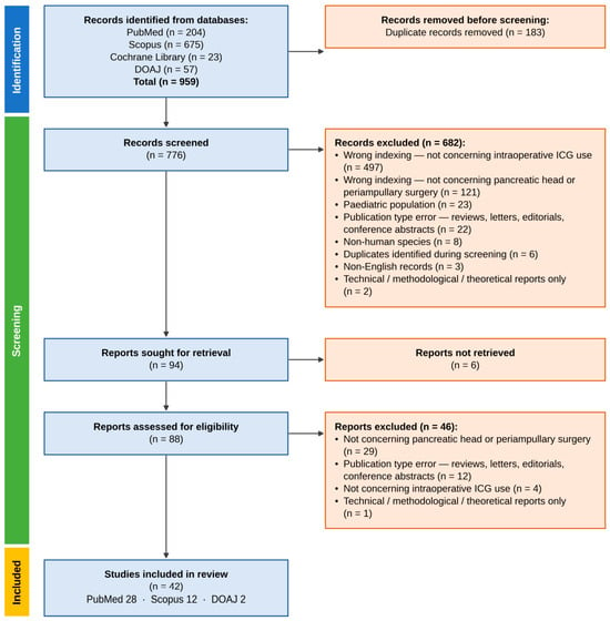
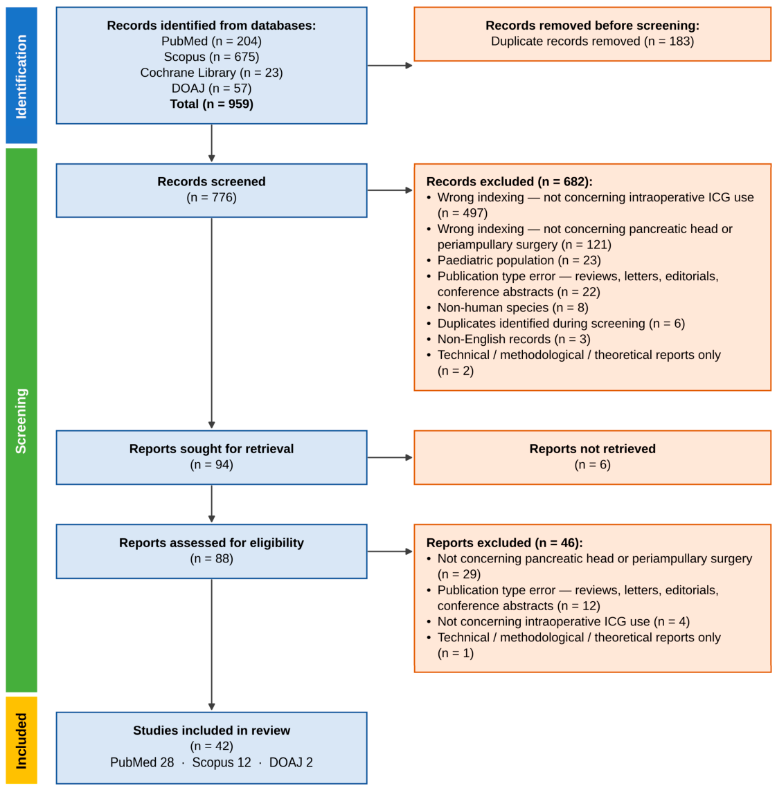
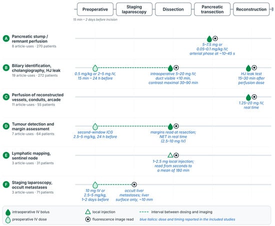
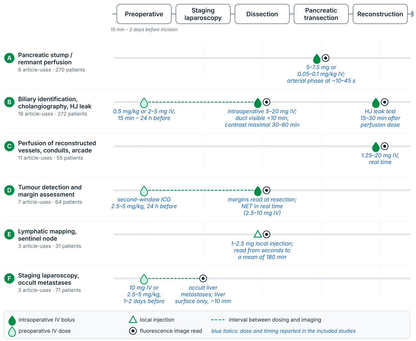

Disclaimer/Publisher’s Note: The statements, opinions and data contained in all publications are solely those of the individual author(s) and contributor(s) and not of MDPI and/or the editor(s). MDPI and/or the editor(s) disclaim responsibility for any injury to people or property resulting from any ideas, methods, instructions or products referred to in the content. |
© 2026 by the authors. Licensee MDPI, Basel, Switzerland. This article is an open access article distributed under the terms and conditions of the Creative Commons Attribution (CC BY) license.