


Abstract
Background/Objectives: This study assessed diagnostic performance, explored diagnostic barriers, and developed an assessment tool for case-based diagnostic performance and associated challenges specific to pediatric OFCs. Methods: An exploratory sequential mixed-methods study was conducted. The qualitative phase included semi-structured interviews with Saudi pediatric dentists, selected using criterion (purposive) sampling until thematic saturation was reached. Findings informed the development and validation of a structured questionnaire, targeting pediatric dentists from various regions of Saudi Arabia. The questionnaire assessed case-based diagnostic performance, appropriate management, and diagnostic barriers. Descriptive statistics were performed using SPSS. Findings from both phases were triangulated. Results: Thirteen interviews were conducted with pediatric dentists (specialists and consultants). Themes and subthemes, including key barriers, were identified. Among 180 pediatric dentists who answered the questionnaire, diagnostic performance was high, with mean diagnosis and management scores of 9.03 ± 1.16 and 8.84 ± 1.32, respectively. Triangulation demonstrated convergence regarding the importance of clinical exposure and interdisciplinary collaboration in improving diagnostic competence. Conclusions: The study developed a valid assessment tool specific to pediatric OFCs in Saudi Arabia, which may have limited generalizability to other countries. Pediatric dentists demonstrated high diagnostic performance and management competency, while key diagnostic barriers highlighted the need for enhanced hands-on training and interdisciplinary collaboration to further support accurate diagnosis and effective management of pediatric OFCs.
1. Introduction
Orofacial conditions (OFCs) are frequently encountered in children and adolescents and include a diverse range of conditions, such as neoplasms, hyperplastic lesions, reactive lesions, salivary gland abnormalities, bone lesions, and oral mucosal lesions [1]. These lesions can significantly impact physical functioning, psychosocial well-being, and overall quality of life [2,3].
Several retrospective studies conducted across different geographic locations have reported the prevalence of OFCs. In Poland, Owczarek-Drabińska et al. analyzed 2747 charts from 143 pediatric patients, ranging from 0 to 17 years of age, finding that aphthous ulcers were the most common lesion (18.9%), followed by mucoceles (10.5%), geographic tongue (5.6%), papilloma (4.9%), pyogenic granulomas (4.2%), and traumatic erosions and ulcers (4.2%) [4]. A study by Aly et al. involved a sample of 1108 specimens from Egyptian pediatric patients aged under 18 years and identified the most prevalent lesions: inflammatory radicular cysts (18.8%), followed by pyogenic granulomas (14.4%), peripheral giant cell granulomas (8.9%), irritational fibromas (8.1%), mucoceles (5.1%), periapical granulomas (4.0%), central giant cell lesions (3.7%), and dentigerous cysts (3.4%) [5]. Additionally, two studies conducted in Brazil revealed similar findings. Costa et al. analyzed 1480 biopsies from pediatric patients under 14 years of age, while Rezende et al. reviewed 93,950 reports from patients aged 0 to 3 years [6,7]. Both studies found mucoceles to be the most common lesions, occurring in 13.6% and 12% of cases, respectively. Similarly, a study conducted by Binmadi and Aldehlawi in Saudi Arabia found that mucoceles (12%) were more prevalent, with a notable predominance in females. The study included 183 pediatric cases of oral pathologies in patients aged 0 to 18 years. Periapical granulomas (7.7%) ranked second, followed by dentigerous cysts (6.6%), radicular cysts (6.6%), and pyogenic granulomas (4.9%) [8].
Pediatric dentists face many challenges in diagnosing OFCs. Recent literature highlights that oral ulcers are highly nonspecific symptoms with a wide range of potential causes, including infectious diseases, nutritional deficiencies, autoimmune disorders, medication side effects, and systemic diseases. This diversity of etiological factors complicates the diagnostic process [9]. A study conducted in Brazil evaluated the clinicopathological features of 315 oral lesions in 307 patients under the age of 18, revealing that only 61.5% of clinical hypotheses aligned with histopathological findings [10]. This indicates that nearly 40% of lesions were misdiagnosed or underdiagnosed based solely on clinical assessment, often due to a lack of adequate clinical and radiographic information. Additionally, challenges such as children’s developing anatomy, patient cooperation issues, and limitations in performing invasive diagnostics or obtaining imaging further hinder accurate diagnosis [11]. Other challenges related to dentists include limited access to oral medicine consultants within the dental institute [12].
To date, there are no available tools to investigate the diagnostic performance and challenges faced by dentists in diagnosing OFCs. Therefore, this study aimed to assess the diagnostic performance of pediatric dentists in identifying OFCs, explore the common barriers and challenges encountered during diagnosis, and develop an assessment tool to evaluate diagnostic performance and associated diagnostic challenges.
2. Materials and Methods
2.1. Ethical Considerations
Ethical approval for this research was obtained from the Research Ethics Committee at the Faculty of Dentistry at King Abdulaziz University (136-11-24; approved on 16 January 2025). Written consent was obtained from all pediatric dentists participating in each phase of the study.
2.2. Study Design
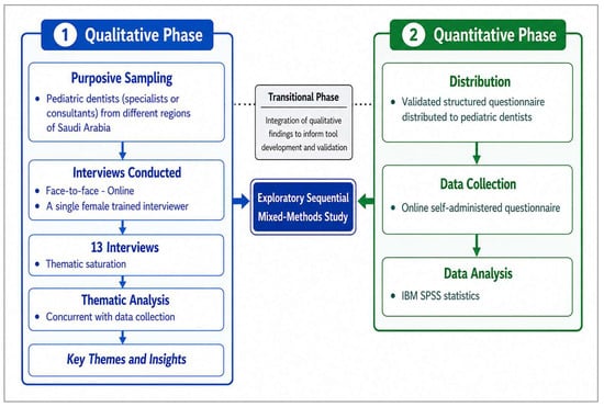
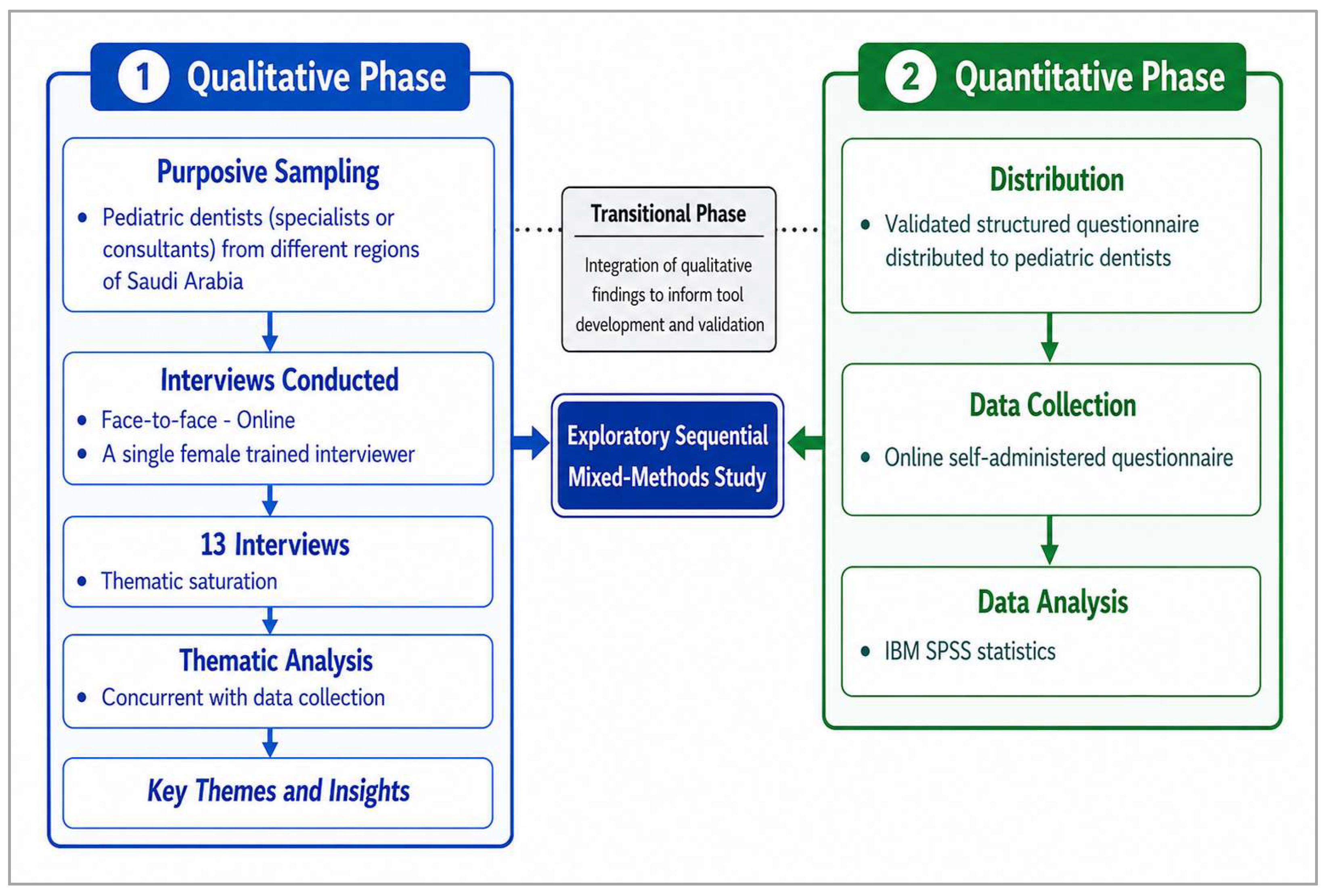
This study used an exploratory mixed-methods design that had two sequential phases. A mixed-methods study design flowchart is shown in Figure 1.
Figure 1.
A mixed-methods study design process flowchart.
2.2.1. Qualitative Phase (I)
Phase Design
A phenomenological qualitative phase was conducted to understand pediatric dentists’ knowledge and experiences regarding OFC cases and the challenges faced during their diagnosis and management.
Eligibility Criteria
Licensed pediatric dentists (either specialists or consultants) of both genders from various centers, including educational institutions, private clinics, and hospitals across multiple cities in Saudi Arabia, who had encountered OFCs within the past year and who consented to participate were included. Participants were required to be proficient in English. Rare cases and OFCs associated with syndromes were excluded.
Participants and Data Collection Procedure
A criterion (purposive) sample of pediatric dentists who met the inclusion criteria was invited to participate in semi-structured, audio-recorded interviews via WhatsApp. Participants were contacted in advance to provide a brief overview of the study, and a reminder message was sent 10 min before the interview. The semi-structured interviews were conducted either face-to-face or online via Google Meet (Google LLC, Mountain View, CA, USA), a web-based video-conferencing platform. Available at: https://meet.google.com/ (accessed on 2 January 2026), depending on participants’ preferences, with reminder texts sent 10 to 15 min prior to the interviews. The interviews were conducted in English using a standardized interview guide (Appendix A) consisting of 19 open-ended questions. The interviews lasted between 30 and 55 min. To ensure consistency, a single female resident in pediatric dentistry (Author 1) conducted all interviews. The resident was trained in qualitative research and was observed by a senior research expert in qualitative studies. Participants were asked the same questions in the same sequence, though the interviewer adapted probing questions based on participants’ key responses. The first interview was conducted as a pilot interview with a pediatric dentistry specialist. Minor modifications were made to the interview guide based on the pilot findings. The pilot interview was included in the analysis. Notes were recorded following each interview, and verbatim transcription was completed using transcription services. Data collection occurred from October 2025 to January 2026.
Sample Size
The sample size was determined by data saturation. Previous studies [13,14] suggest that 12–14 interviews are sufficient to achieve data saturation.
Data Analysis
Data analysis was conducted concurrently after each interview to monitor emerging themes until thematic saturation was achieved. Qualitative data from the verbatim transcripts were analyzed using NVivo software (version 15; Denver, CO, USA: Lumivero, 2024) to support coding, data organization, and theme development. Data analysis was performed by two coders (A.S. and K.B.), and an audit trail was used. Member checking was conducted. The COREQ (Consolidated Criteria for Reporting Qualitative Research) checklist was followed for reporting this study [15].
2.2.2. Questionnaire Development and Validation
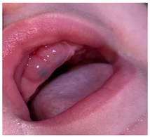
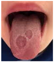
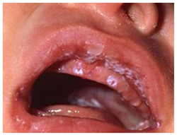
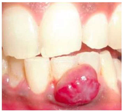
A structured, self-administered questionnaire was developed based on the qualitative phase, with the identified general and challenge-related themes directly informing its content. General themes guided items on the frequency of encountering OFCs, training received, curriculum, and nature of exposure, while opinions, recommendations, and challenges were incorporated based on the corresponding qualitative findings and themes. The common OFCs identified in phase I were compared with the existing literature, verified, and used to develop case scenarios. The qualitative findings also informed the relevant response options, while demographic items were adapted from previously published surveys. The questionnaire content was divided into three sections (Appendix B). The first section included sixteen items related to general demographic and professional characteristics. The second section included 10 case scenarios of common OFCs identified from the interviews and verified based on the existing literature [4,8,9], with 20 case-based items. For each case, participants were asked questions related to diagnosis and management. The third section included five items exploring the challenges faced by pediatric dentists when managing such cases, as well as their opinions and decision-making processes in diagnosis and management.
The questionnaire underwent content validation by five experts from King Abdulaziz University to assess its relevance, feasibility, practicality, validity, and clarity of interpretation: one associate professor in oral medicine and four associate professors in pediatric dentistry. Each item was evaluated by the five experts using a four-point scale, with a rating of 4 indicating high content relevance. The Content Validity Index (CVI) for each item was calculated as the proportion of experts assigning a rating of 4 (i.e., the number of experts who rated the item as 4 divided by the total number of experts). The overall scale-level CVI was calculated as the mean of the item-level CVI values, yielding an overall CVI of 0.82. Items with high agreement scores (rated 4) were retained, while items with lower agreement were revised or removed based on the experts’ feedback.
The questionnaire was then digitized using Google Forms and pilot-tested for face validity among five participants in pediatric dentistry before distribution, including two residents, two specialists, and one faculty member. The participants assessed the questionnaire for clarity and ease of completion. Minor modifications were made based on their feedback. The pilot participants were excluded from the main study.
2.2.3. Quantitative Phase (II)
Phase Design
This phase involved the distribution of the validated questionnaire and subsequent collection and analysis of the quantitative data.
Eligibility Criteria
Pediatric dentists, including consultants, specialists, and residents enrolled in pediatric dentistry programs, from different regions across Saudi Arabia who were actively practicing within the country, were included.
Participants and Procedure
A non-probability maximum variation convenience sampling technique was employed in this phase to capture diverse perspectives and ensure representation of participants with varying backgrounds and characteristics. The final version of the questionnaire was distributed to participants through the WhatsApp and LinkedIn platforms. To enhance the response rate, multiple follow-up reminders were sent periodically to potential participants throughout the data collection phase. Data collection was conducted from the beginning of March 2026 to the end of May 2026. The questionnaire required approximately five to seven minutes to complete.
Sample Size
No statistical sample-size calculation was performed. The target population was estimated based on the reported 257 Saudi pediatric dentists registered with the Saudi Commission for Health Specialties (SCFHS) across Saudi Arabia in 2019 [16]. All accessible pediatric dentists who met the eligibility criteria were invited to participate. A 70% response rate was anticipated based on a previous study [17] and was used for recruitment planning only.
Data Analysis
Data were analyzed using IBM SPSS Statistics version 27 (IBM Corp., Armonk, NY, USA). Descriptive statistics were used to summarize participant characteristics and questionnaire responses. Continuous variables were presented as means and standard deviations (SD), while categorical variables were presented as frequencies and percentages.
Diagnostic and management scores were calculated separately for each participant as the number of correct responses across the 10 clinical cases, with each score ranging from 0 to 10. For the multiple-response questions, participants could select more than one option; therefore, responses were analyzed separately for each option and presented as frequencies and percentages based on the number of participants selecting each option.
3. Results
3.1. Results of Qualitative Phase (I)
3.1.1. Sample Characteristics
A total of 13 interviews were conducted with pediatric dentists (Table 1). Of the 15 invited participants, two declined to participate, indicating that the interview approach was unsuitable for them. Seven interviews were conducted with consultants and six with specialists. Ten participants (76.9%) graduated from Saudi Arabia. More than two-thirds (nine participants; 69.2%) held board certification. The majority (11 participants; 84.6%) were female. Two-thirds (nine participants; 69.2%) worked in Jeddah, while four participants (30.8%) worked in other cities, including Jazan, Riyadh, Al Khobar, and Tabuk. Almost half (seven participants; 53.8%) had five to ten years of experience.
Table 1.
Demographic characteristics of the interviewed participants.
3.1.2. Resulting General Themes
Following thematic analysis of the transcribed semi-structured interviews, nine themes and seven subthemes emerged regarding barriers faced by pediatric dentists in diagnosing OFCs.
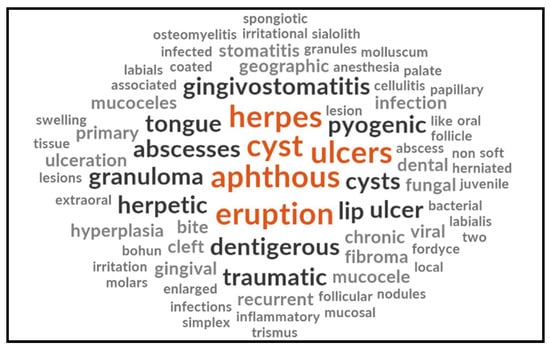
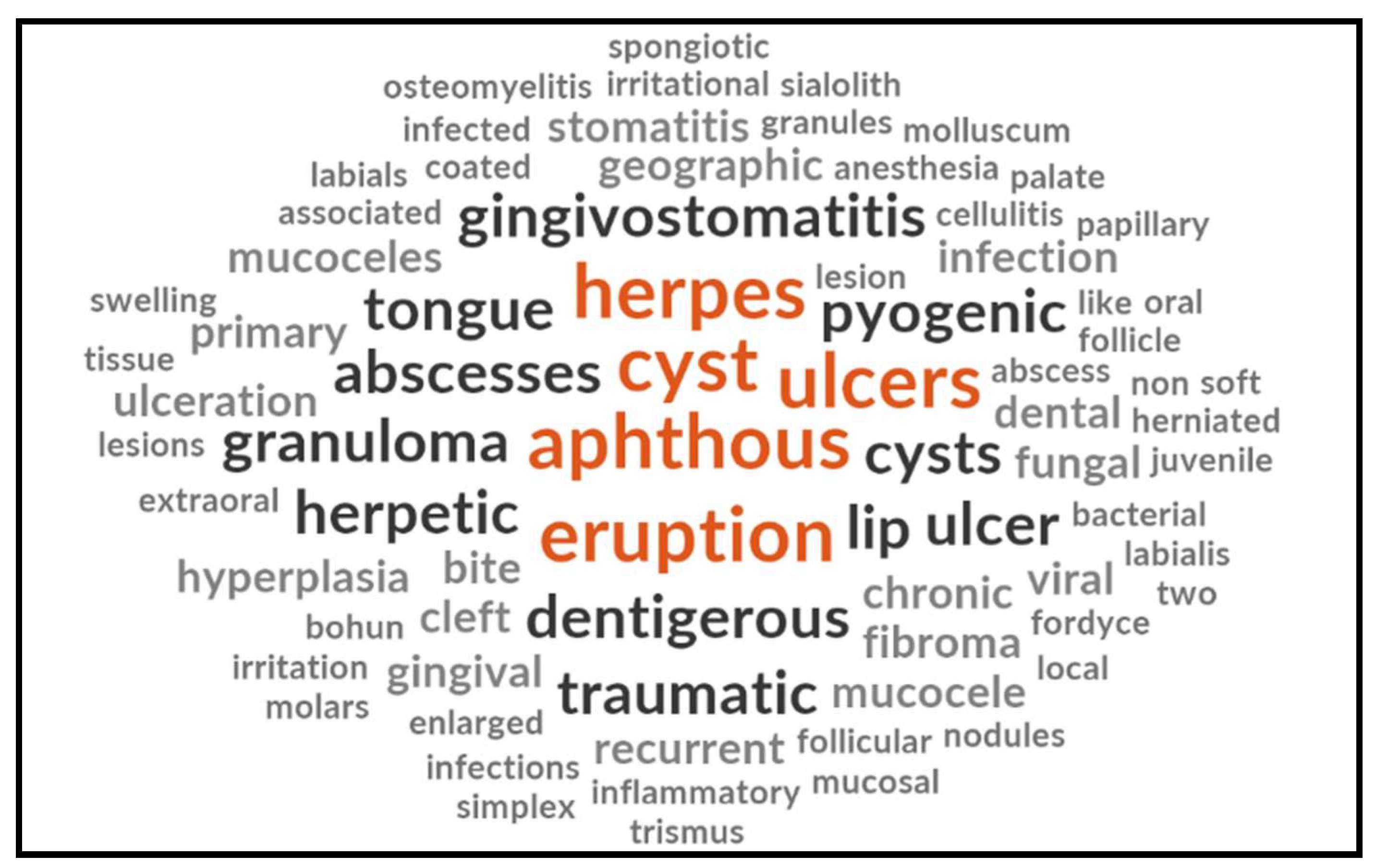
Words from the most frequently reported OFCs in children by pediatric dentists over the last 12 months are presented in Figure 2.
Figure 2.
Word cloud of the most common OFCs reported by pediatric dentists.
Theme 1: Diagnostic Competence
For the case scenario representing severe primary herpetic gingivostomatitis, including a comprehensive history and detailed description of the clinical presentation, some participants provided the correct diagnosis, while others provided incorrect diagnoses, included the correct condition in their differential diagnoses, or were unable to make a diagnosis at all. The selected quote is listed below:
“This needs a differential. Some sort of ulcerative disease could be considered. It could be leukoplakia, pemphigus vulgaris, or any other ulcerative disorder. I would do further investigations, of course. I would ask about Behçet’s, allergies, it could also be from the appliance.”[Participant 2 N C]
Table 2 shows that specialists and consultants were represented equally. Overall, the findings indicate that diagnostic performance was not 100%, highlighting variability in case evaluation among participants.
Table 2.
Diagnostic performance by pediatric dentists’ rank.
Theme 2: Diagnostic Methods
The most frequently reported diagnostic approaches included patient history, clinical examination, and imaging (e.g., cone-beam computed tomography [CBCT]), depending on the presenting condition. The following quotation illustrates how participants approached the diagnostic process and reached a final diagnosis:
“There was no chief complaint. It was an incidental radiographic finding. We took a panoramic radiograph, conducted a clinical examination, requested a CBCT, consulted with the surgery team, and we will take an aspiration biopsy to reach the final diagnosis. We put a differential diagnosis of a dentigerous cyst and OKC.”[Participant 7 S C]
Theme 3: Frequently Encountered Orofacial Conditions
Participants reported variation in the frequency with which they encountered pediatric OFCs in clinical practice. While some participants reported seeing these conditions relatively frequently, others described these cases as rare and encountered them only sporadically. This is reflected in the following participant quotations:
“Very frequently, actually. Once in every other week maybe, once one in every two weeks I receive a case that relates to oral lesions.”[Participants 10 A S]
“Very rare honestly, how many do we see? it’s not daily or weekly. It’s monthly. You know, every month you can say we can see one case, for example. Sometimes more than one month.”[Participants 5 O C]
Theme 4: System of Referral
Most pediatric dentists reported that they initiate internal referrals through the institutional electronic-based system and adhere strictly to established hospital protocols. However, many participants emphasized that, beyond the formal referral pathway, they prefer to directly contact the receiving physician or department by phone. This step was described as a strategy to prevent delays, minimize the risk of patients being “lost” within the system, and ensure timely coordination of care.
“I work in a place where we have many other medical specialties, and this would involve an internal referral system through the hospital. However, in private practice, I refer patients to my colleague in dentistry. This would be either a direct referral or a written referral. So, it honestly depends on the case.”[Participant 10 A S]
Theme 5: Nature of Curriculum Training
Most participants reported that their residency training included education in oral pathology or oral medicine. While some participants described receiving primarily didactic courses, others emphasized that their training was reinforced by extensive clinical exposure to patients with orofacial lesions. Participants described their experiences as follows:
“My program training was a tertiary care center. It was heavy loads of patients. so, we tend to see lots of lesions. Because you know, the incidence of oral pathology, per se, is not much. The incidence of general oral diseases is not much. So, in our training, we used to have heavy loads of patients, so we tend to see a little bit more lesions than others.”[Participant 2 N C]
3.1.3. Resulting Subthemes of Challenges Faced by Pediatric Dentists
- A.
- Dentist-Related Themes
- 1.
- Insufficient Training or Experience
One perspective expressed by a participant emphasized that insufficient training in residency programs, as well as limited exposure at the undergraduate level, contributes to gaps in clinical experience and presents challenges in diagnosing and managing orofacial lesions, often leading to referrals to the oral medicine department.
“Maybe all of what we studied as didactic in the book, including the cases in the photos, is relevant, but I definitely mean the challenge that we don’t see it in the clinic, so it doesn’t get reinforced.”[Participant 9 I C]
- B.
- Lesion-Related Themes
- 2.
- Complexity and Atypical Presentation of Orofacial Lesions
Many participants highlighted the challenges posed by the variability of oral lesions, noting that even the same type of lesion can differ significantly, sometimes making it difficult to identify. Some quotes related to this theme are listed below:
“The main challenges any pediatric dentist will face are the oral lesions. As much as I try to read or attend seminars, whenever you see a lesion, it’s never the same. I think if anyone wants to improve themselves, it should be in oral medicine.”[Participant 1 H S]
- 3.
- Diagnostic Uncertainty
Some participants encountered difficulties in diagnosing certain lesions due to their unknown nature or cause, making it challenging to establish a definitive diagnosis. In such cases, they listed differential diagnoses until confirmation could be obtained through further investigations, such as consultation with a surgeon or a biopsy, as noted by several respondents.
“There was a lesion in the mandible, and it was multilocular. There were many differential diagnoses for the case, and it was difficult to diagnose, so it needed a biopsy.”[Participant 12 A S]
- 4.
- Rarity of Oral Lesions
Most participants had difficulty diagnosing or recognizing lesions because they rarely encounter such cases. This lack of exposure causes confusion when seeing a lesion for the first time and increases the risk of misdiagnosis; in some cases, there are no available references in textbooks.
“These things are rare or we don’t see them daily like caries. So still, we might misdiagnose or miss some information that leads us to a good or proper diagnosis.”[Participant 6 R C]
- C.
- Other Themes
- 5.
- Parental Attitude and Behavioral Challenges
Several pediatric dentists noted that parents’ unwillingness to answer questions, reluctance to cooperate with referrals for further investigations, or refusal of necessary treatments can pose significant challenges. Some quotes illustrating this theme include:
“One challenge is convincing the family to go to the pediatric when they inform about GI problem to go to the pediatric physician and examine to see what’s happening for further examination and management. Most of the time they are not convinced to go and see a pediatric or maybe they are convinced but it’s hard or it’s expensive for them to go, especially if it’s not in governmental sector.”[Participant 4 H S]
- 6.
- Patient Age and Behavioral Challenges
Several participants reported that conducting examinations and managing even simple dental procedures are challenging in uncooperative children, especially in younger ones. They highlighted barriers such as difficulties in communication and a lack of patient cooperation, which often complicate routine care. Representative participant quotes are listed below:
“As we know, the younger they are, the worse the cooperation can be. If he had dental trauma or phobia, it makes the case worse. The behavior of the patient will make the procedure difficult for me. Maybe the dental treatment that would be easy to do without sedation with a cooperative child, even if it was a biopsy, would be very challenging and difficult for a patient who is young age and severely uncooperative, whether in time or in the procedure itself.”[Participant 11 J S]
- 7.
- System-Level Barriers
Many participants believed that system-level barriers, particularly those related to the referral system, resource constraints, and teamwork and interdisciplinary communication challenges, heavily impacted the diagnosis and management of pediatric OFCs.
- Referral System Issues
Most pediatric dentists mentioned issues with the referral protocol to other departments and expressed concern about the time it takes to refer a patient through the system.
“Having no slots to see the patient immediately, especially if it’s urgent or the doctor is not on the floor, no consultant, only the resident or the fellows are dealing with the cases. Sometimes the referral is missed. The patient got an appointment for referral, and he did not show up. We didn’t know that he didn’t go. From their side, they don’t know that the patient is urgent or he need to be seen, so they don’t book him again.”[Participant 8 RB C]
- Resource Constraints
Many participants highlighted that some hospitals lacked oral medicine specialists. They also noted limitations in the availability of certain diagnostic tools and materials. Representative participant quotes include:
“Some hospitals and clinics don’t have an oral medicine department, so it will be difficult to do examination and diagnosis.”[Participant 12 A S]
- Teamwork and Interdisciplinary Communication Challenges
Some participants shared their experiences with the lack of a coordinated team, the resulting need to transfer patients to other hospitals, the absence of a team in the governmental sector, and problems with a lack of direct communication between the dental and medical departments.
“There is no direct communication. I talk about private; for example, I don’t have the extension. I am in the dental center; there is no pediatrician. So, I must talk to the other clinic to know the extension and the working hours of the doctor. So, when there is no such direct access, I refer them to the ER or to a pediatric hospital.”[Participant 7 S C]
3.2. Results of Quantitative Phase (II)
Participant Characteristics
A total of 180 pediatric dentists completed the questionnaire (Table 3). Female participants comprised 110 (61.1%) of the sample. Participants were relatively evenly distributed according to years of professional experience, with 60 (33.3%) reporting 2–5 years of experience and another 60 (33.3%) reporting more than 10 years of experience. Specialists represented 69 (38.3%) of the participants, while 64 (35.6%) held board certification. Most participants graduated from Saudi Arabia (114; 63.3%).
Table 3.
Sociodemographic and professional characteristics of participants (N = 180).
Most participants encountered OFCs sometimes (every 2–4 months) (40.0%) and rated their training as 4 (36.7%). Most reported receiving both theoretical and clinical exposure (62.8%) and had encountered OFCs during the previous 12 months (77.2%). Regarding diagnosed cases, 67.2% reported diagnosing 0–3 cases, while 32.8% diagnosed 4–6 or more cases (Table 4).
Table 4.
Participants’ clinical exposure to OFCs (N = 180).
The distribution of correct answers for the diagnosis and management of the cases is shown in Table 5. The highest diagnostic scores were observed for traumatic ulcers and hand, foot, and mouth disease, with 178 participants (98.9%) correctly identifying each condition. The correct management of a traumatic ulcer was identified by 176 participants (97.8%), whereas only 137 participants (76.1%) correctly selected the appropriate management for hand, foot, and mouth disease. Overall diagnostic and management scores were calculated for each participant based on the number of correct responses across the 10 cases and are presented as the mean ± SD and median, with scores ranging from 0 to 10.
Table 5.
Diagnostic and management performance across clinical cases (N = 180).
Table 6 presents an analysis of the response frequency of the most helpful strategies in a question that allowed participants to select more than one answer. The most frequently reported strategy was gaining more clinical exposure and hands-on experience, with 118 responses (32.7%).
Table 6.
Participants’ responses on helpful strategies (multiple responses allowed) (N = 361).
3.3. Triangulation
Triangulation was conducted to integrate and interpret the findings of both phases (Table 7). Triangulation offers several advantages by incorporating different methods, perspectives, or sources to support the interpretation of findings. It strengthens the study design by integrating quantitative and qualitative findings instead of depending on a single approach, allowing for a broader understanding of the research phenomenon and supporting a more comprehensive interpretation of the results [18].
Table 7.
Triangulation of qualitative and quantitative phases.
4. Discussion
The present exploratory sequential mixed-methods study aimed to assess the diagnostic performance of pediatric dentists in identifying OFCs, explore the common barriers and challenges encountered during diagnosis, and develop an assessment tool to evaluate case-based diagnostic performance and associated diagnostic challenges. The rationale for starting with the qualitative phase was to explore pediatric dentists’ knowledge, experiences, barriers and approaches to identifying and diagnosing OFCs, particularly given the lack of previous studies assessing diagnostic performance in this population. Including certified pediatric dentists (specialists and consultants) from different centers and with varying years of experience across regions of Saudi Arabia provided a deeper understanding of the knowledge gaps, diagnostic approaches, and potential challenges and barriers encountered when diagnosing OFCs in children [19,20,21,22]. The findings from the qualitative phase were then used to develop an assessment tool that was validated for the subsequent quantitative phase. Combining quantitative and qualitative approaches in one study resulted in stronger and more comprehensive findings than using either method independently [23].
Our qualitative findings suggested that no clear difference in diagnostic performance was observed between specialists and consultants and that consultants with many years of experience may still make incorrect diagnoses. These findings are consistent with previous studies that show similar performance between specialists and general dentists or less-experienced clinicians in diagnosing OFCs [24]. On the other hand, other studies have reported that specialists showed better diagnostic performance scores than general dental practitioners in the diagnosis of OFCs [25,26]. This may indicate factors beyond clinical rank and years of experience, such as clinical exposure to a broad spectrum of lesions and variability in continuing education and training opportunities [27].
In the present study, although participants reported using an institutional electronic referral system, many still relied on phone-based referrals to reduce appointment delays. This result is supported by previous reports, where referral communication for oral potentially malignant lesions or malignancy included written electronic correspondence and patient-carried information, whereas referral appointments were also organized through telephone communication [28]. In contrast, previous studies have shown that teleconsultation significantly reduced unnecessary in-person referrals by 35% to 80% and shortened waiting times through structured digital platforms that facilitate the exchange of clinical data and images [29].
Dentist-related factors, particularly insufficient training and experience, emerged as an important finding in this study. This challenge has been reported by several studies as contributing to the difficulty of diagnosing OFCs due to a lack of skills or confidence, often attributed to insufficient training and experience and lack of adequate knowledge [24,30,31,32]. This may result in delayed referrals or treatment.
In addition to dentist-related factors, lesion-related challenges were also identified in this study, including the complexity and atypical presentation of orofacial lesions, diagnostic uncertainty, and the rarity of oral lesions. These findings are consistent with previous studies showing that nonspecific features and variability in clinical presentations make diagnosis challenging and increase the risk of misdiagnosis and improper treatment [33,34,35]. Similarly, it has been reported that oral ulceration may have multiple etiologies, with variations in nature and clinical presentation making accurate diagnosis particularly challenging [9,36]. Furthermore, it has been reported that the low incidence of oral lesions may further contribute to reduced familiarity among practitioners, which can negatively affect both timely diagnosis and appropriate management strategies [37].
Although participants reported several challenges and barriers to diagnosis, the present study did not assess whether these factors were associated with lower diagnostic performance. Therefore, these findings should be interpreted as participants’ perceived barriers to diagnosis rather than as confirmed determinants of diagnostic performance.
Participants in the present study emphasized clinical exposure and hands-on experience as essential for improving diagnostic and management skills. This is consistent with evidence that supervised clinical experience helps translate theoretical knowledge into practice and enhances diagnostic competence and confidence, while continuing professional development (CPD) alone may be insufficient without adequate clinical exposure [38,39]. Hussein et al. similarly highlighted the need for structured curricula integrating theoretical and practical training to address educational gaps in diagnosing and managing OFCs [40]. Strengthening oral medicine and pathology education, incorporating relevant medical content, and providing CPD can further improve dentists’ diagnostic performance [24,41].
Emerging artificial intelligence (AI) technologies may offer complementary approaches to some of the challenges identified in the present study. Large language models (LLMs) have demonstrated potential in reducing the workload associated with preparing educational materials [42]. An LLM-based Anki tool substantially reduced flashcard preparation time and cognitive load among dental students, potentially facilitating self-directed learning and allowing greater focus on conceptual understanding [42]. Furthermore, vision–language models (VLMs) have shown potential as zero-shot tools for supporting the interpretation of condylar osseous changes on CBCT and generating structured diagnostic reports [43]. Such tools may provide supplementary support for learners with limited radiological training, although expert supervision and further clinical validation remain essential [43]. These developments highlight opportunities for integrating AI-assisted learning and diagnostic support into dental education to complement conventional training and address identified educational barriers.
The findings of this study have important clinical implications. Enhancing practical clinical training, promoting CPD, and establishing clear diagnostic protocols may help reduce variability in diagnostic performance. Furthermore, strengthening interdisciplinary collaboration among pediatric dentists, oral medicine specialists, and oral pathologists may facilitate more accurate diagnoses and ultimately improve patient outcomes.
Several strengths of this study should be acknowledged. The mixed-method design provided both breadth and depth by integrating questionnaire data with interview findings, enhancing understanding of and confidence in the conclusions [44,45]. Triangulation between the two phases further strengthened the validity and credibility of the findings [46]. Credibility was ensured through multiple interviews, member checking, established methods, an appropriate study design, and interviewer training. Transferability was supported by purposeful sampling of specialists and consultants from multiple institutions and regions across Saudi Arabia, as well as inclusion of participants who had encountered OFCs within the previous year to minimize recall bias. Dependability was maintained through consistent research procedures and thorough documentation throughout the study. Confirmability was strengthened through piloting and revising the interview guide, maintaining written field notes, and comparing findings with existing literature. Additionally, the questionnaire’s wide geographical coverage and large sample of pediatric dentists across Saudi Arabia enhanced the diversity and representativeness of the study population.
This study has some limitations. The qualitative phase often involved lengthy interviews lasting more than 30 min, which could lead to participant fatigue. Moreover, consultants were often busy and answered phone calls during the interviews. Furthermore, some online interviews experienced technical challenges, leading to interruptions. The interviewer was a single female resident in pediatric dentistry with a background in OFCs, which may have introduced reflexivity bias by influencing participants’ responses throughout the qualitative phase [47,48,49]. Additionally, the quantitative phase evaluated diagnostic performance using hypothetical case scenarios rather than real-time clinical encounters, which may not accurately reflect true clinical performance under pressure. The exclusion of difficult, rare and syndromic conditions may reduce the applicability of the findings to the specialist rank. Furthermore, the online administration of the questionnaire without supervision may have allowed participants to seek assistance from external sources, such as ChatGPT or Google, when completing the questionnaire, potentially inflating the diagnostic performance scores. Finally, the study findings and validated assessment tool are derived exclusively from participants across Saudi Arabia, limiting their applicability to international dental practice settings.
Future research should focus on having participants complete the questionnaire on the spot to ensure that no external sources are used and to ensure the accuracy of the results. Additionally, investigating the impact of digital diagnostic tools and AI on the identification of OFCs may provide valuable insights into enhancing clinical practice.
5. Conclusions
This mixed-methods study resulted in the development of a tool for assessing pediatric dentists’ diagnostic performance and associated diagnostic challenges. The qualitative findings identified key barriers, including insufficient training or experience, lesion complexity and atypical presentation, diagnostic uncertainty, behavioral challenges, system-level barriers, and lesion rarity. The quantitative findings provided evidence supporting the questionnaire’s validity for its intended purpose. The questionnaire findings also indicated high diagnostic performance and management scores among pediatric dentists in Saudi Arabia in identifying pediatric OFCs. The tool is specific to pediatric OFCs in Saudi Arabia and may not be generalizable to other settings. The findings highlight the importance of improving hands-on training and interdisciplinary collaboration, including joint clinics with oral medicine physicians and oral and maxillofacial surgeons to support accurate diagnosis and effective management of pediatric OFCs.
Author Contributions
Conceptualization, K.B. and K.A.; methodology, N.O.B., K.B. and K.A.; software, A.S.; validation, A.S.; formal analysis, A.S. and K.B.; investigation, A.S. and K.B.; resources, A.S.; data curation, A.S.; writing—original draft preparation, A.S.; writing—review and editing, A.S., N.O.B., K.B., K.A., and R.S.; visualization, A.S.; supervision, K.B.; project administration, A.S. and, K.B.; funding acquisition, K.B. All authors have read and agreed to the published version of the manuscript.
Funding
This project was funded by the Deanship of Scientific Research (DSR) at King Abdulaziz University, Jeddah, under grant no. (IPP: 99-165-2026).
Institutional Review Board Statement
The study was conducted in accordance with the Declaration of Helsinki and approved by the Institutional Review Board and Research Ethics Committee of the Faculty of Dentistry, under approval number 136-11-24. Date: 16 January 2025.
Informed Consent Statement
Written informed consent was obtained from all subjects involved in both phases of the study.
Data Availability Statement
The original contributions presented in this study are included in the article. Further inquiries can be directed to the corresponding author.
Acknowledgments
The authors would like to sincerely thank the pediatric dentists from King Abdulaziz University, King Fahad Armed Forces Hospital, Princess Nourah bint Abdulrahman University, the Ministry of Health, Jazan Health Cluster, National Guard Health Affairs, and JedMed Health Center for their valuable participation and support in this study. The authors gratefully acknowledge the Deanship of Scientific Research (DSR) at King Abdulaziz University, Jeddah, Saudi Arabia, for funding this project under grant No. (IPP: 99-165-2026) and for providing technical support.
Conflicts of Interest
The authors declare no conflicts of interest.
Abbreviations
The following abbreviations are used in this manuscript:
| OFCs | Orofacial conditions |
| CPD | Continuing professional development |
| CBCT | Cone-beam computed tomography |
| AI | Artificial intelligence |
| LLMs | Large language models |
| VLMs | vision–language models |
Appendix A
Table A1.
Content of the interview guide.
Appendix B
Table A2.
An online questionnaire.
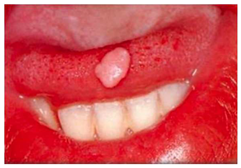
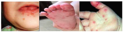
References
- Lima, G.; Fontes, S.; Araújo, L.; Etges, A.; Tarquino, S.; Gomes, A. A Survey of Oral and Maxillofacial Biopsies in Children: A Single-Center Retrospective Study of 20 Years in Pelotas-Brazil. J. Appl. Oral Sci. 2008, 16, 397–402. [Google Scholar] [CrossRef] [Scilit] [PubMed]
- Jokovic, A.; Locker, D.; Tompson, B.; Guyatt, G. Questionnaire for Measuring Oral Health-Related Quality of Life in Eight- to Ten-Year-Old Children. Pediatr. Dent. 2004, 26, 512–518. [Google Scholar] [PubMed]
- Omara, M.; Stamm, T.; Bekes, K. Four-dimensional Oral Health-related Quality of Life Impact in Children: A Systematic Review. J. Oral Rehabil. 2021, 48, 293–304. [Google Scholar] [CrossRef] [Scilit] [PubMed]
- Owczarek-Drabińska, J.E.; Nowak, P.; Zimoląg-Dydak, M.; Radwan-Oczko, M. The Prevalence of Oral Mucosa Lesions in Pediatric Patients. Int. J. Environ. Res. Public Health 2022, 19, 11277. [Google Scholar] [CrossRef] [Scilit] [PubMed]
- Aly, M.M.; Abdul-Aziz, M.A.-W.M.; Elchaghaby, M.A. A Retrospective Analysis of Oral and Maxillofacial Pathological Lesions in a Group of Egyptian Children over 21 Years. BMC Oral Health 2022, 22, 2. [Google Scholar] [CrossRef] [Scilit] [PubMed]
- Costa, A.R.G.F.; Duarte, P.V.S.; Moreira, M.R.; Mello, F.A.A.; Ferreira, M.C.; de Faria, P.R.; Cardoso, S.V.; Loyola, A.M. Histopathological Diagnosis in Pediatric Stomatology: A 43-Year Retrospective Study of 1,480 Cases from a Brazilian Institution. Int. J. Pediatr. Otorhinolaryngol. 2023, 166, 111481. [Google Scholar] [CrossRef] [Scilit] [PubMed]
- Rezende, K.M.; De Barros Gallo, C.; Nogueira, G.P.; Corraza, A.C.; Haddad, A.E.; Gallottini, M.; Bönecker, M. Retrospective Study of Oral Lesions Biopsied in Babies and Toddlers. Oral Dis. 2024, 30, 1242–1244. [Google Scholar] [CrossRef] [Scilit] [PubMed]
- Binmadi, N.O.; AlDehlawi, H. Pediatric Oral Pathology in Saudi Arabia: A 10-Year Retrospective Study at an Academic Dental Hospital. Saudi Dent. J. 2024, 36, 751–755. [Google Scholar] [CrossRef] [Scilit] [PubMed]
- Légeret, C.; Furlano, R. Oral Ulcers in Children- a Clinical Narrative Overview. Ital. J. Pediatr. 2021, 47, 144. [Google Scholar] [CrossRef] [Scilit] [PubMed]
- Vale, E.; Ramos-Perez, F.; Rodrigues, G.; Carvalho, E.; Castro, J.; Perez, D. A Review of Oral Biopsies in Children and Adolescents: A Clinicopathological Study of a Case Series. J. Clin. Exp. Dent. 2013, 5, e144–e149. [Google Scholar] [CrossRef] [Scilit] [PubMed]
- Bilodeau, E. Oral Pathology in Pediatric Patients: Common Conditions and Challenges. J. Oral Med. Surg. 2023, 6, 156. [Google Scholar]
- Akeel, S.; Almazrooa, S.; Ali, S.A.; Alhindi, N.A.; Alhamed, S.; Felemban, O.M.; Mansour, G.; Sabbahi, D.; Binmadi, N.; Mawardi, H. Assessing Tele-Oral Medicine in Saudi Arabia: A Cross-Sectional Study on Specialists’ Experiences and Effectiveness in Oral Healthcare. Healthcare 2023, 11, 3089. [Google Scholar] [CrossRef] [Scilit] [PubMed]
- Guest, G.; Bunce, A.; Johnson, L. How Many Interviews Are Enough? An Experiment with Data Saturation and Variability. Field Methods 2006, 18, 59–82. [Google Scholar] [CrossRef] [Scilit]
- Alrumaim, S.; Farsi, N.; Farsi, N.; Baghlaf, K.; Farsi, D. Feasibility and Acceptability of Pediatric Teledentistry: A Mixed-Methods Study. Int. J. Paediatr. Dent. 2025, 35, 954–963. [Google Scholar] [CrossRef] [Scilit] [PubMed]
- Tong, A.; Sainsbury, P.; Craig, J. Consolidated Criteria for Reporting Qualitative Research (COREQ): A 32-Item Checklist for Interviews and Focus Groups. Int. J. Qual. Health Care 2007, 19, 349–357. [Google Scholar] [CrossRef] [Scilit] [PubMed]
- Saudi Commission for Health Specialties. Saudi Board in Pediatric Dentistry Curriculum; Saudi Commission for Health Specialties: Riyadh, Saudi Arabia, 2020. [Google Scholar]
- AlBaker, A.M.; Al-Ruthia, Y.S.H.; AlShehri, M.; Alshuwairikh, S. The Characteristics and Distribution of Dentist Workforce in Saudi Arabia: A Descriptive Cross-Sectional Study. Saudi Pharm. J. 2017, 25, 1208–1216. [Google Scholar] [CrossRef] [Scilit] [PubMed]
- Renz, S.M.; Carrington, J.M.; Badger, T.A. Two Strategies for Qualitative Content Analysis: An Intramethod Approach to Triangulation. Qual. Health Res. 2018, 28, 824–831. [Google Scholar] [CrossRef] [Scilit] [PubMed]
- Alase, A. The Interpretative Phenomenological Analysis (IPA): A Guide to a Good Qualitative Research Approach. Int. J. Educ. Lit. Stud. 2017, 5, 9–19. [Google Scholar] [CrossRef] [Scilit]
- Renjith, V.; Yesodharan, R.; Noronha, J.A.; Ladd, E.; George, A. Qualitative Methods in Health Care Research. Int. J. Prev. Med. 2021, 12, 20. [Google Scholar] [CrossRef] [Scilit] [PubMed]
- Isik, O. The Power of Stories: Understanding Human Experience Through Qualitative Research. Int. J. Engl. Lit. Soc. Sci. 2025, 10, 622–630. [Google Scholar] [CrossRef] [Scilit]
- Lim, W.M. What Is Qualitative Research? An Overview and Guidelines. Australas. Mark. J. 2025, 33, 199–229. [Google Scholar] [CrossRef] [Scilit]
- Creswell, J.W. Research Design: Qualitative, Quantitative, and Mixed Methods Approaches, 4th ed.; Sage Publication: London, UK, 2014. [Google Scholar]
- Idrees, M.; Halimi, R.; Gadiraju, S.; Frydrych, A.M.; Kujan, O. Clinical Competency of Dental Health Professionals and Students in Diagnosing Oral Mucosal Lesions. Oral Dis. 2024, 30, 3108–3116. [Google Scholar] [CrossRef] [Scilit] [PubMed]
- Chiang, T.-E.; Lin, Y.-C.; Wu, C.-T.; Yang, C.-Y.; Wu, S.-T.; Chen, Y.-W. Comparison of the Accuracy of Diagnoses of Oral Potentially Malignant Disorders with Dysplasia by a General Dental Clinician and a Specialist Using the Taiwanese Nationwide Oral Mucosal Screening Program. PLoS ONE 2021, 16, e0244740. [Google Scholar] [CrossRef] [Scilit] [PubMed]
- Morales-Gómez, C.; Ojeda-Uribe, G.; Adorno-Farías, D.; Maturana-Ramirez, A.; Espinoza-Santander, I. Telediagnosis Performance of Specialists in Oral Medicine and General Dental Practitioner Using Images of Oral Mucosa Lesions in Chile. Med. Oral Patol. Oral Cir. Bucal 2025, 30, e49–e55. [Google Scholar] [CrossRef] [Scilit] [PubMed]
- Poelman, M.R.; Brand, H.S.; Asadi, M.; Remmelzwaal, S.; Jager, D.H.J.; de de Visscher, J.G. Knowledge of Diagnosis and Management of Selected Oral Mucosal Lesions among Dentists in The Netherlands. Med. Oral Patol. Oral Cir. Bucal 2023, 28, e362–e370. [Google Scholar] [CrossRef] [Scilit] [PubMed]
- Psoter, W.J.; Kerr, A.R.; Tomar, S.L.; Psoter, J.A.; Morse, D.E.; Aguilar, M.L.; Burton, V.A. Diagnostic Approach Used by US General Dental Practitioners Following Discovery of Oral Lesions Suspicious for Malignancy/Premalignancy: Findings from the National Dental Practice-Based Research Network. Quintessence Int. 2024, 55, 652–659. [Google Scholar] [CrossRef] [Scilit] [PubMed]
- Dos Santos, R.T.N.; Cavalcanti, J.F.D.S.; Duda Júnior, L.G.D.S.; De Vasconcelos, A.J.A.; Alvares, P.R.; Sobral, A.P.V. Teleconsultation Actions in Reducing In-Person Referrals for Oral Lesions: A Scoping Review. Oral Dis. 2025, 31, 2404–2418. [Google Scholar] [CrossRef] [Scilit] [PubMed]
- Sbricoli, L.; Zago, R.; Cavallin, F.; Stellini, E.; Bacci, C. Diagnostic Ability in Oral Pathology among Different Population Clusters. Oral Dis. 2024, 30, 2278–2284. [Google Scholar] [CrossRef] [Scilit] [PubMed]
- Schiavo-Di Flaviano, V.; González-Navarro, B.; Martín Carreras-Presa, C.; Jané-Salas, E.; López-López, J. Attitude of General Dental Practitioners, Oral Surgeons, and Dentistry Students towards Oral Lesions and Oral Biopsy. Med. Oral Patol. Oral Cir. Bucal 2025, 30, e31–e40. [Google Scholar] [CrossRef] [Scilit] [PubMed]
- Reis, D.T.; Póvoa, L.S.D.d.A.; Santos, D.d.C.; Verner, F.S.; Ribeiro, H.J.; Ferreira, I.V.; Aquino, S.N.D. Influence of Dentists’ Training and Access to Oral Pathology Services on Biopsy Decisions for Oral Mucosal Lesions. Eur. J. Dent. Educ. 2026, 30, 126–132. [Google Scholar] [CrossRef] [Scilit] [PubMed]
- Bansal, R.; Jain, A.; Mittal, S. Orofacial Tuberculosis: Clinical Manifestations, Diagnosis and Management. J. Fam. Med. Prim. Care 2015, 4, 335. [Google Scholar] [CrossRef] [Scilit] [PubMed]
- Troiano, G.; Dioguardi, M.; Giannatempo, G.; Laino, L.; Testa, N.F.; Cocchi, R.; De Lillo, A.; Lo Muzio, L. Orofacial Granulomatosis: Clinical Signs of Different Pathologies. Med. Princ. Pract. 2015, 24, 117–122. [Google Scholar] [CrossRef] [Scilit] [PubMed]
- Suaza, C.; Torres-Osorio, L.; Plazas Román, J.E.; Martinez Martinez, A.; Diaz, A.; Ardila, C.M. Oral Lesions with Identical Clinical Presentation and Different Histopathological Diagnoses: A Case Series of Mucocele, Schwannoma, and Hamartoma. Cureus 2025, 17, e93203. [Google Scholar] [CrossRef] [Scilit] [PubMed]
- Minhas, S.; Sajjad, A.; Kashif, M.; Taj, F.; Alwadaani, H.; Khurshid, Z. Oral Ulcers Presentation in Systemic Diseases: An Update. Open Access Maced. J. Med. Sci. 2019, 7, 3341–3347. [Google Scholar] [CrossRef] [Scilit] [PubMed]
- Kalaitsidou, I.G.; Astreidis, I.T.; Kontos, K.I.; Lazaridou, M.N.; Bourlidou, E.T.; Gerasimidou, D.K.; Vladika, N.P.; Mangoudi, D.L. Metastatic Tumours to the Oral Cavity: Report of Three Cases. J. Oral Maxillofac. Res. 2015, 6, e5. [Google Scholar] [CrossRef] [Scilit] [PubMed]
- Brailo, V.; Freitas, M.D.; Posse, J.L.; Monteiro, L.; Silva, L.M.; Fricain, J.C.; Catros, S.; Fénelon, M.; Lodi, G.; Ariyaratnam, R.; et al. Oral Potentially Malignant Disorders—An Assessment of Knowledge and Attitude to Future Education in Undergraduate Dental Students. Eur. J. Dent. Educ. 2022, 27, 622–632. [Google Scholar] [CrossRef] [Scilit] [PubMed]
- Gaballah, K.; Kujan, O. The Impact of Educational Intervention on Junior Dentists’ Capacity to Detect Oral Mucosal Lesions and Suspect Malignant Potential. Asian Pac. J. Cancer Prev. 2022, 23, 3673–3676. [Google Scholar] [CrossRef] [Scilit] [PubMed]
- Hussein, S.A. Assessment of Knowledge and Practice Regarding Oral Lesions among a Group of Iraqi Dental Practitioners—A Cross-Sectional Study. Clin. Epidemiol. Glob. Health 2025, 36, 102226. [Google Scholar] [CrossRef] [Scilit]
- Gaffar, B.; Farooqi, F.A.; Nazir, M.A.; Bakhurji, E.; Al-Khalifa, K.S.; Alhareky, M.; Virtanen, J.I. Oral Health-Related Interdisciplinary Practices among Healthcare Professionals in Saudi Arabia: Does Integrated Care Exist? BMC Oral Health 2022, 22, 75. [Google Scholar] [CrossRef] [Scilit] [PubMed]
- Chen, K.; Zhang, A.; Xie, X.; Bai, Y. LLM-Based Anki Card Generation: Solving the “Creation Bottleneck” in Dental Education? J. Dent. Educ. 2026, 90, S185–S187. [Google Scholar] [CrossRef] [Scilit] [PubMed]
- Chen, K.; Zhang, A.; Xie, X.; Bai, Y. Vision-Language Model as a ‘Zero-Shot’ Assistant for Evaluating Condylar Osseous Changes in Cone-Beam Computed Tomography. Int. Dent. J. 2026, 76, 109817. [Google Scholar] [CrossRef] [Scilit] [PubMed]
- Johnson, R.B.; Onwuegbuzie, A.J.; Turner, L.A. Toward a Definition of Mixed Methods Research. J. Mix. Methods Res. 2007, 1, 112–133. [Google Scholar] [CrossRef] [Scilit]
- Wasti, S.P.; Simkhada, P.; Van Teijlingen, E.; Sathian, B.; Banerjee, I. The Growing Importance of Mixed-Methods Research in Health. Nepal J. Epidemiol. 2022, 12, 1175–1178. [Google Scholar] [CrossRef] [Scilit] [PubMed]
- Denzin, N.K. The Research Act: A Theoretical Introduction to Sociological Methods; Routledge: London, UK, 2017. [Google Scholar]
- Berger, R. Now I See It, Now I Don’t: Researcher’s Position and Reflexivity in Qualitative Research. Qual. Res. 2015, 15, 219–234. [Google Scholar] [CrossRef] [Scilit]
- Olmos-Vega, F.M.; Stalmeijer, R.E.; Varpio, L.; Kahlke, R. A Practical Guide to Reflexivity in Qualitative Research: AMEE Guide No. 149. Med. Teach. 2023, 45, 241–251. [Google Scholar] [CrossRef] [Scilit] [PubMed]
- Palaganas, E.C.; Sanchez, M.C.; Molintas, M.V.P.; Caricativo, R.D. Reflexivity in Qualitative Research: A Journey of Learning. Qual. Rep. 2017, 22, 426–438. [Google Scholar] [CrossRef] [Scilit]

Disclaimer/Publisher’s Note: The statements, opinions and data contained in all publications are solely those of the individual author(s) and contributor(s) and not of MDPI and/or the editor(s). MDPI and/or the editor(s) disclaim responsibility for any injury to people or property resulting from any ideas, methods, instructions or products referred to in the content. |
© 2026 by the authors. Licensee MDPI, Basel, Switzerland. This article is an open access article distributed under the terms and conditions of the Creative Commons Attribution (CC BY) license.