


Abstract
Background/Objectives: Obesity is increasingly common among patients with acute ST-segment elevation myocardial infarction (STEMI), potentially influencing both clinical evaluation and outcomes. Traditional echocardiographic metrics may be suboptimal for prognosis estimation in this population. Left ventricular myocardial work (LVMW) represents an emerging, load-adjusted marker of myocardial performance. This study aimed to assess the prognostic relevance of LVMW in obese STEMI patients. Methods: A total of 143 patients presenting with STEMI were prospectively enrolled and categorized based on their obesity status (body mass index ≥30 kg/m2). LVMW parameters were measured using echocardiography within 72 ± 24 h of hospital admission. The patients were monitored for major adverse cardiovascular events (MACE), defined as cardiovascular death, malignant ventricular arrhythmias, or unplanned hospitalizations due to heart failure or acute coronary syndrome. Results: During a median follow-up of 13 months (interquartile range: 6–28 months), MACE occurred in 30 patients (21%). Among obese individuals, left ventricular global work efficiency (LVGWE) emerged as the most robust predictor of adverse events, with an area under the receiver operating characteristic curve of 0.736 (95% confidence interval [CI]: 0.559–0.914; p = 0.009). A threshold value of 79% for LVGWE was identified as optimal for predicting MACE. Kaplan–Meier analysis revealed significantly lower event rates in obese patients with LVGWE ≥79% (log-rank p = 0.006). In univariate Cox regression analysis, LVGWE <79% was associated with a markedly elevated risk of MACE in obese patients (hazard ratio [HR] = 5.59; 95% CI: 1.33–23.50; p = 0.019), and remained a significant predictor in the overall cohort (HR = 2.73; 95% CI: 1.26–5.90; p = 0.010). Conclusions: LVGWE demonstrates strong prognostic utility in STEMI, particularly among obese patients. The incorporation of myocardial work indices into routine evaluation may enhance risk stratification and guide management in this high-risk subgroup.
1. Introduction
Obesity, defined as a body mass index (BMI) ≥30 kg/m2, has emerged as a global health epidemic with steadily rising prevalence and significant cardiovascular consequences [1]. Current projections suggest that nearly two-thirds of adults aged ≥25 years will be affected by overweight or obesity in the near future [2].
Obesity triggers complex metabolic derangements that adversely affect myocardial function. A key alteration is the shift toward predominant fatty acid oxidation, accompanied by impaired glucose utilization. Since fatty acid metabolism is less oxygen-efficient, the myocardium requires increased oxygen consumption to generate equivalent amounts of adenosine triphosphate, thereby reducing mechanical efficiency. This maladaptive shift results in greater myocardial oxygen demand without proportional enhancement in contractile performance. Moreover, excessive fatty acid oxidation promotes the accumulation of lipotoxic intermediates, such as ceramides and diacylglycerol, which contributes to myocardial insulin resistance, impaired calcium handling, and reduced contractility [3,4,5]. These metabolic abnormalities are often accompanied by structural cardiac changes frequently observed in obesity, including elevated filling pressures, left atrial enlargement, and left ventricular (LV) hypertrophy, further aggravating myocardial dysfunction [6].
Clinical data indicate that patients with acute myocardial infarction and BMI ≥30 kg/m2 are at increased risk for adverse outcomes, including heart failure-related rehospitalization and cardiovascular death [7]. However, the relationship between BMI and outcomes is nuanced and varies with the specific clinical endpoint. These observations highlight the limitations of BMI as a solitary prognostic marker and underscore the need for individualized risk stratification strategies in post-infarction care.
Although LV ejection fraction (LVEF) remains the most widely used echocardiographic parameter for assessing systolic function—even in individuals with obesity [8]—it may not fully capture the complexity of myocardial injury, particularly in acute settings, such as ST-segment elevation myocardial infarction (STEMI). Advanced echocardiographic modalities have improved the assessment of myocardial function [9]. Left ventricular myocardial work (LVMW), derived from non-invasive pressure–strain loop analysis, integrates deformation imaging with blood pressure data to provide a more physiologic assessment of myocardial performance [10,11,12,13].
Among myocardial work indices, left ventricular global work efficiency (LVGWE) has gained increasing attention as a comprehensive marker of cardiac performance [14]. LVGWE reflects the proportion of total myocardial work that contributes to effective ejection, discounting energy wasted during isovolumic contraction and relaxation [15]. By integrating afterload and myocardial deformation into a single, load-adjusted index, LVGWE offers a physiologically meaningful measure of LV function that may better capture early or subclinical dysfunction, particularly in pathophysiologic states, such as obesity, where standard metrics like LVEF may be preserved despite underlying inefficiency [16].
Given these considerations, we hypothesized that LVMW indices, particularly LVGWE, could provide incremental prognostic value in obese patients presenting with STEMI. The objective of this study was to evaluate the predictive utility of LVMW parameters for major adverse cardiac events (MACEs) in this high-risk population.
2. Materials and Methods
2.1. Study Population
This prospective observational study included 150 consecutive patients with acute STEMI admitted to our tertiary center over a 12-month period. All underwent primary percutaneous coronary intervention (PCI) within 12 h of symptom onset and received a comprehensive transthoracic echocardiographic (TTE) evaluation. The exclusion criteria were significant primary valvular disease, atrial fibrillation or significant R–R interval variability, presence of a pacemaker, life expectancy <1 year due to comorbid conditions, or suboptimal echocardiographic image quality. After exclusions, 143 patients were included in the final analysis.
The patients were stratified based on BMI at admission:
- Group 1: Non-obese (BMI < 30 kg/m2);
- Group 2: Obese (BMI ≥ 30 kg/m2).
All patients received evidence-based treatment per current STEMI guidelines [17] and were followed longitudinally in a structured outpatient cardiology program. The primary endpoint was the occurrence of MACE, defined as a composite of unplanned cardiovascular hospitalization (for heart failure or acute coronary syndrome), malignant ventricular arrhythmias, or cardiovascular death.
The study protocol was approved by the institutional ethics committee (approval No. 10163/5 December 2023) and the university ethics board (No. 07/31 January 2024). All participants provided written informed consent.
2.2. Demographic and Clinical Characteristics
Demographic and clinical data, including age, sex, heart rate and blood pressure on admission, BMI, and body surface area, were extracted from electronic medical records. Cardiovascular risk factors, such as smoking status, diabetes mellitus, hypertension, dyslipidemia, and family history of cardiovascular disease, were also recorded. The biochemical parameters assessed included fasting glucose, creatinine, hemoglobin, leukocyte and neutrophil counts, total cholesterol, low-density lipoprotein cholesterol, triglycerides, erythrocyte sedimentation rate, and peak levels of myocardial necrosis biomarkers (high-sensitivity troponin I and creatine kinase–MB).
2.3. Echocardiographic Assessment
All patients underwent a TTE within 72 ± 24 h of admission using a Vivid 9 ultrasound system (GE Vingmed Ultrasound, Milwaukee, Brookfield, WI, USA) equipped with an M5S probe. Images were acquired in the left lateral decubitus position at rest, and electrocardiographic gating was used. Blood pressure was measured immediately prior to the examination to estimate LV systolic pressure. All echocardiographic measurements followed the current recommendations of the European Association of Cardiovascular Imaging and the American Society of Echocardiography [18].
Tissue Doppler imaging was performed in pulsed-wave mode from the apical four-chamber view, using a high frame rate (typically 150–200 frames per second) to ensure optimal temporal resolution for myocardial velocity measurements. Peak early diastolic velocity (e’), late diastolic velocity (a’), and systolic velocity (s’) were recorded from the septal and lateral mitral annulus. Signals were acquired at end-expiratory apnea over five consecutive cardiac cycles and averaged. A sweep speed of 100 mm/s was used. The average of septal and lateral segmental values was used for analysis.
For the speckle-tracking echocardiography, standard apical four-, two-, and three-chamber views were used. Global longitudinal strain (GLS) was automatically calculated and adjusted manually when necessary to ensure accurate delineation of endocardial borders. Segments that could not be tracked were excluded. Any apical view with two or more non-trackable segments was omitted from the analysis. The remaining apical views were then averaged to calculate GLS.
2.4. Myocardial Work Analysis
LVMW indices were computed using the EchoPac software (version 206, GE Vingmed Ultrasound, Horten, Norway). The opening and closing times of the mitral and aortic valves were determined using continuous-wave Doppler recordings, visually verified in the apical three-chamber view, and manually adjusted when necessary. The software generated a pressure–strain loop using GLS and brachial cuff systolic blood pressure.
The following parameters were calculated:
- LV Global Work Index (LVGWI)—total myocardial work from mitral valve closure to opening;
- LV Global Constructive Work (LVGCW)—work contributing to LV ejection (systolic shortening and isovolumic relaxation lengthening);
- LV Global Wasted Work (LVGWW)—energy expenditure that does not contribute to ejection (systolic lengthening and isovolumic shortening);
- LV Global Work Efficiency (LVGWE)—calculated as follows:
LVGWE = LVGCW/(LVGCW + LVGWW) × 100%
Echocardiographic assessments were independently performed by two experienced cardiologists blinded to the clinical data.
2.5. Statistical Analysis
The normality of the data distribution was assessed using the Shapiro–Wilk test. Categorical variables are reported as frequencies and percentages. Continuous variables are expressed as the mean ± standard deviation for normally distributed data or as the median (interquartile range) for skewed distributions. Group comparisons were made using the chi-squared or Fisher’s exact test for categorical variables, and the independent t-test or Mann–Whitney U test for continuous variables, as appropriate.
Receiver operating characteristic (ROC) analysis and the Youden index were used to determine the optimal cut-off values of LVMW indices for predicting MACE. Kaplan–Meier survival analysis and the log-rank test were used to compare outcomes. Cox proportional hazards regression was performed to identify independent predictors of adverse events. Spearman correlation analysis was performed using BMI as a continuous variable to explore potential linear relationships with LVGWE.
Intraclass correlation coefficients were calculated to assess intra- and interobserver variability, based on a random sample of 21 patients. Intraobserver variability was assessed by repeating measurements at two time points, while interobserver agreement was determined by comparing results from two independent readers.
Statistical analysis was performed using SPSS (version 29.0.2.0, IBM Corp., Armonk, NY, USA). A two-tailed p-value < 0.05 was considered statistically significant.
3. Results
3.1. Characteristics of the Patient Population
A total of 143 patients (77.7% male; mean age of 59 ± 11 years) who met the inclusion criteria were enrolled in the study and underwent TTE evaluation within 72 ± 24 h of hospital admission. Thrombolytic therapy prior to angiocoronarography was administered in 39 patients (27.3%).
Coronary angiography revealed single-vessel disease in 63 patients, two-vessel disease in 47, and three-vessel disease in 33. Infarct localization showed anterior STEMI in 38.5% of cases, inferior in 51.7%, posterolateral in 4.2%, and lateral in 5.6%. All patients underwent successful PCI within 12 h of symptom onset.
Based on BMI classification [19], 38 patients (26.6%) had class I obesity (BMI 30.0–34.9 kg/m2), 9 patients (6.3%) had class II obesity (BMI 35.0–39.9 kg/m2), and 6 patients (4.2%) had class III obesity (BMI ≥ 40 kg/m2), corresponding to morbid obesity.
During a median follow-up of 13 months (interquartile range: 6–28 months), 30 patients (21%) experienced an adverse outcome, including 3 deaths (2.1%), 22 re-hospitalizations due to decompensated heart failure or acute coronary syndrome (15.4%), and 5 cases of malignant ventricular arrhythmia (3.5%). The incidence of MACE did not differ significantly between obese and non-obese patients (15.1% vs. 24.4%, p = 0.185).
Baseline clinical characteristics and laboratory parameters are summarized in Table 1. In the overall cohort of 143 patients, the average systolic and diastolic blood pressures at admission were 146 ± 23 mmHg and 89 ± 15 mmHg, respectively, with a mean heart rate of 82 ± 16 bpm. The average BMI was 28.8 kg/m2. Elevated fasting glucose levels were observed, with a mean value of 134 mg/dL. Regarding cardiovascular risk factors, there was a high prevalence of dyslipidemia (95.8%), hypertension (75.5%), and smoking (68.5%). Most patients presented in Killip class I at admission (83.9%).
Table 1.
Overview of clinical, biochemical, thrombolysis, and angiographic data.
Compared with non-obese individuals, patients with a BMI ≥ 30 kg/m2 were significantly younger and exhibited higher systolic and diastolic blood pressure, as well as elevated blood glucose and triglyceride levels. Additionally, obesity was associated with a higher prevalence of hypertension and diabetes mellitus. No significant differences were observed between the two groups regarding sex distribution, heart rate, serum creatinine, estimated glomerular filtration rate, low-density lipoprotein cholesterol, erythrocyte sedimentation rate, or peak concentrations of cardiac necrosis biomarkers.
During hospitalization, patients with STEMI and obesity were more frequently prescribed beta-blockers and renin–angiotensin–aldosterone system inhibitors. No significant differences were observed between obese and non-obese patients regarding the prescription of other evidence-based cardiovascular medications (Table 2). Consistent with our institution’s treatment protocol and current clinical guidelines [17], neprilysin inhibitors (ARNIs) were not utilized in the management of patients during hospitalization at the time of study enrollment.
Table 2.
Treatment at discharge.
3.2. Echocardiographic Results
Echocardiographic image quality was adequate for quantitative analysis in all patients ultimately included in the study. Although mild image degradation was more frequently observed in obese individuals, it did not hinder the acquisition of essential measurements in any case. Notably, obesity remains a recognized challenge for transthoracic echocardiography; in this context, five patients presenting with suboptimal image quality were excluded from the final analysis. Compared to non-obese STEMI patients, those with obesity exhibited significantly higher LV end-diastolic volume, end-systolic volume, interventricular septal thickness, posterior LV wall thickness, and late diastolic transmitral flow velocity (A wave). However, there were no significant differences between the two groups in terms of LVEF or GLS (Table 3).
Table 3.
Transthoracic echocardiographic measurements: conventional 2D, tissue Doppler, and speckle tracking imaging.
Regarding the LVMW parameters, obese patients demonstrated a significantly reduced LVGWE and increased LVGWW compared to their non-obese counterparts (Table 4).
Table 4.
Myocardial work parameters.
3.3. Receiver Operating Characteristics (ROC) Analysis
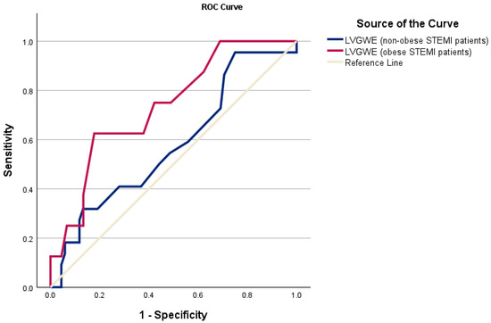
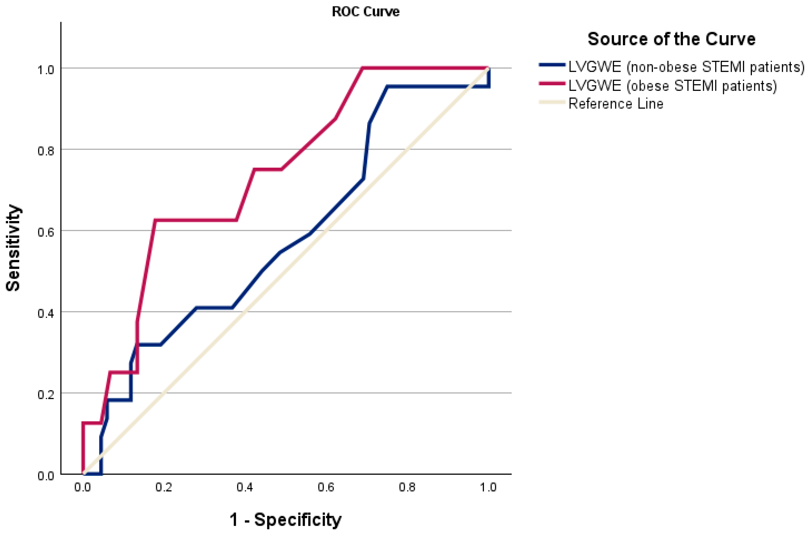
The ROC analysis was conducted to evaluate the predictive performance of various echocardiographic parameters for identifying MACEs in obese and non-obese STEMI patients. Among the parameters evaluated, LVGWE demonstrated the highest predictive value for adverse outcomes in obese STEMI patients (Table 5), with an area under the curve (AUC) of 0.736 (95% confidence interval [CI]: 0.559–0.914; p = 0.009). The optimal cutoff value for LVGWE, determined by the Youden index, was 79%, with a sensitivity of 62% and a specificity of 82%. In contrast, none of the evaluated parameters achieved statistical significance in predicting MACEs among non-obese STEMI patients (Table 6).
Table 5.
ROC analysis for the detection of MACEs in obese STEMI patients.
Table 6.
ROC analysis for the detection of MACEs in non-obese STEMI patients.
Figure 1 illustrates the ROC curves of LVGWE for detecting MACEs in obese and non-obese STEMI patients.
Figure 1.
ROC curves of LVGWE for detecting major adverse cardiac events in non-obese (blue line) and obese STEMI patients (red line). ROC, receiver operating characteristic; LVGWE, left ventricular global work efficiency; STEMI, ST-elevation myocardial infarction.
3.4. Kaplan–Meier Survival Analysis
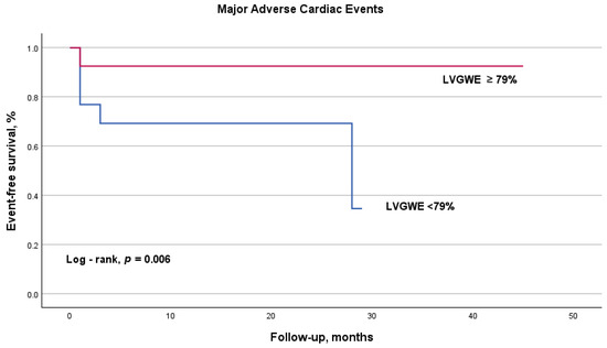
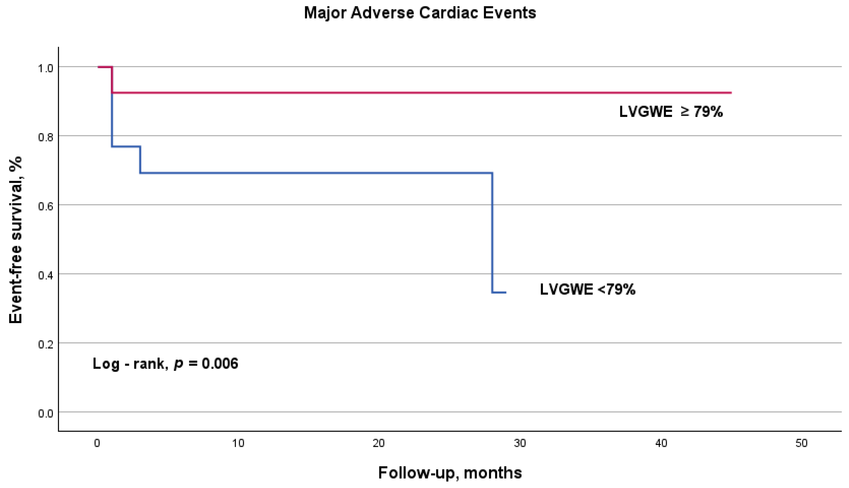
Kaplan–Meier survival analysis showed that obese STEMI patients with a LVGWE ≥ 79% exhibited a significantly better prognosis compared to those with lower LVGWE values, as demonstrated by the log-rank test (p = 0.006). These findings suggest that LVGWE serves as a meaningful prognostic marker, effectively distinguishing between patients with favorable and poorer outcomes within the obese STEMI cohort (Figure 2).
Figure 2.
Kaplan–Meier curve for major adverse cardiac events in obese STEMI patients according to the cutoff value of left ventricular global work efficiency. The curve illustrates improved survival rates for patients with LVGWE ≥ 79% (red line) compared to those with LVGWE < 79% (blue line) (log-rank, p = 0.006). LVGWE, left ventricular global work efficiency.
3.5. Cox Regression Analysis
In obese STEMI patients, univariate Cox proportional hazards regression revealed a significant association between LVGWE—using a cutoff value of 79%—and clinical outcomes (hazard ratio [HR]: 5.59; 95% CI: 1.33–23.50; p = 0.019), indicating an increased risk of adverse events for those with LVGWE < 79%. Specifically, patients with LVGWE < 79% experienced a 5.59-fold higher risk of adverse outcomes compared to those with LVGWE ≥ 79% (Table 7).
Table 7.
Univariate Cox regression analysis in obese STEMI patients.
To assess the broader prognostic value of the LVGWE cutoff, we extended the analysis to the entire study population (Table 8). In both univariate and multivariate models, LVGWE < 79% remained a significant predictor of adverse clinical outcomes. This association persisted even after adjustment for key confounders, including obesity status and infarct size, as reflected by peak creatine kinase-MB levels (adjusted HR: 2.73; 95% CI: 1.26–5.90; p = 0.010). These findings suggest that the prognostic utility of LVGWE extends beyond the obese population, supporting its potential value as a valuable risk stratification tool in the broader STEMI population.
Table 8.
Univariate and multivariate Cox regression analysis for the entire study cohort.
3.6. Correlation Analysis
To explore whether a linear relationship existed between BMI and LVGWE, we performed a Spearman correlation analysis using both BMI and LVGWE as continuous variables. The analysis demonstrated a weak inverse correlation (r = −0.147), which did not reach statistical significance (p = 0.079).
3.7. Reliability of MW Parameters
All MW parameters demonstrated excellent intraobserver agreement, with intraclass correlation coefficients as follows: 0.987 (95% CI: 0.967–0.995) for LVGWI, 0.986 (95% CI: 0.965–0.994) for LVGCW, 0.974 (95% CI: 0.935–0.989) for LVGWW, and 0.975 (95% CI: 0.939–0.990) for LVGWE. Interobserver variability was also satisfactory for all MW indices, with the following values: 0.968 (95% CI: 0.923–0.987) for LVGWI, 0.962 (95% CI: 0.910–0.984) for LVGCW, 0.963 (95% CI: 0.913–0.985) for LVGWW, and 0.962 (95% CI: 0.909–0.984) for LVGWE.
4. Discussion
The present study investigated the prognostic significance of myocardial work indices in patients with acute STEMI, with a particular focus on individuals with obesity. Our data demonstrates that LVGWE serves as a robust predictor of adverse outcomes in STEMI patients, particularly among those with BMI ≥ 30 kg/m2.
Notably, the threshold of LVGWE < 79% reliably identified a subgroup at increased risk for MACEs, making it a promising prognostic marker. This cut-off remained prognostically relevant even when applied to the broader cohort, independent of obesity and infarct size, thereby underscoring its potential value in comprehensive STEMI risk stratification.
4.1. Prognostic Value of LVGWE in STEMI
Numerous studies have examined echocardiographic parameters in relation to STEMI prognosis. Traditionally, LVEF has been the cornerstone for assessing post-infarction systolic function and outcome prediction [20,21,22]. However, technological advances in echocardiography—particularly speckle-tracking and tissue Doppler imaging—have enabled a more sophisticated evaluation of myocardial performance. Among emerging modalities, GLS and myocardial work indices have shown superior prognostic performance [23,24,25,26].
Our findings reinforce this paradigm shift, demonstrating that LVGWE surpasses LVEF in predicting adverse outcomes in STEMI patients. Neither LVEF nor other conventional metrics reached statistical significance in univariate or ROC analyses, whereas LVGWE emerged as a robust prognostic marker. This superiority is biologically plausible: LVGWE reflects the proportion of total myocardial work that contributes to effective ejection, correlating more closely with myocardial oxygen consumption and energy efficiency than LVEF [27,28,29].
Comparable results have been reported in prior investigations. Lustosa et al. showed that an LVGWE < 86% independently predicted all-cause mortality in STEMI, with added prognostic value over LVEF and GLS [30]. Similarly, Coisne et al. identified an LVGWE threshold of 91% as predictive of adverse events one month after acute myocardial infarction, enhancing early post-acute myocardial infarction risk stratification [31].
Our study adds to this growing body of evidence by demonstrating that a lower LVGWE (<79%) is especially informative in obese STEMI patients. The heterogeneity in threshold values reported across studies likely reflects differences in population characteristics, infarct severity, timing of echocardiographic assessment, and methodological variability, underscoring the need for further research to refine clinically applicable cut-offs.
4.2. Impact of Obesity on STEMI Outcomes
We also observed that obese patients with STEMI had significantly lower LVGWE values than their non-obese counterparts, aligning with established pathophysiological mechanisms linking obesity to myocardial dysfunction [32,33,34,35].
Obesity contributes to an adverse myocardial metabolic profile, characterized by increased fatty acid oxidation, impaired glucose metabolism, elevated myocardial oxygen consumption, and structural remodeling [4], all of which compromise contractile efficiency and increase vulnerability to ischemic injury.
Consistent with previous studies, our obese subgroup exhibited a higher prevalence of cardiometabolic comorbidities, including hypertension, diabetes mellitus, and dyslipidemia, as well as elevated systolic and diastolic blood pressures and hyperglycemia—all known contributors to adverse outcomes in the context of STEMI [36,37].
Obesity is also associated with increased risks of heart failure, arrhythmias, and cardiovascular mortality in patients with acute coronary syndromes [38,39].
Our findings support the hypothesis that LVGWE captures obesity-related myocardial dysfunction beyond conventional risk factors. Its ability to detect subclinical inefficiencies in myocardial performance may render it particularly useful for identifying obese STEMI patients at high risk of complications, potentially guiding targeted therapeutic strategies and resource allocation.
Although our primary analysis was based on BMI categories, an exploratory assessment using BMI as a continuous variable did not reveal a statistically significant association with LVGWE. These findings highlight the complexity of the relationship between adiposity and myocardial work efficiency and suggest that additional parameters beyond BMI may be needed to capture this relationship more accurately.
4.3. Limitations and Future Directions
Despite its strengths, this study has limitations that must be acknowledged. The relatively small sample size, particularly within the obese subgroup, limited the statistical power and precluded robust multivariate analyses in that group. Additionally, the short-to-intermediate follow-up duration (median 13 months) may not fully capture long-term prognostic implications of myocardial work parameters. Other important variables, such as medication adherence, post-discharge interventions, and longitudinal functional recovery, were not assessed and may confound outcome interpretation.
Although BMI remains a widely used parameter for classifying obesity, it does not fully reflect the complexity of excess adiposity or its functional consequences. Recent expert recommendations [40] support a shift toward defining obesity based on adiposity-related organ dysfunction, incorporating parameters such as waist circumference, body fat percentage, or echocardiographic markers of fat distribution. Our study protocol was developed prior to the publication of these updated definitions and reflects the standard criteria in use at the time. Another relevant limitation is the lack of standardized protocols for myocardial work quantification in routine clinical practice. Although pressure–strain loop analysis using speckle-tracking echocardiography is increasingly utilized, variability in image acquisition, frame rate optimization, and post-processing software may hinder reproducibility across centers. To facilitate clinical translation, future studies should aim to standardize acquisition protocols and validate myocardial work indices in multicenter prospective cohorts.
5. Conclusions
LVGWE represents a novel and promising prognostic marker in patients with STEMI, particularly in those with obesity—a population known to exhibit more complex pathophysiological and clinical profiles. Its capacity to integrate myocardial deformation with energetic efficiency allows for a more comprehensive assessment of LV performance than traditional metrics. Incorporating LVGWE into routine post-STEMI evaluation may enhance risk stratification and enable more personalized management strategies, especially in high-risk subgroups.
Author Contributions
Conceptualization, A.-C.F. and C.M.; methodology, A.M. and C.M.; software, M.S.; validation, C.-T.L. and D.-M.B.; formal analysis, M.S.; investigation, A.-C.F., M.-A.L., R.Ș., and C.M.; resources, A.M. and S.C.; data curation, M.S. and A.-R.M.; writing—original draft preparation, A.-C.F. and C.M.; writing—review and editing, R.Ș., S.C., and C.M.; visualization, A.-C.F., A.-R.M., and D.-M.B.; supervision, M.-A.L., R.Ș., and S.C.; project administration, D.-M.B. and C.M.; funding acquisition, C.-T.L. All authors have read and agreed to the published version of the manuscript.
Funding
This research received no external funding.
Institutional Review Board Statement
The study was conducted in accordance with the Declaration of Helsinki. Ethical approval was obtained from the institutional review board (approval number 10163/5 December 2023) and the university ethics committee (approval number 07/31 January 2024).
Informed Consent Statement
Written informed consent was obtained from all participants.
Data Availability Statement
The data presented in this study is available upon request from the corresponding author for the protection of the participants’ privacy.
Conflicts of Interest
The authors declare no conflicts of interest.
Abbreviations
The following abbreviations are used in this manuscript:
| AUC | Area under the curve |
| BMI | Body mass index |
| GLS | Global longitudinal strain |
| LV | Left ventricle |
| LVEF | Left ventricular ejection fraction |
| LVGCW | Left ventricular global constructive work |
| LVGWE | Left ventricular global work efficiency |
| LVGWI | Left ventricular global work index |
| LVGWW | Left ventricular global wasted work |
| LVMW | Left ventricular myocardial work |
| MACE | Major adverse cardiac events |
| PCI | Percutaneous coronary intervention |
| ROC | Receiver operating characteristics |
| STEMI | ST-elevation myocardial infarction |
| TTE | Transthoracic echocardiography |
References
- Fruh, S.M. Obesity: Risk factors, complications, and strategies for sustainable long-term weight management. J. Am. Assoc. Nurse Pract. 2017, 29, S3–S14. [Google Scholar] [CrossRef] [Scilit] [PubMed]
- GBD 2021 Adult BMI Collaborators. Global, regional, and national prevalence of adult overweight and obesity, 1990–2021, with forecasts to 2050: A forecasting study for the Global Burden of Disease Study 2021. Lancet 2025, 8, 813–838. [Google Scholar]
- Kolwicz, S.C., Jr.; Purohit, S.; Tian, R. Cardiac metabolism and its interactions with contraction, growth, and survival of cardiomyocytes. Circ. Res. 2013, 113, 603–616. [Google Scholar] [CrossRef] [Scilit] [PubMed]
- Cole, M.A.; Murray, A.J.; Cochlin, L.E.; Heather, L.C.; McAleese, S.; Knight, N.S.; Sutton, E.; Jamil, A.A.; Parassol, N.; Clarke, K. A high fat diet increases mitochondrial fatty acid oxidation and uncoupling to decrease efficiency in rat heart. Basic Res. Cardiol. 2011, 106, 447–457. [Google Scholar] [CrossRef] [Scilit]
- Huang, J.; Li, G.A.; Wang, J.; Jiao, Y.W.; Qian, Z.F.; Fan, L.; Tang, L.M. Evaluation of subclinical left ventricular systolic dysfunction in obese patients by global myocardial work. Diabetol. Metab. Syndr. 2023, 15, 254. [Google Scholar] [CrossRef] [Scilit]
- Parto, P.; Lavie, C.J. Obesity and CardiovascularDiseases. Curr. Probl. Cardiol. 2017, 42, 376–394. [Google Scholar] [CrossRef] [Scilit]
- Amir, O.; Elbaz-Greener, G.; Carasso, S.; Claggett, B.; Barbarash, O.; Zaman, A.; Christersson, C.; Kiatchoosakun, S.; Anonuevo, J.; Opolski, G.; et al. Association between body mass index and clinical outcomes in patients with acute myocardial infarction and reduced systolic function: Analysis of PARADISE-MI trial data. Eur. J. Heart Fail. 2025, 27, 558–565. [Google Scholar] [CrossRef] [Scilit]
- Basha, M.; Stavropoulou, E.; Nikolaidou, A.; Dividis, G.; Peteinidou, E.; Tsioufis, P.; Kamperidis, N.; Dimitriadis, K.; Karamitsos, T.; Giannakoulas, G.; et al. Diagnosing Heart Failure with Preserved Ejection Fraction in Obese Patients. J. Clin. Med. 2025, 14, 1980. [Google Scholar] [CrossRef] [Scilit]
- Galderisi, M.; Cosyns, B.; Edvardsen, T.; Cardim, N.; Delgado, V.; Di Salvo, G.; Donal, E.; Sade, L.E.; Ernande, L.; Garbi, M.; et al. 2016–2018 EACVI Scientific Documents Committee. Standardization of adult transthoracic echocardiography reporting in agreement with recent chamber quantification, diastolic function, and heart valve disease recommendations: An expert consensus document of the European Association of Cardiovascular Imaging. Eur. Heart J. Cardiovasc. Imaging 2017, 18, 1301–1310. [Google Scholar]
- Russell, K.; Eriksen, M.; Aaberge, L.; Wilhelmsen, N.; Skulstad, H.; Remme, E.W.; Haugaa, K.H.; Opdahl, A.; Fjeld, J.G.; Gjesdal, O.; et al. A novel clinical method for quantification of regional left ventricular pressure-strain loop area: A non-invasive index of myocardial work. Eur. Heart J. 2012, 33, 724–733. [Google Scholar] [CrossRef] [Scilit]
- Edwards, N.F.A.; Scalia, G.M.; Shiino, K.; Sabapathy, S.; Anderson, B.; Chamberlain, R.; Khandheria, B.K.; Chan, J. Global myocardial work is superior to global longitudinal strain to predict significant coronary artery disease in patients with normal left ventricular function and wall motion. J. Am. Soc. Echocardiogr. 2019, 32, 947–957. [Google Scholar] [CrossRef] [Scilit] [PubMed]
- Ilardi, F.; D’Andrea, A.; D’Ascenzi, F.; Bandera, F.; Benfari, G.; Esposito, R.; Malagoli, A.; Mandoli, G.E.; Santoro, C.; Russo, V.; et al. Myocardial Work by Echocardiography: Principles and Applications in Clinical Practice. J. Clin. Med. 2021, 10, 4521. [Google Scholar] [CrossRef] [Scilit] [PubMed]
- Papadopoulos, K.; Ozden, T.O.; Mitrousi, K.; Ikonimidis, I. Myocardial work: Methodology and clinical applications. Diagnostics 2021, 11, 573. [Google Scholar] [CrossRef] [Scilit] [PubMed]
- El Mahdiui, M.; van der Bijl, P.; Abou, R.; Ajmone, M.N.; Delgado, V.; Bax, J.J. Global Left Ventricular Myocardial Work Efficiency in Healthy Individuals and Patients with Cardiovascular Disease. J. Am. Soc. Echocardiogr. 2019, 32, 1120–1127. [Google Scholar] [CrossRef] [Scilit]
- Marzlin, N.; Hays, A.G.; Peters, M.; Kaminski, A.; Roemer, S.; O’Leary, P.; Kroboth, S.; Harland, D.R.; Khandheria, B.K.; Tajik, A.J.; et al. Myocardial Work in Echocardiography. Circ. Cardiovasc. Imaging 2023, 16, e014419. [Google Scholar] [CrossRef] [Scilit]
- Zhao, H.; Jiang, M.; Wang, W.; Tao, Z.; Wang, X.; Chai, Y.; Han, Y.; Liu, Q.; Chen, Y.; Yue, J.; et al. Subclinical myocardial work impairment in non-diabetic overweight and obese individuals: Impact of cardiometabolic traits. Int. J. Cardiol. 2025, 26, 133321. [Google Scholar] [CrossRef] [Scilit]
- Byrne, R.A.; Rossello, X.; Coughlan, J.J.; Barbato, E.; Berry, C.; Chieffo, A.; Claeys, M.J.; Dan, G.A.; Dweck, M.R.; Galbraith, M.; et al. 2023 ESC Guidelines for the management of acute coronary syndromes. Eur. Heart J. 2023, 44, 3720–3826. [Google Scholar] [CrossRef] [Scilit]
- Lang, R.M.; Badano, L.P.; Mor-Avi, V.; Afilalo, J.; Armstrong, A.; Ernande, L.; Flachskampf, F.A.; Foster, E.; Goldstein, S.A.; Kuznetsova, T.; et al. Recommendations for cardiac chamber quantification by echocardiography in adults: An update from the American Society of Echocardiography and the European Association of Cardiovascular Imaging. Eur. Heart J. Cardiovasc. Imaging 2015, 16, 233–271. [Google Scholar] [CrossRef] [Scilit]
- Pasquali, R.; Casanueva, F.; Haluzik, M.; van Hulsteijn, L.; Ledoux, S.; Monteiro, M.P.; Salvador, J.; Santini, F.; Toplak, H.; Dekkers, O.M. European Society of Endocrinology Clinical Practice Guideline: Endocrine work-up in obesity. Eur. J. Endocrinol. 2020, 182, G1–G32. [Google Scholar] [CrossRef] [Scilit]
- Lei, Z.; Li, B.; Li, B.; Peng, W. Predictors and prognostic impact of left ventricular ejection fraction trajectories in patients with ST-segment elevation myocardial infarction. Aging Clin. Exp. Res. 2022, 34, 1429–1438. [Google Scholar] [CrossRef] [Scilit]
- Yahud, E.; Tzuman, O.; Fink, N.; Goldenberg, I.; Goldkorn, R.; Peled, Y.; Lev, E.; Asher, E. Trends in long-term prognosis according to left ventricular ejection fraction after acute coronary syndrome. J. Cardiol. 2020, 76, 303–308. [Google Scholar] [CrossRef] [Scilit] [PubMed]
- Otero-García, O.; Cid-Álvarez, A.B.; Juskova, M.; Álvarez-Álvarez, B.; Tasende-Rey, P.; Gude-Sampedro, F.; García-Acuña, J.M.; Agra-Bermejo, R.; López-Otero, D.; Sanmartín-Pena, J.C.; et al. Prognostic impact of left ventricular ejection fraction recovery in patients with ST-segment elevation myocardial infarction undergoing primary percutaneous coronary intervention: Analysis of an 11-year all-comers registry. Eur. Heart J. Acute Cardiovasc. Care 2021, 10, 898–908. [Google Scholar] [CrossRef] [Scilit] [PubMed]
- Holzknecht, M.; Reindl, M.; Tiller, C.; Reinstadler, S.J.; Lechner, I.; Pamminger, M.; Schwaiger, J.P.; Klug, G.; Bauer, A.; Metzler, B.; et al. Global longitudinal strain improves risk assessment after ST-segment elevation myocardial infarction: A comparative prognostic evaluation of left ventricular functional parameters. Clin. Res. Cardiol. 2021, 110, 1599–1611. [Google Scholar] [CrossRef] [Scilit] [PubMed]
- Akkuş, Ö.F.; Gürdoğan, M. Effect of Global Longitudinal Strain at Discharge Period on Predicting Cardiac Defibrillator Implantation in STEMİ Patients with Impaired Left Ventricle Systolic Functions. Medicina 2025, 61, 545. [Google Scholar] [CrossRef] [Scilit]
- Lustosa, R.P.; Fortuni, F.; van der Bijl, P.; Mahdiui, M.E.; Montero-Cabezas, J.M.; Kostyukevich, M.V.; Knuuti, J.; Marsan, N.A.; Delgado, V.; Bax, J.J. Changes in Global Left Ventricular Myocardial Work Indices and Stunning Detection 3 Months After ST-Segment Elevation Myocardial Infarction. Am. J. Cardiol. 2021, 157, 15–21. [Google Scholar] [CrossRef] [Scilit]
- Lustosa, R.P.; van der Bijl, P.; El Mahdiui, M.; Montero-Cabezas, J.M.; Kostyukevich, M.V.; Ajmone Marsan, N.; Bax, J.J.; Delgado, V. Noninvasive Myocardial Work Indices 3 Months after ST-Segment Elevation Myocardial Infarction: Prevalence and Characteristics of Patients with Postinfarction Cardiac Remodeling. J. Am. Soc. Echocardiogr. 2020, 33, 1172–1179. [Google Scholar] [CrossRef] [Scilit]
- Sabatino, J.; De Rosa, S.; Leo, I.; Strangio, A.; Spaccarotella, C.; Polimeni, A.; Sorrentino, S.; Di Salvo, G.; Indolfi, C. Prediction of Significant Coronary Artery Disease Through Advanced Echocardiography: Role of Non-invasive Myocardial Work. Front. Cardiovasc. Med. 2021, 24, 719603. [Google Scholar] [CrossRef] [Scilit]
- Pan, J.C.; Lyu, L.J.; Liu, Q.D.; Yang, W.; Li, X.H.; Han, Y.M.; Sun, J.Y.; Dong, M.; Zhang, P.F.; Zhang, M. Association between resting myocardial work indices and stress myocardial perfusion in patients with angina and non-obstructive coronary artery disease. Quant. Imaging Med. Surg. 2023, 13, 4563–4577. [Google Scholar] [CrossRef] [Scilit]
- Qin, Y.; Wu, X.; Wang, J.; Li, Y.; Ding, X.; Guo, D.; Jiang, Z.; Zhu, W.; Cai, Q.; Lu, X. Value of territorial work efficiency estimation in non-ST-segment-elevation acute coronary syndrome: A study with non-invasive left ventricular pressure-strain loops. Int. J. Cardiovasc. Imaging 2021, 37, 1255–1265. [Google Scholar] [CrossRef] [Scilit]
- Lustosa, R.P.; Butcher, S.C.; van der Bijl, P.; El Mahdiui, M.; Montero-Cabezas, J.M.; Kostyukevich, M.V.; Rocha De Lorenzo, A.; Knuuti, J.; Ajmone, M.N.; Bax, J.J.; et al. Global Left Ventricular Myocardial Work Efficiency and Long-Term Prognosis in Patients After ST-Segment-Elevation Myocardial Infarction. Circ. Cardiovasc. Imaging 2021, 14, e012072. [Google Scholar] [CrossRef] [Scilit]
- Coisne, A.; Fourdinier, V.; Lemesle, G.; Delsart, P.; Aghezzaf, S.; Lamblin, N.; Schurtz, G.; Verdier, B.; Ninni, S.; Delobelle, A.; et al. Clinical significance of myocardial work parameters after acute myocardial infarction. Eur. Heart J. Open 2022, 2, oeac037. [Google Scholar] [CrossRef] [Scilit] [PubMed]
- Ren, J.; Wu, N.N.; Wang, S.; Sowers, J.R.; Zhang, Y. Obesity cardiomyopathy: Evidence, mechanisms, and therapeutic implications. Physiol. Rev. 2021, 101, 1745–1807. [Google Scholar] [CrossRef] [Scilit] [PubMed]
- Koliaki, C.; Liatis, S.; Kokkinos, A. Obesity and cardiovascular disease: Revisiting an old relationship. Metabolism 2019, 92, 98–107. [Google Scholar] [CrossRef] [Scilit] [PubMed]
- El Hadj Othmane, T.; El Hadj Othmane, O.; Nizar, H. Obesity-Related Phenotype of Heart Failure with Preserved Ejection Fraction: A Comprehensive Review. Cureus 2025, 17, e81512. [Google Scholar] [CrossRef] [Scilit]
- Taverna, G.; Trimarchi, G.; Lofrumento, F.; Mancinelli, A.; Teresi, L.; Alagna, G.; Carerj, S.; Zito, C.; Di Bella, G. The impact of BMI on myocardial work and left atrial strain in young overweight patients with preserved left ventricular ejection fraction. Eur. Heart J. 2022, 24, jead119-230. [Google Scholar] [CrossRef] [Scilit]
- Alansari, H.; Lazzara, G.; Taha, M.B.; Gorthi, J.R. The Impact of Obesity on Cardiovascular Diseases: Heart Failure. Methodist. Debakey Cardiovasc. J. 2025, 21, 44–52. [Google Scholar] [CrossRef] [Scilit]
- Welsh, A.; Hammad, M.; Piña, I.L.; Kulinski, J. Obesity and cardiovascular health. Eur. J. Prev. Cardiol. 2024, 31, 1026–1035. [Google Scholar] [CrossRef] [Scilit]
- Abid, A.R.; El-Menyar, A.; Singh, R.; Gomaa, M.; Habib, S.; Abdelrahman, A.S.; Asaad, N.; AlQahtani, A.; Al-Thani, H.; AlBinali, H.; et al. Patterns and Outcomes of Obesity Using Body Mass Index in Patients Hospitalized with Acute Cardiovascular Disorders: A Retrospective Analysis of 7284 Patients in a Middle Eastern Country. J. Clin. Med. 2023, 12, 7263. [Google Scholar] [CrossRef] [Scilit]
- Pop, A.D.; Pop, D.; Buzoianu, A. Particularities of arrhythmias and obesity in heart failure. Rom. J. Cardiol. 2020, 30, 7–11. [Google Scholar]
- Rubino, F.; Cummings, D.E.; Eckel, R.H.; Cohen, R.V.; Wilding, J.P.H.; Brown, W.A.; Stanford, F.C.; Batterham, R.L.; Farooqi, I.S.; Farpour-Lambert, N.J.; et al. Definition and diagnostic criteria of clinical obesity. Lancet Diabetes Endocrinol. 2025, 13, 221–262. [Google Scholar] [CrossRef] [Scilit]

Disclaimer/Publisher’s Note: The statements, opinions and data contained in all publications are solely those of the individual author(s) and contributor(s) and not of MDPI and/or the editor(s). MDPI and/or the editor(s) disclaim responsibility for any injury to people or property resulting from any ideas, methods, instructions or products referred to in the content. |
© 2025 by the authors. Licensee MDPI, Basel, Switzerland. This article is an open access article distributed under the terms and conditions of the Creative Commons Attribution (CC BY) license (https://creativecommons.org/licenses/by/4.0/).