


Abstract
We depict the rare case of a patient with aneurysmatic subarachnoid hemorrhage previously treated with deep brain stimulation for Parkinson’s disease. Initial CT scans showed a Fisher grade 4 subarachnoid hemorrhage with lead displacement due to midline-shift. CT angiogram revealed a supra-ophthalmic aneurysm of the internal carotid artery. The patient subsequently underwent clipping of the aneurysm and decompressive hemicraniecomy.
A 70-year-old patient was admitted to our emergency room with a history of sudden loss of consciousness and epileptic seizure. An initial GCS (Glasgow Coma Scale) was 3, and pupils were isochoric with intact direct and consensual light reflex. Sixteen years prior, he had been implanted with bilateral stimulation leads for deep brain stimulation of the subthalamic nucleus due to Parkinson’s disease. An initial head CT scan (Philips Icon, 1mm head scan) showed a predominantly left-sided subarachoid hemorrhage (SAH) Fisher grade 4 with intra-ventricular hemorrhage, intra-parenchymal hemorrhage within the left frontal lobe, subdural hematoma, and midline-shift of 12 mm. The SAH was classified as WFNS (World Federation of Neurological Surgeons) grade 5. The CT angiogram (Philips Icon, 1mm head scan) of the supra-aortal vessels revealed a left-sided supra-ophthalmic aneurysm of the internal carotid artery as the supposed source of hemorrhage. An external ventricular drain was placed and the patient subsequently brought to the operating room for clipping of the aneurysm and decompressive hemicraniectomy. During the latter, the left-sided DBS stimulation lead had to be removed. Eventually, the therapy was switched to best supportive care due to massive parenchyma destruction.
A subarachnoid hemorrhage (SAH) is an extravasation of blood into the space between the arachnoid membrane and the pia mater filled with cerebrospinal fluid. The most common cause of a non-traumatic SAH is due to a ruptured intracranial aneurysm [1].
Whilst aneurysmal SAH can be a devastating condition associated with neurological complications up to death, nonaneurysmal SAH is associated with a good prognoses and is rarely associated with neurological complications [2].
Diagnosis of a SAH is primarily performed by computed tomography (CT) of the head, in which the SAH appears characteristically with hyperdense extravasated blood sometimes associated with intraparenchymal hematoma and hydrocephalus [3,4].
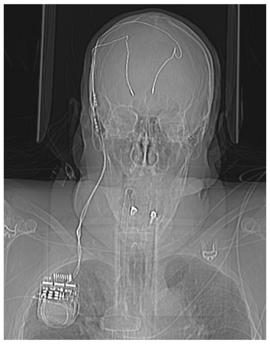
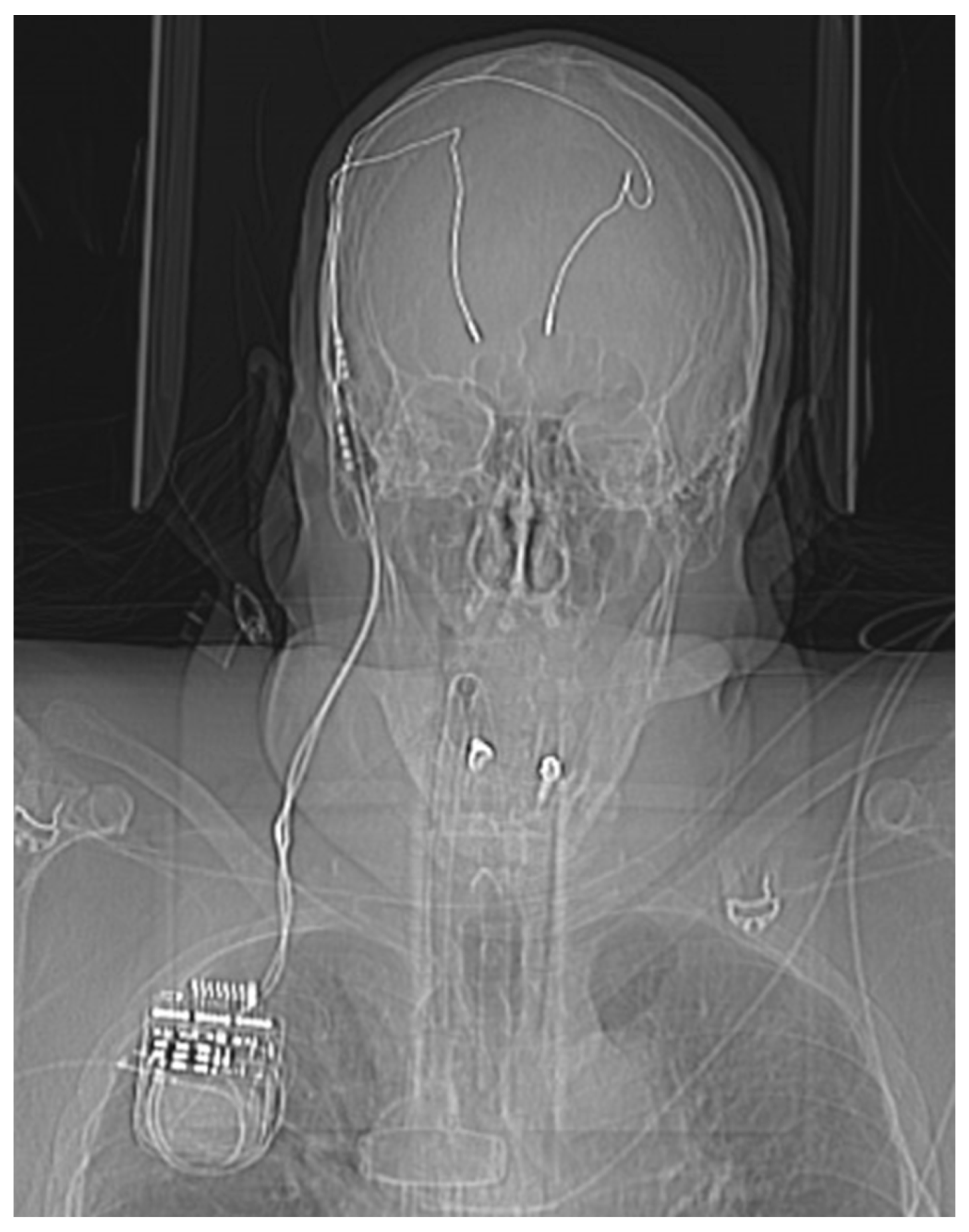
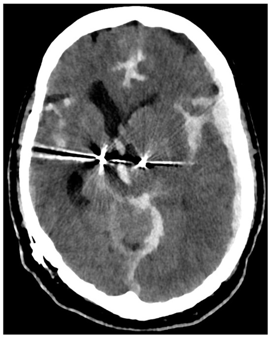
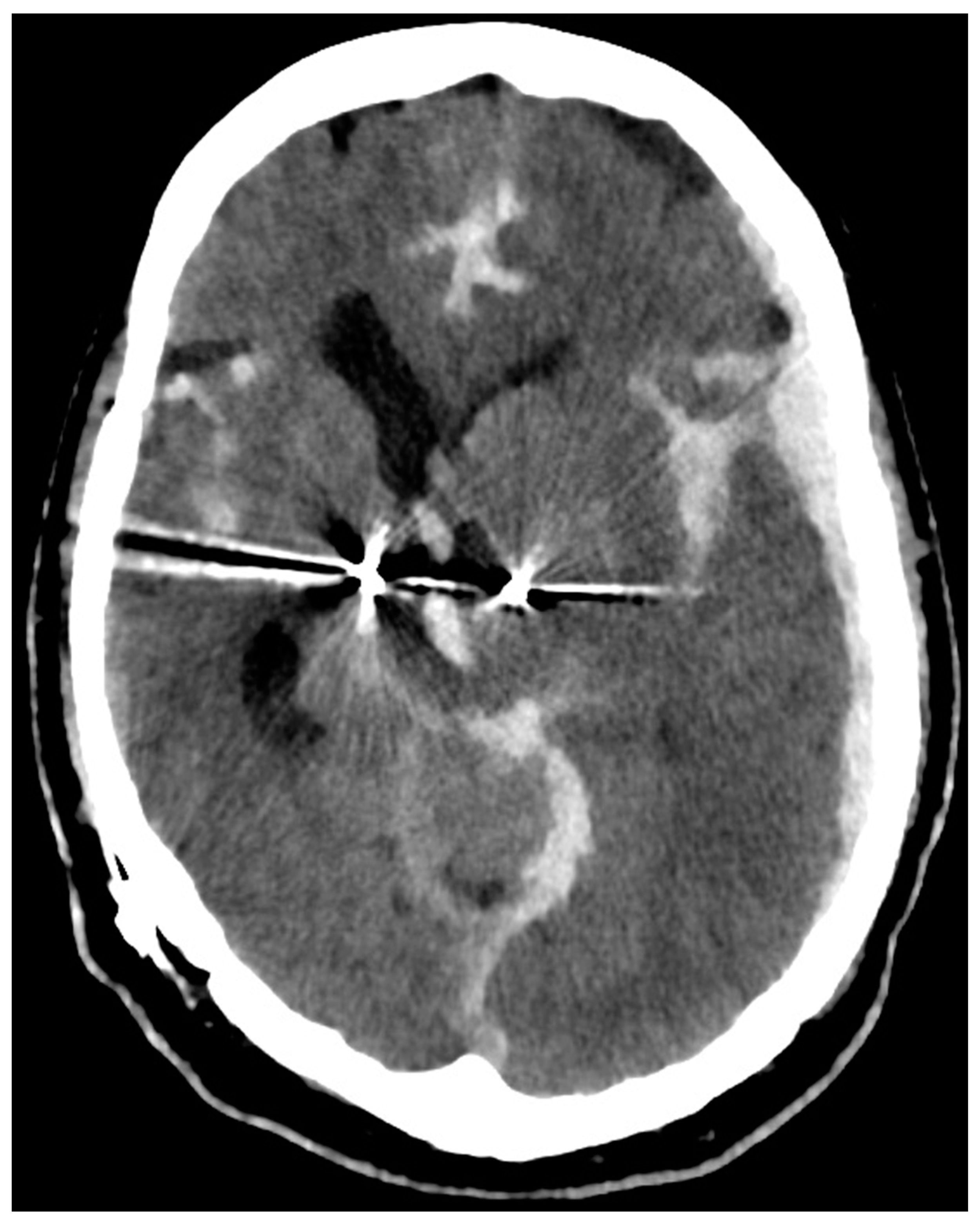
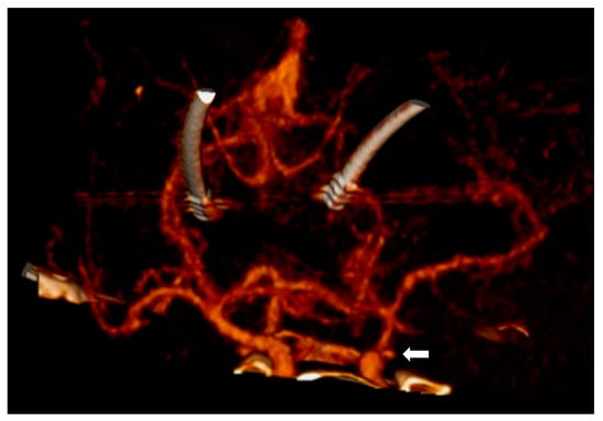
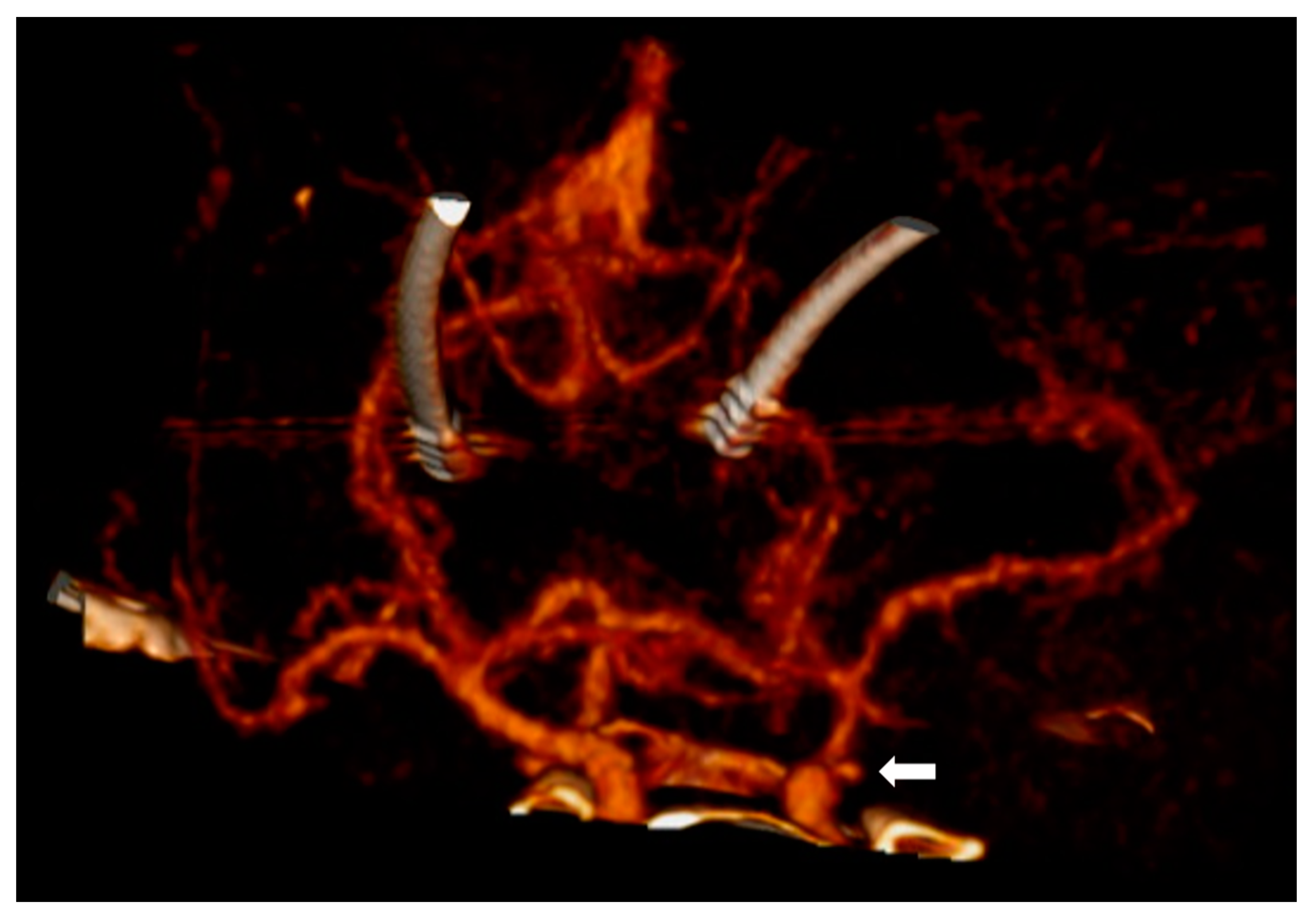
In this case, however, first signs of the SAH could be identified in the initial head X-ray of the CT scout image due to the patient having been implanted with bilateral stimulation leads for deep brain stimulation (Figure 1). The differential diagnosis of an intracranial mass affect includes pathologies of traumatic (epidural hematoma, acute subdural hematoma, chronic subdural hematoma, traumatic intracerebral hemorrhage), tumorous (brain derived tumors such as glioblastoma multiforma or meningioma, metastases) as well as vascular origin (subarachnoid hemorrhage, intracranial hemorrhage). Table 1 depicts the most common differential diagnoses in respect to pathological origin, entity, CT morphology and clinical dynamic. In this particular case, the CT scan revealed the brain shift to be due to the subarachnoid hemorrhage with an associated hematoma which displaced the stimulation leads to the contralateral side (Figure 2). The CT angiogram showed a supra-ophthalmic aneurysm of the internal carotid artery to be the source of hemorrhage (Figure 3).
Figure 1.
Initial scout image of the head CT scan, essentially mimicking an AP cranial X-ray. Note the implanted stimulations leads for deep brain stimulation, which are connected to an infra-clavicular neurostimulator. The DBS stimulation leads are displaced to the right side.
Table 1.
Differential diagnosis of mass effect.
Figure 2.
Initial head CT scan shows the predominantly left-sided subarachnoid hemorrhage with intra-ventricular hemorrhage, subdural hematoma, and midline-shift. The DBS stimulation leads are displaced to the contralateral side.
Figure 3.
A 3D reconstruction of the CT angiogram showing a supra-ophthalmic aneurysm of the internal carotid artery as the source of hemorrhage (marked by the arrow). DBS leads are displaced to the contralateral side.
The amount of blood within the subarachnoid space and the ventricles, as well as the parenchyma, can be easily graded on a head CT scan and is correlated with poor outcome [5,6]. Further, the prognosis is correlated to the initial clinical exam which is most widely measured on the clinical scale of Hunt and Hess and the World Federation of Neurological Surgeons [7,8]. Given the clinical and radiological presentation of this patient, this was defined as poor grade.
Once an aneurysm has been identified by CT angiogram and/or digital subtraction angiography, treatment can be performed either with endovascular coiling or microsurgical clipping [9].
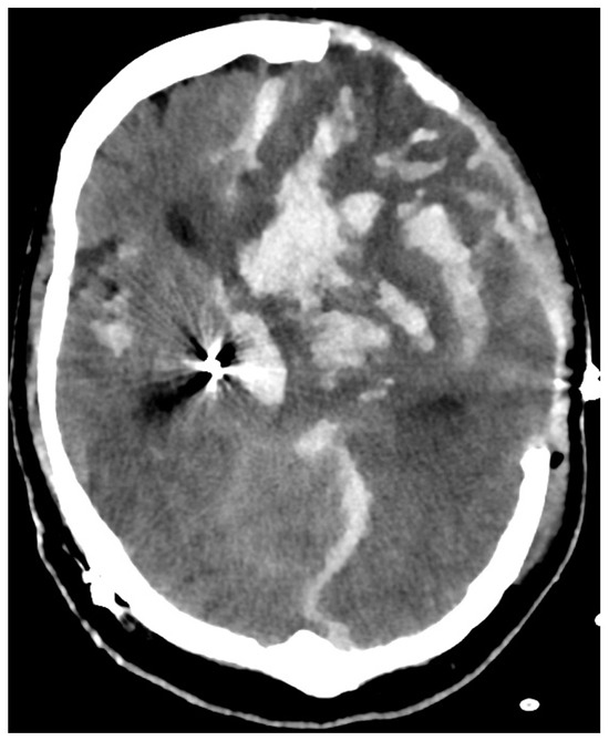
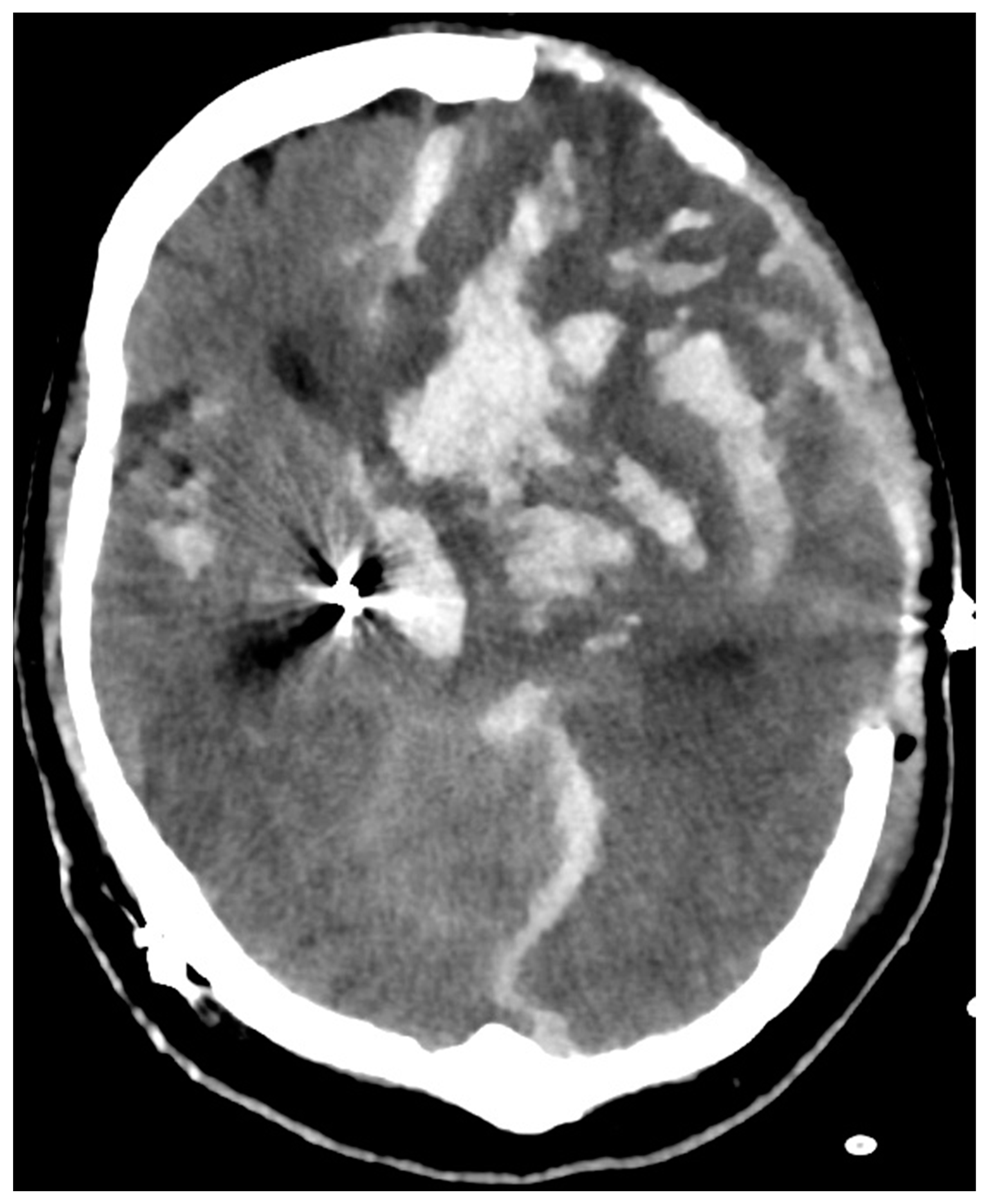
Further complications can be managed by placement of an external ventricular drain for hydrocephalus and decompressive hemicraniectomy in case of otherwise uncontrollable intracranial hypertension, as performed in this case report (Figure 4) [10].
Figure 4.
Postoperative head CT scan after clipping of the supra-ophthalmic aneurysm, left-sided DBS lead removal and decompressive hemicraniectomy. Eventually, therapy was switched to best supportive care due to massive parenchyma destruction.
Author Contributions
Conceptualization, G.B.; methodology, G.B.; investigation, G.B.; data curation, G.B.; writing—original draft preparation, G.B.; writing—review and editing, V.B. and J.M.; supervision, J.M. All authors have read and agreed to the published version of the manuscript.
Funding
This research received no external funding.
Institutional Review Board Statement
The study was conducted in accordance with the Declaration of Helsinki, and approved by the Institutional Review Board of the University of Bonn (protocol code 204/21 and date of 17 August 2021).
Informed Consent Statement
Due to fatality, patient could not give his consent. Written informed consent has been obtained from patient’s next of kin to publish this paper.
Data Availability Statement
No new data was created.
Conflicts of Interest
The authors declare no conflicts of interest.
References
- van Gijn, J.; Rinkel, G.J. Subarachnoid haemorrhage: Diagnosis, causes and management. Brain 2001, 124, 249–278. [Google Scholar] [CrossRef] [Scilit] [PubMed]
- Adams, H.P., Jr.; Gordon, D.L. Nonaneurysmal subarachnoid hemorrhage. Ann. Neurol. 1991, 29, 461–462. [Google Scholar] [CrossRef] [Scilit] [PubMed]
- Latchaw, R.E.; Silva, P.; Falcone, S.F. The role of CT following aneurysmal rupture. Neuroimaging Clin. N. Am. 1997, 7, 693–708. [Google Scholar] [PubMed]
- Hijdra, A.; van Gijn, J.; Nagelkerke, N.J.; Vermeulen, M.; van Crevel, H. Prediction of delayed cerebral ischemia, rebleeding, and outcome after aneurysmal subarachnoid hemorrhage. Stroke 1988, 19, 1250–1256. [Google Scholar] [CrossRef] [Scilit] [PubMed]
- Claassen, J.; Bernardini, G.L.; Kreiter, K.; Bates, J.; Du, Y.E.; Copeland, D.; Connolly, E.S.; Mayer, S.A. Effect of cisternal and ventricular blood on risk of delayed cerebral ischemia after subarachnoid hemorrhage: The Fisher scale revisited. Stroke 2001, 32, 2012–2020. [Google Scholar] [CrossRef] [Scilit] [PubMed]
- Fisher, C.M.; Kistler, J.P.; Davis, J.M. Relation of cerebral vasospasm to subarachnoid hemorrhage visualized by computed tomographic scanning. Neurosurgery 1980, 6, 1–9. [Google Scholar] [CrossRef] [Scilit] [PubMed]
- Hunt, W.E.; Hess, R.M. Surgical risk as related to time of intervention in the repair of intracranial aneurysms. J. Neurosurg. 1968, 28, 14–20. [Google Scholar] [CrossRef] [Scilit] [PubMed]
- Drake, C.G. Report of World Federation of Neurological Surgeons committee on a universal subarachnoid hemorrhage grading scale. J. Neurosurg. 1988, 68, 985–986. [Google Scholar]
- Molyneux, A.; Kerr, R.; Yu, L.-M.; Clarke, M.; Sneade, M.; Yarnold, J.; Stratton, I. International Subarachnoid Aneurysm Trial (ISAT) of neurosurgical clipping versus endovascular coiling in 2143 patients with ruptured intracranial aneurysms: A randomised trial. Lancet 2002, 360, 1267–1274. [Google Scholar] [CrossRef] [Scilit] [PubMed]
- Dorfer, C.; Frick, A.; Knosp, E.; Gruber, A. Decompressive Hemicraniectomy after Aneurysmal Subarachnoid Hemorrhage. World Neurosurg. 2010, 74, 465–471. [Google Scholar] [CrossRef] [Scilit] [PubMed]

Disclaimer/Publisher’s Note: The statements, opinions and data contained in all publications are solely those of the individual author(s) and contributor(s) and not of MDPI and/or the editor(s). MDPI and/or the editor(s) disclaim responsibility for any injury to people or property resulting from any ideas, methods, instructions or products referred to in the content. |
© 2024 by the authors. Licensee MDPI, Basel, Switzerland. This article is an open access article distributed under the terms and conditions of the Creative Commons Attribution (CC BY) license (https://creativecommons.org/licenses/by/4.0/).