


Abstract
Axial MR image demonstrating multiple small gallbladder polypoid lesions characterized by contrast enhancement in a 78-year-old male hospitalized for acute chest pain due to coronary artery disease who showed fever and emesis during hospitalization and had signs of acute acalculous cholecystitis at computed tomography. Given the overall clinical conditions and the MR features, the inflammatory origin of the polyps was considered. The patient underwent cholecystectomy and the histological diagnosis of gallbladder inflammatory pseudopolyps was confirmed. This rare entity represents 5–10% of all gallbladder polyps, and their differentiation from benign and malignant tumors might be challenging especially in acalculous patients, thus surgery is often performed.
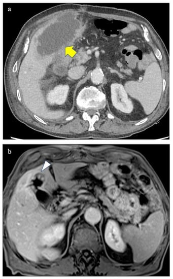
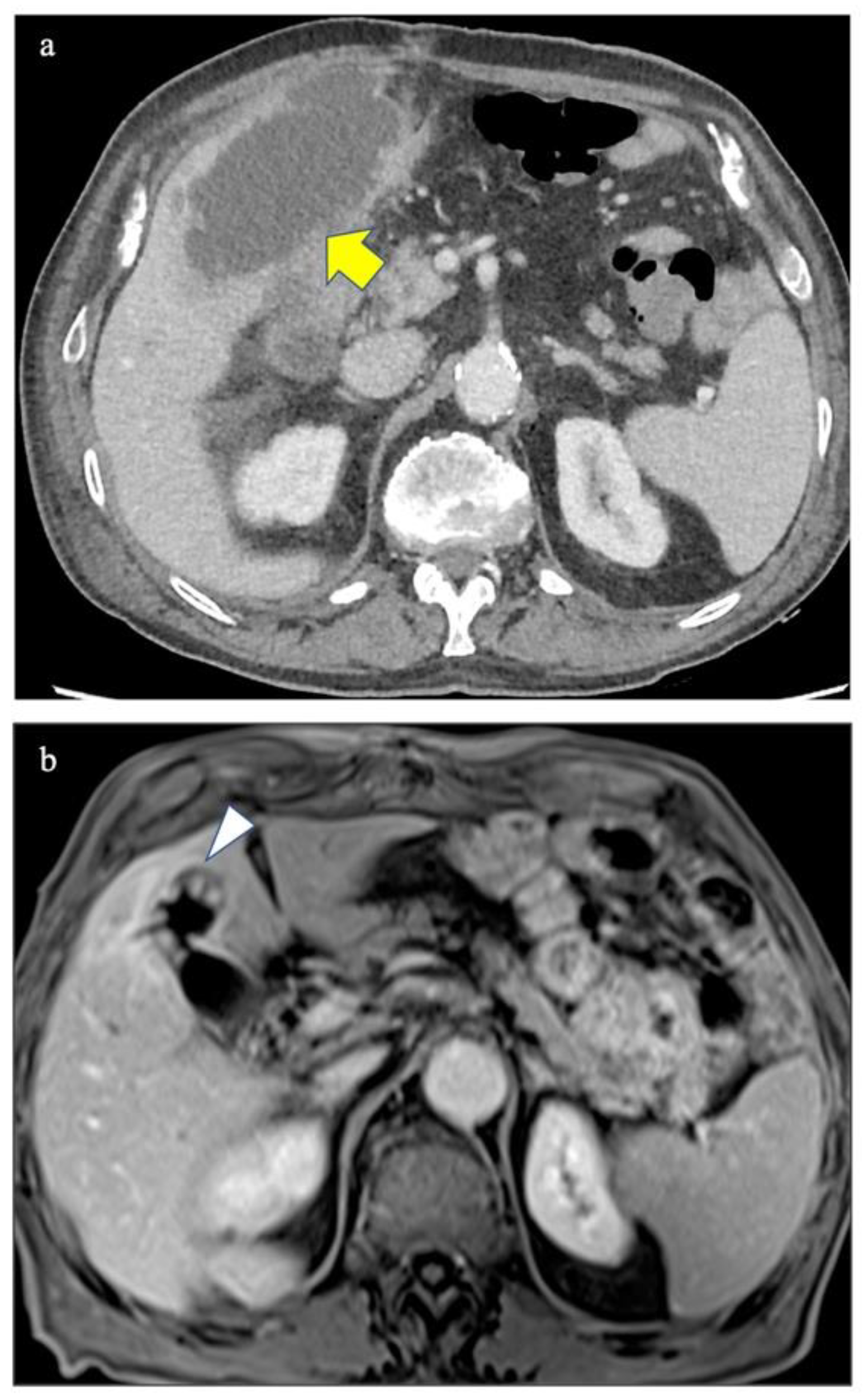
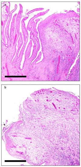
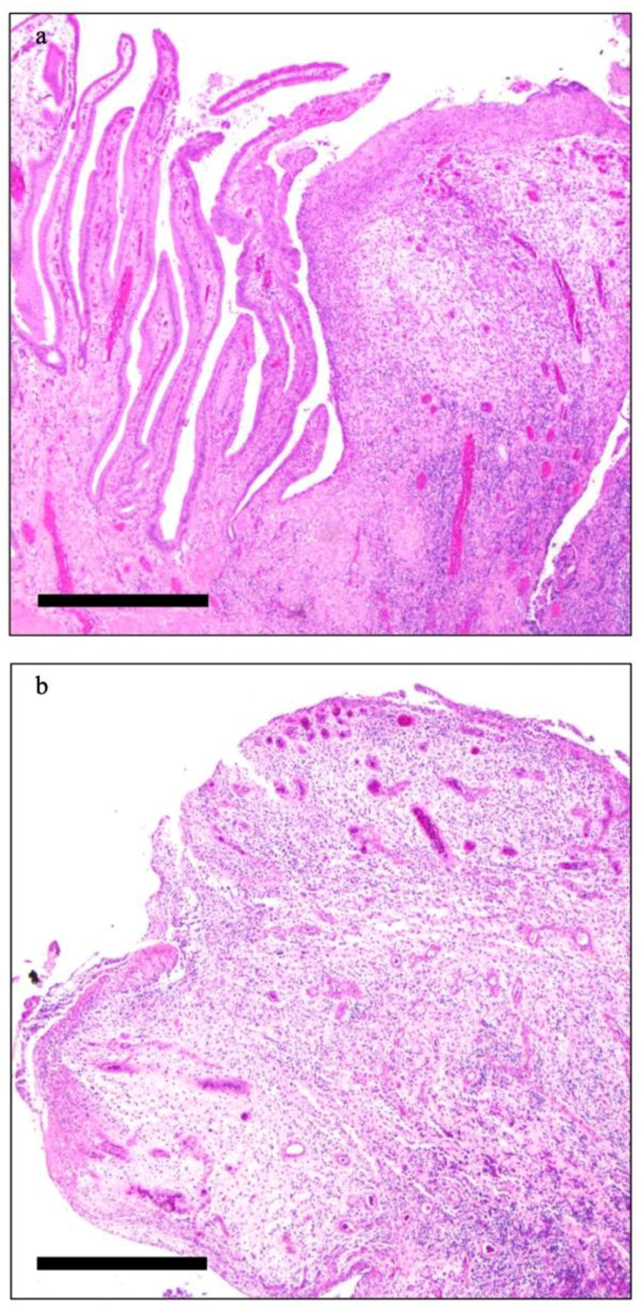
A 78-year-old man was admitted to the emergency room of our tertiary center for acute chest pain due to coronary artery disease and underwent coronary artery bypass. During the hospitalization he had fever and emesis. Thus, he underwent contrast enhanced computed tomography demonstrating acute acalculous cholecystitis with localized gallbladder perforation (Figure 1a). Ultrasound-guided percutaneous transhepatic cholecystostomy was performed and antibiotic therapy administered. Ninety days later, a Magnetic Resonance (MR) scan showed multiple small gallbladder polyps characterized by contrast enhancement without any evidence of gallbladder stones (Figure 1b). The radiological findings were compatible with an inflammatory etiology of the polypoid lesions although, especially because of the lack of gallbladder stones and previous MR scans, neoplastic polyps could not be completely excluded. The patient underwent cholecystectomy and the histological diagnosis of gallbladder inflammatory pseudopolyps (GIP) was confirmed (Figure 2a,b).
Figure 1.
Axial contrast-enhanced Computed Tomography image of the upper abdomen well-demonstrating the acute cholecystitis with localized perforation (yellow arrow in (a)) and fat-saturated Volume Interpolated Breath-Hold Examination (VIBE) Magnetic Resonance image after contrast injection performed 90 days later showing multiple small pseudopolyps in the fundus of the gallbladder (white arrow in (b)).
Figure 2.
Histological image demonstrating papillary hyperplasia and an inflammatory pseudopolyp (a) and particular representative picture of one of the described inflammatory pseudopolyps observed in the gallbladder showing an ulcerated sessile mucosal projection characterized by edematous vascularized stroma with mixed type inflammatory infiltration (b) (scale bar 500 um).
Acute acalculous cholecystitis, even complicated by perforation, often occurs in post-surgical adults while in children it is often caused by infectious disease or immune-mediated disorders. Moreover, it can be associated with cardiovascular diseases, in particular with coronary artery disease and in children with Kawasaki Disease [1,2,3,4]. The etiology of acalculous cholecystitis is often unknown, even though it has been correlated with biliary hypokinesia and local ischemia [5,6,7,8].
The radiological literature regarding rare pseudotumors like GIP, which represents 5–10% of all gallbladder polyps, is scarce, and, even if they are usually multiple and small (<10 mm), a differentiation from benign and malignant tumors might be challenging. Mucosal irritation, granulation, and fibrous tissue, usually due to gallstones and/or chronic inflammation may cause GIPs [9,10,11].
The rarity of our case in which the occurrence of GIP is probably related to acalculous cholecystitis subsequent to acute coronary artery disease, is highlighted by the results of the brief literature search, without any restrictions on language and publication date, we conducted on Pubmed on the 4 January 2022. Indeed, using the keywords “(gallbladder inflammatory pseudopolyps) AND (coronary artery disease)” no records could be identified while applying the keywords “(acute acalculous cholecystitis) AND (coronary artery disease)” and “(acalculous cholecystitis) AND (inflammatory polyps)”, only 16 and one records were identified, respectively [12,13,14,15,16,17,18,19,20,21,22,23,24,25,26,27,28]. Three of the 16 records were excluded because they were not fully matching [25,26,27] and the single record deriving from the second search is not reported in Table 1 since it is a narrative review [28]. The records of the search are summarized in Table 1.
Table 1.
Summary of the records collected by a brief Pubmed literature search using the keywords “(acute acalculous cholecystitis) AND (coronary artery disease)”.
Thus, our case demonstrates that, although rarely, GIP may occur in patients with acalculous cholecystitis even associated with coronary artery disease and can be easily diagnosed at MR. Given the clinical course of our patient, the etiology and the benign nature of the pseudopolyps could have been assumed. Nevertheless, since the malignant behavior of gallbladder lesions is not easily excluded at imaging, especially in acalculous and symptomatic patients with an unknown/partially known clinical history, surgery still plays a dominant role and histology remains the gold standard for a precise characterization.
Author Contributions
M.F. (Margherita Fosio) and C.G.: Conceptualization. G.C. and R.S.: collected clinical information; M.F. (Margherita Fosio): writing—original draft preparation; C.G., G.C., R.S. and M.F. (Matteo Fassan): writing—review and editing; G.C. and M.F. (Matteo Fassan): visualization; C.G.: supervision. All authors have read and agreed to the published version of the manuscript.
Funding
This research received no external funding.
Institutional Review Board Statement
Not applicable.
Informed Consent Statement
The patient provided informed consent for the description and publication of the case.
Conflicts of Interest
The authors declare no conflict of interest.
References
- Tana, M.; Tana, C.; Cocco, G.; Iannetti, G.; Romano, M.; Schiavone, C. Acute acalculous cholecystitis and cardiovascular disease: A land of confusion. J. Ultrasound 2015, 18, 317–320. [Google Scholar] [CrossRef] [Scilit] [PubMed]
- Cher, D.J. Myocardial Infarction and Acute Cholecystitis: An Application of Sequence Symmetry Analysis. Epidemiology 2000, 11, 446–449. [Google Scholar] [CrossRef] [Scilit] [PubMed]
- Rezkallah, K.N.; Barakat, K.; Farrah, A.; Rao, S.; Sharma, M.; Chalise, S.; Zdunek, T. Acute Acalculous Cholecystitis due to primary acute Epstein-Barr virus infection treated with laparoscopic cholecystectomy: A case report. Ann. Med. Surg. 2018, 35, 189–191. [Google Scholar] [CrossRef] [Scilit] [PubMed]
- Huffmann, J.L.; Schenker, S. Acute acalculous cholecystitis: A review. Clin. Gastroenterol. Hepatol. 2010, 8, 15–22. [Google Scholar] [CrossRef] [Scilit]
- Chen, P.F.; Nimeri, A.; Pham, Q.H.; Yuh, J.N.; Gusz, J.R.; Chung, R.S. The clinical diagnosis of chronic acalculous cholecystitis. Surgery 2001, 130, 578–581. [Google Scholar] [CrossRef] [Scilit]
- Wiles, R.; Thoeni, R.F.; Barbu, S.T.; Vashist, Y.K.; Rafaelsen, S.R.; Dewhurst, C.; Arvanitakis, M.; Lahaye, M.; Soltes, M.; Perinel, J.; et al. Management and follow-up of gallbladder polyps. Eur. Radiol. 2017, 27, 3856–3866. [Google Scholar] [CrossRef] [Scilit]
- Maheshwarappa, R.P.; Menda, Y.; Graham, M.M.; Boukhar, S.A.; Zamba, G.K.D.; Samuel, I. Association of gallbladder hyperkinesia with acalculous chronic cholecystitis: A case-control study. Surgery 2020, 168, 800–808. [Google Scholar] [CrossRef] [Scilit]
- Poddighe, D.; Sazonov, V. Acute acalculous cholecystitis in children. World J. Gastroenterol. 2018, 24, 4870–4879. [Google Scholar] [CrossRef] [Scilit]
- Mellnick, V.M.; Menias, C.O.; Sandrasegaran, K.; Hara, A.K.; Kielar, A.Z.; Brunt, E.M.; Doyle, M.B.M.; Dahiya, N.; Elsayes, K.M. Polypoid lesions of the gallbladder: Disease spectrum with pathologic correlation. Radiographics 2015, 35, 387–399. [Google Scholar] [CrossRef] [Scilit]
- Kwon, W.; Jang, J.Y.; Lee, S.E.; Hwang, D.W.; Kim, S. Clinicopathologic features of polypoid lesions of the gallbladder and risk factors of gallbladder cancer. J. Korean Med. Sci. 2009, 24, 481–487. [Google Scholar] [CrossRef] [Scilit]
- Lee, K.F.; Wong, J.; Li, J.C.; Lai, P.B.S. Polypoid lesions of the gallbladder. Am. J. Surg. 2004, 188, 186–190. [Google Scholar] [CrossRef] [Scilit]
- Welling, R.E.; Rath, R.; Albers, J.E.; Glaser, R.S. Gastrointestinal complications after cardiac surgery. Arch. Surg. 1986, 121, 1178–1180. [Google Scholar] [CrossRef] [Scilit]
- Varma, D.G.; Faust, J.M. Computed tomography of gangrenous acute postoperative acalculous cholecystitis. J. Comput. Tomogr. 1988, 12, 29–31. [Google Scholar] [CrossRef] [Scilit]
- Berger, H.; Pratschke, E.; Arbogast, H.; Stäbler, A. Percutaneous cholecystostomy in acute acalculous cholecystitis. Hepatogastroenterology 1989, 36, 346–348. [Google Scholar] [PubMed]
- Teranishi, K.; Murase, M.; Maeda, M.; Murakami, F. A case of acute hemorrhagic gangrenous acalculous cholecystitis with bile peritonitis during anti-coagulant therapy after coronary-artery bypass grafting. Nihon Kyobu Geka Gakkai 1993, 41, 83–87. [Google Scholar]
- Saito, A.; Shirai, Y.; Ohzeki, H.; Hayashi, J.; Eguchi, S. Acute acalculous cholecystitis after cardiovascular surgery. Surg. Today 1997, 27, 907–909. [Google Scholar] [CrossRef] [Scilit] [PubMed]
- Fujiii, H.; Kubo, S.; Tokuhara, T.; Suehiro, S.; Yamamoto, T.; Kinoshita, H. Acute acalculous cholecystitis complicated by penetration into the liver after coronary artery bypass grafting. Jpn. J. Thorac. Cardiovasc. Surg. 1999, 47, 518–521. [Google Scholar] [CrossRef] [Scilit] [PubMed]
- Funabiki, K.; Masuoka, H.; Shimizu, H.; Emi, Y.; Mori, T.; Ito, M.; Nakano, T. Cholesterol crystal embolization (CCE) after cardiac catheterization: A case report and a review of 36 cases in the Japanese literature. Jpn. Heart J. 2003, 44, 767–774. [Google Scholar] [CrossRef] [Scilit]
- Chen, C.J.; Huang, F.C.; Tiao, M.M.; Huang, Y.H.; Lin, L.Y.; Yu, H.R.; Yang, K.D.; Huang, Y.C.; Chen, C.C.; Chang, W.C.; et al. Sonographic gallbladder ab-normality is associated with intravenous immunoglobulin resistance in Kawasaki disease. Sci. World J. 2012, 2012, 485758. [Google Scholar] [CrossRef] [Scilit]
- Van Steijn, J.H.M.; Roeloffzen, W.W.H.; Appeltans, B.M.G.; Jager, P.L.; Gans, R.O.B.; Bijl, M. Acute acalculous cholecystitis: Not only in the intensive care department. Ned. Tijdschr. Voor Geneeskd. 2002, 146, 1305–1308. [Google Scholar]
- Yi, D.; Kim, J.Y.; Choi, E.Y.; Choi, J.Y.; Yang, H.R. Hepatobiliary risk factors for clinical outcome of Kawasaki disease in children. BMC Pediatr. 2014, 14, 51. [Google Scholar] [CrossRef] [Scilit]
- Kang, D.W.; Kim, S.H. Clinical aspects of splenomegaly as a possible predictive factor of coronary artery changes in Kawasaki disease. Cardiol. Young 2019, 29, 297–302. [Google Scholar] [CrossRef] [Scilit] [PubMed]
- Lipe, D.N.; Bridges, L.C. Kawasaki Disease Presenting as Acute Acalculous Cholecystitis. Clin. Pract. Cases Emerg. Med. 2019, 3, 383–386. [Google Scholar] [CrossRef] [Scilit] [PubMed]
- Chen, B.Q.; Chen, G.D.; Xie, F.; Li, X.; Mao, L.; Jia, B. Percutaneous cholecystostomy as a definitive treatment for moderate and severe acute acalculous cholecystitis: A retrospective observational study. BMC Surg. 2021, 21, 439. [Google Scholar] [CrossRef] [Scilit]
- Shapiro, M.J.; Luchtefeld, W.B.; Kurzweil, S.; Kaminski, D.L.; Durham, R.M.; Mazuski, J.E. Acute acalculous cholecystitis in the critically ill. Am. Surg. 1994, 60, 335–339. [Google Scholar]
- Tsuboi, I.; Hayashi, M.; Miyauchi, Y.; Iwasaki, Y.K.; Yodogawa, K.; Hayashi, H.; Uetake, S.; Takahashi, K.; Shimizu, W. Anatomical factors associated with periesophageal vagus nerve injury after catheter ablation of atrial fibrillation. J. Nippon. Med. Sch. 2014, 81, 248–257. [Google Scholar] [CrossRef] [Scilit] [PubMed]
- Udekwu, P.O.; Sullivan, W.G. Contemporary experience with cholecystectomy: Establishing ‘benchmarks’ two decades after the introduction of laparoscopic cholecystectomy. Am. Surg. 2013, 79, 1253–1257. [Google Scholar] [CrossRef] [Scilit]
- Owen, C.; Bilhartz, L.E. Gallbladder polyps, cholesterolosis, adenomyomatosis, and acute acalculous cholecystitis. Semin. Gastrointest. Dis. 2003, 14, 178–188. [Google Scholar]

Publisher’s Note: MDPI stays neutral with regard to jurisdictional claims in published maps and institutional affiliations. |
© 2022 by the authors. Licensee MDPI, Basel, Switzerland. This article is an open access article distributed under the terms and conditions of the Creative Commons Attribution (CC BY) license (https://creativecommons.org/licenses/by/4.0/).