


Abstract
Probiotic interventions for inflammatory bowel disease (IBD) have become a hot research topic in this field. However, no previous studies have investigated whether fox-derived probiotics exert therapeutic effects against colitis. The present investigation evaluated the effects of Lactiplantibacillus plantarum (syn. Lactobacillus plantarum) J3, sourced from the intestines of healthy Arctic foxes, on colitis. In an in vitro LPS-stimulated Caco-2 cell model, strain J3 exhibited prominent anti-inflammatory and antioxidant capacities, indicating potential to preserve intestinal barrier integrity. Furthermore, in an in vivo DSS-induced colitis mouse model, oral J3 supplementation markedly alleviated body weight loss, lowered disease activity index (DAI), and ameliorated histopathological lesions in colon tissues. Mechanistic analyses revealed that the J3 group displayed significantly decreased colonic levels of TNF-α, IL-1β, IL-6, MPO, PGE2, along with downregulated COX-2 mRNA expression (p < 0.05). By contrast, the levels of IL-10, TJ protein mRNA, and MUC2 mRNA were markedly upregulated, whereas serum LPS and D-Lac concentrations were significantly reduced (p < 0.05). In an exploratory analysis of 16S rDNA and short-chain fatty acids (SCFAs) in a subset of mice, J3 treatment showed a trend toward colonic microbial remodeling, accompanied by a significant increase in short-chain fatty acid production (p < 0.05). Overall, these findings indicate that Lactiplantibacillus plantarum J3 derived from foxes may alleviate DSS-induced colitis in mice, providing a basis for future translational research.
1. Introduction
Posing a considerable health challenge, inflammatory bowel disease (IBD) extends from humans to companion animal species like dogs and cats [1]. Ulcerative colitis (UC), a major clinical subtype of IBD, mainly targets the colon and rectum and presents with clinical manifestations including abdominal pain, hematochezia, and body weight loss [2,3]. Nevertheless, current clinical interventions for IBD remain suboptimal. Conventional therapeutic regimens, such as aminosalicylates, corticosteroids, and fecal microbiota transplantation [4,5], frequently lead to primary or secondary treatment failure, accompanied by safety risks, multiple side effects, high medical costs, and the absence of accurate predictive biomarkers [6,7]. Therefore, identifying safe, effective, and cost-effective interventions is key to addressing the current challenges in IBD treatment.
Probiotics have attracted extensive research attention owing to their capacities to maintain a balanced intestinal environment [8], emerging as a core research direction for IBD therapeutic development [9,10]. Lactiplantibacillus plantarum has been widely investigated for its favorable safety profile and versatile probiotic properties. This species exerts multiple gut-beneficial effects by secreting organic acids to reduce luminal pH. Moreover, Lactiplantibacillus plantarum exhibits robust intestinal colonization capacity: after attaching to the intestinal wall via adhesins, it can colonize and exert long-term probiotic effects [11]. Certain strains of Lactiplantibacillus plantarum also secrete antimicrobial compounds (such as bacteriocins) to directly inhibit pathogenic microorganisms [12] and modulate the host’s gut microbiota [13]. In the context of UC, the barrier-protective effect exerted by this strain is typically associated with elevated levels of tight junction (TJ) proteins and mucins [14]. Beyond this, Lactiplantibacillus plantarum has also demonstrated significant efficacy in suppressing inflammatory signaling pathways and regulating cytokine balance [15].
As strict carnivores, Arctic foxes harbor a unique intestinal ecosystem that may host functionally distinctive probiotic strains. In addition, considering the pathological similarities between IBD and weaning stress syndrome in Arctic fox cubs [16,17,18], we hypothesized that fox-origin probiotics could alleviate IBD-associated intestinal inflammation. We identified a strain derived from the Arctic fox—Lactiplantibacillus plantarum J3. Whole-genome sequencing revealed that the J3 strain possesses 196 strain-specific genes, as well as five plasmids, and multiple genes involved in quorum sensing, carbohydrate metabolism, and fatty acid biosynthesis; these characteristics may be related to its gut colonization and probiotic functions. In our experiments, the J3 strain exhibited potent anti-inflammatory, antioxidant, and barrier-enhancing effects in vitro and significantly alleviated DSS-induced colitis in vivo. Our results suggest that J3 may represent a promising candidate within the emerging class of microbiota-based therapeutics for IBD.
2. Materials and Methods
The animal experimentation and sample collection protocols used in this study were approved by the Experimental Animal Ethics Committee of Northeast Agricultural University (No.: NEAUEC20230258). Detailed information regarding the reagents and materials is summarized in Supplementary Material File S3.
2.1. Strain Preparation and Probiotic Properties
2.1.1. Separation and Identification
Colonic contents (1.0 g) from Arctic foxes were thoroughly homogenized with 9 mL of 0.9% NaCl, followed by a 10-fold serial dilution (10−1 to 10−4). Aliquots of each dilution were then spread onto de Man, Rogosa, and Sharpe (MRS) agar plates containing 1.5% CaCO3, and the plates were incubated at 37 °C for 48 h under anaerobic conditions using gas-generating sachets (AnaeroPack, Mitsubishi Gas Chemical, Tokyo, Japan) in sealed anaerobic jars (Oxoid, Basingstoke, UK). Nineteen colonies with distinct calcium-dissolving zones were selected and purified through three consecutive streaking procedures. Following the method described in Reference [19], all strains were identified as Lactiplantibacillus plantarum through 16S rDNA gene sequencing.
2.1.2. In Vitro Screening and Genome-Wide Sequencing
Using gastrointestinal fluid tolerance (pH 3.0 saline + 3 g/L pepsin, 4 h; pH 8.0 saline + 1 g/L trypsin + 3 g/L bile salts, 3 h) and Caco-2 cell adhesion rate (approximately 1 × 10⁸ CFU/well, 2 h), strain J3 was selected from 19 isolates as the superior strain (see Supplementary Material File S1 for detailed procedures and data). Whole-genome sequencing of J3 was performed using the Illumina NovaSeq 6000 platform. The genomic safety assessment was conducted following the strategy described by Chokesajjawatee et al. [20]. Details of the assembly and annotation workflows, as well as the specific methods and results of the safety assessment, are provided in Supplementary Material File S2 (Section S2.11). J3 was stored at −80 °C in 30% glycerol-MRS.
2.1.3. Formulations
Following 18 h of propagation in MRS medium at 37 °C, the J3 cells were harvested via centrifugation (8000× g, 10 min), rinsed twice with sterile Phosphate-Buffered Saline (PBS), and collected for further use. The resulting cell pellets were resuspended separately in sterile PBS (for mouse gavage, 1 × 10¹⁰ CFU/mL) or antibiotic-free Dulbecco’s Modified Eagle Medium (DMEM) (for cell experiments, 1 × 10⁸ CFU/mL). Solutions were prepared immediately before use.
2.2. Test Management and Sample Collection
2.2.1. In Vitro Assay Using Caco-2 Cells
Caco-2 cells, obtained from the Cell Bank of the Chinese Academy of Sciences, were subsequently maintained in DMEM supplemented with 10% fetal bovine serum and 1% penicillin-streptomycin at 37 °C under 5% CO2. Cells from passages 25–35 were used for all experiments. Prior to the experiments, the cells were transferred to antibiotic-free DMEM and allowed to acclimate for 24 h.
A total of 48 subjects were randomly divided into four groups (n = 12 per group). No animals died or were removed during the experiment; all mice survived to the endpoint:
Based on the treatments applied, three groups were designated:
Control group: Cells were maintained in antibiotic-free DMEM medium under standard conditions for 24 h.
LPS group: Cells received 1 μg/mL lipopolysaccharide (LPS) from Escherichia coli O55:B5 (Sigma-Aldrich, St. Louis, MO, USA) stimulation for 24 h. The LPS concentration of 1 μg/mL was selected based on previous studies demonstrating that this concentration effectively induces inflammatory responses in Caco-2 cells without causing cytotoxicity [21,22,23,24].
J3 + LPS group: Cells were pretreated for 1 h with a J3 preparation at a final concentration of 1 × 10⁸ CFU/mL. After pretreatment, they were co-cultured with 1 μg/mL LPS for 24 h.
The supernatant and cells from each treatment group were collected separately for subsequent experiments.
2.2.2. Experimental Study Using a Murine Model of DSS-Induced Colitis
Forty-eight male C57BL/6J mice, aged four weeks, were equilibrated under a duration of 1 week under standardized environmental conditions (humidity: 50 ± 10%, temperature: 22 ± 2 °C, lighting: 12 h light/dark cycle).
A total of 48 subjects underwent randomization into four experimental cohorts:
Control group: Days 1–21: 0.2 mL of sterile PBS administered via oral gavage daily.
DSS group: Days 1–7: 3% DSS (36–50 kDa) was provided ad libitum in the drinking water. Days 8–21: 0.2 mL of sterile PBS was administered via oral gavage daily.
J3 + DSS group: Days 1–7: Modeling with DSS; Days 8–21: Daily intragastric administration of J3 (0.2 mL, 1 × 10¹⁰ CFU/mL) was conducted. This dose was selected based on the effective dose of Lactiplantibacillus plantarum strains previously reported in DSS-induced colitis models [13,25].
Positive drug control group: Days 1–7: Modeling with DSS; Days 8–21: Sulfasalazine (SASP) was administered orally at 200 mg/kg per day.
In the mouse experiment, mice were weighed daily, and their droppings’ texture and rectal bleeding in the feces were observed, from which a disease activity index (DAI) was calculated according to the criteria listed in Table 1. The DAI scores were assessed independently by two observers who were unaware of the experimental group assignments, and the average score was used for statistical analysis. Upon completion of the study, the mice were deeply sedated and euthanized according to the method described in [26], and samples were collected. Colon length was recorded. Serum was collected, with tissue aliquots and colon contents snap-frozen at –80 °C, and additional colonic segments fixed for histopathological examination.
Table 1.
Disease Activity Index (DAI) Scoring Criteria.
2.3. Histopathological Analysis of Mouse Colon Tissue
Fixed colon tissues were paraffin-embedded and microtomed (7 μm). Sections were then subjected to Hematoxylin and Eosin (H&E) staining to observe morphological changes, and with Alcian Blue-Periodic Acid-Schiff (AB-PAS) (pH 2.5) to count goblet cells. From each section, 5 fields of view were randomly selected at a magnification of 200×. Using ImageJ software (version 1.53), the total mucosal area (mm2) was measured in each field of view, and the number of AB-PAS-positive cells was counted. The results were expressed as the number of goblet cells per mm2 of mucosa, and the mean of the 5 fields of view was taken as the final value for that sample.
2.4. Enzyme-Linked Immunosorbent Assay (ELISA)
Caco-2 cell supernatant was assayed for tumor necrosis factor-α (TNF-α), interleukin-6 (IL-6), interleukin-1β (IL-1β), and interleukin-10 (IL-10) using the kit as per the manufacturer’s protocol.
With this kit, and per the manufacturer’s protocols, quantify myeloperoxidase (MPO) catalytic activity, along with the concentrations of Prostaglandin E2 (PGE2), TNF-α, IL-1β, IL-6, and IL-10 in mouse colon tissues. Circulating LPS and D-lactate abundances in murine serum were also determined simultaneously.
2.5. Profiling Reactive Oxygen Species (ROS) and Mitochondrial Functional Traits in Caco-2 Cells
After the Caco-2 cells were administered the interventions specified in Section 2.2.1, they were rinsed using PBS. The fluorescent probe 2′,7′-dichlorodihydrofluorescein diacetate (DCFH-DA) was prepared to 10 μM in phenol red- and serum-deprived DMEM, followed by incubation with cells under dark conditions at 37 °C and 5% CO2 over a 30 min period. After washing three times, the signal intensity of intracellular fluorescence was acquired using 485 nm excitation and 528 nm emission filters to quantify ROS. Under the same conditions, incubate with 5 μM MitoSOX Red and 10 μg/mL Rhodamine 123 (Rh-123), respectively. Upon washing, acquire fluorescent signals using dual-wavelength settings: 510 nm (excitation)/580 nm (emission) and 507 nm (excitation)/529 nm (emission), which enables detection of mitochondrial superoxide radicals and the membrane potential (ΔΨm). All fluorescence intensities were measured using a SpectraMax iD3 microplate reader, and relative fluorescence intensities were calculated with the control group as the reference.
2.6. Determination of Trans-Epithelial Resistance (TEER) in Caco-2 Cells
Trypsinized Caco-2 cells were plated onto 0.4 μm-pore Transwell chambers (Corning, New York, NY, USA) at 1 × 10⁵ cells per well. Incubation proceeded at 37 °C with 5% carbon dioxide, and fresh culture medium was replenished on alternate days. Periodic recordings of transepithelial electrical resistance (TEER) were performed via a Millicell ERS-2 epithelial volt-ohm meter (Millipore, Billerica, MA, USA) until readings plateaued above 500 Ω·cm2, an indicator of intact tight monolayer formation. Results were calculated using the following formula:
where the blank resistance is the resistance value of the Transwell membrane without cells (approximately 220 Ω).
2.7. Gene Expression Analysis (qPCR)
Total RNA was extracted from Caco-2 cell cultures using TRIzol, with RNase-free DNase I added during the extraction process to remove residual genomic DNA contamination. The RNA was quantified using a NanoDrop 2000 spectrophotometer (Thermo Fisher Scientific, Waltham, MA, USA); the A260/A280 ratios for all samples ranged from 1.8 to 2.0, indicating good purity. RNA integrity was assessed using an Agilent 2100 Bioanalyzer (Agilent Technologies, Santa Clara, CA, USA). The RNA Integrity Number (RIN) values for all samples were greater than 8.0, indicating that the RNA showed no significant degradation. The resulting total RNA was then reverse-transcribed into cDNA using the PrimeScript™ RT Kit. Mouse colon tissue samples were homogenized under liquid nitrogen conditions, and total RNA was extracted following the same procedure. After DNase I treatment and verification of the A260/A280 ratio and RIN value to confirm quality, cDNA synthesis was performed using the GoScript™ reverse transcription kit. qPCR analysis was conducted on the ABI 7500 system. Transcription levels of the candidate genes were normalized using the independent internal reference genes GAPDH and β-actin. Relative expression profiles were calculated using the 2−ΔΔCt method based on the cycle threshold comparison; raw data were calibrated against control samples. The nucleotide sequences of all target amplification primers are listed in Supplementary Material File S3.
2.8. 16S rDNA Gene Amplicon Profiling of Murine Colon Microbial Contents
Illumina MiSeq high-throughput sequencing was adopted to characterize microbial communities recovered from mouse colonic luminal specimens. The main workflow included: genomic DNA was purified using the QIAamp DNA Kit; amplification of the 16S rDNA V3–V4 region using primers 338F/806R; and 2 × 300 bp paired-end sequencing following purification and quantification. Raw sequencing data were analyzed using the QIIME 2 platform (version 2024.2). The DADA2 plugin was used to perform quality filtering, denoising, merging, and chimera removal on paired-end sequences, generating an amplicon sequence variant (ASV) feature table. For species annotation, a pretrained Naive Bayes classifier (based on the Silva 138 database, with 99% of OTU reference sequences) was used to classify representative ASV sequences. α-diversity indices (Chao1, Shannon, Simpson) and β-diversity (Principal Coordinate Analysis (PCoA) based on Bray–Curtis distance) were calculated using the normalized ASV feature table. 16S sequencing was performed on an exploratory subset of 3 mice per group. Due to the small sample size (n = 3), microbial abundance data are presented descriptively only, and no formal hypothesis tests were conducted. Detailed statistical test results (Mann–Whitney U test and FDR-corrected p-values) are provided in the Supplementary Materials File S4. The Linear Discriminant Analysis Effect Size (LEfSe) method (Linear Discriminant Analysis (LDA) threshold > 2.0) was performed on the Meiji Bio Cloud Platform (https://cloud.majorbio.com) to identify taxonomic biomarkers capable of distinguishing between the study groups.
2.9. Determination of Short-Chain Fatty Acids in Mice
Weigh 0.80 ± 0.01 g of mouse colon contents and homogenize them in sterile PBS to prepare a 10% (w/v) homogenate suspension. The 500-μL slurry was mixed with 100 μL of crotonic acid solution as an internal standard and stored at −30 °C for 24 h. After thawing, the mixture was centrifuged at 4 °C and 8000× g for 3 min. Post filtration of the collected liquid phase, the specimen was loaded onto the apparatus for measurement.
Chromatographic partitioning was accomplished with an Agilent DB-FFAP capillary tubing column (30 m × 0.25 mm × 0.25 μm) mounted on an Agilent 7890B gas chromatography unit (Santa Clara, CA, USA) fitted with a flame ionization sensing module calibrated to 250 °C. Comprehensive inlet, carrier gas, and oven thermal gradient specifications are documented in Supplementary Material File S3. Short-Chain Fatty Acids (SCFAs) concentrations were calculated using calibration curves.
For SCFAs data, comparisons among the four groups were performed using the Kruskal–Wallis test, followed by Dunn’s post hoc test, with Benjamini–Hochberg FDR correction for multiple comparisons; a corrected p-value < 0.05 was considered statistically significant. The SCFAs analysis data were derived from an exploratory subset (3 mice per group), and the results should be considered preliminary findings.
2.10. Statistical Analysis
All statistical computations were executed via the SPSS 23.0 statistical package. Normality distribution of datasets was assessed through the Shapiro–Wilk procedure, whereas Levene’s assay served for the validation of variance homogeneity. Prerequisites for normal distribution and equal variance among study cohorts were fully satisfied across all measured data. Inter-group disparity evaluation was carried out via one-way ANOVA, and Tukey’s HSD multiple comparison tests were executed as subsequent validation steps. A p-value cutoff of less than 0.05 was adopted to identify statistically relevant discrepancies. In vitro experiments were performed in three independent replicates, each with three replicate wells. In animal experiments, each mouse was considered an independent biological replicate (n = 12 per group). See the captions for each figure for the specific sample sizes for each indicator, and three technical replicates were performed for each sample, with the mean calculated, and resultant readings are reported as mean ± standard deviation (SD).
3. Results
3.1. LPS-Induced Inflammation and Oxidative Stress in Enterocyte Monolayers
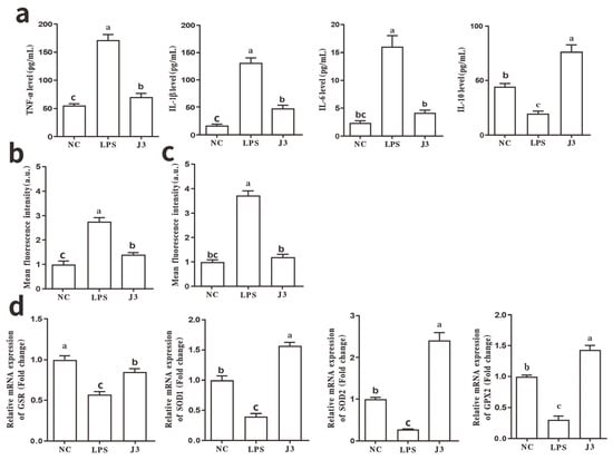
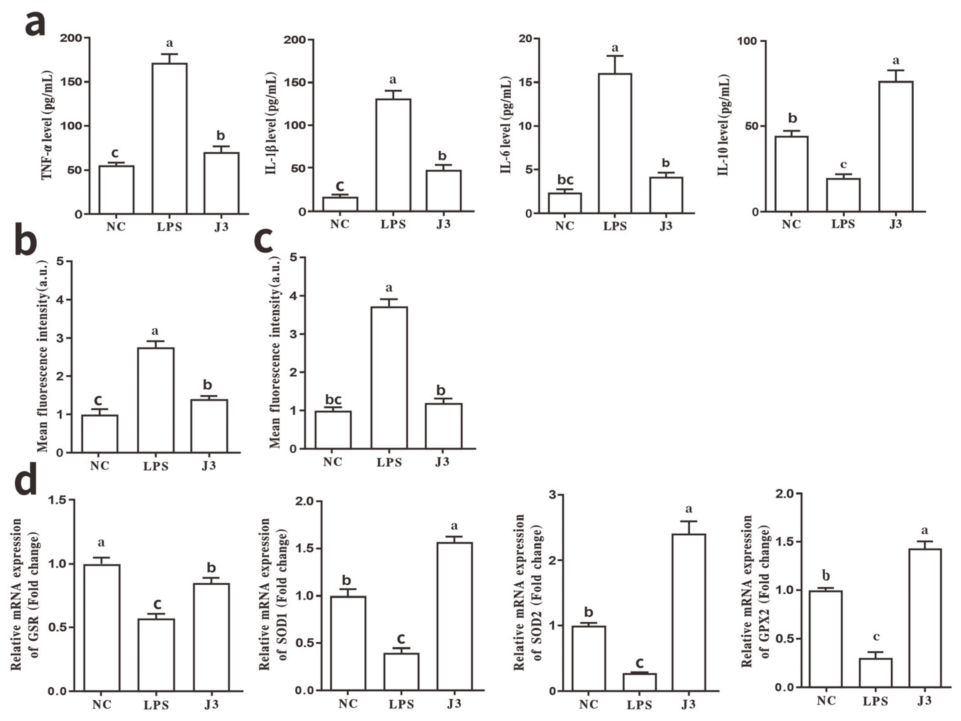
The cytoprotective potential of strain J3 was evaluated in an LPS-stimulated model by assessing the cytokine milieu. Caco-2 epithelial monolayers challenged with LPS showed significantly increased levels of pro-inflammatory cytokines (TNF-α, IL-1β, IL-6) versus blank cellular cultures. (p < 0.05, Figure 1a). Lactiplantibacillus plantarum J3 administration brought about remarkable downregulation of such inflammatory effector contents and facilitated elevated IL-10 secretion (p < 0.05). Strain J3 treatment significantly reduced the overall intracellular ROS levels and mitochondrial superoxide species, alongside elevating transcript abundance of antioxidant enzymatic genes including Glutathione Reductase (GSR), Superoxide Dismutase 1 (SOD1), Superoxide Dismutase 2 (SOD2), and Glutathione Peroxidase 2 (GPX2) (p < 0.05, Figure 1b–d).
Figure 1.
Lactiplantibacillus plantarum J3 intervention mitigates LPS-triggered inflammatory and oxidative responses in polarized Caco-2 enterocytes: (a) Concentrations of pro-inflammatory mediators (TNF-α, IL-1β, IL-6, and IL-10); (b) Concentrations of ROS; (c) Concentrations of mitochondrial superoxide; (d) mRNA expression levels of antioxidant enzymes (GSR, SOD1, SOD2, and GPX2). Measurable continuous readouts are displayed in the form of mean ± SD (n = 3). Disparate letter annotations (a, b, c) mark any statistically meaningful variance across experimental cohorts (p < 0.05).
3.2. Caco-2 Cell Intestinal Barrier Function
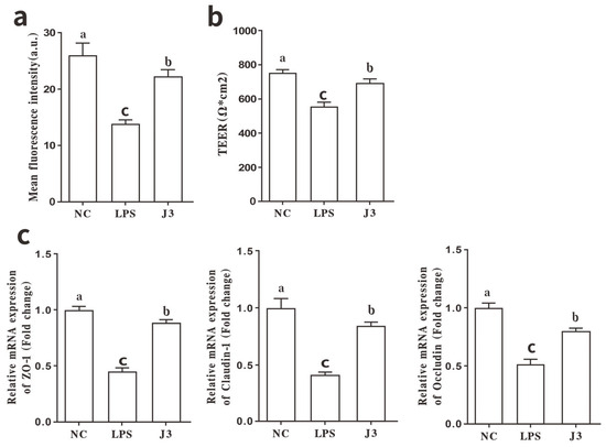
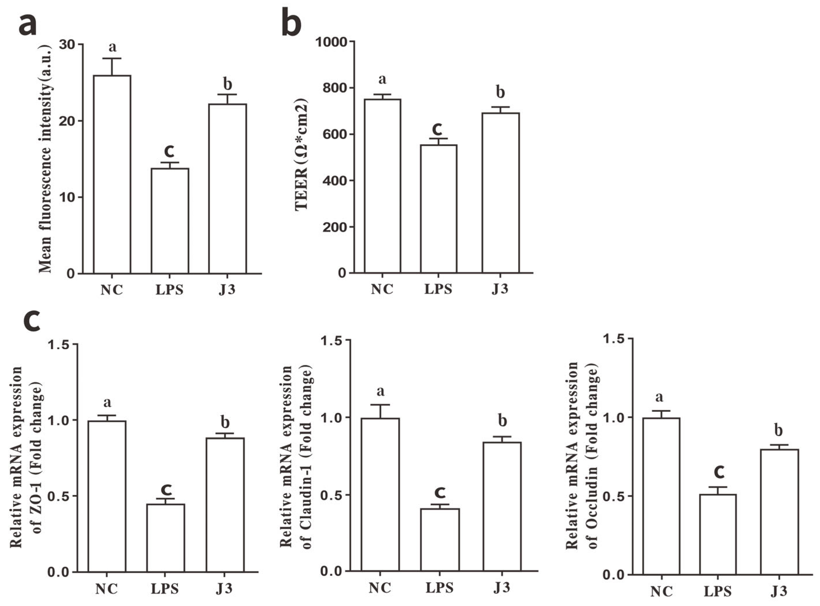
Disruption of mitochondrial membrane potential impairs cellular energy production, induces oxidative stress, and compromises cellular health [27]. UC also causes mitochondrial dysfunction in the enteric mucosa. The integrity and permeability of the epithelial barrier are reflected by the cell membrane potential [28]. TEER and mitochondrial membrane potential were evaluated in Caco-2 cell cultures. Compared with untreated cells, Caco-2 monolayers subjected to LPS treatment showed a significant decrease in mitochondrial membrane potential (p < 0.05, Figure 2a). In contrast, treatment with live J3 significantly increased mitochondrial membrane potential (p < 0.05). Coincident with this, LPS exposure caused a significant decrease in transepithelial electrical resistance metrics (p < 0.05, Figure 2b), whereas J3 conditioning produced prominent TEER level gains over the LPS group (p < 0.05). Further analysis of tight junction protein mRNA expression revealed that J3 treatment reversed the LPS-induced downregulation of TJ-related genes. Zonula Occludens-1 (ZO-1), Occludin, and Claudin-1 mRNA levels were significantly increased following J3 exposure in comparison to the LPS group (p < 0.05, Figure 2c), reaching levels comparable to those of the control group.
Figure 2.
Impact of Lactiplantibacillus plantarum J3 on LPS-Induced gut barrier competence in Caco-2 Cell Models: (a) Mitochondrial membrane potential; (b) TEER; (c) mRNA expression levels of tight junction genes (ZO-1, Occludin, and Claudin-1). Measurable continuous readouts are displayed in the form of mean ± SD (n = 3). Disparate letter annotations (a, b, c) mark any statistically meaningful variance across experimental cohorts (p < 0.05).
3.3. Pathological Manifestations of DSS Colitis in a Murine Model
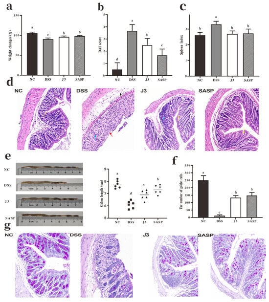
J3 treatment significantly alleviated the colitis phenotype induced by DSS in murine models. DAI was used to monitor colitis progression, while the spleen coefficient served as a marker of systemic inflammation and immune activation [29]. Colonic shortening is a characteristic feature of colitis [30]. J3 administration mitigated weight loss in DSS-treated mice, with effects comparable to those of SASP (p > 0.05). This treatment markedly lowered the DAI, attenuated the elevation of the spleen index, and attenuated colon shortening (p < 0.05; Figure 3a–e). Histopathological examination of the colon (Figure 3d) showed that the control group had intact structure, abundant goblet cells (yellow arrows), and no inflammatory infiltration or submucosal edema. In contrast, the DSS group exhibited severe mucosal damage, with complete epithelial detachment and exposure of the lamina propria (red arrows), destruction of crypts, widespread infiltration of inflammatory cells and fibroblasts (blue arrows), submucosal edema accompanied by mild inflammatory cell infiltration (black arrows), and a significant reduction in goblet cells. The J3 group showed neatly arranged epithelial cells, partial restoration of crypt structure, only focal inflammatory cell infiltration (blue arrows), a mild reduction in goblet cells (yellow arrows), and no submucosal edema or inflammation. The SASP group exhibited near-normal colonic histological features, with an intact epithelium, abundant goblet cells (yellow arrows), and no inflammatory infiltration or submucosal edema. Intestinal inflammation reduces the number of goblet cells and inhibits their secretion of acidic mucins, thereby impairing the mucus layer [31,32]. The number of goblet cells was significantly reduced in the DSS group (p < 0.05, Figure 3f,g). The number of goblet cells increased in both the J3 and SASP groups (p < 0.05). There was no significant difference in treatment efficacy between the J3 and SASP groups (p > 0.05).
Figure 3.
Regulatory Outcomes of J3 over Clinical Features within DSS-Caused Murine Colitis. NC: Control group; DSS: DSS group; J3: J3 group; SASP: SASP group (same below): (a) Variation in body mass; (b) Colon tissue DAI score; (c) Spleen index; (d) Colon tissue histopathology H&E staining (200×); (e) Quantitative analysis of colon length; (f) Quantification of goblet cells within colonic tissue; (g) AB-PAS histochemistry was applied to murine colonic specimens and examined (200×). Measurable continuous readouts are displayed in the form of mean ± SD (n = 12). Disparate letter annotations (a, b, c, d) mark any statistically meaningful variance across experimental cohorts (p < 0.05).
3.4. Inflammatory Response in Mouse Colon Tissue
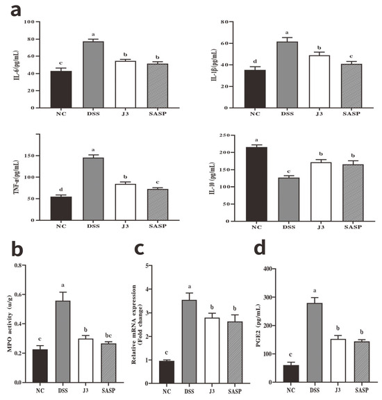
Compared with the DSS group, both J3 and SASP caused significant decreases in the detected contents of MPO, PGE2, IL-1β, IL-6, and TNF-α (p < 0.05; Figure 4a,b,d), while IL-10 levels were markedly elevated (p < 0.05). No significant difference between groups was found in the determined quantities of PGE2, IL-6, or IL-10 between J3- and SASP-treated mice (p > 0.05). Cyclooxygenase-2 (COX-2) mRNA levels were decreased—the key rate-limiting catalyst of PGE2 generation [33]—in the J3 or SASP groups. (p < 0.05, Figure 4c).
Figure 4.
Regulatory action of J3 over DSS-elicited colonic mucosal inflammation in mouse specimens: (a) Concentrations of pro-inflammatory mediators (TNF-α, IL-1β, IL-6, and IL-10); (b) MPO catalytic activity values; (c) COX-2 mRNA expression levels; (d) PGE2 tissue burdens. Measurable continuous readouts are displayed in the form of mean ± SD (n = 6). Disparate letter annotations (a, b, c, d) mark any statistically meaningful variance across experimental cohorts (p < 0.05).
3.5. Intestinal Barrier Function in Mice
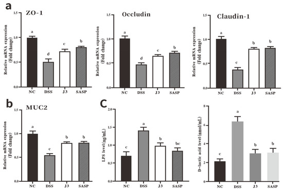
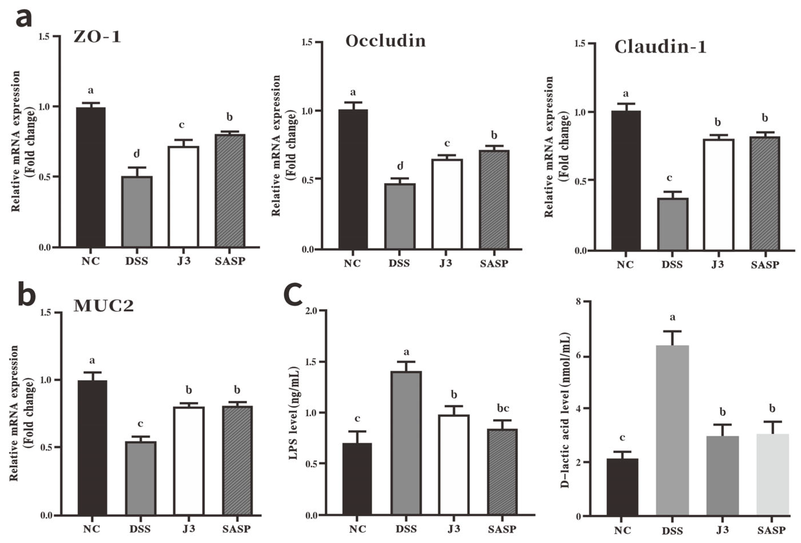
mRNA levels of Mucin 2 (MUC2) and TJ proteins (ZO-1, Occludin, Claudin-1) in colonic mucosa were analyzed to assess intestinal barrier function. J3 treatment significantly increased mRNA levels for all four genes (p < 0.05, Figure 5a,b). MUC2 and Claudin-1 expression levels were equivalent between the J3 and SASP groups (p > 0.05). In line with the above observation, serum LPS levels and D-lactate within the J3 group cohorts were significantly reduced compared with the DSS group (p < 0.05, Figure 5c), further confirming the improvement in intestinal barrier function.
Figure 5.
Impact of J3 on Mucosal Barrier Competence in Mice Subjected to DSS-Provoked Mucosal Damage: (a) TJ mRNA expression levels (ZO-1, Occludin, and Claudin-1); (b) MUC2 mRNA expression levels; (c) Serum component levels (LPS and D-lactic acid). Measurable continuous readouts are displayed in the form of mean ± SD (n = 6). Disparate letter annotations (a, b, c, d) mark any statistically meaningful variance across experimental cohorts (p < 0.05).
3.6. SCFAs Production via Gut Microbiota Modulation in Mice
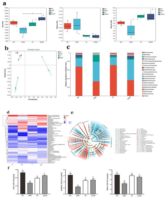
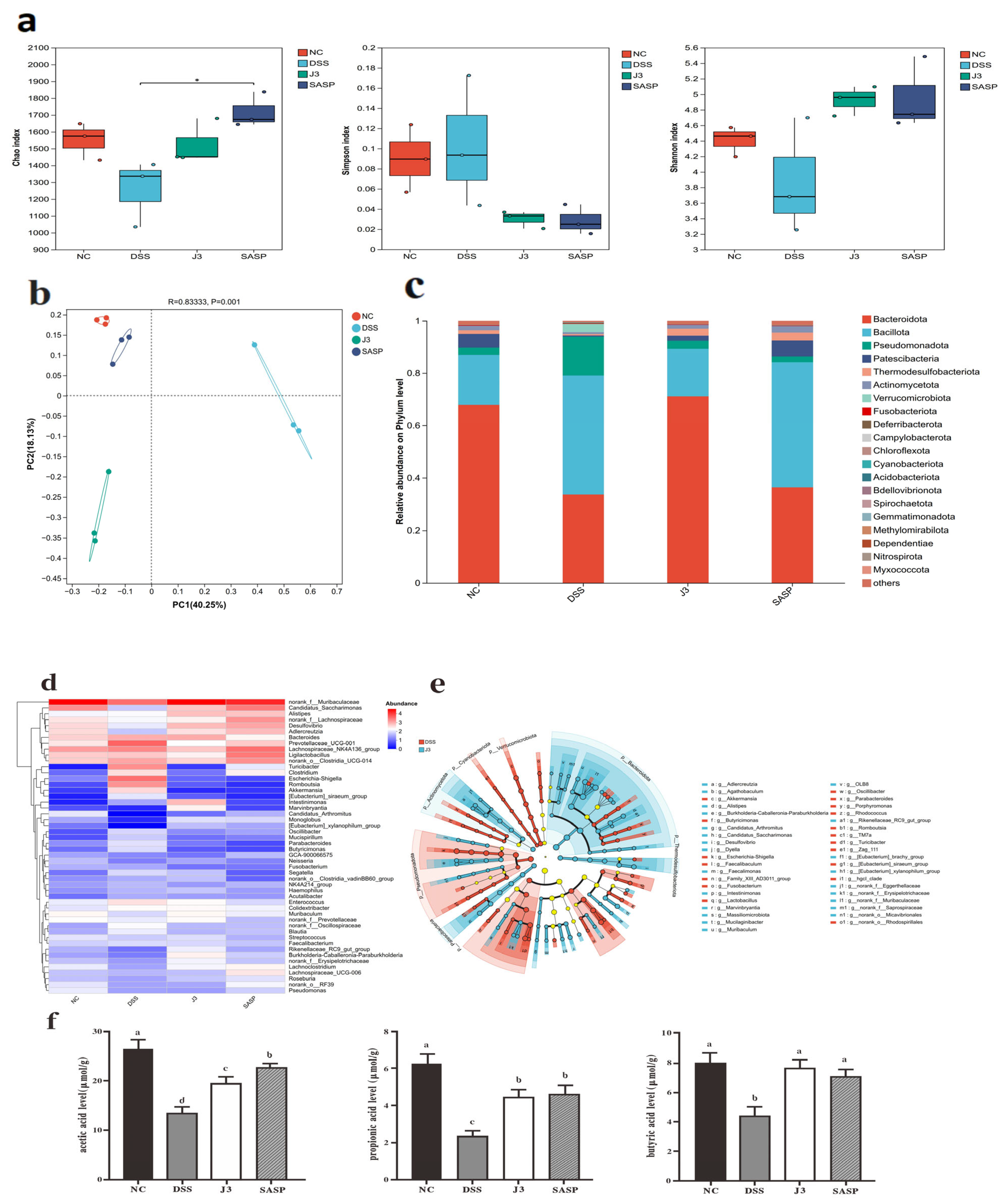
The results of 16S rDNA gene sequencing of the colonic microbiota showed the α-diversity indices across the groups, as shown in Figure 6a. β-diversity analysis indicated that the microbial community underwent structural reorganization (Figure 6b). At the phylum level, the J3 group exhibited higher proportions of Bacteroidota (mean ± SD, 71.05 ± 12.89% vs. 33.60 ± 13.58%), Patescibacteria (1.89 ± 1.31% vs. 0.41 ± 0.38%), and Actinomycetota (1.45 ± 0.29% vs. 0.73 ± 0.34%), whereas the relative abundances of Bacillota (18.24 ± 8.97% vs. 45.46 ± 26.05%), Pseudomonadota (3.10 ± 0.47% vs. 14.86 ± 14.01%), and Verrucomicrobiota (0.01 ± 0.01% vs. 3.12 ± 3.27%) were lower than those in the DSS group (Figure 6c). At the genus level, the J3 group exhibited lower relative abundances of Escherichia-Shigella (0.03 ± 0.01% vs. 12.42 ± 13.94%), Romboutsia (0.03 ± 0.008% vs. 7.41 ± 8.34%), Clostridia_UCG-014 (1.22 ± 0.14% vs. 4.78 ± 4.28%), Prevotellaceae_UCG-001 (0.74 ± 0.72% vs. 19.25 ± 15.71%) and Lachnospiraceae_NK4A136_group (1.91 ± 1.55% vs. 7.88 ± 9.19%), while Candidatus_Saccharimonas (1.86 ± 1.30% vs. 0.34 ± 0.38%), Marvinbryantia (0.78 ± 0.41% vs. 0.01 ± 0.003%), Muribaculaceae (60.96 ± 13.60% vs. 8.13 ± 2.32%), and Desulfovibrio (2.54 ± 2.38% vs. 0.39 ± 0.22%) were all higher than those in the DSS group (Figure 6d). LEfSe multilevel discriminant analysis (score > 2.0) confirmed that J3 treatment enriched Actinobacteria, Patescibacteria, and multiple genera within Bacteroidota (Figure 6e). We hypothesize that this microbial restructuring may enhance the biosynthesis of health-promoting, microbiota-derived metabolites. A targeted metabolic profiling assay was conducted to substantiate the above inference. Compared with the DSS group, J3 and SASP groups showed significantly higher colonic levels of acetate, propionate, and butyrate. (p < 0.05, Figure 6f).
Figure 6.
Impact of J3 on gut microbiota composition and SCFAs production in mice subjected to DSS-induced colitis: (a) Colon microbiota α-diversity indices (Chao1, Shannon, and Simpson); (b) Colon microbiota β-diversity (PCOA); (c) Species profiling bar chart at the phylum level; (d) Heatmap of species distribution among genera; (e) LEfSe cladogram of differentially abundant taxa between the DSS and J3 groups (LDA > 2.0); (f) Profiling of colonic SCFAs storage magnitudes (acetate, propionate, butyrate) Measurable continuous readouts are displayed in the form of mean ± SD (n = 3). Disparate letter annotations (a, b, c, d) mark any statistically meaningful variance across experimental cohorts (p < 0.05).
4. Discussion
IBD is a multifactorial intestinal disorder [34], and the challenges in its treatment have spurred the search for alternative approaches. Probiotics enhance the host’s resistance to disease through symbiotic interactions with the gut microbiota and hold great promise [35,36]. This study found that strain J3, isolated from the intestines of healthy Arctic foxes, exhibits several unique genomic features, as revealed by whole-genome analysis, and effectively alleviated intestinal inflammation in a DSS-induced colitis model in mice. These findings provide preliminary experimental evidence for probiotic-based interventions for intestinal inflammation and suggest that J3 is a functional candidate strain warranting further evaluation; however, these results still need to be validated in additional experimental models and preclinical studies.
The Caco-2 intestinal epithelial cell line is a classic model for studying intestinal barrier function and the dynamics of bacterial adhesion and invasion [37]. Hyperactivated macrophage populations produce precursor IL-1β, whose bioactivity is unlocked through caspase-1-driven proteolytic truncation [38]; membrane receptor engagement launches NF-κB signal transduction cascades following ligand interaction, ultimately boosting the transcription of IL-6 and TNF-α [39]. J3 restored the inflammatory homeostasis of Caco-2 cells by synergistically downregulating TNF-α, IL-1β, and IL-6 while upregulating IL-10, which is consistent with the anti-inflammatory effects of Lactobacillus species [25]. It is hypothesized that J3 may inhibit the secretion of pro-inflammatory factors and enhance IL-10 production by modulating this pathway; the latter further suppresses NF-κB activation, thereby constituting a negative feedback loop [40]. Oxidative stress frequently coexists with inflammation [41]. In agreement with Liu et al. [42], J3 treatment elevated antioxidant enzyme mRNA (SOD1, SOD2, GSR, GPX2) and decreased cellular ROS and mitochondrial superoxide levels, suggesting its antioxidant potential, while TEER and TJ protein expression were used as reliable indicators of intestinal barrier integrity [43]. Augmented mitochondrial membrane polarization induced by J3 fuels adequate adenosine triphosphate (ATP) synthesis inside Caco-2 epithelial monolayers [27]. Dampened LPS-driven degradation of TJ components conferred by J3 bolsters intestinal protective barrier robustness, yielding a prominent elevation in TEER readings. This is consistent with the known role of Lactobacillus in enhancing intestinal barrier function [44]. These in vitro findings provide a mechanistic basis for the efficacy of J3 in subsequent in vivo experiments.
DSS can lead to the translocation of bacteria and antigens into the mucosa and trigger cytokine dysregulation [45]. This model is widely used for mechanistic studies of IBD [46]. The J3 group showed significant remission of DSS-induced colitis, as indicated by reduced weight loss, lowered DAI scores and spleen index, and attenuated colon shortening. Histological analysis revealed that J3 restored mucosal architecture and significantly increased goblet cell numbers. Neutrophil infiltration was largely inhibited, as shown by the significant decrease in MPO activity [47]. The J3 group significantly downregulated pro-inflammatory factors and upregulated IL-10 levels (consistent with Caco-2 results), indicating that it can promote the resolution of colonic inflammation. Furthermore, its modulatory effects on the COX-2/PGE2 axis suggest additional benefits in alleviating pain and local inflammation. Impaired intestinal barrier function is a hallmark pathological manifestation of IBD [17]. TJ proteins dictate the epithelial barrier’s permeability [48], while MUC2 prevents microbial invasion and maintains mucosal homeostasis [49]. Probiotic-induced upregulation of TJ and mucin mRNA can synergistically enhance intestinal defense capabilities [8,50]. Serum LPS and D-lactic acid are commonly used markers for assessing intestinal barrier permeability. LPS is a major component of the outer membrane of Gram-negative bacteria, while D-lactic acid is a metabolic byproduct of intestinal bacteria; when the intestinal epithelial barrier is compromised, serum levels of LPS and D-lactic acid typically rise. In this study, J3 treatment significantly increased the mRNA levels of MUC2 and tight junction proteins and reduced serum LPS and D-lactic acid levels, indicating improved intestinal barrier function and reduced systemic inflammation [51]. Probiotics can improve host health by modulating the gut microbiota [8]. IBD typically disrupts the host’s microbial diversity [52] and reduces levels of SCFAs [53]. Despite having no marked effect on α-diversity, J3 treatment restructured the gut microbial profile. As the primary degraders of dietary polysaccharides, increased abundance of the Bacteroidota phylum is closely associated with elevated levels of acetate, propionate, and butyrate in colonic contents [54]. As major components of SCFAs, acetic acid, propionic acid, and butyric acid can all suppress NF-κB signaling by activating G Protein-Coupled Receptor 43 (GPR43), thereby alleviating intestinal inflammation. Among these, butyric acid further enhances the anti-inflammatory effect by inhibiting histone deacetylases and activating G Protein-Coupled Receptor 109a (GPR109a), while propionic acid plays a complementary role by promoting the differentiation of regulatory T cells [55]. It should be noted, however, that under certain conditions, excessive production of acetic acid and propionic acid may be associated with adverse effects such as pro-inflammatory responses and dysregulation of immune function [56]. Nevertheless, within the scope of this experimental model, the elevation in these SCFA levels was accompanied by an improvement in colitis symptoms, suggesting that their overall effect is beneficial. The reduction in the Verrucomicrobia community and its representative genus, Akkermansia, may depend on the specific context: despite the strain’s beneficial potency in countering metabolic dysfunctions, an acute inflammatory colonic insult can drive A. muciniphila to worsen impairment through unabated proteolytic degradation of mucus layers [57,58]. The enrichment of Candidatus Saccharimonas further confirms the resolution of inflammation [59], while the reduction in pathogenic E. coli and Shigella (Pseudomonadota) indicates alleviated oxidative stress [60]. The increase in Patescibacteria (a group of genomic-minimalist commensal bacteria) [61] and the reversal of the Bacillota/Bacteroidetes ratio imbalance [62,63,64] may reflect improvements in the inflammatory microenvironment and the overall ecosystem following J3 treatment. Changes in the Desulfobacterota phylum are noteworthy. Compared with the DSS group, the relative abundance of Desulfobacterota was elevated in the J3 treatment group. Although this group was previously thought to be associated with mucosal toxicity due to H2S production [65], recent studies suggest that H2S at physiological concentrations may actually alleviate inflammation [66]. This may explain the reduced colonic inflammation observed in mice in the J3 treatment group compared to the DSS group; it is likely related to the fact that H2S in the J3 group modulates the local inflammatory environment without causing mucosal damage. At the same time, the Pseudomonadota phylum (a class of Gram-negative bacteria) is the source of LPS; its reduced abundance in the J3 group may have directly reduced LPS production in the gut, providing a possible microbiome-level explanation for the decrease in serum LPS levels.
One limitation of this study is that we used only male mice in our experiments, so we were unable to investigate potential sex differences. Furthermore, the causal relationship still needs to be established through mechanistic studies using germ-free animal models or fecal microbiota transplants. Second, although the DSS-induced mouse model is a classic system for studying colitis, it cannot fully replicate the complexity of human IBD; therefore, its applicability to other models and clinical studies requires further validation. In addition, this study did not include groups in which healthy cells or healthy mice were treated with J3 alone in either in vitro or in vivo experiments; therefore, the effects of J3 on the intestinal barrier and microbiota homeostasis under normal physiological conditions remain to be elucidated. Finally, both the 16S sequencing and SCFAs analysis in this study were based on small exploratory subsets (three animals per group), which have limited statistical power; therefore, the associated microbiota and metabolite data should be considered preliminary observations and require further validation in larger samples. In our discussion, we treat these findings as suggestive clues rather than causal evidence.
5. Conclusions
In conclusion, the Arctic fox-derived Lactiplantibacillus plantarum J3 alleviates DSS-induced colitis in mice; its protective effect is associated with the suppression of intestinal inflammation and the restoration of epithelial barrier integrity. In exploratory analyses, treatment with J3 was also associated with changes in the colonic microbiota composition and increased levels of SCFAs. These results suggest that this strain possesses certain probiotic potential and research value; however, its mechanism of action and limitations of application require further investigation.
Supplementary Materials
The following supporting information can be downloaded at: https://www.mdpi.com/article/10.3390/life16081355/s1, File S1: Figure S1: Tolerance of Lactiplantibacillus plantarum strains to simulated gastrointestinal fluid; Figure S2: Adherent rate of Lactiplantibacillus plantarum strains to Caco-2 cells; Figure S3: Effect of Lactiplantibacillus plantarum strains on cytokine concentrations in Caco-2 cells (TNF-α, IL-1β, IL-6 and IL-10); Figure S4: Effects of Lactiplantibacillus plantarum strains on oxidative stress in Caco-2 cells; Figure S5: Effect of Lactiplantibacillus plantarum strains on the expression levels of antioxidant enzyme mRNA in Caco-2 cells. (GSR, SOD1, SOD2 and GPX2); Figure S6: Effect of Lactiplantibacillus plantarum strains on the TEER in LPS-induced Caco-2 cells; Figure S7: Effect of Lactiplantibacillus plantarum strains on the expression levels of tight junction protein mRNA in Caco-2 cells. (ZO-1, Occludin and Claudin-1). File S2: Table S1: Preliminary genome assembly results of L. plantarum J3; Table S2: General feathures of L. plantarum J3; Table S3: Scattered repeat sequence results; Table S4: Tandem repeat sequence results; Figure S1: Genomics Islands of L. plantarum J3; Figure S2: GO functional annotation of L. plantarum J3; Figure S3: KEGG functional classification map of L. plantarum J3; Figure S4: Glycolysis/Gluconeogenesis of KEGG functional classification map of L. plantarum J3; Figure S5: Fatty acid biosynthesis of KEGG functional classification map of L. plantarum J3; Figure S6: COG functional annotation of L. plantarum J3; Figure S7: Genome drawing of L. plantarum J3; Figure S8: Circular plasmid maps of L. plantarum J3; Figure S9: Venn diagram of 16 L. plantarum strains by corepan analysis; Figure S10: Phylogenetic tree based on Core Gene; Figure S11: Synteny blocks of L. plantarum J3; Figure S12: KEGG functional classification map of L. plantarum J3; Table S5: Genes related to quorum sensing, carbohydrate and fatty acid metabolic pathways in L. plantarum J3. File S3: Table S1: Design of RT-qPCR Primers for Cell Assay; Table S2: Design of RT-qPCR Primers for Mouse Experiments; Table S3: Reagent Sources. File S4: Table S1: lefse_LDA; Table S2: mann_result at the genus level; Table S3: mann_result at the phylum level; Table S4: PCoA Principal Component Explanatory Power Table; Table S5: Table of Relative Abundance Results for Genus-Level Species; Table S6: Table of Relative Abundance Results for Species at the Phylum Level; Table S7: Table of T-Test Results for the Diversity Index.
Author Contributions
Y.S. (Yiwen Sun): Investigation, Formal Analysis, Data Curation, Visualization, Writing—Original Draft; J.L.: Methodology, Data Curation; X.M.: Investigation; Formal analysis; Writing—review and editing; Y.S. (Yanqiu Sun): Visualization; Formal analysis; Writing—original draft; H.F.: Investigation; Data curation; Writing—review and editing; W.X.: Investigation; Formal analysis; Writing—review and editing; Z.D.: Conceptualization; Methodology; Funding acquisition; Supervision; Writing—review and editing. All authors have read and agreed to the published version of the manuscript.
Funding
This work was supported by the National Key R&D Program of China [Grant No. 2023YFD1302000].
Institutional Review Board Statement
Our research was conducted in accordance with the guidelines of the Animal Welfare and Ethics Committee of Northeast Agricultural University. Northeast Agricultural University Animal Care and Use Committee approved all the protocols (NEAUEC20230258; 2023-06-08).
Informed Consent Statement
Not applicable.
Data Availability Statement
The raw 16S rDNA gene sequencing data generated in this study have been submitted to the NCBI SRA database under accession number PRJNA1501693 and are publicly available. Other data may be reasonably requested from the corresponding author.
Acknowledgments
The authors assert that the handling and treatment of captured animals strictly adhered to the animal welfare guidelines and underwent thorough ethical review by Northeast Agricultural University. This work was supported by the National Key R&D Program of China [Grant No. 2023YFD1302000].
Conflicts of Interest
The authors declare no conflicts of interest.
Abbreviations
Key Symbol Comparison Table:
| Abbreviations | Full Name in English |
| IBD | Inflammatory Bowel Disease |
| LPS | Lipopolysaccharide |
| UC | Ulcerative Colitis |
| DSS | Dextran Sulfate Sodium |
| MRS | de Man Rogosa and Sharpe medium |
| PBS | Phosphate-Buffered Saline |
| DMEM | Dulbecco‘s Modified Eagle’s Medium |
| ELISA | Enzyme-Linked Immunosorbent Assay |
| ROS | Reactive Oxygen Species |
| DCFH-DA | 2′7′-Dichlorodihydrofluorescein Diacetate |
| Rh-123 | Rhodamine 123 |
| TEER | Transepithelial Electrical Resistance |
| SASP | Sulfasalazine |
| DAI | Disease Activity Index |
| H&E | Hematoxylin and Eosin |
| AB-PAS | Alcian Blue-Periodic Acid-Schiff |
| MPO | Myeloperoxidase |
| TNF-α | Tumor Necrosis Factor-alpha |
| IL-1β | Interleukin-1 beta |
| IL-6 | Interleukin-6 |
| IL-10 | Interleukin-10 |
| PGE2 | Prostaglandin E2 |
| COX-2 | Cyclooxygenase-2 |
| MUC2 | Mucin 2 |
| ZO-1 | Zonula Occludens-1 |
| LDA | Linear Discriminant Analysis |
| LEfSe | Linear Discriminant Analysis Effect Size |
| SCFAs | Short-Chain Fatty Acids |
| PCOA | Principal Coordinate Analysis |
| SOD1 | Superoxide Dismutase 1 |
| SOD2 | Superoxide Dismutase 2 |
| GSR | Glutathione Reductase |
| GPX2 | Glutathione Peroxidase 2 |
| ATP | Adenosine Triphosphate |
| GPR43 | G Protein-Coupled Receptor 43 |
| GPR109a | G Protein-Coupled Receptor 109a |
| TJ | tight junction |
| SD | standard deviation |
References
- Zha, M.; Zhu, S.; Chen, Y. Probiotics and cat health: A review of progress and prospects. Microorganisms 2024, 12, 1080. [Google Scholar] [CrossRef] [Scilit] [PubMed]
- Ng, S.C.; Shi, H.Y.; Hamidi, N.; Underwood, F.E.; Tang, W.; Benchimol, E.I.; Panaccione, R.; Ghosh, S.; Wu, J.C.Y.; Chan, F.K.L.; et al. Worldwide incidence and prevalence of inflammatory bowel disease in the 21st century: A systematic review of population-based studies. Lancet 2017, 390, 2769–2778. [Google Scholar] [CrossRef] [Scilit] [PubMed]
- Li, Q.; Wang, J. The effect of protein nutritional support on inflammatory bowel disease and its potential mechanisms. Nutrients 2024, 16, 2302. [Google Scholar] [CrossRef] [Scilit] [PubMed]
- Vieujean, S.; Jairath, V.; Peyrin-Biroulet, L.; Dubinsky, M.; Iacucci, M.; Magro, F.; Danese, S. Understanding the therapeutic toolkit for inflammatory bowel disease. Nat. Rev. Gastroenterol. Hepatol. 2025, 22, 371–394. [Google Scholar] [CrossRef] [Scilit] [PubMed]
- Bai, R.; Jie, X.; Yao, C.; Xie, Y. Discovery of small-molecule candidates against inflammatory bowel disease. Eur. J. Med. Chem. 2020, 185, 111805. [Google Scholar] [CrossRef] [Scilit] [PubMed]
- Ben-Horin, S.; Kopylov, U.; Chowers, Y. Optimizing anti-TNF treatments in inflammatory bowel disease. Autoimmun. Rev. 2014, 13, 24–30. [Google Scholar] [CrossRef] [Scilit] [PubMed]
- Imbrizi, M.; Azevedo, M.F.C.; Baima, J.P.; Queiroz, N.S.F.; Parra, R.S.; Ferreira, S.D.C.; Sassaki, L.Y.; Chebli, J.M.F. Positioning and sequencing of advanced therapies in inflammatory bowel disease: A guide for clinical practice. World J. Gastroenterol. 2025, 31, 107745. [Google Scholar] [CrossRef] [Scilit] [PubMed]
- Plaza-Diaz, J.; Ruiz-Ojeda, F.J.; Gil-Campos, M.; Gil, A. Mechanisms of action of probiotics. Adv. Nutr. 2019, 10, S49–S66. [Google Scholar] [CrossRef] [Scilit] [PubMed]
- Shin, S.; Chen, S.; Xie, K.; Duhun, S.A.; Ortiz-Cerda, T. Evaluating the anti-inflammatory and antioxidant efficacy of complementary and alternative medicines (CAM) used for management of inflammatory bowel disease: A comprehensive review. Redox Rep. 2025, 30, 2471737. [Google Scholar] [CrossRef] [Scilit] [PubMed]
- Lim, C.B.; Cho, S.; Jeong, Y.J.; Kim, I.H. PSVI-25 The benefits of probiotics (L. plantarum and L. acidophilus) supplement in growth, nutrient absorption, gas emission, and gut microbiome diversity in weaning pigs. J. Anim. Sci. 2024, 102, 709. [Google Scholar] [CrossRef] [Scilit]
- Yang, D.; Hao, L.; Wenlong, X.; Xiaoman, H.; Ting, W.; Jiong, C. Cell surface-associated protein elongation factor Tu interacts with fibronectin mediating the adhesion of Lactobacillus plantarum HC-2 to Penaeus vannamei intestinal epithelium and inhibiting the apoptosis induced by LPS and pathogen in Caco-2 cells. Int. J. Biol. Macromol. 2023, 224, 32–47. [Google Scholar] [CrossRef] [Scilit] [PubMed]
- Peng, S.; Song, J.; Zeng, W.; Wang, H.; Zhang, Y.; Xin, J.; Suo, H. A broad-spectrum novel bacteriocin produced by Lactobacillus plantarum SHY 21–2 from yak yogurt: Purification, antimicrobial characteristics and antibacterial mechanism. LWT 2021, 142, 110955. [Google Scholar] [CrossRef] [Scilit]
- Wu, Y.; Jha, R.; Li, A.; Liu, H.; Zhang, Z.; Zhang, C.; Zhang, J. Probiotics (Lactobacillus plantarum HNU082) supplementation relieves ulcerative colitis by affecting intestinal barrier functions, immunity-related gene expression, gut microbiota, and metabolic pathways in mice. Microbiol. Spectr. 2022, 10, e0165122. [Google Scholar] [CrossRef] [Scilit] [PubMed]
- Wu, Y.; Li, A.; Liu, H.; Zhang, Z.; Zhang, C.; Ma, C.; Zhang, J. Lactobacillus plantarum HNU082 alleviates dextran sulfate sodium-induced ulcerative colitis in mice through regulating gut microbiome. Food Funct. 2022, 13, 10171–10185. [Google Scholar] [CrossRef] [Scilit] [PubMed]
- Shi, R.; Ye, J.; Fan, H.; Xiao, C.; Wang, D.; Xia, B.; Liu, X. Lactobacillus plantarum LLY-606 supplementation ameliorates hyperuricemia via modulating intestinal homeostasis and relieving inflammation. Food Funct. 2023, 14, 12. [Google Scholar] [CrossRef] [Scilit] [PubMed]
- Franzosa, E.A.; Sirota-Madi, A.; Avila-Pacheco, J.; Fornelos, N.; Haiser, H.J.; Reinker, S.; Vatanen, T.; Hall, A.B.; Mallick, H.; McIver, L.J.; et al. Gut microbiome structure and metabolic activity in inflammatory bowel disease. Nat. Microbiol. 2019, 4, 293–305. [Google Scholar] [CrossRef] [Scilit] [PubMed]
- Xavier, R.J.; Podolsky, D.K. Unravelling the pathogenesis of inflammatory bowel disease. Nature 2007, 448, 427–434. [Google Scholar] [CrossRef] [Scilit] [PubMed]
- Upadhaya, S.D.; Kim, I.H. The impact of weaning stress on gut health and the mechanistic aspects of several feed additives contributing to improved gut health function in weanling piglets—A review. Animals 2021, 11, 2418. [Google Scholar] [CrossRef] [Scilit] [PubMed]
- Lou, X.; Lin, L.; Zhu, W.; Zhang, X.; Xiong, J.; Gao, Y. Biological activities of lactic acid bacteria isolated from Chinese traditional cheese and the application in antioxidant foods. Microorganisms 2025, 13, 2743. [Google Scholar] [CrossRef] [Scilit] [PubMed]
- Chokesajjawatee, N.; Santiyanont, P.; Chantarasakha, K.; Kocharin, K.; Thammarongtham, C.; Lertampaiporn, S.; Vorapreeda, T.; Srisuk, T.; Wongsurawat, T.; Jenjaroenpun, P.; et al. Safety Assessment of a Nham Starter Culture Lactobacillus plantarum BCC9546 via Whole-genome Analysis. Sci. Rep. 2020, 10, 10241. [Google Scholar] [CrossRef] [Scilit] [PubMed]
- Pizzarello, C.R.; Nelson, A.; Verekhman, I.; Seppo, A.E.; Järvinen, K.M. Human milk affects TLR4 activation and LPS-induced inflammatory cytokine expression in Caco-2 intestinal epithelial cells. Sci. Rep. 2024, 14, 13448. [Google Scholar] [CrossRef] [Scilit] [PubMed]
- Chen, X.W.; Wu, J.H.; Huang, Y.H.; Wang, X.Z.; Li, J.Y. Lactobacillus plantarum improves LPS-induced Caco-2 cell line intestinal barrier damage via cyclic AMP-PKA signaling. PLoS ONE 2022, 17, e0267831. [Google Scholar] [CrossRef] [Scilit] [PubMed]
- Oliveira, R.G.; Damazo, A.S.; Antonielli, L.F.; Miyajima, F.; Pavan, E.; Duckworth, C.A.; Lima, J.C.S.; Arunachalam, K.; Martins, D.T.O. Dilodendron bipinnatum Radlk. extract alleviates ulcerative colitis induced by TNBS in rats by reducing inflammatory cell infiltration, TNF-α and IL-1β concentrations, IL-17 and COX-2 expressions, supporting mucus production and promotes an antioxidant effect. J. Ethnopharmacol. 2020, 269, 113735. [Google Scholar] [CrossRef] [Scilit] [PubMed]
- Yang, H.S.; Haj, F.G.; Lee, M.; Kang, I.; Zhang, G.; Lee, Y. Laminaria japonica Extract Enhances Intestinal Barrier Function by Altering Inflammatory Response and Tight Junction-Related Protein in Lipopolysaccharide-Stimulated Caco-2 Cells. Nutrients 2019, 11, 1001. [Google Scholar] [CrossRef] [Scilit] [PubMed]
- Peng, Y.; Ke, C.; Guo, J.; Zhang, X.; Li, B. Lactobacillus plantarum L15 alleviates colitis by inhibiting LPS-mediated NF-κB activation and ameliorates DSS-induced gut microbiota dysbiosis. Front. Immunol. 2020, 11, 575173. [Google Scholar] [CrossRef] [Scilit] [PubMed]
- Xu, J.; Han, X.; Nie, Z.; Cao, Q.; Zhang, M.; Liu, Y.; Han, S. Crocin supplementation mitigates DSS-induced ulcerative colitis by promoting autophagy and modulating gut microbiota in mice. Food Sci. Nutr. 2026, 14, e71711. [Google Scholar] [CrossRef] [Scilit]
- Zhou, J.; Xi, Y.; Wu, T.; Zeng, X.; Yuan, J.; Peng, L.; Fu, H.; Zhou, C. A potential therapeutic approach for ulcerative colitis: Targeted regulation of mitochondrial dynamics and mitophagy through phytochemicals. Front. Immunol. 2025, 15, 1506292. [Google Scholar] [CrossRef] [Scilit] [PubMed]
- Duan, W.; Zheng, B.; Li, T.; Liu, R.H. Effects of polyphenols from oat and oat bran on anti-inflammatory activity and intestinal barrier function in Raw264.7 and Caco-2 models. Nutrients 2025, 17, 1962. [Google Scholar] [CrossRef] [Scilit] [PubMed]
- Lei, Y.Y.; Ye, Y.H.; Liu, Y.; Xu, J.L.; Zhang, C.L.; Lyu, C.M.; Feng, C.G.; Jiang, Y.; Yang, Y.; Ke, Y. Achyranthes bidentata polysaccharides improve cyclophosphamide-induced adverse reactions by regulating the balance of cytokines in helper T cells. Int. J. Biol. Macromol. 2024, 265, 130736. [Google Scholar] [CrossRef] [Scilit] [PubMed]
- Liu, Y.; Gao, J.; Chen, L.; Chen, Y.; Jiang, J.; Chen, H.; Ma, L. Lithocholic acid ameliorates ulcerative colitis via the PXR/TLR4/NF-κB/NLRP3 signaling pathway and gut microbiota modulation. Cell. Mol. Life Sci. 2025, 82, 336. [Google Scholar] [CrossRef] [Scilit] [PubMed]
- Cheng, S.; Li, H.; Huang, Y.; Su, Y.; Li, Y.; Jia, A.; Jiang, Y.; Zhang, Y.; Man, C. Lactobacillus gasseri JM1 isolated from infant feces alleviates colitis in mice via protecting the intestinal barrier. Nutrients 2022, 15, 139. [Google Scholar] [CrossRef] [Scilit] [PubMed]
- Yang, Y.; Yang, J.; Tang, H. Dietary broccoli alleviates intestinal inflammation in mice by mitigating ferroptosis induced through activating AhR. J. Funct. Foods 2025, 127, 106770. [Google Scholar] [CrossRef] [Scilit]
- Liu, Y.; Li, T.; Niu, C.; Yuan, Z.; Sun, S.; Liu, D. Hyperoxia-activated Nrf2 regulates ferroptosis in intestinal epithelial cells and intervenes in inflammatory reaction through COX-2/PGE2/EP2 pathway. Mol. Med. 2025, 31, 1. [Google Scholar] [CrossRef] [Scilit] [PubMed]
- Jostins, L.; Ripke, S.; Weersma, R.K.; Duerr, R.H.; McGovern, D.P.; Hui, K.Y.; Lee, J.C.; Schumm, L.P.; Sharma, Y.; Anderson, C.A.; et al. Host-microbe interactions have shaped the genetic architecture of inflammatory bowel disease. Nature 2012, 491, 119–124. [Google Scholar] [CrossRef] [Scilit] [PubMed]
- Chen, L.; Wang, D.; Garmaeva, S.; Kurilshikov, A.; Vich Vila, A.; Gacesa, R.; Sinha, T.; Segal, E.; Weersma, R.K.; Wijmenga, C.; et al. The long-term genetic stability and individual specificity of the human gut microbiome. Cell 2021, 184, 2302–2315.e12. [Google Scholar] [CrossRef] [Scilit] [PubMed]
- Jakubczyk, D.; Leszczyńska, K.; Górska, S. The effectiveness of probiotics in the treatment of inflammatory bowel disease (IBD)—A critical review. Nutrients 2020, 12, 1973. [Google Scholar] [CrossRef] [Scilit] [PubMed]
- Anjum, M.; Laitila, A.; Ouwehand, A.C.; Forssten, S.D. Current perspectives on gastrointestinal models to assess probiotic-pathogen interactions. Front. Microbiol. 2022, 13, 831455. [Google Scholar] [CrossRef] [Scilit] [PubMed]
- Latz, E.; Xiao, T.S.; Stutz, A. Activation and regulation of the inflammasomes. Nat. Rev. Immunol. 2013, 13, 397–411. [Google Scholar] [CrossRef] [Scilit] [PubMed]
- Lee, H.R.; Jeong, Y.J.; Park, S.A.; Kim, H.J.; Heo, T.H. Geraniin alleviates inflammation in Caco-2 cells and dextran sulfate sodium-induced colitis mice by targeting IL-1β. J. Agric. Food Chem. 2024, 72, 7882–7893. [Google Scholar] [CrossRef] [Scilit] [PubMed]
- McCoy, C.E.; Sheedy, F.J.; Qualls, J.E.; Doyle, S.L.; Quinn, S.R.; Murray, P.J.; O’ Neill, L.A. IL-10 inhibits miR-155 induction by toll-like receptors. J. Biol. Chem. 2010, 285, 20492–20498. [Google Scholar] [CrossRef] [Scilit] [PubMed]
- Muro, P.; Zhang, L.; Li, S.; Zhao, Z.; Jin, T.; Mao, F.; Mao, Z. The emerging role of oxidative stress in inflammatory bowel disease. Front. Endocrinol. 2024, 15, 1390351. [Google Scholar] [CrossRef] [Scilit] [PubMed]
- Liu, Z.; Dong, L.; Jia, K.; Zhan, H.; Zhang, Z.; Shah, N.P.; Tao, X.; Wei, H. Sulfonation of Lactobacillus plantarum WLPL04 exopolysaccharide amplifies its antioxidant activities in vitro and in a Caco-2 cell model. J. Dairy Sci. 2019, 102, 5922–5932. [Google Scholar] [CrossRef] [Scilit] [PubMed]
- Zihni, C.; Mills, C.; Matter, K.; Balda, M.S. Tight junctions: From simple barriers to multifunctional molecular gates. Nat. Rev. Mol. Cell Biol. 2016, 17, 564–580. [Google Scholar] [CrossRef] [Scilit] [PubMed]
- Sun, M.; Liu, Y.; Song, Y.; Gao, Y.; Zhao, F.; Luo, Y.; Qian, F.; Mu, G.; Tuo, Y. The ameliorative effect of Lactobacillus plantarum -12 on DSS-induced murine colitis. Food Funct. 2020, 11, 5205–5222. [Google Scholar] [CrossRef] [Scilit] [PubMed]
- Kiesler, P.; Fuss, I.J.; Strober, W. Experimental models of inflammatory bowel diseases. Cell. Mol. Gastroenterol. Hepatol. 2015, 1, 154–170. [Google Scholar] [CrossRef] [Scilit] [PubMed]
- Kwon, J.; Lee, C.; Heo, S.; Kim, B.; Hyun, C.K. DSS-induced colitis is associated with adipose tissue dysfunction and disrupted hepatic lipid metabolism leading to hepatosteatosis and dyslipidemia in mice. Sci. Rep. 2021, 11, 5283. [Google Scholar] [CrossRef] [Scilit] [PubMed]
- Lu, H.; Shen, M.; Chen, Y.; Yu, Q.; Chen, T.; Xie, J. Alleviative effects of natural plant polysaccharides against DSS-induced ulcerative colitis via inhibiting inflammation and modulating gut microbiota. Food Res. Int. 2023, 167, 112630. [Google Scholar] [CrossRef] [Scilit] [PubMed]
- Chelakkot, C.; Ghim, J.; Ryu, S.H. Mechanisms regulating intestinal barrier integrity and its pathological implications. Exp. Mol. Med. 2018, 50, 1–9. [Google Scholar] [CrossRef] [Scilit] [PubMed]
- Peterson, L.W.; Artis, D. Intestinal epithelial cells: Regulators of barrier function and immune homeostasis. Nat. Rev. Immunol. 2014, 14, 141–153. [Google Scholar] [CrossRef] [Scilit] [PubMed]
- Liu, Y.; Liu, G.; Fang, J. Progress on the mechanisms of Lactobacillus plantarum to improve intestinal barrier function in ulcerative colitis. J. Nutr. Biochem. 2024, 124, 109505. [Google Scholar] [CrossRef] [Scilit] [PubMed]
- Huang, R.; Wu, F.; Zhou, Q.; Wei, W.; Yue, J.; Xiao, B.; Luo, Z. Lactobacillus and intestinal diseases: Mechanisms of action and clinical applications. Microbiol. Res. 2022, 260, 127019. [Google Scholar] [CrossRef] [Scilit] [PubMed]
- Ananthakrishnan, A.N.; Luo, C.; Yajnik, V.; Khalili, H.; Garber, J.J.; Stevens, B.W.; Cleland, T.; Xavier, R.J. Gut microbiome function predicts response to anti-integrin biologic therapy in inflammatory bowel diseases. Cell Host Microbe 2017, 21, 603–610. [Google Scholar] [CrossRef] [Scilit] [PubMed]
- Liu, S.; Zhao, W.; Lan, P.; Mou, X. The microbiome in inflammatory bowel diseases: From pathogenesis to therapy. Protein Cell 2021, 12, 331–345. [Google Scholar] [CrossRef] [Scilit] [PubMed]
- Rowland, I.; Gibson, G.; Heinken, A.; Scott, K.; Swann, J.; Thiele, I.; Tuohy, K. Gut microbiota functions: Metabolism of nutrients and other food components. Eur. J. Nutr. 2018, 57, 1–24. [Google Scholar] [CrossRef] [Scilit] [PubMed]
- Parada Venegas, D.; De la Fuente, M.K.; Landskron, G.; González, M.J.; Quera, R.; Dijkstra, G.; Harmsen, H.J.M.; Faber, K.N.; Hermoso, M.A. Short chain fatty acids (SCFAs)-mediated gut epithelial and immune regulation and its relevance for inflammatory bowel diseases. Front. Immunol. 2019, 10, 277. [Google Scholar] [CrossRef] [Scilit] [PubMed]
- Ahmad, S.; Ali, S.; Rehman, A.U.; Haq, I.U.; Liaqat, I.; Aftab, M.N.; Shume, T. SCFAs’ pleiotropic role in pathogenesis and salutogenesis: Mechanisms in exacerbation and regulation of inflammation and fibrosis from gut to host. J. Med. Microbiol. 2026, 75, 002117. [Google Scholar] [CrossRef] [Scilit] [PubMed]
- Kuang, J.; Huang, Y.; Hu, J.; Yu, J.-J.; Zhou, Q.-Y.; Liu, D.-M. Exopolysaccharides from Bacillus amyloliquefaciens DMBA-K4 ameliorate dextran sodium sulfate-induced colitis via gut microbiota modulation. J. Funct. Foods 2020, 75, 104212. [Google Scholar] [CrossRef] [Scilit]
- Zheng, M.; Han, R.; Yuan, Y.; Xing, Y.; Zhang, W.; Sun, Z.; Liu, Y.; Li, J.; Mao, T. The role of Akkermansia muciniphila in inflammatory bowel disease: Current knowledge and perspectives. Front. Immunol. 2023, 13, 1089600. [Google Scholar] [CrossRef] [Scilit] [PubMed]
- Zhao, L.; Yu, J.; Liu, Y.; Liu, Y.; Zhao, Y.; Li, M.Y. The major roles of intestinal microbiota and TRAF6/NF-κB signaling pathway in acute intestinal inflammation in mice, and the improvement effect by Hippophae rhamnoides polysaccharide. Int. J. Biol. Macromol. 2025, 296, 139710. [Google Scholar] [CrossRef] [Scilit] [PubMed]
- Fan, L.; Qi, Y.; Qu, S.; Chen, X.; Li, A.; Hendi, M.; Xu, C.; Wang, L.; Hou, T.; Si, J.; et al. B. adolescentis ameliorates chronic colitis by regulating Treg/Th2 response and gut microbiota remodeling. Gut Microbes 2021, 13, 1–17. [Google Scholar] [CrossRef] [Scilit] [PubMed]
- Yang, F.; Zhang, M.; Xu, R.; Yu, Y.; Feng, H.; Li, D.; Li, L.; Zhang, B.; Liu, G.; Wang, Y.; et al. SDH, a novel diarylheptane compound, alleviates dextran sulfate sodium (DSS)-induced colitis by reducing Th1/Th2/Th17 induction and regulating the gut microbiota in mice. Int. Immunopharmacol. 2024, 134, 112234. [Google Scholar] [CrossRef] [Scilit] [PubMed]
- Lin, H.; Ma, X.; Yang, X.; Chen, Q.; Wen, Z.; Yang, M.; Fu, J.; Yin, T.; Lu, G.; Qi, J.; et al. Natural shikonin and acetyl-shikonin improve intestinal microbial and protein composition to alleviate colitis-associated colorectal cancer. Int. Immunopharmacol. 2022, 111, 109097. [Google Scholar] [CrossRef] [Scilit] [PubMed]
- Guo, F.; Tsao, R.; Li, C.; Wang, X.; Zhang, H.; Jiang, L.; Sun, Y.; Xiong, H. Green pea (Pisum sativum L.) hull polyphenol extracts ameliorate DSS-induced colitis through Keap1/Nrf2 pathway and gut microbiota modulation. Foods 2021, 10, 2765. [Google Scholar] [CrossRef] [Scilit] [PubMed]
- Yang, W.; Huang, Z.; Xiong, H.; Wang, J.; Zhang, H.; Guo, F.; Wang, C.; Sun, Y. Rice protein peptides alleviate dextran sulfate sodium-induced colitis via the Keap1-Nrf2 signaling pathway and regulating gut microbiota. J. Agric. Food Chem. 2022, 70, 12469–12483. [Google Scholar] [CrossRef] [Scilit] [PubMed]
- Stummer, N.; Feichtinger, R.G.; Weghuber, D.; Kofler, B.; Schneider, A.M. Role of hydrogen sulfide in inflammatory bowel disease. Antioxidants 2023, 12, 1570. [Google Scholar] [CrossRef] [Scilit] [PubMed]
- Zhao, H.; Yan, R.; Zhou, X.; Ji, F.; Zhang, B. Hydrogen sulfide improves colonic barrier integrity in DSS-induced inflammation in Caco-2 cells and mice. Int. Immunopharmacol. 2016, 39, 121–127. [Google Scholar] [CrossRef] [Scilit] [PubMed]

Disclaimer/Publisher’s Note: The statements, opinions and data contained in all publications are solely those of the individual author(s) and contributor(s) and not of MDPI and/or the editor(s). MDPI and/or the editor(s) disclaim responsibility for any injury to people or property resulting from any ideas, methods, instructions or products referred to in the content. |
© 2026 by the authors. Licensee MDPI, Basel, Switzerland. This article is an open access article distributed under the terms and conditions of the Creative Commons Attribution (CC BY) license.