


Abstract
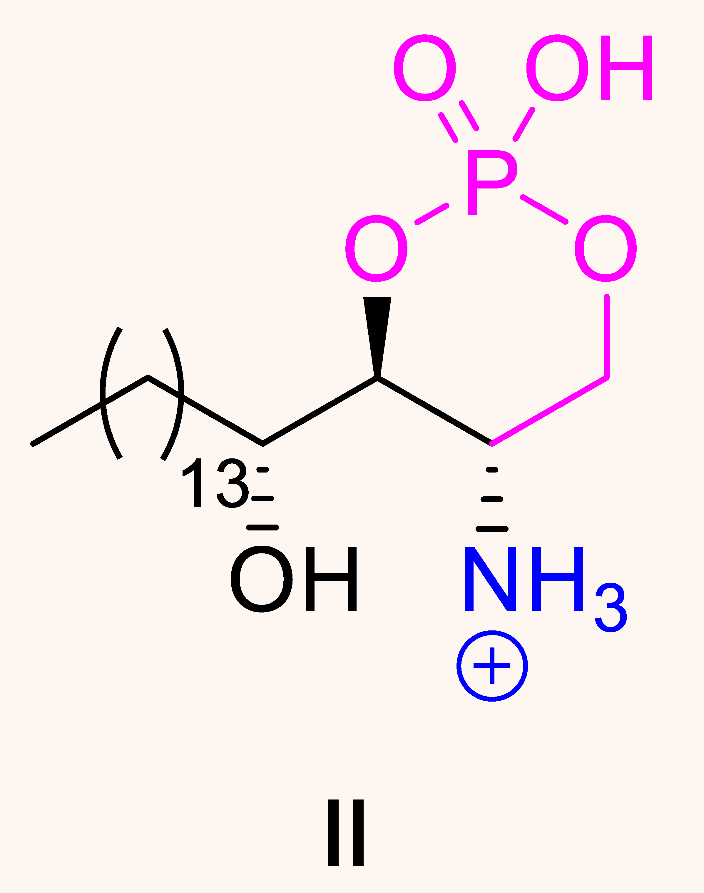
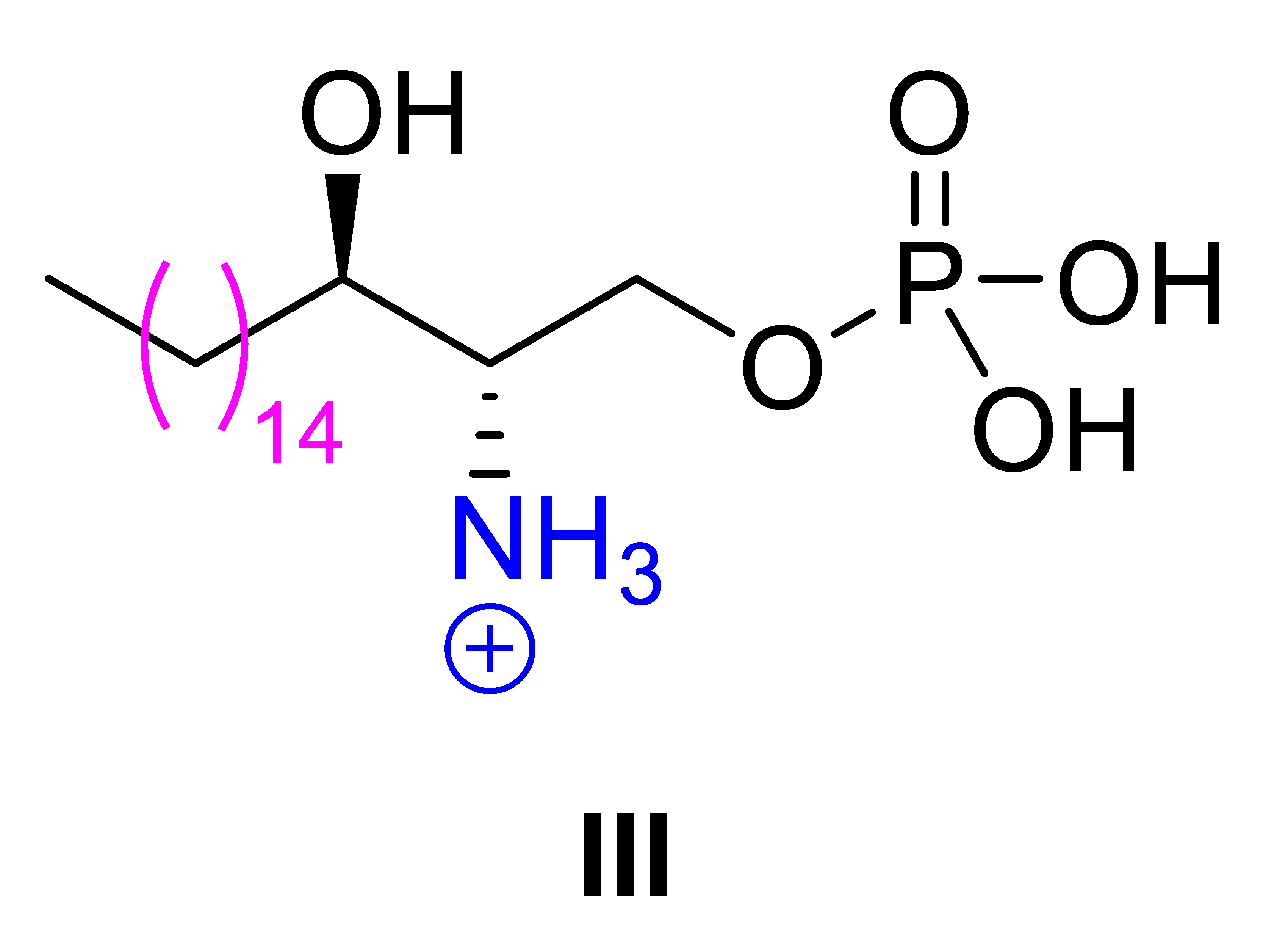
Phytosphingosine, a naturally occurring long-chain fatty alcohol present in the upper layers of the skin, is a key building block for ceramides essential to skin structure. In this study, three phytosphingosine derivatives, (2S,3S,4R)-3,4-dihydroxy-1-(phosphonooxy)octadecan-2-aminium chloride (I), (4S,5S)-2-hydroxy-4-((R)-1-hydroxypentadecyl)-1,3,2-dioxaphosphinan-5-aminium 2-oxide chloride (II), and (2S,3R)-3-hydroxy-1-(phosphonooxy)octadecan-2-aminium chloride (III), were synthesized to evaluate their potential as fungicides. Phytosphingosine-1-phosphate salt I was prepared via a novel four-step phosphorylation process using dimethyl chlorophosphate. O-Cyclic phytosphingosine-1-phosphate salt II was obtained by cyclizing Boc-protected phytosphingosine with POCl3, while Boc-protected sphinganine was phosphorylated under mild conditions using dimethyl chlorophosphate to yield sphinganine-1-phosphate salt III. The antifungal activities of compounds I–III were assessed in vitro against four phytopathogenic fungi: Botrytis cinerea, Fusarium asiaticum, Fusarium solani, and Rhizoctonia solani. Among these, compound II exhibited the most potent growth inhibitory activity against Fusarium asiaticum (IC50 = 27.5 μg/mL). In vivo evaluations using infected apples further confirmed the concentration-dependent inhibitory activity of compound II against F. asiaticum (IC50 = 108 μg/mL). Morphological analyses revealed shortened hyphal structures and abnormal branching in F. asiaticum, while compound II treatment was associated with a concentration-dependent shift toward predominantly yeast-form morphology in adherent Candida albicans populations. In silico docking against five selected fungal enzymes suggested a potential interaction between compound II and N-myristoyltransferase (NMT), characterized by predicted hydrophobic interactions within the acyl-chain binding pocket and a possible hydrogen bond involving LEU171. Molecular dynamics simulations further supported the relative stability of the predicted NMT–compound II complex. Together, these findings highlight the O-cyclic phytosphingosine-1-phosphate salt II as a promising antifungal candidate and suggest NMT as a potential molecular target warranting further experimental validation.
1. Introduction
A phytopathogenic fungus is a type of fungus that causes diseases in plants. These fungi are harmful because they infect various parts such as roots, stems, leaves, flowers, or fruits, disrupting normal growth and leading to plant disease [1]. Phytopathogenic fungi can induce a wide range of symptoms, including wilting, leaf spots, blights, rots, and cankers, all of which negatively impact plant health and agricultural yields [2]. The fungi used in the antifungal evaluation, including Botrytis cinerea, Fusarium asiaticum, Fusarium solani, and Rhizoctonia solani, are widespread and destructive plant pathogens that affect a diverse range of plants. Fungi of the genus Botrytis engage in complex interactions with their host plants and are responsible for diseases in more than 1400 plant species [3]. Particularly, B. cinerea is a notorious pathogen in agriculture, causing substantial losses by reducing both crop yield and quality. The main toxic compound produced by B. cinerea is botrydial [4], which is crucial for infection as it rapidly necrotizes the host plant, leading to tissue collapse. This process enables the fungus to penetrate more rapidly into the plant interior and works synergistically with other substances secreted by Botrytis to orchestrate a coordinated attack on plant tissues [5,6]. In addition, fungi of the genus Fusarium can significantly disrupt plant growth, reduce yields, and lower crop quality. These fungi reproduce through spores that spread quickly and efficiently, allowing rapid infection of numerous plants over large agricultural areas [7]. In particular, F. asiaticum is the primary pathogen causing fusarium head blight and is known to produce mycotoxins such as deoxynivalenol (Vomitoxin), its derivatives, and nivalenol. These mycotoxins not only pose a potential threat to food and feed but are also harmful to humans and animals, resulting in strict regulatory controls in many countries [8]. Another member of the Fusarium genus, F. solani, can persist in soil for extended periods due to its adaptability to a wide range of temperatures and moisture levels. This fungus causes problems in agricultural systems and is also frequently reported as an opportunistic pathogen in humans, causing infections such as onychomycosis [9]. Lastly, Rhizoctonia solani thrives in warm and humid environments common in many agricultural regions. As a saprophyte, R. solani can persist in the soil until conditions become favorable for plant infection. One significant factor contributing to the damaging nature of R. solani is its resistance to many fungicides, such as azoxystrobin [10], which tend to be less effective once the fungus has established itself in the soil or within the plant [11]. Therefore, developing novel and environmentally friendly fungicides that effectively target phytopathogenic fungi is essential in modern pesticide research.
Phytosphingosine is a long-chain carbon backbone featuring a 2-amino-1,3,4-triol functionality, serving as a fundamental building block of sphingolipids naturally present in the upper layers of the skin [12]. Among its derivatives, phytosphingosine-1-phosphate (PhS1P) is the phosphorylated product of phytosphingosine, generated by the catalytic action of sphingosine kinase. This metabolite belongs to the sphingolipid family and is abundantly found in fungi, plants, and certain mammalian tissues [13,14,15]. PhS1P has been recognized as a critical regulator of various physiological and pathophysiological processes, including cellular signaling pathways and immune responses [16,17,18,19]. Despite its biological significance and potential applications across diverse fields, research into concise and scalable synthetic methods for PhS1P remains limited [20,21,22].
In this study, we describe a new and practical approach for synthesizing PhS1P. The process involves the selective phosphorylation of phytosphingosine using dimethyl chlorophosphate in a four-step procedure, enabling a gram-scale synthesis with an overall yield of 61% from phytosphingosine. Due to the limited applicability of PhS1P itself—attributable to its low solubility in both organic solvents and water—phytosphingosine-1-phosphate salt I was prepared to enhance solubility. To further explore its potential for novel biological activities, two additional derivatives of phytosphingosine were synthesized. One derivative, O-cyclic phytosphingosine-1-phosphate salt II, was prepared via cyclization of Boc-protected phytosphingosine using POCl3. Under mild reaction conditions, another derivative, sphinganine-1-phosphate salt III, was efficiently synthesized by phosphorylating Boc-protected sphinganine with dimethyl chlorophosphate.
As part of our ongoing efforts to discover biologically active compounds from diverse resources [23,24,25,26,27,28], the antifungal activities of newly synthesized phytosphingosine derivatives were evaluated against agriculturally important pathogenic strains, including Botrytis cinerea, Fusarium asiaticum, Fusarium solani, and Rhizoctonia solani. The compounds were first screened in vitro for antifungal activity against these four strains, and the most active compound was subsequently tested in vivo to determine its ability to protect fruit or inhibit disease. To further broaden their potential, we also assessed cytotoxicity in mammalian cells and compared their activity with representative antifungal drug classes. Morphological analyses were performed to characterize the effects of the active compound on fungal growth and filamentous morphology, while molecular docking and dynamics simulations were conducted to explore potential interactions with selected fungal proteins. This study aimed to identify phytosphingosine derivatives with antifungal potential and to provide preliminary insights into their biological activities and possible molecular interactions.
2. Materials and Methods
2.1. General Experimental Procedures
1H NMR, 13C NMR and 31P NMR spectra were recorded on a JEOL JNM-AL400 spectrometer (JEOL, Peabody, MA, USA). Chemical shifts are reported in parts per million (ppm) relative to tetramethylsilane (TMS) as an internal standard. The NMR data are presented as follows: chemical shift, multiplicity (s = singlet, d = doublet, t = triplet, q = quartet, quint = quintet, m = multiplet), coupling constants (in Hz), and integration. High-resolution electrospray ionization mass spectrometry (HR-ESI-MS) data were acquired using an Agilent 6545 Q-TOF LC/MS spectrometer (Agilent Technologies, Santa Clara, CA, USA) equipped with an Eclipse Plus C18 95 Å column (50 × 2.1 mm, 1.8 µm), with a flow rate of 0.3 mL/min and a gradient elution from 30% to 100% MeCN in H2O over 10 min. Unless otherwise noted, all reactions were conducted using distilled solvents under an atmosphere of dry N2 in oven-dried (130 °C) glassware. All solvents were freshly distilled from the appropriate drying agents prior to use. Reagents of the highest commercially available grade were employed without further purification. Phytosphingosine was purchased from TCI and used as received.
2.2. Synthetic Procedures
2.2.1. Synthesis of tert-Butyl ((2S,3S,4R)-1,3,4-trihydroxyoctadecan-2-yl)carbamate (1)
To a solution of phytosphingosine (1.50 g, 4.72 mmol) in tert-butanol (4.7 mL), di-tert-butyl dicarbonate (1.03 g, 4.72 mmol) was added. The reaction mixture was stirred at 22 °C for 3 h. Upon completion, the solution was concentrated under reduced pressure, and hexanes (20 mL) were added to precipitate a white solid. The resulting precipitate was filtered and washed with hexanes (2 × 20 mL) to afford product 1 (1.90 g, 4.55 mmol, 96%) as a white solid. This compound has been previously reported, and its spectral data are consistent with those described in the literature [22]. mp = 88–89 °C (lit. 86 °C) [29].
1H NMR (400 MHz, CDCl3): δ 5.41 (br s, 1H, NH), 3.90–3.84 (m, 2H, CHN, CH2OH), 3.76–3.73 (m, 2H, CH2OH, CHOH), 3.68–3.64 (m, 3H, CHOH, OH), 3.25 (br s, 1H, OH), 1.69–1.67 (m, 1H, CH2), 1.52–1.50 (m, 1H, CH2), 1.45 (s, 9H, t-Bu), 1.30–1.26 (m, 24H, CH2), 0.88 (t, J = 6.4 Hz, 3H, CH3); 13C NMR (100 MHz, CDCl3): δ 156.5 (C=O), 80.1 (OC(CH3)3), 76.2 (CHOH), 72.3 (CHOH), 61.9 (CH2OH), 53.0 (CHN), 33.1 (CH2), 31.9 (CH2), 29.7 (CH2), 29.6 (CH2), 29.4 (CH2), 28.4 (OC(CH3)3), 25.9 (CH2), 22.7 (CH2), 14.0 (CH3).
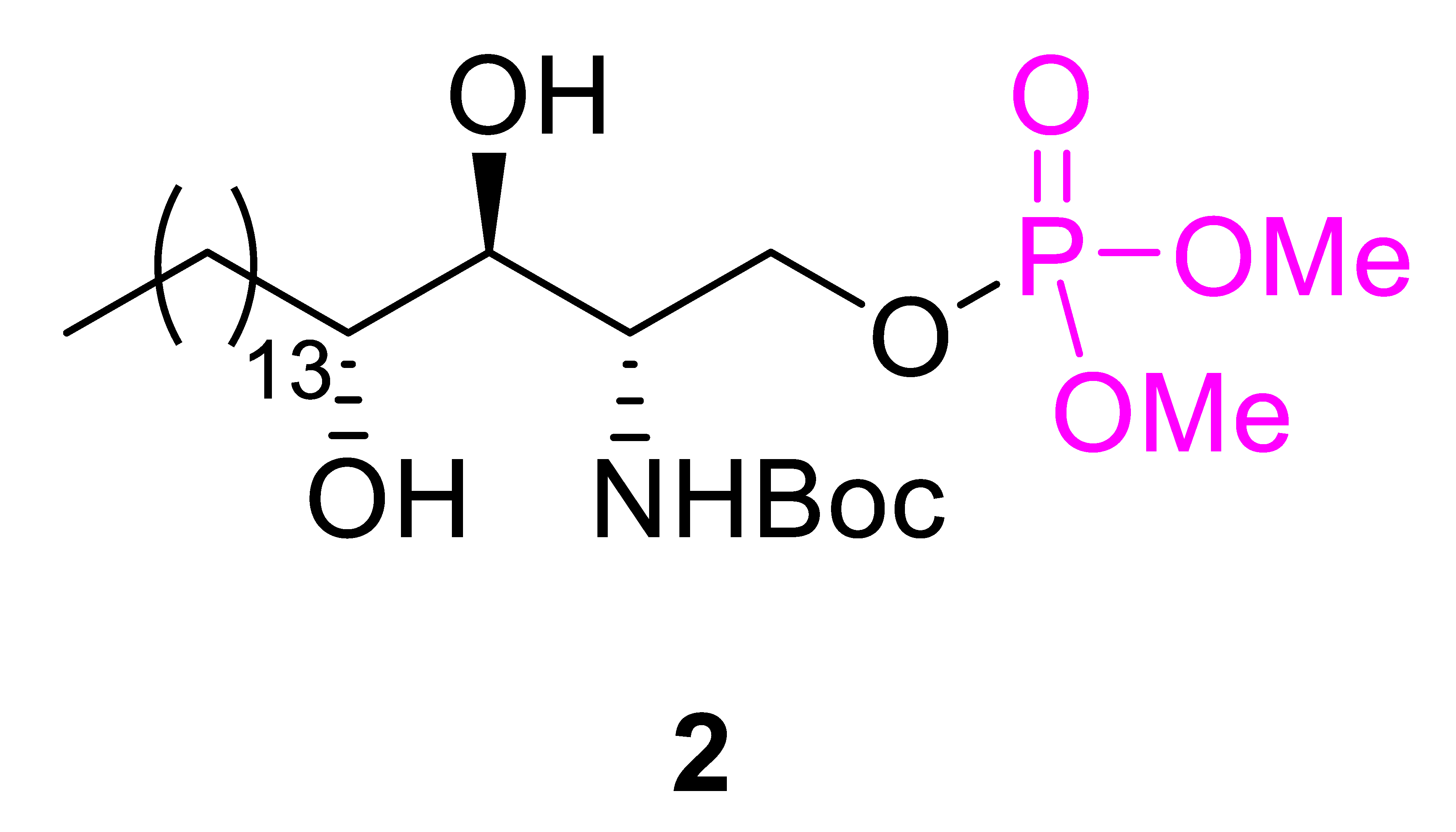
2.2.2. Synthesis of tert-Butyl ((2S,3S,4R)-1-((dimethoxyphosphoryl)oxy)-3,4-dihydroxyoctadecan-2-yl)carbamate (2)
To a solution of N-Boc protected compound 1 (1.90 g, 4.55 mmol) in dichloromethane (45 mL), 1-methylimidazole (544 μL, 6.82 mmol) and dimethyl chlorophosphate (589 μL, 5.46 mmol) were added. The mixture was stirred at 22 °C for 3 h, after which it was quenched by adding a saturated aqueous solution of NH4Cl (40 mL). The resulting solution was extracted with dichloromethane (2 × 20 mL), and the combined organic layers were dried over MgSO4, filtered, and concentrated under reduced pressure. The crude product was purified by silica gel column chromatography using 100% EtOAc as the eluent, affording product 2 (2.10 g, 3.99 mmol, 88%) as a white solid. This compound has been previously reported, and its spectral data are consistent with those described in the literature [21]. mp = 97–98 °C.
1H NMR (400 MHz, CDCl3): δ 5.35 (br s, 1H, NH), 4.41 (br s, 1H, CH2OP), 4.22–4.17 (m, 1H, CH2OP), 3.92 (br s, 1H, CHN), 3.80 (s, 3H, OCH3), 3.77 (s, 3H, OCH3), 3.65–3.63 (m, 2H, CHOH), 3.17 (br s, 2H, OH), 1.62–1.50 (m, 2H, CH2), 1.43 (s, 9H, t-Bu), 1.30–1.24 (m, 24H, CH2), 0.87 (t, J = 6.4 Hz, 3H, CH3); 13C NMR (100 MHz, CDCl3): δ 155.8 (C=O), 79.9 (OC(CH3)3), 73.8 (CHOH), 72.7 (CHOH), 67.5 (d, JC-P = 4.8 Hz, CH2OP), 54.6 (d, JC-P = 5.8 Hz, OCH3), 54.5 (d, JC-P = 5.8 Hz, OCH3), 52.1 (d, JC-P = 6.8 Hz, CHN), 31.9 (CH2), 31.9 (CH2), 29.7 (CH2), 29.6 (CH2), 29.5 (CH2), 29.3 (CH2), 28.3 (OC(CH3)3), 26.0 (CH2), 22.6 (CH2), 14.1 (CH3). = +13.1 (c = 0.25, MeOH).
2.2.3. Synthesis of (2S,3S,4R)-2-Amino-3,4-dihydroxyoctadecyl dihydrogen phosphate (PhS1P)
The phosphate compound 2 (2.10 g, 3.99 mmol), trifluoroacetic acid (18 mL), and CH2Cl2 (0.85 mL) were added to a 50 mL round-bottomed flask. The mixture was stirred at room temperature for 1 h. Upon completion of the reaction, a saturated aqueous solution of Na2CO3 was slowly added at 0 °C (using an ice bath) until a white precipitate formed. The precipitate was filtered and washed with water (3 × 10 mL) and diethyl ether (3 × 10 mL) to afford the amine intermediate (1.70 g, 3.99 mmol, >98%) as a white solid. The obtained amine intermediate (1.70 g, 3.99 mmol) was then dissolved in dichloromethane (38 mL), and the solution was cooled to −20 °C using a dry ice-ethanol bath. Trimethylsilyl bromide (3.61 mL, 27.9 mmol) was slowly added to the solution, and the mixture was stirred at −20 °C for 1 h. Subsequently, the reaction temperature was increased to 0 °C (using an ice bath), and the mixture was stirred for an additional 2 h. After this period, half of the solvent was removed under reduced pressure, and water was added to precipitate a white solid. The precipitate was collected by centrifugation at 25 °C for 15 min at 800 rpm, and the supernatant was discarded. Following the same procedure (vortexing and centrifuging at 25 °C for 15 min at 800 rpm), the resulting white pellet was sequentially washed with water (3 × 10 mL), acetone (3 × 10 mL), and diethyl ether (3 × 10 mL). Finally, the product was dried under vacuum, yielding PhS1P as a white solid (1.14 g, 2.87 mmol, 72% yield). This compound has been previously reported, and its spectral data are consistent with those described in the literature [30]. mp = 170–171 °C (lit. 169–170 °C).
1H NMR (400 MHz, CD3OD-CD3COOD, 1:1): δ 4.29–4.17 (m, 2H, CH2OP), 3.73 (ddd, J = 7.8, 4.1, 4.1 Hz, 1H, CHN), 3.65–3.55 (m, 2H, CHOH), 1.81–1.75 (m, 1H, CH2), 1.52–1.50 (m, 1H, CH2), 1.41–1.26 (m, 24H, CH2), 0.86 (t, J = 6.4 Hz, 3H, CH3); 13C NMR (100 MHz, CD3OD-CD3COOD, 1:1): δ 73.9 (CHOH), 72.6 (CHOH), 63.0 (d, JC-P = 3.9 Hz, CH2OP), 56.1 (d, JC-P = 5.8 Hz, CHN), 35.1 (CH2), 33.1 (CH2), 31.0 (CH2), 30.8 (CH2), 30.8 (CH2), 30.7 (CH2), 30.5 (CH2), 26.1 (CH2), 23.8 (CH2), 14.6 (CH3); 31P NMR (162 MHz, CD3OD-CD3COOD, 1:1): δ 2.76.
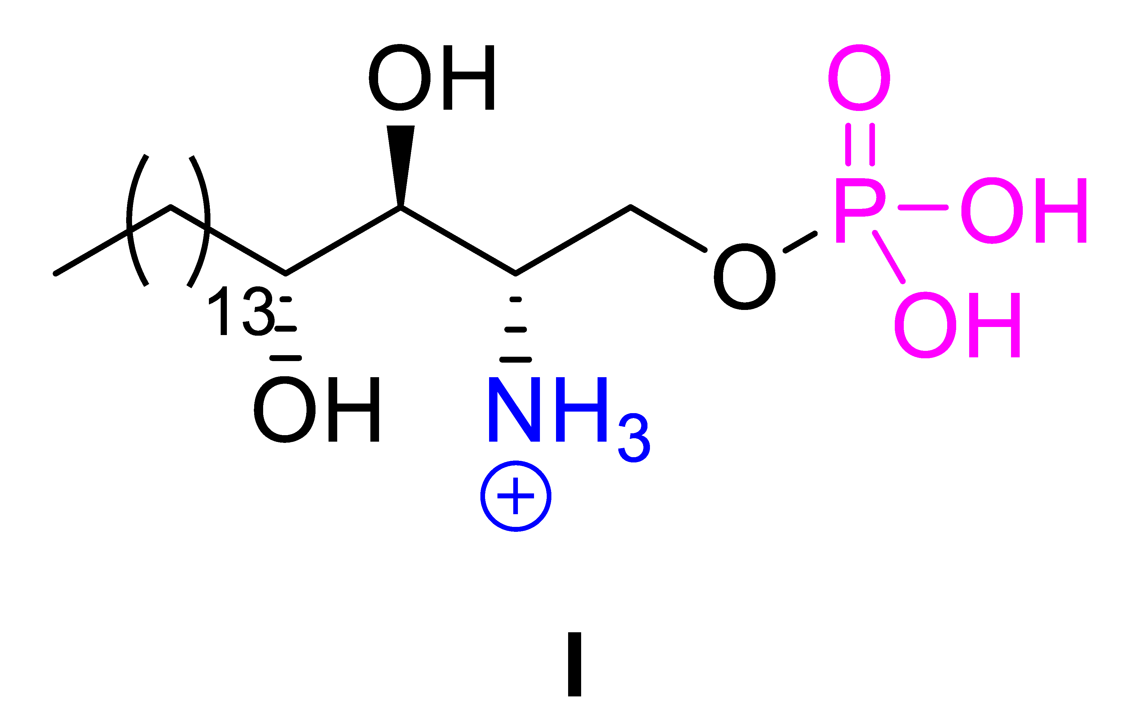
2.2.4. Synthesis of (2S,3S,4R)-3,4-Dihydroxy-1-(phosphonooxy)octadecan-2-aminium chloride (I)
A solution of HCl in dioxane (4 M, 32 mL) and PhS1P (1.14 g, 2.87 mmol) were added to a 100 mL round-bottom flask and stirred at 22 °C for 1 h. After this period, the volatiles were removed under reduced pressure. The remaining solid was washed with acetone (10 mL) and collected by centrifugation at 25 °C for 15 min at 800 rpm, after which the supernatant was discarded. Following the same procedure (vortexing and centrifuging at 25 °C for 15 min at 800 rpm), the resulting white pellet was sequentially washed with diethyl ether (3 × 10 mL) and acetone (3 × 10 mL). Finally, the product was dried under vacuum, yielding the desired compound I as a white solid (0.947 g, 2.18 mmol, 76% yield); mp > 225 °C (decomp.).
1H NMR (400 MHz, CD3OD-CD3COOD, 1:1): δ 4.29–4.18 (m, 2H, CH2OP), 3.74 (ddd, J = 7.8, 4.1, 4.1 Hz, 1H, CHN), 3.67–3.58 (m, 2H, CHOH), 1.81–1.75 (m, 1H, CH2), 1.51–1.49 (m, 1H, CH2), 1.41–1.25 (m, 24H, CH2), 0.85 (t, J = 6.4 Hz, 3H, CH3); 13C NMR (100 MHz, CD3OD-CD3COOD, 1:1): δ 73.9 (CHOH), 72.6 (CHOH), 63.0 (d, JC-P = 4.8 Hz, CH2OP), 56.1 (d, JC-P = 5.8 Hz, CHN), 35.2 (CH2), 33.1 (CH2), 30.8 (CH2), 30.8 (CH2), 30.7 (CH2), 30.5 (CH2), 26.1 (CH2), 23.8 (CH2), 14.6 (CH3); 31P NMR (162 MHz, CD3OD-CD3COOD, 1:1): δ 2.78; HRESIMS: m/z calculated for C18H39NO6P 396.2515; Found 396.2518 [M − H]−; = −135.0 (c = 0.7, MeOH:AcOH = 1:1).
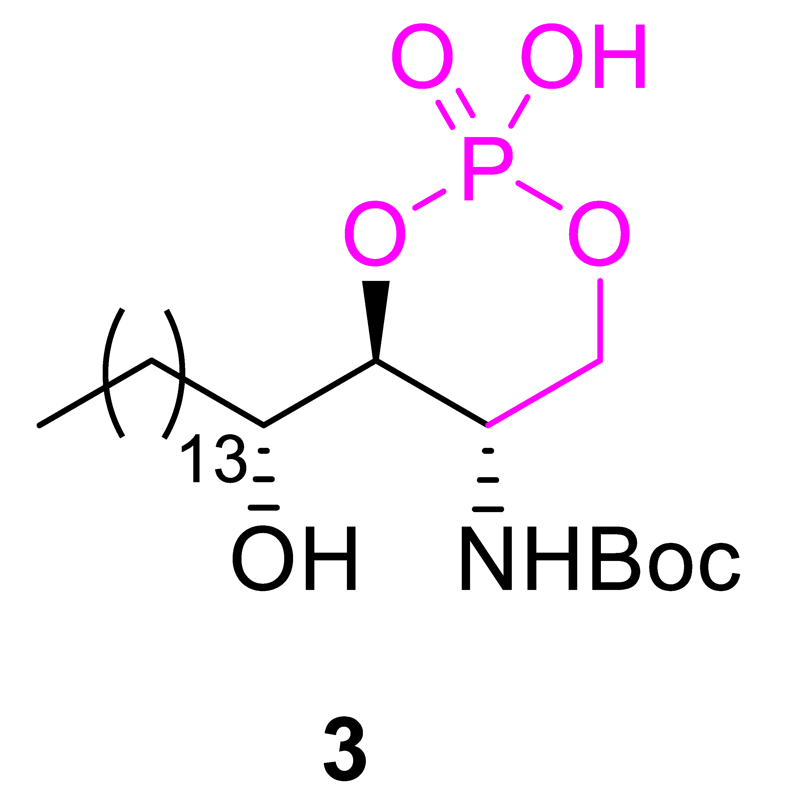
2.2.5. Synthesis of tert-Butyl ((4S,5S)-2-hydroxy-4-((R)-1-hydroxypentadecyl)-2-oxido-1,3,2-dioxaphosphinan-5-yl)carbamate (3)
A solution of N-Boc protected compound 1 (1.84 g, 4.41 mmol) in pyridine (44 mL) was cooled to −20 °C using a dry ice–ethanol bath. Phosphorus oxychloride (825 μL, 8.82 mmol) was then added dropwise, and the reaction mixture was stirred at −20 °C for 1 h. After stirring, the solvent was removed under reduced pressure. The resulting residue was dissolved in 6 N HCl (40 mL) and ethyl acetate (80 mL), and the solution was extracted with ethyl acetate (3 × 50 mL). The combined organic layers were dried over anhydrous MgSO4, filtered, and concentrated under reduced pressure. The crude solid was washed with H2O (100 mL) and collected by centrifugation at 25 °C for 15 min at 800 rpm, discarding the supernatant. The white pellet was then sequentially washed—first with diethyl ether (100 mL) and then with ethyl acetate (100 mL)—using vortexing followed by centrifugation at 25 °C for 15 min at 800 rpm after each wash. The purified product was dried under vacuum to yield compound 3 as a white solid (2.02 g, 4.21 mmol, 95% yield). This compound has been previously reported, and its spectral data are consistent with those described in the literature [21]. mp > 215 °C (decomp.).
1H NMR (400 MHz, CDCl3): δ 4.33–4.28 (m, 1H, CH2OP), 4.18–4.06 (m, 3H, CH2OP, CHOP, CHN), 3.75 (dd, J = 8.3, 2.3 Hz, 1H, CHOH), 1.94–1.88 (m, 1H, OH), 1.67–1.59 (m, 1H, CH2), 1.56–1.50 (m, 1H, CH2), 1.47 (s, 9H, t-Bu), 1.32–1.25 (m, 24H, CH2), 0.88 (t, J = 6.4 Hz, 3H, CH3); 13C NMR (100 MHz, CDCl3): δ 157.9 (C=O), 81.3 (OC(CH3)3), 77.2 (CHOP), 75.1 (CHOH), 65.8 (CH2OP), 52.9 (CHN), 31.9 (CH2), 31.5 (CH2), 29.7 (CH2), 29.7 (CH2), 29.7 (CH2), 29.5 (CH2), 29.4 (CH2), 28.2 (OC(CH3)3), 24.6 (CH2), 22.7 (CH2), 14.1 (CH3). = +6.8 (c = 0.05, MeOH).
2.2.6. Synthesis of (4S,5S)-2-Hydroxy-4-((R)-1-hydroxypentadecyl)-1,3,2-dioxaphosphinan-5-aminium 2-oxide chloride (II)
A solution of cyclophosphate 3 (2.02 g, 4.21 mmol) in ethyl acetate (21 mL) was treated with HCl (2 M in Et2O, 10.5 mL, 21.1 mmol) and stirred at 22 °C for 1 h. Upon completion, the resulting precipitate was filtered and washed with ethyl acetate (3 × 100 mL). The obtained product was dried under vacuum to afford the desired compound II (1.52 g, 3.65 mmol, 87%) as a white solid. mp > 236 °C (decomp.).
1H NMR (400 MHz, CD3OD): δ 4.28 (dd, J = 12.8, 10.0 Hz, 1H, CH2OP), 4.07–3.93 (m, 3H, CH2OP, CHOP), 3.70 (dd, J = 10.1, 4.4 Hz, 1H, CHOH), 3.56 (t, J = 4.1 Hz, 1H, CHN), 1.93–1.86 (m, 1H, OH), 1.61–1.57 (m, 1H, CH2), 1.49–1.41 (m, 1H, CH2), 1.33–1.26 (m, 24H, CH2), 0.91 (t, J = 6.5 Hz, 3H, CH3); 13C NMR (100 MHz, CD3OD): δ 74.0 (CHOP), 71.9 (CHOH), 62.7 (CH2OP), 56.6 (CHN), 33.7 (CH2), 33.2 (CH2), 31.0 (CH2), 31.0 (CH2), 30.9 (CH2), 30.9 (CH2), 30.8 (CH2), 30.6 (CH2), 25.9 (CH2), 23.9 (CH2), 14.6 (CH3); 31P NMR (162 MHz, CD3OD): δ 2.50; HRESIMS: m/z calculated for C18H37NO5P 378.2409, Found 378.2417 [M − H]−; = −134.9 (c = 0.7, MeOH:AcOH = 1:1).
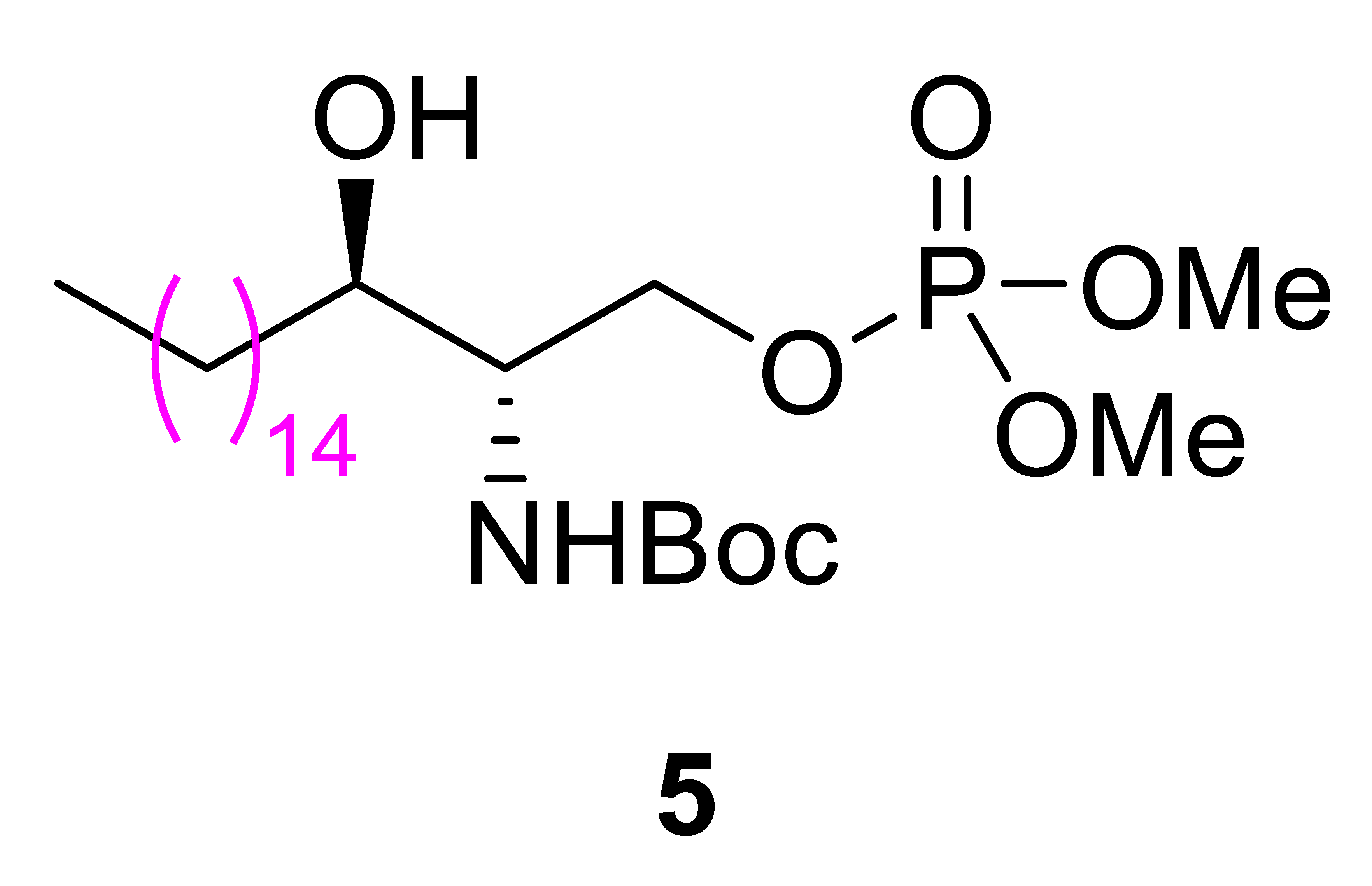
2.2.7. Synthesis of tert-Butyl ((2S,3R)-1-((dimethoxyphosphoryl)oxy)-3-hydroxyoctadecan-2-yl)carbamate (5)
To a solution of N-Boc protected compound 4 (200 mg, 0.498 mmol) in dichloromethane (5 mL), 1-methylimidazole (60.0 μL, 0.747 mmol) and dimethyl chlorophosphate (64.0 μL, 0.598 mmol) were added. The mixture was stirred at 22 °C for 4 h, after which the reaction was quenched by adding a saturated aqueous solution of NH4Cl (5 mL). The resulting solution was extracted with dichloromethane (2 × 5 mL). The combined organic layers were dried over MgSO4, filtered, and concentrated under reduced pressure. The crude product was purified by silica gel column chromatography (100% EtOAc), affording product 5 (223 mg, 0.438 mmol, 88%) as a white solid. This compound has been previously reported, and its spectral data are consistent with those described in the literature [31]. mp = 94–95 °C.
1H NMR (400 MHz, CDCl3): δ 5.11 (d, J = 8.3 Hz, 1H, NH), 4.39 (ddd, J = 11.0, 8.2, 4.1 Hz, 1H, CH2OP), 4.15–4.09 (m, 1H, CH2OP), 3.79 (d, J = 2.7 Hz, 3H, OCH3), 3.77 (d, J = 2.7 Hz, 3H, OCH3), 3.69 (br s, 1H, CHN), 3.61–3.60 (m, 1H, CHOH), 3.13 (d, J = 5.0 Hz, 1H, OH), 1.59–1.49 (m, 2H, CH2), 1.44 (s, 9H, t-Bu), 1.25 (br s, 26H, CH2), 0.87 (t, J = 6.5 Hz, 3H, CH3); 13C NMR (100 MHz, CDCl3): δ 155.6 (C=O), 79.7 (OC(CH3)3), 71.3 (CHOH), 66.9 (d, JC-P = 5.8 Hz, CH2OP), 54.9 (d, JC-P = 4.8 Hz, CHN), 54.6 (d, JC-P = 5.8 Hz, OCH3), 33.7 (CH2), 31.9 (CH2), 29.7 (CH2), 29.6 (CH2), 29.6 (CH2), 29.5 (CH2), 29.3 (CH2), 28.3 (OC(CH3)3), 25.9 (CH2), 22.6 (CH2), 14.1 (CH3). = +15.9 (c = 0.20, MeOH).
2.2.8. Synthesis of (2S,3R)-3-Hydroxy-1-(phosphonooxy)octadecan-2-aminium chloride (III)
The phosphate compound 5 (15.0 mg, 0.0294 mmol) was dissolved in CH2Cl2 (1.2 mL) and cooled to 0 °C using an ice bath. Trimethylsilyl bromide (17.0 μL, 0.132 mmol) was then added dropwise, and the reaction mixture was stirred at 0 °C for 2 h. Upon completion of the reaction, the volatile components were removed under reduced pressure. The resulting residue was dried under vacuum, and HCl (4 M in dioxane, 100 μL) was added. The mixture was stirred at 22 °C for 1 h, after which the volatiles were again removed under reduced pressure. The remaining solid was washed with tetrahydrofuran (8 mL) and collected by centrifugation at 25 °C for 15 min at 800 rpm, discarding the supernatant. Using the same procedure (vortexing and centrifugation at 25 °C for 15 min at 800 rpm), the resulting white pellet was sequentially washed with H2O (2 × 5 mL), diethyl ether (2 × 5 mL), and acetone (2 × 5 mL). Finally, the purified product was dried under vacuum, yielding the desired compound III as a white solid (10.2 mg, 0.0244 mmol, 83% yield); mp > 194 °C (decomp.).
1H NMR (400 MHz, CD3OD): δ 4.30–4.25 (m, 1H, CH2OP), 4.15 (ddd, J = 11.3, 8.4, 7.3 Hz, 1H, CH2OP), 3.83–3.80 (m, 1H, CHOH), 3.44–3.40 (m, 1H, CHN), 1.54–1.49 (m, 3H, CH2, OH), 1.32–1.29 (m, 26H, CH2), 0.90 (t, J = 6.9 Hz, 3H, CH3); 13C NMR (100 MHz, CD3OD): δ 70.2 (CHOH), 64.1 (CH2OP), 57.2 (CHN), 34.4 (CH2), 33.2 (CH2), 30.9 (CH2), 30.9 (CH2), 30.9 (CH2), 30.8 (CH2), 30.7 (CH2), 30.6 (CH2), 27.1 (CH2), 23.9 (CH2), 14.6 (CH3); 31P NMR (162 MHz, CD3OD): δ 0.44; HRESIMS: m/z calculated for C18H39NO5P 380.2566, Found 380.2572 [M − H]−; = −135.5 (c = 0.7, MeOH:AcOH = 1:1).
2.3. Fungi Material
For the antifungal activity assay, four phytopathogenic fungi provided by the Korean Agricultural Culture Collection (KACC; Jeonju, Republic of Korea) were used: Botrytis cinerea (KACC 40965), Fusarium solani (KACC 44891), Fusarium asiaticum (KACC 46429), and Rhizoctonia solani (KACC 48921). The fungi were cultivated on 25 mL of malt extract agar (MEA; Difco, MD, USA) at 25 °C for 14 days in the dark.
2.4. Antifungal Activity Assay In Vitro
The antifungal activities of phytosphingosine derivatives (I–III), along with the Boc-protected intermediates 2, 3, and 5, were evaluated against agricultural pathogenic fungi, including Botrytis cinerea, Fusarium solani, Fusarium asiaticum, and Rhizoctonia solani, using a microtiter assay [32]. The effects of the compounds were assessed in a 96-well microplate, where each well contained 2 μL of the compound, 20 μL of a spore suspension, and 178 μL of malt extract peptone dextrose broth (MPD) medium, resulting in final compound concentrations of 100, 50, 25, 12.5, and 6.25 μg/mL. Benomyl (Sigma-Aldrich, St. Louis, MO, USA; 99% purity), a commercial fungicide dissolved in dimethyl sulfoxide (DMSO), served as the positive control, while DMSO alone was used as the negative control. Benomyl was tested using a separate dilution series at final concentrations of 10, 5, 2.5, 1.25, and 0.625 μg/mL. All test compounds were dissolved in DMSO, with a final DMSO concentration of 1% (v/v) in the assay wells, and the spore suspension of the plant pathogenic fungi was adjusted to 4 × 10⁶ spores/mL. The microplates were incubated in a growth chamber at 27 °C, and absorbance was measured at 600 nm every 12 h for 96 h. All experiments were performed in triplicate. Concentration–response curves were fitted by nonlinear regression in GraphPad Prism 8. The IC50 was defined as the concentration producing 50% growth inhibition and is reported with its 95% confidence interval (Figure S28). The percentage of growth inhibition (GI%) was calculated using the formula: GI% = (Acontrol − Atest sample)/(Acontrol) × 100%.
Broth microdilution assays were performed using methods adapted from the Clinical and Laboratory Standards Institute (CLSI) guidelines M27-A3 (for yeasts) and M38-A2 (for filamentous fungi), using Candida albicans SC5314 and F. asiaticum as representative species. Compounds were serially diluted in RPMI 1640 medium buffered with MOPS (pH 7.0), and standard antifungal agents, including polyene, echinocandins, and azoles, were tested in parallel. Plates were incubated at 35 °C for 48 h. Fungal growth and morphology were evaluated by visual inspection and EVOS M7000 microscopy (Thermo Fisher Scientific, Waltham, MA, USA). Drug concentrations associated with marked suppression of visible fungal growth or distinct morphological alterations were selected for comparative microscopic analysis.
To assess the effects of phytosphingosine derivatives on the morphology of adherent C. albicans cells, non-adherent C. albicans cells were removed by washing after 48 h of compound exposure. The remaining adherent cells were examined microscopically to assess changes in yeast-form and filamentous morphology. Cells bearing germ tubes or filaments longer than the major axis of the associated mother cell were classified as filamentation-positive. The prevalence of filamentation-positive cells was expressed using a semiquantitative filamentation score ranging from 0 to 4.
2.5. Cytotoxicity Assay
Cytotoxicity was evaluated in mammalian cells to assess the safety of compound II. 3T3-L1 fibroblasts and HEK293A kidney epithelial cells were cultured in DMEM supplemented with 10% FBS at 37 °C in 5% CO2. Cells were seeded in 96-well plates (1 × 10⁴ cells/well) and treated with compound II. For 3T3-L1 fibroblasts, cells were treated with compound II at concentrations of 12.5, 25, 50, and 100 μg/mL for 24 h. Cell viability was measured using a WST-1 assay, with DMSO as a solvent control. For HEK293A kidney cells, compound II was tested across a concentration range of 0.1–100 μM (final DMSO 1%). Cell viability was measured after 24, 48, and 72 h of incubation using an XTT Cell Viability Assay Kit (Invitrogen, Carlsbad, CA, USA), with absorbance read at 450 nm. Viability was expressed as a percentage relative to untreated controls [33]. All experiments were performed in triplicate.
2.6. Antifungal Activity Assay In Vivo
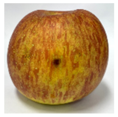
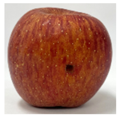
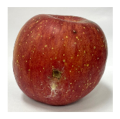
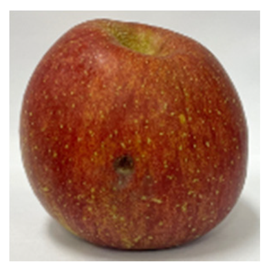
Following the in vitro antifungal results, compound II with the best activity against Fusarium asiaticum was further tested in vivo. The same type of mature apples with similar shapes and sizes were harvested from the same orchard for testing. The apples were soaked in 1% sodium hypochlorite for 2 min to disinfect them, and then rinsed thoroughly under running water for 5 min. After drying, 3 × 3 mm holes were punched in the surface of the washed apples, and 20 μL of a solution of each compound in acetone was applied to the apples at five different concentrations (500, 200, 100, 50, and 25 μg/mL). Once the acetone solutions had evaporated, a 20 μL spore suspension (4 × 10⁶ spores/mL) was added to each apple. A spore suspension was used as a blank, and sterile water served as a negative control [34]. The commercial agricultural fungicide benomyl, applied at five concentrations (100, 50, 25, 12.5, and 6.25 μg/mL), was used as a positive control. Solutions of different concentrations were prepared using the same method as for the in vitro antifungal activity assay. After drying, all apples were placed in a greenhouse maintained at 27 °C under constant moisture and observed for 8–9 days. For each treatment concentration, three independent apples were used (n = 3), with two inoculation sites prepared on each apple under identical treatment conditions. The two inoculation sites served as within-apple technical replicates, and their measurements were averaged to obtain a single value for each apple. Thus, the individual apple was considered the independent experimental unit for data analysis. Once the mycelium had fully developed, the diameter of the fungal growth was measured. The percentage of growth inhibition (GI%) was calculated using the following formula: GI% = (Acontrol − Atest sample)/(Acontrol − 3 mm) × 100%, where Acontrol is the diameter of fungal growth on untreated apples and Atest sample is the diameter of fungal growth on treated apples [34].
2.7. Molecular Docking
Five fungal target proteins were selected based on their essential roles in fungal viability and pathogenicity. The corresponding crystal structures were obtained from the RCSB Protein Data Bank (http://www.pdb.org; accessed on 10 July 2025), and the 3D structures of compounds I–III were generated using the 2D Sketcher tool in Schrödinger Maestro (Schrödinger Release 2025-2) [35]. Protein structures were prepared using Schrödinger’s Protein Preparation Workflow (Schrödinger Release 2025-2) [35], which included the removal of crystallographic water molecules, addition of missing side chains, and optimization of H-bond networks. Ligand structures were processed using LigPrep to optimize tautomeric and ionization states and to refine ring conformations, all within the OPLS4 force field (Schrödinger Release 2025-2) [36]. The receptor grid boxes (20 × 20 × 20 Å) were generated around the druggable pockets identified within the crystal structures of the target proteins. Molecular docking was conducted using the Glide SP mode (Schrödinger Release 2025-2) [37], with full ligand flexibility enabled through torsional refinement under a distance-dependent dielectric environment. Post-docking minimization was applied to further refine the docked poses within the force field. The binding free energy of each protein–ligand complex was estimated using the Molecular Mechanics–Generalized Born Surface Area (MM-GBSA) method.
2.8. Molecular Dynamics
MD simulations were performed for 200 ns using Desmond integrated with Maestro (Schrödinger Release 2025-4) [38] to assess the structural stability and binding interactions of the complexes formed between NMT and compounds I–III, which were selected based on their favorable docking scores.
Simulation systems were prepared using the System Builder module in Desmond, wherein each protein–ligand complex was placed in an orthorhombic, buffer-generated box, solvated with TIP3P water, and neutralized with Na+ ions. The OPLS4 force field was employed to evaluate complex stability. Following completion of the simulations, trajectory analyses, including root mean square deviation (RMSD) and root mean square fluctuation (RMSF), were performed to characterize the stability and dynamic behavior of the protein–ligand interactions.
3. Results and Discussion
3.1. Chemical Synthesis
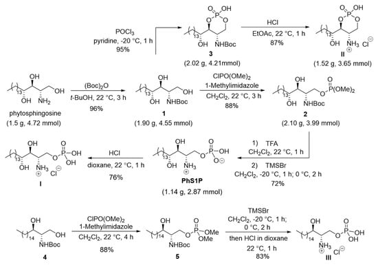
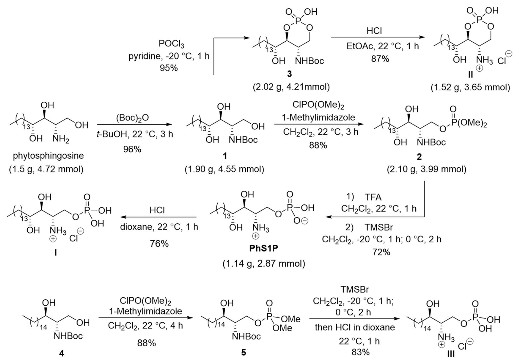
Gram-scale synthesis of phytosphingosine-1-phosphate (PhS1P) was initiated with the N-Boc protection of commercially available phytosphingosine (1.50 g, 4.72 mmol) in tert-butanol. For clarity, synthetic intermediates are designated by Arabic numerals (1–5), whereas the final synthesized compounds are designated by Roman numerals (I–III) throughout the manuscript (Scheme 1). The resulting Boc-protected phytosphingosine 1 was then selectively phosphorylated using dimethyl chlorophosphate and 1-methylimidazole, yielding product 2 in 88% yield under mild reaction conditions. Initially, simultaneous removal of the N-Boc protecting group and the methyl ester of compound 2 was attempted using trimethylsilyl bromide; however, this approach proved problematic due to difficulties in eliminating unexpected side products on a gram scale. Consequently, a stepwise deprotection strategy was adopted: first, the N-Boc group of compound 2 was removed using trifluoroacetic acid, and then the methyl ester was hydrolyzed in the presence of trimethylsilyl bromide, leading to PhS1P in 72% yield (1.14 g, 2.87 mmol). Finally, PhS1P was treated with HCl to produce (2S,3S,4R)-3,4-dihydroxy-1-(phosphonooxy)octadecan-2-aminium chloride (I), obtained as the hydrochloride salt of PhS1P. The gram-scale synthesis of (4S,5S)-2-hydroxy-4-((R)-1-hydroxypentadecyl)-1,3,2-dioxaphosphinan-5-aminium 2-oxide chloride (II) involved the cyclization of Boc-protected phytosphingosine 1 (1.84 g, 4.41 mmol) using POCl3 in the presence of pyridine, followed by treatment of the resulting intermediate 3 with HCl to afford the O-cyclic phytosphingosine-1-phosphate salt II in 83% yield for two steps (1.52 g, 3.65 mmol). Notably, compound II was obtained from naturally occurring phytosphingosine via a concise three-step sequence, delivering an overall yield of 79% on a gram scale under mild conditions and with short reaction times. Furthermore, all intermediates and the final product were purified by simple solidification rather than silica gel column chromatography, highlighting the scalability and cost-effectiveness of this synthetic route. (2S,3R)-3-Hydroxy-1-(phosphonooxy)octadecan-2-aminium chloride (III) was synthesized by first phosphorylating the known N-Boc-protected sphinganine 4 [22] using dimethyl chlorophosphate and 1-methylimidazole. The phosphorylated intermediate 5 was then sequentially treated with trimethylsilyl bromide and HCl, enabling the simultaneous removal of the N-Boc group and the methyl ester, and ultimately yielding the sphinganine-1-phosphate HCl salt product III (Scheme 1). The structures of the newly synthesized compounds I, II, and III were thoroughly analyzed using a combination of 1H, 13C, and 31P NMR spectroscopy as well as high-resolution mass spectrometry (HRMS).
Scheme 1.
Synthesis of phytosphingosine derivatives I–III.
3.2. Evaluation of Antifungal Activity for the Synthetic Compounds (I–III) In Vitro
The synthetic compounds (I–III) were evaluated for their in vitro antifungal activity against four plant pathogens, including Botrytis cinerea, Fusarium solani, Fusarium asiaticum, and Rhizoctonia solani, using a microtiter growth-inhibition assay at 6.25, 12.5, 25, 50, and 100 μg/mL. Growth inhibition was assessed at different time points for each strain: 96 h for B. cinerea, 96 h for F. solani, 72 h for F. asiaticum, and 96 h for R. solani. Concentration-dependent growth inhibition was quantified relative to the vehicle-treated control, and IC50 values were estimated from the corresponding concentration–response data. Among the three phytosphingosine derivatives (I–III), only compound II demonstrated the strongest activity against F. asiaticum, with an IC50 of 27.5 μg/mL (Table 1). Compounds I and III did not reach 50% growth inhibition against F. asiaticum within the tested concentration range, with IC50 values greater than 100 μg/mL. Compound II also showed limited activity against B. cinerea and R. solani, for which 50% inhibition was not reached at 50 μg/mL, and against F. solani, for which 50% inhibition was not reached at 100 μg/mL. These results indicate that compound II exhibited preferential activity against F. asiaticum within the tested pathogen panel.
Table 1.
In vitro antifungal activity of the Boc-protected intermediates (2, 3, 5) and final compounds (I–III) against plant pathogens.
Following the initial antifungal screening against phytopathogenic fungi, compound II was further evaluated using CLSI guideline-based broth microdilution assays, which are widely employed in clinical antifungal susceptibility testing, to compare its activity with conventional antifungal agents and characterize its effects on fungal morphology. Compound II markedly suppressed visible growth of F. asiaticum at 32 μg/mL. The reference antifungal agents such as amphotericin B, posaconazole, and voriconazole exhibited substantial growth suppression at 1, 1, and 4 μg/mL, respectively. In contrast, caspofungin was evaluated based on morphological changes in fungal hyphae, with marked alterations observed at 32 μg/mL. In Candida albicans SC5314, compound II produced detectable reductions in filamentous morphology at concentrations as low as 2 μg/mL. These observations were used to guide subsequent microscopic comparisons of fungal morphology following treatment with compound II and conventional antifungal agents.
In addition, the cytotoxicity of compound II was evaluated in mammalian cells as a preliminary assessment relevant to its potential agricultural application. In preliminary assays using 3T3-L1 fibroblasts, compound II exhibited negligible cytotoxicity up to 100 μg/mL (Figure S27). To further confirm its safety profile, cytotoxicity was assessed in HEK293A kidney cells across a concentration range of 0.1–100 μM. Compound II did not display persistent toxicity within this range: an initial dose-dependent reduction in metabolic activity was observed at 24 h, but cells recovered by 48 h, and only the highest concentration (100 μM, 37.92 μg/mL) showed residual reduction at 72 h (Figure S27). These preliminary findings suggest limited mammalian cytotoxicity at concentrations overlapping the antifungal activity range of compound II, although further safety evaluation is required.
3.3. Structure–Activity Relationship (SAR) Analysis
To facilitate a visual comparison of the preliminary structure–activity relationships (SAR), the chemical structures of the synthetic intermediates (2, 3, and 5) and final compounds (I–III) are presented alongside their corresponding antifungal activities against F. asiaticum in Table 2, using the IC50 values already reported in Table 1. Compound II was the only member of the six-compound set to afford a finite IC50 value (27.5 μg/mL), whereas the other five compounds had IC50 values >100 μg/mL. The resulting SAR was therefore interpreted as a qualitative active–inactive relationship rather than as a quantitative potency ranking (Table 2). Given that only one of the six compounds exhibited a measurable IC50 within the tested concentration range, the present dataset supports only a preliminary qualitative SAR analysis, and its limited size precludes the identification of generalizable structural requirements for antifungal activity.
Table 2.
Antifungal activities (IC50, μg/mL) of the Boc-protected intermediates and final compounds against Fusarium asiaticum.
The 3/II pair provides the most direct assessment of the C2 nitrogen. Both compounds contain the same six-membered cyclic phosphate framework, and conversion of 3 to II removes the N-Boc group while retaining the phosphate scaffold. The difference in activity between compounds 3 and II suggests that the deprotected C2 amine may contribute to the antifungal activity observed for compound II within the cyclic phosphate series. The complementary I/II pair retains the phytosphingosine backbone and the deprotected amine while changing the phosphate from an acyclic to a cyclic form. The inactivity of I therefore shows that amine deprotection alone is insufficient. Within the compounds examined, activity was associated with the coexistence of the cyclic phosphate and the deprotected C2 amine, rather than with either feature alone.
Compounds 2 and 5, and compounds I and III, provide comparisons of the phytosphingosine and sphinganine backbones in the N-Boc-protected and deprotected acyclic-phosphate series, respectively. Because both members of each pair were inactive, these comparisons do not establish whether the C4 hydroxyl contributes to activity. A cyclic sphinganine analogue would be required to examine this substituent in the structural context in which activity was observed. The conserved long-chain sphingoid backbone is likewise insufficient to account for the activity of II, because it is present throughout the series. The combination of a long hydrophobic backbone and a deprotected amine is compatible with membrane interaction; however, the inactivity of compounds I and III indicates that these features alone are insufficient. Thus, the cyclic phosphate framework appears to be associated with antifungal activity within the series examined. Overall, these findings suggest an association between antifungal activity and the combination of the cyclic phosphate framework and the deprotected C2 amine within the limited compound series examined. However, because only compound II exhibited measurable activity, these observations should be regarded as preliminary SAR findings rather than established structural determinants. Further investigation of a broader range of analogues will be necessary to determine the generalizability of these structure–activity relationships.
3.4. Evaluation of Antifungal Activity of Compound II In Vivo
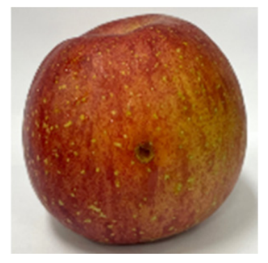
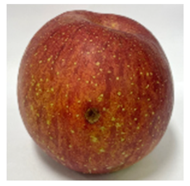
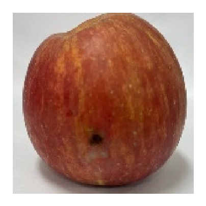
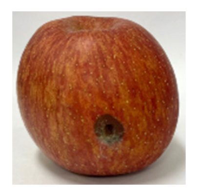
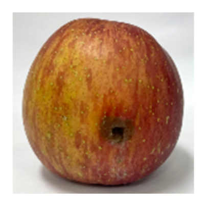
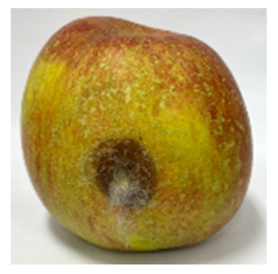
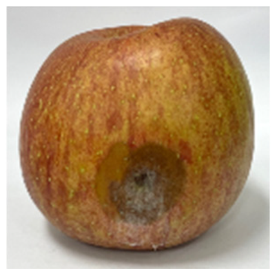
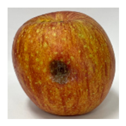
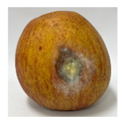
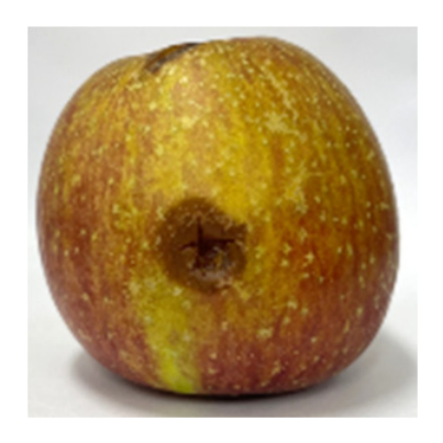
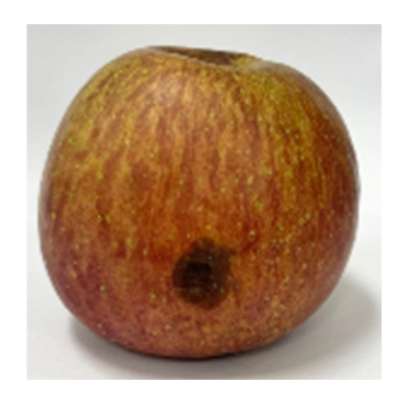
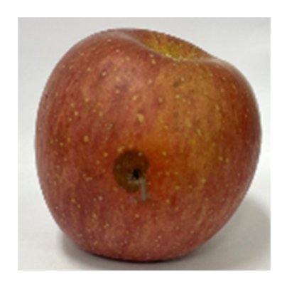
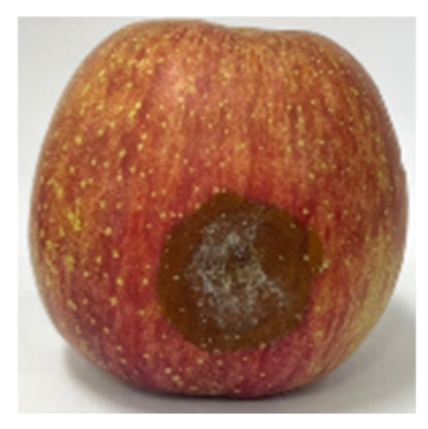
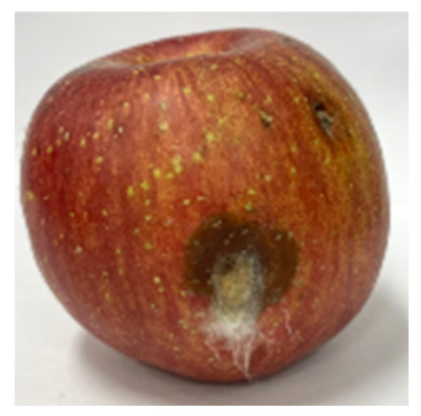
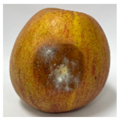
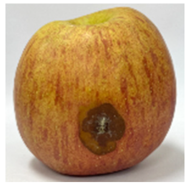
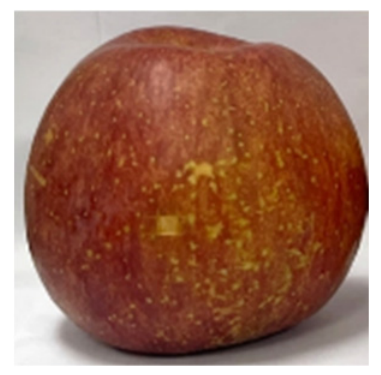
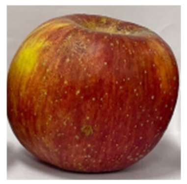
Following the in vitro antifungal results, the in vivo antifungal activity of compound II was further investigated, focusing on its efficacy against F. asiaticum, the fungal strain that exhibited significant sensitivity to compound II in vitro. The in vivo tests were conducted under carefully controlled temperature and humidity conditions over a period of 9 days to ensure consistency. The results obtained on the last day (day 9) are summarized in Table 3. In these studies, compound II exhibited an IC50 value of 108 μg/mL, indicating that a higher concentration was necessary to achieve levels of inhibition comparable to those observed in vitro. A comparison between the in vitro and in vivo data revealed a higher apparent IC50 for compound II in the apple assay. Nevertheless, a clear dose-dependent inhibition of F. asiaticum growth was observed, with compound II effectively protecting the fruit from fungal infection. Similarly, the positive control, benomyl, showed reduced efficacy in vivo, with an IC50 value of 7.71 μg/mL. These observations suggest that the higher apparent in vivo IC50 of compound II may reflect differences in assay endpoints and exposure conditions between the in vitro and apple infection models. In the present study, the in vitro assay assessed fungal growth inhibition by measuring broth absorbance, whereas the apple assay evaluated fungal growth diameter after 9 days. Therefore, the IC50 values obtained from these different experimental systems should not be interpreted as directly comparable measures of intrinsic antifungal potency. Similar differences between in vitro and in vivo antifungal efficacy have been reported for griseofulvin derivatives against Colletotrichum gloeosporioides in a wounded-apple model [34]. Furthermore, following compound application and solvent evaporation, interactions with fruit tissue components or limited diffusion from the wound site may reduce the local availability of compound II to the fungus. Consistent with this possibility, diffusion capacity has been associated with in vivo antifungal efficacy in pear fruit [39]. Compound stability during the 9-day assay period may also influence the observed activity, although degradation cannot be identified as a contributing factor without direct experimental evidence. In addition, host defense responses may influence antifungal efficacy in fruit tissues, as suggested by previous studies involving ursolic acid treatment in apples [40], although their specific contribution to the activity of compound II remains unknown. Accordingly, future studies should investigate formulation and application strategies aimed at improving compound dispersion, retention at the infection site, and treatment timing to enhance antifungal efficacy under in vivo conditions.
Table 3.
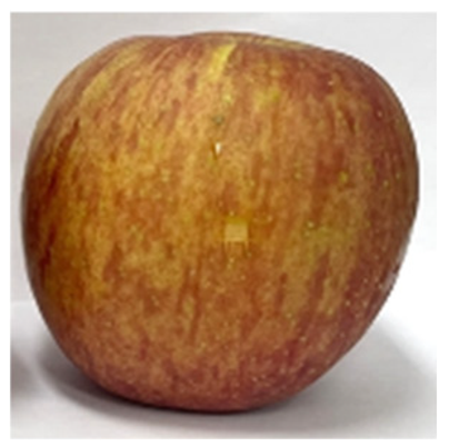
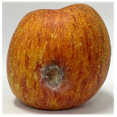
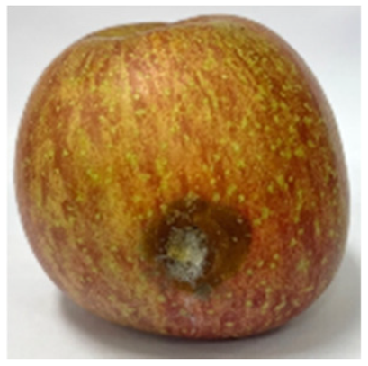
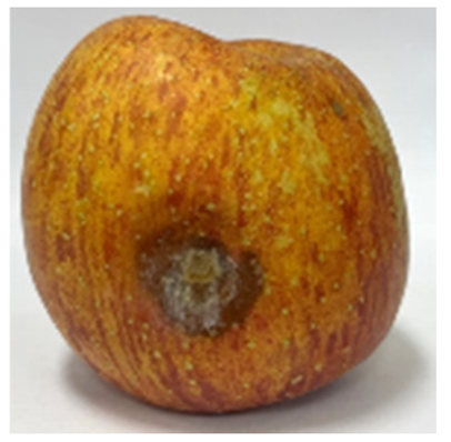
In vivo antifungal assay of compound II against Fusarium asiaticum on apples stored for 9 days.
This study reports the efficient synthesis and antifungal assessment of phytosphingosine derivatives, among which compound II, a structurally unique cyclic analogue, demonstrated selective and significant inhibitory activity, particularly against F. asiaticum. The differential bioactivity observed between compound II and its linear counterparts suggests a potential association between the cyclic phosphate framework and antifungal activity within the limited series examined. Furthermore, the dose-dependent inhibition of fungal growth observed in vivo, despite a higher apparent IC50 than in the in vitro assay, highlights the potential of compound II as a promising lead compound for further development. These findings emphasize the critical influence of molecular architecture on biological activity and indicate that continued optimization, through formulation strategies and mechanistic elucidation, may enhance the practical application of such compounds in agricultural disease management.
3.5. Effects of Phytosphingosine Derivatives on Fungal Morphology
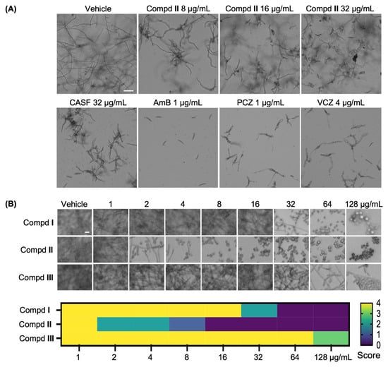
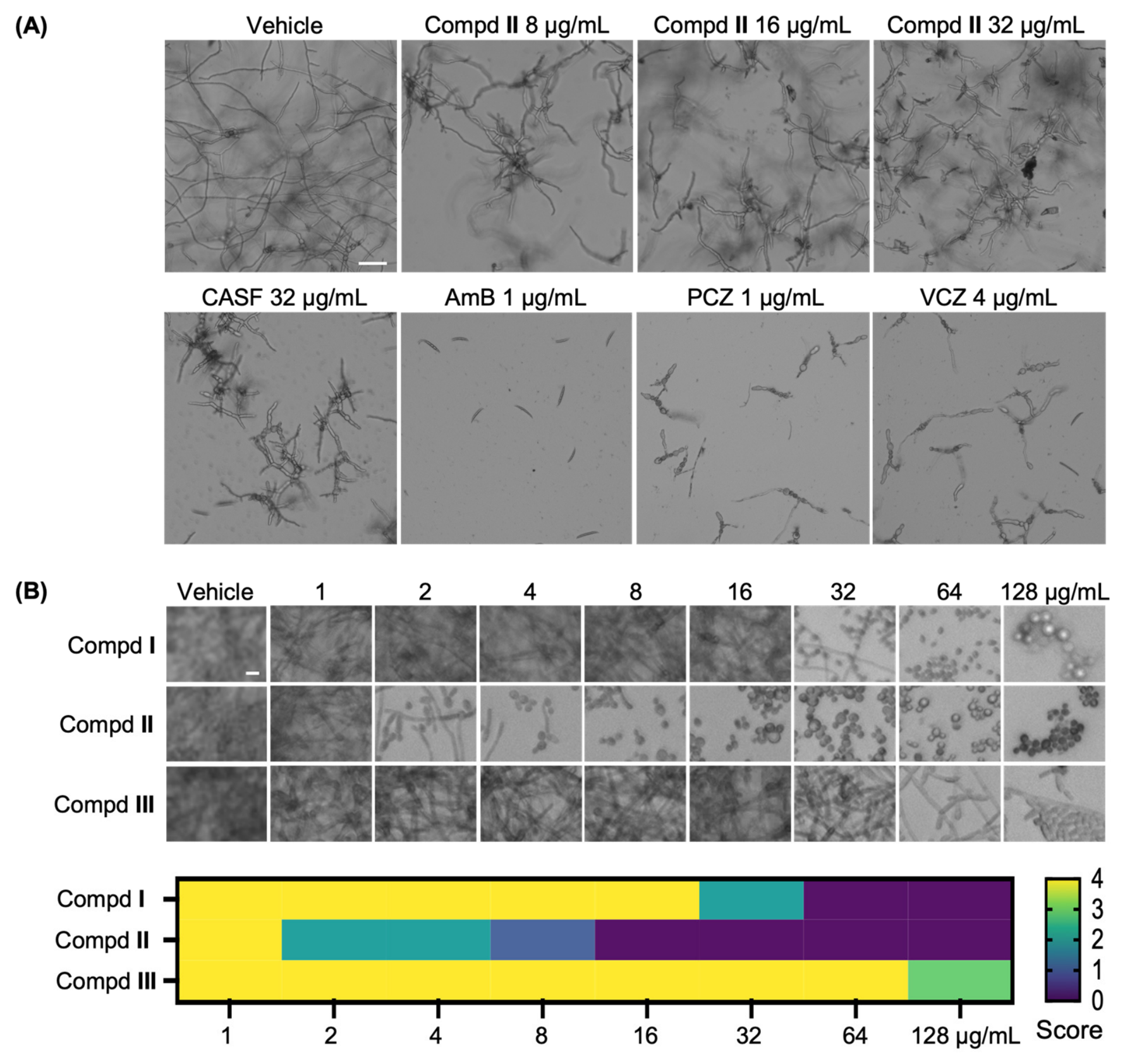
To characterize the antifungal and morphogenetic effects of compound II, we examined its effects on fungal growth and morphology and compared its morphological effects with those of standard antifungal agents representing the three major classes: the polyene amphotericin B, the echinocandin caspofungin, and triazole antifungals (fluconazole, itraconazole, voriconazole, and posaconazole). F. asiaticum exhibited morphological alterations following treatment with the polyene amphotericin B (1 μg/mL) and the triazoles posaconazole and voriconazole (1 μg/mL and 4 μg/mL, respectively). Compound II was further examined at 8–32 μg/mL to characterize its effects on fungal morphology. Compound II induced distinct morphological changes in F. asiaticum, including reduced hyphal extension and aberrant branching (Figure 1A). Exploratory image analysis provided additional descriptive support for these observations, with shorter hyphal segments numerically observed following compound II treatment, particularly at 32 μg/mL (Table S1).
Figure 1.
Morphological changes in fungal cells following treatment with phytosphingosine derivatives and conventional antifungal agents. (A) Fusarium asiaticum filamentous morphology under treatment with compound II (top row) and standard antifungal agents (bottom row), including caspofungin (CASF), amphotericin B (AmB), posaconazole (PCZ), and voriconazole (VCZ), at selected concentrations associated with visible growth suppression or morphological alterations. Scale bar = 100 μm. (B) Concentration-dependent changes in the morphology of adherent Candida albicans populations following treatment with compounds I–III. Representative microscopic images were acquired after 48 h of treatment and subsequent washing. Filamentous morphology was evaluated using predefined criteria, with cells bearing a germ tube or filament longer than the major axis of the associated mother cell classified as filamentation-positive. The accompanying heatmap shows semiquantitative filamentation scores based on the prevalence of filamentation-positive cells: 0, absent or <1%; 1, 1–9%; 2, 10–49%; 3, 50–74%; and 4, ≥75%. Scale bar = 10 μm.
To further assess whether these effects extend beyond filamentous plant pathogens, we also examined Candida albicans as a representative dimorphic human pathogen in which hyphal transition and biofilm formation are tightly linked to pathogenicity. Following 48 h of compound II exposure and subsequent washing, the remaining adherent C. albicans populations exhibited a concentration-dependent morphological shift from filament-rich growth toward predominantly yeast-form cells, with detectable changes observed at concentrations as low as 2 μg/mL (Figure 1B). Filamentous morphology was evaluated using predefined morphological criteria: cells bearing a germ tube or filament longer than the major axis of the associated mother cell were classified as filamentation-positive. The prevalence of filamentation-positive cells was subsequently expressed using a semiquantitative filamentation score ranging from 0 to 4. Compound II produced a detectable reduction in filamentous morphology at 2 μg/mL, a progressive shift toward yeast-form predominance at 4–8 μg/mL, and nearly complete absence of filamentous cells among the remaining adherent population at concentrations of ≥16 μg/mL. Comparison with the related compounds revealed substantially weaker effects for compound I, which partially reduced filamentation at 32 μg/mL and nearly completely suppressed it at ≥64 μg/mL. In contrast, compound III showed no clear reduction in filamentous morphology at concentrations up to 128 μg/mL. These findings demonstrate differences in the morphological responses of adherent C. albicans populations to the three phytosphingosine derivatives, with compound II exhibiting the strongest reduction in filamentous morphology (Figure 1B). Together with the morphological alterations observed in F. asiaticum, these results suggest that compound II affects fungal morphology across different fungal species, although the underlying molecular mechanisms remain to be elucidated.
3.6. In Silico Antifungal Activity of Compound II
To generate structural hypotheses regarding the potential mechanism of action of compound II, which exhibited significant antifungal efficacy in both in vitro and in vivo assays, molecular docking studies were conducted against five key antifungal target enzymes. These candidates represent distinct functional categories critical to fungal survival and pathogenicity. Chitinase and sterol 14α-demethylase are essential for cell wall and membrane integrity [41,42]. Specifically, chitin serves as a key structural component of the fungal cell wall, and targeting chitin metabolism provides a potential antifungal strategy [41,43]. Sterol 14α-demethylase mediates ergosterol biosynthesis, maintaining membrane fluidity [42]. Endoglucanase I and endopolygalacturonase (pectinase) facilitate host invasion by degrading plant cell wall components-catalyzing the cleavage of β-1,4-glycosidic bonds in cellulose and hydrolyzing polygalacturonate in pectin, respectively [44,45]. N-myristoyltransferase (NMT) catalyzes the myristoylation of proteins involved in fungal signal transduction and vegetative growth [46]. NMT was considered a candidate molecular target because of its important role in fungal growth, well-characterized ligand-binding pocket, and established precedent for inhibition by small molecules. In particular, crystallographic studies of Candida albicans NMT–inhibitor complexes and structure-based optimization of benzofuran-derived inhibitors have demonstrated its potential as a druggable antifungal target [47,48]. These findings provided a rationale for including NMT among the candidate enzymes investigated in the present molecular docking study. These enzymes were therefore selected to explore potential binding interactions of compound II with functionally distinct fungal proteins.
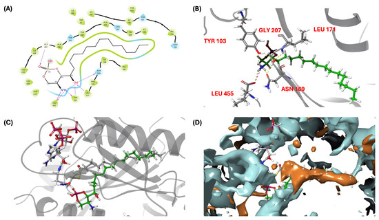
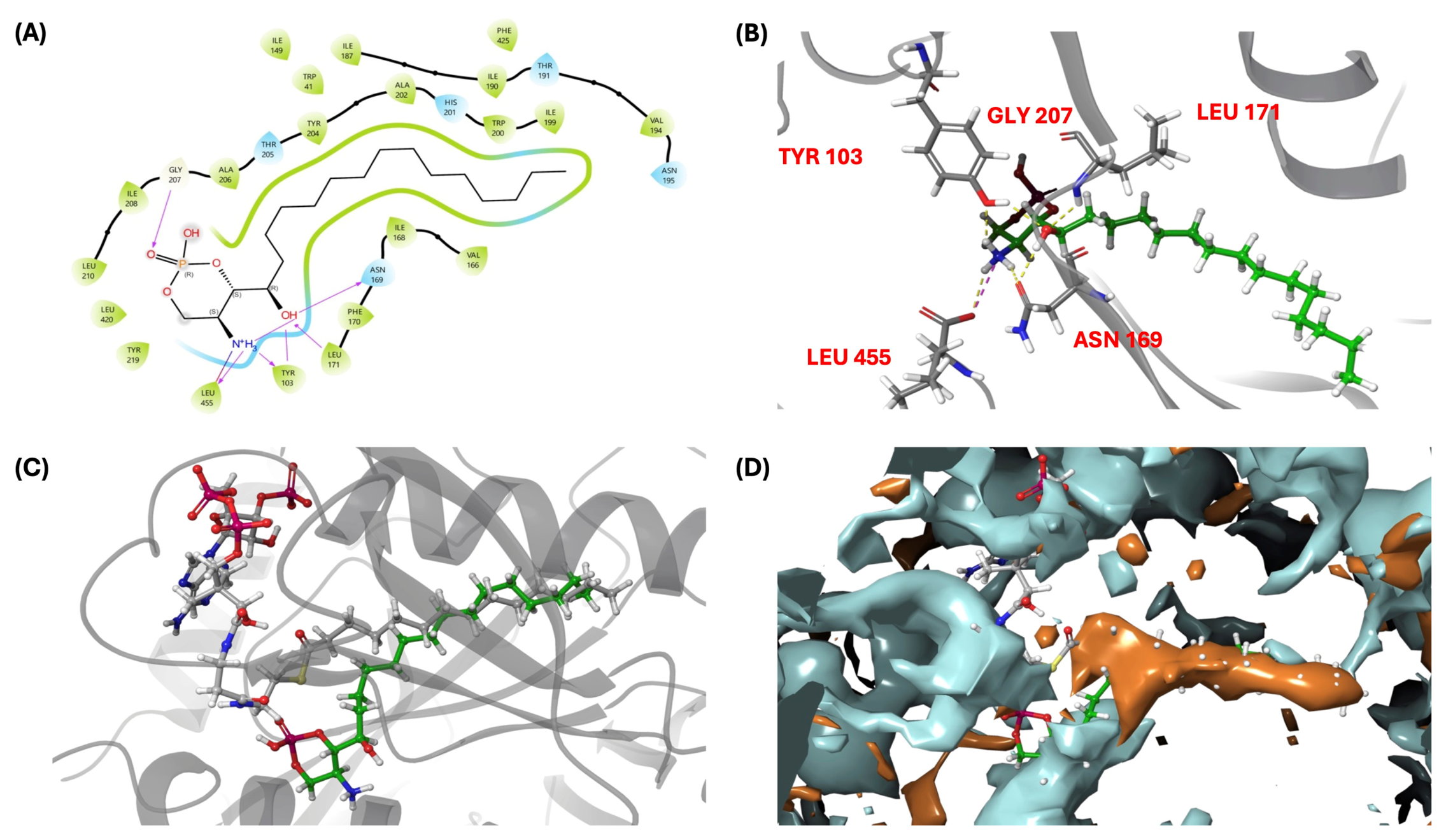
Among the five target enzymes, compound II exhibited the most favorable Glide docking score (−9.5 kcal/mol) and MM-GBSA estimated binding energy (−111.7 kcal/mol) for NMT (Table 4). However, compared with the co-crystallized ligand, compound II showed a less favorable Glide docking score (−9.5 vs. −13.8 kcal/mol), whereas its MM-GBSA estimated binding energy was marginally more favorable (−111.7 vs. −109.4 kcal/mol). This discrepancy may arise from differences in the scoring functions, particularly their treatment of solvation and receptor–ligand interactions. Given the inherent approximations and uncertainties associated with these computational methods, the observed rank-order divergence precludes a definitive conclusion regarding the relative binding affinities of compound II and the reference ligand. Therefore, these results should be interpreted as consistent with a potential interaction with NMT, rather than demonstrating superior binding affinity or direct NMT inhibition. Compound II shares similar structural features with myristoyl-CoA, particularly the presence of a long aliphatic chain. Accordingly, the results of molecular docking analysis predicted that the aliphatic chain of compound II could occupy the hydrophobic pocket of NMT and interact with residues lining the myristoyl-CoA binding site (Figure 2A,B). The docking pose of compound II aligns well with the acyl chain-binding region of myristoyl-CoA, which suggests it may occupy the same hydrophobic pocket engaged by the native acyl chain (Figure 2C,D). Although compounds I and III also exhibited similar binding modes due to their extended hydrophobic chains (Figure S24), only compound II was predicted to engage in H-bonding with LEU171 (Figure 2B), a conserved key residue that contributes to the oxyanion hole, thereby replicating a critical interaction of the native acyl donor [49]. This may contribute to a more favorable binding orientation of compound II and could be associated with the conformational rigidity of the cyclic phosphate moiety, compared to the linear phosphates in compounds I and III. To test whether the static docking poses and putative hydrogen bonds remain stable over time, we performed molecular dynamics (MD) simulations in an equilibrated system.
Table 4.
Docking scores and MM-GBSA estimated binding energies of docked poses of compound II, including reference ligand in complex with chitinase (PDB ID: 4txe), endoglucanase I (PDB ID: 2ovw), transferase (N-myristoyltransferase, PDB ID: 2p6g), demethylase (sterol 14α-demethylase (CYP51), PDB ID: 5eah), and pectinase (endopolygalacturonase, PDB ID: 1czf).
Figure 2.
Binding interactions and conformations of compound II within N-myristoyltransferase (NMT, PDB ID: 2P6G). (A) 2D interaction diagram of compound II in complex with NMT. H-bonds are shown as pink arrows, hydrophobic contacts as green arcs, and polar residues as blue. (B) 3D interaction diagram highlighting key residues involved in H-bonds between compound II and NMT. (C) Binding conformations of compound II and native myristoyl-CoA (MYA) within the NMT active site. (D) Hydrophobic and hydrophilic surface representation of NMT in complex with compound II and MYA. Hydrophilic regions are shown in blue, and hydrophobic regions are shown in orange.
3.7. Molecular Dynamics Simulation
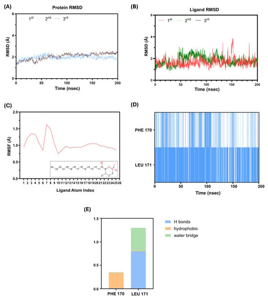
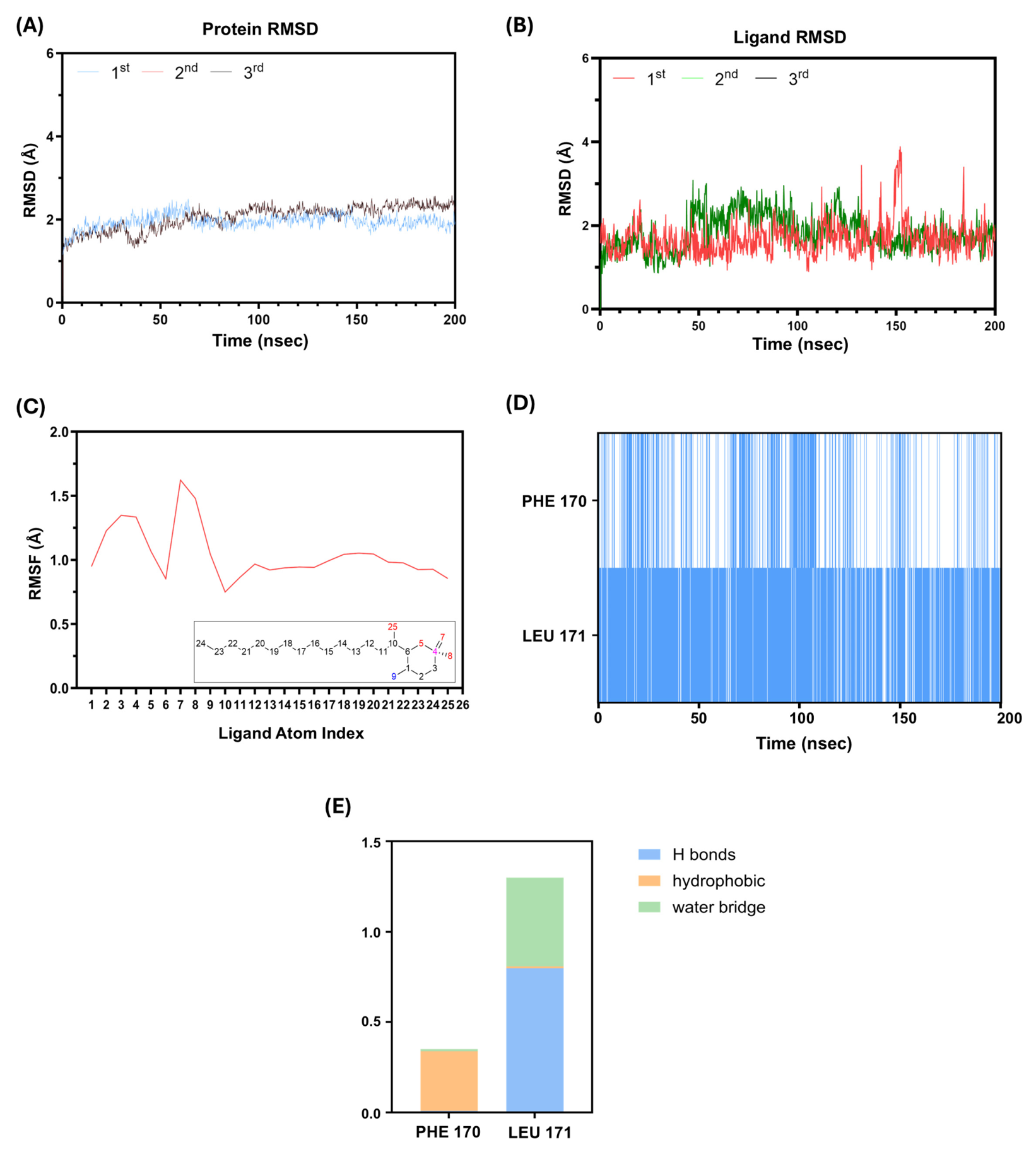
Molecular dynamics (MD) simulations of compounds I–III in complex with N-myristoyltransferase (NMT) were conducted over 200 ns to investigate the dynamic behavior and stability of the predicted protein–ligand complexes. To assess the reproducibility of the results for compound II, two additional independent 200 ns simulations were performed, yielding three independent trajectories (n = 3). Across these simulations, the protein RMSD generally remained within 1–3 Å, suggesting consistent overall structural stability of NMT under the simulated conditions. In the original trajectory, the ligand RMSD exhibited a brief increase to approximately 3.80 Å near 150 ns before returning toward its previous range. The two additional trajectories showed relatively stable ligand RMSD profiles without comparable fluctuations (Figure 3B). Although these observations support the reproducibility of the overall conformational behavior, the transient RMSD excursion cannot be conclusively attributed to a specific conformational event without further structural analysis.
Figure 3.
MD simulation results of compound II in complex with NMT over 200 ns. (A) Protein root mean square deviation (RMSD) profiles of the NMT–compound II complex. (B) Ligand RMSD profiles of compound II. (C) Root mean square fluctuation (RMSF) of compound II. (D) Timeline representation of the protein–ligand interactions and contacts involving residues PHE170 and LEU171 of NMT. (E) Stacked bar chart summarizing the normalized interaction fractions of PHE170 and LEU171 over the course of the trajectory.
In the original trajectory, the ligand-contacting residues exhibited a mean RMSF of 0.85 Å, suggesting relatively limited flexibility within the binding pocket (Figure S25A). In contrast, atoms 7–8 of compound II displayed greater conformational flexibility (Figure 3C). Compounds I and III exhibited greater RMSD and RMSFs than compound II in the simulations examined (Figure S26A–D), potentially reflecting the greater conformational flexibility of their acyclic phosphate-containing regions.
Interaction analysis further revealed that compound II maintained contacts with PHE170 and LEU171, residues associated with the NMT oxyanion hole [49]. In the original 200 ns trajectory, PHE170 and LEU171 exhibited contact occupancies of 34.23% and 88.82%, respectively (Figure 3D,E). These interactions included a π–cation interaction involving PHE170 and hydrogen-bonding or water-bridge interactions involving LEU171. In comparison, compounds I and III showed lower LEU171 contact occupancies, with no detectable PHE170 contacts under the analyzed simulation conditions (Figure S26E). In addition, the long aliphatic chain of compound II maintained hydrophobic interactions within the NMT binding cavity, with individual residue contact occupancies ranging from 23.85% to 52.55% (Figure S25B).
Collectively, these computational results suggest that compound II may adopt a relatively stable binding orientation within the NMT active site, supported by interactions involving the hydrophobic pocket and residues associated with the oxyanion hole. Although these findings provide a preliminary structural hypothesis for a potential interaction between compound II and NMT, they do not establish NMT inhibition as its mechanism of action or explain the observed differences in antifungal potency among the compounds. Further biochemical and genetic validation will be necessary to determine whether NMT contributes to the antifungal activity of compound II.
4. Conclusions
In summary, phytosphingosine-1-phosphate salt derivatives (I–III) were efficiently synthesized via tailored phosphorylation and cyclization strategies. Evaluation of the antifungal activities of compounds I–III demonstrated the potential of compound II as a protective agent against fungal pathogens such as F. asiaticum. Compound II altered hyphal morphology in F. asiaticum and reduced filamentous morphology in adherent C. albicans populations, highlighting its impact on fungal morphology. Furthermore, its dose-dependent efficacy in suppressing fungal growth in infected apples supports its potential application as an agricultural fungicide. In addition to these phenotypic effects, computational modeling suggests that compound II may interact with N-myristoyltransferase (NMT) through predicted hydrophobic and hydrogen-bonding interactions, offering a plausible hypothesis for its potential molecular target. Future research should prioritize biochemical and genetic validation, including recombinant NMT enzymatic inhibition assays and competitive binding assays involving myristoyl-CoA, to determine whether NMT contributes to the antifungal activity of compound II.
Supplementary Materials
The following supporting information can be downloaded at: https://www.mdpi.com/article/10.3390/life16111829/s1, Figure S1: 1H NMR spectrum of compound 1 in CDCl3, 400 MHz; Figure S2: 13C NMR spectrum of compound 1 in CDCl3, 100 MHz; Figure S3: 1H NMR spectrum of compound 2 in CDCl3, 400 MHz; Figure S4: 13C NMR spectrum of compound 2 in CDCl3, 100 MHz; Figure S5: 1H NMR spectrum of compound PhS1P in CD3OD–CD3COOD (1:1), 400 MHz; Figure S6: 13C NMR spectrum of compound PhS1P in CD3OD–CD3COOD (1:1), 100 MHz; Figure S7: 31P NMR spectrum of compound PhS1P in CD3OD–CD3COOD (1:1), 162 MHz; Figure S8: 1H NMR spectrum of compound I in CD3OD–CD3COOD (1:1), 400 MHz; Figure S9: 13C NMR spectrum of compound I in CD3OD–CD3COOD (1:1), 100 MHz; Figure S10: 31P NMR spectrum of compound I in CD3OD–CD3COOD (1:1), 162 MHz; Figure S11: 1H NMR spectrum of compound 3 in CDCl3, 400 MHz; Figure S12: 13C NMR spectrum of compound 3 in CDCl3, 100 MHz; Figure S13: 1H NMR spectrum of compound II in CD3OD, 400 MHz; Figure S14: 13C NMR spectrum of compound II in CD3OD, 100 MHz; Figure S15: 31P NMR spectrum of compound II in CD3OD, 162 MHz; Figure S16: 1H NMR spectrum of compound 5 in CDCl3, 400 MHz; Figure S17: 13C NMR spectrum of compound 5 in CDCl3, 100 MHz; Figure S18: 1H NMR spectrum of compound III in CD3OD, 400 MHz; Figure S19: 13C NMR spectrum of compound III in CD3OD, 100 MHz; Figure S20: 31P NMR spectrum of compound III in CD3OD, 162 MHz; Figure S21: HR-ESIMS of compound I; Figure S22: HR-ESIMS of compound II; Figure S23: HR-ESIMS of compound III; Figure S24: Binding interactions and conformations of compounds I and III within N-myristoyltransferase (NMT, PDB ID: 2P6G); Figure S25: Additional MD simulation results of compound II in complex with NMT over 200 ns; Figure S26: MD simulation results of compounds I and III in complex with NMT over 200 ns; Figure S27: Cytotoxicity of compound II in 3T3-L1 fibroblast and HEK293A kidney cells; Figure S28: Dose–response curves of the tested compounds and benomyl against four phytopathogenic fungi; Table S1. Quantitative analysis of hyphal segment lengths following compound II treatment.
Author Contributions
Conceptualization, Y.L. and K.H.K.; methodology, S.Y.L., K.L., B.J., E.J.H. and S.L. (Seeun Lim); formal analysis, B.J., E.J.H., S.L. (Seeun Lim), E.-S.K., K.-T.L., S.L. (Seulah Lee), Y.L. and K.H.K.; investigation, S.Y.L., K.L., B.J., E.J.H. and S.L. (Seeun Lim); resources, K.L. and Y.L.; data curation, E.-S.K., K.-T.L. and S.L. (Seulah Lee); writing—original draft preparation, S.Y.L., K.L., K.-T.L., S.L. (Seulah Lee), Y.L. and K.H.K.; writing—review and editing, K.-T.L., S.L. (Seulah Lee), Y.L. and K.H.K.; visualization, K.-T.L., S.L. (Seulah Lee), Y.L. and K.H.K.; supervision, Y.L. and K.H.K.; project administration, Y.L. and K.H.K. All authors have read and agreed to the published version of the manuscript.
Funding
This work was supported by National Research Foundation of Korea (NRF) grants funded by the Korean government (MSIT; grant numbers RS-2022-NR072215). This work was also partially supported by a Research Grant from Kwangwoon University in 2025. This work was also supported by the Creative-Pioneering Researchers Program through Seoul National University (800-20240448). This research was supported by the National Research Foundation of Korea (NRF) grant funded by the Korea government (MSIT) (RS-2024-00440614). This work was carried out with the support of “Research Program for Agriculture Science and Technology Development (Project No. RS-2026-25518677)” Rural Development Administration, Republic of Korea.
Institutional Review Board Statement
Not applicable.
Informed Consent Statement
Not applicable.
Data Availability Statement
The original contributions presented in this study are included in the article and Supplementary Materials. Further inquiries can be directed to the corresponding authors.
Conflicts of Interest
The authors declare no conflicts of interest.
References
- Hyde, K.D.; Cai, L.; McKenzie, E.H.C.; Yang, Y.L.; Zhang, J.Z.; Prihastuti, H. Colletotrichum: A catalogue of confusion. Fungal Divers. 2009, 39, 1–17. [Google Scholar]
- Ekwomadu, T.I.; Mwanza, M. Fusarium fungi pathogens, identification, adverse effects, disease management, and global food security: A review of the latest research. Agriculture 2023, 13, 1810. [Google Scholar] [CrossRef] [Scilit]
- Fillinger, S.; Elad, Y. (Eds.) Botrytis—The Fungus, the Pathogen and Its Management in Agricultural Systems; Springer International Publishing: Cham, Switzerland, 2016. [Google Scholar]
- Durán-Patrón, R.; Cantoral, J.M.; Hernández-Galán, R.; Hanson, J.R.; Collado, I.G. The biodegradation of the phytotoxic metabolite botrydial by its parent organism, Botrytis cinerea. J. Chem. Res. 2004, 2004, 441–443. [Google Scholar] [CrossRef] [Scilit]
- Deighton, N.; Muckenschnabel, I.; Colmenares, A.J.; Collado, I.G.; Williamson, B. Botrydial is produced in plant tissues infected by Botrytis cinerea. Phytochemistry 2001, 57, 689–692. [Google Scholar] [CrossRef] [Scilit] [PubMed]
- Rossi, F.R.; Gárriz, A.; Marina, M.; Romero, F.M.; Gonzalez, M.E.; Collado, I.G.; Pieckenstain, F.L. The sesquiterpene botrydial produced by Botrytis cinerea induces the hypersensitive response on plant tissues and its action is modulated by salicylic acid and jasmonic acid signaling. Mol. Plant-Microbe Interact. 2011, 24, 888–896. [Google Scholar] [CrossRef] [Scilit] [PubMed]
- Chen, X.; Liu, H.; Dong, L.; Shi, J.; Ma, Z.; Yan, L.; Yin, Y. Identification and characterization of Fusarium incarnatum causing leaf spot and fruit rot on luffa in China. Plants 2025, 14, 845. [Google Scholar] [CrossRef] [Scilit] [PubMed]
- Qiu, J.; Yu, M.; Yin, Q.; Xu, J.; Shi, J. Molecular characterization, fitness, and mycotoxin production of Fusarium asiaticum strains resistant to fludioxonil. Plant Dis. 2018, 102, 1759–1765. [Google Scholar] [CrossRef] [Scilit] [PubMed]
- Godoy, P.; Nunes, F.; Silva, V.; Tomimori-Yamashita, J.; Zaror, L.; Fischman, O. Onychomycosis caused by Fusarium solani and Fusarium oxysporum in Sao Paulo, Brazil. Mycopathologia 2004, 157, 287–290. [Google Scholar] [CrossRef] [Scilit] [PubMed]
- Cheng, X.; Zhang, J.; Liang, Z.; Wu, Z.; Liu, P.; Hao, J.; Liu, X. Multidrug resistance of Rhizoctonia solani determined by enhanced efflux for fungicides. Pestic. Biochem. Physiol. 2023, 195, 105525. [Google Scholar] [CrossRef] [Scilit] [PubMed]
- Zhang, C.; Liu, Y.; Ma, X.; Feng, Z.; Ma, Z. Characterization of sensitivity of Rhizoctonia solani, causing rice sheath blight, to mepronil and boscalid. Crop Prot. 2009, 28, 381–386. [Google Scholar] [CrossRef] [Scilit]
- Howell, A.R.; Ndakala, A.J. The preparation and biological significance of phytosphingosines. Curr. Org. Chem. 2002, 6, 365–391. [Google Scholar] [CrossRef] [Scilit]
- Dickson, R.C. Sphingolipid functions in Saccharomyces cerevisiae: Comparison to mammals. Annu. Rev. Biochem. 1998, 67, 27–48. [Google Scholar] [CrossRef] [Scilit] [PubMed]
- Mashima, R.; Okuyama, T.; Ohira, M. Biosynthesis of long chain base in sphingolipids in animals, plants and fungi. Future Sci. OA 2020, 6, FSO434. [Google Scholar] [CrossRef] [Scilit] [PubMed]
- Pata, M.O.; Hannun, Y.A.; Ng, C.K.Y. Plant sphingolipids: Decoding the enigma of the Sphinx. New Phytol. 2010, 185, 611–630. [Google Scholar] [CrossRef] [Scilit] [PubMed]
- Coursol, S.; Le Stunff, H.; Lynch, D.V.; Gilroy, S.; Assmann, S.M.; Spiegel, S. Arabidopsis sphingosine kinase and the effects of phytosphingosine-1-phosphate on stomatal aperture. Plant Physiol. 2005, 137, 724–737. [Google Scholar] [CrossRef] [Scilit] [PubMed]
- Cowart, L.A.; Shotwell, M.; Worley, M.L.; Richards, A.J.; Montefusco, D.J.; Hannun, Y.A.; Lu, X. Revealing a signaling role of phytosphingosine-1-phosphate in yeast. Mol. Syst. Biol. 2010, 6, 349. [Google Scholar] [CrossRef] [Scilit] [PubMed]
- Maceyka, M.; Milstien, S.; Spiegel, S. Sphingosine kinases, sphingosine-1-phosphate and sphingolipidomics. Prostaglandins Other Lipid Mediat. 2005, 77, 15–22. [Google Scholar] [CrossRef] [Scilit] [PubMed]
- Wu, J.; Qin, X.; Tao, S.; Jiang, X.; Liang, Y.K.; Zhang, S. Long-chain base phosphates modulate pollen tube growth via channel-mediated influx of calcium. Plant J. 2014, 79, 507–516. [Google Scholar] [CrossRef] [Scilit] [PubMed]
- Gao, Y.; He, X.; Ding, F.; Zhang, Y. Recent progress in chemical syntheses of sphingosines and phytosphingosines. Synthesis 2016, 48, 4017–4037. [Google Scholar] [CrossRef] [Scilit]
- Jo, S.-Y.; Kim, H.-C.; Woo, S.-W.; Seo, M.-J.; Lee, G.-H.; Kim, H.-R. Synthesis of 1-substituted-phytosphingosine: Novel protection of phytosphingosine. Bull. Korean Chem. Soc. 2003, 24, 267–268. [Google Scholar] [CrossRef] [Scilit]
- Li, S.; Wilson, W.K.; Schroepfer, G.J., Jr. Chemical synthesis of D-ribo-phytosphingosine-1-phosphate, a potential modulator of cellular processes. J. Lipid Res. 1999, 40, 117–125. [Google Scholar] [CrossRef] [Scilit]
- Bridget, A.F.; Budhathoki, R.; Huo, C.; Joshi, S.; Parajuli, N.; Sohng, J.K.; Kim, K.H. Activation of cryptic biosynthetic pathways in Saccharopolyspora spinosa through deletion of the spinosyn gene cluster: Induction of cryptic and bioactive natural products. Arch. Pharm. Res. 2025, 48, 514–527. [Google Scholar] [CrossRef] [Scilit] [PubMed]
- Cho, C.H.; Chae, S.H.; Kim, S.H.; Kim, K.H. Phenolic compounds isolated from Juncus decipiens and their effects on osteoblast differentiation in the mouse mesenchymal stem cell line C3H10T1/2. Nat. Prod. Sci. 2024, 30, 135–142. [Google Scholar] [CrossRef] [Scilit]
- Gil, T.-Y.; Kim, H.-J.; Kim, H.-M.; Sim, H.-Y.; Choi, W.; Lee, B.S.; Kim, K.H.; An, H.-J. Aster glehni ethanol extract inhibits inflammatory responses regulating skin barrier molecules in human keratinocytes. Nat. Prod. Sci. 2024, 30, 262–267. [Google Scholar] [CrossRef] [Scilit]
- Glick, V.J.; Webber, C.A.; Simmons, L.E.; Martin, M.C.; Ahmad, M.; Kim, C.H.; Adams, A.N.D.; Bang, S.; Chao, M.C.; Howard, N.C.; et al. Vaginal lactobacilli produce anti-inflammatory β-carboline compounds. Cell Host Microbe 2024, 32, 1897–1909.e7. [Google Scholar] [CrossRef] [Scilit] [PubMed]
- Kim, Y.; Chae, S.H.; Lee, D.; Lee, B.S.; Lim, J.; Jung, H.I.; Kim, K.H.; Kwak, B. Alternative models for anticancer drug discovery from natural products using binary tumor-microenvironment-on-a-chip. Adv. Sci. 2025, 12, e07944. [Google Scholar] [CrossRef] [Scilit] [PubMed]
- Lee, S.; Jang, M.; Ryoo, R.; Roh, J.; Ko, S.-K.; Kim, K.H. New autophagy-modulating lanostane-type triterpenoids from a hallucinogenic poisonous mushroom Gymnopilus orientispectabilis. Arch. Pharm. Res. 2024, 47, 272–287. [Google Scholar] [CrossRef] [Scilit] [PubMed]
- van den Berg, R.J.; Boltje, T.J.; Verhagen, C.P.; Litjens, R.E.; van der Marel, G.A.; Overkleeft, H.S. An efficient synthesis of the natural tetrahydrofuran pachastrissamine starting from D-ribo-phytosphingosine. J. Org. Chem. 2006, 71, 836–839. [Google Scholar] [CrossRef] [Scilit] [PubMed]
- Alcaide, A.; Llebaria, A. Aziridine ring opening for the synthesis of sphingolipid analogues: Inhibitors of sphingolipid-metabolizing enzymes. J. Org. Chem. 2014, 79, 2993–3029. [Google Scholar] [CrossRef] [Scilit] [PubMed]
- Sanllehí, P.; Abad, J.-L.; Bujons, J.; Casas, J.; Delgado, A. Studies on the inhibition of sphingosine-1-phosphate lyase by stabilized reaction intermediates and stereodefined azido phosphates. Eur. J. Med. Chem. 2016, 123, 905–915. [Google Scholar] [CrossRef] [Scilit] [PubMed]
- Fokialakis, N.; Cantrell, C.L.; Duke, S.O.; Skaltsounis, A.L.; Wedge, D.E. Antifungal activity of thiophenes from Echinops ritro. J. Agric. Food Chem. 2006, 54, 1651–1655. [Google Scholar] [CrossRef] [Scilit] [PubMed]
- Lee, K.; Park, J.H.; Jo, S.M.; Park, Y.; Kang, T.J. Corchorus olitorius ethanolic extract has anti-inflammatory and wound healing effects in vitro. Nat. Prod. Sci. 2023, 29, 291–296. [Google Scholar] [CrossRef] [Scilit]
- Bai, Y.-B.; Gao, Y.-Q.; Nie, X.-D.; Tuong, T.-M.-L.; Li, D.; Gao, J.-M. Antifungal activity of griseofulvin derivatives against phytopathogenic fungi in vitro and in vivo and three-dimensional quantitative structure–activity relationship analysis. J. Agric. Food Chem. 2019, 67, 6125–6132. [Google Scholar] [CrossRef] [Scilit] [PubMed]
- Schrödinger. Maestro, Schrödinger Release 2025-2; Schrödinger, LLC: New York, NY, USA, 2025; Available online: https://www.schrodinger.com/platform/products/maestro/ (accessed on 31 July 2026).
- Schrödinger. LigPrep, Schrödinger Release 2025-2; Schrödinger, LLC: New York, NY, USA, 2025; Available online: https://www.schrodinger.com/platform/products/ligprep/ (accessed on 31 July 2026).
- Schrödinger. Glide, Schrödinger Release 2025-2; Schrödinger, LLC: New York, NY, USA, 2025; Available online: https://www.schrodinger.com/platform/products/glide/ (accessed on 31 July 2026).
- Schrödinger. Desmond Molecular Dynamics System, Schrödinger Release 2025-4; Maestro-Desmond Interoperability Tools; Schrödinger, LLC: New York, NY, USA, 2025; Available online: https://www.schrodinger.com/platform/products/desmond/ (accessed on 7 August 2026).
- Zhu, G.-Y.; Chen, Y.; Wang, S.-Y.; Shi, X.-C.; Herrera-Balandrano, D.D.; Polo, V.; Laborda, P. Peel diffusion and antifungal efficacy of different fungicides in pear fruit: Structure-diffusion-activity relationships. J. Fungi 2022, 8, 547. [Google Scholar] [CrossRef] [Scilit] [PubMed]
- Shu, C.; Jiao, W.; Cui, K.; Cao, J.; Jiang, W. Ursolic acid induces multifaceted defense responses against postharvest blue mold rot in apple fruit. Foods 2025, 14, 761. [Google Scholar] [CrossRef] [Scilit] [PubMed]
- Gastebois, A.; Clavaud, C.; Aimanianda, V.; Latgé, J.-P. Aspergillus fumigatus: Cell wall polysaccharides, their biosynthesis and organization. Future Microbiol. 2009, 4, 583–595. [Google Scholar] [CrossRef] [Scilit] [PubMed]
- Zhang, R.; Wang, Y.; Wu, A.; Wang, J.; Zhang, J. Strategies of targeting CYP51 for IFIs therapy: Emerging prospects, opportunities and challenges. Eur. J. Med. Chem. 2023, 259, 115658. [Google Scholar] [CrossRef] [Scilit] [PubMed]
- Ruiz-Herrera, J.; San-Blas, G. Chitin synthesis as a target for antifungal drugs. Curr. Drug Targets Infect. Disord. 2003, 3, 77–91. [Google Scholar] [CrossRef] [Scilit] [PubMed]
- van Santen, Y.; Benen, J.A.; Schröter, K.-H.; Kalk, K.H.; Armand, S.; Visser, J.; Dijkstra, B.W. 1.68-Å crystal structure of endopolygalacturonase II from Aspergillus niger and identification of active site residues by site-directed mutagenesis. J. Biol. Chem. 1999, 274, 30474–30480. [Google Scholar] [CrossRef] [Scilit] [PubMed]
- Nühse, T.S. Cell wall integrity signaling and innate immunity in plants. Front. Plant Sci. 2012, 3, 280. [Google Scholar] [CrossRef] [Scilit] [PubMed]
- Wu, J.; Tao, Y.; Zhang, M.; Howard, M.H.; Gutteridge, S.; Ding, J. Crystal structures of Saccharomyces cerevisiae N-myristoyltransferase with bound myristoyl-CoA and inhibitors reveal the functional roles of the N-terminal region. J. Biol. Chem. 2007, 282, 22185–22194. [Google Scholar] [CrossRef] [Scilit] [PubMed]
- Sogabe, S.; Masubuchi, M.; Sakata, K.; Fukami, T.A.; Morikami, K.; Shiratori, Y.; Ebiike, H.; Kawasaki, K.; Aoki, Y.; Shimma, N. Crystal structures of Candida albicans N-myristoyltransferase with two distinct inhibitors. Chem. Biol. 2002, 9, 1119–1128. [Google Scholar] [CrossRef] [Scilit] [PubMed]
- Masubuchi, M.; Ebiike, H.; Kawasaki, K.-i.; Sogabe, S.; Morikami, K.; Shiratori, Y.; Tsujii, S.; Fujii, T.; Sakata, K.; Hayase, M. Synthesis and biological activities of benzofuran antifungal agents targeting fungal N-myristoyltransferase. Bioorg. Med. Chem. 2003, 11, 4463–4478. [Google Scholar] [CrossRef] [Scilit] [PubMed]
- Farazi, T.A.; Waksman, G.; Gordon, J.I. The biology and enzymology of protein N-myristoylation. J. Biol. Chem. 2001, 276, 39501–39504. [Google Scholar] [CrossRef] [Scilit] [PubMed]

Disclaimer/Publisher’s Note: The statements, opinions and data contained in all publications are solely those of the individual author(s) and contributor(s) and not of MDPI and/or the editor(s). MDPI and/or the editor(s) disclaim responsibility for any injury to people or property resulting from any ideas, methods, instructions or products referred to in the content. |
© 2026 by the authors. Licensee MDPI, Basel, Switzerland. This article is an open access article distributed under the terms and conditions of the Creative Commons Attribution (CC BY) license.