


Abstract
Lung-protective mechanical ventilation using lower airway pressures remains a cornerstone of the management of acute respiratory distress syndrome (ARDS). However, positive pressure ventilation may contribute to positive fluid balance, which has been associated with adverse clinical outcomes. We prospectively enrolled patients with ARDS and collected clinical characteristics and mechanical ventilation parameters during the first 3 days after ARDS onset. The associations between airway pressures and cumulative fluid balance (CFB) were evaluated. The analysis included 287 patients with ARDS, and the all-cause hospital mortality rate was 45.6%. Patients were divided into the positive CFB group (n = 173, 60.3%) and the negative CFB group (n = 114, 39.7%). Compared with patients in the negative CFB group, those in the positive CFB group had a higher risk of organ failure, higher white blood cell counts, higher C-reactive protein levels, more severe hypoxemia, higher peak inspiratory pressures (Ppeak), and higher hospital mortality (all p < 0.05). Multivariable logistic regression analysis showed that higher Ppeak was independently associated with increased odds of positive CFB (adjusted OR, 1.076; p = 0.011). These findings highlight the potential relationship between airway pressures and fluid accumulation during mechanical ventilation in patients with ARDS.
1. Introduction
Fluid resuscitation is crucial for restoring perfusion and maintaining hemodynamic stability in critically ill patients, including those with acute respiratory distress syndrome (ARDS). However, increased capillary permeability and reduced plasma oncotic pressure are common in critical illness and may promote fluid extravasation and tissue edema. Therefore, optimal fluid management should provide adequate oxygen delivery while avoiding excessive fluid accumulation and pulmonary or distant organ edema, which may further impair gas exchange and contribute to organ dysfunction [1,2]. Previous studies have reported that positive cumulative fluid balance (CFB) is associated with worse clinical outcomes, including a higher risk of weaning failure from mechanical ventilation and increased mortality in critically ill patients [3,4,5,6,7]. Among patients with ARDS, positive fluid balance has been associated with prolonged mechanical ventilation, longer intensive care unit (ICU) and hospital stays, and increased mortality [1,2,8,9,10]. These findings highlight the importance of maintaining an appropriate fluid balance while ensuring adequate perfusion in patients with ARDS.
ARDS is a life-threatening form of acute respiratory failure characterized by severe hypoxemia, dysregulated alveolar and systemic inflammatory responses, and increased cytokine activation. Disruption of the alveolar–capillary barrier leads to increased permeability, resulting in non-cardiogenic, protein-rich pulmonary edema and severe impairment of gas exchange [11,12]. Lung-protective mechanical ventilation strategies using lower tidal volumes and airway pressures to reduce stress and strain and minimize ventilator-induced lung injury (VILI) remain the mainstay of ARDS management and have been associated with improved survival [13,14,15,16,17].
Positive pressure ventilation can increase intrathoracic pressure, impair right ventricular function, and reduce cardiac output, potentially contributing to systemic organ congestion and hypoperfusion. Moreover, injurious mechanical ventilation settings may aggravate biotrauma and promote systemic inflammatory responses, which can contribute to distant organ dysfunction, including reduced renal blood flow and acute kidney injury (AKI). Renal dysfunction may subsequently impair fluid excretion and promote fluid accumulation [18,19,20,21,22,23].
Therefore, this study aimed to investigate the association between airway pressures and CFB during the early phase of ARDS and to identify factors associated with positive CFB in patients with ARDS.
2. Materials and Methods
2.1. Study Design and Patients
Patients admitted to the medical ICUs of Chang Gung Memorial Hospital, Linkou Branch, a tertiary referral center in Taiwan, between October 2020 and May 2026 were prospectively screened. All consecutive participants receiving invasive mechanical ventilation in medical ICUs were assessed, and patients fulfilling the Berlin criteria for ARDS were included [24]. CFB was defined as all documented cumulative total fluid input minus all documented cumulative total fluid output over the first 3 days following ARDS onset. AKI was defined according to the Kidney Disease: Improving Global Outcomes (KDIGO) criteria based on serum creatinine levels and/or urine output [25]. AKI occurrence was assessed during the first 3 days following ARDS onset. Patients with ARDS were stratified into positive CFB or negative CFB groups according to the cumulative fluid status within the first 3 days following ARDS onset, and further categorized into survivors and non-survivors based on survival status at hospital discharge. At our hospital, decisions regarding the initiation of renal replacement therapy (RRT) were determined by the primary intensivist in consultation with a nephrologist. Participants were excluded if they met any of the following conditions: (1) younger than 20 years of age; (2) pre-existing end-stage renal disease necessitating maintenance hemodialysis; (3) presentation in a moribund state accompanied by refractory multiorgan failure; (4) absence of informed consent from either the individual or their legally authorized representative; or (5) inability to obtain complete data required to calculate CFB or determine airway pressures based on ventilator settings during the first three days following ARDS onset. Patients who died, were transferred, or were discharged before completion of the three-day assessment period were excluded only if these events resulted in incomplete CFB or airway pressures data required for the primary analysis. This study protocol was reviewed and approved by the Institutional Review Board for Human Research of CGMH (CGMH IRB No. 202000760A3, 202100595A3, 202201833A3, 202300897A3, and 202401982B0) and was conducted in accordance with the Declaration of Helsinki.
2.2. Data Collection
Baseline data including demographic profiles, ARDS etiologies, preexisting comorbidities, and clinical parameters such as Sequential Organ Failure Assessment (SOFA) scores were systematically documented upon study enrollment. Arterial blood gas profiles and invasive mechanical ventilator settings parameters were collected at approximately 10:00 a.m. on days 1, 3, and 7 following ARDS onset. Pressure-controlled ventilation mode was routinely used for patients with ARDS as part of standard clinical practice at our institution. Peak inspiratory pressure (Ppeak) was obtained directly from ventilator-displayed measurements and routinely documented by respiratory therapists. Deep sedation and continuous neuromuscular blockade were not routinely administered to all patients with ARDS during the initial phase of ARDS; their use depended on clinical indications, particularly in patients with moderate or severe ARDS. Consequently, Ppeak measurements may have been influenced by spontaneous respiratory effort and airway resistance. For the primary analysis, Ppeak and other ventilatory parameters were calculated as the arithmetic mean of the measurements obtained on days 1 and 3. Daily fluid balance was derived from total fluid intake (comprising enteral/parenteral fluids, blood products, and RRT replacement solutions) minus total fluid output (including urine volume, gastrointestinal losses, drainage, RRT effluent dialysate, and estimated insensible fluid loss), and CFB was calculated over the first 3 days following ARDS onset. Accordingly, Ppeak and CFB were assessed over overlapping observation periods. Patients were classified into the positive CFB group if their CFB during the first 3 days following ARDS onset was greater than zero, whereas patients with a CFB of zero or less were classified into the negative CFB group. For patients receiving RRT, ultrafiltration volume was included as fluid output. Insensible water loss (IWL) was estimated based on body weight. For patients receiving mechanical ventilation, IWL was estimated using the following formula: body weight (kg) × 10 mL/kg/day × 0.6. The estimated IWL was incorporated into total fluid output when calculating CFB. The dataset also included the timing of ICU and hospital admission and discharge, ARDS onset, initiation and liberation from mechanical ventilation, the presence of shock, and death. Therapeutic interventions, including vasopressor use, RRT use during the ICU stay, prone positioning, and extracorporeal membrane oxygenation (ECMO) support, were also documented.
2.3. Outcome Measurements
The primary outcome was positive CFB during the first three days following ARDS onset. Secondary outcomes included 28-day, 60-day, 90-day, and all-cause hospital mortality. Hospital mortality was defined as death from any cause before discharge from the index hospitalization. Patients discharged alive were classified as survivors. Additional clinical outcomes included shock, vasopressor support, duration of mechanical ventilation, ICU length of stay, and hospital length of stay.
2.4. Statistical Analysis
Continuous variables were summarized using means with standard deviations or medians with interquartile range for normally distributed or non-normally distributed data, respectively. Categorical variables were summarized using numbers and percentages. To determine the differences between groups, Student’s t-test or Mann–Whitney U test for normally or non-normally distributed continuous variables, and the chi-square or Fisher exact test for categorical variables were conducted, as appropriate. The Jonckheere–Terpstra test was used to assess the trend in CFB across the five ordered Ppeak categories. Univariable logistic regression analyses were first performed to identify factors associated with positive CFB during the first 3 days following ARDS onset. Multivariable logistic regression was then used to evaluate the association between Ppeak and positive CFB. Covariates were selected based on clinical relevance and their potential to confound the association between airway pressures and fluid balance. All selected covariates were simultaneously entered into the multivariable model using the Enter method. Sensitivity analyses were performed using alternative model specifications to assess the robustness of the association between Ppeak and positive CFB. Effect estimates are presented as odds ratios (ORs) with 95% confidence intervals (CIs). Kaplan–Meier curves were employed to describe the cumulative incidence of 90-day hospital survival according to positive or negative CFB category, with groups compared by the log-rank test. All statistical analyses were performed in SPSS version 29.0 (IBM Inc., Armonk, NY, USA), and two-sided p-values < 0.05 were considered statistically significant.
3. Results
From a total of 7312 patients admitted to medical ICUs during the study period, a total of 287 patients with ARDS were included in the final analysis (Figure 1), with an overall all-cause in-hospital mortality rate of 45.6%. During the first 3 days after ARDS onset, positive CFB occurred in 173 patients (60.3%). Among these patients, the all-cause in-hospital mortality rate was 56.1%, compared with 29.8% among patients with negative CFB (n = 114, 39.7%).
3.1. Comparisons Between Patients with ARDS with Positive and Negative CFB
Patients with ARDS were stratified into the positive CFB group (n = 173, 60.3%) and negative CFB group (n = 114, 39.7%) based on the cumulative fluid status following the first 3 days after ARDS onset (Table 1).
No significant differences were observed in age, gender, ARDS etiology, or underlying comorbidities between patients with positive and negative CFB. Patients in the positive CFB group had significantly higher SOFA scores and levels of inflammatory markers, including white blood cell (WBC) counts and C-reactive protein (CRP), than those in the negative CFB group (all p < 0.05). Patients in the positive CFB group also had more severe hypoxemia, as reflected by lower PaO2/FiO2 ratios, than those in the negative CFB group (p = 0.033).
In terms of mechanical ventilation parameters, Ppeak was significantly higher in the positive CFB group than in the negative CFB group (p = 0.013). The incidence of AKI and the requirement for RRT were also significantly higher in the positive CFB group. No significant differences were observed between the two groups in the use of prone positioning or ECMO support.
Figure 1.
Flowchart of patient enrollment with ARDS. ARDS, acute respiratory distress syndrome; CFB, cumulative fluid balance; ICU, intensive care unit.
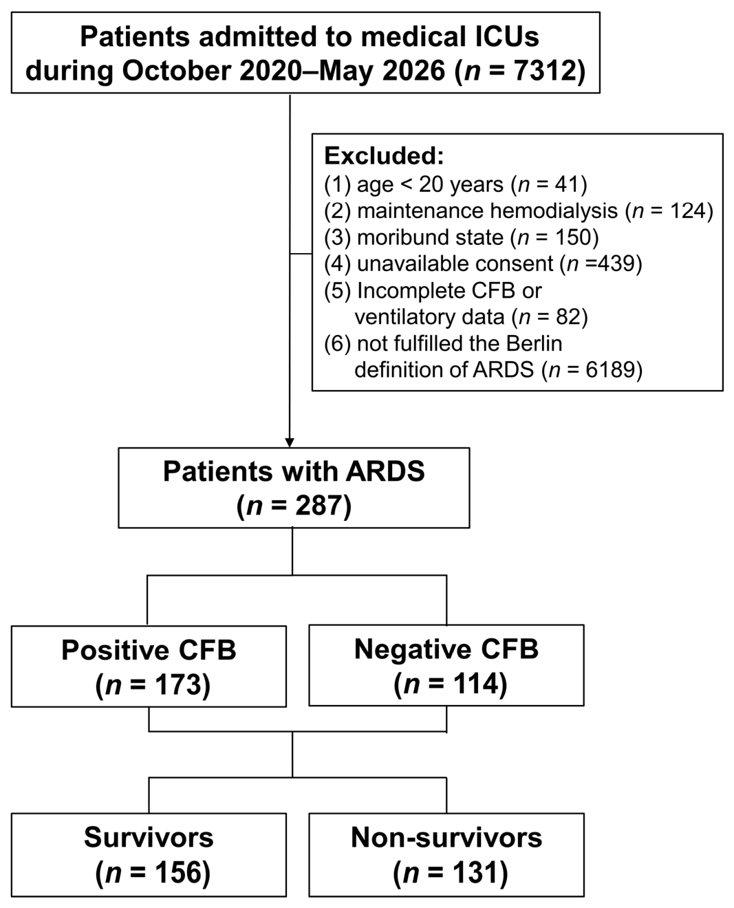
Table 1.
Baseline characteristics and clinical variables of ARDS patients: positive CFB versus negative CFB.
3.2. Distribution of Peak Inspiratory Pressures and CFB
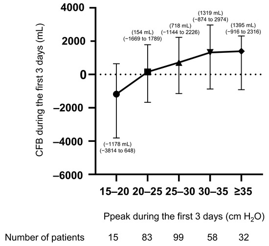
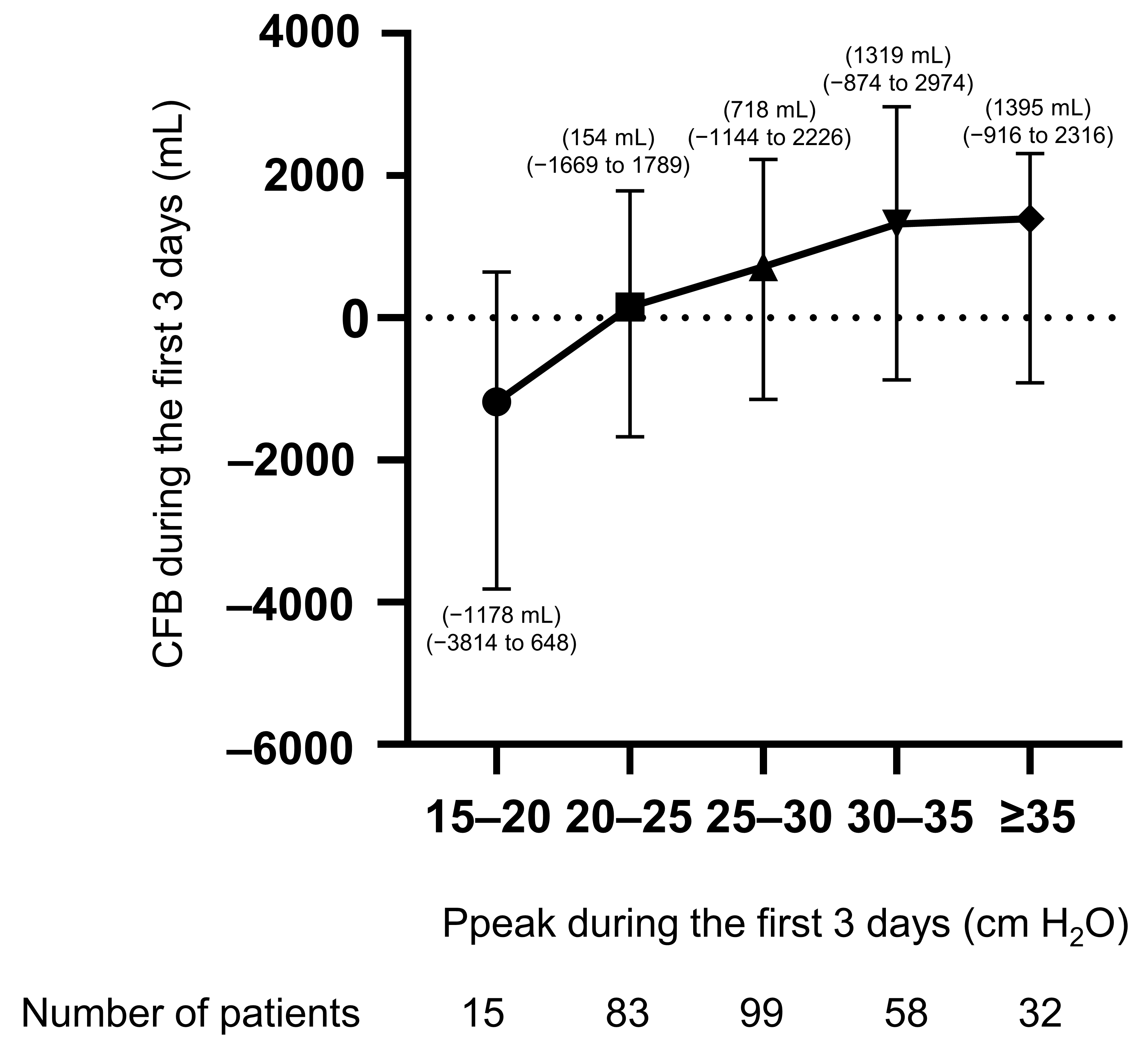
As shown in Figure 2, among a total of 287 patients with ARDS, the distribution of Ppeak and median CFB values during the first 3 days after ARDS onset was presented. An increase in Ppeak values was consistently associated with an increasing trend in the total amount of CFB. CFB increased progressively with higher Ppeak values. The median and interquartile range (IQR) of CFB was −1178 mL (IQR, −3814 to 648) among patients with a Ppeak of 15–20 cm H2O and increased to 154 mL (IQR, −1669 to 1789), 718 mL (IQR, −1144 to 2226), 1319 mL (IQR, −874 to 2974), and 1395 mL (IQR, −916 to 2316) among those with Ppeak values of 20–25, 25–30, 30–35, and ≥35 cm H2O, respectively. A Jonckheere–Terpstra test was performed to assess the trend in CFB across the five ordered Ppeak categories, and CFB showed a statistically significant increasing trend across Ppeak categories (p = 0.007).
Figure 2.
Distribution of Ppeak and median (interquartile range) values of CFB during the first 3 days of ARDS onset. CFB: cumulative fluid balance; Ppeak: peak inspiratory pressure.
3.3. Comparisons Between Survivors and Non-Survivors of Patients with ARDS
As shown in Table 2, survivors and non-survivors had similar age and gender distributions. No significant differences in the etiologies of ARDS or underlying comorbidities were observed between the two groups. Non-survivors had significantly higher SOFA scores and presented more severe hypoxemia, as reflected by lower PaO2/FiO2 ratios than did survivors (both p < 0.05).
Table 2.
Baseline characteristics and clinical variables of patients with ARDS: survivors versus non-survivors.
Regarding mechanical ventilator settings, non-survivors underwent significantly higher Ppeak and mean airway pressures, and lower dynamic compliance values than those in survivors (all p < 0.05). Non-survivors had a significantly higher incidence of AKI development and required RRT more frequently than survivors. Non-survivors exhibited a significantly higher CFB from day 1 to day 3 following ARDS onset (p < 0.001). The use of prone positioning or ECMO support revealed no significant difference between the two groups.
3.4. Clinical Outcomes of Patients with ARDS Stratified by CFB
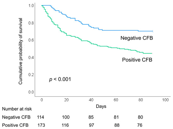
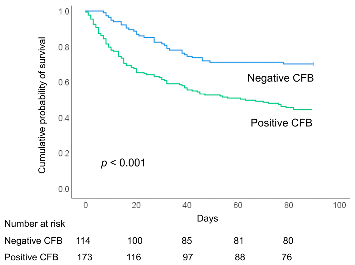
Patients in the positive CFB group had significantly higher 28-, 60-, and 90-day mortality and higher all-cause hospital mortality (all p < 0.05) (Table 3 and Figure 3). Shock and vasopressor use were also more frequent in the positive CFB group. No significant differences were observed in terms of the duration of mechanical ventilation, ICU length of stay, and total hospital length of stay.
Table 3.
Clinical outcomes as a function of CFB in ARDS patients.
Figure 3.
Kaplan–Meier 90-day survival curves for ARDS patients stratified by cumulative fluid balance status during the first 3 days of ARDS onset. ARDS: acute respiratory distress syndrome; CFB: cumulative fluid balance.
3.5. Factors Associated with Positive CFB
After adjusting for significant confounding variables, a multivariable logistic regression analysis showed that higher SOFA scores and higher Ppeak values were independently associated with positive CFB occurrence during the first 3 days of ARDS (Table 4). Specifically, patients with ARDS who underwent higher Ppeak values faced significantly increased odds of positive CFB occurrence (adjusted OR 1.078, [95% CI 1.019–1.140], p = 0.009). Sensitivity analyses were performed to assess the robustness of the association between Ppeak and positive CFB. After additional adjustment for AKI, serum creatinine at ARDS onset, or PaO2/FiO2 at ARDS onset, mean Ppeak on days 1 and 3 remained significantly associated with positive CFB. However, when Ppeak measured on day 1 was used instead of the mean Ppeak on days 1 and 3, the association was not statistically significant (Table 5).
Table 4.
Multivariable logistic regression analysis of factors associated with positive CFB in ARDS patients.
Table 5.
Sensitivity analyses evaluating the association between Ppeak and positive CFB in ARDS patients.
4. Discussion
The principal finding of this study was that higher Ppeak during the first 3 days after ARDS onset was independently associated with increased odds of positive CFB in patients with ARDS. However, the causality or temporal directionality cannot be established. Furthermore, positive CFB during the first 3 days after ARDS onset was associated with worse clinical outcomes and higher in-hospital mortality.
Lung-protective mechanical ventilation strategies using optimized tidal volumes and inspiratory pressures are evidence-based approaches to the management of ARDS and have been associated with improved survival [13,14,15]. However, excessive airway pressures may provoke pulmonary and systemic inflammation and adversely affect hemodynamics by increasing intrathoracic pressure, increasing right ventricular afterload, impairing venous return and lymphatic drainage, reducing left ventricular preload, and decreasing cardiac output. These hemodynamic effects may contribute to systemic venous congestion and impaired organ perfusion, including reduced renal blood flow, thereby increasing the risk of acute kidney injury (AKI) and fluid accumulation. AKI is common among patients receiving invasive mechanical ventilation, and its potential mechanisms include gas exchange abnormalities, such as hypoxemia and hypercapnia; biotrauma associated with ventilator-induced lung injury (VILI) and inflammatory responses; and hemodynamic disturbances leading to reduced renal perfusion [18,19,20,21,22,23,26,27,28,29].
In this study, patients in the positive CFB group had significantly higher Ppeak and higher rates of AKI and RRT requirement, as well as more frequent shock and vasopressor use, than those in the negative CFB group. These findings suggest a potential interplay between higher airway pressures, hemodynamic disturbances, and renal dysfunction during mechanical ventilation. Elevated airway pressures may increase intrathoracic pressure and impair venous return and cardiac output, potentially compromising effective renal perfusion and increasing the risk of AKI. Conversely, renal dysfunction may impair fluid excretion and thereby contribute to fluid accumulation. In addition, patients in the positive CFB group had significantly higher levels of inflammatory markers, including WBC counts and CRP, suggesting a greater degree of systemic inflammation. Increased capillary permeability and impaired lymphatic drainage during critical illness may further promote interstitial fluid accumulation and reduce the efficiency of fluid redistribution and clearance, potentially contributing to positive CFB in addition to inflammation associated with VILI [19].
In addition, the interaction between invasive mechanical ventilation and kidney injury may be bidirectional, and lung–kidney cross-talk is complex. AKI may contribute to lung injury through the accumulation of uremic toxins, disruption of acid–base balance and fluid homeostasis, and exacerbation of systemic inflammation [20,21,28,29]. Conversely, fluid overload may adversely affect pulmonary mechanics, impair respiratory system compliance, and worsen oxygenation in mechanically ventilated patients with ARDS. These effects may necessitate higher airway pressures to achieve adequate gas exchange and oxygen delivery, potentially increasing ventilatory stress and strain. Excessive fluid accumulation may also increase hydrostatic pressure and interstitial edema, thereby reducing organ perfusion and further impairing organ function [1,18,25,30].
In this study, patients in the positive CFB group had more severe hypoxemia, as indicated by lower PaO2/FiO2 ratios, as well as significantly higher Ppeak, SOFA scores, and in-hospital mortality than those in the negative CFB group. These findings suggest that greater fluid accumulation may be associated with more severe pulmonary dysfunction and the need for higher inspiratory pressures to achieve adequate oxygenation. Higher airway pressures may, in turn, increase ventilatory stress and strain and potentially contribute to VILI and systemic inflammatory responses. These processes may be associated with subsequent multiple organ dysfunction and worse clinical outcomes. However, the bidirectional relationship between fluid accumulation, respiratory dysfunction, and mechanical ventilation parameters should also be considered, as more severe illness may independently contribute to both positive CFB and the need for higher airway pressures.
The association between mechanical ventilation parameters and fluid balance has not been clearly established. A recent retrospective study reported that positive end-expiratory pressure (PEEP) appeared to modify the association between fluid balance and hospital mortality in patients with sepsis undergoing invasive mechanical ventilation. However, that study focused on a single ventilator parameter, PEEP, and did not evaluate other mechanical ventilation parameters, particularly inspiratory airway pressures, in relation to fluid balance and clinical outcomes [6]. In the present study, CFB increased progressively with increasing Ppeak during the first 3 days following the onset of ARDS, and multivariable logistic regression analysis demonstrated that higher Ppeak was independently associated with positive CFB in patients with ARDS (adjusted OR, 1.076; p = 0.011). These findings suggest that inspiratory airway pressure may be an important mechanical ventilation parameter associated with fluid accumulation in patients with ARDS.
The interpretation of Ppeak may vary depending on the ventilation mode and airway resistance. Pressure-controlled ventilation was routinely used for patients with ARDS at our hospital. Under this ventilation mode, Ppeak represents the maximum inspiratory airway pressure and is primarily determined by the set inspiratory pressure level and PEEP. Therefore, Ppeak should be interpreted as an indicator of applied airway pressure rather than as a direct measure of lung mechanical stress or its associated hemodynamic effects. In this study, patients with ARDS and positive CFB exhibited significantly higher Ppeak values, whereas PEEP and mean airway pressure did not differ significantly between groups. Higher Ppeak may indicate a greater inspiratory pressure requirement, potentially reflecting altered respiratory mechanics associated with fluid accumulation. In contrast, PEEP is generally adjusted according to oxygenation requirements and may not necessarily change in parallel with inspiratory pressure. Mean airway pressure is influenced by several factors, including inspiratory pressure, PEEP, and inspiratory time. These differences may partly explain why Ppeak, but not PEEP or mean airway pressure, was significantly associated with CFB. Nevertheless, in the absence of detailed respiratory mechanics data, the underlying mechanisms remain uncertain, and the observed association does not establish a causal relationship between Ppeak and fluid accumulation.
This study has a number of limitations. First, this was a single-center observational study from a tertiary referral hospital in Taiwan, which no doubt limits the generalizability of our findings, and external validation in independent populations would be required to confirm. Second, the study population was derived exclusively from medical ICUs, which may have resulted in a non-representative cohort and introduced potential selection bias, and does not adequately reflect the general ICU population. Third, detailed information on fluid management, including the composition of administered fluids such as colloids, crystalloids, or blood products, doses and durations of vasopressors and diuretics, and RRT dose and frequency, was not systematically evaluated. We therefore could not determine whether specific fluid management strategies influenced the observed clinical outcomes. In addition, the lack of a standardized definition of fluid overload and an established optimal time point for assessing cumulative fluid balance may have affected the interpretation of our findings. Finally, our objective in this observational cohort study was to evaluate the associations between airway pressures and positive CFB without considering a cause-and-effect relationship. Although a multivariable analysis was performed to adjust for potential confounding, residual unmeasured factors may still exist, and our findings may not serve as independent prognostic factors or direct causal effects. Whether higher inspiratory airway pressure (i.e., Ppeak) accelerates positive CFB or vice versa can be difficult to examine clearly in this observational study. In addition, Ppeak and CFB were assessed over overlapping observation periods; the temporal direction of their association cannot be established. Although a sensitivity analysis using Ppeak measured on day 1 may help assess the consistency of the findings using an earlier exposure measurement, it cannot fully establish temporal precedence because fluid accumulation may already have occurred before that measurement. Therefore, the observed association should not be interpreted as evidence of a causal relationship. The proposed mechanisms linking Ppeak to fluid accumulation remain speculative without comprehensive respiratory mechanics data, and our findings do not establish a therapeutic target or a safe Ppeak threshold. Further prospective clinical studies incorporating comprehensive respiratory mechanics and detailed ventilatory data, together with experimental studies, are warranted to clarify the mechanistic links between airway pressure and fluid accumulation and to determine their clinical implications.
5. Conclusions
Our findings showed that higher Ppeak was independently associated with positive CFB during the early phase of ARDS. Optimizing mechanical ventilation settings while maintaining safe airway pressures and accurately assessing fluid requirements remains challenging in patients with ARDS. The observed associations between higher Ppeak and positive CFB highlight the potential interplay between mechanical ventilation, fluid accumulation, and organ dysfunction and underscore the importance of considering both airway pressures and fluid balance when managing patients with ARDS.
Author Contributions
Conceptualization, T.-M.C., H.-H.L., and L.-C.C.; methodology, L.-C.C.; software, H.-H.L. and L.-C.C.; validation, T.-M.C. and L.-C.C.; formal analysis, T.-M.C., H.-H.L., and L.-C.C.; investigation, H.-W.K., S.C.-H.K., and T.-H.Y.; resources, T.-M.C., H.-H.L., S.C.-H.K., T.-H.Y., and L.-C.C. data curation, T.-M.C., H.-H.L., C.-S.L., and P.-C.H.; writing—original draft preparation, T.-M.C., H.-H.L., and L.-C.C.; writing—review and editing, T.-M.C., H.-H.L., and L.-C.C.; visualization, C.-S.L., P.-C.H., and S.C.-H.K.; supervision, T.-H.Y. and L.-C.C.; project administration, L.-C.C.; funding acquisition, L.-C.C. All authors have read and agreed to the published version of the manuscript.
Funding
This research was funded by grants from the Chang Gung Memorial Hospital (CMRPG3L0821, CMRPG3L0822, CORPG3M0331, and CMRPG3N1121), the Taiwan Ministry of Science and Technology (MOST 111-2314-B-182A-148), and the Taiwan National Science and Technology Council (NSTC114-2314-B-182-065-).
Institutional Review Board Statement
The study was conducted in accordance with the Declaration of Helsinki and was approved by the Institutional Review Board of Chang Gung Medical Foundation (IRB No. 202000760A3, 202100595A3, 202201833A3, 202300897A3, and 202401982B0, Approval date: 26 May 2020, 28 May 2021, 16 January 2023, 28 August 2023, and 13 January 2025).
Informed Consent Statement
Informed consent was obtained from all subjects involved in the study.
Data Availability Statement
The datasets used or analyzed in the study are available from the corresponding author upon reasonable request.
Acknowledgments
The authors would like to express their appreciation for the patients and staff in the ICUs of Chang Gung Memorial Hospital.
Conflicts of Interest
The authors declare no conflicts of interest.
Abbreviations
| AKI | acute kidney injury |
| ARDS | acute respiratory distress syndrome |
| CI | confidence interval |
| CFB | cumulative fluid balance |
| CRP | C-reactive protein |
| ECMO | extracorporeal membrane oxygenation |
| ICU | intensive care unit |
| IWL | insensible water loss |
| PEEP | positive end-expiratory pressure |
| OR | odds ratio |
| RRT | renal replacement therapy |
| SOFA | sequential organ failure assessment |
| Ppeak | peak inspiratory pressure |
| VILI | ventilator-induced lung injury |
| WBC | white blood cell |
References
- Vignon, P.; Evrard, B.; Asfar, P.; Busana, M.; Calfee, C.S.; Coppola, S.; Demiselle, J.; Geri, G.; Jozwiak, M.; Martin, G.S.; et al. Fluid administration and monitoring in ARDS: Which management? Intensive Care Med. 2020, 46, 2252–2264. [Google Scholar] [CrossRef] [Scilit] [PubMed]
- Ziaka, M.; Exadaktylos, A. Fluid management strategies in critically ill patients with ARDS: A narrative review. Eur. J. Med. Res. 2025, 30, 401. [Google Scholar] [CrossRef] [Scilit] [PubMed]
- Sakr, Y.; Rubatto Birri, P.N.; Kotfis, K.; Nanchal, R.; Shah, B.; Kluge, S.; Schroeder, M.E.; Marshall, J.C.; Vincent, J.L. Intensive Care Over Nations Investigators. Higher Fluid Balance Increases the Risk of Death From Sepsis: Results From a Large International Audit. Crit. Care Med. 2017, 45, 386–394. [Google Scholar] [PubMed]
- Messmer, A.S.; Zingg, C.; Müller, M.; Gerber, J.L.; Schefold, J.C.; Pfortmueller, C.A. Fluid Overload and Mortality in Adult Critical Care Patients-A Systematic Review and Meta-Analysis of Observational Studies. Crit. Care Med. 2020, 48, 1862–1870. [Google Scholar] [CrossRef] [Scilit] [PubMed]
- Ke, J.; Deng, J.; Yang, X.; Shang, W.; Xiao, Q.; Zou, D.; Xiong, J. Association between fluid balance and weaning outcomes in patients with mechanical ventilation: A systematic review and meta-analysis. Heart Lung 2026, 79, 102830. [Google Scholar] [CrossRef] [Scilit] [PubMed]
- Zhou, D.; Lv, Y.; Wang, C.; Li, D. The modified effect of mechanical ventilation setting on relationship between fluid balance and hospital mortality for sepsis patients: A retrospective study. BMC Anesthesiol. 2025, 25, 91. [Google Scholar] [CrossRef] [Scilit] [PubMed]
- Ruste, M.; Rodemund, N.; Subtil, F.; Amaz, C.; Fellahi, J.L.; Jacquet-Lagrèze, M. Association of cumulative fluid balance trajectories with haemodynamics, inflammation, and long-term mortality in critically ill patients with circulatory failure. Br. J. Anaesth. 2026, 136, 542–551. [Google Scholar] [CrossRef] [Scilit] [PubMed]
- Silversides, J.A.; Major, E.; Ferguson, A.J.; Mann, E.E.; McAuley, D.F.; Marshall, J.C.; Blackwood, B.; Fan, E. Conservative fluid management or deresuscitation for patients with sepsis or acute respiratory distress syndrome following the resuscitation phase of critical illness: A systematic review and meta-analysis. Intensive Care Med. 2017, 43, 155–170. [Google Scholar] [CrossRef] [Scilit] [PubMed]
- Van Mourik, N.; Metske, H.A.; Hofstra, J.J.; Binnekade, J.M.; Geerts, B.F.; Schultz, M.J.; Vlaar, A.P.J. Cumulative fluid balance predicts mortality and increases time on mechanical ventilation in ARDS patients: An observational cohort study. PLoS ONE 2019, 14, e0224563. [Google Scholar] [CrossRef]
- Chiu, L.C.; Chuang, L.P.; Lin, S.W.; Chiou, Y.C.; Li, H.H.; Chen, Y.C.; Lin, Y.J.; Chang, C.J.; Tsai, F.C.; Chang, K.W.; et al. Cumulative Fluid Balance during Extracorporeal Membrane Oxygenation and Mortality in Patients with Acute Respiratory Distress Syndrome. Membranes 2021, 11, 567. [Google Scholar] [CrossRef] [Scilit] [PubMed]
- Bos, L.D.J.; Ware, L.B. Acute respiratory distress syndrome: Causes, pathophysiology, and phenotypes. Lancet 2022, 400, 1145–1156. [Google Scholar] [CrossRef] [Scilit] [PubMed]
- Ma, W.; Tang, S.; Yao, P.; Zhou, T.; Niu, Q.; Liu, P.; Tang, S.; Chen, Y.; Gan, L.; Cao, Y. Advances in acute respiratory distress syndrome: Focusing on heterogeneity, pathophysiology, and therapeutic strategies. Signal Transduct. Target. Ther. 2025, 10, 75. [Google Scholar] [CrossRef] [Scilit] [PubMed]
- Fan, E.; Del Sorbo, L.; Goligher, E.C.; Hodgson, C.L.; Munshi, L.; Walkey, A.J.; Adhikari, N.K.J.; Amato, M.B.P.; Branson, R.; Brower, R.G.; et al. American Thoracic Society, European Society of Intensive Care Medicine, and Society of Critical Care Medicine. An Official American Thoracic Society/European Society of Intensive Care Medicine/Society of Critical Care Medicine Clinical Practice Guideline: Mechanical Ventilation in Adult Patients with Acute Respiratory Distress Syndrome. Am. J. Respir. Crit. Care Med. 2017, 195, 1253–1263. [Google Scholar] [CrossRef] [Scilit] [PubMed]
- Gorman, E.A.; O’Kane, C.M.; McAuley, D.F. Acute respiratory distress syndrome in adults: Diagnosis, outcomes, long-term sequelae, and management. Lancet 2022, 400, 1157–1170. [Google Scholar] [CrossRef] [Scilit] [PubMed]
- Qadir, N.; Sahetya, S.; Munshi, L.; Summers, C.; Abrams, D.; Beitler, J.; Bellani, G.; Brower, R.G.; Burry, L.; Chen, J.T.; et al. An Update on Management of Adult Patients with Acute Respiratory Distress Syndrome: An Official American Thoracic Society Clinical Practice Guideline. Am. J. Respir. Crit. Care Med. 2024, 209, 24–36. [Google Scholar] [CrossRef] [Scilit] [PubMed]
- Slutsky, A.S.; Ranieri, V.M. Ventilator-induced lung injury. N. Engl. J. Med. 2013, 369, 2126–2136. [Google Scholar] [CrossRef] [Scilit] [PubMed]
- Curley, G.F.; Laffey, J.G.; Zhang, H.; Slutsky, A.S. Biotrauma and Ventilator-Induced Lung Injury: Clinical Implications. Chest 2016, 150, 1109–1117. [Google Scholar] [PubMed]
- Kusirisin, P.; Bagshaw, S.M. Kidney-ventilator interaction and kidney-protective ventilation. Curr. Opin. Crit. Care 2026, 32, 42–51. [Google Scholar] [CrossRef] [Scilit] [PubMed]
- Dull, R.O.; Hahn, R.G. Hypovolemia with peripheral edema: What is wrong? Crit. Care 2023, 27, 206. [Google Scholar] [CrossRef] [Scilit] [PubMed]
- Benites, M.H.; Suarez-Sipmann, F.; Kattan, E.; Cruces, P.; Retamal, J. Ventilation-induced acute kidney injury in acute respiratory failure: Do PEEP levels matter? Crit. Care 2025, 29, 130. [Google Scholar] [CrossRef] [Scilit] [PubMed]
- Mendes, R.S.; Silva, P.L.; Robba, C.; Battaglini, D.; Lopes-Pacheco, M.; Caruso-Neves, C.; Rocco, P.R.M. Advancements in understanding the mechanisms of lung-kidney crosstalk. Intensive Care Med. Exp. 2024, 12, 81. [Google Scholar] [CrossRef] [Scilit] [PubMed]
- Grieco, D.L.; Coudroy, R.; Jonkman, A.H.; Liu, L.; Collino, F.; Menga, L.S.; Rosà, T.; Alcala, G.; Vaporidi, K.; Telias, I.; et al. PEEP and alveolar recruitment after 60 years of acute respiratory distress syndrome. Intensive Care Med. 2026, 52, 1833–1852. [Google Scholar] [CrossRef] [Scilit] [PubMed]
- Sbaraini Zernini, I.; Nocera, D.; D’Albo, R.; Tonetti, T. Acute Respiratory Distress Syndrome and Fluid Management: Finding the Perfect Balance. J. Clin. Med. 2025, 14, 2067. [Google Scholar] [CrossRef] [Scilit] [PubMed]
- ARDS Definition Task Force; Ranieri, V.M.; Rubenfeld, G.D.; Thompson, B.T.; Ferguson, N.D.; Caldwell, E.; Fan, E.; Camporota, L.; Slutsky, A.S. Acute respiratory distress syndrome: The Berlin Definition. JAMA 2012, 307, 2526–2533. [Google Scholar] [CrossRef] [Scilit] [PubMed]
- Kellum, J.A.; Lameire, N. KDIGO AKI Guideline Work Group. Diagnosis, evaluation, and management of acute kidney injury: A KDIGO summary (Part 1). Crit. Care 2013, 17, 204. [Google Scholar] [PubMed]
- Manca, B.; Forni, L.; Booke, H.; Meuth, S.; Koyner, J.; La, A.; Zarbock, A. AKI as a systemic syndrome and its impact on other organ systems. Crit. Care 2026, 30, 442. [Google Scholar] [CrossRef] [Scilit] [PubMed]
- Benites, M.H.; Papazian, L.; Retamal, J. Balancing lung recruitment and venous congestion in ARDS: Rethinking the systemic effects of PEEP. Intensive Care Med. 2026, 52, 2019–2022. [Google Scholar] [CrossRef] [Scilit] [PubMed]
- Husain-Syed, F.; Slutsky, A.S.; Ronco, C. Lung-Kidney Cross-Talk in the Critically Ill Patient. Am. J. Respir. Crit. Care Med. 2016, 194, 402–414. [Google Scholar] [CrossRef] [Scilit] [PubMed]
- Kumar, A.; Epler, K.; DeWolf, S.; Barnes, L.; Hepokoski, M. Bidirectional pressure: A mini review of ventilator-lung-kidney interactions. Front. Physiol. 2024, 15, 1428177. [Google Scholar] [CrossRef] [Scilit] [PubMed]
- Vemuri, S.V.; Rolfsen, M.L.; Sykes, A.V.; Takiar, P.G.; Leonard, A.J.; Malhotra, A.; Spragg, R.G.; Macedo, E.; Hepokoski, M.L. Association Between Acute Kidney Injury During Invasive Mechanical Ventilation and ICU Outcomes and Respiratory System Mechanics. Crit. Care Explor. 2022, 4, e0720. [Google Scholar] [CrossRef] [Scilit] [PubMed]

Disclaimer/Publisher’s Note: The statements, opinions and data contained in all publications are solely those of the individual author(s) and contributor(s) and not of MDPI and/or the editor(s). MDPI and/or the editor(s) disclaim responsibility for any injury to people or property resulting from any ideas, methods, instructions or products referred to in the content. |
© 2026 by the authors. Licensee MDPI, Basel, Switzerland. This article is an open access article distributed under the terms and conditions of the Creative Commons Attribution (CC BY) license.