


Abstract
Rapid identification of possible vascular compromise in free flap reconstruction to minimize time to reoperation improves achieving free flap salvage. Subjective clinical assessment, often complemented with handheld Doppler, is the golden standard for flap monitoring; but this lacks consistency and may be variable. Non-invasive optical methods such as near-infrared spectroscopy (NIRS) and hyperspectral imaging (HSI) could facilitate objective flap monitoring. A systematic review was conducted to compare NIRS with HSI in detecting vascular compromise in reconstructive flap surgery as compared to standard monitoring. A literature search was performed using PubMed and Embase scientific database in August 2021. Studies were selected by two independent reviewers. Sixteen NIRS and five HSI studies were included. In total, 3662 flap procedures were carried out in 1970 patients using NIRS. Simultaneously; 90 flaps were performed in 90 patients using HSI. HSI and NIRS flap survival were 92.5% (95% CI: 83.3–96.8) and 99.2% (95% CI: 97.8–99.7). Statistically significant differences were observed in flap survival (p = 0.02); flaps returned to OR (p = 0.04); salvage rate (p < 0.01) and partial flap loss rate (p < 0.01). However, no statistically significant difference was observed concerning flaps with vascular crisis (p = 0.39). NIRS and HSI have proven to be reliable; accurate and user-friendly monitoring methods. However, based on the currently available literature, no firm conclusions can be drawn concerning non-invasive monitoring technique superiority
1. Introduction
One of the most feared complications in reconstructive flap surgery is flap failure as a consequence of microvascular thrombosis. Usually, vascular compromise occurs within 48 h after surgery [1,2]. Achieving free flap salvage is improved by rapid identification of possible complications to minimize time to reoperation [3]. In theory, the ideal method of monitoring would be continuous, non-invasive, sensitive enough to detect vascular compromise instantly, sufficiently reliable to make specialized nursing care dispensable, easy to use, harmless to the patient and flap, applicable to all types of flaps, and inexpensive [4,5,6,7].
Monitoring traditionally consists of the subjective assessment of skin color, capillary refill time, temperature and tissue turgor. Frequently, techniques such as handheld Doppler ultrasound, implantable Doppler probes, temperature probes and color duplex sonography are used in conjunction. However, differences in level of clinical experience in free flap monitoring of medical staff influences the consistency of recordings and increases variability. Additionally, these methods are labour intensive, performed intermittently, and one is not clearly superior to another [8,9,10,11,12]. Therefore, more objective methods are desired for flap monitoring.
Near-infrared spectroscopy (NIRS) is a non-invasive continuous bedside monitoring technique of flap tissue oxygenation that could potentially live up to this demand. Selective absorption of near-infrared light during transmission through the tissue by oxygen-dependent chromophores (hemoglobin) is measured. The percentage of saturated hemoglobin (StO2) is calculated based on the ratio of oxygenated (HbO2) and deoxygenated (Hb) hemoglobin. StO2 is associated with tissue oxygenation determined by the balance between oxygen delivery and consumption. Therefore, it indirectly reflects the status of tissue perfusion [7,11,13,14]. NIRS can be used to monitor buried flaps as long as the thickness of the overlying skin does not exceed the maximum depth range of the sensor used [11]. However, measuring tissue oxygenation intraoperatively is currently not possible, since sterile sensors are not available.
Another promising method that can be applied to assess the quality of tissue perfusion is hyperspectral imaging (HSI). HSI is a non-invasive, contactless monitoring technique that combines the principles of imaging and spectroscopy. The technique processes the optical properties of the flap area in a wavelength spectrum from visual to near-infrared light. Consequently, a three-dimensional data set is acquired. HSI provides objective, precise, reproducible and relevant information about 4 parameters in tissue perfusion measurements. The cutaneous and subcutaneous oxygenation patterns are analyzed with hemoglobin oxygenation (StO2) and Near-infrared Perfusion index (NPI or NIR (PI)), measuring the superficial hemoglobin oxygen saturation with a penetration depth of consecutively 1 mm and of 3–5 mm. Tissue Hemoglobin Index (THI) displays the distribution of hemoglobin in the flap microcirculation. Tissue Water Index (TWI) provides information concerning water content and distribution in the flap [15,16,17,18,19,20]. Despite the measurement not being continuous, this monitoring technique enables the assessment of flap viability intraoperatively.
This review aims to compare NIRS with HSI in detecting vascular compromise in reconstructive flap surgery compared to standard monitoring.
2. Materials and Methods
This literature review is reported in accordance with the Preferred Reporting Items for Systematic Reviews and Meta-Analyses (PRISMA) guideline. The PRISMA checklist is provided in Appendix B. The review was registered prospectively on Prospero (receipt number 274,196; formal approval is pending). A systematic literature search was performed by two reviewers independently (AL/AS) utilizing the National library of medicine (PubMed) database and Embase scientific database (via OvidSP). The literature search was completed in August 2021. The search was performed separately for both databases. Various medical subject heading (MeSH) terms combined with free search terms were used as depicted in Table 1. Studies conducted other than in humans, reviews and studies published in languages other than Dutch, English, German, French and Spanish were excluded from this review. A detailed search query is provided in Appendix B. For the selection of the studies included in this study the Population, Intervention, Comparison, Outcome and Study Design (PICOS) approach was used. After removal of the duplicates, eligibility of the remaining articles was primarily determined by screening based on title. Subsequently, studies were screened based on the abstract. Remaining studies were screened by reading the full text; those that did not answer the research question of this review were excluded. In case of disagreement between the two reviewers AL/AS a third researcher (RS) was consulted.
Table 1.
Search strategy.
2.1. Data Extraction
From the included studies, the following information was extracted: the surname of the first author, country of origin, year of publication, study design, study period, researched monitoring tool, monitoring protocol, study objective, number of patients, number of flaps, age, sex, Body Mass Index (BMI), flap survival, monitoring control technique, bilateral flaps, flap weight, mean ischemia time, types of flaps, vascular disease, diabetes mellitus, smoking, radiotherapy, chemotherapy, prior abdominal surgery, use of inotropes, decisive monitoring tool, warning value, flaps with vascular crisis, flaps returned to OR, salvage rate, average time to discharge, total flap loss rate, partial flap loss rate, sensitivity and specificity.
2.2. Data Synthesis
Systematic review methodology and standard summary statistics overall were used to summarize available evidence. Study-level data was analyzed using meta-regression using a random-effects model. The analysis was performed in R 4.1.1 (R Foundation for Statistical Computing, Vienna, Austria) with the ‘meta’ package. Meta-regression was carried out for the following outcomes: flap survival, flaps with vascular crisis, flaps returned to OR, salvage rate and partial flap loss. Because of significant methodological and statistical heterogeneity between the included studies, further meta-analytic methods were not applied.
3. Results
3.1. Literature Search
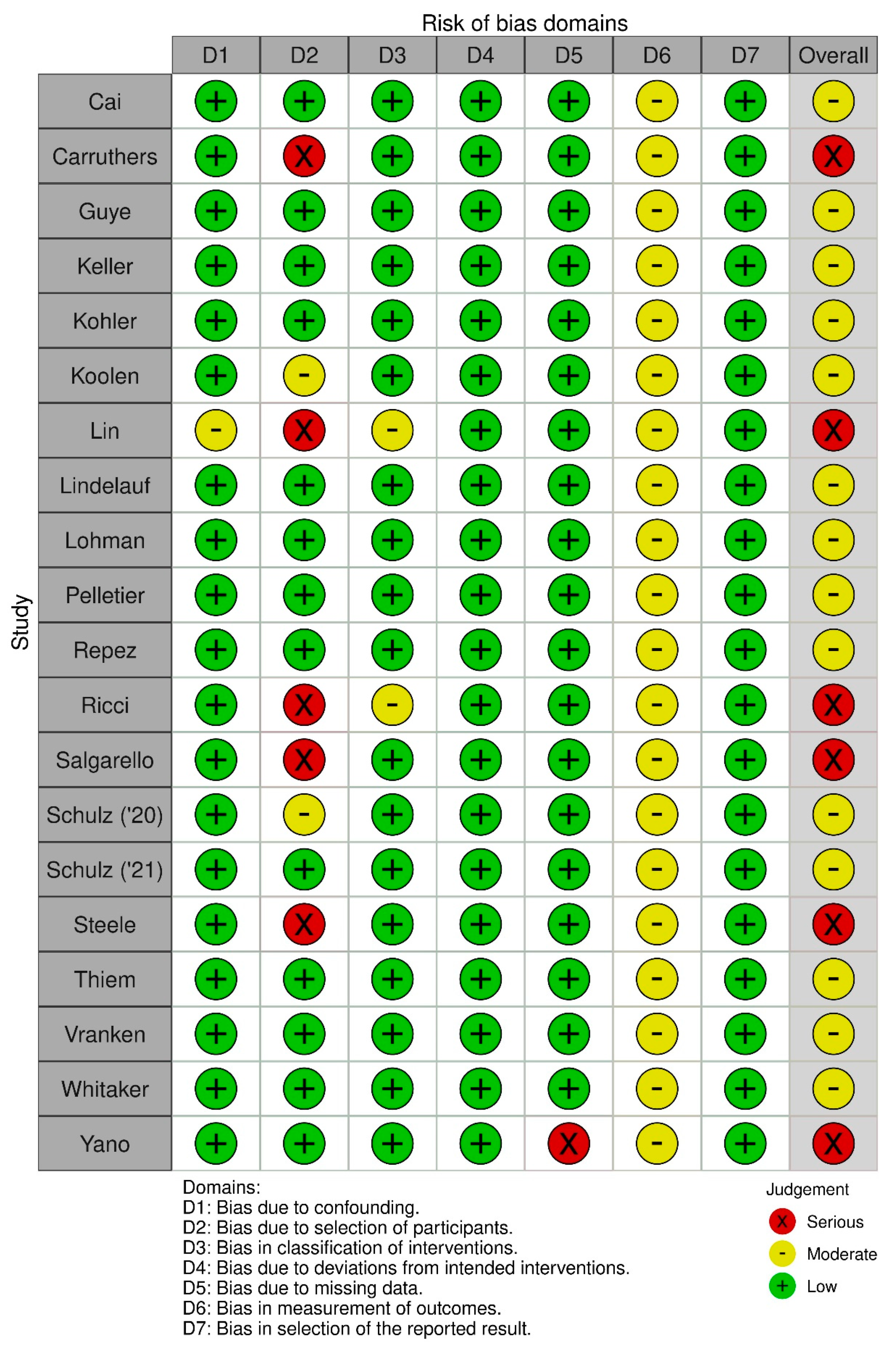
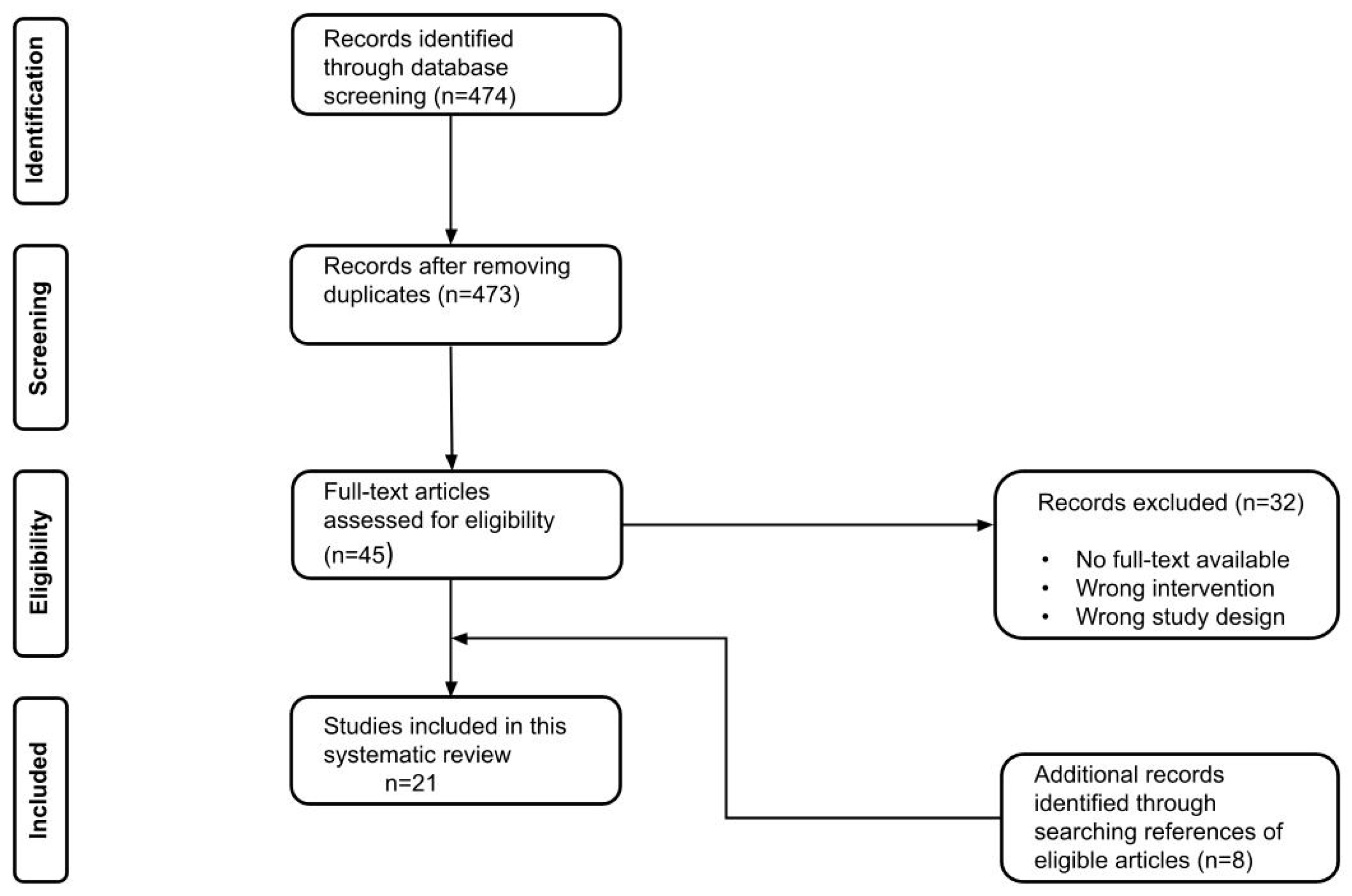
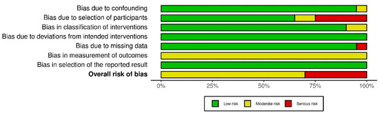
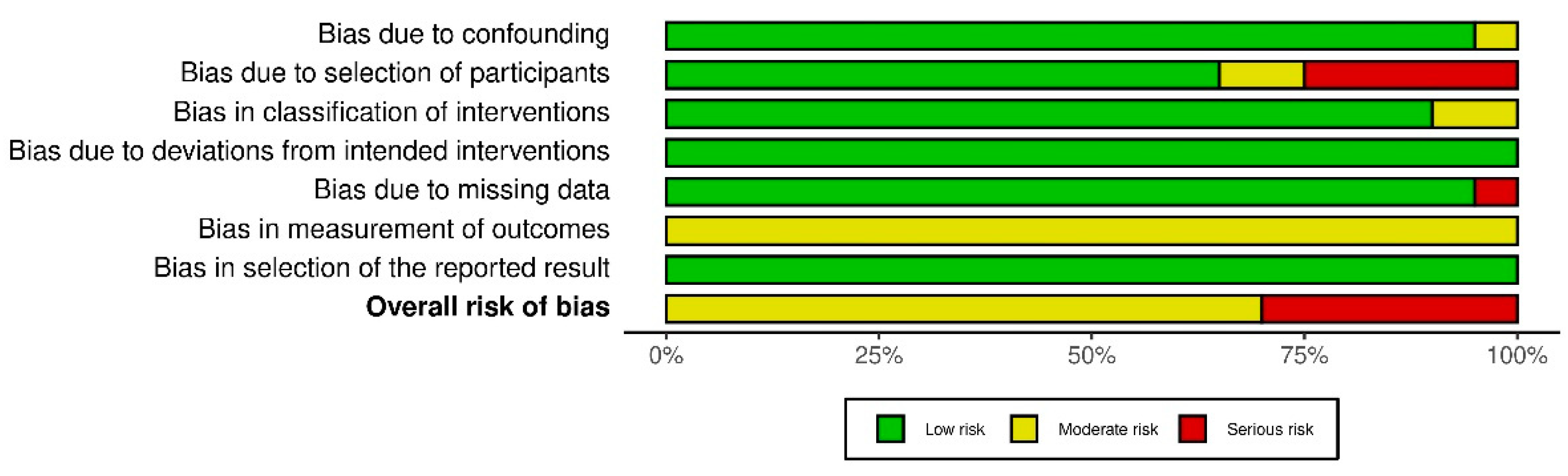
In total, twenty one of the 428 studies that were found with our search strategy qualified for inclusion in this study, see Figure 1. All twenty-one studies were single center studies, except one. Eleven studies were performed in Europe, two in Asia, and eight in the USA during the period from January 2004 to January 2020. Sixteen studies reported on the use of NIRS to detect flap failure and five studies reported on the use of HSI to prevent flap failure. Twenty studies had a cohort study design. Most of the included studies had a prospective design; ten of the NIRS and three of the HSI studies. Seven studies reported retrospectively collected data: six of the NIRS and one of the HSI studies. One HSI study was a case report. No randomized trials were identified. According to the ROBINS-I, the risk of bias assessment of the observational studies is presented in Figure 2 and Figure A1. The bias assessment of one case report was carried out with the Newcastle-Ottawa Scale [20]. Among these studies, inclusion criteria were comparable (Table A1)
Figure 1.
Flow chart of the included studies.
Figure 2.
Summary of the risk of bias assessment of the included observational studies according to the ROBINS-I.
3.2. Overall Flap Surgery Patient Profiles
Data of 2686 patients extracted from 21 studies who underwent flap surgery and were consequently monitored were analyzed (Table 2). The flaps were monitored with either NIRS in 1970 (73.3%), HSI in 90 (3.4%) or standard monitoring alone in 626 (23.3%) patients. The devices used for NIRS monitoring were ViOptix in eight (T.Ox 6, ODIsey 1), Inspectra in three (M325 1, M650 2), INVOS in two (5000C 1, 5100C 1), TSNIR-3 in one and TOS-96/TOS-OR in one study. For HSI TIVITA was used in four and ImSpector V8E in one study. The control monitoring technique consisted of clinical examination in twenty and indocyanine green (ICG) fluorescence [21] imaging in one. Control monitoring was carried out in conjunction with Doppler in eleven studies and with ICG imaging in one. The average/median ages in the included studies are depicted in Table 2. Females accounted for 91.8% (1861/2027) overall; 19.3% (11/57) of the HSI and 93.9% (1850/1970) of the NIRS study population. Gender data weren’t described in one HSI study [22], age wasn’t described in two studies [22,23]. Data on body mass index (BMI) was reported by nine NIRS studies. 3662 flaps were monitored with NIRS in 2759 (75.3%), HSI in 90 (2.5%) or standard monitoring alone in 813 (22.2%) flaps. The overall flap survival was 98.8% (95% CI: 97.1–99.5); HSI and NIRS flap survival respectively were 92.5% (95% CI: 83.3–96.8) and 99.2% (95% CI: 97.8–99.7). This difference was statistically significant (p = 0.02).
3.3. Flap-Related Characteristics
Data depicting flap related characteristics were reported inconsistently, except for types of flaps. Therefore, substantial amount of data was not available. Flap types, ischemia time, vascular disease, Diabetes Mellitus, smoking, radiotherapy and chemotherapy are described in Table 3. In one study 14 (47%) patients received (neo)-adjuvant therapy prior to surgery consisting of immunotherapy, endocrine therapy, radiation therapy, chemotherapy or a combination of these [24]. Prior abdominal surgery is described in two studies: 52 (26.5%) [25] and 214 (56.5%) in the control group, 356 (53.1%) in the NIRS group [26]. None of the included studies described use of inotropes.
3.4. Detection of Flap Failure
In at least nine out of sixteen studies, NIRS was the first tool indicating flap failure. Data regarding the first monitoring tool to detect complication was not provided by five studies. A faster detection with standard monitoring was observed in one study and with ICG imaging or standard monitoring in another compared with NIRS [27,28]. HSI was the first tool to indicate a vascular crisis in at least two out of five studies [22,29]; the other three didn’t provide data concerning the first tool to detect flap complication (Table 4). Time to detection was not mentioned in any of the included studies. The cut-off value for detection of flap complication was mentioned in the majority of studies. Proposed warning values for specific flap monitoring models according to recent studies and parameters to distinguish venous congestion from arterial occlusion are indicated in Table 5. Overall, 6.0% (95% CI: 4.0–8.9) of flaps had vascular crisis. Flaps monitored using HSI presented with vascular crisis in 10.0% (95% CI: 5.3–18.1) and using NIRS in 5.5% (95% CI: 3.4–8.8). This difference was not statistically significant (p = 0.39). Overall, 6.3% (95% CI: 4.3–9.1) of flaps were returned to the OR. In the HSI studies 12.7% (95% CI: 5.5–26.7) and in the NIRS studies 5.6% (95% CI: 3.8–8.2) of flaps were returned to the OR. This difference was statistically significant (p = 0.04). Salvage rate, the percentage of flaps with vascular crisis that could be saved, overall was 81.1% (95% CI: 65.1–90.8). HSI salvage rate was 22.2% (95% CI: 5.6–57.9) and NIRS salvage rate was 88.3% (95% CI: 80.1–93.4). This difference was statistically significant (p < 0.01). Average time to discharge was mentioned in 6 studies and is depicted in Table 4. Partial loss rate overall was 0.57% (95% CI: 0.13–2.52), for HSI 6.64% (0.44–53.36) and for NIRS 0.60% (95% CI: 0.19–1.89). This difference was statistically significant (p < 0.01). Sensitivity and Specificity were described in 13 studies.
Table 2.
Baseline characteristics of included population (n = 2686) and outcomes.
Table 3.
Flap-related characteristics of included flaps (n = 3662).
Table 4.
Detection of flap complication.
Table 5.
Proposed warning values for vascular crisis, parameters indicative of vascular crisis and parameters to distinguish venous from arterial crisis using NIRS versus HSI.
4. Discussion
Flap loss is a severe and feared complication after free tissue transfer in reconstructive microsurgery. Alongside the clinical assessment to detect signs of flap failure (either partial or total flap loss) in the early postoperative phase, objective monitoring of free flaps is expedient [11,40]. The ideal monitoring technique most importantly is objective, but also reliable, accurate, sensitive, continuous and user friendly, as defined by Creech and Miller [4]. NIRS and HSI are two different non-invasive monitoring methods that meet (almost) all criteria as described and have also proven to be suitable for detection of vascular compromise [9,23,37]. This study provides a systematic review in which a comparison between NIRS and HSI is made in detecting vascular compromise in reconstructive flap surgery compared to standard monitoring.
For NIRS, several commercial devices are available, such as FORE-SIGHT (Edwards Lifesciences, Irvin, CA, USA), INVOS (Medtronic, Minneapolis, MN, USA), EQUANOXTM (Nonin Medical Inc., Plymouth, MN, USA) and ViOptix (ViOptix Inc., Fremont, Ca, USA). With these devices tissue oxygenation is measured continuously using non-invasive sensors, which need to be applied on the skin in the area of interest. Despite its proven added value in detection of vascular compromise, the technique is only implemented in 5% of the DIEP-flap procedures in clinical practice [8,9,10]. Recently, more research has been performed on implementing HSI to monitor flap viability after free flap surgery. Although data on the use of HSI in the clinical setting is scarce, several studies concluded HSI to be reliable and accurate [22,26,29,38]. In addition, in a recent study by Thiem et al. HSI showed to be able to detect malperfusion of flaps before clinical monitoring [41]. Measuring tissue oxygenation with this imaging modality is discontinuous but contactless: no sensors need to be applied on the skin.
A lack of knowledge concerning the interpretation of values presented by the different devices could be an explanation for the low percentage in daily clinical use of NIRS measurements. Manufacturers use different algorithms to assess the tissue saturation values, apply different fixed ratios between arterial to venous blood volume and incorporate varying number and different wavelengths of near-infrared light [42]. Furthermore, they develop sensors with different transmitter-receiver spacing, resulting in different penetration depths, which also affects estimation and calculation of rStO2 [43]. Hence, it is difficult to define universal cut-off values necessitating prompt intervention [24,44]. In the included studies, most research was performed using the ViOptix device. For this particular device, Keller defined a threshold for rStO2 of an absolute value below 30% as predictive values for detection of vascular compromise [23]. For HSI, the diversity in used devices is currently limited. In all HSI observational studies included in this review, the Tivita system (Diaspective Vision GmbH, Am Salzhaff, Germany) was used for tissue oxygenation measurement. For this device no general cut-off values are defined yet, but most studies concluded a StO2 value below 30% to be an indication for circulatory compromise for which intervention would be recommended and justified [22,29,38,45]. Using continuous NIRS, measurement changes in tissue oxygenation can be monitored over time. A decrease of 20% from baseline for more than 60 minutes in duration is considered to be an indication for a lack in tissue perfusion. By HSI this continuity in monitoring is unfortunately not possible. Nevertheless, using HSI it has recently been shown feasible to detect circulatory compromise before standard clinical detection [41]. Therefore, both monitoring methods can be used to detect vascular compromise in the early postoperative period.
Implementing NIRS as a monitoring tool is less labor intensive for the medical staff. Because measurements are continuous, only one member of the team needs to be trained in performing the measurements. When values decrease below a certain threshold, this member receives a text message stating an extra clinical examination of the flap needs to be performed [40,46]. For using HSI extra medical staff needs to be trained before using the device, because photos need to be taken on different time-points during the day in a standardized manner.
Since sensors need to be used to measure tissue oxygenation with NIRS, not all flaps can be monitored with NIRS. For example, when using the FORE-SIGHT system a flap dimension of at least 50 mm by 30 mm was necessary for proper sensor placement [24]. Furthermore, these sensors are not sterile. Therefore, measuring saturation can only be performed in the postoperative phase. These could also be reasons for the scarce implementation of NIRS in clinical practice. With HSI being a contactless measurement, all types of flaps (e.g., fascio-cutaneous, muscle, intestinal) can be included. For example, a probe fixation of NIRS for an intraoral flap is difficult, although the contactless measurement by HSI may be suitable for intraoral flap monitoring. On the other hand, a buried flap monitoring would be difficult by a contactless way. Furthermore, without applying sensors on the skin, the HSI technique is friendlier for the patient and more importantly, the measurements can also be performed during surgery.
With HSI four different parameters (StO2, rStO2, THI, TWI) are measured. When these parameters are combined it is possible to determine whether the observed changes in values are caused by an arterial inflow or a venous outflow track problem [18,29,38]. For monitoring free flaps this could be of added value. When using NIRS, this distinction can only be made with a few devices. For example, with the ViOptix, which is unavailable in Europe. Therefore, the number of available devices in this area are limited.
The limited use of NIRS could also be due to the fact that implementing this technique comes with a price [9,26]. Implementing tissue oximetry costs $16,500 for the device and $150 per sensor according to Smit et al. In a different study, costs up to $30,000 for a device and $700–$1200 for a sensor are documented. Nevertheless, by implementing NIRS in standard protocol, vascular compromise could be detected in an early phase. Total flap loss could potentially be prevented and consequently duration of hospital stays shortened, resulting in a decrease of $1350–1700 per DIEP-flap procedure [33,34,47]. The costs for an HSI device are approximately $40,000 [38]. Initially implementing HSI would be more expensive than NIRS, but in the long term it could be more cost effective because no extra costs are required for buying the single use sensors. However, literature concerning cost effectiveness of HSI as a monitoring tool for flap viability is currently not available.
A limitation of the current literature study is the amount and quality of the included studies. In this review 21 studies were included. Sixteen reported on NIRS (n = 1970 patients) and five reported on HSI (n = 90 patients). All studies were observational cohort studies; accordingly, the average risk of bias was moderate. For this reason, randomized clinical trials with a larger patient population comparing the two monitoring techniques are mandatory. Moreover, defining solid cut-off values and performing an up-to-date cost-effectiveness evaluation regarding NIRS and HSI are required.
In conclusion, the authors believe that NIRS and HSI can have an added value in the detection of flap failure in the early postoperative phase. Both techniques have proven to be reliable, accurate and user-friendly monitoring methods, but do not (yet) replace the gold standard of clinical flap assessment. Based on the currently available literature, no firm conclusions can be drawn on which technique would be superior as an adjunct tool in free flap monitoring.
Author Contributions
Conceptualization, A.G.S., A.A.M.A.L. and R.M.S.; methodology, A.G.S. and A.A.M.A.L.; software, A.G.S.; validation, A.G.S., A.A.M.A.L. and R.M.S.; formal analysis, A.G.S. and S.M.J.v.K.; investigation, A.G.S. and A.A.M.A.L.; resources, A.G.S. and A.A.M.A.L.; data curation, A.G.S.; writing—original draft preparation, A.G.S. and A.A.M.A.L.; writing—review and editing, A.G.S., A.A.M.A.L., S.M.J.v.K., R.R.W.J.v.d.H. and R.M.S.; visualization, A.G.S. and A.A.M.A.L.; supervision, R.M.S. and R.R.W.J.v.d.H. All authors have read and agreed to the published version of the manuscript.
Funding
This research received no external funding.
Institutional Review Board Statement
Not applicable.
Informed Consent Statement
Not applicable.
Acknowledgments
Not applicable.
Conflicts of Interest
The authors declare no conflict of interest.
Appendix A
Figure A1.
Risk of Bias assessment of the observational studies according to the ROBINS-I.
Newcastle-Ottawa scale; Calin 6 stars (Selection ***, Comparability *, Exposure **).
Table A1.
Inclusion criteria of the studies.
Appendix B
Figure A2.
PRISMA checklist.
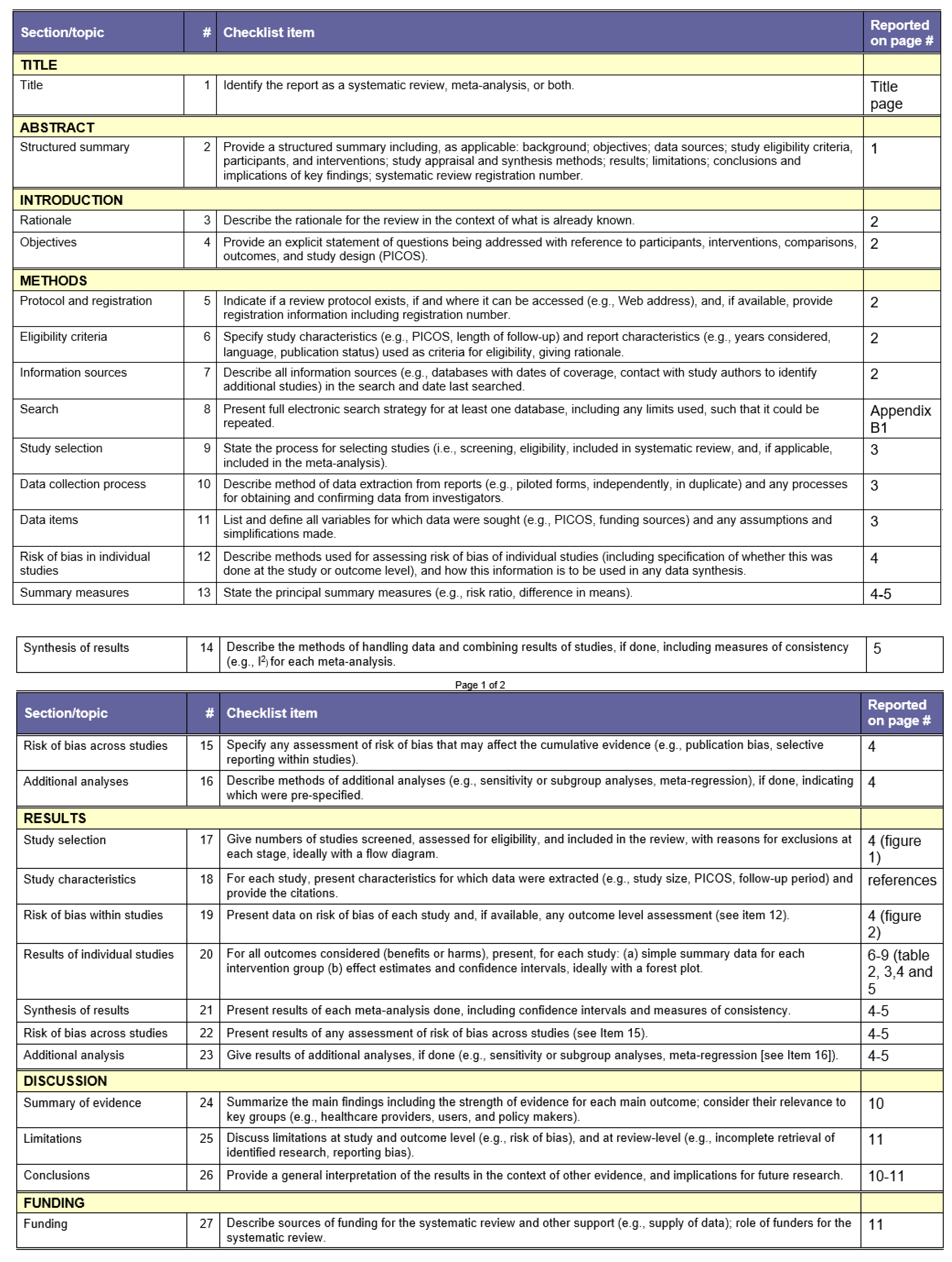
Table A2.
PubMed search strategy.
References
- Chen, K.-T.; Mardini, S.; Chuang, D.C.-C.; Lin, C.-H.; Cheng, M.-H.; Lin, Y.-T.; Huang, W.-C.; Tsao, C.-K.; Wei, F.-C. Timing of presentation of the first signs of vascular compromise dictates the salvage outcome of free flap transfers. Plast. Reconstr. Surg. 2007, 120, 187–195. [Google Scholar] [CrossRef] [Scilit]
- Hidalgo, D.A.; Jones, C.S. The role of emergent exploration in free-tissue transfer: A review of 150 consecutive cases. Plast. Reconstr. Surg. 1990, 86, 492–498. [Google Scholar] [CrossRef] [Scilit] [PubMed]
- Mirzabeigi, M.N.; Wang, T.; Kovach, S.J.; Taylor, J.A.; Serletti, J.M.; Wu, L.C. Free flap take-back following postoperative microvascular compromise: Predicting salvage versus failure. Plast. Reconstr. Surg. 2012, 130, 579–589. [Google Scholar] [CrossRef] [Scilit] [PubMed]
- Creech, B.; Miller, S. Evaluation of circulation in skin flaps. In Skin Flaps; Grabb, W.C., Myers, M., Eds.; Little, Brown: Boston, MA, USA, 1975; Volume 21. [Google Scholar]
- Sloan, G.M.; Sasaki, G.H. Noninvasive monitoring of tissue viability. Clin. Plast. Surg. 1985, 12, 185–195. [Google Scholar] [CrossRef] [Scilit]
- Yuen, J.C.; Feng, Z. Monitoring free flaps using the laser Doppler flowmeter: Five-year experience. Plast. Reconstr. Surg. 2000, 105, 55–61. [Google Scholar] [CrossRef] [Scilit] [PubMed]
- Keller, A. Noninvasive tissue oximetry for flap monitoring: An initial study. J. Reconstr. Microsurg. 2007, 23, 189–197. [Google Scholar] [CrossRef] [Scilit]
- Lohman, R.F.; Langevin, C.-J.; Bozkurt, M.; Kundu, N.; Djohan, R. A prospective analysis of free flap monitoring techniques: Physical examination, external Doppler, implantable Doppler, and tissue oximetry. J. Reconstr. Microsurg. 2013, 29, 051–056. [Google Scholar] [CrossRef] [Scilit]
- Smit, J.M.; Zeebregts, C.J.; Acosta, R.; Werker, P.M. Advancements in free flap monitoring in the last decade: A critical review. Plast. Reconstr. Surg. 2010, 125, 177–185. [Google Scholar] [CrossRef] [Scilit]
- Colwell, A.S.; Craft, R.O. Near-infrared spectroscopy in autologous breast reconstruction. Clin. Plast. Surg. 2011, 38, 301–307. [Google Scholar] [CrossRef] [Scilit]
- Repež, A.; Oroszy, D.; Arnež, Z.M. Continuous postoperative monitoring of cutaneous free flaps using near infrared spectroscopy. J. Plast. Reconstr. Aesthetic Surg. 2008, 61, 71–77. [Google Scholar] [CrossRef] [Scilit]
- Machens, H.G.; Mailaender, P.; Rieck, B.; Berger, A. Techniques of postoperative blood flow monitoring after free tissue transfer: An overview. Microsurgery 1994, 15, 778–786. [Google Scholar] [CrossRef] [Scilit]
- Irwin, M.; Thorniley, M.; Dore, C.; Green, C. Near infra-red spectroscopy: A non-invasive monitor of perfusion and oxygenation within the microcirculation of limbs and flaps. Br. J. Plast. Surg. 1995, 48, 14–22. [Google Scholar] [CrossRef] [Scilit]
- Thorniley, M.S.; Sinclair, J.; Barnett, N.; Shurey, C.; Green, C. The use of near-infrared spectroscopy for assessing flap viability during reconstructive surgery. Br. J. Plast. Surg. 1998, 51, 218–226. [Google Scholar] [CrossRef] [Scilit]
- Lu, G.; Fei, B. Medical hyperspectral imaging: A review. J. Biomed. Opt. 2014, 19, 010901. [Google Scholar] [CrossRef] [Scilit]
- Goetz, A.F.; Vane, G.; Solomon, J.E.; Rock, B.N. Imaging spectrometry for earth remote sensing. Science 1985, 228, 1147–1153. [Google Scholar] [CrossRef] [Scilit] [PubMed]
- Grambow, E.; Dau, M.; Holmer, A.; Lipp, V.; Frerich, B.; Klar, E.; Vollmar, B.; Kämmerer, P.W. Hyperspectral imaging for monitoring of perfusion failure upon microvascular anastomosis in the rat hind limb. Microvasc. Res. 2018, 116, 64–70. [Google Scholar] [CrossRef] [Scilit]
- Holmer, A.; Marotz, J.; Wahl, P.; Dau, M.; Kämmerer, P.W. Hyperspectral imaging in perfusion and wound diagnostics–methods and algorithms for the determination of tissue parameters. Biomed. Eng. Biomed. Tech. 2018, 63, 547–556. [Google Scholar] [CrossRef] [Scilit] [PubMed]
- Kulcke, A.; Holmer, A.; Wahl, P.; Siemers, F.; Wild, T.; Daeschlein, G. A compact hyperspectral camera for measurement of perfusion parameters in medicine. Biomed. Eng. Biomed. Tech. 2018, 63, 519–527. [Google Scholar] [CrossRef] [Scilit] [PubMed]
- Calin, M.-A.; Coman, T.; Parasca, S.V.; Bercaru, N.; Savastru, R.S.; Manea, D. Hyperspectral imaging-based wound analysis using mixture-tuned matched filtering classification method. J. Biomed. Opt. 2015, 20, 046004. [Google Scholar] [CrossRef] [Scilit]
- Pruimboom, T.; Schols, R.M.; Van Kuijk, S.M.; Van der Hulst, R.R.; Qiu, S.S. Indocyanine green angiography for preventing postoperative mastectomy skin flap necrosis in immediate breast reconstruction. Cochrane Database Syst. Rev. 2020, 4, CD013280. [Google Scholar] [CrossRef] [Scilit]
- Thiem, D.G.; Frick, R.W.; Goetze, E.; Gielisch, M.; Al-Nawas, B.; Kämmerer, P.W. Hyperspectral analysis for perioperative perfusion monitoring—a clinical feasibility study on free and pedicled flaps. Clin. Oral Investig. 2021, 25, 933–945. [Google Scholar] [CrossRef] [Scilit]
- Keller, A. A new diagnostic algorithm for early prediction of vascular compromise in 208 microsurgical flaps using tissue oxygen saturation measurements. Ann. Plast. Surg. 2009, 62, 538–543. [Google Scholar] [CrossRef] [Scilit]
- Lindelauf, A.A.; Vranken, N.; Schols, R.M.; Bouman, E.A.; Weerwind, P.W.; van der Hulst, R.R. Exploring personalized postoperative non-invasive tissue oximetry in DIEP flap breast reconstruction. Eur. J. Plast. Surg. 2021, 1–9. [Google Scholar] [CrossRef] [Scilit]
- Carruthers, K.H.; Tiwari, P.; Yoshida, S.; Kocak, E. Inpatient flap monitoring after deep inferior epigastric artery perforator flap breast reconstruction: How long is long enough? J. Reconstr. Microsurg. 2019, 35, 682–687. [Google Scholar] [CrossRef] [Scilit]
- Koolen, P.G.; Vargas, C.R.; Ho, O.A.; Ibrahim, A.; Ricci, J.A.; Tobias, A.M.; Winters, H.A.; Lin, S.J.; Lee, B.T. Does increased experience with tissue oximetry monitoring in microsurgical breast reconstruction lead to decreased flap loss? The learning effect. Plast. Reconstr. Surg. 2016, 137, 1093–1101. [Google Scholar] [CrossRef] [Scilit] [PubMed]
- Vranken, N.; Weerwind, P.; Van Onna, M.; Bouman, E.; Van der Hulst, R. Non-invasive tissue oximetry following unilateral DIEP-flap reconstruction: A pilot evaluation. JPRAS Open 2017, 12, 59–65. [Google Scholar] [CrossRef] [Scilit]
- Yano, A.; Orihashi, K.; Yoshida, Y.; Kuriyama, M. Near-infrared spectroscopy for monitoring free jejunal flap. J. Plast. Reconstr. Aesthetic Surg. 2021, 74, 108–115. [Google Scholar] [CrossRef] [Scilit] [PubMed]
- Kohler, L.H.; Köhler, H.; Kohler, S.; Langer, S.; Nuwayhid, R.; Gockel, I.; Spindler, N.; Osterhoff, G. Hyperspectral Imaging (HSI) as a new diagnostic tool in free flap monitoring for soft tissue reconstruction: A proof of concept study. BMC Surg. 2021, 21, 1–9. [Google Scholar] [CrossRef] [Scilit]
- Cai, Z.-g.; Zhang, J.; Zhang, J.-g.; Zhao, F.-y.; Yu, G.-y.; Li, Y.; Ding, H.-s. Evaluation of near infrared spectroscopy in monitoring postoperative regional tissue oxygen saturation for fibular flaps. J. Plast. Reconstr. Aesthetic Surg. 2008, 61, 289–296. [Google Scholar] [CrossRef] [Scilit]
- Guye, M.-L.; Motamed, C.; Chemam, S.; Leymarie, N.; Suria, S.; Weil, G. Remote peripheral tissue oxygenation does not predict postoperative free flap complications in complex head and neck cancer surgery: A prospective cohort study. Anaesth. Crit. Care Pain Med. 2017, 36, 27–31. [Google Scholar] [CrossRef] [Scilit]
- Lin, S.J.; Nguyen, M.-D.; Chen, C.; Colakoglu, S.; Curtis, M.S.; Tobias, A.M.; Lee, B.T. Tissue oximetry monitoring in microsurgical breast reconstruction decreases flap loss and improves rate of flap salvage. Plast. Reconstr. Surg. 2011, 127, 1080–1085. [Google Scholar] [CrossRef] [Scilit] [PubMed]
- Pelletier, A.; Tseng, C.; Agarwal, S.; Park, J.; Song, D. Cost analysis of near-infrared spectroscopy tissue oximetry for monitoring autologous free tissue breast reconstruction. J. Reconstr. Microsurg. 2011, 27, 487–494. [Google Scholar] [CrossRef] [Scilit] [PubMed]
- Ricci, J.A.; Vargas, C.R.; Ho, O.A.; Lin, S.J.; Tobias, A.M.; Lee, B.T. Evaluating the use of tissue oximetry to decrease intensive unit monitoring for free flap breast reconstruction. Ann. Plast. Surg. 2017, 79, 42–46. [Google Scholar] [CrossRef] [Scilit] [PubMed]
- Salgarello, M.; Pagliara, D.; Rossi, M.; Visconti, G.; Barone-Adesi, L. Postoperative monitoring of free DIEP flap in breast reconstruction with near-infrared spectroscopy: Variables affecting the regional oxygen saturation. J. Reconstr. Microsurg. 2018, 34, 383–388. [Google Scholar] [CrossRef] [Scilit]
- Steele, M.H. Three-year experience using near infrared spectroscopy tissue oximetry monitoring of free tissue transfers. Ann. Plast. Surg. 2011, 66, 540–545. [Google Scholar] [CrossRef] [Scilit] [PubMed]
- Whitaker, I.S.; Pratt, G.F.; Rozen, W.M.; Cairns, S.A.; Barrett, M.D.; Hiew, L.Y.; Cooper, M.A.; Leaper, D.J. Near infrared spectroscopy for monitoring flap viability following breast reconstruction. J. Reconstr. 2012, 28, 149–154. [Google Scholar] [CrossRef] [Scilit]
- Schulz, T.; Marotz, J.; Stukenberg, A.; Reumuth, G.; Houschyar, K.S.; Siemers, F. Hyperspectral imaging for postoperative flap monitoring of pedicled flaps. Handchir. Mikrochir. Plast. Chir. Organ. Der Dtsch. Arb. Fur Handchir.:Organ. Der Dtsch. Arb. Fur Mikrochir. Der Peripher. Nerven Und Gefasse Organ. Der V. 2020, 52, 316–324. [Google Scholar]
- Schulz, T.; Leuschner, S.; Siemers, F.; Marotz, J.; Houschyar, K.; Corterier, C.C. Assessing flap perfusion after free tissue transfer using hyperspectral imaging (HSI). Eur. J. Plast. Surg. 2021, 1–10. [Google Scholar] [CrossRef] [Scilit]
- Keller, A. Noninvasive tissue oximetry. Clin. Plast. Surg. 2011, 38, 313–324. [Google Scholar] [CrossRef] [Scilit]
- Thiem, D.G.; Römer, P.; Blatt, S.; Al-Nawas, B.; Kämmerer, P.W. New Approach to the Old Challenge of Free Flap Monitoring—Hyperspectral Imaging Outperforms Clinical Assessment by Earlier Detection of Perfusion Failure. J. Pers. Med. 2021, 11, 1101. [Google Scholar] [CrossRef] [Scilit]
- Yoshitani, K.; Kawaguchi, M.; Tatsumi, K.; Kitaguchi, K.; Furuya, H. A comparison of the INVOS 4100 and the NIRO 300 near-infrared spectrophotometers. Anesth. Analg. 2002, 94, 586–590. [Google Scholar] [CrossRef] [Scilit] [PubMed]
- Bevan, P.J. Should cerebral near-infrared spectroscopy be standard of care in adult cardiac surgery? Heart Lung Circ. 2015, 24, 544–550. [Google Scholar] [CrossRef] [Scilit] [PubMed]
- Hyttel-Sorensen, S.; Hessel, T.W.; Greisen, G. Peripheral tissue oximetry: Comparing three commercial near-infrared spectroscopy oximeters on the forearm. J. Clin. Monit. Comput. 2014, 28, 149–155. [Google Scholar] [CrossRef] [Scilit]
- Jafari-Saraf, L.; Wilson, S.E.; Gordon, I.L. Hyperspectral image measurements of skin hemoglobin compared with transcutaneous PO2 measurements. Ann. Vasc. Surg. 2012, 26, 537–548. [Google Scholar] [CrossRef] [Scilit]
- Ricci, J.A.; Vargas, C.R.; Lin, S.J.; Tobias, A.M.; Taghinia, A.H.; Lee, B.T. A novel free flap monitoring system using tissue oximetry with text message alerts. J. Reconstr. Microsurg. 2016, 32, 415–420. [Google Scholar] [CrossRef] [Scilit] [PubMed]
- Lindelauf, A.A.; Vranken, N.P.; Rutjens, V.G.; Schols, R.M.; Heijmans, J.H.; Weerwind, P.W.; van der Hulst, R.R. Economic Analysis of Noninvasive Tissue Oximetry for Postoperative Monitoring of Deep Inferior Epigastric Perforator Flap Breast Reconstruction: A Review. Surg. Innov. 2020, 27, 534–542. [Google Scholar] [CrossRef] [Scilit]

Publisher’s Note: MDPI stays neutral with regard to jurisdictional claims in published maps and institutional affiliations. |
© 2022 by the authors. Licensee MDPI, Basel, Switzerland. This article is an open access article distributed under the terms and conditions of the Creative Commons Attribution (CC BY) license (https://creativecommons.org/licenses/by/4.0/).