


Abstract
As the largest known ancient jade mining site, the Mazongshan Site is crucial for understanding the “West–to–East Jade Transportation” system in ancient China. However, its vast nephrite materials remain poorly characterized mineralogically and geochemically. This study employs a multi-technique approach, including polarized light microscopy, SEM-EDS, XRD, CRS, EPMA, and ICP-MS to analyze Mazongshan nephrite. The results identify tremolite as the principal mineral, with accessory minerals including diopside, apatite, serpentine, calcite, dolomite, graphite, hornblende, epidote, forsterite, and albite, as well as limonite occurring as a secondary mineral formed by oxidation. Its rare earth element patterns show significant negative Eu anomalies, low total REE concentrations, and low levels of Cr, Ni, and Co. These results confirm a metamorphic origin for the deposit. Most significantly, the high compositional affinity it exhibits with Hetian nephrite from Xinjiang, together with evidence of ancient mining, has led us to reconsider the prevalence of nephrite materials used during the Warring States to Han periods.
1. Introduction
As a symbol of ritual and power, jade played a paramount role in the social complexity of ancient East Asia from the Middle Neolithic onward [1]. Archeological studies have identified three main nephrite transmission routes: North-to-South, East-to-West, and West-to-East [2]. A significant shift occurred at the end of the Neolithic period: with the rise in early states and possible local resource depletion, the former two routes declined, while the West-to-East route became the dominant “Jade Road.” This transition laid the foundation for the flourishing of jade culture during the Warring States and Han periods, when the functions of jade extended far beyond ritual to burial, adornment, daily use, and health cultivation [3]. Such prosperity required a stable and efficient supply system, making the West-to-East route critical for understanding ancient jade consumption [4].
Long-dominant in academia, the “Hetian Nephrite Centric Theory” posited Xinjiang’s Hetian deposits as the primary source of high-quality nephrite for early China [5]. However, the identification of dozens of gem-quality nephrite deposits across China [6], together with mineralogical and geochemical research on archeological jade [7,8,9,10,11], has invalidated the sole attribution of ancient nephrite artifacts to Xinjiang sources. Despite modern scientific methods, provenance research remains contentious, hampered by disparities between modern and ancient deposits and a lack of traceable archeological samples. The key to resolving these disputes lies in the discovery and study of concrete archeological evidence from ancient mining sites.
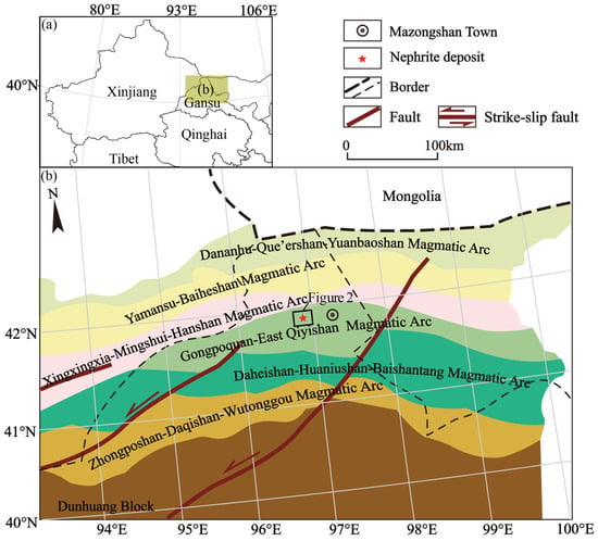
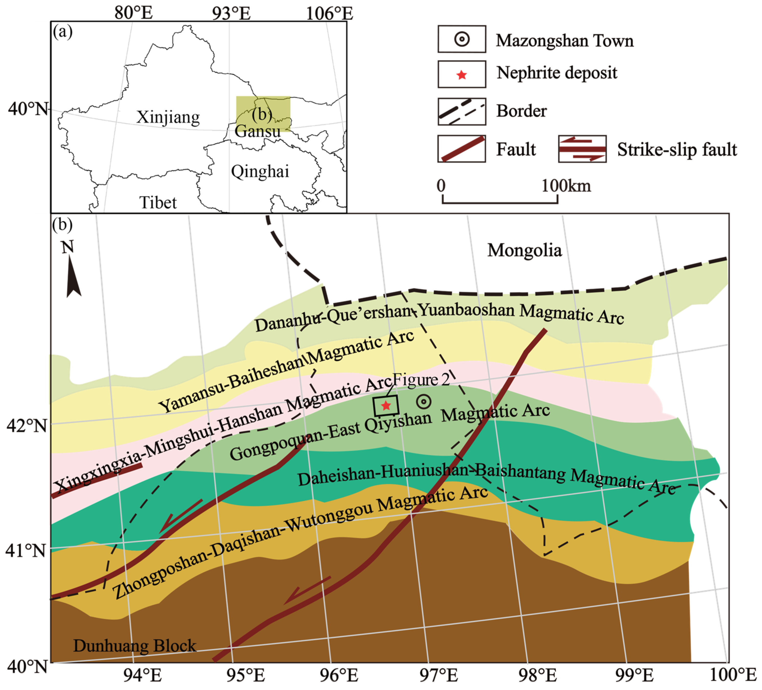
Against this background, ancient jade mining sites discovered in the Hexi Corridor have provided new insights into early jade resource systems [12,13]. The Mazongshan site is particularly notable as the largest ancient jade mine identified in China. Figure 1a shows the location of nephrite deposit. Radiocarbon dating indicates that mining began as early as ca. 390 BCE (Early–Middle Warring States). Spatially and temporally, Mazongshan confirms that the Hexi Corridor was not only a transportation hub but also a major center for large-scale nephrite production. A detailed study of the Mazongshan mine can clarify the relationships among jade resources, technology, and cultural expansion.
Figure 1.
Schematic location of the Mazongshan Jade Mining Site. (a). Location of the Mazongshan nephrite deposit in Northwest China; (b). Main tectonic units around the deposit (modified from Reference [14]).
This study conducts a systematic mineralogical and geochemical analysis of nephrite specimens from the Mazongshan Mining Deposit. By characterizing the mineralogical properties of Mazongshan nephrite, we aim to refine geochemical tracing parameters for northwest China’s nephrite deposits, elucidate the geological conditions governing the formation of the Mazongshan deposit, and provide a foundational mineralogical dataset for future provenance studies of Warring States to Han period nephrite artifacts.
2. Geological Setting
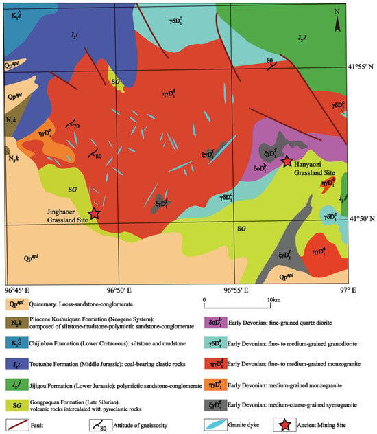
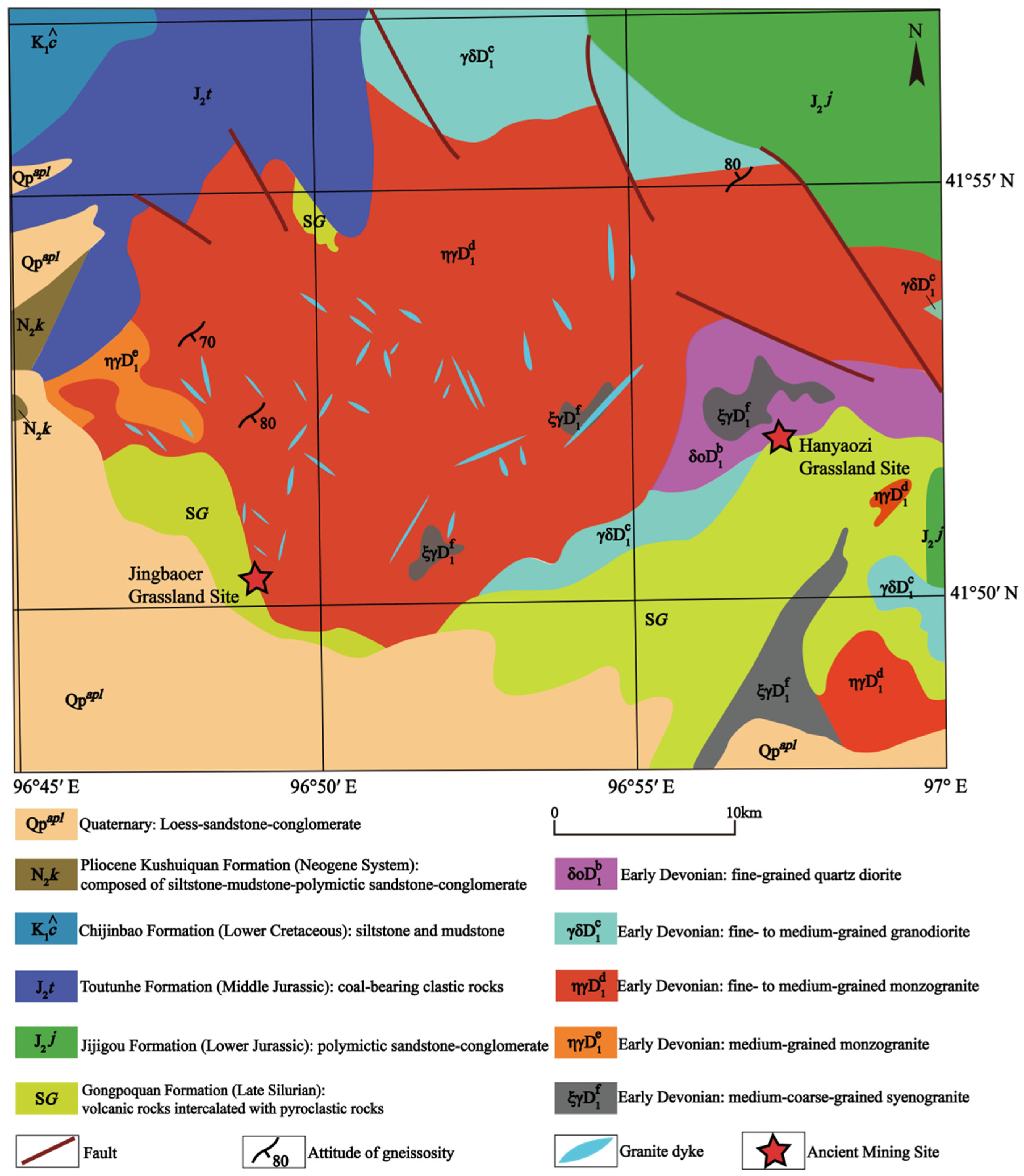
Situated in the Beishan Orogenic Belt, the Mazongshan nephrite deposit lies within the Gongpoquan–East Qiyishan Early Paleozoic magmatic arc (Figure 1b). Ordovician to Carboniferous plutonism extensively emplaced granitic rocks in the region (Figure 2). Jade-bearing veins exhibit two principal orientations: NW–SE–trending in the Jingbaoer Grassland and E–W–trending in the Hanyaozi Grassland, interpreted as secondary extensional fractures related to the regional NE–SW-trending fault system [15]. Both sites are located at the critical lithological contact zones.
Figure 2.
1: 250,000 Geological structure map of Mazongshan Area. This map is based on the spatial database of serial suite-tectonic map-sheets of mainland China with drawing number K47C003001 downloaded from the standard map service website of the Ministry of Natural Resources of the People’s Republic of China [16].
Jingbaoer Grassland Site is situated at the contact between the Late Silurian Gongpoquan Formation, which consists of volcanic rocks intercalated with pyroclastic rocks (including andesite, dacite, andesitic breccia lava, and andesitic volcanic breccia interbedded with sedimentary breccia) and thin dolomitic marble, and the Early Devonian fine- to medium-grained monzogranite. Distinct from the Jingbaoer Grassland Site, the Hanyaozi Grassland Site is situated at the contact between the Gongpoquan Formation of the Late Silurian and the Early Devonian fine–grained quartz diorite. Additional Early Devonian granitoids, including fine- to medium-grained granodiorite and medium-coarse-grained syenogranite, are also exposed in the mining area and are in intrusive contact with the Gongpoquan Formation.
The entire mining area is characterized by intense structural deformation, with intensive weathering and erosion that expose extensive acidic magmatic rocks (predominantly Early Devonian granitoids). Within the mineralized zones, the wall rocks at both contact zones exhibit typical Mg-rich skarn alteration, with diopsidization and tremolitization widely developed along the intrusion–volcanic rock boundaries. The tremolite jade orebodies occur as veins or stratiform bodies, and despite the deformation, they preserve primary metasomatic textures.
3. Materials and Methods
To assess the quality and composition of Mazongshan nephrite, we analyzed fifteen representative samples from the Jingbaoer Grassland Site and Hanyaozi Grassland Site, which varied in color and luster, numbered JBE 1-10 and HYZ 1-5.
Gemological tests were conducted at the Key Laboratory of Cultural Heritage Research and Conservation (School of Cultural Heritage, Northwest University, Xi’an, China). The experimental instruments included a gem refractometer, an electronic balance, and a Mohs hardness pen. These tests were applied to obtain the basic gemological parameters (refractive index, density, and hardness) for the fundamental characterization of Mazongshan nephrite. Each experiment was performed three times, and the arithmetic average of the results was used as the final measurement, as shown in Table 1.
Polarizing light microscopic analyses were carried out at the Regional Geological and Mineral Resources Survey Institute of Hebei Province. This method was adopted to identify the mineral composition, microtextures, and crystallization stages of the nephrite through petrographic observation. The samples were prepared as thin sections, with a 0.03 mm thickness, and analyzed using an OLYMPUS-BX51 polarizing light microscope (Olympus Corporation, Tokyo, Japan). The testing environment was controlled at 29 °C with a relative humidity of 70%.
X-ray diffraction (XRD) analysis was utilized to accurately determine the mineral phases and bulk mineral composition of the samples. The test was conducted at the Key Laboratory of Cultural Heritage Research and Conservation (School of Cultural Heritage, Northwest University, China). The samples were prepared by surface polishing and analyzed using a SmartLab rotating anode X-ray diffractometer (Rigaku Corporation, Tokyo, Japan). Cu Kα radiation (40 kV, 150 mA) was used for the measurements, with a scanning range of 5–90°, a step size of 0.02°, and a scan rate of 10°/min. The five samples from the Hanyaozi Grassland Site, which initially yielded unsatisfactory results from analysis on the polished surface, were subsequently ground into powder and re-analyzed under the same instrumental conditions.
The confocal Raman spectrometer (CRS) was used to characterize the molecular structure and precise mineral species of the tremolite jade. The test was conducted at the School of Physics, Northwest University. The polished samples were analyzed using a LabRAM Odyssey laser confocal Raman spectrometer (HORIBA Corporation, Palaiseau, France). Raman shifts were calibrated using a single-crystal silicon standard (main band at 520.5 cm−1). Measurement conditions included a 785 nm excitation laser at 50 mW power, a grating with 1200 grooves/mm, a 100 × objective, a confocal aperture of 100 μm, an estimated spatial spot size of ~1 μm, a spectral resolution of 1 cm−1, a measured Raman shift range from 100 cm−1 to 2000 cm−1, and two accumulations with 20 s integration per accumulation.
The scanning electron microscope–energy dispersive spectroscopy (SEM-EDS) observation was applied to reveal the micro-morphology, mineral assemblage, and contact relationships of the ore-forming minerals. The test was conducted at the Key Laboratory of Cultural Heritage Research and Conservation (School of Cultural Heritage, Northwest University, China). The samples were prepared by embedding in resin, polishing, and sputter-coating with gold. Analyses were performed using a TESCAN VEGA3 scanning electron microscope (TESCAN Corporation, Brno, Czech Republic), coupled with an X-ACT energy dispersive spectrometer (Oxford Instruments, Abingdon, UK). Operating conditions included both secondary electron (SE) and backscattered electron (BSE) detectors, an accelerating voltage of 10–20 kV, an SE resolution of 1.3 nm, a beam current of ~1 nA, a spot size of 30–50 nm, a working distance of 15–20 mm, a spot scan mode for EDS analysis, a magnification range of 50× to 500×, and a dwell time of 120 s. The field of view (FoV) varied with magnification.
The electron microprobe analyzer (EPMA) was employed to determine the major element compositions. The test was conducted at Wuhan SampleSolution Testing Technology Co., Ltd., using a JXA-8230 electron probe microanalyzer (JEOL Corporation, Akishima, Japan). Analytical conditions included an accelerating voltage of 15 kV, a beam current of 20 nA, and a spot size of 3 μm. For quantitative analysis, the following major elements were targeted: Ca, Ti, Al, Si, Zn, Ni, Co, Fe, Mn, Cr, Na, Mg, K, and P. Peak counting times were set to 10 s, with background counting times of 5 s for all elements to ensure measurement precision. Calibration was performed using a series of natural and synthetic SPI standards, specifically: chrome diopside for Ca, rutile for Ti, yttrium aluminum garnet for Al, quartz for Si, sphalerite for Zn, native nickel for Ni, native cobalt for Co, hematite for Fe, rhodonite for Mn, native chromium for Cr, jadeite for Na, olivine for Mg, K-feldspar for K, and fluorapatite for P. All data were corrected with the ZAF (atomic number, absorption, fluorescence) method.
The inductively coupled plasma mass spectrometry (ICP-MS) was used to quantify the trace element compositions, which offer critical evidence for tracing the material source and metallogenic mechanism. Analysis was performed at Tianjing Createch Testing Technology Co., Ltd., using a PlasmaQuant MS inductively coupled plasma mass spectrometer (Analytik Jena AG, Jena, Germany), following GB/T 14506.30–2010 [17]. The system operated in STD mode under a 0.6 MPa argon atmosphere, with the ground potential maintained below 5 V. Sample preparation followed the closed-vessel acid digestion method specified in the standard. Approximately 2.5 g of each sample was ground to <200 mesh and dried at 105 °C for 2–4 h. A 25 mg aliquot was accurately weighed (0.01 mg precision) and placed in a PTFE-lined stainless steel bomb. After adding 1 mL HF and 0.5 mL HNO3, the bomb was sealed and heated at 185 °C for 24 h. The solution was then evaporated to near dryness, re-dissolved in 0.5 mL HNO3 (evaporated again), and finally taken up in 5 mL HNO3 (1 + 1) and heated at 130 °C for 3 h. The cooled solution was transferred to a plastic bottle and diluted to 25 mL with ultrapure water. External calibration was performed with multi-element standards, and an online Rh-Re internal standard corrected for matrix effects and signal drift. Forty-four elements (Li to U) were quantified, with detection limits (0.003–3 μg/g) and precision (RSD < 10%) meeting GB/T 14506.30–2010 requirements. Polyatomic interferences were corrected using single-element solutions (Ba, Ce, Zr, Nd, Sn) and the interference coefficient method. Data were processed with ICPMSDataCal (version 10.7).
4. Results
4.1. Gemological Properties
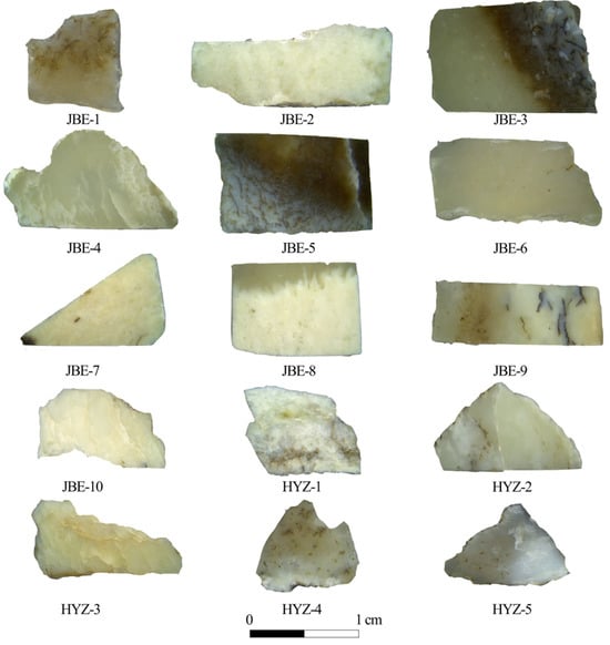
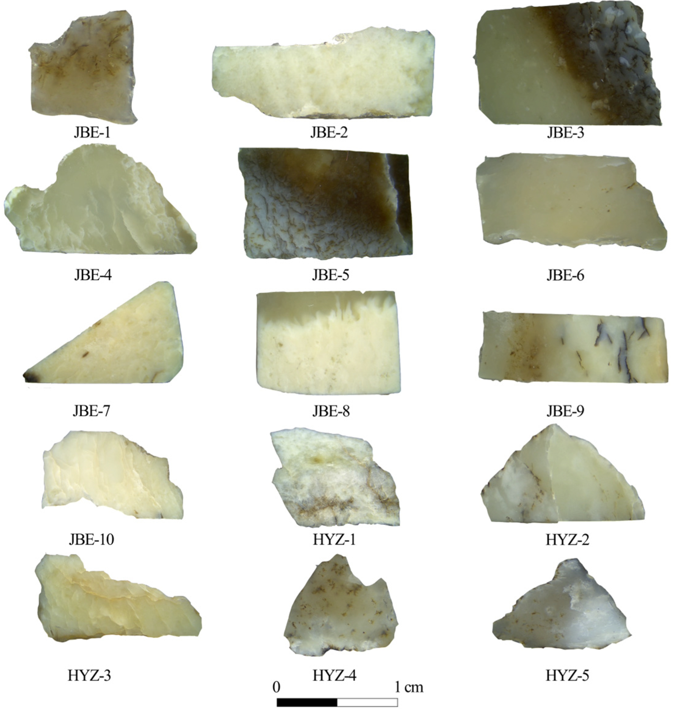
The conventional gemological properties of the fifteen Mazongshan nephrite samples are listed in Table 1. These nephrite samples closely resemble typical nephrite, except for their color. All the samples exhibit a range of white to brownish-yellow hues with a fine texture (Figure 3) and display translucency with a sub-vitreous to greasy luster. Their refractive index (RI) ranges from 1.57 to 1.62, while their specific gravity (SG) ranges from 2.81 to 3.17. The Mohs hardness ranges from 5.9 to 6.2. Overall, these gemological properties are consistent with those of typical nephrite jade.
Figure 3.
Nephrite samples from Jingbaoer Grassland Site (JBE-1~10) and Hanyaozi Grassland Site (HYZ-1~5) in Subei County, Gansu Province, China.
Table 1.
Gemological properties of the nephrite jade samples from the Mazongshan Site.
4.2. Crystal Structure and Chemical Bonding/Molecular Vibrational Information
4.2.1. XRD Characterization
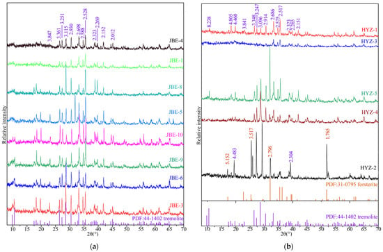
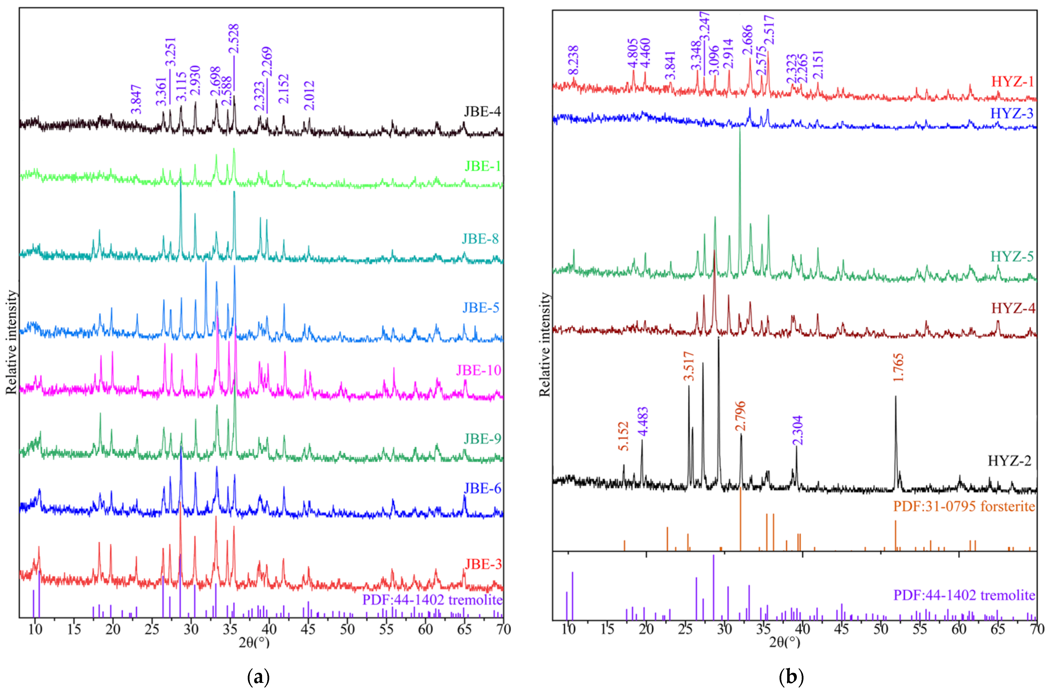
Figure 4 shows the XRD analysis of Mazongshan nephrite samples, revealing a diffraction pattern that aligns with the standard tremolite reference (PDF: 44-1402). Band position shifts and variations in band intensity may result from the isomorphous substitution of other ions and variations in crystallinity. Additionally, diffraction bands of forsterite were identified insample HYZ-2 from the Hanyaolizi Grassland Site.
Figure 4.
XRD Diffraction Patterns of Mazongshan Nephrite. (a). Samples from Jingbaower Grassland Site. (b). Samples from Hanyaozi Grassland Site. The numbers distributed vertically in the figure are d-values.
4.2.2. CRS Characterization
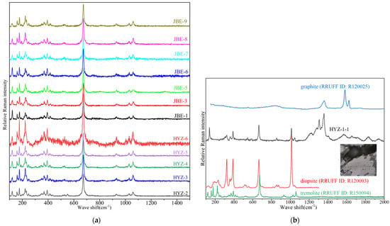
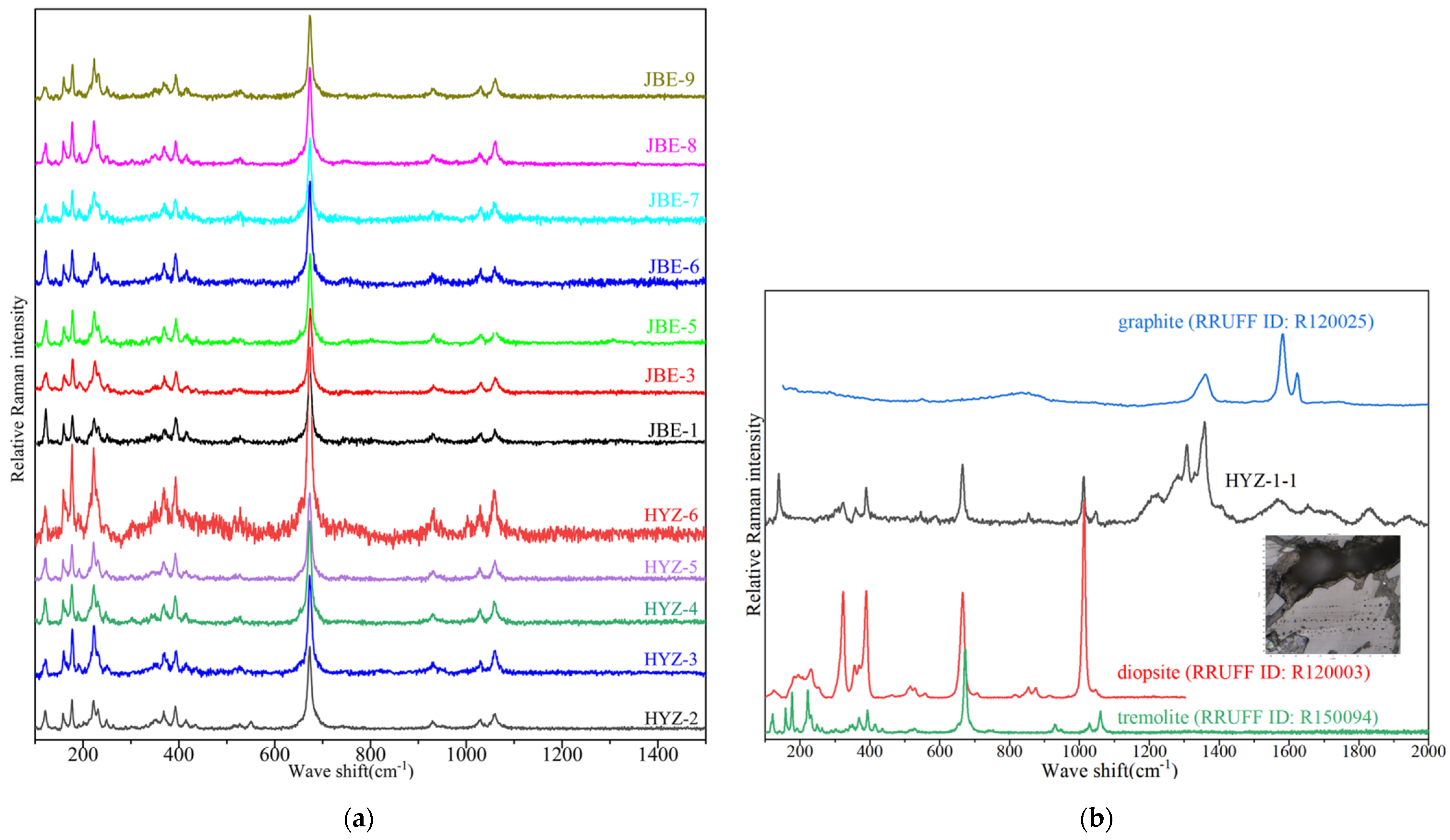
CRS analysis of samples, as shown in Figure 5, reveals characteristic spectroscopic features of tremolite. The primary vibration bands occur near 673–674 cm−1, corresponding to the symmetric stretching vibrations of Si–O–Si. Secondary strong vibration bands are observed in the range of 100–400 cm−1, with specific bands near 159 cm−1 corresponding to lattice vibrations of [SiO4]4−, 223 cm−1 corresponding to bending vibrations of O–Si–O, and 371 cm−1 and 394 cm−1, representing the bending and lattice vibration of M (metal)–O. Weak bands at 437 cm−1 correspond to the bending vibration of Si-O-Si, while bands at 530 cm−1 correspond to oxygen bridge-bonding. Stretching vibrations of Si–O are observed in 900–1100 cm−1 with prominent bands at 1060 cm−1, and weaker bands at 930 and 1029 cm−1. These Raman spectral bands align with the standard spectrum of tremolite [18].
Figure 5.
Raman Spectra of Mazongshan nephrite. (a). tremolite; (b). diopside and graphite in Sample HYZ-1.
Characteristic spectral bands of diopside and graphite were identified in the inclusion of sample HYZ-1, as shown in Figure 5b. The bands below 1200 cm−1 include the lattice vibrations and translational vibrations of Ca2+ and Mg2+ cations appearing at between 130 and 140 cm−1, bending vibrations of M–O appearing at 322 cm−1 and 388 cm−1, stretching vibrations of M–O and bending vibrations of Si–O–Si at 508 cm−1, symmetric stretching vibrations of Si–O–Si at 665 cm−1, and symmetric stretching vibrations of Si–O at 1012 cm−1. These spectral features align closely with the standard diopside bands [19], confirming the coexistence of a tremolite–diopside mineral assemblage in the Mazongshan nephrite samples. Meanwhile, the bands above 1200 cm−1 can be attributed to graphite identified in the inclusion. Previous research has indicated that the Raman spectrum of graphite is highly sensitive to its crystallinity [20]: graphite with high crystallinity exhibits sharp spectral bands near 1580 cm−1 (i.e., the ordered “G (graphite)” bands), whereas carbonaceous material with low crystallinity displays broad spectral bands near 1355 cm−1 (i.e., the disordered “D (defect)” band); its “G” band shifts closer to 1600 cm−1. As the crystallization temperature increases, the positions of the “D” and “G” bands gradually converge, with the “D” band becoming sharper, and an “intensity inversion” occurs where the “D” band surpasses the “G” band in intensity, marking the key transformation of carbonaceous material into microcrystalline graphite [21]. The Raman spectrum of the inclusion in HYZ-1 closely resembles that of a standard graphite spectrum (RRUFF ID: R120025), confirming the presence of graphite. Specifically, a pattern characterized by broad bands at 1564 cm−1 and 1653 cm−1 alongside a sharp band at 1358 cm−1 reflects structural characteristics specific to a certain stage of graphitization, indicating the complex thermal evolution conditions experienced by the graphite inclusion of HYZ-1.
4.3. Petrographic Characteristics
Polarizing microscopic observation and SEM analyses identify tremolite as the dominant mineral in Mazongshan nephrite samples (Tr: >95 vol.%). In samples from the Jingbaoer Grassland Site, accessory minerals include diopside, apatite, serpentine, calcite, graphite, and limonite. In samples of the Hanyaozi Grassland Site, accessory minerals include diopside, apatite, graphite, hornblende, dolomite, epidote, and albite. The occurrences of these minerals are described below.
4.3.1. Tremolite
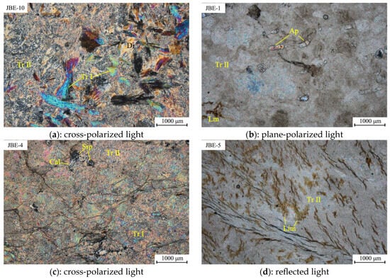
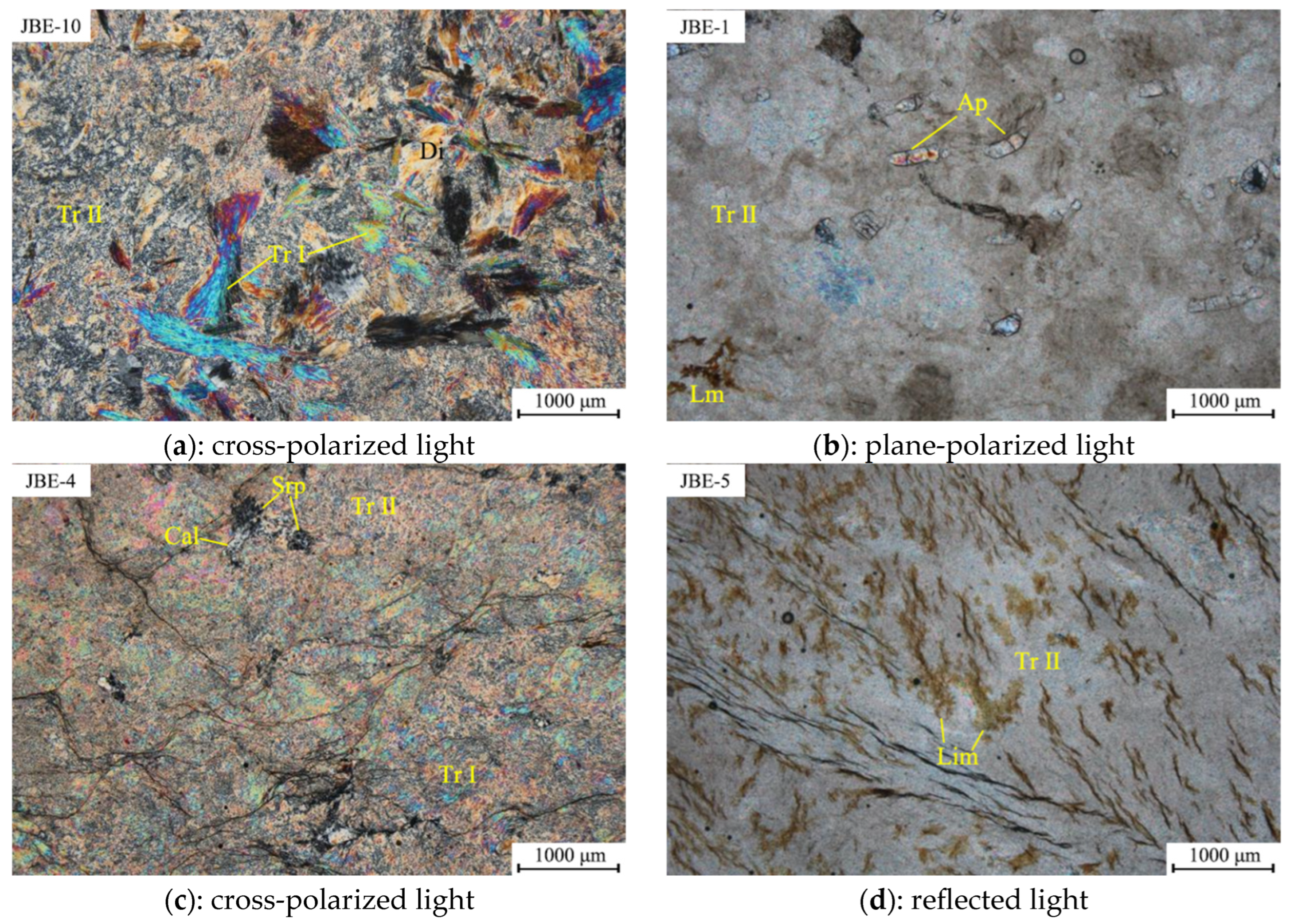
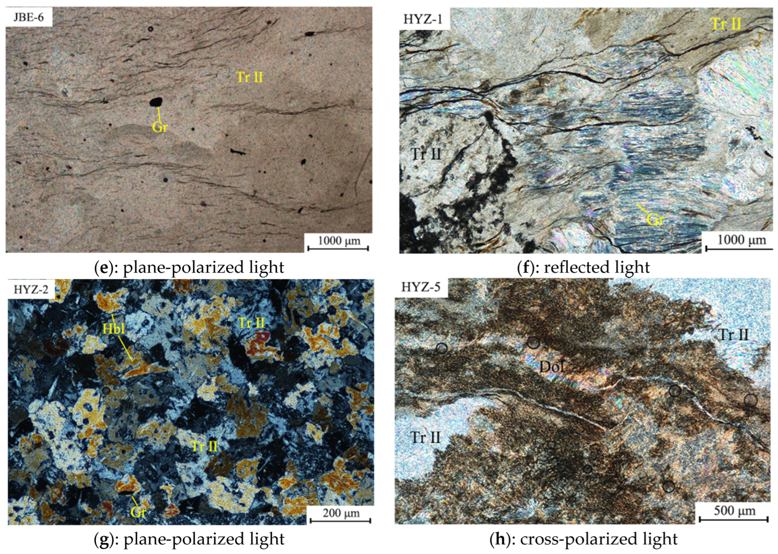
Tremolite occurs primarily as fibrous or cryptocrystalline aggregates, with minor coarse-grained occurrences, indicating multi-stage mineralization [22]. Fibrous aggregates (grain size: <10 μm) or cryptocrystalline aggregates (grain size: 10–200 μm) represent the late mineral phase (TrII). Coarse-grained occurrences (grain size: 100–1500 μm), which represent the early product of the Skarn Stage (TrI), are primarily found as metasomatic remnants within the TrII matrix, illustrating the generational replacement of minerals. Some of the coarse-grains retain their original crystal orientation (Figure 6a), while others show no directional structure. The optical properties of tremolite are consistent across stages: it exhibits second–order interference colors from blue to blue–green under cross-polarized light, and appears colorless under plane–polarized light.
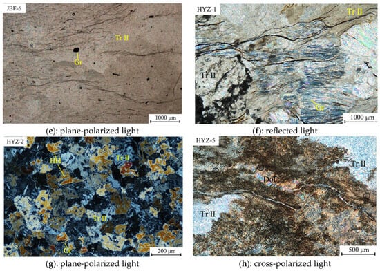
Figure 6.
Polarized light micro photographs of Mazongshan nephrite samples. (a). Coarse-grained crystals of tremolite (TrI: early tremolite) and diopside are embedded within fibrous tremolite (TrII: late tremolite), with their edges replaced by fibrous tremolite. (b). Apatite grains are sporadically embedded in tremolite (TrII) matrix, and arranged along the long axis of the grain itself. (c). Serpentine and calcite jointly replace tremolite. (d). Limonite infills the fractures within tremolite. (e). Graphite grains are embedded in the late-stage tremolite matrix. (f). Radial aggregates of acicular-prismatic graphite are distributed within the tremolite matrix. (g). Hornblende occurs semi-oriented within the tremolite matrix. (h). The edges of the dolomite have been replaced by tremolite, resulting in island-shaped remnants of dolomite. Mineral abbreviations: Tr—tremolite, Di—diopside, Lim—limonite, Ap—apatite, Cal—calcite, Srp—serpentine, Gr—graphite, Hbl—hornblende, Dol—dolomite.
4.3.2. Accessory Minerals
Dolomite occurs as irregular, island-shaped aggregates within the tremolite matrix, and its edges are replaced by tremolite (Figure 6h). It exhibits high-order white to iridescent interference colors under cross-polarized light.
Diopside is the predominant accessory mineral in Mazongshan nephrite. With grain sizes ranging from 10 to 1000 μm, it occurs as coarse-grained aggregates (Figure 6a and Figure 7c). It is weakly oriented within the multi-stage tremolite matrix, with edges frequently replaced by tremolite, and displays second–order interference colors from blue–green to orange under cross-polarized light.
Figure 7.
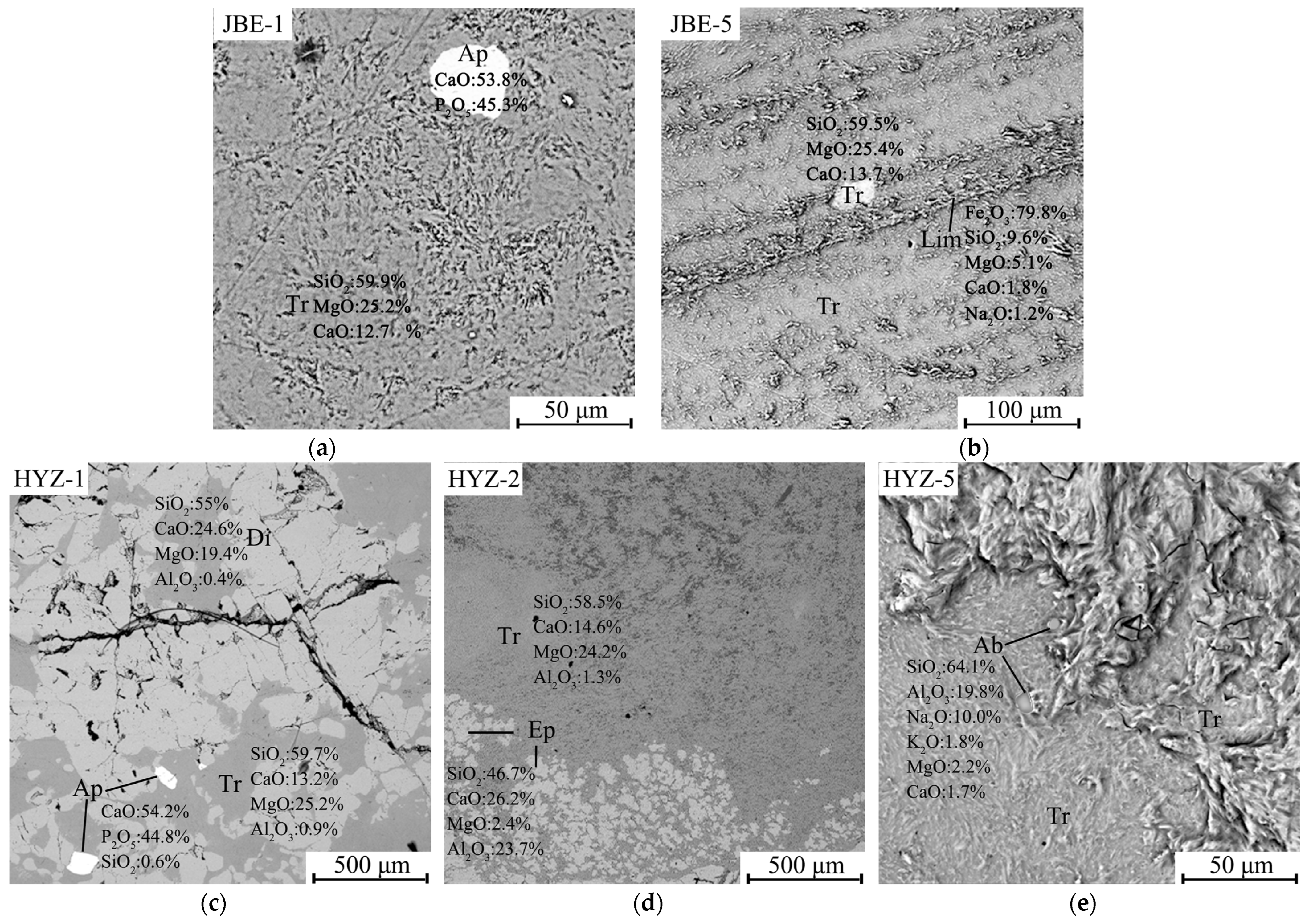
Scattered electron images of Mazongshan nephrite samples. (a). Subrounded apatite grain is embedded in tremolite matrix. (b). Limonite occurs as infillings in the fractures of tremolite. (c). Apatite grains are sporadically embedded in tremolite matrix, and the edges of diopside are replaced by tremolite. (d). Epidote occurs as scaly grains dispersed in tremolite. (e). Albite grains are sporadically embedded in the tremolite matrix, and the tremolite in sample HYZ-5 exhibits a fibrous interwoven texture. Mineral abbreviations: Hbl—hornblende, Dol—dolomite, Ep—epidote, Ab—albite.
The serpentine–calcite assemblage exhibits alteration features: serpentine forms island-shaped aggregates intermixed with anhedral granular calcite (grain size: 50–500 μm), with edges replaced by tremolite (Figure 6c). Serpentine shows first–order gray to gray–white interference colors under cross-polarized light. Calcite displays high-order white to iridescent interference colors.
Apatite, a common accessory mineral at both sites, occurs as long columnar or sub-rounded grains, ranging from 50 to 500 μm in size (Figure 6b and Figure 7a,c). It occurs within the late-stage tremolite and exhibits first–order gray under cross-polarized light and appears colorless under plane-polarized light.
Graphite occurs in two forms: as equiaxed individual crystals (approximately 200 μm in size, Figure 6e) or as acicular-prismatic radial aggregates (Figure 6f). Both forms are embedded within the tremolite matrix. Graphite appears black under cross-polarized light and lead gray under reflected light.
Hornblende is weakly oriented within the tremolite matrix in sample HYZ-2, as shown in Figure 6g, and exhibits distinct green to yellowish-green pleochroism under plane-polarized light.
Limonite, a secondary mineral, infills fractures in tremolite (Figure 6d and Figure 7b). It appears brown in reflected light and lacks metallic luster. The composition determined by EDS analysis, as shown in Figure 7b, closely matches the values reported in Reference [23] on a loss-on-ignition-free basis.
Epidote forms irregular and massive aggregates within the tremolite matrix. The composition determined by EDS analysis, as shown in Figure 7b, closely matches the values reported in Reference [24] on a loss-on-ignition-free basis.
Albite, with grain sizes ranging from 5 to 10 μm, occurs sporadically within the fibrous tremolite matrix. The composition determined by EDS analysis, as shown in Figure 7e, closely matches the values reported in Reference [25].
4.4. Major Chemistry
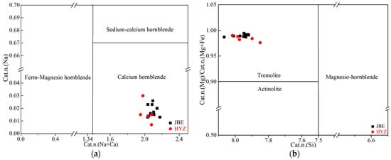
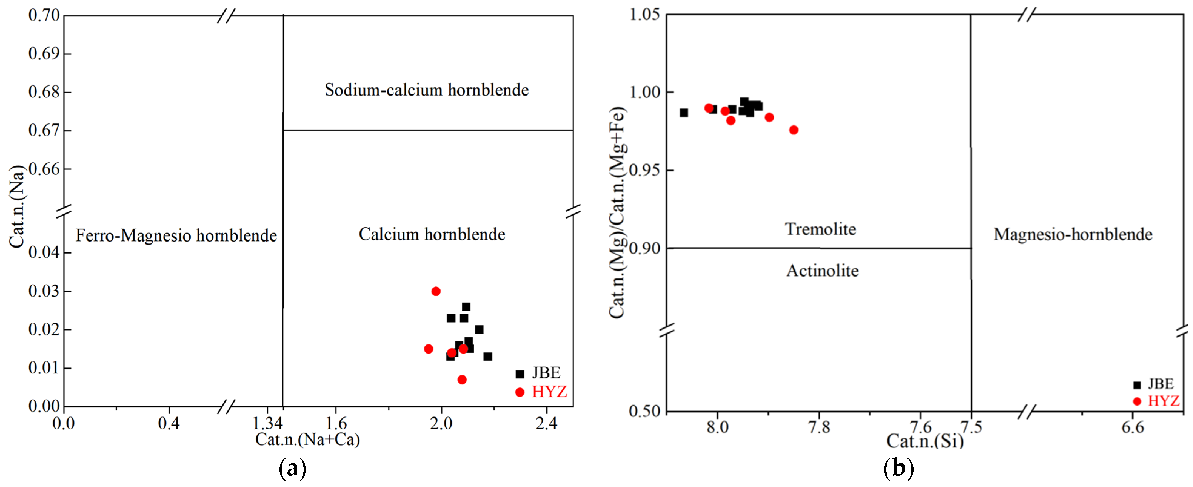
The primary theoretical compositions of tremolite are as follows: SiO2 at 58.80%, MgO at 24.60%, and CaO at 13.80%. As shown in Table 2, EPMA analysis of samples from the Jingbaoer Site exhibits average values of 58.2% SiO2, 23.76% MgO, and 12.76% CaO. Similarly, samples from Hanyaozi Site display average values of 58.06% SiO2, 23.73% MgO, and 12.43% CaO. The calculated cations are shown in Table 3. According to the amphibole classification diagram [26], all the Mazongshan nephrite belongs to the calcic group (Figure 8a). Furthermore, based on the calculated Mg/ (Mg + Fe2+) ratios, they are classified as tremolite (Figure 8b).
Table 2.
The EPMA analyses of Mazongshan nephrite (wt%).
Table 3.
The calculated cations of Mazongshan nephrite.
Figure 8.
Amphibole composition classification diagram of Mazongshan nephrite samples. Modifications were made based on Leake, B.E. et al., 1997 [26]. (a) Classification of calcic amphiboles, showing that all samples plot within the calcic amphibole group. (b) Classification based on Mg/(Mg + Fe2+) ratios, indicating that all samples are classified as tremolite.
4.5. Trace Elements and Rare Earth Element (REE) Analysis
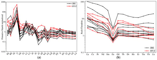
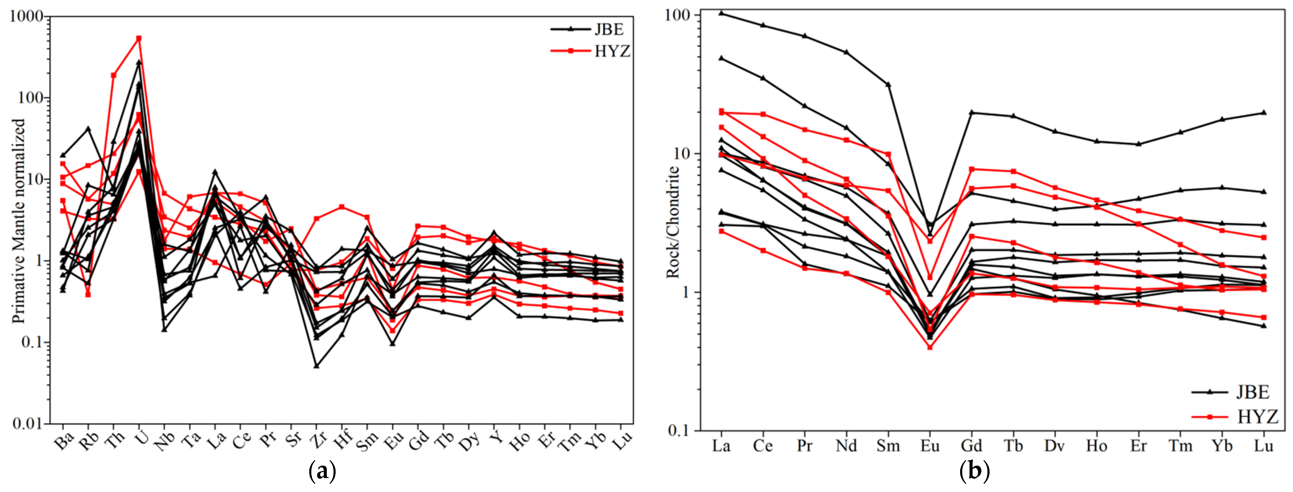
As shown in Table 4, the trace element compositions of Cr, Ni, and Co are significant geochemical indicators for the nephrite mineralization sources [27]. The Cr, Ni, and Co contents in both JBE and HYZ samples are lower than those in ultrabasic nephrites (Cr: 366–1184 µg/g, Ni: 14–47 µg/g, Co: 287–591 µg/g [28]), and are more similar to those in marble–related nephrite (Cr: 2–179 µg/g, Ni: 0.0 5–471 µg/g [29]). The concentrations of Ti, Mn, and Zn are relatively high, likely because Mazongshan nephrite was formed in the contact metasomatic zone between magnesium marble and intermediate-acidic magmatic rocks, where mineral-rich hydrothermal fluids introduced these elements [30]. The primitive mantle-normalized trace elements spider diagrams for the Mazongshan nephrite samples reveal enrichment in large-ion lithophile elements (Rb, U, Sr) and depletion in high-field-strength elements (Nb, Ta, Zr, Hf). This pattern likely results from metasomatism by granitic magmatic fluids during mineralization.
Table 4.
Bulk–rock trace element composition obtained by ICP-MS of Mazongshan nephrite. (µg/g).
The Mazongshan nephrite samples exhibit low ΣREE contents (JBE samples: 4.45–12.50 µg/g; HYZ samples: 3.64–30.22 µg/g). Chondrite–normalized REE patterns are characterized by steep patterns for the LREE and flat patterns for the HREE (Figure 9b), with LREE/ HREE ratios ranging from 2.90 to 10.83 (JBE: 2.90–9.70; HYZ: 3.16–10.83). The Ce anomalies (δCe) are weak or absent, with values ranging from 0.89 to 1.36 for JBE samples and 0.98 to 1.12 for HYZ samples, indicating deposition in an oxygen-rich environment. Significant negative Eu anomalies (δEu = 0.10–0.56 for JBE and 0.15–0.45 for HYZ) are observed, suggesting that tremolite underwent fractional crystallization during mineralization.
Figure 9.
(a) Spider diagram of trace elements of Mazongshan nephrite. (b) Rare earth element distribution curve of Mazongshan nephrite. (digitized standard value data: Sun & McDonough, 1989 [31]).
5. Discussion
5.1. Mineralization Type of the Mazongshan Nephrite Deposit
The mineral assemblages suggest a complex, multi-stage mineralization process. A prominent feature is the replacement of diopside by tremolite, displaying a typical residual structure (Figure 6a). Diopside likely formed during an earlier prograde metasomatic event, as expressed in reaction Equations (1) and (2) [32]. Although diopside and calcite were not observed to coexist in the samples, reference [32] indicates that, as metasomatism advanced, during the transition from the coarse-grained tremolite to fine randomly oriented tremolite (TrII), calcite could form as an intermediate reactant and subsequently be replaced by tremolite, as expressed in Equations (3) and (4). Following this early metasomatic phase, continued temperature increases during prograde metamorphism eventually destabilized the existing tremolite within siliceous dolomitic marbles. Equation (5) [33] captures this breakdown reaction, which produces forsterite (Fo) and accounts for its common coexistence with tremolite in such deposits.
CaMg[CO3]2 (Dol) + 2SiO2 → CaMgSi2O6 (Di) + 2CO2
5CaMgSi2O6 (Di) + H2O → Ca2Mg5(Si4O11)2(OH)2 (Tr) + 3Ca2+ + 6SiO2
5CaMg[CO3]2 (Dol) + 8SiO2 + H2O → Ca2Mg5(Si4O11)2(OH)2 (Tr) + 3CaCO3 (Cal) + 7CO2
2CaCO3 (Cal) + 5Mg2+ + 8SiO2 + 6H2O → Ca2Mg5(Si4O11)2(OH)2 (Tr) + 10H+ + 2CO2
Ca2Mg5Si8O22(OH)2 (Tr) + 11CaMg (CO3)2 (Dol) → 8Mg2SiO4 (Fo) + 13CaCO3 (Cal) + 9CO2 + H2O
Additionally, the sporadic occurrence of apatite suggests a later, distinct hydrothermal stage characterized by the influx of fluids enriched in phosphorus. The precipitation of apatite likely utilized calcium ions (Ca2+) that were either liberated during the earlier tremolitization of carbonate rocks (e.g., Equations (3) and (4)) or derived from renewed metasomatism [34]. This process, which consumed calcite, is described by Equation (6) and is consistent with petrographic observations (Figure 6b and Figure 7c).
5CaCO3 (Cal) + 3PO43− + F− → Ca5(PO4)3F (Ap) + 5CO2
The serpentine–calcite–tremolite assemblage in sample JBE-4 reveals additional complex mineral reactions during mineralization processes. Dolomite is altered by silica-rich hydrothermal fluids [24], resulting in a serpentine and calcite matrix that facilitates subsequent tremolite mineralization (Figure 6e). The relevant reactions are illustrated by Equations (7)–(9). Although this superimposed metasomatism is common in ultrabasic nephrites, such as the green nephrite in Manas [35], its occurrence in Mazongshan nephrite samples as minor serpentine + calcite/tremolite assemblages suggests a transitional mineralization mechanism.
CaMg[CO3]2 (Dol) + 2H+ → Mg2+ + CaCO3 (Cal) + CO2 + H2O
6Mg2+ + 4SiO2 + 6H2O + 4OH− → 2Mg3SiO5(OH)4 (Srp)
2Mg3SiO5(OH)4 (Srp) + 3SiO2 + 2Ca2+ → Ca2Mg5Si8O22(OH)2 (Tr) + 2H+ + 3H2O
Other secondary minerals also reveal the complexity of the mineralization. Graphite, along with epidote, albite [36], and hornblende [37], also indicates that the Mazongshan nephrite was formed through polyphase hydrothermal alteration and metamorphism. Additionally, the presence of limonite as an oxidation-derived secondary mineral reveals that the primary iron-bearing deposits underwent oxidation and hydration after being exposed to the surface [38].
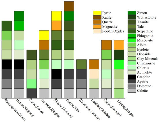
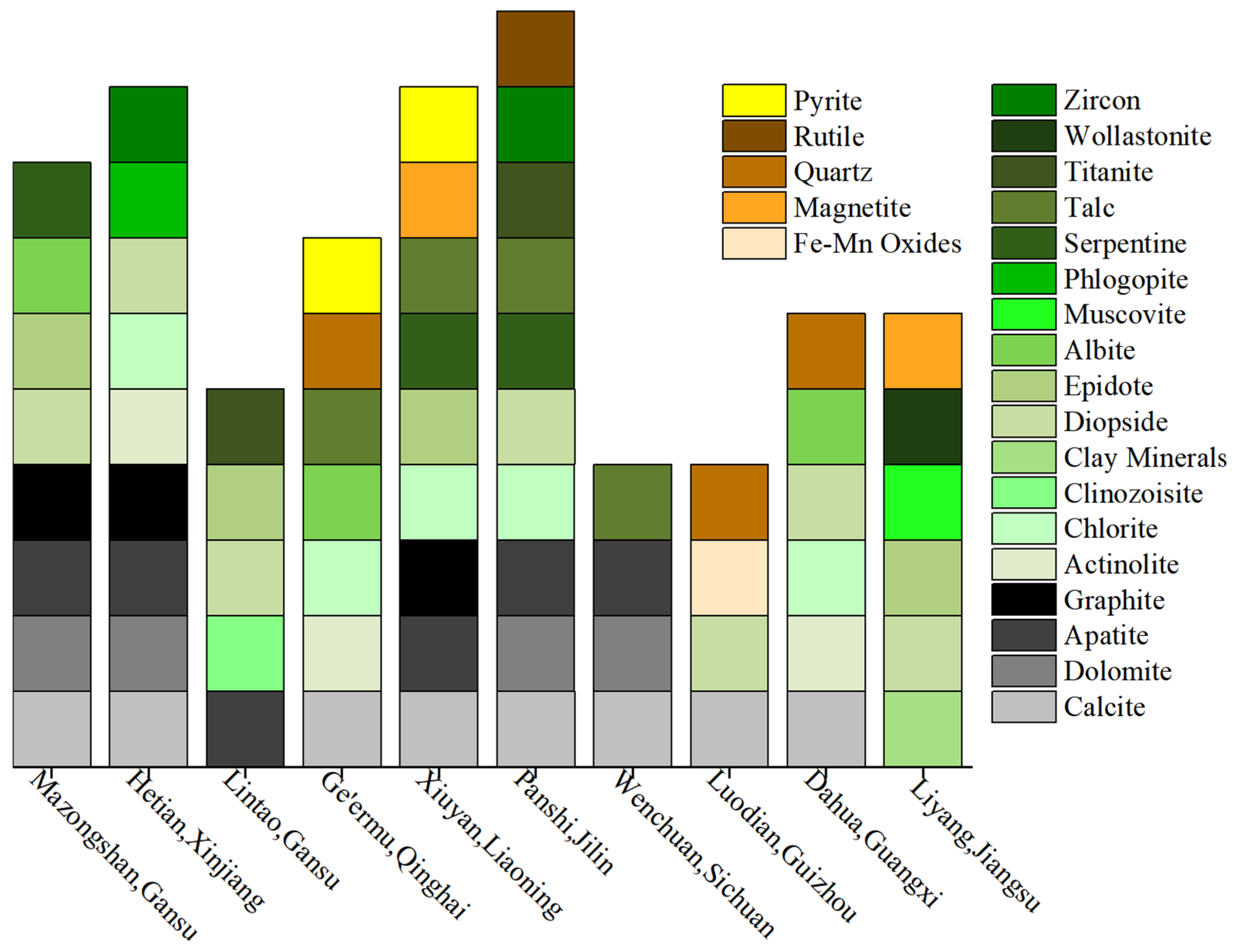
The analysis of minerals in gem-quality magnesian marble-type nephrite deposits in China reveals their composition [39,40,41,42,43,44], as shown in Figure 10. The accessory mineral characteristics of Mazongshan nephrite closely resemble those of nephrite from Hetian and Lintao, providing a reference for determining the provenance characteristics of Mazongshan nephrite, supporting its classification into a “Northwest Nephrite Group”. While this group is supported by accessory minerals, its broader geochemical and tectonic basis requires further verification. In fact, Zhang [45] analyzed 352 nephrite samples from 15 key localities across East Asia by using LA-ICP-MS, focusing on significant variations in lithium (Li) and beryllium (Be) concentrations, as well as cerium (Ce) anomalies, and categorized these samples into the North China Craton type, the Yangtze Craton type, and the Phanerozoic Orogenic belt type. It is noteworthy that the Northwest Nephrite Group, which includes classic sources such as Hetian, Lintao, and Mazongshan, is geotectonically situated within Phanerozoic orogenic belts.
Figure 10.
Accessory Mineral Assemblages of Chinese Nephrites in Magnesian Marbles.
Based on microscopic and geological evidence, the Mazongshan nephrite deposit is identified as a typical marble-hosted metasomatic nephrite deposit. Key factors contributing to this classification include:
(1) The Late Silurian Gongpoquan Formation volcanic-pyroclastic rocks (including andesite, dacite, and volcanic breccia) and Early Devonian intermediate-acidic intrusive rocks (monzonitic granite, syenogranite, and granodiorite) provided essential materials for metasomatism.
(2) The deposit is located in the Beishan Orogenic Belt, characterized by well-developed intrusive rocks and frequent volcanic activities from the Ordovician to Carboniferous. This prolonged magmatic-tectonic activity created a pervasive fracture network that served as conduits for multi-stage hydrothermal fluids, providing the spatial architecture necessary for large-scale metasomatism.
(3) The intermediate-acidic magmatic rocks exposed in the area are the direct source of the silica-rich hydrothermal fluids. These fluids formed tremolite through interaction with Mg- and Ca-bearing country rocks, as exemplified by the replacement of diopside.
(4) The mineralization involved polyphase fluid events, as evidenced by: (a) the replacement of diopside by coarse-grained tremolite; (b) the assemblage of serpentine, calcite, and tremolite indicating more complex mineral reactions during mineralization processes; (c) the sporadic distribution of apatite, marking a distinct later stage of phosphorus-rich fluid influx; and (d) the diverse secondary minerals including graphite, epidote, albite, and hornblende. These minerals collectively attest to a history of polyphase hydrothermal alteration and metamorphism.
5.2. Characteristics from the Perspective of REE
Rare earth elements (REE) act as essential geochemical tracers, commonly migrating as a group during geological processes. Despite extensive reworking, including magma melting and fluid metasomatism, the geochemical stability of REE provides a reliable framework for analyzing rock and ore formation mechanisms [46]. Their distribution and fractionation patterns effectively reflect petrogenesis and mineralization processes. By analyzing ΣREE contents and REE distribution patterns in tremolite from various deposits, Zhong [47] identified distinct REE characteristics for each deposit. This confirms that REE signatures serve as effective geochemical fingerprints for tracing nephrite provenance.
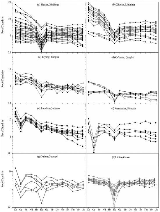
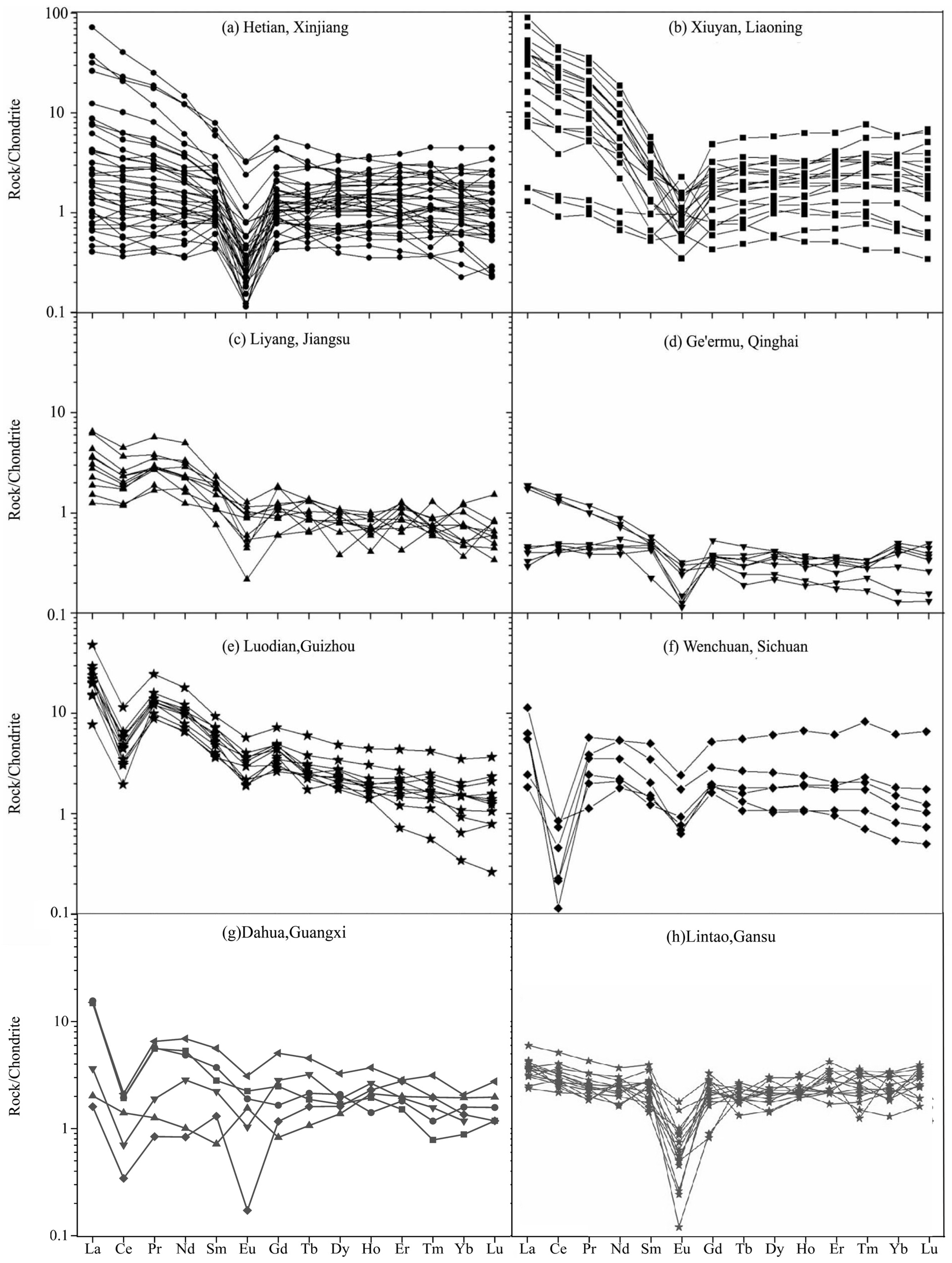
This Section presents a comprehensive analysis of the REE characteristics of Mazongshan nephrite, with comparisons to nephrite from Hetian, Ge’ermu, Xiuyan, Liyang, Luodia, Wenchua, Dahua, and Lintao, as cited in References [43,47,48]. The ΣREE contents in samples from Luodian and Xiuyan are significantly higher than those from other deposits. The LREE/ HREE ratios reveal the extent of rare earth fractionation: samples from Xiuyan and Luodian exhibit a pronounced rightward inclination on the REE distribution curves, indicating strong enrichment in light rare earth elements. In contrast, the distribution curves of samples from Lintao, Dahua, Wenchuan, and Ge’ermu display stable “seagull-shaped” patterns. Nephrite samples from Mazongshan, Hetian, and Liyang show a slight rightward distribution, as shown in Figure 11. The Ce anomalies (δCe) of Mazongshan nephrite exhibit weak to absent negative anomalies, similar to those observed in samples from Hetian, Lintao, Ge’ermu, and Xiuyan. Additionally, the Eu anomalies (δEu) of Mazongshan nephrite and the first three deposits display significant negative anomalies. In contrast, Ce anomalies in samples from Dahua, Liyang, Wenchuan, and Luodian exhibit significant negative anomalies, with particularly strong anomalies observed in Wenchuan and Luodian.
Figure 11.
Chondrite-normalized REE patterns of nephrite from different deposits in China. (Modifications were made based on References [43,47,48]). (a) Hetian, Xinjiang; (b) Xiuyan, Liaoning; (c) Liyang, Jiangsu; (d) Ge’ermu Qinghai; (e) Luodian, Guizhou; (f) Wenchuan, Sichuan; (g) Dahua, Guangxi; (h) Lintao, Gansu.
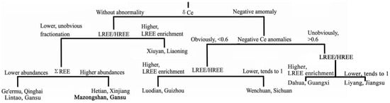
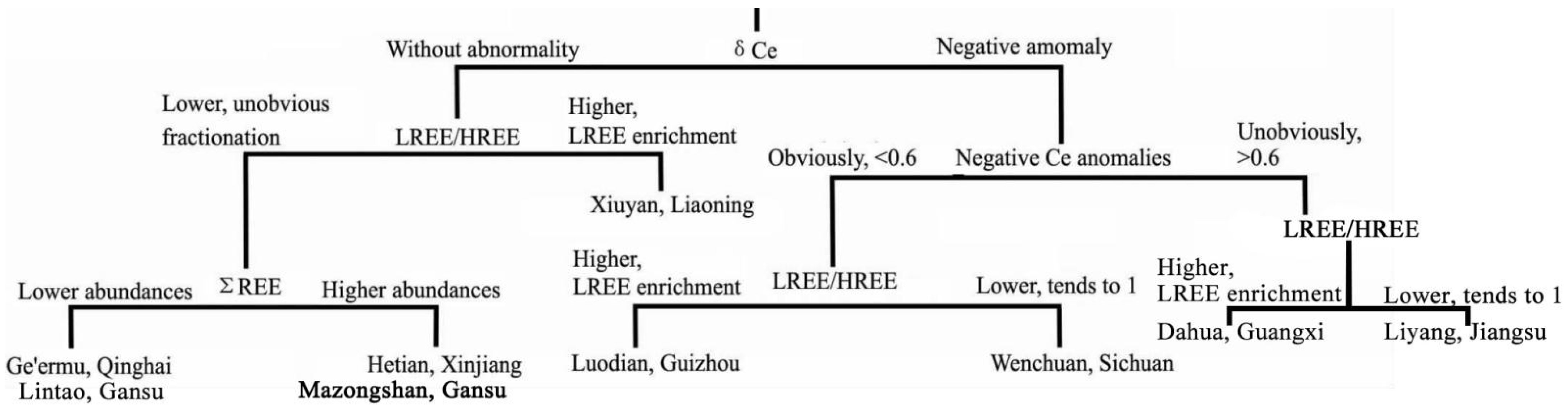
The REE characteristics of Mazongshan nephrite were classified, as shown in Figure 12. The results indicate that Mazongshan nephrite exhibits REE patterns most similar to Hetian nephrite, as evidenced by the LREE/HREE ratios, ΣREE contents, δCe, and δEu. This finding strengthens the scientific basis for the “Northwest Nephrite Group”.
Figure 12.
CART (classification and regression tree) for the recognition of nephrite from different deposits. Modifications were made based on References [43,47,48].
However, this raises a crucial geological and archeological issue: based on REE characteristics alone, distinguishing Mazongshan nephrite from Hetian nephrite is practically impossible. This compels a reconsideration of the discrimination patterns based on geochemical characteristics of nephrite sources within the “Northwest Nephrite Group”. Although the Kunlun Mountains host a typical Hetian nephrite metallogenic belt, the highly similar REE distribution patterns of Mazongshan nephrite strongly suggest that both may be governed by similar geochemical backgrounds or fluid evolution processes. Therefore, when investigating regional mineralization patterns and material sources, a scientifically open approach should be maintained towards nephrite exhibiting such REE characteristics. Given the confirmed evidence of ancient nephrite mining activities in Northwest China, it is necessary to re-evaluate the geological correlations for nephrite artifacts distributed along the Hexi Corridor and even in the Central Plains that possess these “Hetian-like” geochemical features. They could very well originate from neighboring metallogenic systems like Mazongshan. Consequently, the Mazongshan mine should not be regarded as a secondary supplementary source. It functioned as one of the primary, strategically significant sources within the “West-to-East” jade transmission system.
6. Conclusions
This paper provides new insights into the mineralogical and chemical composition of Mazongshan nephrite. The results reveal that the nephrite samples are predominantly composed of tremolite, with accessory minerals including diopside, apatite, serpentine, calcite, dolomite, graphite, hornblende, epidote, and albite, as well as limonite occurring as a secondary mineral formed by oxidation. The samples mainly exhibit fibrous textures, with common relict textures of metamorphic origin. A comparison of the accessory mineral assemblages, concentrations of Cr, Ni, and Co, REE characteristics, and the composition of other nephrite samples suggests that Mazongshan nephrite was formed by contact metamorphism between marble and igneous rock.
It is important to note that the accessory mineral assemblage of Mazongshan nephrite is more similar to that of Hetian (Xinjiang) and Lintao (Gansu) nephrite. Additionally, its REE characteristics closely align with those of Hetian nephrite, including a low ΣREE, slightly right-inclined distribution curves, weak to absent negative Ce anomalies, and pronounced negative Eu anomalies. These features, combined with the archeological significance of the Mazongshan Mining site—currently the earliest identified nephrite mining industrial site in China—establish a critical benchmark for determining nephrite provenance during the Pre-Qin Period, particularly associated with the “West–to–East Jade Transportation” system from the Warring States to the Han periods.
Author Contributions
Conceptualization, L.Y., G.C. and Y.X.; methodology, L.Y., X.L. and Y.X.; validation, L.Y.; formal analysis, L.Y.; investigation, Y.Y. and G.C.; resources, G.C. and Y.Y.; data curation, L.Y.; writing—original draft preparation, L.Y. and G.C.; writing—review and editing, L.Y., Y.Y., X.L. and Y.X.; visualization, L.Y.; supervision, Y.Y., X.L. and Y.X.; project administration, Y.Y.; funding acquisition, Y.Y. and G.C. All authors have read and agreed to the published version of the manuscript.
Funding
This Research is funded by the Archaeological Talent Promotion Program of China: 2025–192; National Key R&D Program of China: 2022YFE0203800.
Data Availability Statement
The data presented in this study are available in the article.
Conflicts of Interest
The authors declare no conflicts of interest.
Abbreviations
| Ab | albite |
| Ap | apatite |
| Cal | calcite |
| Di | diopside |
| Ep | epidote |
| Gr | graphite |
| Fo | forsterite |
| Hbl | hornblende |
| Lim | limonite |
| Srp | serpentine |
| TrI | early-stage tremolite |
| TrII | late-stage tremolite |
References
- Tung, C. East Asian Jade: Symbol of Excellence; Centre for Chinese Archaeology and Art, The Chinese University of Hong Kong: Hong Kong, 1998; ISBN 978-962-85303-2-8. [Google Scholar]
- Yang, B. On the Position & Network of “The Jade Road”. Acad. Forum Nan Du (J. Humanit. Soc. Sci.) 2004, 3, 113–117. [Google Scholar] [CrossRef]
- Lu, J.; Li, Y.; Ouyang, M. General History of Chinese Jade Ware; Haitian Publishing House: Shenzhen, China, 2014; ISBN 978-7-5507-1188-4. [Google Scholar]
- Xu, L. On Ancient China’s Jade Material Sources Going along The Course from Diversity to Integration. Palace Mus. J. 2020, 2, 94–107+111. [Google Scholar] [CrossRef]
- Xia, N. Han Jades. Acta Archaeol. Sin. 1983, 2, 125–150. [Google Scholar]
- Liu, F.; Yu, X. Classification and Mineralogical Characteristics of Nephrite Deposits in China. Miner. Resour. Geol. 2009, 23, 375–380. [Google Scholar]
- Hung, H.-C.; Iizuka, Y.; Bellwood, P.; Nguyen, K.D.; Bellina, B.; Silapanth, P.; Dizon, E.; Santiago, R.; Datan, I.; Manton, J.H. Ancient Jades Map 3000 Years of Prehistoric Exchange in Southeast Asia. Proc. Natl. Acad. Sci. USA 2007, 104, 19745–19750. [Google Scholar] [CrossRef] [Scilit]
- Bao, Y.; Yun, X.; Zhao, C.; Wang, F.; Li, Y. Nondestructive Analysis of Alterations of Chinese Jade Artifacts from Jinsha, Sichuan Province, China. Sci. Rep. 2020, 10, 18476. [Google Scholar] [CrossRef] [Scilit]
- Li, P.; Liao, Z.; Zhou, Z.; Wu, Q. Evidence from Infrared and Raman Spectra: Xiaomeiling Is One Reasonable Provenance of Nephrite Materials Used in Liangzhu Culture. Spectrochim. Acta Part A Mol. Biomol. Spectrosc. 2021, 261, 120012. [Google Scholar] [CrossRef] [Scilit]
- Liu, J.; Yang, M.; Su, Y.; Liu, Y. Analysis of Material and Source of Archaic Jade from the Tomb of Marquis Yi of Zeng in Suizhou, Hubei Province. Spectrosc. Spectr. Anal. 2022, 42, 215–221. [Google Scholar]
- Hu, Q.; Yang, M.; Liu, Y.; Liu, J.; Dai, L. Study on the Material and Mineral Source Characteristics of Jade Excavated from Longwangshan Tomb in Jingmen. Spectrosc. Spectr. Anal. 2022, 42, 3736–3744. [Google Scholar]
- Zhao, J.; Wang, H.; Chen, G.; Lu, G.; Sun, M. Report of the 2011 Excavation of the Mazongshan Ancient Jade Mine Site in Subei County, Gansu. Cult. Relics 2012, 8, 38–44+1. [Google Scholar] [CrossRef]
- Chen, G.; Yang, Y.; Zhang, P.; Zhang, F.; Kang, Y. A Brief Report on the Excavation of the Jade Mine Site in Hanxia, Dunhuang. Dunhuang Res. 2021, 5, 74–84. [Google Scholar]
- Liu, Y.; Fang, T.; Zheng, X.; Mao, Q.; Ao, S. Discovery of the Queershan Ophiolitic Mélange in the Beishan Orogen and the Implication for the Tectonic Evolution of the Altaids. Chin. J. Geol. (Sci. Geol. Sin.) 2021, 56, 471–482. [Google Scholar]
- Liu, J.; Cao, Y.; Chang, Y.; Su, Y.; Yu, X.; Yang, M. Mineralogy and Geochemistry Characteristics of Nephrite from Jingbaoer Grassland Jade Mine Site in Mazongshan Town, Gansu Province, China: Implications for the Provenance of Excavated Jade Artifacts. Minerals 2025, 15, 1186. [Google Scholar] [CrossRef] [Scilit]
- Zuo, Q.; Ye, T.; Feng, Y.; Ge, Z.; Wang, Y. Spatial Database of Serial Suite-Tectonic Map-Sheets of Mainland China (1:250,000). Geol. China 2018, 45, 1–26. [Google Scholar] [CrossRef]
- GB/T 14506.30—2010; Methods for Chemical Analysis of Silicate Rocks—Part 30: Determination of 44 Elements. Standardization Administration of China: Beijing, China, 2010.
- Khan, R.A.; Anwar-ul-Haq, M.; Qasim, M.; Afgan, M.S.; Haq, S.U.; Hussain, S.Z. Spectroscopic and Crystallographic Analysis of Nephrite Jade Gemstone Using Laser-Induced Breakdown Spectroscopy, Raman Spectroscopy, and X-Ray Diffraction. Heliyon 2022, 8, e11493. [Google Scholar] [CrossRef] [Scilit] [PubMed]
- Luo, B.; Yu, Y.; Liao, J. Nondestructive Testing Spectral Library and Analysis for Jewelry and Jade; China University of Geosciences Press: Wuhan, China, 2019; ISBN 978-7-5625-4199-8. [Google Scholar]
- Xiong, D.; Sun, X.; Zhai, W.; Shi, G.; Wang, S. Graphite Inclusions with High Crystallinity in the Auriferous Quartz Veins from Daping Gold Deposit in Yunnan Province, China: Evidence for Involvement of Metamorphic Fluid from Granulite-Facies Lower Crust in the Mineralization. Acta Geol. Sin. 2006, 9, 1448–1456. [Google Scholar]
- Lünsdorf, N.K.; Dunkl, I.; Schmidt, B.C.; Rantitsch, G.; von Hilmar, E. Towards a Higher Comparability of Geothermometric Data Obtained by Raman Spectroscopy of Carbonaceous Material. Part 2: A Revised Geothermometer. Geostand. Geoanalytical Res. 2017, 41, 593–612. [Google Scholar] [CrossRef] [Scilit]
- Tarling, M.S.; Smith, S.A.F.; Negrini, M.; Kuo, L.-W.; Wu, W.-H.; Cooper, A.F. An Evolutionary Model and Classification Scheme for Nephrite Jade Based on Veining, Fabric Development, and the Role of Dissolution–Precipitation. Sci. Rep. 2022, 12, 7823. [Google Scholar] [CrossRef] [Scilit]
- Chen, P.; Chen, T.; Xie, Q.; Xu, L.; Liu, H.; Zhou, Y. Mineralogy and Geochemistry of Limonite as a Weathering Product of Ilvaite in the Yeshan Iron Deposit, Tongling, China. Clays Clay Miner. 2018, 66, 190–207. [Google Scholar] [CrossRef] [Scilit]
- Kislov, E.V.; Goncharuk, I.S.; Vanteev, V.V.; Posokhov, V.F. Voimakan Deposit of Dolomite-Type Nephrite, Middle-Vitim Mountain Country: Formation Conditions. Geol. Ore Depos. 2024, 66, 752–768. [Google Scholar] [CrossRef] [Scilit]
- Hofmann, M.H.; Sherry, M.C. Synchronous Authigenic Albite and Dolomite Formation in Carbonates. Sedimentology 2025, 72, 2393–2423. [Google Scholar] [CrossRef] [Scilit]
- Leake, B.E.; Woolley, A.; Birch, W.; Grice, J.D.; Hawthorne, F.C.; Kato, A.; Kisch, H.J.; Vladimir, K.; Laird, J.O.; Mandarino, J.A.; et al. Nomenclature of Amphiboles—Report of the Subcommittee on Amphiboles of the International Mineralogical Association Commission on New Minerals and Mineral Names. Can. Mineral. 1997, 219–246. [Google Scholar]
- Li, N.; Bai, F.; Peng, Q.; Liu, M. Geochemical Characteristics of Nephrite from Chuncheon, South Korea: Implications for Geographic Origin Determination of Nephrite from Dolomite-Related Deposits. Crystals 2023, 13, 1468. [Google Scholar] [CrossRef] [Scilit]
- Yu, H.; Yang, X. Geochemical Characteristics, Mineralization Resource and Processes of Jasper from the World’s Major Producer. J. Guilin Univ. Technol. 2019, 39, 311–320. [Google Scholar]
- Jing, Y.; Liu, Y.; Zhang, Y.; Abuduwayiti, M. Metallogenic Age, Formation Process and Prospecting Direction of Marble-Related Nephrite Deposit in China. Acta Petrol. Mineral. 2022, 41, 651–667. [Google Scholar]
- Stanienda-Pilecki, K.J. Metasomatism of the Carbonate Rocks of the Contact Zone with the Basalt. Sci. Rep. 2025, 15, 14096. [Google Scholar] [CrossRef] [Scilit]
- Sun, S.; McDonough, W.F. Chemical and Isotopic Systematics of Oceanic Basalts: Implications for Mantle Composition and Processes. Geol. Soc. 1989, 313–345. [Google Scholar] [CrossRef] [Scilit]
- Jiang, Y.; Shi, G.; Xu, L.; Li, X. Mineralogy and Geochemistry of Nephrite Jade from Yinggelike Deposit, Altyn Tagh (Xinjiang, NW China). Minerals 2020, 10, 418. [Google Scholar] [CrossRef] [Scilit]
- Trommsdorff, V.; Evans, B.W. Antigorite-Ophicarbonates: Phase Relations in a Portion of the System CaO-MgO-SiO2-H2O-CO2. Contr. Mineral. Petrol. 1977, 60, 39–56. [Google Scholar] [CrossRef] [Scilit]
- Wang, L.; Zhang, Z.; Xie, Q. Geochemical Characteristics of Apatite and Their Geological Significance in the Jinling Skarn-Type Iron Deposit, Shandong Province. Acta Petrol. Mineral. 2025, 44, 1267–1284. [Google Scholar] [CrossRef]
- Tang, Y.; Liu, D.; Zhou, R. Geological Characteristics of Manasi Green Jade in Xinjiang. Acta Petrol. Mineral. 2002, 22–25. [Google Scholar]
- Zhang, C. The Chemical Component and Indication of Graphite Inclusions in Tremolite Jade from Sangpiyu. Liaoning. Master’s Thesis, China University of Geosciences (Beijing), Beijing, China, 2018. [Google Scholar]
- Feng, Z. Mineral Composition and Symbiosis of Tungsten Deposits in Malipo County, Yunnan Province. World Nonferrous Met. 2018, 9, 140–142. [Google Scholar]
- Han, W.; Hong, H.; Wu, Y.; Yin, K. Color Genesis of Brown Jade from Hetian Nephrite. Spectrosc. Spectr. Anal. 2013, 33, 1446–1450. [Google Scholar]
- Yang, X. Mineralization of Nephrite Metallogenic Belt in Hetian, Xinjiang. Doctoral Dissertation, China University of Geosciences (Beijing), Beijing, China, 2013. [Google Scholar]
- Yang, L. Study on Petro-Mineral Features and Genetic Mechanism of Luodian Jade, Guizhou Province. Doctoral Dissertation, Chengdu University of Technology, Chengdu, China, 2013. [Google Scholar]
- Bi, S. The Gemological Characteristic and Cause Study of Nephrite in Nachitai Golmud Qinghai, China. Doctoral Dissertation, China University of Geosciences (Beijing), Beijing, China, 2015. [Google Scholar]
- Lei, J. The Study on Geochemistry and Mineralization Age of Nephrite from Panshi, Jilin, Southeast China. Master’s Thesis, China University of Geosciences (Beijing), Beijing, China, 2020. [Google Scholar]
- Jiang, C. Gemmological Characteristics of Nephrite from Dahua, Hechi, Guangxi, and Implications on the Origin Identification. Doctoral Dissertation, China University of Geosciences (Wuhan), Wuhan, China, 2020. [Google Scholar]
- Wang, W.; Liao, Z.; Zhou, Z.; Shang, J.; Li, P.; Cui, D.; Li, L.; Chen, Q. Gemmological and Mineralogical Characteristics of Nephrite from Longxi, Sichuan Province. J. Gems Gemmol. 2022, 24, 20–27. [Google Scholar] [CrossRef]
- Zhang, Y.; Yang, J.; Qiu, Z.; Gu, X.; Luo, H.; Zhong, L.; Li, F.; Wang, H.; Chen, G.; Qin, S.; et al. A New Tectonic-Geochemical Approach Using LA-ICP-MS to Constrain the Provenance of East Asian Carbonate-Related Nephrite. J. Anal. At. Spectrom. 2025, 40, 2702–2715. [Google Scholar] [CrossRef] [Scilit]
- Zhou, X.; Zhao, E.; Yue, M.; Cao, D. Geological Significance of Rare Earth Elements in Geochemical Analysis. Geol. Resour. 2014, 23, 495–499. [Google Scholar] [CrossRef]
- Zhong, Y.; Qiu, Z.; Li, L.; Gu, X.; Luo, H.; Chen, Y.; Jiang, Q. REE Composition of Nephrite Jades from Major Mines in China and Their Significance for Indicating Origin. J. Chin. Rare Earth Soc. 2013, 31, 738–748. [Google Scholar]
- Zhang, Y.; Qiu, Z.; Yang, J.; Zhang, Y.; Yang, H.; Yang, J.; Li, L. The Geological and Geochemical Characteristics of Nephrites in Maxianshan, Gansu Province, and Their Implication for Raw Material Source of the Qijia Culture Jadewares. Acta Sci. Nat. Univ. Sunyatseni 2018, 57, 1–11. [Google Scholar] [CrossRef]

Disclaimer/Publisher’s Note: The statements, opinions and data contained in all publications are solely those of the individual author(s) and contributor(s) and not of MDPI and/or the editor(s). MDPI and/or the editor(s) disclaim responsibility for any injury to people or property resulting from any ideas, methods, instructions or products referred to in the content. |
© 2026 by the authors. Licensee MDPI, Basel, Switzerland. This article is an open access article distributed under the terms and conditions of the Creative Commons Attribution (CC BY) license.