


Abstract
Antiviral substances are considered emerging contaminants. Once released into the environment, they may affect organisms through complex and often still-unknown mechanisms. This study focuses on a class of antiviral substances with potential use in treating COVID-19 patients, aiming to identify specific structural characteristics that significantly contribute to their ecotoxicity. An empirical approach called quantitative structure–activity relationship (QSAR) was used for this purpose. The study examined 13 antiviral substances: atazanavir, daclatasvir, darunavir, emtricitabine, favipiravir, lopinavir, nirmatrelvir, oseltamivir, remdesivir, ribavirin, ritonavir, and sofosbuvir. The ecotoxicity of these antivirals was assessed using three tests: the Aliivibrio fischeri test, the Chlorella sp. test, and the Pseudomonas putida test. These three microorganisms represent different trophic levels in aquatic and soil ecosystems. Ecotoxicity was expressed as EC20 and EC50, and these values served as the dependent variables in the QSAR models. A large set of numerical descriptors calculated from the molecular structures of the antivirals was used as an independent variable. EC20-based QSAR models offer insight into the effects of antivirals under sub-lethal exposure conditions. The results indicated that sub-lethal exposure in Aliivibrio fischeri was associated with favorable electronic properties and compact structures that promote cellular accumulation, while long-range fragments reduced toxicity. In Chlorella sp., sub-lethal exposure was driven by optimal molecular size, chain length, and specific electronic groups enabling cell penetration and biochemical inhibition. For sub-lethal exposure in P. putida, lipophilicity and reactive group geometry enhanced toxicity, while high short-range polarity mitigated it by limiting membrane permeability. Acute toxicity patterns showed similar trade-offs: strong electronic reactivity increased potency, but steric bulk, long-range polarity, or unfavorable mass distribution frequently restricted bioavailability and reduced toxic effects. Overall, the models demonstrated that antiviral toxicity results from a balance of electronic activity, structural accessibility, and physicochemical constraints, providing a mechanistic basis for predicting the environmental risk of selected antiviral substances.
1. Introduction
The high quality-of-life enjoyed by modern humans is largely due to advances in medicine. Today, drugs are available to alleviate, control, or resolve various health problems. Given the importance of medicine for quality-of-life, the adverse effects of drugs entering the environment were long overlooked. However, legislation in many countries, especially developed ones, now recognizes that pollution of water and soil with pharmaceuticals or pharmaceutical residues is an emerging environmental issue [1,2]. The presence of pharmaceuticals in the environment can cause various adverse effects, including the development of microbial resistance, bioaccumulation, and potential toxic effects on organisms [3,4].
Drug consumption results in about 30–40% of the unmetabolized active substance being excreted from the body into wastewater [5]. Unfortunately, conventional wastewater treatment plants cannot completely remove pharmaceuticals from wastewater [6,7,8]. Therefore, drug consumption is one way these substances enter the environment. Other sources of environmental pollution include improper drug disposal and effluents from pharmaceutical industries [7].
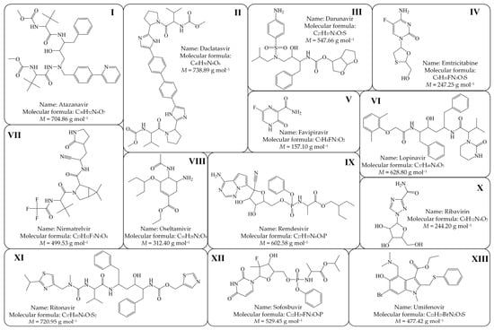
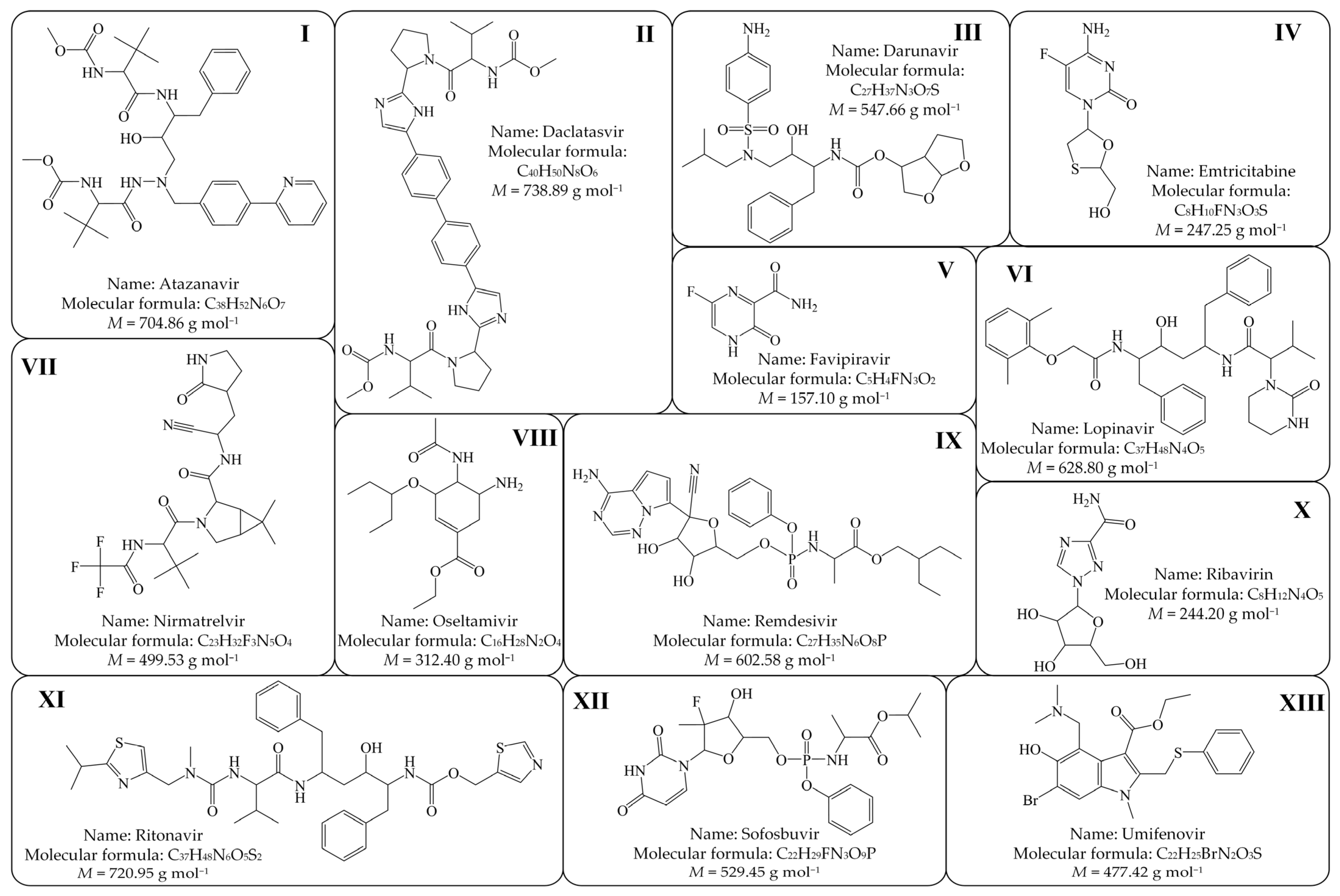
The recent COVID-19 pandemic posed a significant health challenge for the global population. Although the COVID-19 pandemic officially ended in 2023, the SARS-CoV-2 virus continues to circulate among the population. During the pandemic, especially in its early stages, no drugs were specifically developed to treat SARS-CoV-2 infection. Consequently, some existing antivirals, such as atazanavir, daclatasvir, darunavir, emtricitabine, favipiravir, lopinavir, oseltamivir, remdesivir, ribavirin, ritonavir, sofosbuvir, and umifenovir, were used because they demonstrated certain positive effects in COVID-19 patients. Over time, the use of some of these repurposed antivirals for treating COVID-19 patients has declined, primarily due to insufficient efficacy or potential adverse side effects [9,10,11,12,13]. Meanwhile, new drugs developed specifically to target SARS-CoV-2, such as nirmatrelvir, have been introduced. In this paper, we focused on the 13 antivirals mentioned above and analyzed their structural features responsible for toxicity. We applied the QSAR (quantitative structure–activity relationship) methodology in our analysis. The molecular structures and basic information for the antivirals used in this study are shown in Figure 1.
Figure 1.
Molecular structures and basic information on the 13 antivirals used in this study: (I) Atazanavir, (II) Daclatasvir, (III) Darunavir, (IV) Emtricitabine, (V) Favipiravir, (VI) Lopinavir, (VII) Nirmatrelvir, (VIII) Oseltamivir, (IX) Remdesivir, (X) Ribavirin, (XI) Ritonavir, (XII) Sofosbuvir, (XIII) Umifenovir.
Information on the occurrence of some of the 13 selected antivirals in the aquatic environment is available in the literature. A brief overview of related studies is presented in Table 1. No information was found on the concentrations of daclatasvir, nirmatrelvir, remdesivir, or sofosbuvir in the aquatic environment. Said et al. [14] mention the existence of reports in the literature regarding the presence of daclatasvir in various aquatic media but do not cite these reports, which prevents confirmation of this statement with concrete concentration values.
Table 1.
A brief review of available studies on the presence of the 13 tested antivirals in the aquatic environment.
Based on the above, conducting a systematic assessment of the ecotoxicity of 13 selected antiviral agents is crucial to better understand their environmental fate and potential ecological hazards. In one of our previous studies, we evaluated the ecotoxicity of six antivirals toward the bacterium Aliivibrio fischeri [26]. Although several additional toxicity studies on Aliivibrio fischeri exist [27,28,29], the available toxicity data for most of these 13 antivirals are predominantly derived from studies on higher organisms (e.g., mice, rats, rabbits) conducted primarily to establish safe dosage levels for humans [30,31,32,33,34,35]. Consequently, comprehensive ecotoxicity data for these pharmaceuticals remain scarce. This lack of empirical information has become particularly evident since the emergence of SARS-CoV-2 and the global COVID-19 pandemic, during which computational drug-discovery efforts increasingly leveraged QSAR techniques to identify or repurpose antiviral molecules.
However, developing reliable QSAR models for antiviral ecotoxicity remains challenging. One of the main obstacles is the limited availability of empirical aquatic toxicity data, which restricts model training and external validation. Additionally, antiviral compounds represent a chemically diverse group—including protease and polymerase inhibitors as well as numerous repurposed drugs—making it difficult to establish generalized predictive relationships. Simplified QSAR frameworks further tend to overlook environmentally relevant processes such as degradation, metabolite formation, and changes in bioavailability. Despite these challenges, recent progress in in silico risk assessment demonstrates the growing potential of computational ecotoxicology [36].
In this study, the toxicity of selected antivirals was evaluated using three representative test organisms: the bacteria Pseudomonas putida and Aliivibrio fischeri, and the freshwater microalgae Chlorella sp. QSAR modeling was used to establish correlations between the molecular structure characteristics of the antivirals and the assessed toxicity values. Toxicity was assessed at two levels: EC20 and EC50. EC20 represents the concentration that causes a 20% effect in the tested population and is typically associated with sub-lethal exposure conditions, capturing subtle but ecologically relevant long-term responses such as reduced growth or reproduction. EC50 corresponds to a 50% effect in the tested population and reflects acute toxicity and overall hazardous potential, indicating stronger short-term impacts on the test organisms. Based on the assessed toxicity values, predictive toxicity models were developed separately for each test organism.
The primary objective of this study is to fill the current data gap on the ecotoxicological profiles of selected antiviral drugs.
2. Materials and Methods
2.1. Antiviral Solutions Preparation
To minimize the impact of potential data extrapolation when determining ecotoxicity values, the antiviral concentrations in stock solutions were significantly higher than those reported for aquatic ecosystems (Table 1). Solutions of eight antivirals were prepared at the maximum concentrations we were able to dissolve in water: atazanavir at 0.0030 mM; daclatasvir at 0.1000 mM; darunavir and favipiravir at 0.2000 mM; lopinavir, remdesivir, and ritonavir at 0.0020 mM; and umifenovir at 0.0001 mM. The other antivirals have much higher solubility than these eight, so we prepared their stock solutions at a concentration of 1.0000 mM. The concentration of each prepared stock solution was checked by LC analysis (Vanquish UHPLC, Thermo Fisher Scientific, Waltham, MA, USA).
2.2. Ecotoxicity Measurement
2.2.1. Luminescence Inhibition Test with Marine Bacteria Aliivibrio fischeri
The bacterium Aliivibrio fischeri was cultivated according to ISO 11348-3:2007 [37]. Freeze-dried bacterial cells were rehydrated in a standard-defined resuspension solution and subsequently subcultured for 24 h before testing in a growth medium optimized for its growth. The initial luminescence of the bacterial culture was verified to be about 3000 relative light units before testing to ensure consistent and comparable results. As a control solution, a 2% NaCl solution was used. A linear dilution series was applied to the samples, and measurements were taken over a 30 min testing period. Temperature control of the solutions was achieved using a LUMIStherm thermostat, and bacterial luminescence was quantified with a LUMIStox 300 luminometer (Dr. Lange GmbH, Hannover, Germany). The inhibition of the luminescence (INH) was calculated according to Equation (1).
L0 stands for initial luminescence, L30 for luminescence after a 30 min exposure, and f for the factor used to correct the initial value of bacterial luminescence in the test solutions.
2.2.2. Growth Inhibition Test with Soil Bacteria Pseudomonas putida
Pseudomonas putida bacteria were cultivated according to the guidelines ISO 10712 [38]. The culture was cultivated on nutrient agar at 37 °C for 24 h in a thermostat. The resulting 24 h culture was used to perform the ecotoxicity test. Prior to the start of the experiment, the bacterial culture was pre-cultivated for 5 ± 0.1 h to allow adaptation to the conditions of the mineral medium [39]. The exception from the standard was the mineral medium employed in the experiment, which was prepared without the addition of glucose. The optical density of the bacterial suspension was determined using a DR2400 spectrophotometer (Hach, Loveland, CO, USA), measuring the absorbance at a wavelength of 436 nm. The initial optical density of the bacterial suspension prepared was OD = 0.2. The experiment was conducted in 100 mL Erlenmeyer flasks with a working volume of 20 mL. Each flask was filled with the appropriate volumes of bacterial suspension, mineral medium, and antiviral solution. The prepared flasks were placed on a rotary shaker at room temperature and agitated at 150 rpm to ensure homogenization and aeration of the samples. Control flasks (blanks) were also prepared without the addition of the antiviral solution. After 16 h of incubation, the total number of colony-forming units (CFUs) was determined. Serial tenfold dilutions (10−1 to 10−6) were prepared and 1 mL of the final dilution was transferred to a Petri dish, overlaid with nutrient agar, and incubated in a thermostat at 37 °C for 24 h. Following incubation, the number of bacterial colonies was counted to determine the colony-forming units at each antiviral concentration. Inhibition was calculated based on the CFU value in each sample and the CFU value in the blank sample according to Equation (2).
2.2.3. Growth Inhibition Test with Freshwater Microalgae Chlorella sp.
The ecotoxicity test using the microalgae Chlorella sp. was carried out according to OECD guidelines [40]. The microalgae were cultivated for two weeks in basal medium under continuous aeration which promoted homogenization of the suspension, prevented microalgal sedimentation, and maintained the dissolved oxygen concentration required for growth. Optical density of the suspension was measured spectrophotometrically (DR2400 spectrophotometer, Hach, Loveland, CO, USA) at a wavelength of 670 nm. The concentrated suspension was afterwards diluted as needed to obtain an initial optical density of OD = 0.02 for the experiment. The experiment was set up in 100 mL Erlenmeyer flasks with a working volume of 20 mL. The appropriate volume of microalgae suspension, basal medium, and antiviral solution was added to each flask. The solutions thus prepared were placed on a rotary shaker at room temperature and 150 rpm for homogenization. Blanks were also set up, which contained everything except the antiviral solution. The experiment was conducted over a 72 h period, during which the total number of viable microalgal cells (CFUs) was determined every 24 h with a Thoma counting chamber. Cell counts were performed in three large squares, and the number of cells within each square was recorded. The number of viable cells per milliliter (N) was calculated according to Equation (3), where n is the reciprocal of the dilution factor, m is the total number of counted cells, 16 represents the total number of large squares, 104 is the volume correction factor, and K is the number of large squares in which the counting was performed. Inhibition was then calculated based on Equation (3):
2.2.4. Calculation of Ecotoxicities
After the experiments, the EC20 and EC50 values were calculated from the linearization of the experimental data—inhibition (INH) and the tested antiviral concentrations (c). Therefore, calculations were made from the relationship of ln γ and ln c, where ln γ was calculated according to Equation (4). (INH represents the inhibition of the observed feature, e.g., luminescence or bacterial/algal growth, for a specific tested concentration of antiviral drug.)
2.2.5. QSAR Modeling
The dataset for QSAR modeling of ecotoxicity includes data on 13 selected antivirals obtained from three ecotoxicity tests (Aliivibrio fischeri, Chlorella sp. and Pseudomonas putida). Despite the small number of compounds, the dataset provides a focused chemical space useful for preliminary mechanistic insight [41].
For each antiviral molecule, structural descriptors and physicochemical parameters were compiled. Experimentally determined ecotoxicity values served as the dependent variables in modeling, and molecular descriptors as independent variables. The dataset was cleaned, standardized, and checked for consistency to ensure suitability for QSAR model development.
Molecular structures of the antivirals studied were built using Chem3D Pro software (ChemOffice 15.0, PerkinElmer, Waltham, MA, USA) and optimized by the AM1 method using the MOPAC2012 interface. The semiempirical quantum-chemical descriptors including the dipole moment (total µ and its X, Y, and Z–Cartesian components), EHOMO (energy of the highest occupied molecular orbital), ELUMO (energy of the lowest unoccupied molecular orbital), the HOMO–LUMO gap (HLG), final heat of formation (ΔHf), and ionization potential, were calculated. The molecular descriptors were computed using DRAGON 6.0 software (Milano Chemometrics & QSAR Research Group, TALETE, Milano, Italy). Of the 4885 available descriptors in DRAGON 6.0, 4870 were calculated using AM1-optimized molecular structures. These descriptors capture key structural features of the compounds used to develop QSAR models and effectively represent the chemical diversity of the studied antivirals. Before calculation and variable selection, the correlation coefficients for all pairs of descriptor variables used in the models were evaluated to identify highly correlated descriptors and detect redundancy in the data set. As a result, many highly correlated and constant descriptors (R ≥ 0.99) were excluded, leaving a total of 3165 DRAGON descriptors and 9 previously mentioned semiempirical quantum-chemical descriptors in the calculation matrix.
The QSAR models were developed using three independent variables (three descriptors) and a Multiple Linear Regression (MLR) approach. Models were generated separately for EC20 and EC50 values. The most significant descriptors were selected using a Genetic Algorithm (GA) optimization method. Optimization was performed with QSARINS 2.2 (QSAR Group, University of Insubria, Como, Italy) software. The GA procedure was initialized with a population of 100 random models and evolved over 1000 iterations, using a mutation probability of 20%.
To prevent using models with highly correlated descriptors or with weak correlations between descriptors and responses, we first applied the QUIK rule in QSARINS. Then, before selecting final models, we used additional built-in tools such as “CI,” which ranks models based on the size of their confidence intervals and the significance of their coefficients [42]. This process removed all models with strongly inter-correlated descriptors or with non-significant coefficients from further consideration.
The selection and validation of models were conducted by comparing values of selected statistical performances: R2 (the model-explained variance), R2adj (R2 adjusted for the number of predictors in the model), F (F-ratio between the variances of observed and calculated activity), RMSEtr and RMSEcv (root mean square error of the training set and cross-validation set, respectively), and Q2loo (leave-one-out cross-validation coefficient). Validation of the models selected as best for each chosen response was also performed using the Leave Many Out (LMO) approach and Y-scramble tests [43].
3. Results and Discussion
3.1. Ecotoxicity Measurements
Ecotoxicity measurements were conducted using stock solutions of antivirals, and the results are shown in Table 2.
Table 2.
Toxicity values for three different toxicity tests. Experimentally determined EC20 and EC50 toxicities are presented as the determined toxicity value ± standard deviation.
The assessed EC20 and EC50 values show that the three test microorganisms differ in their sensitivity to the antiviral substance. However, to confirm that the three toxicity tests yield statistically different results, a one-way analysis of variance (ANOVA) was performed at a significance level of 0.05. For this analysis, we selected EC20 values because their assessment was less susceptible to extrapolation. The ANOVA results are presented in Table 3. As shown in the table, the calculated p-value is below 0.05 and the F-value is slightly higher than the critical F-value, confirming the hypothesis that the three applied ecotoxicity tests are statistically independent.
Table 3.
The results of the analysis of variance for three applied ecotoxicity tests. The analysis was conducted at a significance level of p = 0.05.
It is noteworthy (Table 2) that there is inconsistency in the estimated toxicities for some related antivirals. For example, atazanavir, darunavir, lopinavir, and ritonavir were primarily developed as HIV-1 protease inhibitors and can therefore be considered related. However, the order of toxicity for these three antivirals varies among the Aliivibrio fischeri, Chlorella sp., and Pseudomonas putida tests. This supports our assumption of different mechanisms of toxic action in these three microorganisms.
For the antivirals studied, we attempted to find experimentally available ecotoxicity values that could be compared with those estimated in this work. However, in addition to the general lack of ecotoxicity data, a major issue is that assessments are conducted using different tests or refer to different levels of toxic effect, making it difficult to find comparable measurements. Among the few comparable reports we found, Bartels et al. [28] reported an EC50 value for favipiravir toxicity to Aliivibrio fischeri of 86.2 mg/L and greater than 19.3 mg/L for remdesivir. Escher et al. [44] reported an EC50 value of oseltamivir against Aliivibrio fischeri as 3124 mg/L, while Straub [45] reported an EC50 value of the same compound against Chlorella vulgaris as 343.64 mg/L.
3.2. Modeling
Because the observed toxicity values for Aliivibrio fischeri, Chlorella sp., and Pseudomonas putida varied widely and did not allow straightforward interpretation, QSAR modeling was applied to elucidate the underlying chemical properties and bioavailability factors contributing to these differences.
To elucidate the chemical mechanisms underlying toxicity across different microorganisms, a three-variable model was used, as it provided the most information about the structural features responsible for toxicity, despite the general rule of thumb that the ratio of molecules to variables should be at least five to one to avoid overfitting [46]. All QSAR models were confirmed to be reliable, with a Concordance Index (CI) of 1, indicating that all compounds fell within the applicability domain, and a QUIK value of 0.50, demonstrating an appropriate balance between the number of descriptors and the size of the dataset [42]. The best selected models and their statistical evaluations are presented in Table 4, while Table 5 provides basic information about the descriptors used in the developed models.
Table 4.
Model equations and statistical analysis of models with internal validation.
Table 5.
Names and definitions of descriptors included in the best 3-variable models for prediction of EC20 and EC50.
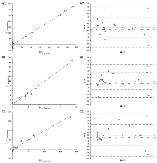
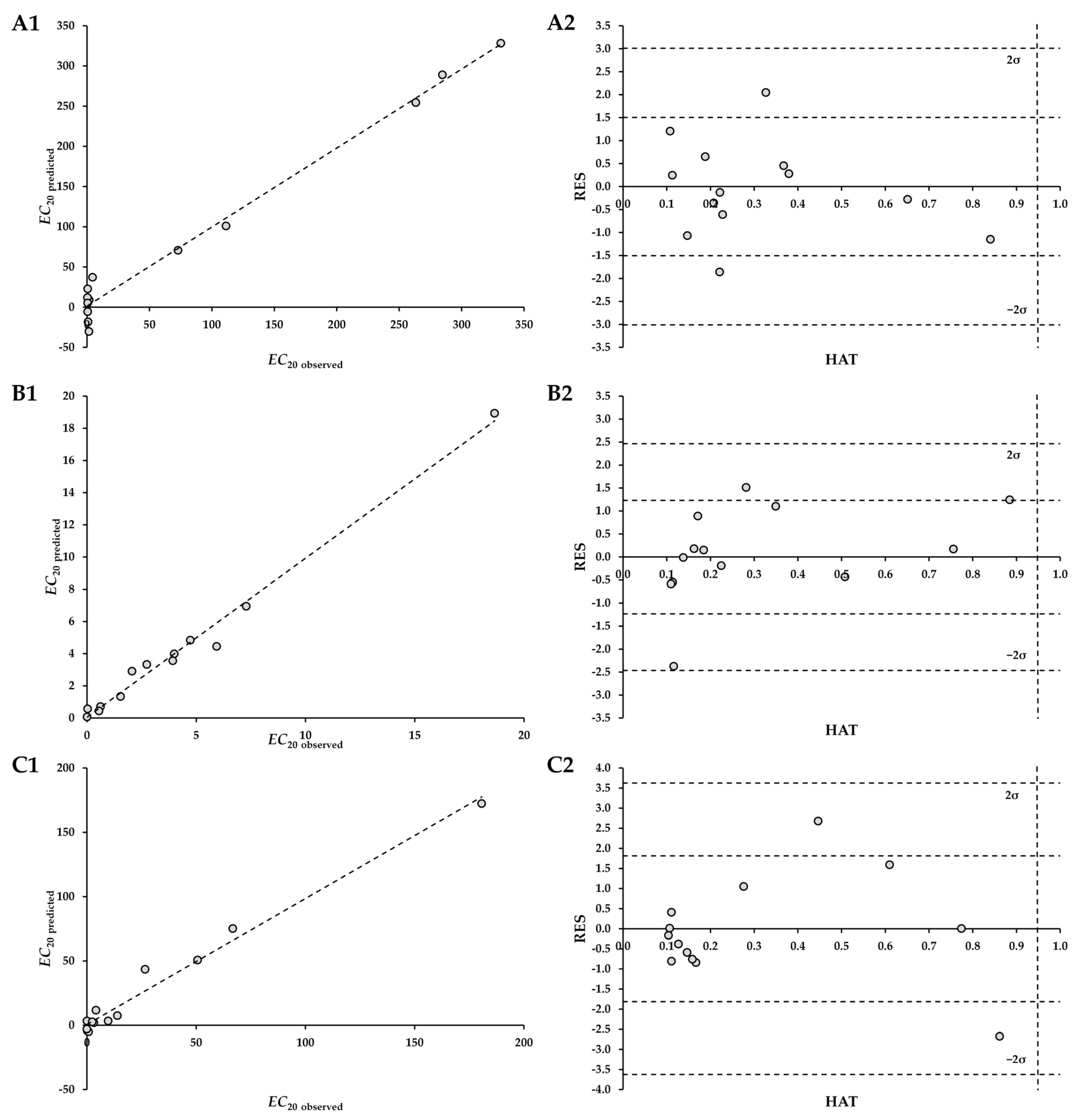
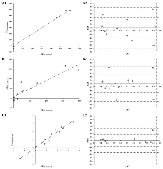
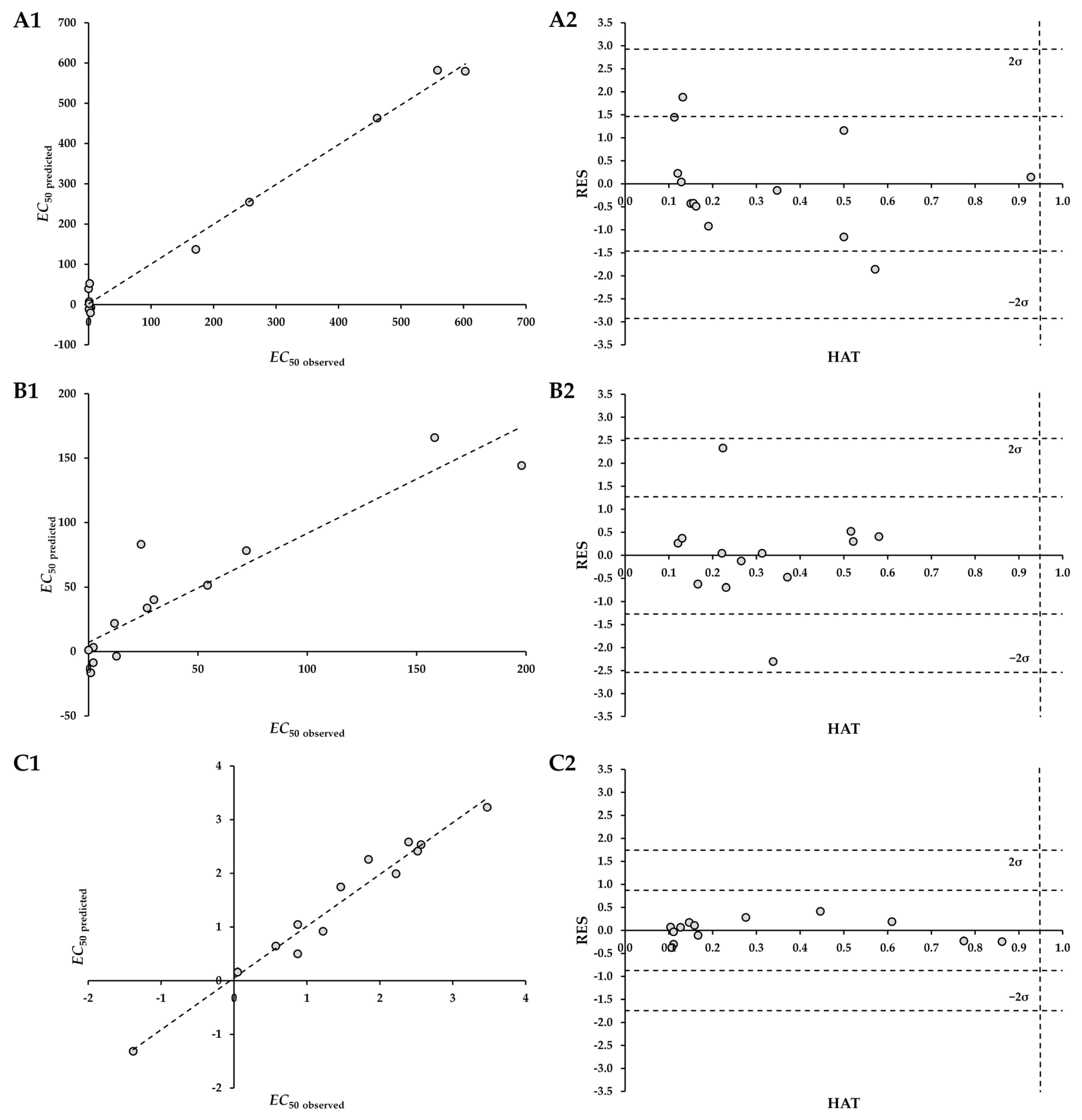
The developed QSAR models have been evaluated and the results are shown in Figure 2 and Figure 3 for the EC20 and EC50 models, respectively. Cases A in these figures show the accuracy of the models by illustrating the relationship between the predicted antiviral toxicities (Ypred) and the experimentally determined values (Yobs). The line Y = X represents the ideal scenario, indicating perfect agreement between prediction and observation. Cases B in the same figures show the Williams plot. Analysis of the Williams plots confirmed that all compounds fall within the model’s applicability domain. Specifically, no Y outliers were detected, as all standardized residual values were within the ±3.0σ limits. Furthermore, with all leverage HAT values below the warning limit hii = 0.5, the absence of X outliers was also confirmed [47].
Figure 2.
Observed vs. predicted plot (Y = X plot, case 1) and Williams plot (case 2) of developed EC20 QSAR models. The models were developed for Aliivibrio fischeri (A1,A2), Chlorella sp. (B1,B2), and Pseudomonas putida (C1,C2).
Figure 3.
Observed vs. predicted plot (Y = X plot, case 1) and Williams plot (case 2) of developed EC50 QSAR models. The models were developed for Aliivibrio fischeri (A1,A2), Chlorella sp. (B1,B2), and Pseudomonas putida (C1,C2).
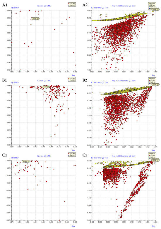
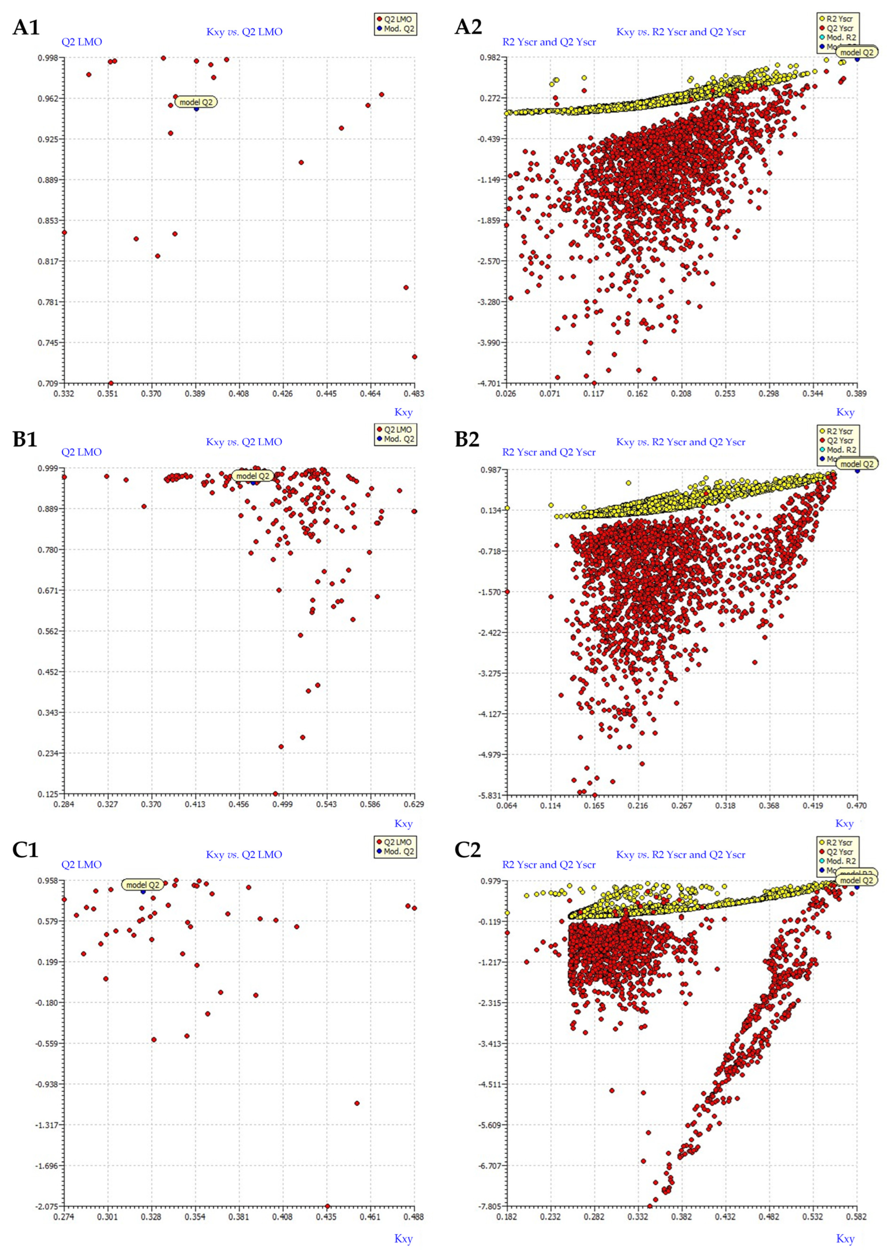
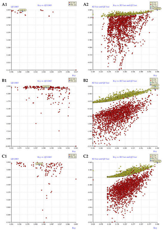
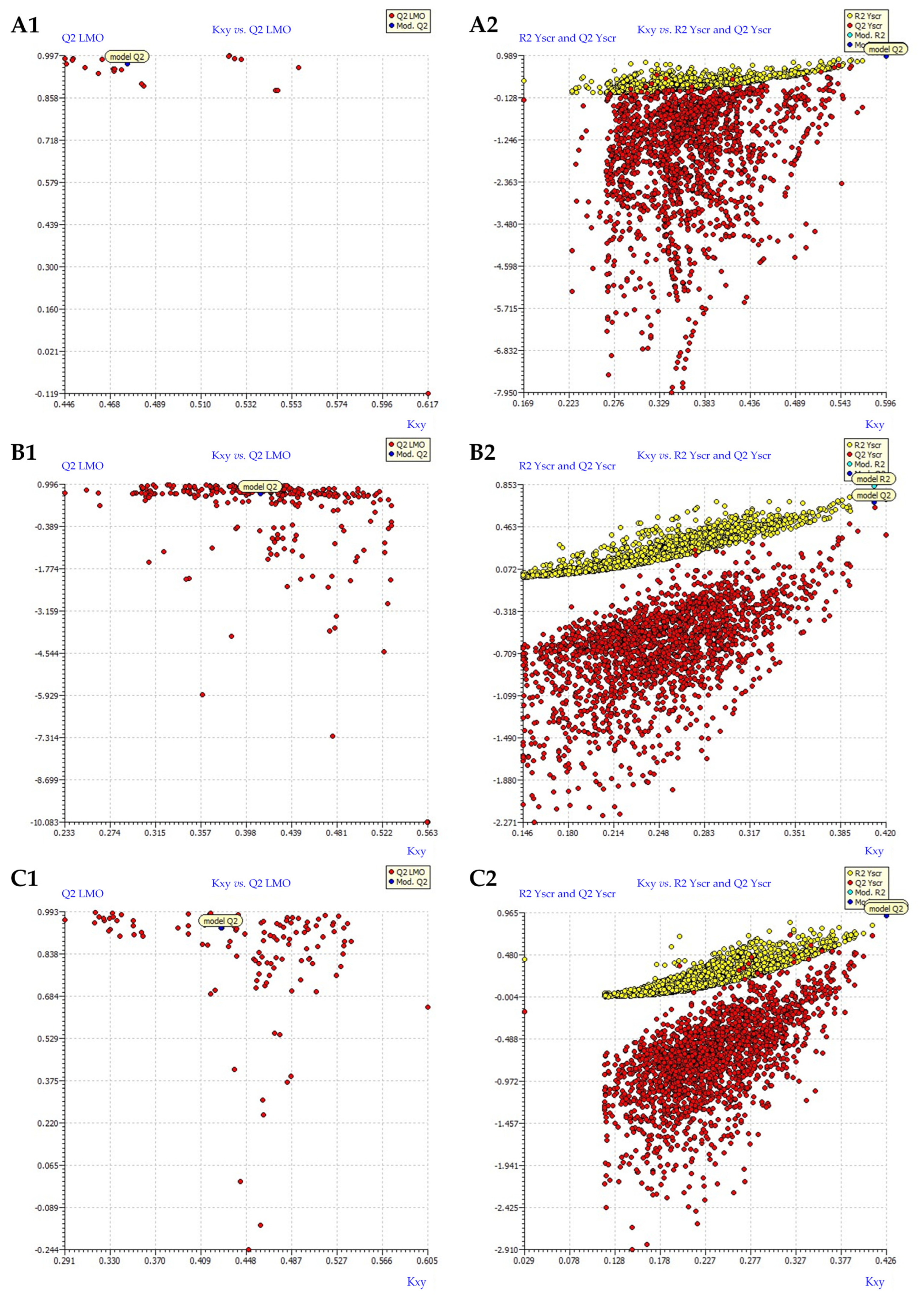
Validation of the models, selected as the best for each chosen response, was also performed using the Leave Many Out (LMO) test and the Y-scramble test (Figure 4 and Figure 5; cases A and B, respectively).
Figure 4.
Validation of developed EC20 QSAR models. The models were developed for Aliivibrio fischeri (A1,A2), Chlorella sp. (B1,B2), and Pseudomonas putida (C1,C2). Cases 1 and 2 represent the LMO and Y-scramble plots, respectively.
Figure 5.
Validation of developed EC50 QSAR models. The models were developed for Aliivibrio fischeri (A1,A2), Chlorella sp. (B1,B2), and Pseudomonas putida (C1,C2). Cases 1 and 2 represent the LMO and Y-scramble plots, respectively.
All LMO plots for both: EC20 and EC50 models (Figure 4 and Figure 5, Case 1) show tight clustering with low scatter, confirming that the developed three-variable models are robust and not overfitted to the small dataset. The relationships between molecular structure and toxicity remain reliable and trustworthy even when large amounts of data are excluded, providing strong confidence in the mechanistic interpretations. Meanwhile, the Y-scramble plots (Figure 4 and Figure 5, Case 2) show a dense cluster of randomized models with very low R2 values (near zero), while the original (non-scrambled) model point stands out distinctly with the highest values on both the x- and y-axes. This clear separation confirms that the three-variable models capture genuine structure–toxicity relationships rather than chance correlations, even in a small dataset of 13 compounds. The strong performance of the true model is therefore attributable to meaningful molecular descriptors, providing solid confidence in the mechanistic interpretations of antiviral ecotoxicity.
As mentioned in the Introduction chapter, EC20 values are typically associated with sub-lethal exposure conditions. The EC20 model established for Aliivibrio fischeri (Table 4, Equation (5)) suggests that the sub-lethal exposure conditions are primarily governed by a combination of three structural features: AVS_B(i) (representing 2D matrix-based properties), RDF075m (RDF descriptor), and F09[C–F] (2D atom pair descriptor), which collectively provided the best statistical fit for the experimental data. AVS_B(i) has negative sign in the equation which has inverse relationship with EC20 value. Generally, 2D matrix-based properties are descriptors that describe topological structure, how its atoms are connected, without considering its 3D spatial conformation. AVS_B(i) reflects the molecule’s structural complexity and the degree of branching or compactness [48]. Molecular size and shape affect bioavailability shown as the rate at which a substance can enter an environmental organism and reach its site of action [49]. A change in branching captured by this descriptor can alter membrane permeability [50]. The descriptor is weighted by the first ionization potential of the atoms, meaning it encodes information about the electronic richness and releasability of electrons within the molecule. These properties, like the presence of polarized bonds, determine the compound’s hydrophilic/lipophilic balance and its ability to partition into the bacterial cell membrane, which is the first barrier to toxicity [51]. RDF075m is from RDF descriptor group. Unlike 2D topological descriptors, RDF descriptors encode information about the 3D spatial arrangement of atoms in a molecule, providing insight into its size, shape, and flexibility [52]. The negative sign of the coefficient associated with this descriptor in the regression equation indicates an inverse relationship with the EC20 value. Consequently, an increase in this descriptor’s value leads to a decrease in EC20. RDF075m is Radial Distribution Function at 0.75 Å, weighted by mass gives an information that molecules with high local densities or compactness or that have heavy atoms near each other increases toxicity due to the fact that dense spatial arrangement creates a feature that fits better into a toxicological target site within the Aliivibrio fischeri cell (e.g., a hydrophobic pocket on the luciferase enzyme) [53]. F09[C–F] is part of 2D atom pair descriptor. The F09[C–F] descriptor quantifies the number of Carbon-Fluorine atom pairs separated by nine bonds, providing insight into the long-range topology and electronic influence of fluorine substitution. Due to its positive coefficient in the model, an increase in F09[C–F] leads to an increase in the EC20 value, suggesting that this specific structural motif reduces the compound’s toxicity to Aliivibrio fischeri. Mechanistically, this long-range C–F separation may reduce the molecule’s overall lipophilicity or hinder the optimal orientation required for chronic binding to the bacterial target site [54].
The sub-lethal QSAR model for Chlorella sp. is given by Equation (7) (Table 4). The sub-lethal toxicity of Chlorella sp. is explained by MPC07 (walk and path counts), MATS6m (2D autocorrelations) and F05[O–S] (2D atom pairs). The descriptor MPC07 is a 2D topological descriptor that counts the number of paths spanning seven bonds between non-hydrogen atoms in the molecule. When an increase in this descriptor leads to increased toxicity to Chlorella sp. (lower EC20), it suggests that this specific molecular length and structure is highly advantageous for the toxic mechanism. The MATS6m descriptor is a Molecular Accession Type Structure descriptor, while six indicates a distance of six bonds which represents medium-range topological information, weighted by an atomic mass. The mass-weighted medium-range connectivity (six bonds) is a critical structural characteristic that promotes the molecule’s overall bioavailability and long-term activity within the Chlorella sp. as possibly enhances the ability to interact with binding on active spots [55]. F05[O-S] descriptor is a 2D atom pair descriptor that counts the number of times a specific pair of chemical features occurs at a defined topological distance. A positive correlation means that molecules with more instances of an O-S pair separated by five bonds are more toxic [56]. This is highly relevant for biological activity, especially in an organism like Chlorella sp. The specific O-S spacing may create a unique electronic environment that facilitates a necessary chemical reaction (like electrophilic attack or redox cycling) at the same time that specific bond perfectly balances solubility and lipophilicity which can interrupt long-term photosynthesis [57].
The use of QSAR models for the explanation of EC20 endpoint for the bacterium Pseudomonas putida (Table 4, Equation (9)) provides crucial information regarding a compound’s potential impact under sub-lethal exposure conditions. C-034 (atom-centered fragments) is fragment count descriptor that enumerates the presence of aliphatic carbon atoms bonded to exactly three other non-hydrogen atoms (tertiary aliphatic carbons). This feature often enhances a compound’s lipophilicity and reduces its water solubility [58], which is critical for traversing the cell membrane of Pseudomonas putida [59]. The compact nature facilitates bioaccumulation over time, leading to a sustained toxic concentration inside the cell and thus a highly effective sub-lethal exposure. B04[N–O] is a 2D atom pair descriptor that counts the number of N and O atom pairs separated by exactly four bonds. The presence of these highly polar/reactive atoms, arranged at this precise distance, likely provides the ideal fit for binding as a hydrogen-bond-type composition [60] to a specific protein or enzyme target-critical for Pseudomonas Putida growth or metabolism [61]. This specific molecular recognition results in potent, sustained inhibition characteristic of sub-lethal exposure. CATS2D_03_PL shows a negative correlation with toxicity, meaning an increase in this feature leads to a higher concentration being required for the exposure. CATS2D_03_PL describes topological distance (in this case a path length of three bonds) between atoms characterized by a specific electronic property such as PL which means Polarizability/Polar Surface Area. This indicates a greater presence of specific polar interactions distributed at a three-bond distance. This increase in molecular polarity/hydrophilicity acts to reduce toxicity by significantly hindering the compound’s ability to partition into and cross the hydrophobic bacterial membrane of Pseudomonas putida [62]. This reduced bioavailability means a much higher external concentration is needed to achieve the threshold internal dose, thereby increasing the EC20 value.
Acute toxicity is represented by the EC50 values. The EC50 model for Aliivibrio fischeri is given by Equation (6) (Table 4). The model includes Eig15_EA(ed) (edge adjacency indices), CATS2D_07_PL, and B08[C–F]. Eig15_EA(ed) is an eigenvalue descriptor derived from a matrix weighted by electronic properties, related to electron affinity (EA). Eigenvalues encode the global topological and electronic architecture of the entire molecule. The bioluminescence inhibition assay is highly sensitive to compounds that interfere with the electron transport chain (respiration) [63]. A favorable Eig15_EA(ed) value suggests the compound possesses an optimal global electronic structure that facilitates rapid electron transfer or redox cycling [64]. This electronic profile enables the molecule to quickly interfere with the respiratory process of Aliivibrio fischeri, leading to an immediate collapse of energy production and light output [65]. This is a potent mechanism for acute toxicity. CATS2D_07_PL has positive correlation to the EC50. Similarly to the earlier-explained CATS2D_03_PL, this descriptor reduces toxicity due to the fact highly polar molecules over long distances hinder the compound’s ability to rapidly partition and cross the hydrophobic bacterial membrane. Aliivibrio fischeri is a Gram-negative bacterium, and its membrane containing lipopolysaccharides (LPSs) is a primary barrier [66]. B08[C–F] is a 2D atom pair descriptor counting C–F pairs separated by a very long path length (eight bonds). It reduces toxicity due to the fact that the molecule’s specific large size prevents optimal interaction or binding to the active site of the luciferase enzyme or the respiratory chain complexes [67].
In the case of acute toxicity toward Chlorella sp. (Table 4, model described by Equation (8)), GATS5m (2D autocorrelations), Mor27v (3D-MoRSE descriptors), and P1e (WHIM descriptors) have the main effect. GATS5m and Mor27v positively correlate with EC50, while P1e reduces EC50 and has positive effect toward toxicity. GATS5m (2D autocorrelation, mass-weighted): This descriptor belongs to the Generalized Atom-Type Spacing (GATS) family, which quantifies the distribution of mass at a specific topological distance (five bonds). A high value suggests an increased presence of heavier atoms at this medium range [67]. This complexity may introduce significant steric hindrance, preventing the molecule from optimally aligning or binding to the critical target sites [68,69] involved in the acute toxicity mechanism (like enzymes in the rapid photosynthetic cycle or cell division), thus lowering its effective potency [70]. Mor27v (3D-MoRSE, Volume-Weighted): This is a 3D-MoRSE (Molecular Representation of Structures based on Electronic distribution) descriptor weighted by atomic volume (v) and calculated at a specific scattering angle (related to index 27). This feature encodes information about global molecular size, shape, and flexibility [70]. Its positive correlation with EC50 implies that structures with this specific large volume or conformational arrangement are poorly suited for membrane penetration or target binding in Chlorella sp., reducing the overall rate of accumulation and, therefore, mitigating acute toxicity. Conversely, the P1e descriptor strongly enhances toxicity, overriding the mitigating effects of the other two. P1e is a WHIM (Weighted Holistic Invariant Molecular) descriptor, which represents the molecule’s size and shape invariance projected along its principal axes, weighted by an electronic property. The P1 component specifically relates to the molecule’s first principal component or molecular length [71]. Its negative correlation with EC50 suggests that an electronically influenced molecular length is ideally suited for toxic action.
The EC50 model for Pseudomonas putida (Table 4, Equation (10) indicates a clear competition between two structural features that enhance toxicity (Eig02_EA(dm) and F05[O-S]) and one that acts as a strong mitigating factor (Mor27m). Eig02_EA(dm) indicating it quantifies the global topology, branching, and symmetry of the entire molecule. It is weighted by electron affinity (EA) and electronic density (dm). This descriptor suggests that the compound’s overall electronic architecture is crucial. This specific global electronic distribution likely optimizes the molecule’s ability to interfere in the Pseudomonas putida lifecycle through facile electron transfer or redox cycling [57] through the respiratory chain or key enzymes such as Cytochrome c [72]. This electronic suitability drives the compound’s intrinsic potency. F05[O–S] counts the number of O–S pairs separated by exactly five bonds. The strong positive correlation with toxicity indicates that the five-bond separation between O and S creates an optimal electronic and steric structure [56,57]. On the other hand, Mor27m has positive effect on EC50 due to the fact that the specific large size or rigid shape may hinder the efficient passage of the compound across the Gram-negative bacterial membrane, severely limiting the rate of bioavailability [73].
4. Conclusions
The aim of this study was to identify structural features responsible for toxicity of a group of 13 antivirals with potential to treat COVID-19 patients: atazanavir, daclatasvir, darunavir, emtricitabine, favipiravir, lopinavir, nirmatrelvir, oseltamivir, remdesivir, ribavirin, ritonavir, and sofosbuvir. To do this, their toxicities toward three different microorganisms: Aliivibrio fischeri, Chlorella sp., and Pseudomonas putida, were assessed, and the obtained data were used for the development of QSAR models.
The analysis of descriptors included in developed models revealed that toxicity at sub-lethal exposure (EC20) of Aliivibrio fischeri is enhanced by compounds with favorable electronic characteristics and compact structural organization, while long fragments disrupt optimal geometric or electronic requirements, thereby reducing toxicity. The primary driver of acute toxicity (EC50) is strong global electronic reactivity. This effect is counteracted by long-range polarity and steric complexity.
The toxicity toward Chlorella sp. at sub-lethal exposure results from a synergistic combination of molecular size and mass distribution, optimal chain length, and specific electronic groups that facilitate cell entry and effective biochemical inhibition. Acute toxicity increases with electronically favorable and length-dependent structures but decreases with excessive mass distribution or unfavorable overall size or shape.
For Pseudomonas putida, sub-lethal toxicity is influenced by lipophilicity, structural compactness, and reactive group geometry, while high short-range polarity serves as a protective factor by limiting membrane permeability and reducing long-term effects. Acute toxicity is driven by optimized electronic reactivity and a characteristic scaffold; however, this effect is limited by excessive mass and volume, which restrict efficient access to intracellular targets.
Collectively, these findings demonstrate that toxicity across all three microorganisms arises from a dynamic interplay between electronic properties, structural accessibility, lipophilicity, and molecular size. The developed QSAR models highlight that the most toxic molecules are those that simultaneously possess favorable electronic reactivity and structural compatibility with the biological target, while avoiding excessive steric or polar features that impede uptake.
This study focused only on short-term effects and does not address the lasting impacts of long-term drug exposure. Therefore, further research should examine the chronic toxicity of the selected antivirals to determine whether it is influenced by the same structural features as acute toxicity or if a different form of toxic action is involved.
Author Contributions
Conceptualization, M.C., D.K.G., Š.U. and T.B.; methodology, M.C., D.K.G., L.V., D.L., M.M.N. and V.M.; formal analysis, L.F., M.M.N. and D.L.; investigation, M.C. and V.M.; writing—original draft preparation, M.C.; writing—review and editing, M.C., D.K.G., Š.U. and T.B.; visualization, M.C. and Š.U. All authors have read and agreed to the published version of the manuscript.
Funding
The authors would like to acknowledge the financial support of the Croatian Science Foundation through the project Environmental Aspects of SARS-CoV-2 Antiviral Substances (EnA-SARS; IP-2022-10-2822).
Data Availability Statement
The data are contained within the article.
Conflicts of Interest
Author Dajana Kučić Grgić is affiliated to the company Comprehensive Water Technology d.o.o. The remaining authors declare that the research was conducted in the absence of any commercial or financial relationships that could be construed as a potential conflict of interest.
References
- Mudgal, S.; De Toni, A.; Lockwood, S.; Salès, K.; Backhaus, T.; Halling Sorensen, B. Study on the Environmental Risks of Medicinal Products FINAL REPORT. BIO Intelligence Service. 2013. Available online: https://health.ec.europa.eu/system/files/2016-11/study_environment_0.pdf (accessed on 4 November 2025).
- United States Environmental Protection Agency. Contaminants of Emerging Concern Including Pharmaceuticals and Personal Care Products; United States Environmental Protection Agency: Washington, DC, USA, 2025. Available online: https://www.epa.gov/wqc/contaminants-emerging-concern-including-pharmaceuticals-and-personal-care-products (accessed on 4 November 2025).
- Chaturvedi, P.; Shukla, P.; Giri, B.S.; Chowdhary, P.; Chandra, R.; Gupta, P.; Pandey, A. Prevalence and hazardous impact of pharmaceutical and personal care products and antibiotics in environment: A review on emerging contaminants. Environ. Res. 2021, 194, 110664. [Google Scholar] [CrossRef] [Scilit]
- Pinto, I.; Simões, M.; Gomes, I.B. An Overview of the Impact of Pharmaceuticals on Aquatic Microbial Communities. Antibiotics 2022, 11, 1700. [Google Scholar] [CrossRef] [Scilit]
- Moratalla, A.; Cotillas, S.; Lacasa, E.; Cañizares, P.; Rodrigo, M.A.; Sáez, C. Electrochemical technologies to decrease the chemical risk of hospital wastewater and urine. Molecules 2021, 26, 6813. [Google Scholar] [CrossRef] [Scilit]
- Gros, M.; Petrović, M.; Ginebreda, A.; Barceló, D. Removal of pharmaceuticals during wastewater treatment and environmental risk assessment using hazard indexes. Environ. Int. 2010, 36, 15–26. [Google Scholar] [CrossRef] [Scilit] [PubMed]
- Papac Zjačić, J.; Morović, S.; Košutić, K.; Ašperger, D. Combined Application of Membrane and Advanced Oxidation Processes for Removal of Pharmaceuticals from Water. Kem. Ind. 2022, 71, 719–727. [Google Scholar] [CrossRef] [Scilit]
- Giouni, E.A.; Kyriazi, M.; Malamis, D.; Loizidou, M. Design of an innovative system for the detoxification of pharmaceutical wastewater. Tech. Ann. 2023, 1, 35979. [Google Scholar] [CrossRef] [Scilit]
- Chaves, O.A.; Sacramento, C.Q.; Ferreira, A.C.; Mattos, M.; Fintelman-Rodrigues, N.; Temerozo, J.R.; Vazquez, L.; Pinto, D.P.; da Silveira, G.P.E.; da Fonseca, L.B.; et al. Atazanavir Is a Competitive Inhibitor of SARS-CoV-2 Mpro, Impairing Variants Replication In Vitro and In Vivo. Pharmaceuticals 2022, 15, 21. [Google Scholar] [CrossRef] [Scilit] [PubMed]
- Feng, J.Y.; Du Pont, V.; Babusis, D.; Gordon, C.J.; Tchesnokov, E.P.; Perry, J.K.; Duong, V.; Vijjapurapu, A.; Zhao, X.; Chan, J.; et al. The Nucleoside/Nucleotide Analogs Tenofovir and Emtricitabine Are Inactive against SARS-CoV-2. Molecules 2022, 27, 4212. [Google Scholar] [CrossRef] [Scilit] [PubMed]
- Tan, Q.; Duan, L.; Ma, Y.; Wu, F.; Huang, Q.; Mao, K.; Xiao, W.; Xia, H.; Zhang, S.; Zhou, E.; et al. Is oseltamivir suitable for fighting against COVID-19: In silico assessment, in vitro and retrospective study. Bioorg. Chem. 2020, 104, 104257. [Google Scholar] [CrossRef] [Scilit]
- Unal, M.A.; Bitirim, C.V.; Summak, G.Y.; Bereketoglu, S.; Cevher Zeytin, I.; Besbinar, O.; Gurcan, C.; Aydos, D.; Goksoy, E.; Kocakaya, E.; et al. Ribavirin shows antiviral activity against SARS-CoV-2 and downregulates the activity of TMPRSS2 and the expression of ACE2 in vitro. Can. J. Physiol. Pharmacol. 2021, 99, 449–460. [Google Scholar] [CrossRef] [Scilit]
- Vegivinti, C.T.R.; Evanson, K.W.; Lyons, H.; Akosman, I.; Barrett, A.; Hardy, N.; Kane, B.; Keesari, P.R.; Pulakurthi, Y.S.; Sheffels, E.; et al. Efficacy of antiviral therapies for COVID-19: A systematic review of randomized controlled trials. BMC Infect. Dis. 2022, 22, 107. [Google Scholar] [CrossRef] [Scilit]
- Said, M.I.; Sayed, M.; Aly, A.A.M.; Atef, M. Unveiling potential capability of PEG6000@Al-BDC nanocomposite as an efficient adsorbent for pharmaceutical remediation from wastewaters: A case study of daclatasvir removal. J. Phys. Chem. Sol. 2026, 208, 113105. [Google Scholar] [CrossRef] [Scilit]
- Brauns, B.; Chandra, S.; Civil, W.; Lapworth, D.J.; MacDonald, A.M.; McKenzie, A.A.; Read, D.S.; Sekhar, M.; Singer, A.C.; Thankachan, A.; et al. Presence of emerging organic contaminants and microbial indicators in surface water and groundwater in urban India. Environ. Pollut. 2024, 362, 124983. [Google Scholar] [CrossRef] [Scilit]
- Teixeira Ramos, L.; Roveri, V.; Toma, W.; Barreiros Kiyotani, R.; de Assis Junior, W.R.A.; Metropolo, A.P.; Aparecido dos Santos, G.; Zogaib Rodrigues, A.; Lopes Guimarães, L. Predicted environmental concentration (PEC), environmental risk assessment (ERA) and prioritization of antiretroviral drugs (ARVs) in seawater from Guarujá (Brazilian coastal zone). Mar. Environ. Res. 2025, 204, 106964. [Google Scholar] [CrossRef] [Scilit]
- Abafe, O.A.; Späth, S.; Fick, J.; Jansson, S.; Buckley, C.; Stark, A.; Pietruschka, B.; Martincigh, B.S. LC-MS/MS determination of antiretroviral drugs in influents and effluents from wastewater treatment plants in KwaZulu-Natal, South Africa. Chemosphere 2018, 200, 660–670. [Google Scholar] [CrossRef] [Scilit] [PubMed]
- Boulard, L.; Dierkes, G.; Ternes, T. Utilization of large volume zwitterionic hydrophilic interaction liquid chromatography for the analysis of polar pharmaceuticals in aqueous environmental samples: Benefits and limitations. J. Chromatogr. A 2018, 1535, 27–43. [Google Scholar] [CrossRef] [Scilit]
- Azuma, T.; Ishida, M.; Hisamatsu, K.; Yunoki, A.; Otomo, K.; Kunitou, M.; Shimizu, M.; Hosomaru, K.; Mikata, S.; Mino, Y. Fate of new three anti-influenza drugs and one prodrug in the water environment. Chemosphere 2017, 169, 550–557. [Google Scholar] [CrossRef] [Scilit] [PubMed]
- Nibamureke, U.M.C.; Barnhoorn, E.J. Screening of Pharmaceuticals in Surface Waters from Vhembe District, Limpopo Province, South Africa. Water 2025, 17, 379. [Google Scholar] [CrossRef] [Scilit]
- Wood, T.P.; Duvenage, C.S.J.; Rohwer, E. The occurrence of anti-retroviral compounds used for HIV treatment in South African surface water. Environ. Pollut. 2015, 199, 235–243. [Google Scholar] [CrossRef] [Scilit]
- Zhang, Z.; Zhou, Y.; Han, L.; Guo, X.; Wu, Z.; Fang, J.; Hou, B.; Cai, X.; Jiang, J.; Yang, Z. Impacts of COVID-19 pandemic on the aquatic environment associated with disinfection byproducts and pharmaceuticals. Sci. Total Environ. 2022, 811, 151409. [Google Scholar] [CrossRef] [Scilit]
- Takamami, R.; Ozaki, H.; Giri, R.R.; Taniguchi, S.; Hayashi, S. Antiviral drugs zanamivir and oseltamivir found in wastewater and surface water in Osaka, Japan. J. Water Environ. Technol. 2012, 10, 57–68. [Google Scholar] [CrossRef] [Scilit]
- Chen, X.; Lei, L.; Liu, S.; Han, J.; Li, R.; Men, J.; Li, L.; Wei, L.; Sheng, Y.; Yang, Y.; et al. Occurrence and risk assessment of pharmaceuticals and personal care products (PPCPs) against COVID-19 in lakes and WWTP-river-estuary system in Wuhan, China. Sci. Total Environ. 2021, 792, 148352. [Google Scholar] [CrossRef] [Scilit]
- Ul’yanovskii, N.V.; Kosyakov, D.S.; Sypalov, S.A.; Varsegov, I.S.; Shavrina, I.S.; Lebedev, A.T. Antiviral drug Umifenovir (Arbidol) in municipal wastewater during the COVID-19 pandemic: Estimated levels and transformation. Sci. Total Environ. 2022, 805, 150380. [Google Scholar] [CrossRef] [Scilit]
- Martinjak, V.; Miloloža, M.; Markić, M.; Furač, L.; Cvetnić, M.; Bolanča, T.; Kučić Grgić, D.; Ukić, Š. Revealing the Adverse Potential of Six SARS-CoV-2 Antivirals by Aliivibrio fischeri Assay: Toxicity Analysis of Single Agent Solutions and Binary Mixtures. Water 2024, 16, 3607. [Google Scholar] [CrossRef] [Scilit]
- Czech, B.; Krzyszczak, A.; Boguszewska-Czubara, A.; Opielak, G.; Jośko, I.; Hojamberdiev, M. Revealing the toxicity of lopinavir- and ritonavir-containing water and wastewater treated by photo-induced processes to Danio rerio and Allivibrio fischeri. Sci. Total Environ. 2022, 824, 153967. [Google Scholar] [CrossRef] [Scilit]
- Bartels, I.; Nahar, N.; Smollich, E.; Zimmermann, S.; Schmidt, T.C.; Jaeger, M.; Sures, B. Ecotoxicological data of selected antiviral drugs acting against SARS-CoV-2: Aliivibrio fischeri bioluminescence inhibition, Daphnia magna immobilization, and comparison with in silico predictions. Environ. Sci. Eur. 2025, 37, 110. [Google Scholar] [CrossRef] [Scilit]
- Dogruel, S.; Chavoshi, N.; Bilgin-Saritas, N.; Khataee, A.; Topuz, E.; Pehlivanoglu, E. Degradation and ecotoxicity of favipiravir and oseltamivir in the presence of microplastics during ozonation and catalytic ozonation of synthetic municipal wastewater effluents. J. Chem. Technol. Biotechnol. 2025, 100, 955–966. [Google Scholar] [CrossRef] [Scilit]
- Ülger, M.; Turan, I.T.; Sipahi, A.; Önder, G.Ö. Experimental evaluation of favipiravir (T-705)-induced liver and kidney toxicity in rats. Food Chem. Toxicol. 2025, 202, 115472. [Google Scholar] [CrossRef] [Scilit] [PubMed]
- Mendes, S.; Cordeiro Guimarães, L.; Camilo de Oliveira, L.; Carvalho Costa, P.A.; Alves da Silva, N.J.; Silva Alves Pessim Pereira, G.; Couto Fernandez, C.; Figueiredo, M.M.; Souza dos Santos, R.A.; Teixeira, M.M.; et al. Intranasal liposomal remdesivir induces SARS-CoV-2 clearance in K18-hACE2 mice and ensures survival. J. Control. Release 2025, 379, 558–573. [Google Scholar] [CrossRef] [Scilit] [PubMed]
- Chan, C.C.-Y.; Guo, Q.; Chan, J.F.-W.; Tang, K.; Cai, J.-P.; Chik, K.K.-H.; Huang, Y.; Dai, M.; Qin, B.; Ong, C.P.; et al. Identification of novel small-molecule inhibitors of SARS-CoV-2 by chemical genetics. Acta Pharm. Sin. B 2024, 14, 4028–4044. [Google Scholar] [CrossRef] [Scilit]
- Murray, M. The Role of CYPs and Transporters in the Biotransformation and Transport of the Anti-hepatitis C Antiviral Agents Asunaprevir, Daclatasvir, and Beclabuvir: Impact of Liver Disease, Race and Drug-drug Interactions on Safety and Efficacy. Curr. Drug Metab. 2024, 25, 96–109. [Google Scholar] [CrossRef] [Scilit]
- Szczech, G.M.; Wang, L.H.; Walsh, J.P.; Rousseau, F.S. Reproductive toxicology profile of emtricitabine in mice and rabbits. Reprod. Toxicol. 2003, 17, 95–108. [Google Scholar] [CrossRef] [Scilit]
- Mohamed, A.F.; Abo-Ouf, A.M.; Arafa, M.A.A. Histological and biochemical studies on effect of Sofosbuvir (Sovaldi) on adult male albino rat kidney. Ultrastruct. Pathol. 2021, 45, 286–296. [Google Scholar] [CrossRef] [Scilit] [PubMed]
- Cvetnic, M.; Juretic Perisic, D.; Kovacic, M.; Ukic, S.; Bolanca, T.; Rasulev, B.; Kusic, H.; Loncaric Bozic, A. Toxicity of aromatic pollutants and photooxidative intermediates in water: A QSAR study. Ecotoxicol. Environ. Saf. 2019, 169, 918–927. [Google Scholar] [CrossRef] [Scilit]
- ISO 11348-3:2007; Water Quality—Determination of the Inhibitory Effect of Water Samples on the Light Emission of Vibrio fischeri (Luminescent Bacteria Test). Part 3: Method Using Freeze Dried Bacteria. International Organization for Standardization: Geneva, Switzerland, 2007.
- ISO 10712; Water Quality—Pseudomonas Putida Growth Inhibition Test Pseudomonas Cell Multiplication Inhibition Test. International Organization for Standardization: Geneva, Switzerland, 1995.
- Hafner, C. Pseudomonas putida growth inhibition test. In Ecotoxicological Characterization of Waste; Moser, H., Römbke, J., Eds.; Springer: New York, NY, USA, 2009; pp. 153–159. [Google Scholar]
- OECD. OECD Test No. 201: Freshwater Alga and Cyanobacteria, Growth Inhibition Test, OECD Guidelines for the Testing of Chemicals, Section 2; OECD Publishing: Paris, France, 2011. [Google Scholar] [CrossRef] [Scilit]
- Cvetnić, M.; Tomić, A.; Sigurnjak, M.; Novak Stankov, M.; Ukić, Š.; Kušić, H.; Bolanča, T.; Lončarić Božić, A. Structural features of contaminants of emerging concern behind empirical parameters of mechanistic models describing their photooxidative degradation. J. Water Process Eng. 2020, 33, 101053. [Google Scholar] [CrossRef] [Scilit]
- Todeschini, R.; Consonni, V.; Maiocchi, A. The K correlation index: Theory development and its application in chemometrics. Chemom. Intell. Lab. Syst. 1999, 46, 13–29. [Google Scholar] [CrossRef] [Scilit]
- Gramatica, P.; Sangion, A. A Historical Excursus on the Statistical Validation Parameters for QSAR Models: A Clarification Concerning Metrics and Terminology. J. Chem. Inf. Model. 2016, 56, 1127–1131. [Google Scholar] [CrossRef] [Scilit] [PubMed]
- Escher, B.I.; Bramaz, N.; Lienert, J.; Neuwoehner, J.; Straub, J.O. Mixture toxicity of the antiviral drug Tamiflu((R)) (oseltamivir ethylester) and its active metabolite oseltamivir acid. Aquat. Toxicol. 2010, 96, 194–202. [Google Scholar] [CrossRef] [Scilit]
- Straub, J.O. An environmental risk assessment for oseltamivir (Tamiflu) for sewage works and surface waters under seasonal influenza and pandemic use conditions. Ecotoxicol. Environ. Saf. 2009, 72, 1625–1634. [Google Scholar] [CrossRef] [Scilit]
- Sun, G.; Zhang, Y.; Pei, L.; Lou, Y.; Mu, Y.; Yun, J.; Li, F.; Wang, Y.; Hao, Z.; Xi, S.; et al. Chemometric QSAR modeling of acute oral toxicity of Polycyclic Aromatic Hydrocarbons (PAHs) to rat using simple 2D descriptors and interspecies toxicity modeling with mouse. Ecotoxicol. Environ. Saf. 2021, 222, 112525. [Google Scholar] [CrossRef] [Scilit]
- Gramatica, P.; Chirico, N.; Papa, E.; Cassani, S.; Kovarich, S. QSARINS: A new software for the development, analysis, and validation of QSAR MLR models. J. Comput. Chem. 2013, 34, 2121–2132. [Google Scholar] [CrossRef] [Scilit]
- Khan, A.U.; Porta, G.M.; Riva, M.; Guadagnini, A. In-silico mechanistic analysis of adsorption of Iodinated Contrast Media agents on graphene surface. Ecotoxicol. Environ. Saf. 2024, 280, 116506. [Google Scholar] [CrossRef] [Scilit]
- Mok, Z.H. The effect of particle size on drug bioavailability in various parts of the body. Pharm. Sci. Adv. 2024, 2, 100031. [Google Scholar] [CrossRef] [Scilit] [PubMed]
- Shinoda, W.; Mikami, M.; Baba, T.; Hato, M. Molecular Dynamics Study on the Effects of Chain Branching on the Physical Properties of Lipid Bilayers: 2. Permeability. J. Phys. Chem. B 2004, 108, 9346–9356. [Google Scholar] [CrossRef] [Scilit]
- Manallack, D.T.; Prankerd, R.J.; Yuriev, E.; Oprea, T.I.; Chalmers, D.K. The Significance of Acid/Base Properties in Drug Discovery. Chem. Soc. Rev. 2013, 42, 485–496. [Google Scholar] [CrossRef] [Scilit] [PubMed]
- González, M.P.; Gándara, Z.; Fall, Y.; Gómez, G. Radial Distribution Function descriptors for predicting affinity for vitamin D receptor. Eur. J. Med. Chem. 2008, 43, 1360–1365. [Google Scholar] [CrossRef] [Scilit]
- Campbell, Z.T.; Weichsel, A.; Montfort, W.R.; Baldwin, T.O. Crystal Structure of the Bacterial Luciferase/Flavin Complex Provides Insight into the Function of the beta Subunit. Biochemistry 2009, 48, 6085–6094. [Google Scholar] [CrossRef] [Scilit]
- Wang, Z.; Felstead, H.R.; Troup, R.I.; Linclau, B.; Williamson, P.T.F. Lipophilicity Modulations by Fluorination Correlate with Membrane Partitioning. Angew. Chem. Int. Ed. 2023, 62, e202301077. [Google Scholar] [CrossRef] [Scilit]
- Fan, T.; Sun, G.; Zhao, L.; Cui, X.; Zhong, R. QSAR and Classification Study on Prediction of Acute Oral Toxicity of N-Nitroso Compounds. Int. J. Mol. Sci. 2018, 19, 3015. [Google Scholar] [CrossRef] [Scilit]
- Pandey, S.K.; Roy, K. Development of hybrid models by the integration of the read-across hypothesis with the QSAR framework for the assessment of developmental and reproductive toxicity (DART) tested according to OECD TG 414. Toxicol. Rep. 2024, 13, 101822. [Google Scholar] [CrossRef] [Scilit]
- De Sciscio, M.L.; D’Annibale, V.; D’Abramo, M. Theoretical Evaluation of Sulfur-Based Reactions as a Model for Biological Antioxidant Defense. Int. J. Mol. Sci. 2022, 23, 14515. [Google Scholar] [CrossRef] [Scilit]
- Nour, B. The Role of Alkyl Groups in Organic Chemistry and Drug Design. J. Anal. Bioanal. Tech. 2025, 16, 735. [Google Scholar]
- Figueroa-Valverde, L.; Díaz-Cedillo, F.; Garcia-Cervera, E.; Pool-Gómez, J.E.M.; Cardeña-Arredondo, C. Synthesis of a Carbamazepine Derivative as Antibacterial Agent. Asian J. Chem. 2012, 24, 699–704. [Google Scholar]
- Madushanka, A.; Moura, R.T.; Verma, N.; Kraka, E. Quantum Mechanical Assessment of Protein–Ligand Hydrogen Bond Strength Patterns: Insights from Semiempirical Tight-Binding and Local Vibrational Mode Theory. Int. J. Mol. Sci. 2023, 24, 6311. [Google Scholar] [CrossRef] [Scilit]
- Schmidt, M.; Pearson, A.N.; Incha, M.R.; Thompson, M.G.; Baidoo, E.E.K.; Kakumanu, R.; Mukhopadhyay, A.; Shih, P.M.; Deutschbauer, A.M.; Blank, L.M.; et al. Nitrogen Metabolism in Pseudomonas putida: Functional Analysis Using Random Barcode Transposon Sequencing. Appl. Environ. Microbiol. 2022, 88, e02430-21. [Google Scholar] [CrossRef] [Scilit]
- Leus, I.V.; Weeks, J.W.; Bonifay, V.; Shen, Y.; Yang, L.; Cooper, C.J.; Nath, D.; Duerfeldt, A.S.; Smith, J.C.; Parks, J.M.; et al. Property space mapping of Pseudomonas aeruginosa permeability to small molecules. Sci. Rep. 2022, 12, 8220. [Google Scholar] [CrossRef] [Scilit]
- Abbas, M.; Adil, M.; Ehtisham-ul-Haque, S.; Munir, B.; Yameen, M.; Ghaffar, A.; Shar, G.A.; Tahir, M.A.; Iqbal, M. Vibrio fischeri bioluminescence inhibition assay for ecotoxicity assessment: A review. Sci. Total Environ. 2018, 626, 1295–1309. [Google Scholar] [CrossRef] [Scilit]
- Bolton, J.L.; Trush, M.A.; Penning, T.M.; Dryhurst, G.; Monks, T.J. Role of quinones in toxicology. Chem. Res. Toxicol. 2000, 13, 135–160. [Google Scholar] [CrossRef] [Scilit] [PubMed]
- Sushko, E.S.; Zenkov, A.V.; Kudryasheva, N.S. Effects of Selenite-Ions on a Luminescence Enzymatic System. Eurasian J. Chem. 2024, 29, 65–71. [Google Scholar] [CrossRef] [Scilit]
- Chu, J.; Liu, J.; Hoover, T.R. Phylogenetic Distribution, Ultrastructure, and Function of Bacterial Flagellar Sheaths. Biomolecules 2020, 10, 363. [Google Scholar] [CrossRef] [Scilit] [PubMed]
- Todeschini, R.; Consonni, V. Molecular Descriptors for Chemoinformatics; Wiley-VCH: Weinheim, Germany, 2009; Volume 1. [Google Scholar]
- Cherkasov, A.; Muratov, E.N.; Fourches, D.; Varnek, A.; Baskin, I.I.; Cronin, M.; Dearden, J.; Gramatica, P.; Martin, Y.C.; Todeschini, R.; et al. QSAR Modeling: Where Have You Been? Where Are You Going To? J. Med. Chem. 2014, 57, 4977–5004. [Google Scholar] [CrossRef] [Scilit]
- Ahmadinejad, N.; Shafiei, F. Quantitative Structure-Activity Relationship Study of Camptothecin Derivatives as Anticancer Drugs Using Molecular Descriptors. Comb. Chem. High Throughput Screen. 2019, 22, 387–399. [Google Scholar] [CrossRef] [Scilit] [PubMed]
- Zhao, F.; Xiang, Q.; Zhou, Y.; Xu, X.; Qiu, X.; Yu, Y.; Ahmad, F. Evaluation of the toxicity of herbicide topramezone to Chlorella vulgaris: Oxidative stress, cell morphology and photosynthetic activity. Ecotoxicol. Environ. Saf. 2017, 143, 129–135. [Google Scholar] [CrossRef] [Scilit] [PubMed]
- Kubinyi, H.; Folkers, G.; Martin, Y.C. 3D QSAR in Drug Design: Ligand-Protein Interactions and Molecular Similarity; Springer: Dordrecht, The Netherlands, 1998. [Google Scholar]
- Lai, B.; Bernhardt, P.V.; Krömer, J.O. Cytochrome c Reductase is a Key Enzyme Involved in the Extracellular Electron Transfer Pathway towards Transition Metal Complexes in Pseudomonas Putida. ChemSusChem 2020, 13, 5308–5317. [Google Scholar] [CrossRef] [Scilit] [PubMed]
- Yamamoto, K.; Yamamoto, N.; Ayukawa, S.; Yasutake, Y.; Ishiya, K.; Nakashima, N. Scaffold size-dependent effect on the enhanced uptake of antibiotics and other compounds by Escherichia coli and Pseudomonas aeruginosa. Sci. Rep. 2022, 12, 5609. [Google Scholar] [CrossRef] [Scilit]

Disclaimer/Publisher’s Note: The statements, opinions and data contained in all publications are solely those of the individual author(s) and contributor(s) and not of MDPI and/or the editor(s). MDPI and/or the editor(s) disclaim responsibility for any injury to people or property resulting from any ideas, methods, instructions or products referred to in the content. |
© 2026 by the authors. Licensee MDPI, Basel, Switzerland. This article is an open access article distributed under the terms and conditions of the Creative Commons Attribution (CC BY) license.