


Abstract
Unsafe water sources contaminated with fecal bacteria and antibiotic-resistant pathogens represent a critical public health challenge in rapidly urbanizing regions. This study investigates the occurrence, distribution, and antimicrobial resistance (AMR) patterns of Escherichia coli across interconnected water sources in Guwahati, India, including river water, wells, municipal supply, and groundwater. A total of 87 samples were analyzed using membrane filtration, biochemical identification, and automated confirmation, followed by antibiotic susceptibility testing and phenotypic detection of β-lactamase production. Total coliforms were detected in 64.4% of samples, while E. coli was confirmed in 51.7%. According to World Health Organization risk thresholds, 93.3% of river water samples and 50% of groundwater samples fell within the very-high-risk category (>1000 CFU/100 mL), indicating widespread fecal contamination in sources used for domestic purposes. Antibiotic-resistant E. coli was identified across all water types, with 14.1% of isolates exhibiting multidrug resistance (MAR index > 0.2). Notably, 20.6% of isolates produced β-lactamases, including one extended-spectrum β-lactamase (ESBL) producer, while no carbapenemase-producing strains were detected. The co-occurrence of multidrug-resistant E. coli across surface and groundwater systems is consistent with the interconnected nature of urban water contamination and suggests the potential for widespread exposure through drinking water pathways, though direct transfer between water systems was not experimentally confirmed in this study. These findings underscore the urgent need for improved wastewater management, protection of groundwater resources, and strengthened antibiotic stewardship within a One Health framework to mitigate the environmental dissemination of AMR.
1. Introduction
Antimicrobial resistance (AMR) is increasingly recognized as a global environmental crisis that extends beyond clinical boundaries and is shaped by complex interactions across human, animal, and ecological systems. Recent global estimates attribute nearly 5 million deaths annually to bacterial AMR, highlighting its scale and urgency [1]. While clinical misuse of antibiotics remains a primary driver, growing evidence indicates that environmental compartments, particularly aquatic systems, play a critical role in the persistence, amplification, and dissemination of antibiotic-resistant bacteria (ARB) and resistance genes (ARGs) [2,3]. In addition to clonal dissemination, antimicrobial resistance in Escherichia coli can also spread through horizontal gene transfer, enabling the exchange of resistance determinants within environmental microbial communities [4,5,6,7,8]. In this context, water systems function not only as sinks but also as dynamic reservoirs and transmission pathways, integrating inputs from multiple sectors within a One Health framework. These pathways are central to the One Health perspective, which recognizes the interconnected roles of human, animal, and environmental systems in the emergence and spread of AMR [9].
Urban environments in low- and middle-income countries represent hotspots for environmental AMR dissemination due to rapid and often unplanned development. Inadequate wastewater treatment, leakage from sanitation infrastructure, and diffuse inputs from domestic, industrial, and agricultural sources result in the continuous release of antibiotics, resistant bacteria, and genetic material into urban water systems [10,11]. Surface waters receiving untreated or partially treated effluents frequently exhibit high loads of fecal contamination and clinically relevant resistance phenotypes, including extended-spectrum β-lactamase (ESBL)-producing Escherichia coli [12,13,14]. Among fecal indicator organisms, E. coli is widely used to assess microbial contamination in water and is recognized as an important reservoir and potential vector of resistance genes [2,7,15,16]. However, these observations are typically confined to individual environmental compartments, limiting understanding of how AMR propagates across interconnected water systems. Most previous investigations have focused on isolated environmental compartments such as rivers, wastewater systems, or drinking water sources individually, while comparatively fewer studies have examined the interconnected dynamics linking surface water, groundwater, wells, and municipal water supplies.
A critical but underexplored dimension is the role of hydrological connectivity in facilitating the transfer of contamination between surface water and groundwater systems. Groundwater is widely regarded as a safer and more protected source of drinking water due to natural filtration processes; however, this assumption may not hold in urban settings characterized by shallow aquifers, high recharge rates, and compromised sanitation infrastructure. Processes such as river–aquifer exchange, infiltration of contaminated surface water during flooding, and leakage from unlined drains and septic systems have been proposed as potential pathways for the downward transport of fecal bacteria and associated resistance determinants in comparable urban settings [17,18]. Despite this, empirical evidence linking AMR occurrence across surface and subsurface water compartments remains limited, particularly in rapidly urbanizing regions. Urban floodplain environments characterized by shallow aquifers, recurrent flooding, and inadequate sanitation infrastructure may be especially vulnerable to the transfer and persistence of fecal contamination and associated resistance determinants.
India represents a critical setting for examining these dynamics due to high antibiotic consumption, heterogeneous wastewater treatment capacity, and widespread reliance on decentralized water sources [14,19]. Additional contributing factors include incomplete wastewater treatment, inequalities in access to safely managed water and sanitation, non-prescription antibiotic access in some settings, and varying levels of antibiotic stewardship across human and animal sectors [20,21,22]. Several studies have documented ARB in major Indian river systems and urban aquatic environments, including resistant Gram-negative bacteria and extended-spectrum β-lactamase (ESBL) producers in riverine systems and other water resources [12,13,23,24,25,26,27,28]. The detection of carbapenemases and ESBL genes such as bla_NDM-1 and bla_CTX-M variants further indicates that clinically relevant resistance phenotypes occur in environmental matrices [14,25]. Collectively, these studies demonstrate that Indian aquatic systems can serve as environmental reservoirs for clinically relevant resistance phenotypes with potential implications for public health.
Floods and other extreme events may further compromise water quality by mobilizing fecal material and contaminants into wells and aquifers, thereby increasing the exposure risk [10,17,18,20,28,29]. Also, untreated wastewater discharge and hydrological exchange processes can create multiple pathways for fecal contamination to enter surface waters, also affecting groundwater quality, in urbanizing floodplain environments [10,17,18,20,28,29]. Although several studies have documented ARB in Indian aquatic environments, evidence from northeastern region remains comparatively limited relative to other regions of the country [20,23]. Existing evidence from Guwahati suggests resistance to several commonly used antibiotics among microorganisms in urban aquatic environments; however, systematic assessments across multiple interconnected water-source types, particularly including groundwater, remain scarce [23]. Guwahati, the largest city in Northeast India, exemplifies these conditions. Situated along the Brahmaputra River, the city experiences rapid urban expansion, recurrent flooding, and substantial dependence on untreated groundwater for domestic use.
To our knowledge, this study provides one of the first integrated assessments of antimicrobial-resistant E. coli across multiple interconnected urban water compartments in a rapidly urbanizing city of northeastern India. Unlike previous studies that have primarily examined isolated environmental compartments such as rivers, wastewater systems, or groundwater independently, this study simultaneously evaluates faecal contamination, antimicrobial resistance patterns, and β-lactamase production across linked urban water sources, including river water, wells, municipal supply, and groundwater, within a flood-prone urban hydrological setting. Against this backdrop, this study quantifies faecal contamination and elucidates antimicrobial resistance patterns in Escherichia coli across interconnected urban water sources, including river water, wells, municipal supply, and groundwater. By integrating microbiological indicators with antibiotic susceptibility profiling and β-lactamase detection, the study demonstrates how antibiotic-resistant E. coli is distributed across linked water compartments and provides evidence that challenges the assumed microbiological security of groundwater in rapidly urbanizing environments.
2. Methodology
2.1. Sampling Strategy
2.1.1. Study Area
Guwahati, the largest city in Assam, with an estimated population of more than 1.5 million, has experienced rapid, largely unplanned urban expansion within its metropolitan area of approximately 328 km2 [30]. The urban landscape is strongly influenced by the surrounding water bodies, including the Brahmaputra River to the north, the Bahini River to the south, and the Bharalu River, which traverses the city. A major concern in this setting is the discharge of untreated or inadequately treated industrial and domestic effluents into these rivers, which ultimately contributes to the contamination of the Brahmaputra River and degrades both surface water and groundwater quality. Because many households, peri-urban communities, and rural areas depend primarily on untreated groundwater for drinking and other domestic uses, the continuous input of wastewater and solid waste into these interconnected water bodies poses a substantial risk to the safety and reliability of local drinking water sources [23].
2.1.2. Sample Collection and Analyses
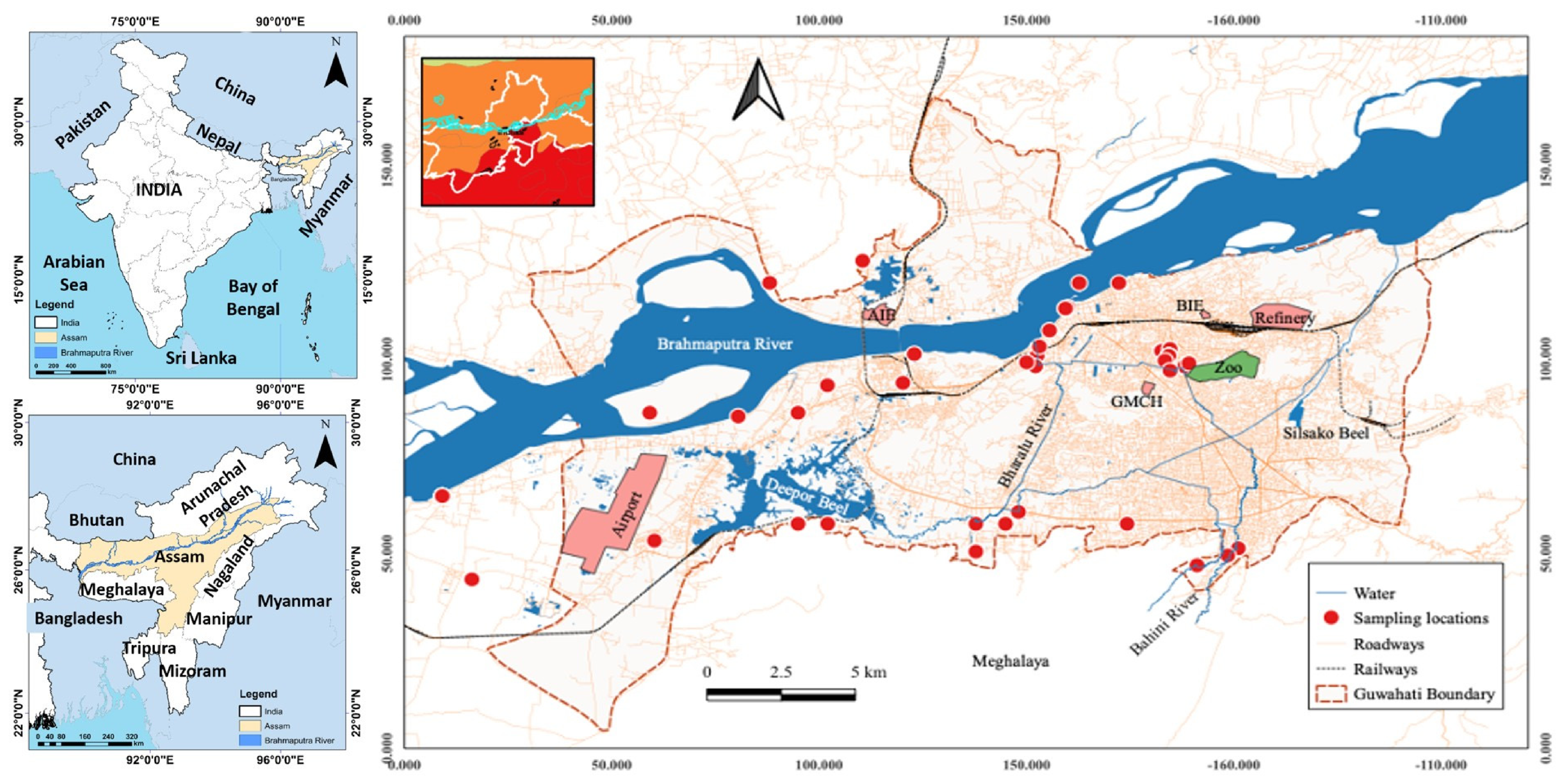
Water samples were collected from different locations distributed across the four administrative zones of Guwahati (North, East, West, and Central) to capture the spatial variability in water quality. A total of 87 samples were obtained between August 2022 and February 2023, spanning two climatically distinct periods: the late monsoon season (August–October 2022) and the post-monsoon/dry season (November 2022–February 2023). The samples comprised river water (RW) (n = 15), municipal supply water (SW) (n = 4), tube well water (n = 20), and groundwater (GW), also including handpump (HP) (n = 48) (Figure 1). Groundwater samples included water from tubewells (TW), handpumps (HP) and bore wells (BW). Both TW and BW are groundwater-derived sources, but TW samples were collected from shallow tube wells (40–60 ft depth), whereas BW samples were collected from deeper borewells (>100 ft depth). Sampling locations along the Brahmaputra River were selected based on a preliminary field survey and hydrologic considerations, i.e., upstream of the city before major urban influence (L1), immediately upstream of the confluence with the Bharalu (urban runoff) tributary (L2), immediately downstream of the confluence (L3), and approximately 10 km downstream of Guwahati (L4). Guwahati experiences a strongly seasonal precipitation regime, with average annual rainfall of approximately 1600–1800 mm, of which more than 70% falls during the monsoon months of June–September [31]. The Brahmaputra River typically reaches peak discharge during July–September, frequently inundating low-lying neighborhoods and increasing hydraulic connectivity between surface water and shallow aquifers. During the August 2022 sampling period, Assam recorded above-normal monsoon precipitation, with widespread urban flooding affecting drainage infrastructure and leading to documented incidents of sewage overflow and floodwater intrusion into domestic wells across Guwahati [32]. By contrast, samples collected between November 2022 and February 2023 corresponded to the dry season, when river levels recede, surface runoff diminishes, and hydrological connectivity between surface water and groundwater is substantially reduced. This seasonal contrast provides relevant hydrological context for interpreting spatial and source-type differences in contamination levels across sampling events, although a formal comparative seasonal analysis was beyond the scope of the present study. All collected samples were transferred into sterile containers, stored on ice in insulated coolers, and transported to the laboratory on the day of sampling. Upon arrival, the samples were stored at 4 °C and processed for microbiological analysis within 24 h. At each site, water samples were analyzed for faecal indicator bacteria, including E. coli and total coliforms. Sample collection, preservation, and handling followed standard procedures for drinking water and environmental microbiological sampling, as recommended by USEPA methods [33].
Figure 1.
Map of the study area and sampling locations. The figure shows the position of sampling points taken from Guwahati in northeastern India and the course of the Brahmaputra River across the city. Individual sampling sites along the Brahmaputra and within the urban area are marked.
2.2. Detection of Total Coliforms, Isolation, and Identification of E. coli
Total coliforms and E. coli were detected using the membrane filtration method [34]. Briefly, water samples were serially diluted tenfold in phosphate buffer (HiMedia Laboratories Pvt. Ltd., Mumbai, India) containing 0.8–0.85% sodium chloride (NaCl) (Merck, Darmstadt, Germany). Appropriate volumes of diluted samples were filtered through sterile 0.45 µm pore-size cellulose nitrate membrane filters (HiMedia Laboratories Pvt. Ltd., Mumbai, India) using a vacuum-filtration apparatus (Borosil Scientific Ltd., Mumbai, India). The filters were placed onto Chromocult® Coliform Agar ES media (HiMedia Laboratories Pvt. Ltd., Mumbai, India) and incubated at 37 °C for 24 h in an incubator (Orbitek, Scigenics Biotech Private Limited, Chennai, India) for coliform enumeration. Colonies with typical coliform morphology were counted using a digital colony counter (Systonic Lab, Delhi, India), and the counts were adjusted for dilution factors. Both total coliform colonies and suspected E. coli colonies (dark blue/violet) were recorded as colony-forming units (CFU) per 100 mL of sample. Suspected E. coli isolates (blue/violet colonies) were subcultured onto MacConkey agar (HiMedia Laboratories Pvt. Ltd., Mumbai, India) and eosin methylene blue (EMB) agar (HiMedia Laboratories Pvt. Ltd., Mumbai, India) to verify the characteristic growth and colony morphology. Species-level confirmation was performed based on standard biochemical tests and automated identification using the VITEK® 2 Compact system (bioMérieux SA, Marcy-I’Etoile, France).
2.3. Phenotypic Detection of β-Lactamases
2.3.1. Detection of AmpC β-Lactamases
AmpC β-lactamase-producing E. coli isolates were initially screened using the disk diffusion method using a cefoxitin (30 μg) disk (HiMedia Laboratories Pvt. Ltd., Mumbai, India). Isolates exhibiting an inhibition zone diameter < 18 mm around cefoxitin were considered presumptive AmpC β-lactamase producers [35]. These isolates were further tested using the Modified Three-Dimensional Extract Test (M3DET) for phenotypic confirmation of AmpC β-lactamase activity [36]. Escherichia coli ATCC 25922 and Enterobacter cloacae P99 were used as negative and positive control strains, respectively.
2.3.2. Detection of ESBL-Producing E. coli and Carbapenemase Production
Extended-spectrum β-lactamase (ESBL) production was screened and confirmed according to the Clinical and Laboratory Standards Institute (CLSI) guidelines [37]. Isolates were first screened on Mueller-Hinton (M-H) agar (HiMedia Laboratories Pvt. Ltd., Mumbai, India) plates containing a single ceftazidime (CAZ, 30 μg) disk (HiMedia Laboratories Pvt. Ltd., Mumbai, India). Isolates non-susceptible to CAZ were further evaluated using a combined disc test with CAZ (30 µg) alone and CAZ (30 µg) plus clavulanic acid (10 µg) (HiMedia Laboratories Pvt. Ltd., Mumbai, India). An increase in the inhibition zone diameter of ≥ 5 mm in the presence of clavulanic acid compared with CAZ alone was interpreted as phenotypic evidence of ESBL production [37]. The isolates were screened using imipenem (IPM, 10 µg) (HiMedia Laboratories Pvt. Ltd., Mumbai, India). Isolates showing reduced susceptibility to IPM were further tested using the modified Hodge test (MHT) [33]. After overnight incubation, the presence of a cloverleaf-like indentation of the indicator strain along the streak of the test organism was interpreted as positive for carbapenemase production [33].
2.3.3. Detection of Inducible AmpC β-Lactamase
Inducible AmpC β-lactamase activity was assessed using imipenem (10 µg) and cefoxitin (30 µg) disks as inducers. Disks were placed 20 mm apart (center-to-center) from a ceftazidime (30 µg) disk on MH agar plates (HiMedia Laboratories Pvt. Ltd., Mumbai, India) inoculated with a 0.5 McFarland suspension of the test isolate. The plates were incubated at 37 °C for 24 h. A distinct blunting or reduction of the inhibition zone around the CAZ disk adjacent to the inducer disk, indicative of antagonism, was considered positive for inducible AmpC β-lactamase production [38].
2.4. Antibiotic Susceptibility Testing
The antimicrobial susceptibility of E. coli isolates was determined using the Kirby–Bauer (KB) disk diffusion method on Mueller–Hinton agar [39]. The following antibiotics (HiMedia Laboratories Pvt. Ltd., Mumbai, India) were tested: ciprofloxacin (CIP), norfloxacin (NFX), tetracycline (TC), levofloxacin (LVX), aztreonam (AT), cefepime (CPM), imipenem (IPM), amikacin (AK), kanamycin (KAN), and tigecycline (TGC). Zone diameters were measured after incubation at 37 °C for 16–18 h, and the isolates were categorized as susceptible, intermediate, or resistant according to CLSI breakpoints [37]. For TGC, the interpretive criteria were based on the European Committee on Antimicrobial Susceptibility Testing (EUCAST) recommendations [40]. The multiple antibiotic resistance (MAR) index was calculated for each isolate using the formula MAR = a/b, where a is the number of antibiotics to which the isolate was resistant and b is the total number of antibiotics tested [41]. MAR index values > 0.2 were indicative of exposure to high-risk sources of contamination, such as environments with frequent antibiotic use [41]. For isolates phenotypically confirmed to produce β-lactamases and showing resistance to selected β-lactam and β-lactam/β-lactamase inhibitor combinations (cefoxitin, CAZ, ceftriaxone, ertapenem, sulfamethoxazole, CPM, AT, IPM, and meropenem), representative isolates from each water source were confirmed as E. coli using the VITEK® 2 Compact system (bioMérieux SA, Marcy-I-Etoile, France). Subsequently, minimum inhibitory concentrations (MICs) were determined using the same system following the manufacturer’s instructions.
3. Results and Discussion
3.1. Prevalence of Total Coliforms and E. coli
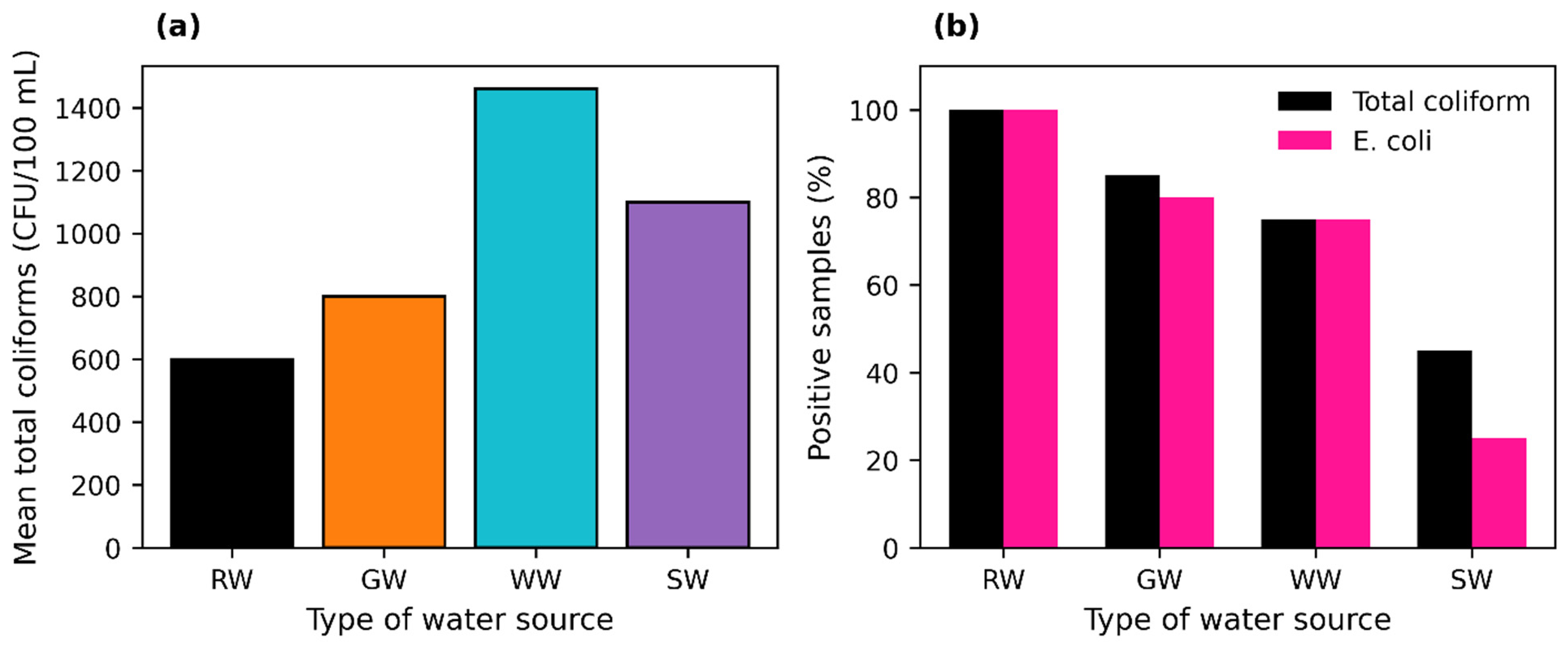
Of the 87 water samples analyzed in Guwahati, 64.4% were positive for total coliforms, indicating widespread fecal contamination (Table 1). This high prevalence reflects a substantial microbial burden across the urban water system. Well water had the highest mean total coliform count (1460 CFU/100 mL), followed by supply water, groundwater, and river water (Figure 2a), showing that shallow point sources used for domestic supply can be as impacted as surface waters. The elevated coliform counts in well water are attributable to the shallow construction of most dug wells in Guwahati’s peri-urban zones, which limits the natural filtration capacity of the vadose zone and increases hydraulic connectivity with contaminated surface water and subsurface leachate. Leaking septic tanks, unlined pit latrines, and open drains in close proximity to these wells represent persistent point sources of fecal contamination, as documented in comparable South and Southeast Asian urban settings [42,43,44]. Well depth, construction quality, and proximity to sanitation infrastructure are therefore critical determinants of microbial water quality at the household level. All river water and well water samples, 75% of supply water samples, and 45.8% of groundwater samples contained detectable total coliforms (Table 1; Figure 2b). High contamination (>103 CFU/100 mL) dominated river water (93.3% of samples) and wells (62.5%) but was less frequent in groundwater (22.9%) and supply water (33.3%), with overall 41.4% of all samples heavily contaminated, 20.7% moderately contaminated, and 2.3% showing low-level contamination (Table 1). The maximum total coliform concentration (9600 CFU/100 mL) occurred in a Brahmaputra River sample near Tarun Nagar, a densely populated neighborhood characterized by inadequate drainage infrastructure and recurrent urban flooding. This site is particularly susceptible to microbial contamination because stormwater runoff and sewage overflows frequently co-mingle in poorly separated drainage systems, delivering concentrated fecal loads directly into the river during and after rainfall events [10]. Additionally, this sampling zone is near the confluence of the Bharalu River, an urban tributary that traverses Guwahati and receives large volumes of inadequately treated domestic and industrial effluents before discharging into the Brahmaputra, further amplifying the microbial burden at this site [23]. In contrast, the upstream site (L1), located before major urban influence, showed the lowest coliform counts among river sampling points, consistent with the well-documented longitudinal gradient of fecal contamination that increases progressively as rivers accumulate urban inputs [26,28]. This spatial pattern reflects how the degree of urban encroachment along the river corridor directly shapes local microbiological water quality. Some well and groundwater sites showed substantially lower coliform counts (Supplementary Information), likely reflecting deeper construction, reduced surface connectivity, or more favorable lithological conditions at those locations that limit downward bacterial transport [42].
Table 1.
Distribution of total coliform concentrations in different water sources in Guwahati.
Figure 2.
Coliform contamination in water samples from Guwahati, India. (a) Mean total coliform counts (CFU/100 mL) in river water (RW), well water (WW), supply water (SW), and groundwater (GW). (b) Percentage of samples positive for total coliforms and E. coli for each water source (RW, WW, SW, GW).
The WHO classifies microbial water quality into five risk bands: 0 CFU/100 mL (conformity), 1–10 CFU/100 mL (low risk), 10–100 CFU/100 mL (intermediate risk), 100–1000 CFU/100 mL (high risk), and >1000 CFU/100 mL (very high risk) [43,44]. In this study, 93.3% of river water samples, 62.5% of well water samples, 50% of groundwater samples, and 33.3% of supply water samples fell into the very-high-risk category (total coliforms >1000 CFU/100 mL), indicating extensive fecal contamination across multiple sources. These findings indicate that a large proportion of water sources are unsuitable for direct human consumption without treatment. Comparable levels of contamination have been reported in other Indian cities; for instance, drinking water in Junagadh (Gujarat) exhibited coliform counts exceeding 2400 CFU/100 mL, largely attributed to contamination during distribution [45]. Urbanization intensity has been identified as the primary watershed-scale driver of fecal coliform variability, with studies showing that catchments with impervious surface coverage above 65% undergo a shift in dominant contamination sources from diffuse agricultural inputs to concentrated urban sewage and stormwater pathways [46]. Guwahati’s rapid and largely unplanned urban expansion, with an estimated metropolitan area of ~328 km2 and a population exceeding 1.5 million, places it firmly within this high-risk urbanization category, where infrastructure deficits outpace population growth [30].
The situation in Guwahati is further exacerbated by recurrent “artificial” flooding in densely populated neighborhoods where inadequate drainage and compromised sanitation infrastructure can promote sewage leakage and cross-contamination between supply and wastewater systems [32]. Consistent with this pattern, river water collected near the Tarun Nagar area exhibited some of the highest coliform loads in the present study, highlighting that localized sanitation failures and flood-induced infrastructure stress may drive microbial contamination of urban surface waters. This spatial pattern underscores how urban infrastructure deficits translate directly into microbiological contamination hotspots at identifiable locations within the water system. A combination of point sources (e.g., direct sewage discharge, overflow from treatment facilities) and non-point pollution sources (e.g., surface runoff from impervious areas, agricultural drainage, leachate from informal waste disposal) simultaneously influences water quality across both surface water and groundwater [10]. In rapidly growing cities such as Guwahati, these pressures are likely to be amplified, as infrastructure investment lags behind population growth and urbanization rate, reinforcing the need for integrated watershed-scale water quality management [46].
E. coli was quantified alongside total coliforms to more specifically attribute the coliform burden to fecal sources. From each coliform-positive sample, at least two presumptive E. coli colonies were randomly selected and confirmed using conventional and automated methods, yielding a total of 99 isolates from 45 of 87 samples (51.7%). The gradient in E. coli detection—100% positivity in river water with progressively lower frequencies in well water, supply water, and groundwater (Figure 2b)—closely paralleled the distribution of total coliforms across the sampled water sources. This concordance strengthens the inference that fecal contamination is the dominant source of microbial pollution. The supply water contamination (75% coliform-positive) despite undergoing treatment likely reflects post-treatment recontamination during distribution, a well-documented mechanism in low- and middle-income country water systems where intermittent supply, pressure fluctuations, and aging pipe infrastructure create conditions for external infiltration of microbial contaminants [10,45].
Groundwater, a key source of drinking water in the region, was also significantly impacted. Groundwater and well sampling locations in this study were situated within densely populated residential and peri-urban areas where they have inadequate drainage infrastructure, in the vicinity of open drains and surface water channels, and recurrent seasonal flooding. In Guwahati, previous studies have also reported that untreated urban sewage discharge, poorly maintained sanitation systems, septic tank leakage, and improper solid waste disposal may contribute to microbial contamination of local water resources, particularly during monsoon periods when runoff and hydraulic connectivity are enhanced [30,46]. Shallow wells and groundwater sources may therefore be especially vulnerable to fecal contamination in such urban floodplain environments.
Approximately 50% of groundwater samples exceeded 103 CFU/100 mL, placing them in the very-high-risk band, in direct violation of WHO drinking water guidelines, which require the absence of E. coli and total coliforms in any 100 mL sample intended for human consumption [47]. This represents a critical public health concern given the reliance on untreated groundwater. The elevated contamination in groundwater is consistent with infiltration of fecal matter through compromised sanitation infrastructure, particularly during flooding events, when hydraulic connectivity between surface water and groundwater is likely enhanced [17,18]. However, as direct hydrological tracing was not performed in this study, this interpretation is based on the spatial co-occurrence of contamination across water compartments and remains inferential. In many urban and peri-urban settings, poorly constructed sanitation systems such as pit latrines and leaky septic tanks may further contribute to groundwater contamination. Pit latrines lack a physical barrier between stored excreta and surrounding soil, allowing pathogens and other contamination to leach into groundwater and contaminate nearby wells used for drinking water [42].
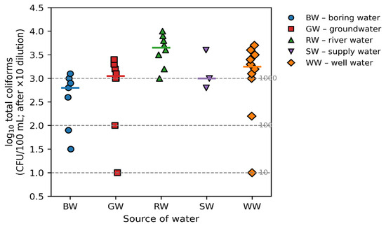
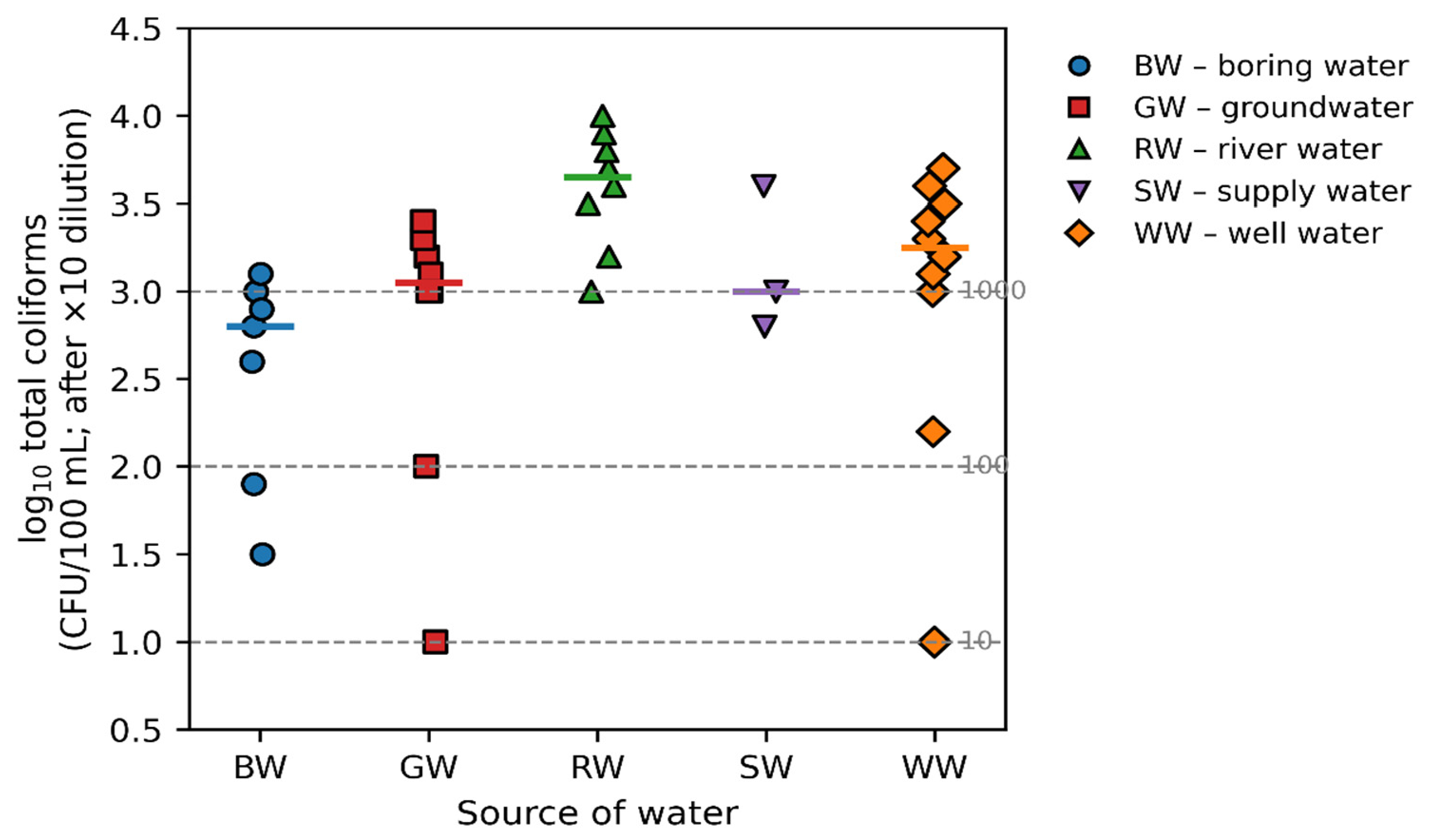
Analysis of total coliform counts (CFU/100 mL, 10-fold dilution-adjusted) further revealed pronounced within-source variability (Figure 3): river water exhibited the broadest range, whereas supply water showed a narrower but still substantial range (Figure 3). As the total coliform data were not normally distributed, non-parametric statistical analysis was applied. The Kruskal-Wallis rank-sum test indicated that total coliform distributions differed significantly among water types (p = 0.0033, p < 0.05), confirming that the differences observed visually between sources in Figure 2 and Figure 3 represent systematic variation rather than random noise. This statistical significance further supports the influence of distinct contamination pathways across different water sources. Even when the absolute E. coli counts in groundwater are lower than those in river water, their presence is epidemiologically relevant [48]. This remains concerning because many residents consume groundwater without treatment.
Figure 3.
Distribution of log10-transformed total coliform counts (CFU/100 mL; after ×10 dilution) by water source. Each point represents a sample from boring water (BW), groundwater (GW), river water (RW), supply water (SW), or well water (WW). The horizontal colored segments indicate the median of each source. Grey dashed lines at log10 = 1, 2 and 3 correspond to 10, 100 and 1000 CFU/100 mL, respectively, marking the WHO thresholds between low, intermediate, high and very high microbiological risk bands for drinking water.
3.2. Antibiotic Susceptibility Profiles of E. coli
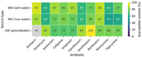
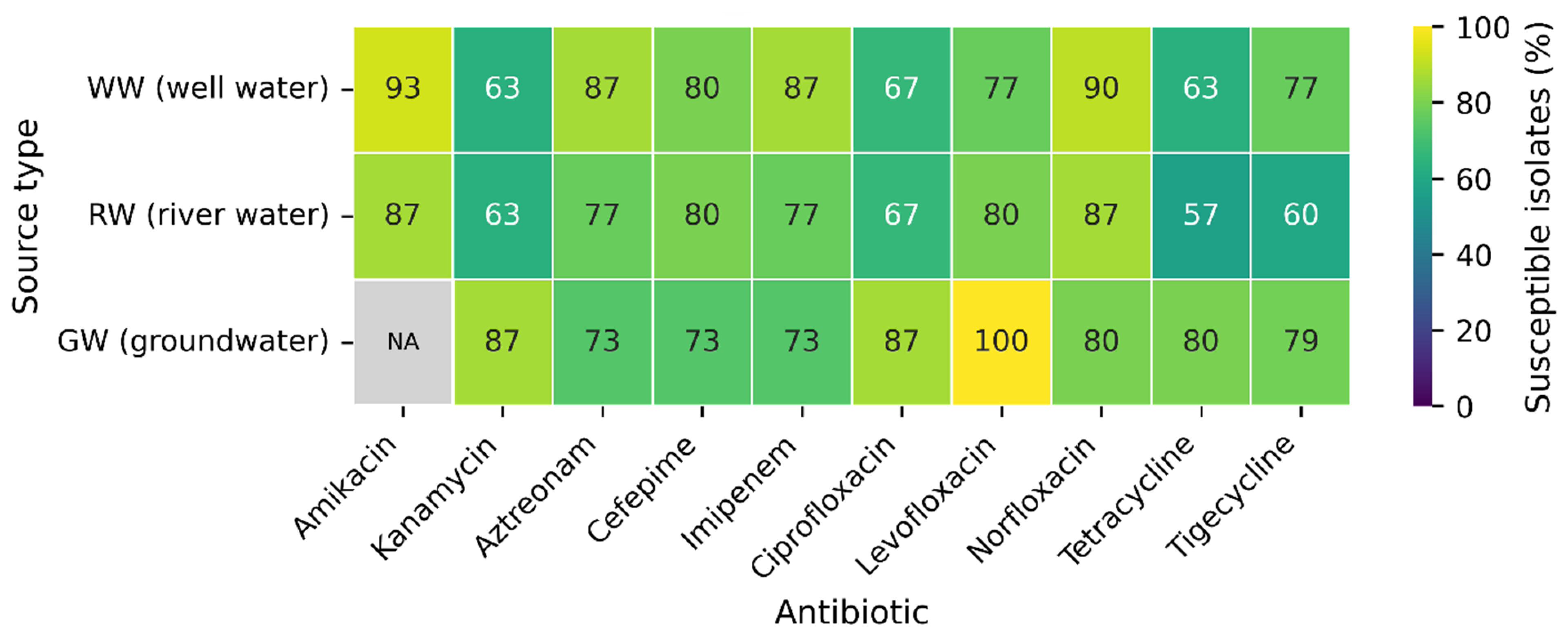
All 99 confirmed E. coli isolates were tested for susceptibility to the selected antibiotic panel (Figure 4). In river water, the highest susceptibility was observed for norfloxacin (NFX) and amikacin (AK), with 86.7% of isolates susceptible to each, whereas resistance was most frequent to kanamycin (KAN), tigecycline (TGC), and tetracycline (TC). Among the well water isolates, susceptibility was highest to AK (93.3%) and NFX (90.0%), but notable resistance was observed to ciprofloxacin (CIP), KAN, and TC. Groundwater-derived E. coli isolates showed complete susceptibility to NFX (100%) and high susceptibility to CIP, KAN, and levofloxacin (LVX) (each 86.7%), yet exhibited comparatively high resistance to aztreonam (AT) and imipenem (IPM) (Figure 4).
Figure 4.
Susceptibility profiles of Escherichia coli isolates. Bar plots show the percentage of susceptible isolates from river water (RW), well water (WW), and groundwater (GW) to amikacin (AK), norfloxacin (NFX), imipenem (IPM), aztreonam (AT), cefepime (CPM), levofloxacin (LVX), tigecycline (TGC), ciprofloxacin (CIP), kanamycin (KAN), and tetracycline (TC). Percentages were calculated within each source group (RW, WW, GW) relative to the total number of E. coli isolates from that source, rather than cumulatively across all sources.
These resistance patterns are not random but reflect identifiable selective pressure operating across the Guwahati urban water system. The occurrence of antibiotic-resistant E. coli across river water, well water, and groundwater samples indicates that resistance determinants have disseminated beyond agricultural settings into interconnected environmental water sources. A primary driver of this dissemination is the continuous discharge of inadequately treated wastewater into the Bharalu River and ultimately into the Brahmaputra, which carries a mixture of hospital effluents, domestic sewage, and pharmaceutical residues, all well-established sources of antibiotic selective pressure in aquatic environments [2,3]. Global surveillance data confirm that 65% of river sites across 72 countries contain antibiotic concentrations exceeding safety thresholds, with the highest burdens recorded in South and Southeast Asia due to limited wastewater treatment capacity [49]. Once sub-inhibitory concentrations of antibiotics are present in water, they exert continuous selective pressure on resident bacterial populations, enriching for resistant genotypes and facilitating horizontal gene transfer (HGT) of resistance determinants among environmental microbial communities [4,7]. Resistance was particularly common to tetracycline and aminoglycosides such as kanamycin, which is consistent with the extensive use of these antibiotics in both human and veterinary medicine. Tetracyclines are widely used due to their low cost and availability and have been repeatedly identified as key drivers of resistance in enteric bacteria in low- and middle-income countries [22,50]. The persistence of tetracycline resistance in environmental E. coli is further reinforced by the long half-life of tetracyclines in aquatic sediments and their ability to bind soil particles, maintaining low-level selective concentrations in the environment long after direct antibiotic input has ceased [51]. In India, unrestricted over-the-counter availability of tetracyclines and their use in livestock feed supplements create multiple entry points into the water cycle through human excretion, agricultural runoff, and direct discharge from animal husbandry operations, all of which converge in urban river systems such as the Brahmaputra and its tributaries [21,22].
The elevated resistance to tetracycline is consistent with its widespread, low-cost use and frequent over-the-counter availability, which are well-recognized drivers of tetracycline resistance in enteric bacteria [22]. Kanamycin resistance, also prevalent across water source types in this study, is similarly explained by its historical and current use as a broad-spectrum antibiotic in both human medicine and veterinary settings in India. Resistance to aminoglycosides such as kanamycin is typically mediated by aminoglycoside-modifying enzymes encoded on mobile genetic elements, enabling rapid horizontal dissemination among environmental bacterial populations [2,6]. The co-occurrence of kanamycin and tetracycline resistance in the same isolates is particularly concerning, as co-resistance on shared mobile elements can maintain and co-select for multiple resistance phenotypes simultaneously, even in the absence of direct antibiotic pressure [8]. These findings align with growing global evidence that aquatic environments act as reservoirs and mixing grounds for antibiotic resistance genes, facilitating interactions between environmental, animal, and human microbial communities. The detection of MDR E. coli in both open dug wells and deeper bore wells suggests that resistance is not confined to shallow, directly contaminated sources and that aquifers may also be affected. This finding challenges the assumption that groundwater is inherently protected from resistance dissemination and suggests that the same hydrological processes (river–aquifer exchange, flooding-driven recharge, and leakage from sanitation infrastructure) that transport fecal bacteria also carry their associated resistance determinants into the subsurface [17,18]. Urban sanitation failures and intensive antibiotic use therefore act as co-drivers of groundwater AMR vulnerability in Guwahati, consistent with patterns reported in other rapidly urbanizing South Asian cities [20,23].
Resistance to ciprofloxacin is also noteworthy because fluoroquinolones are frequently prescribed antibiotics to treat urinary tract infections (UTIs) caused by E. coli [52,53]. The occurrence of ciprofloxacin-resistant isolates in environmental waters suggests that resistant strains circulating in clinical settings may enter aquatic systems through wastewater discharge. This pathway is well-supported by studies documenting the occurrence of fluoroquinolones, including ciprofloxacin, in Indian river water at concentrations exceeding minimum selective concentrations (MSCs), even far from point discharge locations [3,51]. In the Guwahati context, hospital and clinic effluents discharged into the Bharalu River without adequate treatment are a plausible source of both ciprofloxacin residues and fluoroquinolone-resistant E. coli entering the broader water system [23]. Additionally, the detection of imipenem resistance, although less common, is of particular concern because carbapenems are considered last-resort antibiotics for treating infections caused by multidrug-resistant Gram-negative bacteria [54]. Imipenem resistance in environmental E. coli in the absence of carbapenemase production (as confirmed in this study) may be mediated by alternative mechanisms such as overexpression of efflux pumps, loss of outer membrane porins (e.g., OmpF/OmpC), or upregulation of intrinsic AmpC β-lactamases, all of which can confer reduced carbapenem susceptibility without encoding carbapenemase genes [7,35]. The presence of such isolates in groundwater, a source consumed without treatment by many Guwahati households, is especially concerning as it implies potential direct human exposure to last-resort-resistant bacteria through drinking water.
Also, the presence of E. coli in drinking water sources is directly linked to diarrheal disease burden, as multiple studies have shown associations between fecal contamination and increased incidence of gastrointestinal infections [17,30,55,56]. In many cases, such infections are treated with antibiotics, which can further select for resistant strains of bacteria. Alarmingly, some E. coli lineages have acquired resistance to multiple antibiotic classes, limiting the treatment options [2,5,57,58].
The multiple antibiotic resistance (MAR) index was calculated for each isolate. Fourteen of the 99 E. coli isolates (14.1%) had MAR > 0.2, indicating exposure to environments with frequent or intensive antibiotic use and consistent with a multidrug-resistant (MDR) phenotype. These MDR isolates were distributed across river, well, and groundwater sources, suggesting that high-risk contamination sources influence both surface and subsurface components of the urban water system. High MAR indices in the selected isolates reinforce this observation. In particular, E. coli isolates from the Brahmaputra at Pandu port, Ananda Nagar, and Maligaon showed resistance to multiple antibiotics, with MAR values of up to 0.8, indicating exposure to strong and diverse antibiotic selection pressures. In the current study, 14 MDR isolates (14.1% of all E. coli) had MAR indices > 0.2, a commonly used threshold indicative of high-risk-contamination sources such as hospital effluents, intensive livestock operations, or areas of frequent antibiotic misuse [41,59]. Previous studies have similarly reported MDR E. coli in drinking water, with 3–6 drug resistance profiles observed in more than 60% of isolates in some settings [59]. Together, these findings suggest that resistant E. coli in Guwahati’s water system are likely the outcome of cumulative antibiotic misuse and poor waste management. These strains may contribute to a self-reinforcing cycle of infection and transmission in households, healthcare facilities, and the broader community.
Comparative data from other regions highlight that several resistance levels observed in Guwahati were elevated (Table 2). Ciprofloxacin resistance in Guwahati River water (33.3%) was lower than the highest values reported for Sri Lankan rivers and canals (up to 70%) but substantially higher than the 3.2% reported for household water in Peru and the 11–17% reported for drinking water on Greek islands. Tetracycline resistance in Guwahati River water (43.3%) falls within the broad range reported for other polluted rivers and canals (10–90%) but exceeds the values noted for some treated tap waters in South Africa and surface waters in the United States, where resistance was often ≤35% or absent. Most strikingly, cefoxitin resistance among groundwater E. coli isolates in this study (91.7%) was far higher than the 3.2% reported for household water in Peru, underscoring a particularly severe burden of β-lactam-resistance in local groundwater.
Table 2.
Reported percentages of E. coli resistant to selected antibiotics in different water sources worldwide, compared with those in Guwahati (present study). CIP-R, ciprofloxacin resistance; TGC-R, tigecycline resistance; KAN/KM-R, kanamycin resistance (KM in Sri Lankan studies); TC-R, tetracycline resistance; IPM-R, imipenem resistance; ATM-R, aztreonam resistance; FOX-R, cefoxitin resistance; NA, data not available.
Similar patterns of AMR in aquatic environments have been documented across several major river systems in India. Studies of the Ganga River have reported a high abundance of ARB, ARGs, and MGEs, particularly in downstream locations receiving anthropogenic inputs. A significant positive correlation between fecal indicator bacteria and ARGs in these waters suggests that fecal contamination may facilitate the environmental dissemination of antimicrobial resistance [26]. More broadly, antimicrobial-resistant bacteria have been reported from multiple water sources across India, including rivers, ponds, lakes, and groundwater systems, with hospital effluents and pharmaceutical wastewater identified as major contributors to environmental contamination [63]. High proportions of ESBL-producing Gram-negative bacteria have also been documented in major rivers such as the Ganges and Yamuna, highlighting the widespread nature of resistance in aquatic ecosystems. In addition to resistant bacteria, measurable concentrations of antibiotic residues have been detected in river waters. For example, monitoring of the Yamuna River revealed the presence of antibiotics such as ofloxacin, amoxicillin, and erythromycin, with ofloxacin concentrations reaching 145.37 ng/mL in surface water samples [64]. The presence of such pharmaceutical residues can exert selective pressure on environmental microbial communities, promoting the emergence and persistence of antibiotic-resistant strains. Together, these findings support the interpretation that the antibiotic-resistant E. coli detected in the present study may reflect cumulative inputs of fecal contamination, untreated wastewater, and antibiotic residues into aquatic environments.
3.3. β-Lactamase Production
AmpC β-lactamase production was detected in 12 of the 99 E. coli isolates (12.1%). The detection of AmpC β-lactamase-producing E. coli further elevates this concern. In the present study, 12/99 isolates were AmpC positive, a prevalence lower than the 37.2% reported for clinical E. coli in Iran but still substantial for environmental isolates [65]. Most AmpC-positive isolates were recovered from river water, with additional isolates from wells, boring water, and supply water, indicating dissemination across interconnected surface and subsurface sources. AmpC β-lactamases are important in clinical settings because they confer resistance to many penicillins and cephalosporins and can promote nosocomial outbreaks and treatment failure, particularly when expression is inducible or derepressed [66]. In this study, inducible AmpC production was observed in five isolates (S16E1, S20E8, S30E4, S53E1, and S69E2), suggesting that these strains may upregulate AmpC expression in response to specific β-lactam exposure and thereby survive antibiotic therapy.
For detailed β-lactamase characterization and MIC analysis, a subset of 58 representative isolates exhibiting resistance to at least one antibiotic was selected. Among these, isolates originated from river (n = 5), well (n = 3), boring (n = 3), and supply (n = 1) water, indicating that AmpC producers occur across both surface and groundwater-derived sources (Table 3) (Supplementary Information). The co-occurrence of AmpC and ESBL phenotypes in a well water isolate (S20E8) is particularly concerning. Plasmid-mediated ESBLs, such as CTX-M, TEM, and SHV variants, have previously been reported to spread among Enterobacteriaceae via horizontal gene transfer, contributing to the dissemination of resistance across clinical and environmental settings [25,67]. However, as molecular characterization was not performed in the present study, the occurrence of specific resistance genes and associated transfer mechanisms remains inferential. Therefore, the accurate detection and surveillance of ESBL-producing E. coli are essential to prevent unnoticed dissemination. Increasing numbers of ESBL-producing E. coli have been documented not only in hospitals but also in livestock, wildlife, and aquatic environments, illustrating the environmental dimension of ESBL spread [25,67,68]. The detection of such strains in key urban water sources in northeastern India suggests that Guwahati may reflect a broader regional pattern of environmental dissemination of ESBL-producing bacteria.
Table 3.
Characteristics and resistance profiles of phenotypically confirmed β-lactamase-producing E. coli isolates. S, susceptible; R, resistant; CIP, ciprofloxacin; NFX, norfloxacin; TC, tetracycline; LVX, levofloxacin; ATM, aztreonam; CPM, cefepime; IPM, imipenem; AK, amikacin; KAN, kanamycin; TGC, tigecycline; FOX, cefoxitin; CAZ, ceftazidime; CTX, cefotaxime; CRO, ceftriaxone; FEP, cefepime; MEM, meropenem; ERT, ertapenem.
All 12 isolates showed reduced susceptibility to cefoxitin and were positive by the confirmatory M3DET, supporting the presence of AmpC β-lactamase activity. Overall, the predominance of AmpC producers without additional β-lactamase resistance mechanisms and the absence of carbapenemase suggest that advanced β-lactamase resistance remains relatively limited in the studied environmental water sources (Table 3). MIC testing showed that 11 of the 12 AmpC-positive isolates remained susceptible to all β-lactam and β-lactam/β-lactamase inhibitor agents tested (cefotaxime, ceftazidime, ceftriaxone, ertapenem, cefepime, aztreonam, imipenem, and meropenem), with MIC values within the susceptible range (for example, CTX ≤ 4 µg/mL, CAZ ≤ 1–4 µg/mL, IPM ≤ 0.25 µg/mL) (Table 3). Only one well water isolate (S65E2) exhibited high-level resistance to cefotaxime (CTX ≥ 64 µg/mL) while remaining susceptible to the other β-lactams, indicating that overt β-lactam treatment failure in this dataset may be associated with a limited subset of environmental E. coli strains. Nevertheless, the detection of AmpC-positive and ESBL-positive E. coli across multiple water types suggests that Guwahati’s water resources can act as environmental reservoirs for β-lactam resistance determinants, with the potential for human exposure through drinking water and other pathways.
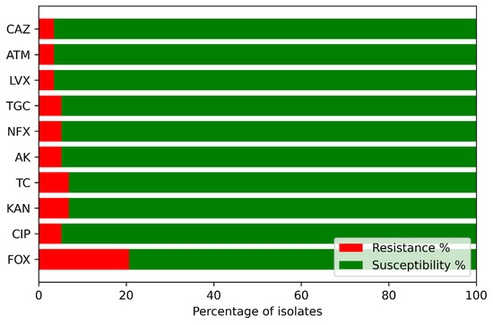
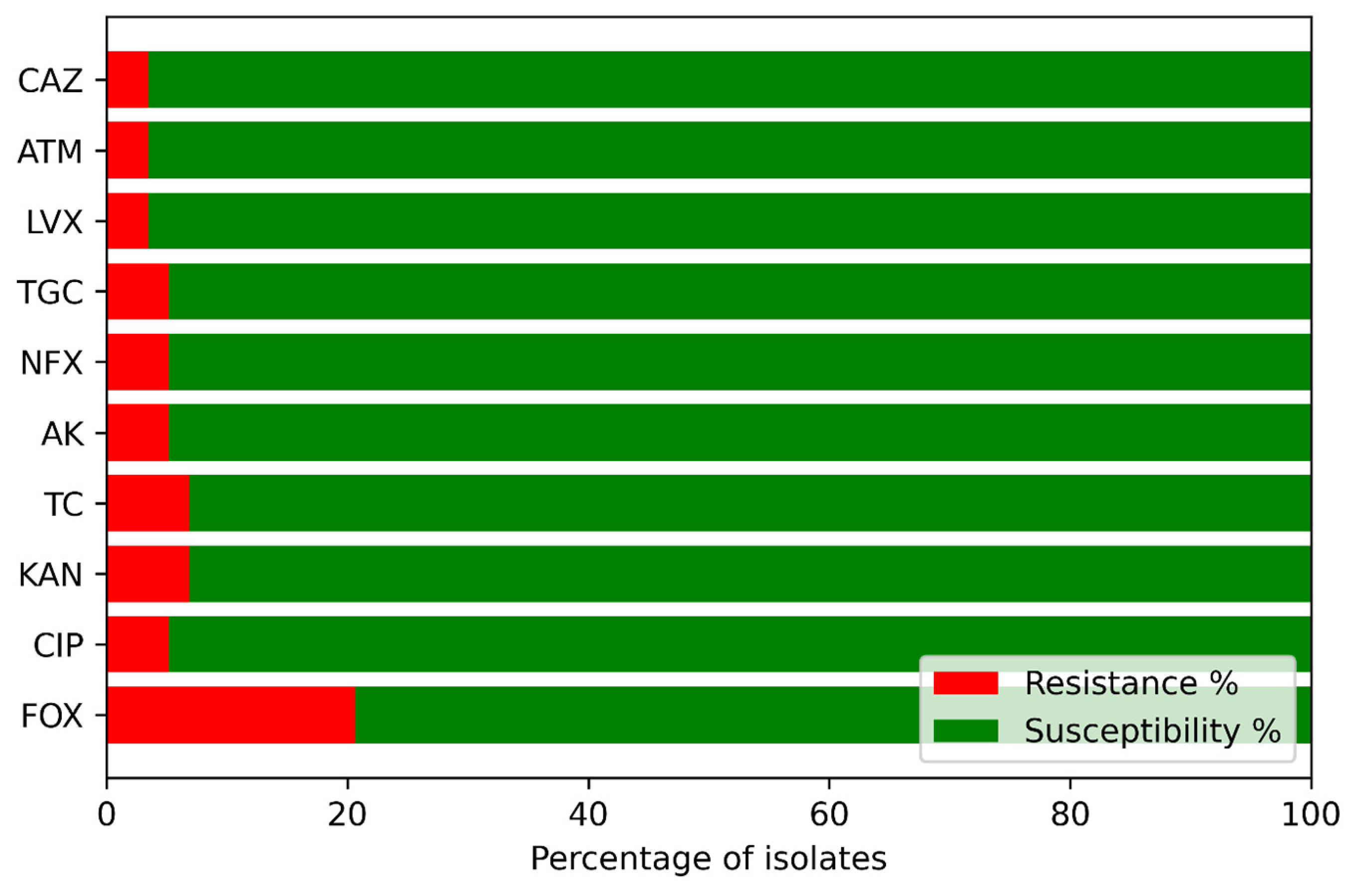
A single-time-point antibiogram was constructed to visualize both isolate-level resistance patterns and the overall proportion of resistant versus susceptible E. coli to each antibiotic (Figure 5). Across all 58 representative isolates, only 12 (20.7%) showed resistance to at least one drug, and the overall prevalence of resistance was low (6.6% of all isolates–antibiotic combinations). Cefoxitin displayed the highest resistance frequency (20.7% of isolates), whereas resistance to the remaining agents was rare (5.2–6.9% for ciprofloxacin, kanamycin, tetracycline, amikacin, norfloxacin, and tigecycline, and only 3.4% for levofloxacin, aztreonam, and ceftazidime), with the corresponding susceptibility proportions ranging from 79.3% (FOX) to 96.6% (LVX, ATM, CAZ). Some isolates from the same source (e.g., S3E3 and S3E6 from the Brahmaputra River) exhibited nearly identical resistance profiles, suggesting possible clonal relatedness or shared selective pressures within that reach. In contrast, the diversity and generally low frequency of resistance across antibiotics and sources is consistent with the complex hydrological interconnections between surface water, groundwater, and supply systems, as well as spatially heterogeneous anthropogenic inputs, such as antibiotic use and wastewater discharge. Unlike conventional clinical antibiograms, this environmental antibiogram provides a cross-sectional snapshot of susceptibility patterns among environmental E. coli isolates.
Figure 5.
Percentage-based-resistance profile of environmental E. coli isolates. For each antibiotic, ciprofloxacin (CIP), tetracycline (TC), aztreonam (ATM), amikacin (AK), kanamycin (KAN), tigecycline (TGC), cefoxitin (FOX), ceftazidime (CAZ), norfloxacin (NFX) and levofloxacin (LVX), horizontal bars show the proportion of isolates classified as resistant (red segment) and susceptible (green segment).
3.4. Overall Implications and Limitations
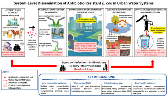
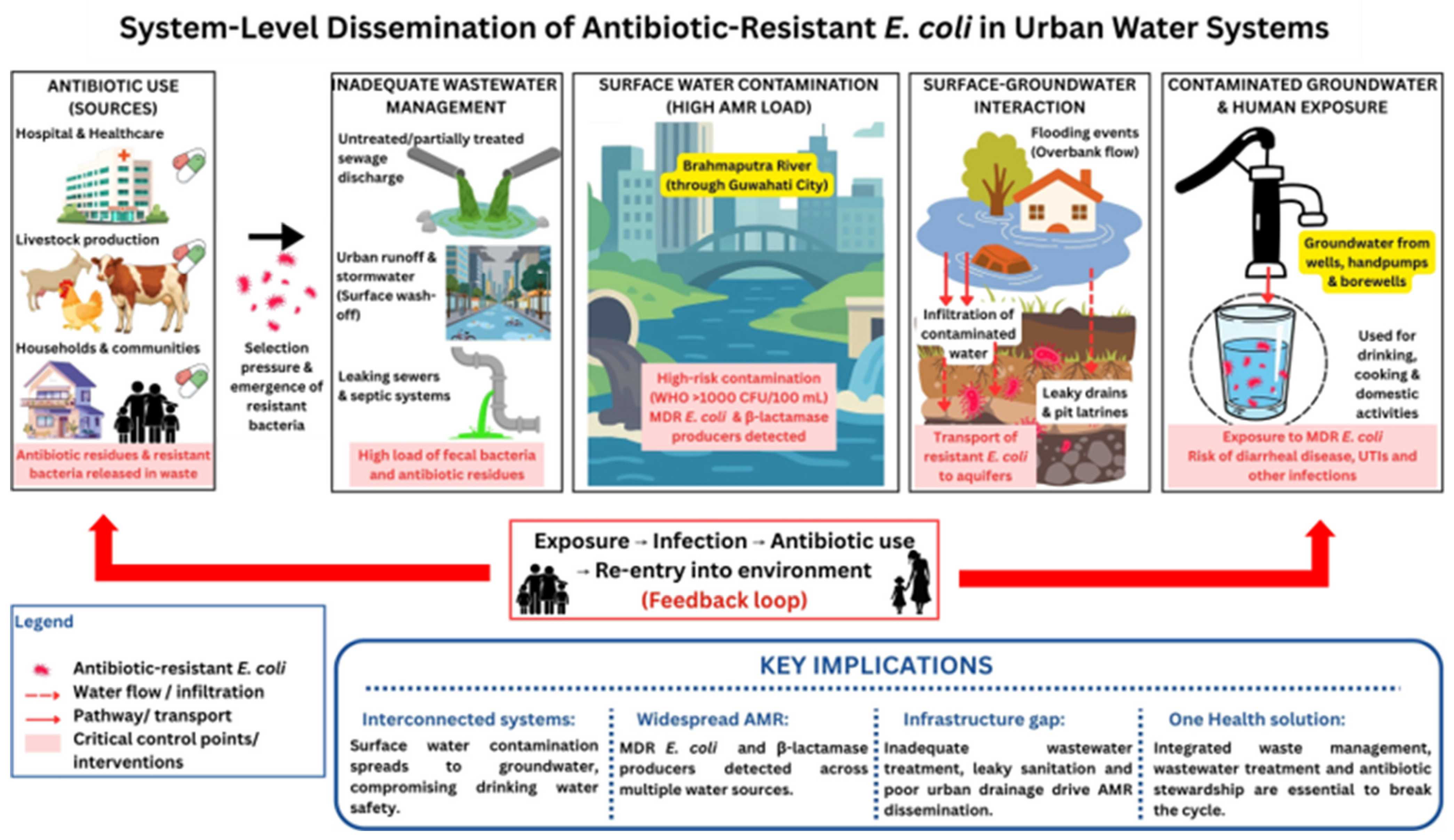
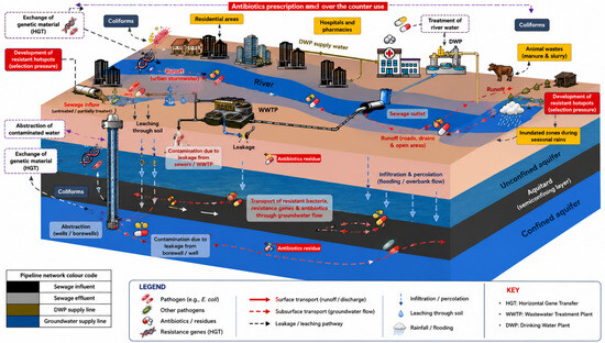
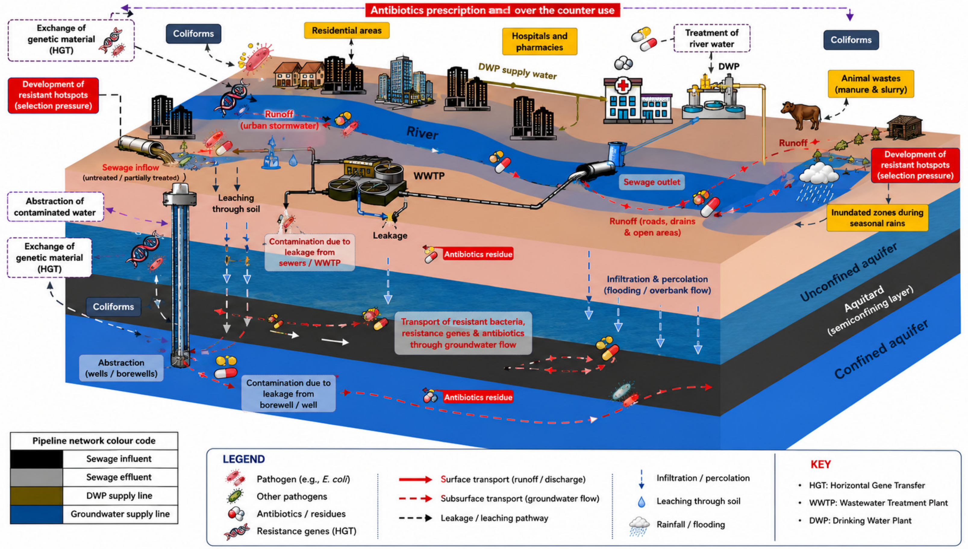
Overall, this study demonstrates that urban water systems function as interconnected pathways for antimicrobial resistance dissemination, with groundwater contamination challenging their assumed microbiological safety in rapidly urbanizing environments. The combination of very high microbial risk levels according to WHO criteria, frequent detection of E. coli in both surface and groundwater, substantial MDR prevalence, and the presence of AmpC- and ESBL-producing E. coli indicates that Guwahati’s water resources represent an important environmental reservoir of antimicrobial resistance. Antibiotic use in human and animal sectors contributes to the release of resistant bacteria into wastewater, which enters surface waters through untreated discharge and urban runoff. Hydrological processes, including infiltration and flooding, facilitate the transfer of contaminants from surface water to groundwater. As a result, groundwater used for drinking may become contaminated, creating pathways for human exposure and reinforcing the environmental cycle of antimicrobial resistance. The public health significance of these findings is considerable because groundwater and tube well water sources are often consumed untreated for drinking, cooking, and other household purposes in several communities in Guwahati. Exposure to AMR E. coli through contaminated water may increase the risk of difficult-to-treat gastrointestinal and opportunistic infections. Previous studies conducted in India have shown that children are particularly vulnerable to groundwater contamination due to their lower body weight, developing immune systems, and higher water intake relative to body mass [69,70]. Furthermore, the occurrence of multidrug-resistant bacteria in groundwater has been recognized as an additional environmental and public health concern in urbanized regions affected by wastewater discharge, runoff, and inadequate sanitation infrastructure [71]. These pathways are conceptually summarized in Figure 6, which illustrates the system-level connectivity linking wastewater inputs, surface water contamination, groundwater infiltration, and human exposure.
Figure 6.
Conceptual framework illustrating the system-level dissemination of antibiotic-resistant Escherichia coli in urban water systems.
The findings call for urgent interventions to improve wastewater collection and treatment, protect groundwater from sewage and industrial leakage, regulate antibiotic use, and expand the routine monitoring of microbial and resistance indicators in both surface and drinking water supplies. Although, to our knowledge, one of the first detailed assessments of antibiotic-resistant E. coli in groundwater in this region provides a baseline for future surveillance and supports the inclusion of northeastern India in national and international AMR monitoring frameworks. Yet, there remains several constraints, viz., the sampling design shows a cross-sectional assessment and therefore does not capture any seasonal variability in microbial contamination, particularly during the monsoon when flooding and sewage overflows, which may further influence water quality. Additionally, key physicochemical water quality parameters, including pH, dissolved oxygen, conductivity, and turbidity, were not measured in the present study. Inclusion of these parameters could have provided further insight into the environmental conditions influencing microbial contamination and the persistence of ARB across different water compartments. Second, although phenotypic methods were used to characterize antibiotic resistance and β-lactamase production, molecular analysis of resistance genes was not performed and could provide deeper insights into the genetic mechanisms underlying the observed resistance patterns. Future studies incorporating molecular analysis and wider spatial sampling would help clarify the dynamics of antimicrobial resistance in Guwahati’s urban water system.
Addressing this situation will require integrated measures (Figure 7), improving wastewater collection and treatment, protecting groundwater from sewage and industrial leakage, tightening antibiotic stewardship in the human and animal sectors, and institutionalizing routine monitoring of microbial indicators and resistance profiles in urban water supplies. Continued research and longitudinal surveillance of E. coli and other resistant pathogens in Guwahati and the broader northeastern region are essential for tracking trends, refining risk assessments, and guiding targeted interventions to safeguard public health.
Figure 7.
Conceptual model of antibiotic resistance in coliforms across interconnected water sources in Guwahati, India. Resistance levels are highest in river water, which receives multiple non-point-inputs (urban runoff, sewage, and industrial effluents), and decrease along pathways to groundwater and drinking water supplies. Seasonal monsoon rains cause inundation and unsanitary conditions, creating localized hotspots of fecal contamination and antibiotic resistance. Excessive antibiotic use and horizontal gene transfer within microbial communities further amplify this resistance. WWTP, wastewater treatment plant; DWP, drinking-water plant.
4. Conclusions
This study demonstrates that antibiotic-resistant Escherichia coli is widely distributed across interconnected urban water sources in Guwahati, with 64.4% of samples positive for total coliforms and 51.7% confirmed for E. coli. Contamination levels were particularly severe in surface waters, where 93.3% of river samples exceeded the World Health Organization’s very-high-risk threshold (>1000 CFU/100 mL). Importantly, groundwater, commonly relied upon for drinking, also showed substantial contamination, with 50% of samples falling within the very-high-risk category, challenging its perceived microbiological safety. Antimicrobial resistance was detected across all water sources, with 14.1% of E. coli isolates exhibiting multidrug resistance (MAR > 0.2), indicating exposure to high-risk, antibiotic-impacted environments. Furthermore, 20.6% of isolates produced β-lactamases, including an extended-spectrum β-lactamase producer, highlighting the environmental presence of clinically relevant resistance mechanisms.
The co-occurrence of fecal contamination and antimicrobial resistance across surface water and groundwater suggests that urban water systems function as interconnected pathways for AMR dissemination. Hydrological connectivity, combined with untreated wastewater discharge, leaky sanitation infrastructure, and urban runoff, represents a plausible mechanism by which resistant bacteria may be transferred from surface environments into aquifers; however, direct confirmation of such inter-compartment transfer would require future studies incorporating hydrological tracer approaches or genomic source-tracking analyses such as whole-genome sequencing. These findings have direct public health implications, as they point to widespread exposure risks through drinking water pathways in a rapidly urbanizing setting. Addressing this challenge requires integrated interventions that move beyond compartmentalized water management. Priorities include strengthening wastewater treatment, protecting groundwater sources from contamination, and promoting responsible antibiotic use across human and animal sectors. Overall, this study provides quantitative evidence that urban water systems can act as conduits for the environmental spread of antimicrobial resistance, emphasizing the need for system-level strategies within a One Health framework to safeguard water quality and public health.
Supplementary Materials
The following supporting information can be downloaded at: https://www.mdpi.com/article/10.3390/w18141725/s1, Table S1. Total Coliform Count (CFU/100 mL) and E. coli isolates in different water samples collected from Guwahati City. Table S2. List of Escherichia coli (E. coli) phenotypically confirmed to be β-lactamase producers.
Author Contributions
R.G.: Methodology, Supervision, Writing and editing, Funding acquisition. B.I.: Investigation, Data curation, review and editing. N.S.: Investigation, Data analysis. H.N.: Writing and editing. T.K.: Data analysis, writing, and editing. J.A.T.-M.: review and editing. S.D.: Methodology, Formal analysis, and editing. K.N.: review and editing. M.K.: Supervision, review, and editing. All authors have read and agreed to the published version of the manuscript.
Funding
This work was funded by the Science and Engineering Research Board (SERB), the Department of Science and Technology (DST), Govt. of India, under the SERB-POWER grant (grant number SPG/2021/002107) awarded to Dr. Ritusmita Goswami.
Data Availability Statement
The original contributions presented in this study are included in the article. Further inquiries can be directed to the corresponding authors.
Conflicts of Interest
The authors declare no conflicts of interest.
References
- Murray, C.J.L.; Ikuta, K.S.; Sharara, F.; Swetschinski, L.; Robles Aguilar, G.; Gray, A.; Han, C.; Bisignano, C.; Rao, P.; Wool, E.; et al. Global burden of bacterial antimicrobial resistance in 2019: A systematic analysis. Lancet 2022, 399, 629–655. [Google Scholar] [CrossRef] [Scilit] [PubMed]
- Marti, E.; Variatza, E.; Balcazar, J.L. The role of aquatic ecosystems as reservoirs of antibiotic resistance. Trends Microbiol. 2014, 22, 36–41. [Google Scholar] [CrossRef] [Scilit] [PubMed]
- Larsson, D.G.J.; Andremont, A.; Bengtsson-Palme, J.; Brandt, K.K.; de Roda Husman, A.M.; Fagerstedt, P.; Fick, J.; Flach, C.-F.; Gaze, W.H.; Kuroda, M.; et al. Critical knowledge gaps and research needs related to the environmental dimensions of antibiotic resistance. Environ. Int. 2018, 117, 132–138. [Google Scholar] [CrossRef] [Scilit] [PubMed]
- Juhas, M. Horizontal gene transfer in human pathogens. Crit. Rev. Microbiol. 2015, 41, 101–108. [Google Scholar] [CrossRef] [Scilit] [PubMed]
- Kraupner, N.; Ebmeyer, S.; Bengtsson-Palme, J.; Fick, J.; Kristiansson, E.; Flach, C.-F.; Larsson, D.G.J. Selective concentration for ciprofloxacin resistance in Escherichia coli grown in complex aquatic bacterial biofilms. Environ. Int. 2018, 116, 255–268. [Google Scholar] [CrossRef] [Scilit] [PubMed]
- Maeusli, M.; Lee, B.; Miller, S.; Reyna, Z.; Lu, P.; Yan, J.; Ulhaq, A.; Skandalis, N.; Spellberg, B.; Luna, B. Horizontal gene transfer of antibiotic resistance from Acinetobacter baylyi to Escherichia coli on lettuce and subsequent antibiotic resistance transmission to the gut microbiome. mSphere 2020, 5, e00329-20. [Google Scholar] [CrossRef] [Scilit] [PubMed]
- Poirel, L.; Madec, J.-Y.; Lupo, A.; Schink, A.-K.; Kieffer, N.; Nordmann, P.; Schwarz, S. Antimicrobial resistance in Escherichia coli. Microbiol. Spectr. 2018, 6, 10-1128. [Google Scholar] [CrossRef] [Scilit] [PubMed]
- Sun, D.; Sun, X.; Hu, Y.; Yamaichi, Y. Editorial: Horizontal gene transfer mediated bacterial antibiotic resistance, volume II. Front. Microbiol. 2023, 14, 1221606. [Google Scholar] [CrossRef] [Scilit] [PubMed]
- Winkler, A.S.; Brux, C.M.; Carabin, H.; Das Neves, C.G.; Häsler, B.; Zinsstag, J.; Lapinski, M.K.; Stone, M.; Agbogbatey, M.K.; Xiao, N.; et al. One Health Commission: Harnessing our interconnectedness for equitable, sustainable, and healthy socioecological systems. Lancet 2025, 406, 501–570. [Google Scholar] [CrossRef] [Scilit] [PubMed]
- Khatri, N.; Tyagi, S. Influences of natural and anthropogenic factors on surface and groundwater quality in rural and urban areas. Front. Life Sci. 2015, 8, 23–39. [Google Scholar] [CrossRef] [Scilit]
- Mutiyar, P.K.; Gupta, S.K.; Mittal, A.K. Fate of pharmaceutical active compounds (PhACs) from River Yamuna, India: An ecotoxicological risk assessment approach. Ecotoxicol. Environ. Saf. 2018, 150, 297–304. [Google Scholar] [CrossRef] [Scilit] [PubMed]
- Ahammad, Z.S.; Sreekrishnan, T.R.; Hands, C.L.; Knapp, C.W.; Graham, D.W. Increased waterborne blaNDM-1 resistance gene abundances associated with seasonal human pilgrimages to the upper Ganges River. Environ. Sci. Technol. 2014, 48, 3014–3020. [Google Scholar] [CrossRef] [Scilit] [PubMed]
- Azam, M.; Jan, A.T.; Haq, Q.M.R. blaCTX-M-152, a novel variant of CTX-M-group-25, identified in a study performed on the prevalence of multidrug resistance among natural inhabitants of River Yamuna, India. Front. Microbiol. 2016, 7, 176. [Google Scholar] [CrossRef] [Scilit] [PubMed]
- Lübbert, C.; Baars, C.; Dayakar, A.; Lippmann, N.; Rodloff, A.C.; Kinzig, M.; Sörgel, F. Environmental pollution with antimicrobial agents from bulk drug manufacturing industries in Hyderabad, South India, is associated with dissemination of extended-spectrum beta-lactamase and carbapenemase-producing pathogens. Infection 2017, 45, 479–491. [Google Scholar] [CrossRef] [Scilit] [PubMed]
- Savichtcheva, O.; Okabe, S. Alternative indicators of fecal pollution: Relations with pathogens and conventional indicators, current methodologies for direct pathogen monitoring and future application perspectives. Water Res. 2006, 40, 2463–2476. [Google Scholar] [CrossRef] [Scilit] [PubMed]
- Tallon, P.; Magajna, B.; Lofranco, C.; Leung, K.T. Microbial indicators of faecal contamination in water: A current perspective. Water Air Soil Pollut. 2005, 166, 139–166. [Google Scholar] [CrossRef] [Scilit]
- Bertuzzo, E.; Mari, L. Hydrology, water resources and the epidemiology of water-related diseases. Adv. Water Resour. 2017, 108, 329–331. [Google Scholar] [CrossRef] [Scilit]
- Bett, B.; Tumusiime, D.; Lindahl, J.; Roesel, K.; Delia, G. The role of floods on pathogen dispersion. In Nature-Based Solutions for Flood Mitigation. The Handbook of Environmental Chemistry; Ferreira, C.S.S., Kalantari, Z., Hartmann, T., Pereira, P., Eds.; Springer International Publishing: Cham, Switzerland, 2021; pp. 139–157. [Google Scholar] [CrossRef] [Scilit]
- Van Boeckel, T.P.; Brower, C.; Gilbert, M.; Grenfell, B.T.; Levin, S.A.; Robinson, T.P.; Teillant, A.; Laxminarayan, R. Global trends in antimicrobial use in food animals. Proc. Natl. Acad. Sci. USA 2015, 112, 5649–5654. [Google Scholar] [CrossRef] [Scilit] [PubMed]
- Biswas, S.; Adhikary, M.; Alam, A.; Islam, N.; Roy, R. Disparities in access to water, sanitation, and hygiene (WASH) services and the status of SDG-6 implementation across districts and states in India. Heliyon 2024, 10, e37646. [Google Scholar] [CrossRef] [Scilit] [PubMed]
- Ganguly, N.K.; Arora, N.K.; Chandy, S.J.; Fairoze, M.N.; Gill, J.P.S.; Gupta, U.; Hossain, S.; Joglekar, S.; Joshi, P.C.; Kakkar, M.; et al. Rationalizing antibiotic use to limit antibiotic resistance in India. Indian J. Med. Res. 2011, 134, 281–294. [Google Scholar] [PubMed]
- Shakya, P.; Barrett, P.; Diwan, V.; Marothi, Y.; Shah, H.; Chhari, N.; Tamhankar, A.J.; Pathak, A.; Lundborg, C.S. Antibiotic resistance among Escherichia coli isolates from stool samples of children aged 3 to 14 years from Ujjain, India. BMC Infect. Dis. 2013, 13, 477. [Google Scholar] [CrossRef] [Scilit] [PubMed]
- Kumar, M.; Ram, B.; Honda, R.; Poopipattana, C.; Canh, V.D.; Chaminda, T.; Furumai, H. Concurrence of antibiotic resistant bacteria (ARB), viruses, pharmaceuticals and personal care products (PPCPs) in ambient waters of Guwahati, India: Urban vulnerability and resilience perspective. Sci. Total Environ. 2019, 693, 133640. [Google Scholar] [CrossRef] [Scilit] [PubMed]
- Kumar, S.; Tripathi, V.R.; Garg, S.K. Antibiotic resistance and genetic diversity in water-borne Enterobacteriaceae isolates from recreational and drinking water sources. Int. J. Environ. Sci. Technol. 2013, 10, 789–798. [Google Scholar] [CrossRef] [Scilit]
- Ensor, V.M.; Shahid, M.; Evans, J.T.; Hawkey, P.M. Occurrence, prevalence and genetic environment of CTX-M β-lactamases in Enterobacteriaceae from Indian hospitals. J. Antimicrob. Chemother. 2006, 58, 1260–1263. [Google Scholar] [CrossRef] [Scilit] [PubMed]
- Prasad, D.K.; Shukla, R.; Ahammad, S.Z. Antibiotic resistance in the Ganga River: Investigation of antibiotic-resistant bacteria and antibiotic resistance genes, and public health risk assessment. J. Environ. Chem. Eng. 2024, 12, 114931. [Google Scholar] [CrossRef] [Scilit]
- Singh, N.S.; Singhal, N.; Kumar, M.; Virdi, J.S. High prevalence of drug resistance and class 1 integrons in Escherichia coli isolated from River Yamuna, India: A serious public health risk. Front. Microbiol. 2021, 12, 621564. [Google Scholar] [CrossRef] [Scilit] [PubMed]
- Skariyachan, S.; Mahajanakatti, A.B.; Grandhi, N.J.; Prasanna, A.; Sen, B.; Sharma, N.; Vasist, K.S.; Narayanappa, R. Environmental monitoring of bacterial contamination and antibiotic resistance patterns of the fecal coliforms isolated from Cauvery River, a major drinking water source in Karnataka, India. Environ. Monit. Assess. 2015, 187, 279. [Google Scholar] [CrossRef] [Scilit] [PubMed]
- Manga, M.; Kolsky, P.; Rosenboom, J.W.; Ramalingam, S.; Sriramajayam, L.; Bartram, J.; Stewart, J. Public health performance of sanitation technologies in Tamil Nadu, India: Initial perspectives based on E. coli release. Int. J. Hyg. Environ. Health 2022, 243, 113987. [Google Scholar] [CrossRef] [Scilit] [PubMed]
- Naik, O.A.; Shashidhar, R.; Rath, D.; Bandekar, J.R.; Rath, A. Characterization of multiple antibiotic resistance of culturable microorganisms and metagenomic analysis of total microbial diversity of marine fish sold in retail shops in Mumbai, India. Environ. Sci. Pollut. Res. 2018, 25, 6228–6239. [Google Scholar] [CrossRef] [Scilit] [PubMed]
- India Meteorological Department (IMD). District-Wise Rainfall Data for Assam. 2023. Available online: https://www.mausam.imd.gov.in (accessed on 6 February 2026).
- Assam Tribune. Guwahati Grapples with Artificial Floods. 2023. Available online: https://www.guwahatiplus.com/exclusive-news/guwahati-grapples-with-artificial-floods (accessed on 17 January 2026).
- Carvalhaes, C.G.; Picão, R.C.; Nicoletti, A.G.; Xavier, D.E.; Gales, A.C. Cloverleaf test (modified Hodge test) for detecting carbapenemase production in Klebsiella pneumoniae: Be aware of false positive results. J. Antimicrob. Chemother. 2010, 65, 249–251. [Google Scholar] [CrossRef] [Scilit] [PubMed]
- Tille, P.M. Bailey & Scott’s Diagnostic Microbiology, 15th ed.; Elsevier Health Sciences: St. Louis, MO, USA, 2021. [Google Scholar]
- Mathers, A.J. Antibiotics in Laboratory Medicine, 6th Edition. Clin. Infect. Dis. 2015, 60, 1446–1447. [Google Scholar] [CrossRef] [Scilit]
- Coudron, P.E.; Moland, E.S.; Thomson, K.S. Occurrence and detection of AmpC beta-lactamases among Escherichia coli, Klebsiella pneumoniae, and Proteus mirabilis isolates at a Veterans Medical Center. J. Clin. Microbiol. 2000, 38, 1791–1796. [Google Scholar] [CrossRef] [Scilit] [PubMed]
- Clinical and Laboratory Standards Institute (CLSI). Performance Standards for Antimicrobial Susceptibility Testing; CLSI: Wayne, PA, USA, 2020. [Google Scholar]
- Ingti, B.; Krishnatreya, D.B.; Maurya, A.P.; Dhar (Chanda), D.; Chakravarty, A.; Bhattacharjee, A. Role of inducers in detection of blaPDC-mediated oxyimino-cephalosporin resistance in Pseudomonas aeruginosa. Indian J. Med. Res. 2017, 145, 659–664. [Google Scholar] [CrossRef] [Scilit] [PubMed]
- Nataro, J.P.; Bopp, C.A.; Fields, P.I.; Kaper, J.B.; Strockbine, N.A. Escherichia, Shigella, and Salmonella. In Manual of Clinical Microbiology; Versalovic, J., Carroll, K.C., Funke, G., Jorgensen, J.H., Landry, M.L., Warnock, D.W., Eds.; Wiley: Hoboken, NJ, USA, 2011; pp. 603–626. [Google Scholar] [CrossRef] [Scilit]
- European Committee on Antimicrobial Susceptibility Testing (EUCAST). Antimicrobial Wild Type Distributions of Microorganisms. 2025. Available online: https://mic.eucast.org/ (accessed on 24 November 2025).
- Jain, P.; Bepari, A.K.; Sen, P.K.; Rafe, T.; Imtiaz, R.; Hossain, M.; Reza, H.M. High prevalence of multiple antibiotic resistance in clinical E. coli isolates from Bangladesh and prediction of molecular resistance determinants using WGS of an XDR isolate. Sci. Rep. 2021, 11, 22859. [Google Scholar] [CrossRef] [Scilit] [PubMed]
- Graham, J.P.; Polizzotto, M.L. Pit latrines and their impacts on groundwater quality: A systematic review. Environ. Health Perspect. 2013, 121, 521–530. [Google Scholar] [CrossRef] [Scilit] [PubMed]
- Van Geen, A.; Ahmed, K.M.; Akita, Y.; Alam, M.J.; Culligan, P.J.; Emch, M.; Escamilla, V.; Feighery, J.; Ferguson, A.S.; Knappett, P.; et al. Impact of low-cost interventions on the reduction of arsenic and microbial contamination in drinking water in Bangladesh. Environ. Sci. Technol. 2011, 45, 1199–1205. [Google Scholar] [CrossRef] [Scilit] [PubMed]
- Dey, N.C.; Parvez, M.; Dey, D.; Saha, R.; Ghose, L.; Barua, M.K.; Islam, A.; Chowdhury, M.R. Microbial contamination of drinking water from risky tubewells situated in different hydrological regions of Bangladesh. Int. J. Hyg. Environ. Health 2017, 220, 621–636. [Google Scholar] [CrossRef] [Scilit] [PubMed]
- Vyas, V.G.; Hassan, M.M.; Vindhani, S.I.; Parmar, H.J.; Bhalani, V.M. Physicochemical and microbiological assessment of drinking water from different sources in Junagadh City, India. Am. J. Microbiol. Res. 2015, 3, 148–154. [Google Scholar] [CrossRef]
- Zhang, X.; Chen, L.; Shen, Z. Impacts of rapid urbanization on characteristics, sources and variation of fecal coliform at watershed scale. J. Environ. Manag. 2021, 286, 112195. [Google Scholar] [CrossRef] [Scilit] [PubMed]
- World Health Organization (WHO). Guidelines for Drinking-Water Quality: Incorporating the First and Second Addenda, 4th ed.; WHO: Geneva, Switzerland, 2022. [Google Scholar]
- Park, J.; Kim, J.S.; Kim, S.; Shin, E.; Oh, K.-H.; Kim, Y.; Kim, C.H.; Hwang, M.A.; Jin, C.M.; Na, K.; et al. A waterborne outbreak of multiple diarrhoeagenic Escherichia coli infections associated with drinking water at a school camp. Int. J. Infect. Dis. 2018, 66, 45–50. [Google Scholar] [CrossRef] [Scilit] [PubMed]
- Wilkinson, J.L.; Boxall, A.B.A.; Kolpin, D.W.; Leung, K.M.Y.; Lai, R.W.S.; Galbán-Malagón, C.; Adell, A.D.; Mondon, J.; Metian, M.; Marchant, R.A.; et al. Pharmaceutical pollution of the world’s rivers. Proc. Natl. Acad. Sci. USA 2022, 119, e2113947119. [Google Scholar] [CrossRef] [Scilit] [PubMed]
- Karami, N.; Nowrouzian, F.; Adlerberth, I.; Wold, A.E. Tetracycline resistance in Escherichia coli and persistence in the infantile colonic microbiota. Antimicrob. Agents Chemother. 2006, 50, 156–161. [Google Scholar] [CrossRef] [Scilit] [PubMed]
- Kümmerer, K. Antibiotics in the aquatic environment—A review—Part I. Chemosphere 2009, 75, 417–434. [Google Scholar] [CrossRef] [Scilit] [PubMed]
- Choudhury, D.; Alanbari, R.; Saveliev, P.; Sokurenko, E.; Fuzi, M.; Tchesnokova, V. Clonal and resistance profiles of fluoroquinolone-resistant uropathogenic Escherichia coli in countries with different practices of antibiotic prescription. Front. Microbiol. 2024, 15, 1446818. [Google Scholar] [CrossRef] [Scilit] [PubMed]
- Stapleton, A.E.; Wagenlehner, F.M.; Mulgirigama, A.; Twynholm, M. Escherichia coli resistance to fluoroquinolones in community-acquired uncomplicated urinary tract infection in women: A systematic review. Antimicrob. Agents Chemother. 2020, 64, e00862-20. [Google Scholar] [CrossRef] [Scilit] [PubMed]
- Sheu, C.C.; Chang, Y.T.; Lin, S.Y.; Chen, Y.H.; Hsueh, P.R. Infections caused by carbapenem-resistant Enterobacteriaceae: An update on therapeutic options. Front. Microbiol. 2019, 10, 80. [Google Scholar] [CrossRef] [Scilit] [PubMed]
- Ghazani, M.; FitzGerald, G.; Hu, W.; Toloo, G.S.; Xu, Z. Temperature variability and gastrointestinal infections: A review of impacts and future perspectives. Int. J. Environ. Res. Public Health 2018, 15, 766. [Google Scholar] [CrossRef] [Scilit] [PubMed]
- Odonkor, S.; Addo, K. Microbiological quality of water sources from the largest district in Greater Accra Region, Ghana: A call for innovational schemes towards rural water. Int. J. Sci. Environ. 2013, 4, 536–555. [Google Scholar] [CrossRef] [Scilit]
- Kim, S.; Covington, A.; Pamer, E.G. The intestinal microbiota: Antibiotics, colonization resistance, and enteric pathogens. Immunol. Rev. 2017, 279, 90–105. [Google Scholar] [CrossRef] [Scilit] [PubMed]
- Maier, L.; Pruteanu, M.; Kuhn, M.; Zeller, G.; Telzerow, A.; Anderson, E.E.; Brochado, A.R.; Fernandez, K.C.; Dose, H.; Mori, H.; et al. Extensive impact of non-antibiotic drugs on human gut bacteria. Nature 2018, 555, 623–628. [Google Scholar] [CrossRef] [Scilit] [PubMed]
- Patoli, A.A.; Patoli, B.B.; Mehraj, V. High prevalence of multidrug-resistant Escherichia coli in drinking water samples from Hyderabad. Gomal J. Med. Sci. 2010, 8, 23–26. [Google Scholar]
- Holt, M.S. Sources of chemical contaminants and routes into the freshwater environment. Food Chem. Toxicol. 2000, 38, S21–S27. [Google Scholar] [CrossRef] [Scilit] [PubMed]
- Sathishkumar, P.; Meena, R.A.A.; Palanisami, T.; Ashokkumar, V.; Palvannan, T.; Gu, F.L. Occurrence, interactive effects and ecological risk of diclofenac in environmental compartments and biota—A review. Sci. Total Environ. 2020, 698, 134057. [Google Scholar] [CrossRef] [Scilit] [PubMed]
- Farré, M.L.; Pérez, S.; Kantiani, L.; Barceló, D. Fate and toxicity of emerging pollutants, their metabolites and transformation products in the aquatic environment. TrAC Trends Anal. Chem. 2008, 27, 991–1007. [Google Scholar] [CrossRef] [Scilit]
- Taneja, N.; Sharma, M. Antimicrobial resistance in the environment: The Indian scenario. Indian J. Med. Res. 2019, 149, 119–128. [Google Scholar] [CrossRef] [Scilit] [PubMed]
- Akhter, S.; Bhat, M.A.; Ahmed, S.; Siddiqi, W.A.; Ahmad, S.; Shrimal, H. Profiling of antibiotic residues in surface water of river Yamuna stretch passing through Delhi, India. Water 2023, 15, 527. [Google Scholar] [CrossRef] [Scilit]
- Mansouri, S.; Kalantar Neyestanaki, D.; Shokoohi, M.; Halimi, S.; Beigverdi, R.; Rezagholezadeh, F.; Hashemi, A. Characterization of AmpC, CTX-M and MBLs types of β-lactamases in clinical isolates of Klebsiella pneumoniae and Escherichia coli producing extended-spectrum β-lactamases in Kerman, Iran. Jundishapur J. Microbiol. 2014, 7, e8756. [Google Scholar] [CrossRef] [Scilit] [PubMed]
- Ogefere, H.O.; Osikobia, J.G.; Omoregie, R. Prevalence of AmpC β-lactamase among Gram-negative bacteria recovered from clinical specimens in Benin City, Nigeria. Trop. J. Pharm. Res. 2016, 15, 1947–1952. [Google Scholar] [CrossRef] [Scilit]
- Bradford, P.A. Extended-spectrum β-lactamases in the 21st century: Characterization, epidemiology, and detection of this important resistance threat. Clin. Microbiol. Rev. 2001, 14, 933–951. [Google Scholar] [CrossRef] [Scilit] [PubMed]
- Kamruzzaman, M.; Shoma, S.; Bari, S.M.N.; Ginn, A.N.; Wiklendt, A.M.; Partridge, S.R.; Faruque, S.M.; Iredell, J.R. Genetic diversity and antibiotic resistance in Escherichia coli from environmental surface water in Dhaka City, Bangladesh. Diagn. Microbiol. Infect. Dis. 2013, 76, 222–226. [Google Scholar] [CrossRef] [Scilit] [PubMed]
- Prasun, A.; Singh, A. Evaluation of potential human health risks arising from nitrate and fluoride in the groundwater of Aurangabad, Bihar using GIS and chemometric analysis. Environ. Geochem. Health 2024, 46, 268. [Google Scholar] [CrossRef] [Scilit] [PubMed]
- Kumar, S.; Maurya, N.S. Assessing probabilistic health risks due to ingestion of fluoride and nitrate contaminated water: A study in Gaya, Bihar. Natl. Acad. Sci. Lett. 2025, 1–4. [Google Scholar] [CrossRef] [Scilit]
- Nair, D.; Varghese, R.; Gopinath, G. Impacts of land use and seasonal variations on occurrence of antibiotic-resistant bacterial contamination in groundwater. Dev. Environ. Sci. 2025, 19, 207–220. [Google Scholar]

Disclaimer/Publisher’s Note: The statements, opinions and data contained in all publications are solely those of the individual author(s) and contributor(s) and not of MDPI and/or the editor(s). MDPI and/or the editor(s) disclaim responsibility for any injury to people or property resulting from any ideas, methods, instructions or products referred to in the content. |
© 2026 by the authors. Licensee MDPI, Basel, Switzerland. This article is an open access article distributed under the terms and conditions of the Creative Commons Attribution (CC BY) license.