


Abstract
Fatty acid-binding proteins (FABPs) are a family of intracellular lipid chaperones that transport fatty acids and other hydrophobic molecules, playing essential roles in cellular lipid metabolism, signaling, and brain function. Within the central nervous system, FABP3, FABP5, and FABP7 facilitate the trafficking of long-chain polyunsaturated fatty acids and endocannabinoids, thereby modulating key regulatory pathways including the endocannabinoid system (ECS), peroxisome proliferator-activated receptor (PPAR) signaling, and dopaminergic neurotransmission. Peripherally, FABP1 and FABP4 contribute to hepatic drug metabolism, kidney excretion, and inflammatory processes in both tissues, with implications for the pharmacokinetics of substances of abuse. This narrative review synthesizes the current literature on FABPs and their involvement in substance use and addiction-related behaviors. Evidence from transgenic knockout models, pharmacological inhibition studies, and adeno-associated virus vector approaches demonstrates that manipulation of FABP subtypes can alter reward-related behaviors across multiple substances, including THC, ethanol, nicotine, and cocaine. Reduction or knockout of FABP7 alters THC metabolite levels in a sex-dependent manner. FABP3 shows involvement with dopamine receptor expression; however, interaction between FABP3 modulation and specific substances has sparsely been investigated. FABP5 has vastly diverging interactions with addictive behavior and appears to be substance dependent, as downregulation reduces cocaine self-administration, but knockout enhances nicotine conditioned place preference (CPP) and increases brain uptake of THC. Combined deletion of FABP5 and 7 additionally reduces cocaine CPP and reinstatement, while showing promising decreases in ethanol consumption paradigms. FABPs may be a potential therapeutic target for treating substance use disorders and underlying reward deficiency mechanisms underlying addiction and further research is required to elucidate specific mechanistic effects and eliminate potential adverse consequences of chronic FABP modulation.
1. Introduction
1.1. What Are Fatty Acid-Binding Proteins?
Fatty acid-binding proteins (FABPs) are a group of lipid chaperones involved in the membrane and intracellular transport of fatty acids and other hydrophobic molecules, making them essential regulators of cellular lipid metabolism [1]. By transporting long-chain polyunsaturated fatty acids (PUFASs), FABPs support brain development and neurotransmission. Alterations in PUFA levels or FABP expression can modulate key regulatory pathways such as peroxisome proliferator-activating receptors (PPARs), retinoid X receptors (RXRs), and the endocannabinoid system (ECS) [2]. These changes can cause aberrant neurodevelopment and various neuropsychiatric disorders [3]. In addition, FABPs may exert systemic effects beyond their sites of expression, with circulating levels often reflecting the physiological state of their tissues of origin [4].
The FABP family consists of 12 proteins, with ten being found in humans: FABP1, FABP2, FABP3, FABP4, FABP5, FABP6, FABP7, FABP8, FABP9, and FABP12 [5]. Although FABPs share a similar structure and ligand-binding capacity, their variation in protein–protein and protein–membrane interactions influence their distinct roles within cells [6]. A summary of the role of FABP subtypes in biophysiology can be seen in Table 1.
FABP subtypes exhibit distinct localization patterns across tissues and cell types. FABP1 (liver FABP) is highly expressed in the liver, and FABP2 (intestinal FABP) is localized to the small intestine, where it is co-expressed with FABP1 [4,7]. FABP3 (heart FABP) is highly expressed in cardiac and skeletal muscles as well as in the brain, particularly in dopaminergic neurons [8,9]. FABP4 (adipocyte FABP) is primarily expressed in adipocytes, macrophages, monocytes, and endothelial cells, making it a marker of adipocyte maturation [4]. Similarly, FABP5 (epidermal FABP) is present in neurons, astrocytes, adipocytes, macrophages, dendritic cells, and airway epithelia, but is also abundantly expressed in the epidermis [10]. FABP6 (ileal bile acid-binding protein) is mainly present in the ileum, which is the site of bile acid uptake [4]. FABP7 (brain FABP) is expressed in astrocytes and radial glial cells, whereas FABP8 (myelin FABP) is a protein of myelin in the central nervous system (CNS) and Schwann cells in the peripheral nervous system [11,12]. FABP9 is found in the salivary gland and mammary gland as well as the testis, while FABP12 is expressed in both the testis and retina [4,13].
FABPs participate in a range of physiological processes related to lipid transport and cellular regulation, with functions varying between isoforms. FABP1 is involved in hepatic and intestinal lipid metabolism and has been shown to influence whole-body homeostasis, in part through regulating endocannabinoid levels. This is demonstrated in FABP1 knockout models showing decreased body weight, reduced hepatic steatosis, and altered fatty acid ethanolamide metabolism [14,15]. In contrast, FABP2 is associated with intestinal integrity and permeability, with circulating levels used as a marker of intestinal damage [7,16]. FABP3 supports fatty acid oxidation in highly metabolic tissues, while also playing a role in neuronal signaling, particularly through dopaminergic pathways [9,17]. More broadly, FABP4 and FABP5 link lipid metabolism with inflammatory and systemic signaling processes, influencing immune responses, metabolic disease, and cancer-related pathways [18,19,20]. FABP6 is involved in bile acid trafficking and participates in regulatory pathways that coordinate lipid and bile acid homeostasis [21]. Within the CNS, FABP3, FABP5, and FABP7 further integrate lipid transport with signaling by facilitating the handling of long-chain PUFAs, which are essential for brain development, synaptic plasticity and neurotransmission [1,9,22]. Through this interaction, they engage in key pathways including PPAR signaling, RXRs activation, and the ECS modulation, thereby linking lipid availability to gene expression and neuronal activity [1]. In particular, FABP7 is strongly associated with neurodevelopmental processes such as neuronal migration and astrocyte function [23]. In contrast, FABP8 contributes to myelin structure and neural integrity, and FABP9 is associated with reproductive and disease-related processes [12,24,25]. FABP12, a more recently identified isoform, has been linked to cancer progression through PPARγ signaling and altered fatty acid utilization [13]. These processes and downstream effects of FABPs and the modulation of such may produce favorable effects regarding reward behaviors. Potentially, with further research, these mechanisms can be further elucidated and effects specific to addiction can be harnessed. In this review, promising mechanistic links and tissues specific effects, as well as promising current data specific to drug-seeking and consumption behaviors on various drugs of abuse will be discussed.
Table 1.
FABP protein-specific roles in biophysiology. FABP, fatty acid-binding protein; CNS, central nervous system; PNS, peripheral nervous system; PUFAs, polyunsaturated fatty acids; PPARs, peroxisome proliferator-activated receptors; RXRs, retinoid X receptors; ECS, endocannabinoid system. Regions expressed in the brain noted at the bottom of the table.
1.2. Role of Fatty Acid-Binding Proteins in Neurochemistry
1.2.1. Endocannabinoid Signaling
The endocannabinoid system (ECS) is a dynamic neuromodulatory network composed of endogenous lipid signaling molecules, receptors, metabolic enzymes, and transport proteins such as FABPs that regulate neuronal excitability and synaptic transmission [1]. The primary ECS receptors, named cannabinoid receptor type 1 (CB1) and type 2 (CB2), are Gi/o-coupled G-protein-coupled receptors that modulate intracellular signaling pathways following activation by dampening signaling [26]. CB1 is mainly expressed in the CNS, whereas CB2 is expressed in the peripheral immune tissue [27]. However, CB2 has also recently been found in lower amounts in the brain [28]. In neural tissues, CB1 and 2 activation modulates both calcium and potassium channels, resulting in decreased firing [29,30]. Being relatively widely expressed in presynaptic terminals of neurons [31], CB1 and 2 act as retrograde signaling receptors to control neuronal excitability and therefore development and influences widespread excitatory and inhibitory firing rates, making it a powerful neuromodulating system [32]. ECS can also be exogenously activated via Δ9-tetrahydrocannabinol (THC), the main psychoactive component in cannabis [1,33].
Endogenously, cannabinoid receptors are activated by either anandamide (AEA) or 2-arachidonoylglycerol (2-AG). These molecules are classified as eicosanoids, meaning they are lipid-based signaling molecules derived from PUFAs [34,35]. AEA and 2-AG are cleaved and degraded by fatty acid amide hydrolase (FAAH) and monoacylglycerol lipase (MAGL), respectively. FAAH is expressed intracellularly, mainly on the endoplasmic reticulum [36], whereas MAGL is expressed on the cellular membrane on presynaptic cells, directly cleaving 2-AG to arachidonic acid (AA) [27,37]. Given the expression of FAAH inside of the cell and the lipophilic nature of AEA, intracellular transport proteins are required to facilitate AEA trafficking for enzymatic degradation. FABP5 is critical for bringing AEA to FAAH for degradation into AA and ethanolamide, and its removal results in intracellular accumulation of AEA [32,36,37]. A graphical representation of the role of FABP5 in endocannabinoid metabolism can be seen in Figure 1. While FABP7 is also localized in the brain, its function seems to modulate overall levels of endocannabinoids and exogenous cannabinoids but does not show a clear link to AEA/FAAH degradation mechanisms [1].
Figure 1.
The role of FABP5 in ECS metabolism. The figure depicts the basic mechanism of the ECS in neurons. 2-AG and AEA are released by the postsynaptic neuron as retrograde signaling molecules. The two ligands bind to Gi/o-coupled CB1 or CB2 GPCRs, modulating presynaptic neuronal firing by influencing ion channel activity. AEA will diffuse into the cell where FABP5 subsequently traffics AEA to the soma, to be cleaved by FAAH expressed on the endoplasmic reticulum (ER). FAAH is shown to be expressed in the presynaptic neuron but can be cleaved in the same way by the postsynaptic neuron if FAAH and FABP5 are expressed.
In the context of substance use, the neurons that are most directly involved in reward are those which release dopamine. Expression of CB1 and 2 receptors on inputs to these dopaminergic neurons are largely inhibitory and act as dampening inputs onto dopaminergic pathways. In this fashion, the inhibitory action of CB1/2 activation on the inhibitory inputs to the dopamine pathways effectively disinhibit dopaminergic release, resulting in an increase in pleasure and attention [38,39,40,41]. This effect on dopamine via CB1 and 2 suggests the ECS is heavily involved in adaptive selection of not only reward-related cues, but aversive cues [39]. Preclinical evidence thereby suggests direct ECS inhibition or indirect inhibition of ECS involvement can attenuate consumption of ethanol, nicotine, opioids, cocaine, and methamphetamine [42,43,44,45,46,47,48,49]. With this in mind, the modulation of the ECS activation via AEA and 2-AG by FABPs 5 and 7 could potentially show promising effects in clinical settings, though they should be further investigated by preclinical studies.
1.2.2. Peroxisome Proliferator-Activated Receptor Signaling
There are three subtypes of peroxisome proliferator-activated receptors (PPARs), all of which are located on the nucleus and act as transcription factors: PPARγ, PPARα, and PPARβ/δ [50]. The PPAR signaling cascade is similar to other nuclear receptor mechanisms, where PPARs must dimerize with co-receptor RXRs to bind to a response element, named PPAR response elements (PPREs), in promoter regions of target genes [50]. PPARα and PPARβ/δ are ubiquitously expressed throughout the brain, with PPARβ/δ having the highest expression in corticolimbic structures such as the prefrontal cortex (PFC) and nucleus accumbens (NAc) [51]. Alternatively, PPARγ expression is sparser, largely localized to the PFC and glial cells, particularly microglia and oligodendrocytes [51,52]. Various lipid-derived molecules, including fatty acids and their derivatives, can act as ligands for PPARs. The activation of PPAR signaling in the brain can modulate neuro/glial cell metabolism, cellular activity, and inflammatory processes particularly through modulation of microglia function [1,52]. In particular, dopaminergic transmission can be influenced by PPAR action. As a result, there is a myriad of promising preclinical evidence of PPAR agonism to modulate addiction and reward-based behavior, offering promising behavioral outcomes for addiction therapies. In this fashion the agonism of different PPARs has been shown to show favorable effects in reducing consumption of food, nicotine, ethanol, cocaine, opioids, and Methamphetamine [53,54].
It is suggested that PPARβ/δ’s main role is to act as a “nexus” for PPARγ and PPARα, influencing the expression of these two subtypes [55]. Given the ligand role of lipid molecules in the PPAR signaling system, it has been suggested that AEA can act as a ligand for PPARβ/δ [56,57]. However, more recently, it has been shown that arachidonic acid (AA), the main metabolite of AEA, may represent the primary endogenous activator. With the involvement of FABP5 in AEA metabolism [36], it is clear that there is crosstalk between the ECS and PPAR signaling system. In this fashion, FABP5’s cognitive function is shown to be mediated by PPARβ/δ activation via AA [58]. Furthermore, it can also be inferred that FABP5’s role in PPARβ/δ signaling can influence the other isoforms [55]. On the other hand, PPARγ’s main roles are in neuroinflammation and lipid regulation [51,52]. It is also known that PPARγ interact with brain-expressed FABP 3, 5 and 7 [59], suggesting that signaling systems associated with these FABPs, including the ECS, can additionally influence neuro-inflammatory and lipid synthesis processes (Figure 2). Given the role of FABPs within the PPAR system and ECS, it can be suggested that there is a crosstalk of the ECS and PPAR systems, both modulating reward processes and therefore behaviors. Although speculative, the current literature suggests that there is mechanistic promise that FABP modulation could offer, as evidenced through subsequent PPAR/ECS effects.
Figure 2.
The role of FABPs in PPAR signaling. PPARs dimerize with RXR and associated co-regulatory proteins within the nucleus to regulate gene expression. PPAR ligands are all lipophilic, where the 3 CNS-expressed FABPs, FABP3, FABP5, and FABP7 can all aid in trafficking PPAR ligands within the cell to the nucleus to bind with PPAR.
1.2.3. Polyunsaturated Fatty Acid Uptake
PUFAs are long-chain fatty acids which include carbon double bonds at specific positions that are essential for neural health, development, and brain function [1,60,61]. Most notably, ω-3 and ω-6, which denote the locations of the carbon double bonds at the 3rd and 6th carbons from the end (ω carbon), are abundant in brain tissue and are the most well studied, having functions that aid in neuronal survival and neural activity. These include ω-3 PUFAs such as alpha-linolenic acid, eicosapentaenoic acid (EPA), and docosahexaenoic acid (DHA), as well as ω-6 PUFAs like linoleic acid (LA) and AA [1,60,61,62]. Deficiencies in PUFA are associated with neuro-inflammatory and neurodevelopmental disorders such as depression, anxiety, attention-deficit/hyperactivity disorder, autism spectrum disorder, dementia, Huntington’s disease, schizophrenia, and mild cognitive impairment [63,64]. In the largely hydrophilic physiological conditions within the CNS, FABP3, 5, and 7 are largely responsible for the trafficking, and therefore functional competency of ω-3 and -6 PUFAs [1]. A graphical representation of involvement of FABPs in CNS PUFA transport can be seen in Figure 3. Along with the aforementioned role of FABP5 in AEA [36], and therefore AA signaling, FABP5 is also known to facilitate DHA uptake into the CNS [65]. The deletion of FABP5 in the context was associated with deficits in working and short-term memory [65,66]. Similarly, FABP7 is also shown to bind to DHA within astrocytes and is also responsible for overall ω-3 PUFA-related processes [1,67,68,69]. Behaviorally, FABP7 deletion in mice was found to rescue alteration in brain activity through positron emission tomography following an unpredictable chronic stress model, which suggests the protein’s involvement neuroplasticity and learning in response to chronic stimuli through ECS/PUFA interplay [69]. FABP5 and dual FABP5/7 deletions have also been implicated in learning processes in this fashion, through PUFA, ECS, and PPAR-mediated pathways [70,71]. In addition, FABP3, being mainly expressed in neuronal tissue [6,72] preferentially binds to ω-6 PUFAs, such as AA [73]. This makes FABP3 also a key player in LA/AA trafficking, contributing to the role of FABP3 in conditions such as ASD and schizophrenia [1]. In essence, the role of FABP3, 5, and 7 in fatty acid trafficking and balance in this manner ultimately influences neuronal survival and excitability through ω-3 and -6, and ECS function. The role of these CNS-expressed FABPs may be a factor in which pharmacokinetic properties of lipophilic drugs are affected within the CNS, and overall behavioral outcomes through FABP involvement in dopaminergic and endocannabinoid function.
Figure 3.
Role of FABPs in PUFA trafficking. FABP3, FABP5, and FABP7 can function as trafficking proteins for PUFAs, which are essential in basic neuronal function and survival. FABP5 aids in ω-3 PUFAs (e.g., DHA) uptake into the CNS from the BBB and further aids in carrying them to neurons along with FABP7. FABP3 has high affinity for ω-6 PUFAs (e.g., AA) and assists in their neuronal uptake.
1.2.4. Role of Fatty Acid-Binding Proteins in Drug Metabolism and Elimination
The principal organs which govern pharmacokinetic properties, namely drug metabolism and excretion, are the liver and kidney. In the liver, FABP1 and FABP4 are prominently expressed and contribute to lipid transport, metabolic regulation, and inflammatory signaling [74,75]. Deficits in these FABPs, along with other fatty acid transporters, can lead to the accumulation of lipid droplets in hepatic tissue. This is a hallmark of hepatic steatosis, which is involved in the pathophysiology of non-alcoholic fatty liver disease, alcoholic fatty liver, acute fatty liver in pregnancy, and hepatitis C [76]. Additionally, FABP1 is also shown to be involved in bile formation, cholesterol homeostasis, and fatty acid metabolism. Genetic knockout of FABP1, especially in tandem with a high-fat diet, increased hepatic accumulation and biliary saturation of cholesterol, altered biliary synthesis, and decreased β-oxidation rate and fatty acid synthesis [77,78,79,80].
On the other hand, FABP4 is involved in hepatic fibrosis [75]. That is because FABP4 can aid in the trafficking of transforming growth factor β (TGFβ), which is a cytokine that can mediate inflammatory factors and influence cell survival by activating Jun-N-terminal kinases (JNK) and subsequently MAPK to influence apoptotic and immune factors and ultimately promote collagen accumulation [75,81,82]. Pharmacologic inhibition of FABP4 can ameliorate liver fibrosis via reduction in JNK and TGFβ1 signaling [83]. Similarly, knockout of FABP4 is shown to reduce hepatic fibrosis whereas overexpression potentiates it, suggesting a direct role of the gene in fibrosis [75]. Given these roles of FABP1 and 4 in the metabolic and immune physiology of hepatic tissue, these proteins may also affect the metabolism distribution, and pharmacokinetics of lipid-soluble drugs and substances of abuse, although further research is required to elucidate these effects and determine therapeutic potential.
Outside of morphological alterations in which FABPs are involved, biochemically, the metabolism of other lipophilic drugs of abuse such as opioids, cocaine, and amphetamines may be affected by alterations in FABP1 and 4 expression. For example, FABP4 expression is upregulated following ethanol metabolism through cytochrome P450 2E1 (CYP2E1)-dependent pathways, promoting lipid accumulation providing a direct mechanistic link to the aforementioned FABP4-mediated fibrosis and alcohol-induced fibrosis [84]. Alternatively, it is suggested that FABP1 facilitates intracellular trafficking of lipophilic drugs within hepatocytes, potentially influencing their metabolism and clearance. These drugs include cannabinoids, NSAIDs, fibrates, benzodiazepines, glitazones, β-blockers, and steroids [79,85,86,87,88]. Specifically, hepatic FABP1 has been shown to influence the metabolism of THC through interactions with cytochrome P450 enzymes, including CYP2C9, CYP2C19, and CYP3A, highlighting a role for FABPs in modulating drug biotransformation [89]. Generally, cytochrome P450 enzymes such as CYP3A4 are also involved in the metabolism of substances including cocaine, further emphasizing the intersection between lipid-binding proteins, hepatic metabolism, and substance-related biochemical pathways [84,86]. Although further investigations are required, it could be inferred that metabolic differences in aberrant FABP genes, or the modulation of such, may be a potential therapeutic target in which substance use could be targeted. In the same manner, caution to chronic modulation of FABPs should be raised due to their apparent roles in metabolism and overall function of metabolic organs.
FABP1 is also found in the kidney, while FABP1, 2 and 3 are also found in urine in pathological states [74,90,91]. Like FABP5 and 7, these FABPs also play a role in fatty acid signaling and trafficking and can also modify these molecules to serve their purpose [74]. Elevated urinary levels of FABP1, 2, and 3 have been proposed as biomarkers of nephropathies and renal injury, as damage to glomerular and tubular structures can result in leakage of intracellular and circulating into the urine [91,92,93]. FABP4 is particularly important, expressed in renal tissue where it is involved in injury-response and repair mechanisms via trafficking of inflammatory mediators, similar to some of its functions in the liver [92,94,95]. In this fashion, excessive accumulation or dysregulated expression of FABP4 in the cytoplasm of renal cells can exacerbate kidney fibrosis, ultimately leading to reduced kidney function and has been linked to a myriad of kidney diseases. Nevertheless, the resulting altered pharmacokinetics of drugs of abuse due to decreased excretion via renal fibrosis or reduced metabolism via hepatic fibrosis and stenosis may be a factor in which FABPs can peripherally alter drug pharmacokinetics and therefore consumption behavior. Further studies are required to elucidate these effects and again, while there is therapeutic potential, caution should be raised in the chronic modulation of FABPs, as they have roles in nearly all tissues and affect lipid metabolism and trafficking throughout the body. A summary of the role of FABPs in liver- and kidney-mediated drug metabolism and excretion can be found in Table 2.
Table 2.
The role of FABPs in drug metabolism and excretion. FABP, fatty acid-binding protein; CYP, cytochrome P450 enzyme; TGFβ, transforming growth factor β; JNK, Jun-N-terminal kinase.
2. Methods of Fatty Acid-Binding Protein Manipulation
2.1. Transgenic Knockout
Transgenic knockout models involve the targeted deletion of a gene of interest, allowing researchers to assess the functional consequences of a complete loss of that specific gene. Most FABP knockout (−/−) studies utilize mouse models on distinct C57BL/6 genetic backgrounds, which helped reveal the effects of chronic FABP deficiency [1,96,97]. These models help infer how missense polymorphisms may affect development and drive physiological adaptations. Effects of transgenic FABP knockout are listed in Table 3.
For example, FABP1−/− mice have been found to increase brain AEA and 2-AG levels, which supports the role of FABP1 in the modulation of endocannabinoid signaling by limiting arachidonic acid levels [98]. Another study examining FABP2−/− mice and FABP1−/− mice, after giving them high-fat diets (HFD), showed that FABP1−/− increased body fat and weight, whereas FABP2−/− decreased body weight relative to WT [99]. Interestingly, a separate study analyzing a double knockout of FABP1 and 2 in HFD mice found these opposing results to balance one another out, resulting in intermediate body weight and composition profiles [7]. The authors suggest this may be driven by opposing alterations in endocannabinoid-associated feeding behavior, where FABP1−/− showed higher endocannabinoid levels and FABP2−/− showed lower. It is also suggested that differences may be influenced by alterations in metabolic fuel utilization. These findings again highlight the potential importance of FABP-related endocannabinoid signaling in motivational and reinforcement seeking behaviors. Additionally, when examining HFD effects on FABP4−/− mice, it has been seen to reduce thermogenesis and whole-body energy [100]. FABP4−/− mice have also been seen to reduce mammary tumor growth and metastasis due to changes in inflammatory pathways, including altered nuclear factor-κB (NF-kB) activity and interleukin-6 (IL6) signaling [101]. A parallel mechanism exists within central nervous system microglia where inhibiting FABP4-dependent immunometabolism in the brain suppresses microglial NF-κB activation, [102]. In addition, genetically knockout FABP3, 5 and 7 increases analgesia and anti-inflammatory effects via a lack of uptake and metabolism of N-acylethanolamines (NAEs), which leads to the elevation of whole-brain AEA tone. The accumulation is due to decreased delivery of AEA to FAAH, resulting in attenuation of nociception via increased ECS activation [70,103,104,105]. Furthermore, FABP3−/− mice were seen to be resistant to 1-methyl-1,2,3,6-tetrahydropiridine (MPTP)-induced dopaminergic neurodegeneration in the substantia nigra pars compacta (SNpc) due to the reduced arachidonic acid mediated α-synuclein oligomerization and accumulation, resulting in improved motor function in the Parkinson’s disease model [106]. This further suggests that FABP-mediated lipid signaling can influence dopaminergic neuronal vulnerability and adaptative neurophysiological responses. Moreover, FABP7−/− disrupts the stability and functional organization of cell membranes by impairing astrocyte signaling responses which may lead to alterations of neuronal activity [107]. These observations suggest that FABP7 associated lipid-mediated signaling may influence long-term neuron–glia communication and synaptic adaptations. FABP7−/− has also been seen to reduce prepulse inhibition (PPI), which reflects a sensorimotor gating function, and neurogenesis while altering startle responses, indicating a role in sensory gating and neurodevelopment relevant to schizophrenia-like phenotypes [96,108]. FABP5−/− exhibited reduced memory compared to wild-type counterparts, which environmental enrichment rescued memory deficits in only male FABP5−/− mice [70]. Distinct from these central effects, studies using nerve injury models demonstrated that FABP8−/− mice exhibit alterations in nodal and internodal structure following nerve injury, indicating a role for FABP8 in peripheral myelin organization and remyelination processes [24].
Table 3.
Summary of FABP transgenic knockouts. HFD, high-fat diet; ND, normal diet; (−/−), KO = knockout; DKO = double knockout; PPI, prepulse inhibition. Symbols: →, indicating or leading to; ↑, increase; ↓, decrease.
2.2. Pharmacological Inhibition
Pharmacological inhibition represents another approach used to study FABP function and develop FABP-based pharmacological therapies. For example, FABP5 inhibitor ART 26.12 has been seen to promote periaqueductal gray and broad plasma proteomic translation [109]. In the same light, the small-molecule inhibitor SBFI26 has been seen to block pro-tumorigenic signaling pathways, which suppresses proliferation, invasion, and metastasis across multiple cancer models [110,111,112]. Inhibition of FABP5 and FABP7 using SBFI26 blocks the transport of the endocannabinoid anandamide (AEA) to fatty acid amide hydrolase (FAAH), thereby increasing AEA availability in the brain [113]. This elevation in endocannabinoid levels triggers cannabinoid receptors that produce significant pain reduction effects, which highlights SBFI26 as a potent anti-nociceptive agent with mild anti-inflammatory activity in mice [104,114]. This is consistent with the role of FABPs as intracellular lipid transporters, as overall this inhibition alters endocannabinoid signaling by limiting ligand access to metabolic enzymes and receptors. In addition to these neural FABPs, pharmacological targeting of FABP4 has been shown to influence metabolic and inflammatory pathways, highlighting a broader role for FABP inhibition in regulating systemic lipid signaling [115]. Notably, these manipulations also produce functional outcomes, as inhibition of FABPs has been associated with reduced ethanol consumption in mice, suggesting that FABPs regulate signaling processes in a manner that extends beyond simple ligand availability [115,116].
Additional inhibitors include BMS309403, which significantly downregulates FABP4 protein expression and is seen to promote locomotor recovery following spinal cord injury [117]. BMS309403 is also considered a competitive inhibitor for FABP5 and has been shown to overcome the retinoic acid (RA) resistance in UW228-2 cells, suppressing the growth of medulloblastoma cells [118]. MF6, a derivative of BMS309403, acts as a FABP5 and 7 inhibitor and has been indicated to have a powerful protective function for the mitochondria in the oligodendrocytes of experimental autoimmune encephalomyelitis (EAE) mice via FABP inhibition [119,120]. Both MF6 and FABP3/5-specific inhibitor HY11-08 (HY08) have also been found to suppress promotor activity in human neuroblastoma cells, suggesting potential neuroprotective applications [121,122]. In multiple system atrophy (MSA), which is characterized by the accumulation of misfolded α-synuclein (αSyn) in glial cells, inhibition of FABP7 using MF6 was seen to reduce α-synuclein aggregation and propagation, improving cellular and neuronal health, and ameliorated motor deficits in MSA models [123].
This was similarly seen using the FABP3-specific inhibitor MF1 (also derivative of BMS309403), where the reduction in αSyn accumulation in dopaminergic neurons lead to inhibition of oligomerization of αSyn, loss of dopaminergic neurons, and Parkinson’s disease (PD)-like motor deficits in MPTP-treated mice, making it a therapeutic candidate for synucleinopathies [124]. This inhibitor has even been seen to prevent spread of αSyn following injection of a αSyn pre-formed fibrils (PFF) into the bilateral SNpc, which seemed to ameliorate cognitive impairments compared to controls with no inhibitor present [125]. MF1 was also seen to enhance GABA currents and act as a mild positive modulator of the GABAA receptor through benzodiazepine recognition sites, suggesting its anti-epileptic effects [126]. Table 4 summarizes the relevant literature on FABP inhibitors, while Table 5 lists the affinity of each inhibitor to FABP subtypes.
Table 4.
Summary of pharmacological FABP inhibitors. Symbols: → leads to; ↑ increase; ↓ decrease.
Table 5.
FABP inhibitor binding affinities. Kd is a pharmacological parameter that describes the concentration of a ligand present in which 50% of available receptors are occupied. This parameter is inversely proportional to the affinity of the ligand to the receptor. Therefore, the smaller the value, the stronger the ligand binds to the receptor.
2.3. Adeno-Associated Virus Vector
Adeno-associated virus (AAV) is a versatile viral vector technology that can be engineered to deliver DNA to targeted cells [132]. It is ultimately a protein shell, surrounding a single-stranded DNA that can be modified to carry a gene of interest. When injected into a specific brain region, the virus infects local cells and induces expression of the inserted gene, allowing for targeted manipulation of protein levels. Unlike knockout models, AAV-mediated approaches enable localized overexpression or knockdown of FABP subtypes in adult animals. For example, in one study, AAV-driven overexpression of FABP5 in the basolateral amygdala (BLA) was achieved through stereotaxic intracranial injection, resulting in alterations in endocannabinoid signaling [133]. Functionally, this manipulation reduced stress-induced reinstatement of cocaine-seeking behavior, highlighting a role for FABP5 in modulating reward-related circuitry and stress responses.
In a separate study, AAV vectors were used to restore FABP3 expression in the substantia nigra pars compacta of FABP3 knockout mice following α-synuclein pre-formed fibril injection [125]. This targeted re-expression of FABP3 led to increased phosphorylation and aggregation of α-synuclein, supporting a role for FABP3 in facilitating the spread of synuclein pathology and contributing to neurodegenerative processes [106,125]. Studies utilizing AAVs to identify brain region-specific roles of FABPs are listed in Table 6. Collectively, these findings demonstrate that AAV-based approaches enable precise region-specific investigation of FABP function, revealing distinct and context-dependent roles of FABP subtypes in both reward-related behavior and disease pathology.
Table 6.
AAV techniques for FABP modulation. AAV, adeno-associated virus; BLA, basolateral amygdala; FABP, fatty acid-binding protein; SNpc, Substantia nigra pars compacta. Symbols: ↑ increase; ↓ decrease.
3. Methods
A narrative literature review was conducted using searches for PubMed. Inclusion of articles required them to pertain to FABPs, the major pathways FABPs affect, methods of experimentally manipulating FABPs, FABPs’ role in reward, and FABPs’ interaction with specific psychoactive drugs. Search terms included (“fatty acid binding protein” OR “FABP”) AND (“subtypes” OR “localization” OR “biophysiology” OR “neurochemistry” OR “endocannabinoid system” OR “PPAR signaling” OR “PUFA” OR “drug metabolism” OR “manipulation” OR “transgenic knockout” OR “pharmacological inhibition” OR “AAV vector” OR “general reward behavior” OR “THC” OR “ethanol” OR “nicotine” OR “cocaine”). The final search was conducted in March 2026. The review aims to detail underlying mechanisms by which FABPs can affect addictive behavior and highlight how these proteins can serve as therapeutic targets for treating substance use disorders. Articles were included if they met the following search criteria: (1) publications written in English; (2) publication date from January 1985 to March 2026; (3) peer-reviewed articles including pediatric as well as adult population studies, rat and/or mice studies, in vitro neuronal network studies, review papers and meta-analyses; (4) outcomes related to behaviors linked to substance use or addiction directly implicating FABPs; and (5) outcomes related to non-FABP proteins were included only if FABPs were directly implicated in their mechanism of action in the full text of article and if authors deemed inclusion appropriate. Case studies and editorials were excluded. Additionally, if inclusion eligibility was unclear from the title and/or abstract, the reviewers independently assessed the full text of the articles for inclusion and exclusion criteria.
Outcomes of included studies were categorized and analyzed separately according to the type of drug-related effect assessed. Behavioral outcomes such as conditioned place preference (CPP), drug self-administration, and reinstatement of drug-seeking behavior offer information on reward, reinforcement, and relapse behaviors respectively. Autoradiography measures such as receptor binding and pharmacokinetic measures such as HPLC-MS are also measures in which drug-related effects can be examined in response to drugs of abuse.
3.1. Role of Fatty Acid-Binding Proteins in Substance Abuse
3.1.1. Effects of FABPs on Drugs of Abuse
THC
FABP1, 5, and 7 have been shown to have critical roles in the pharmacokinetics of THC [66,85,134]. To reiterate, FABP1 is expressed in the liver and contributes to the metabolism of circulating THC, as evidenced by reduced rates of THC metabolism in FABP1 knockout mice [85]. Deficits of FABP 5 and 7 increased brain THC accumulation and enhanced cannabinoid-induced effects. This may be linked to the aforementioned role of FABPs in THC metabolism and indicate impaired systemic clearance. Within the CNS, both FABP5 and 7 are involved in the handling of THC and its metabolites. FABP5 promotes cellular reuptake and hydrolysis of AEA, which regulates AEA levels in the brain, reflecting that FABP5 has been proposed to deliver AEA to its metabolizing enzyme FAAH, located in endoplasmic reticulum membranes [36]. This indicates its ability to prevent excess accumulation of endocannabinoids in the brain [58]. However, FABP5 has also been recently suggested to act as a chaperone that binds to arachidonic acid (AA), a metabolite of AEA breakdown by FAAH, which facilitates its movement to the nucleus to activate and induce the expression of PPARβ/δ target genes, allowing it to potently promote learning and memory [58]. Additionally, loss of FABP5 may result in increased sequestration of THC in the brain, supporting its role in facilitating intracellular trafficking and clearance of THC [66]. In contrast, a study found FABP7 to exhibit sex-dependent effects on cannabinoid metabolism, with females displaying higher THC and metabolite levels compared to males [134]. In particular, FABP7 knockout females show reduced levels of the active metabolite 11-hydroxy-THC (11-OH-THC), suggesting that FABP7 may selectively contribute to metabolite formation or processing in a sex-specific manner [134]. A separate study revealed that genetic deletion of FABP7 decreased CB1 binding in the amygdala, secondary somatosensory cortex, and ventral caudate putamen [135]. Together, these findings demonstrate that FABPs regulate both the metabolism and distribution of THC, linking intracellular lipid-binding mechanisms to cannabinoid pharmacokinetics and downstream physiological effects (Table 7).
Table 7.
Summary of FABP effects on THC. THC, tetrahydrocannabinol; FAAH, fatty acid amide hydrolase; MAGL, monoacylglycerol lipase. Symbols: ↑, increase; ↓, decrease.
Ethanol
In the case of ethanol, the ECS is critical in the regulation of intake, craving, and overall addiction behavior, through its actions within the basolateral amygdala (BLA), NAc, and ventral tegmental area (VTA) [1,137,138]. Genetic knockout or pharmacological inhibition of CB1 [42,139], FAAH [140,141,142], and FABP5/7 [113] have all shown significant effects on the preference and consumption of alcohol. CB1 inhibition and deletion in mice can decrease ethanol preference and consumption [42,139]. In the case of FAAH, genetic deletion in mice and rats exhibits increased ethanol consumption, suggesting an inverse relationship between ECS activation/tone and alcohol reward [140,141,142]. FABP5/7 inhibition (via SBFI26), in particular, reduced alcohol preference and consumption in mice in a two-bottle choice paradigm. CB1 and FABP5/7 inhibition result in similar behavioral phenotypes, and mechanistically both result in decreased AEA binding by CB1 [1,113]. In addition, FABP5 is known to be involved in the trafficking of AEA [32,36,37] which has been suggested to have a bi-directional relationship in the modulation of reward [1]. AEA can activate CB1 receptors, but at higher concentrations is known to activate the transient receptor potential vanilloid 1 (TRPV1) receptor [143]. Consequently, the deletion of TRPV1 increased ethanol consumption in mice [144]. Taken together, the crosstalk between the inhibitory CB1 and excitatory TRPV1 via AEA mediated by FABP5 can influence the addictive action of alcohol. Potentially, pharmacological modulation of FABP5 may represent a promising therapeutic strategy for alcohol use disorders. The specific role of FABP5 and 7 in the modulation of alcohol reward pathways is evidently more complex than the previously considered role in the ECS and warrants further research [1]. A summary of the role of FABPs in alcohol consumption behavior can be seen in Table 8.
Table 8.
Role of FABPs in ethanol addiction. FABP, fatty acid-binding protein; CB1, cannabinoid receptor 1; FAAH, fatty acid amide hydrolase; TRPV1, transient receptor potential vanilloid 1. Symbols: ↑ increase; ↓ decrease.
Nicotine
Nicotine is the main addictive constituent of tobacco products and vapes, acting on nicotinic acetylcholine receptors, and producing quick flooding of dopamine inside the brain [145]. FABP3 can be complexed with dopamine receptor 2 (D2R), where both proteins are heavily implicated in nicotine’s addictive potential [146,147,148]. Genetic deletion or pharmacological inhibition of FABP3 suppressed acquisition of chronic nicotine-induced conditioned place preference as well as relapse [146,147,149]. FABP3−/− mice completely fail to acquire nicotine CPP [147]. This behavioral change was paired with an increase in D2 positive cells as well as decreased c-fos, CaMKII, and ERK response to nicotine [147]. FABP3 inhibition with MF1 attenuated nicotine-induced upregulation of D2 in the NAc and hippocampus in mice [146]. Further, phosphorylation in CaMKII and ERK following administration of a D2 agonist was significantly reduced. MF1 also inhibited the induction of nicotine CPP [146]. Altogether these findings indicate that FABP3 may be an ideal target for modulating the DA system which could help combat addiction.
On the other hand, genetic deletion of FABP5 in mice increased nicotine-induced CPP [150], which is similar to results found for genetic deletion of FAAH [151]. This is suggested to be due to elevated AEA and 2-AG levels in the brain, which may disrupt the metabolism of nicotine and potentiate its reinforcing effects [150].
FABP4 has also been implicated in nicotine-related pathology. FABP4 upregulation in human bronchial epithelium cell culture was associated with the harmful effects of cigarette smoke, including inflammation and oxidative stress in pulmonary tissue mediated by MAPK pathways [152]. A summary of the involvement of FABPs in nicotine seeking and physiological consequences can be seen in Table 9.
Table 9.
Role of FABPs in nicotine addiction. FAAH, fatty acid amide hydrolase; CPP, conditioned place preference; D2R, dopamine receptor 2. Symbols: ↑ increase; ↓ decrease.
Cocaine
Cocaine is a highly addictive substance that blocks the dopamine transporter (DAT), which directly increases dopaminergic tone. It is suggested that FABP2, 5, and 7 are associated with cocaine use and related behaviors. In the nucleus accumbens (NAc), viral vector-mediated downregulation of FABP5 in mice significantly reduced cocaine self-administration, indicating a functional role for FABP5 in reinforcing properties of cocaine [153]. Similarly, genetic deletion of FABP5 and 7 in mice reduces cocaine-seeking behavior as well as preference, even after stress-induced reinstatement, further supporting their involvement in reward and relapse-related processes [154]. Interestingly, AVV-mediated overexpression of FABP5 in the BLA also exhibited a decrease in stress-induced CPP reinstatement [133]. This implies FABP5 has region-specific effects on cocaine-seeking behavior, where global deletion and region-specific upregulation show converging effects. Peripheral markers of FABP activity have also been linked to cocaine exposure. Clinical evidence demonstrated that circulating FABP2, a marker of gut integrity, is elevated in cocaine users and is associated with increased gut permeability and microbial translocation, reflecting systemic effects of cocaine use beyond the central nervous system [155]. Overall, these findings indicate that FABPs may play a role in linking neural reward signaling with peripheral physiological changes in cocaine use (Table 10). Future studies should test if similar links can be found with other FABP subtypes or other psychoactive drugs.
Table 10.
Summary of FABP effects on cocaine. FABP, fatty acid-binding protein; KO, knockout. Symbols: ↑, increase; ↓, decrease.
- Broadly, FABP7 KO alters CB1 binding and THC metabolism, whereas reductions in cocaine seeking and ethanol consumption have been reported following combined FABP5/7 manipulation.
- FABP5 plays a divergent role depending on the drug being used. FABP5 KO blunts cocaine CPP and reduces ethanol consumption and cocaine seeking. Alternatively, BLA-selective upregulation of FABP5 also reduces cocaine seeking. However, FABP5 KO also increases brain concentration of THC and enhances nicotine CPP.
- FABP3 has been shown to alter D2R expression in response to nicotine. Involvement of FABP3 in the reinforcement of other drugs remains to be investigated; however, its interaction with D2R suggests that knockout or inhibition of the protein may be protective.
- Other FABP subtypes have been shown to play a role in drug metabolism and may be indicators of drug use; however, effects on behavioral response to substances remain to be investigated.
3.1.2. Reward Deficiency Syndrome
Reward deficiency syndrome is a proposed theoretical framework suggesting that dysfunction in the dopaminergic and opioidergic reward pathways contribute to a range of neuropsychiatric conditions, including SUDs [156,157]. The RDS construct remains debated as empirical validation of its phenomenological concept is still limited [158].
FABP2 has been proposed as a marker of gut barrier permeability and linked to depressive and anxiety symptoms, as well as severity in stroke patients, suggesting a broader link between FABP-related mood and stress-related pathology relevant to RDS-associated conditions [1,159]. However, this association has been demonstrated in a non-SUD, stroke-related depression cohort, and its relevance to reward dysfunction specifically has not yet been tested. Deletions of FABP5 and 7 have been more directly linked to reward-relevant behavior, as FABP5/7−/− mice have shown increased sucrose consumption and reduced forced-swim immobility relative to wild-type controls [160]. These behavioral changes correlated with increased anandamide and related N-acylethanolamide signaling, though the precise receptor mechanism remains uncertain [160]. FABP7 deletion was correlated with increased striatal N-methyl-D-aspartate receptor binding in the striatum [71]. Striatal glutamate signaling is known to play a role in psychostimulant addiction, specifically reinstatement of CPP; thus, reduced glutamate receptor density could have implications on addiction susceptibility [161]. In the same vein, FABP5 deletions were found to alter social behaviors in a sex-dependent manner and locomotion regardless of sex, of which these effects were thought to be due to alterations in ECS signaling [71]. In other words, removing these FABPs changes how much respond to both pleasure and stress, likely by raising anandamide signaling, which highlights how FABPs may be implicated in shaping reward-related behavior. Taken together, these findings suggest FABP2, FABP5, and FABP7 may interact with reward- and stress-related circuitry relevant to the RDS framework as it applies to SUDs. However, whether FABP function actually relates to RDS-defined outcomes has not been directly tested, leaving this an open area for future research.
3.1.3. Generalized Substance Use
FABPs play a critical role in regulating reward-related behavior through their control of endocannabinoid signaling pathways. By facilitating the intracellular trafficking of lipid ligands such as AEA and 2-arachidonoylglycerol (2-AG), FABPs influence dopamine release within key reward regions including the ventral tegmental area (VTA) and striatum, where eCB signaling modulates glutamatergic and GABAergic inputs [1,113]. Disruption of FABP-mediated transport has been implicated as a shared mechanism underlying multiple substance use disorders, as alterations in eCB availability can shift reinforcement processes. The behavioral outcomes of FABP manipulation appear to depend on the method used: genetic deletion of FABP5 has been associated with increased motivation for nicotine and sucrose consumption, whereas pharmacological inhibition of FABP5 and FABP7 reduces alcohol intake [1,160]. Importantly, pharmacological inhibition of FABPs enhances eCB signaling without producing conditioned place preference or increasing dopaminergic signaling, suggesting a low abuse liability despite their involvement in reward-related pathways [162]. Additionally, FABP3 has been implicated in dopamine receptor-associated processes, indicating that subtype-specific differences may differentially impact reward circuitry and associated behaviors [162]. Together, these findings highlight FABPs as key modulators of reward circuitry, linking intracellular lipid transport to dopaminergic signaling and addictive behavior (Figure 4 and Figure 5).
Figure 4.
Role of FABPs in dopamine signaling. FABP3, FABP5, and FABP7 act as intracellular lipid chaperones that bind and transport endocannabinoids (AEA and 2-AG) within the cell, regulating their intracellular availability and delivery to FAAH for hydrolysis. Endocannabinoids released from the postsynaptic neuron diffuse across the synaptic membrane to activate presynaptic CB1 receptors, suppressing presynaptic neurotransmitter release (glutamate and GABA) along with synaptic input (represented by red X and downward arrows), and enhancing dopamine signaling within mesolimbic reward circuits, resulting in increased dopamine release from the ventral tegmental area (VTA) to the striatum. Genetic deletion of FABP3 reduces D2 receptor binding and nicotine reward, whereas FABP5 deletion increases motivation for nicotine and sucrose. Pharmacological inhibition of FABP5/7 enhances endocannabinoid signaling.
Figure 5.
Mechanisms of FABP impact on substance use disorder. Table summarizing the major mechanisms by which FABP may affect substance use. FABPs can facilitate activation of PPARs and help normalize ECS signaling via their roles in PUFA transport. Theoretically, these mechanisms should help normalize dopamine signaling and attenuate reward sensitivity. FABPs also help modulate lipophilic transport and facilitate CYP450 enzymes in the liver and kidney which helps speed metabolism and excretion of drugs of abuse. This can help reduce toxicity of certain drugs by increasing drug elimination rate. Alternatively, rapid elimination may drive increased frequency of drug use. Since FABP4 can induce fibrosis in these tissues, chronic FABP modulation may warrant caution based on the selectivity of induction. FABPs in pink denote the mechanisms in which the specific involvement of FABPs are proposed in the literature. Parentheses denote speculative mechanisms or indirect links.
FABPs may also contribute to compulsive drug use. While involvement in dopamine release is important for establishing the reinforcement of a substance, compulsive use describes habitual use despite adverse consequences. This is associated with severe addiction and occurs in ~20% of humans and rodents exposed to powerful reinforcers [163,164]. Compulsive drug use is best characterized by exhibition of three criteria: reduced ability to inhibit drug taking upon cue of drug availability; increased motivation for drug under progressive ratio reinforcement; and persistent drug taking despite consequences (such as food shock punishment) [165]. Several behavioral and neurochemical aberrations have been shown to predict the development of compulsive drug seeking and taking.
Compulsive use is also associated with several other changes in neurocircuitry including: increased connection between the orbitofrontal cortex and dorsomedial striatum, decreased connection between the orbitofrontal cortex and dorsomedial striatum, and impaired long-term depression in the nucleus accumbens in the context of long-term drug taking [165]. While the role of FABPs in amygdala function are well documented and have clear implications for compulsive behavior, FABPs may also play a role in these compulsion related circuits. FABP5 has been shown to alter endocannabinoid signaling in the prefrontal cortex [166], which may in turn influence noradrenergic and GABAergic transmission, though it has not been directly measured [167]. FABP5 also plays a role in modulating endocannabinoid signaling at GABAergic synapses in the striatum [168], which may have implications for LTD in the region [169].
Current treatments for SUDs are largely limited to rehabilitation services, drug replacement therapies, cognitive behavioral therapies, and pharmacological aid limited to the management of withdrawals symptoms and cravings [170]. One of a few examples of pharmacological therapies regarding SUDs is the treatment of opioid use disorder. Buprenorphine is a partial agonist of μ-opioid receptors and is used like methadone, a very-slow-acting μ-opioid receptor agonist, to provide relief to individuals experiencing opioid withdrawal [171,172]. Use of methadone and buprenorphine in the treatment of opioid use disorder replaces opioid agonism with a weaker agonist but does not completely abolish opioid signaling. In a very similar fashion, varenicline, better known as Chantix, is a partial agonist at nicotinic acetylcholine receptors and can be used to aid smoking and vaping cessation [173]. This treatment, just like buprenorphine, simply acts on the same mechanisms as the original drug in a weaker and more gradual way to curb cravings. In the treatment of alcohol use disorder, opioid receptor antagonist, naltrexone reduces the rewarding effects of alcohol. Naltrexone also reduces opioid reward though its mechanism and can induce rapid withdrawal in users who are dependent. [174,175]. Similarly, disulfiram can be used to induce sensitivity and aversion to alcohol by causing the buildup of metabolites [174]. The major issue with treatments such as naltrexone and disulfiram is patient adherence, as they induce adverse effects to curb substance use [176,177]. FABPs may be able to curb substance use without inducing rapid withdrawal as theorized mechanisms of action would not directly antagonize receptors.
Discussed therapies are generally more effective than abrupt cessation alone; most primarily target withdrawal symptoms and craving. Although speculative, we see great promise in the pharmacologic modulation of brain-acting FABPs: FABP3, FABP5, and FABP7, to counteract the decreased drive and further aid these patients in the goal of complete cessation. In this fashion, our previous findings, showing that FABP3 can interact directly with dopaminergic circuits via D2R, and showing the action of FABP5 and 7 in modulating the ECS, resulting in altered reward drive, can potentially be manipulated in a way to target SUDs from a new perspective. Individual differences in FABP gene variants may help predict susceptibility to compulsive drug use and could potentially be remedied by administration of FABP-specific pharmacotherapies. While convincing hypothetical mechanisms exist, further research is required to uncover the real-world effects of pharmacological modulation on SUDs.
3.1.4. Future Research and Clinical Implications
Previous preclinical research has demonstrated that FABPs influence synaptic transmission in the context of addiction. FABPs play a role in eCB transmission and release of dopamine, GABA, glutamate, and norepinephrine in circuits involved in reward processing, stress, compulsive drug use, and reinstatement of drug seeking. FABPs have been shown to selectively modulate addictive behavior in a drug-specific manner (THC, ethanol, nicotine, cocaine). However, several gaps remain to be experimentally investigated.
The role of FABPs in the reinforcement of drugs not detailed in this review (such as opioids and benzodiazepines) remains to be investigated. Benzodiazepine reinforcement in particular is likely altered by FABPs. FABP1 attenuates diazepam metabolism by liver tissue [178]. FABP5 and FABP3 colocalize with diazepam binding inhibitor in the intestine and glia respectively [179,180]. Furthermore, FABP3 inhibition mimics the effects of benzodiazepines at GABAA receptors [126]. Yet, the effect of FABP modulation on addiction behaviors such as self-administration and conditioned place preference in response to benzodiazepines or opioids have not been investigated.
While the role of FABP7 in THC metabolism has been investigated [134], the effect of other FABP subtypes and the involvement of all FABP subtypes in the metabolism of other addictive drugs is a significant gap in the literature. In this review we highlight how FABPs may alter liver- and kidney-mediated metabolism of different drugs; however, these hypotheses should be investigated experimentally. Additionally, the sex-specific effect of FABP modulation on addictive behavior has not been widely investigated despite evidence that sex influences the involvement of FABPs in THC metabolism as well as liver FABP expression and function [134,181,182].
An abundance of evidence shows that the effects of FABP in the brain are mediated, at least in part, through regulation of PUFA uptake and PPAR signaling. Despite this, most research on FABPs in the context of addiction focus on how FABPs modulate eCB signaling. Future research should interrogate the roles of each mechanism in addiction-like behavior. PPAR signaling would presumably be the most straightforward to investigate through administration of PPAR agonists (such as pioglitazone, fenofibrate, and GW501516) in combination with FABP inhibitors or within transgenic FABP knockout animals.
The previous literature relies heavily on male rodents as experimental subjects. This is a critical limitation as sex differences have been shown to alter drug consumption patterns in both rodents and humans [183,184]. The dearth of clinical studies of FABPs in addiction behavior limit the translatability of the relatively abundant preclinical research. Moreover, the design of most preclinical studies focus exclusively on reinforcement to a single substance, which ignores polysubstance use which is common in humans with substance use disorder [185]. Future studies should aim to correct these clear research gaps.
While FABP5 has been associated with human cocaine consumption [155] and FABP4 is associated with oxidative damage in smokers [152], the effect of FABP modulation on reward processing, stress, compulsive drug use, and reinstatement of drug seeking have not been tested in humans. A candidate FABP inhibitor for further research would be ART26.12, a FABP5 inhibitor which has been granted investigational new drug clearance by the FDA as a neuropathy treatment. Based on preclinical findings detailed in this review, ART26.12 may help with cocaine and alcohol abuse and may alter metabolism of THC. Further investigation on dosing, effectiveness, and abuse liability in preclinical models should preclude clinical testing as a treatment for substance use disorder.
Beyond efficacy, the systemic distribution of FABPs raises safety considerations for long-term SUD treatment. FABP1 regulates hepatic lipid metabolism and bile acid homeostasis [77,78,79,80] while also participating in renal injury-response pathways and serving as a biomarker of tubular injury [91,92,93,94,95], suggesting that prolonged modulation of this protein warrants evaluation of hepatic and renal safety. FABP4 also functions as a circulating adipokine linked to metabolic and cardiovascular disease [18], and the lack of clinically approved FABP4 inhibitors highlights the challenges associated with translating FABP inhibition into safe therapeutic strategies [186]. In contrast, FABP5-specific inhibition has demonstrated a favorable early preclinical safety profile with limited toxicity reported to date [109,187], although these findings are largely restricted to short-term studies, and the consequences of sustained FABP5/7 inhibition on glial, neuroimmune, and metabolic function remain unknown. As per the other translational gaps discussed above, future studies should systematically evaluate the long-term hepatic, renal, metabolic, immune, and CNS consequences of chronic FABP modulation, as well as pharmacokinetic interactions and isoform-selective targeting, before FABP-directed therapies can be translated into clinical treatment strategies for substance use disorders.
4. Conclusions
The present review highlights the multifaceted roles of fatty acid-binding proteins in the neurobiology of substance use and addiction-related behaviors. The evidence reviewed here demonstrates that FABP modulation produces substance-specific and subtype-dependent effects on addiction-relevant behaviors in the context of THC, ethanol, nicotine, and cocaine. Despite these advances, several gaps remain. The effect of FABPs on the reinforcement of drugs such as benzodiazepines and opioids is a significant gap in the literature. Influence of FABPs on drug metabolism and sex-dependent effects, as demonstrated in FABP7 knockout studies on THC metabolism, require broader characterization across substances and subtypes. While FABPs have been shown to alter brain function through multiple pathways (PUFA, PPAR, and eCB), most addiction research has focused on the effect of FABPs on eCB signaling. Additionally, the translational applicability of current findings, predominantly derived from rodent models, has yet to be established in human populations. While a FABP inhibitor (ART26.12) is under development for pain management, its effect on addiction behavior remains to be investigated. Continued investigation into these research gaps will be essential to advance FABPs from preclinical targets to viable interventions for addiction.
Author Contributions
Conceptualization, P.K.T.; Methodology, A.P. (Aidan Powell), Writing—Original Draft Preparation, A.P. (Aidan Powell), N.Y. and M.D.; Writing—Review and Editing, A.P. (Aidan Powell), N.Y., M.D., P.K.T., K.B., A.P. (Albert Pinhasov) and I.E.; Funding Acquisition, P.K.T. All authors have read and agreed to the published version of the manuscript.
Funding
This research was funded by the NY Research Foundation (RIAQ0940) and the NIH (5R33DA045640-05).
Institutional Review Board Statement
Not applicable.
Data Availability Statement
No new data were created or analyzed in this study. Data sharing is not applicable to this article.
Conflicts of Interest
The authors declare no conflicts of interest.
References
- Powell, A.; Yamaguchi, N.; Lu, H.; Pareek, O.; Elman, I.; Gold, M.S.; Pinhasov, A.; Blum, K.; Thanos, P.K. The Role of Fatty Acid Binding Proteins in Neuropsychiatric Diseases: A Narrative Review. Front. Biosci. (Landmark Ed.) 2025, 30, 26812. [Google Scholar] [CrossRef] [Scilit]
- Liu, R.Z.; Mita, R.; Beaulieu, M.; Gao, Z.; Godbout, R. Fatty acid binding proteins in brain development and disease. Int. J. Dev. Biol. 2010, 54, 1229–1239. [Google Scholar] [CrossRef] [Scilit]
- Picklo, M.J., Sr.; Johnson, L.; Idso, J. PPAR mRNA Levels Are Modified by Dietary n − 3 Fatty Acid Restriction and Energy Restriction in the Brain and Liver of Growing Rats. J. Nutr. 2017, 147, 161–169. [Google Scholar] [CrossRef] [Scilit]
- Xu, H.; Diolintzi, A.; Storch, J. Fatty acid-binding proteins: Functional understanding and diagnostic implications. Curr. Opin. Clin. Nutr. Metab. Care 2019, 22, 407–412. [Google Scholar] [CrossRef] [Scilit]
- Ali, E.T.O.; Min, E.J.; Cho, W.; Park, Y.M. Expression analyses of fatty acid binding proteins (FABPs) in breast cancer subtypes: Implications for immune modulation and clinical outcomes. Transl. Cancer Res. 2026, 15, 92. [Google Scholar] [CrossRef] [Scilit]
- Storch, J.; Thumser, A.E. Tissue-specific functions in the fatty acid-binding protein family. J. Biol. Chem. 2010, 285, 32679–32683. [Google Scholar] [CrossRef] [Scilit]
- Gajda, A.M.; Tawfeeq, H.R.; Lackey, A.I.; Zhou, Y.X.; Kanaan, H.; Pappas, A.; Xu, H.; Kodukula, S.; Storch, J. The proximal intestinal Fatty Acid-Binding Proteins liver FABP (LFABP) and intestinal FABP (IFABP) differentially modulate whole body energy homeostasis but are not centrally involved in net dietary lipid absorption: Studies of the LFABP/IFABP double knockout mouse. Biochim. Biophys. Acta Mol. Cell Biol. Lipids 2023, 1868, 159238. [Google Scholar] [CrossRef] [Scilit]
- Smathers, R.L.; Petersen, D.R. The human fatty acid-binding protein family: Evolutionary divergences and functions. Hum. Genom. 2011, 5, 170–191. [Google Scholar] [CrossRef] [Scilit]
- Yabuki, Y.; Takahata, I.; Matsuo, K.; Owada, Y.; Fukunaga, K. Ramelteon Improves Post-traumatic Stress Disorder-Like Behaviors Exhibited by Fatty Acid-Binding Protein 3 Null Mice. Mol. Neurobiol. 2018, 55, 3577–3591. [Google Scholar] [CrossRef] [Scilit]
- Song, J.; Zhang, H.; Wang, Z.; Xu, W.; Zhong, L.; Cao, J.; Yang, J.; Tian, Y.; Yu, D.; Ji, J.; et al. The Role of FABP5 in Radiation-Induced Human Skin Fibrosis. Radiat. Res. 2018, 189, 177–186. [Google Scholar] [CrossRef] [Scilit]
- Driessen, T.M.; Zhao, C.; Saenz, M.; Stevenson, S.A.; Owada, Y.; Gammie, S.C. Down-regulation of fatty acid binding protein 7 (Fabp7) is a hallmark of the postpartum brain. J. Chem. Neuroanat. 2018, 92, 92–101. [Google Scholar] [CrossRef] [Scilit]
- Laulumaa, S.; Nieminen, T.; Raasakka, A.; Krokengen, O.C.; Safaryan, A.; Hallin, E.I.; Brysbaert, G.; Lensink, M.F.; Ruskamo, S.; Vattulainen, I.; et al. Structure and dynamics of a human myelin protein P2 portal region mutant indicate opening of the β barrel in fatty acid binding proteins. BMC Struct. Biol. 2018, 18, 8. [Google Scholar] [CrossRef] [Scilit]
- Liu, R.Z.; Li, X.; Godbout, R. A novel fatty acid-binding protein (FABP) gene resulting from tandem gene duplication in mammals: Transcription in rat retina and testis. Genomics 2008, 92, 436–445. [Google Scholar] [CrossRef] [Scilit]
- Martin, G.G.; Seeger, D.R.; McIntosh, A.L.; Chung, S.; Milligan, S.; Landrock, D.; Dangott, L.J.; Golovko, M.Y.; Murphy, E.J.; Kier, A.B.; et al. Scp-2/Scp-x ablation in Fabp1 null mice differentially impacts hepatic endocannabinoid level depending on dietary fat. Arch. Biochem. Biophys. 2018, 650, 93–102. [Google Scholar] [CrossRef] [Scilit]
- Newberry, E.P.; Kennedy, S.M.; Xie, Y.; Luo, J.; Crooke, R.M.; Graham, M.J.; Fu, J.; Piomelli, D.; Davidson, N.O. Decreased body weight and hepatic steatosis with altered fatty acid ethanolamide metabolism in aged L-Fabp −/− mice. J. Lipid Res. 2012, 53, 744–754. [Google Scholar] [CrossRef] [Scilit]
- Cox, A.J.; Zhang, P.; Bowden, D.W.; Devereaux, B.; Davoren, P.M.; Cripps, A.W.; West, N.P. Increased intestinal permeability as a risk factor for type 2 diabetes. Diabetes Metab. 2017, 43, 163–166. [Google Scholar] [CrossRef] [Scilit]
- Shingu, Y.; Yokota, T.; Takada, S.; Niwano, H.; Ooka, T.; Katoh, H.; Tachibana, T.; Kubota, S.; Matsui, Y. Decreased gene expression of fatty acid binding protein 3 in the atrium of patients with new onset of atrial fibrillation in cardiac perioperative phase. J. Cardiol. 2018, 71, 65–70. [Google Scholar] [CrossRef] [Scilit]
- Furuhashi, M. Fatty Acid-Binding Protein 4 in Cardiovascular and Metabolic Diseases. J. Atheroscler. Thromb. 2019, 26, 216–232. [Google Scholar] [CrossRef] [Scilit]
- Prentice, K.J.; Saksi, J.; Hotamisligil, G.S. Adipokine FABP4 integrates energy stores and counterregulatory metabolic responses. J. Lipid Res. 2019, 60, 734–740. [Google Scholar] [CrossRef] [Scilit]
- Senga, S.; Kobayashi, N.; Kawaguchi, K.; Ando, A.; Fujii, H. Fatty acid-binding protein 5 (FABP5) promotes lipolysis of lipid droplets, de novo fatty acid (FA) synthesis and activation of nuclear factor-kappa B (NF-kappaB) signaling in cancer cells. Biochim. Biophys. Acta Mol. Cell Biol. Lipids 2018, 1863, 1057–1067. [Google Scholar] [CrossRef] [Scilit]
- Schumacher, J.D.; Kong, B.; Pan, Y.; Zhan, L.; Sun, R.; Aa, J.; Rizzolo, D.; Richardson, J.R.; Chen, A.; Goedken, M.; et al. The effect of fibroblast growth factor 15 deficiency on the development of high fat diet induced non-alcoholic steatohepatitis. Toxicol. Appl. Pharmacol. 2017, 330, 1–8. [Google Scholar] [CrossRef] [Scilit]
- Li, H.; Yang, Q.; Han, X.; Tan, X.; Qin, J.; Jin, G. Low-dose DHA-induced astrocyte proliferation can be attenuated by insufficient expression of BLBP in vitro. Prostaglandins Other Lipid Mediat. 2018, 134, 114–122. [Google Scholar] [CrossRef] [Scilit]
- Elsherbiny, M.E.; Chen, H.; Emara, M.; Godbout, R. ω-3 and ω-6 Fatty Acids Modulate Conventional and Atypical Protein Kinase C Activities in a Brain Fatty Acid Binding Protein Dependent Manner in Glioblastoma Multiforme. Nutrients 2018, 10, 454. [Google Scholar] [CrossRef] [Scilit]
- Stettner, M.; Zenker, J.; Klingler, F.; Szepanowski, F.; Hartung, H.P.; Mausberg, A.K.; Kleinschnitz, C.; Chrast, R.; Kieseier, B.C. The Role of Peripheral Myelin Protein 2 in Remyelination. Cell. Mol. Neurobiol. 2018, 38, 487–496. [Google Scholar] [CrossRef] [Scilit]
- Al Fayi, M.S.; Gou, X.; Forootan, S.S.; Al-Jameel, W.; Bao, Z.; Rudland, P.R.; Cornford, P.A.; Hussain, S.A.; Ke, Y. The increased expression of fatty acid-binding protein 9 in prostate cancer and its prognostic significance. Oncotarget 2016, 7, 82783–82797. [Google Scholar] [CrossRef] [Scilit]
- Howlett, A.C.; Abood, M.E. CB(1) and CB(2) Receptor Pharmacology. Adv. Pharmacol. 2017, 80, 169–206. [Google Scholar] [CrossRef] [Scilit]
- Winters, B.L.; Vaughan, C.W. Mechanisms of endocannabinoid control of synaptic plasticity. Neuropharmacology 2021, 197, 108736. [Google Scholar] [CrossRef] [Scilit]
- Jordan, C.J.; Xi, Z.X. Progress in brain cannabinoid CB(2) receptor research: From genes to behavior. Neurosci. Biobehav. Rev. 2019, 98, 208–220. [Google Scholar] [CrossRef] [Scilit]
- Nogueron, M.I.; Porgilsson, B.; Schneider, W.E.; Stucky, C.L.; Hillard, C.J. Cannabinoid receptor agonists inhibit depolarization-induced calcium influx in cerebellar granule neurons. J. Neurochem. 2001, 79, 371–381. [Google Scholar] [CrossRef] [Scilit]
- Mu, J.; Zhuang, S.-y.; Kirby, M.T.; Hampson, R.E.; Deadwyler, S.A. Cannabinoid receptors differentially modulate potassium A and D currents in hippocampal neurons in culture. J. Pharmacol. Exp. Ther. 1999, 291, 893–902. [Google Scholar] [CrossRef] [Scilit]
- Hillard, C.J. The Endocannabinoid Signaling System in the CNS: A Primer. Int. Rev. Neurobiol. 2015, 125, 1–47. [Google Scholar] [CrossRef] [Scilit]
- Hill, M.N.; Haney, M.; Hillard, C.J.; Karhson, D.S.; Vecchiarelli, H.A. The endocannabinoid system as a putative target for the development of novel drugs for the treatment of psychiatric illnesses. Psychol. Med. 2023, 53, 7006–7024. [Google Scholar] [CrossRef] [Scilit]
- Pertwee, R.G. The diverse CB1 and CB2 receptor pharmacology of three plant cannabinoids: Δ9-tetrahydrocannabinol, cannabidiol and delta9-tetrahydrocannabivarin. Br. J. Pharmacol. 2008, 153, 199–215. [Google Scholar] [CrossRef] [Scilit]
- Devane, W.A.; Hanus, L.; Breuer, A.; Pertwee, R.G.; Stevenson, L.A.; Griffin, G.; Gibson, D.; Mandelbaum, A.; Etinger, A.; Mechoulam, R. Isolation and structure of a brain constituent that binds to the cannabinoid receptor. Science 1992, 258, 1946–1949. [Google Scholar] [CrossRef] [Scilit] [PubMed]
- Mechoulam, R.; Fride, E. The unpaved road to the endogenous brain cannabinoid ligands, the anandamides. In Cannabinoids; Academic Press: London, UK, 1995; pp. 233–258. [Google Scholar]
- Kaczocha, M.; Glaser, S.T.; Deutsch, D.G. Identification of intracellular carriers for the endocannabinoid anandamide. Proc. Natl. Acad. Sci. USA 2009, 106, 6375–6380. [Google Scholar] [CrossRef] [Scilit]
- Deutsch, D.G. A Personal Retrospective: Elevating Anandamide (AEA) by Targeting Fatty Acid Amide Hydrolase (FAAH) and the Fatty Acid Binding Proteins (FABPs). Front. Pharmacol. 2016, 7, 370. [Google Scholar] [CrossRef] [Scilit]
- Lupica, C.R.; Hoffman, A.F. Cannabinoid disruption of learning mechanisms involved in reward processing. Learn. Mem. 2018, 25, 435–445. [Google Scholar] [CrossRef] [Scilit]
- Lujan, M.A.; Young-Morrison, R.; Kashtelyan, V.; Loewinger, G.; Klingenberg, T.D.; Hoffman, A.F.; Gildish, I.; Covey, D.P.; McDonnell, E.E.; Morgado, F.; et al. Midbrain endocannabinoids actuate dopamine-based action selection. Cell Rep. 2026, 45, 117298. [Google Scholar] [CrossRef] [Scilit]
- Riegel, A.C.; Lupica, C.R. Independent presynaptic and postsynaptic mechanisms regulate endocannabinoid signaling at multiple synapses in the ventral tegmental area. J. Neurosci. 2004, 24, 11070–11078. [Google Scholar] [CrossRef] [Scilit]
- Heinsbroek, J.A.; De Vries, T.J.; Peters, J. Glutamatergic Systems and Memory Mechanisms Underlying Opioid Addiction. Cold Spring Harb. Perspect. Med. 2021, 11, a039602. [Google Scholar] [CrossRef] [Scilit]
- Hungund, B.L.; Szakall, I.; Adam, A.; Basavarajappa, B.S.; Vadasz, C. Cannabinoid CB1 receptor knockout mice exhibit markedly reduced voluntary alcohol consumption and lack alcohol-induced dopamine release in the nucleus accumbens. J. Neurochem. 2003, 84, 698–704. [Google Scholar] [CrossRef] [Scilit]
- Thanos, P.K.; Dimitrakakis, E.S.; Rice, O.; Gifford, A.; Volkow, N.D. Ethanol self-administration and ethanol conditioned place preference are reduced in mice lacking cannabinoid CB1 receptors. Behav. Brain Res. 2005, 164, 206–213. [Google Scholar] [CrossRef] [Scilit]
- Castane, A.; Valjent, E.; Ledent, C.; Parmentier, M.; Maldonado, R.; Valverde, O. Lack of CB1 cannabinoid receptors modifies nicotine behavioural responses, but not nicotine abstinence. Neuropharmacology 2002, 43, 857–867. [Google Scholar] [CrossRef] [Scilit]
- Cossu, G.; Ledent, C.; Fattore, L.; Imperato, A.; Bohme, G.A.; Parmentier, M.; Fratta, W. Cannabinoid CB1 receptor knockout mice fail to self-administer morphine but not other drugs of abuse. Behav. Brain Res. 2001, 118, 61–65. [Google Scholar] [CrossRef] [Scilit]
- Melis, M.; Pillolla, G.; Luchicchi, A.; Muntoni, A.L.; Yasar, S.; Goldberg, S.R.; Pistis, M. Endogenous fatty acid ethanolamides suppress nicotine-induced activation of mesolimbic dopamine neurons through nuclear receptors. J. Neurosci. 2008, 28, 13985–13994. [Google Scholar] [CrossRef] [Scilit]
- Xi, Z.X.; Spiller, K.; Pak, A.C.; Gilbert, J.; Dillon, C.; Li, X.; Peng, X.Q.; Gardner, E.L. Cannabinoid CB1 receptor antagonists attenuate cocaine’s rewarding effects: Experiments with self-administration and brain-stimulation reward in rats. Neuropsychopharmacology 2008, 33, 1735–1745. [Google Scholar] [CrossRef] [Scilit]
- Vinklerova, J.; Novakova, J.; Sulcova, A. Inhibition of methamphetamine self-administration in rats by cannabinoid receptor antagonist AM 251. J. Psychopharmacol. 2002, 16, 139–143. [Google Scholar] [CrossRef] [Scilit]
- Serrano, A.; Parsons, L.H. Endocannabinoid influence in drug reinforcement, dependence and addiction-related behaviors. Pharmacol. Ther. 2011, 132, 215–241. [Google Scholar] [CrossRef] [Scilit]
- Berger, J.; Moller, D.E. The mechanisms of action of PPARs. Annu. Rev. Med. 2002, 53, 409–435. [Google Scholar] [CrossRef] [Scilit]
- Warden, A.; Truitt, J.; Merriman, M.; Ponomareva, O.; Jameson, K.; Ferguson, L.B.; Mayfield, R.D.; Harris, R.A. Localization of PPAR isotypes in the adult mouse and human brain. Sci. Rep. 2016, 6, 27618. [Google Scholar] [CrossRef] [Scilit]
- Christofides, A.; Konstantinidou, E.; Jani, C.; Boussiotis, V.A. The role of peroxisome proliferator-activated receptors (PPAR) in immune responses. Metabolism 2021, 114, 154338. [Google Scholar] [CrossRef] [Scilit]
- Le Foll, B.; Di Ciano, P.; Panlilio, L.V.; Goldberg, S.R.; Ciccocioppo, R. Peroxisome proliferator-activated receptor (PPAR) agonists as promising new medications for drug addiction: Preclinical evidence. Curr. Drug Targets 2013, 14, 768–776. [Google Scholar] [CrossRef] [Scilit]
- Matheson, J.; Le Foll, B. Therapeutic Potential of Peroxisome Proliferator-Activated Receptor (PPAR) Agonists in Substance Use Disorders: A Synthesis of Preclinical and Human Evidence. Cells 2020, 9, 1196. [Google Scholar] [CrossRef] [Scilit]
- Aleshin, S.; Strokin, M.; Sergeeva, M.; Reiser, G. Peroxisome proliferator-activated receptor (PPAR)β/δ, a possible nexus of PPARα- and PPARγ-dependent molecular pathways in neurodegenerative diseases: Review and novel hypotheses. Neurochem. Int. 2013, 63, 322–330. [Google Scholar] [CrossRef] [Scilit]
- Pistis, M.; Melis, M. From surface to nuclear receptors: The endocannabinoid family extends its assets. Curr. Med. Chem. 2010, 17, 1450–1467. [Google Scholar] [CrossRef] [Scilit]
- Xu, H.E.; Lambert, M.H.; Montana, V.G.; Parks, D.J.; Blanchard, S.G.; Brown, P.J.; Sternbach, D.D.; Lehmann, J.M.; Wisely, G.B.; Willson, T.M.; et al. Molecular recognition of fatty acids by peroxisome proliferator-activated receptors. Mol. Cell 1999, 3, 397–403. [Google Scholar] [CrossRef] [Scilit]
- Yu, S.; Levi, L.; Casadesus, G.; Kunos, G.; Noy, N. Fatty acid-binding protein 5 (FABP5) regulates cognitive function both by decreasing anandamide levels and by activating the nuclear receptor peroxisome proliferator-activated receptor β/δ (PPARβ/δ) in the brain. J. Biol. Chem. 2014, 289, 12748–12758. [Google Scholar] [CrossRef] [Scilit]
- Glaser, S.T.; Jayanetti, K.; Oubraim, S.; Hillowe, A.; Frank, E.; Jong, J.; Wang, L.; Wang, H.; Ojima, I.; Haj-Dahmane, S.; et al. Fatty acid binding proteins are novel modulators of synaptic epoxyeicosatrienoic acid signaling in the brain. Sci. Rep. 2023, 13, 15234. [Google Scholar] [CrossRef] [Scilit]
- Custers; Emma, E.M.; Kiliaan; Amanda, J. Dietary lipids from body to brain. Prog. Lipid Res. 2022, 85, 101144. [Google Scholar] [CrossRef] [Scilit]
- Melo, H.M.; Santos, L.E.; Ferreira, S.T. Diet-Derived Fatty Acids, Brain Inflammation, and Mental Health. Front. Neurosci. 2019, 13, 265. [Google Scholar] [CrossRef] [Scilit]
- Mallick, R.; Basak, S.; Duttaroy, A.K. Fatty acids and evolving roles of their proteins in neurological, cardiovascular disorders and cancers. Prog. Lipid Res. 2021, 83, 101116. [Google Scholar] [CrossRef] [Scilit]
- Gao, X.; Su, X.; Han, X.; Wen, H.; Cheng, C.; Zhang, S.; Li, W.; Cai, J.; Zheng, L.; Ma, J.; et al. Unsaturated Fatty Acids in Mental Disorders: An Umbrella Review of Meta-Analyses. Adv. Nutr. 2022, 13, 2217–2236. [Google Scholar] [CrossRef] [Scilit]
- Bazinet, R.P.; Laye, S. Polyunsaturated fatty acids and their metabolites in brain function and disease. Nat. Rev. Neurosci. 2014, 15, 771–785. [Google Scholar] [CrossRef] [Scilit]
- Pan, Y.; Short, J.L.; Choy, K.H.; Zeng, A.X.; Marriott, P.J.; Owada, Y.; Scanlon, M.J.; Porter, C.J.; Nicolazzo, J.A. Fatty Acid-Binding Protein 5 at the Blood-Brain Barrier Regulates Endogenous Brain Docosahexaenoic Acid Levels and Cognitive Function. J. Neurosci. 2016, 36, 11755–11767. [Google Scholar] [CrossRef] [Scilit]
- Penman, S.L.; Roeder, N.M.; Berthold, E.C.; Senetra, A.S.; Marion, M.; Richardson, B.J.; White, O.; Fearby, N.L.; McCurdy, C.R.; Hamilton, J.; et al. FABP5 is important for cognitive function and is an important regulator of the physiological effects and pharmacokinetics of acute Δ9 tetrahydrocannabinol inhalation in mice. Pharmacol. Biochem. Behav. 2023, 231, 173633. [Google Scholar] [CrossRef] [Scilit]
- Chen, Y.; Liu, J.; He, Y.; Lu, Y.; Yu, W. The Role of Fatty Acid Binding Protein 7 in Neurological Diseases. Mol. Neurobiol. 2025, 62, 14801–14810. [Google Scholar] [CrossRef] [Scilit]
- Bodnariuc, I.; Lenz, S.; Renaud-Young, M.; Butler, T.M.; Ishida, H.; Vogel, H.J.; MacCallum, J.L. A combined computational-biophysical approach to understanding fatty acid binding to FABP7. Biophys. J. 2023, 122, 741–752. [Google Scholar] [CrossRef] [Scilit]
- Hamilton, J.; Roeder, N.; Richardson, B.; Hammond, N.; Sajjad, M.; Yao, R.; Owada, Y.; Kagawa, Y.; Thanos, P.K. Unpredictable chronic mild stress differentially impacts resting brain glucose metabolism in fatty acid-binding protein 7 deficient mice. Psychiatry Res. Neuroimaging 2022, 323, 111486. [Google Scholar] [CrossRef] [Scilit]
- Marion, M.; Hamilton, J.; Richardson, B.; Roeder, N.; Figueiredo, A.; Nubelo, A.; Hetelekides, E.; Penman, S.; Owada, Y.; Kagawa, Y.; et al. Environmental enrichment sex-dependently rescues memory impairment in FABP5 KO mice not mediated by brain-derived neurotrophic factor. Behav. Brain Res. 2022, 425, 113814. [Google Scholar] [CrossRef] [Scilit]
- Khan, A.; Richardson, B.; Roeder, N.; Hamilton, J.; Marion, M.; Fearby, N.; White, O.; Owada, Y.; Kagawa, Y.; Thanos, P.K. The role of fatty acid-binding protein 5 and 7 on locomotor, anxiety and social behavior: Interaction with NMDA signaling. Neurosci. Lett. 2024, 836, 137862. [Google Scholar] [CrossRef] [Scilit]
- Owada, Y. Fatty acid binding protein: Localization and functional significance in the brain. Tohoku J. Exp. Med. 2008, 214, 213–220. [Google Scholar] [CrossRef] [Scilit]
- Yamamoto, Y.; Owada, Y. Possible involvement of fatty acid binding proteins in psychiatric disorders. Anat. Sci. Int. 2021, 96, 333–342. [Google Scholar] [CrossRef] [Scilit]
- Chmurzynska, A. The multigene family of fatty acid-binding proteins (FABPs): Function, structure and polymorphism. J. Appl. Genet. 2006, 47, 39–48. [Google Scholar] [CrossRef] [Scilit]
- Wu, X.; Shu, L.; Zhang, Z.; Li, J.; Zong, J.; Cheong, L.Y.; Ye, D.; Lam, K.S.L.; Song, E.; Wang, C.; et al. Adipocyte Fatty Acid Binding Protein Promotes the Onset and Progression of Liver Fibrosis via Mediating the Crosstalk between Liver Sinusoidal Endothelial Cells and Hepatic Stellate Cells. Adv. Sci. 2021, 8, e2003721. [Google Scholar] [CrossRef] [Scilit]
- Canbay, A.; Bechmann, L.; Gerken, G. Lipid metabolism in the liver. Z. Gastroenterol. 2007, 45, 35–41. [Google Scholar] [CrossRef] [Scilit]
- Martin, G.G.; Atshaves, B.P.; McIntosh, A.L.; Mackie, J.T.; Kier, A.B.; Schroeder, F. Liver fatty-acid-binding protein (L-FABP) gene ablation alters liver bile acid metabolism in male mice. Biochem. J. 2005, 391, 549–560. [Google Scholar] [CrossRef] [Scilit]
- Martin, G.G.; Huang, H.; Atshaves, B.P.; Binas, B.; Schroeder, F. Ablation of the liver fatty acid binding protein gene decreases fatty acyl CoA binding capacity and alters fatty acyl CoA pool distribution in mouse liver. Biochemistry 2003, 42, 11520–11532. [Google Scholar] [CrossRef] [Scilit]
- Yabut, K.C.B.; Martynova, A.; Nath, A.; Zercher, B.P.; Bush, M.F.; Isoherranen, N. Drugs Form Ternary Complexes with Human Liver Fatty Acid Binding Protein 1 (FABP1) and FABP1 Binding Alters Drug Metabolism. Mol. Pharmacol. 2024, 105, 395–410. [Google Scholar] [CrossRef] [Scilit]
- Storch, J.; Corsico, B. The emerging functions and mechanisms of mammalian fatty acid-binding proteins. Annu. Rev. Nutr. 2008, 28, 73–95. [Google Scholar] [CrossRef] [Scilit]
- Kluwe, J.; Pradere, J.P.; Gwak, G.Y.; Mencin, A.; De Minicis, S.; Osterreicher, C.H.; Colmenero, J.; Bataller, R.; Schwabe, R.F. Modulation of hepatic fibrosis by c-Jun-N-terminal kinase inhibition. Gastroenterology 2010, 138, 347–359. [Google Scholar] [CrossRef] [Scilit]
- Gressner, A.M.; Weiskirchen, R.; Breitkopf, K.; Dooley, S. Roles of TGF-beta in hepatic fibrosis. Front. Biosci. 2002, 7, d793–d807. [Google Scholar] [CrossRef] [Scilit]
- Hoo, R.L.; Lee, I.P.; Zhou, M.; Wong, J.Y.; Hui, X.; Xu, A.; Lam, K.S. Pharmacological inhibition of adipocyte fatty acid binding protein alleviates both acute liver injury and non-alcoholic steatohepatitis in mice. J. Hepatol. 2013, 58, 358–364. [Google Scholar] [CrossRef] [Scilit]
- Attal, N.; Marrero, E.; Thompson, K.J.; McKillop, I.H. Role of AMPK-SREBP Signaling in Regulating Fatty Acid Binding-4 (FABP4) Expression following Ethanol Metabolism. Biology 2022, 11, 1613. [Google Scholar] [CrossRef] [Scilit]
- Elmes, M.W.; Prentis, L.E.; McGoldrick, L.L.; Giuliano, C.J.; Sweeney, J.M.; Joseph, O.M.; Che, J.; Carbonetti, G.S.; Studholme, K.; Deutsch, D.G.; et al. FABP1 controls hepatic transport and biotransformation of Δ9-THC. Sci. Rep. 2019, 9, 7588. [Google Scholar] [CrossRef] [Scilit]
- Yabut, K.C.B.; Winnie Wen, Y.; Simon, K.T.; Isoherranen, N. CYP2C9, CYP3A and CYP2C19 metabolize Δ9-tetrahydrocannabinol to multiple metabolites but metabolism is affected by human liver fatty acid binding protein (FABP1). Biochem. Pharmacol. 2024, 228, 116191. [Google Scholar] [CrossRef] [Scilit]
- Huang, H.; McIntosh, A.L.; Martin, G.G.; Dangott, L.J.; Kier, A.B.; Schroeder, F. Structural and Functional Interaction of Δ9-Tetrahydrocannabinol with Liver Fatty Acid Binding Protein (FABP1). Biochemistry 2018, 57, 6027–6042. [Google Scholar] [CrossRef] [Scilit]
- Chuang, S.; Velkov, T.; Horne, J.; Porter, C.J.; Scanlon, M.J. Characterization of the drug binding specificity of rat liver fatty acid binding protein. J. Med. Chem. 2008, 51, 3755–3764. [Google Scholar] [CrossRef] [Scilit]
- Roque Bravo, R.; Faria, A.C.; Brito-da-Costa, A.M.; Carmo, H.; Mladenka, P.; Dias da Silva, D.; Remiao, F.; on behalf of The OEMONOM Researchers. Cocaine: An Updated Overview on Chemistry, Detection, Biokinetics, and Pharmacotoxicological Aspects including Abuse Pattern. Toxins 2022, 14, 278. [Google Scholar] [CrossRef] [Scilit]
- Kawakami, R.; Matsui, M.; Konno, A.; Kaneko, R.; Shrestha, S.; Shrestha, S.; Sunaga, H.; Hanaoka, H.; Goto, S.; Hosojima, M.; et al. Urinary FABP1 is a biomarker for impaired proximal tubular protein reabsorption and is synergistically enhanced by concurrent liver injury. J. Pathol. 2021, 255, 362–373. [Google Scholar] [CrossRef] [Scilit]
- Tsai, I.T.; Wu, C.C.; Hung, W.C.; Lee, T.L.; Hsuan, C.F.; Wei, C.T.; Lu, Y.C.; Yu, T.H.; Chung, F.M.; Lee, Y.J.; et al. FABP1 and FABP2 as markers of diabetic nephropathy. Int. J. Med. Sci. 2020, 17, 2338–2345. [Google Scholar] [CrossRef] [Scilit]
- Xu, Y.; Xie, Y.; Shao, X.; Ni, Z.; Mou, S. L-FABP: A novel biomarker of kidney disease. Clin. Chim. Acta 2015, 445, 85–90. [Google Scholar] [CrossRef] [Scilit]
- Keller, F.; Denicolo, S.; Leierer, J.; Kruus, M.; Heinzel, A.; Kammer, M.; Ju, W.; Nair, V.; Burdet, F.; Ibberson, M.; et al. Association of Urinary Epidermal Growth Factor, Fatty Acid-Binding Protein 3, and Vascular Cell Adhesion Molecule 1 Levels with the Progression of Early Diabetic Kidney Disease. Kidney Blood Press. Res. 2024, 49, 1013–1025. [Google Scholar] [CrossRef] [Scilit]
- Wen, Y.; Parikh, C.R. Current concepts and advances in biomarkers of acute kidney injury. Crit. Rev. Clin. Lab. Sci. 2021, 58, 354–368. [Google Scholar] [CrossRef] [Scilit]
- Kamijo-Ikemori, A.; Sugaya, T.; Kimura, K. L-type fatty acid binding protein (L-FABP) and kidney disease. Rinsho Byori 2014, 62, 163–170. [Google Scholar]
- Shimamoto-Mitsuyama, C.; Ohnishi, T.; Balan, S.; Ohba, H.; Watanabe, A.; Maekawa, M.; Hisano, Y.; Iwayama, Y.; Owada, Y.; Yoshikawa, T. Evaluation of the role of fatty acid-binding protein 7 in controlling schizophrenia-relevant phenotypes using newly established knockout mice. Schizophr. Res. 2020, 217, 52–59. [Google Scholar] [CrossRef] [Scilit]
- Kirikae, H.; He, X.; Ohnishi, T.; Miyazaki, H.; Yoshikawa, T.; Owada, Y.; Maekawa, M. Gene Expression Profiling in the Cortex of Fabp4 Knockout Mice. Neuropsychopharmacol. Rep. 2025, 45, e70006. [Google Scholar] [CrossRef] [Scilit]
- Martin, G.G.; Chung, S.; Landrock, D.; Landrock, K.K.; Huang, H.; Dangott, L.J.; Peng, X.; Kaczocha, M.; Seeger, D.R.; Murphy, E.J.; et al. FABP-1 gene ablation impacts brain endocannabinoid system in male mice. J. Neurochem. 2016, 138, 407–422. [Google Scholar] [CrossRef] [Scilit]
- Gajda, A.M.; Zhou, Y.X.; Agellon, L.B.; Fried, S.K.; Kodukula, S.; Fortson, W.; Patel, K.; Storch, J. Direct comparison of mice null for liver or intestinal fatty acid-binding proteins reveals highly divergent phenotypic responses to high fat feeding. J. Biol. Chem. 2013, 288, 30330–30344. [Google Scholar] [CrossRef] [Scilit]
- Shu, L.; Hoo, R.L.; Wu, X.; Pan, Y.; Lee, I.P.; Cheong, L.Y.; Bornstein, S.R.; Rong, X.; Guo, J.; Xu, A. A-FABP mediates adaptive thermogenesis by promoting intracellular activation of thyroid hormones in brown adipocytes. Nat. Commun. 2017, 8, 14147. [Google Scholar] [CrossRef] [Scilit]
- Hao, J.; Yan, F.; Zhang, Y.; Triplett, A.; Zhang, Y.; Schultz, D.A.; Sun, Y.; Zeng, J.; Silverstein, K.A.T.; Zheng, Q.; et al. Expression of Adipocyte/Macrophage Fatty Acid-Binding Protein in Tumor-Associated Macrophages Promotes Breast Cancer Progression. Cancer Res. 2018, 78, 2343–2355. [Google Scholar] [CrossRef] [Scilit]
- Kagawa, Y.; Low, Y.L.; Pyun, J.; Doglione, U.; Short, J.L.; Pan, Y.; Nicolazzo, J.A. Fatty Acid-Binding Protein 4 is Essential for the Inflammatory and Metabolic Response of Microglia to Lipopolysaccharide. J. Neuroimmune Pharmacol. 2023, 18, 448–461. [Google Scholar] [CrossRef] [Scilit]
- Lenz, S.; Bodnariuc, I.; Renaud-Young, M.; Butler, T.M.; MacCallum, J.L. Understanding FABP7 binding to fatty acid micelles and membranes. Biophys. J. 2023, 122, 603–615. [Google Scholar] [CrossRef] [Scilit]
- Berger, W.T.; Ralph, B.P.; Kaczocha, M.; Sun, J.; Balius, T.E.; Rizzo, R.C.; Haj-Dahmane, S.; Ojima, I.; Deutsch, D.G. Targeting fatty acid binding protein (FABP) anandamide transporters—A novel strategy for development of anti-inflammatory and anti-nociceptive drugs. PLoS ONE 2012, 7, e50968. [Google Scholar] [CrossRef] [Scilit]
- Kaczocha, M.; Glaser, S.T.; Maher, T.; Clavin, B.; Hamilton, J.; O’Rourke, J.; Rebecchi, M.; Puopolo, M.; Owada, Y.; Thanos, P.K. Fatty acid binding protein deletion suppresses inflammatory pain through endocannabinoid/N-acylethanolamine-dependent mechanisms. Mol. Pain. 2015, 11, 52. [Google Scholar] [CrossRef] [Scilit]
- Shioda, N.; Yabuki, Y.; Kobayashi, Y.; Onozato, M.; Owada, Y.; Fukunaga, K. FABP3 protein promotes α-synuclein oligomerization associated with 1-methyl-1,2,3,6-tetrahydropiridine-induced neurotoxicity. J. Biol. Chem. 2014, 289, 18957–18965. [Google Scholar] [CrossRef] [Scilit]
- Kagawa, Y.; Yasumoto, Y.; Sharifi, K.; Ebrahimi, M.; Islam, A.; Miyazaki, H.; Yamamoto, Y.; Sawada, T.; Kishi, H.; Kobayashi, S.; et al. Fatty acid-binding protein 7 regulates function of caveolae in astrocytes through expression of caveolin-1. Glia 2015, 63, 780–794. [Google Scholar] [CrossRef] [Scilit]
- Watanabe, A.; Toyota, T.; Owada, Y.; Hayashi, T.; Iwayama, Y.; Matsumata, M.; Ishitsuka, Y.; Nakaya, A.; Maekawa, M.; Ohnishi, T.; et al. Fabp7 maps to a quantitative trait locus for a schizophrenia endophenotype. PLoS Biol. 2007, 5, e297. [Google Scholar] [CrossRef] [Scilit]
- Warren, G.; Osborn, M.; Tsantoulas, C.; David-Pereira, A.; Cohn, D.; Duffy, P.; Ruston, L.; Johnson, C.; Bradshaw, H.; Kaczocha, M.; et al. Discovery and Preclinical Evaluation of a Novel Inhibitor of FABP5, ART26.12, Effective in Oxaliplatin-Induced Peripheral Neuropathy. J. Pain. 2024, 25, 104470. [Google Scholar] [CrossRef] [Scilit]
- Al-Jameel, W.; Gou, X.; Forootan, S.S.; Al Fayi, M.S.; Rudland, P.S.; Forootan, F.S.; Zhang, J.; Cornford, P.A.; Hussain, S.A.; Ke, Y. Inhibitor SBFI26 suppresses the malignant progression of castration-resistant PC3-M cells by competitively binding to oncogenic FABP5. Oncotarget 2017, 8, 31041–31056. [Google Scholar] [CrossRef] [Scilit]
- He, G.; Zhang, Y.; Feng, Y.; Chen, T.; Liu, M.; Zeng, Y.; Yin, X.; Qu, S.; Huang, L.; Ke, Y.; et al. SBFI26 induces triple-negative breast cancer cells ferroptosis via lipid peroxidation. J. Cell Mol. Med. 2024, 28, e18212. [Google Scholar] [CrossRef] [Scilit]
- Wang, W.; Liu, Z.; Chen, X.; Lu, Y.; Wang, B.; Li, F.; Lu, S.; Zhou, X. Downregulation of FABP5 Suppresses the Proliferation and Induces the Apoptosis of Gastric Cancer Cells Through the Hippo Signaling Pathway. DNA Cell Biol. 2021, 40, 1076–1086. [Google Scholar] [CrossRef] [Scilit]
- Figueiredo, A.; Hamilton, J.; Marion, M.; Blum, K.; Kaczocha, M.; Haj-Dahmane, S.; Deutsch, D.; Thanos, P.K. Pharmacological Inhibition of Brain Fatty Acid Binding Protein Reduces Ethanol Consumption in Mice. J. Reward. Defic. Syndr. Addict. Sci. 2017, 3, 21–27. [Google Scholar] [CrossRef] [Scilit]
- Kaczocha, M.; Rebecchi, M.J.; Ralph, B.P.; Teng, Y.H.; Berger, W.T.; Galbavy, W.; Elmes, M.W.; Glaser, S.T.; Wang, L.; Rizzo, R.C.; et al. Inhibition of fatty acid binding proteins elevates brain anandamide levels and produces analgesia. PLoS ONE 2014, 9, e94200. [Google Scholar] [CrossRef] [Scilit]
- Huang, R.; Shi, M.; Guo, F.; Feng, Y.; Feng, Y.; Liu, J.; Li, L.; Liang, Y.; Xiang, J.; Lei, S.; et al. Pharmacological Inhibition of Fatty Acid-Binding Protein 4 (FABP4) Protects Against Rhabdomyolysis-Induced Acute Kidney Injury. Front. Pharmacol. 2018, 9, 917. [Google Scholar] [CrossRef] [Scilit]
- Gruber, J.E. Left ventricular enlargement. J. Emerg. Med. 1985, 3, 37–39. [Google Scholar] [CrossRef] [Scilit]
- Licero Campbell, J.; Serrano-Illan, M.; Descorbeth, M.; Cordero, K.; Figueroa, J.D.; De Leon, M. Fatty Acid-Binding Protein 4 Inhibition Promotes Locomotor and Autonomic Recovery in Rats following Spinal Cord Injury. J. Neurotrauma 2022, 39, 1099–1112. [Google Scholar] [CrossRef] [Scilit]
- Zhang, S.; Liu, H.; Li, H.; Wu, M.; Yu, Y.; Li, F.; Cheng, X. Differential CRABP-II and FABP5 expression patterns and implications for medulloblastoma retinoic acid sensitivity. RSC Adv. 2018, 8, 14048–14055. [Google Scholar] [CrossRef] [Scilit]
- Shinoda, Y.; Wang, Y.; Yamamoto, T.; Miyachi, H.; Fukunaga, K. Analysis of binding affinity and docking of novel fatty acid-binding protein (FABP) ligands. J. Pharmacol. Sci. 2020, 143, 264–271. [Google Scholar] [CrossRef] [Scilit]
- Cheng, A.; Jia, W.; Kawahata, I.; Fukunaga, K. A novel fatty acid-binding protein 5 and 7 inhibitor ameliorates oligodendrocyte injury in multiple sclerosis mouse models. EBioMedicine 2021, 72, 103582. [Google Scholar] [CrossRef] [Scilit]
- Guo, Q.; Kawahata, I.; Degawa, T.; Ikeda-Matsuo, Y.; Sun, M.; Han, F.; Fukunaga, K. Fatty Acid-Binding Proteins Aggravate Cerebral Ischemia-Reperfusion Injury in Mice. Biomedicines 2021, 9, 529. [Google Scholar] [CrossRef] [Scilit]
- Guo, Q.; Kawahata, I.; Cheng, A.; Wang, H.; Jia, W.; Yoshino, H.; Fukunaga, K. Fatty acid-binding proteins 3 and 5 are involved in the initiation of mitochondrial damage in ischemic neurons. Redox Biol. 2023, 59, 102547. [Google Scholar] [CrossRef] [Scilit]
- Cheng, A.; Jia, W.; Finkelstein, D.I.; Stefanova, N.; Wang, H.; Sasaki, T.; Kawahata, I.; Fukunaga, K. Pharmacological inhibition of FABP7 by MF 6 counteracts cerebellum dysfunction in an experimental multiple system atrophy mouse model. Acta Pharmacol. Sin. 2024, 45, 66–75. [Google Scholar] [CrossRef] [Scilit]
- Matsuo, K.; Cheng, A.; Yabuki, Y.; Takahata, I.; Miyachi, H.; Fukunaga, K. Inhibition of MPTP-induced alpha-synuclein oligomerization by fatty acid-binding protein 3 ligand in MPTP-treated mice. Neuropharmacology 2019, 150, 164–174. [Google Scholar] [CrossRef] [Scilit]
- Yabuki, Y.; Matsuo, K.; Kawahata, I.; Fukui, N.; Mizobata, T.; Kawata, Y.; Owada, Y.; Shioda, N.; Fukunaga, K. Fatty Acid Binding Protein 3 Enhances the Spreading and Toxicity of α-Synuclein in Mouse Brain. Int. J. Mol. Sci. 2020, 21, 2230. [Google Scholar] [CrossRef] [Scilit]
- Yabuki, Y.; Liu, J.; Kawahata, I.; Izumi, H.; Shinoda, Y.; Koga, K.; Ueno, S.; Shioda, N.; Fukunaga, K. Anti-Epileptic Effects of FABP3 Ligand MF1 through the Benzodiazepine Recognition Site of the GABA(A) Receptor. Int. J. Mol. Sci. 2020, 21, 5525. [Google Scholar] [CrossRef] [Scilit]
- Thanos, P.K.; Clavin, B.H.; Hamilton, J.; O’Rourke, J.R.; Maher, T.; Koumas, C.; Miao, E.; Lankop, J.; Elhage, A.; Haj-Dahmane, S.; et al. Examination of the Addictive and Behavioral Properties of Fatty Acid-Binding Protein Inhibitor SBFI26. Front. Psychiatry 2016, 7, 54. [Google Scholar] [CrossRef] [Scilit]
- Miller, S.; Daily, L.; Dharla, V.; Gertsch, J.; Malamas, M.S.; Ojima, I.; Kaczocha, M.; Ogasawara, D.; Straiker, A. Endocannabinoid metabolism and transport as targets to regulate intraocular pressure. Exp. Eye Res. 2020, 201, 108266. [Google Scholar] [CrossRef] [Scilit]
- Hsu, H.C.; Tong, S.; Zhou, Y.; Elmes, M.W.; Yan, S.; Kaczocha, M.; Deutsch, D.G.; Rizzo, R.C.; Ojima, I.; Li, H. The Antinociceptive Agent SBFI-26 Binds to Anandamide Transporters FABP5 and FABP7 at Two Different Sites. Biochemistry 2017, 56, 3454–3462. [Google Scholar] [CrossRef] [Scilit]
- Cheng, A.; Shinoda, Y.; Yamamoto, T.; Miyachi, H.; Fukunaga, K. Development of FABP3 ligands that inhibit arachidonic acid-induced α-synuclein oligomerization. Brain Res. 2019, 1707, 190–197. [Google Scholar] [CrossRef] [Scilit]
- Cheng, A.; Wang, Y.F.; Shinoda, Y.; Kawahata, I.; Yamamoto, T.; Jia, W.B.; Yamamoto, H.; Mizobata, T.; Kawata, Y.; Fukunaga, K. Fatty acid-binding protein 7 triggers α-synuclein oligomerization in glial cells and oligodendrocytes associated with oxidative stress. Acta Pharmacol. Sin. 2022, 43, 552–562. [Google Scholar] [CrossRef] [Scilit]
- Naso, M.F.; Tomkowicz, B.; Perry, W.L., 3rd; Strohl, W.R. Adeno-Associated Virus (AAV) as a Vector for Gene Therapy. BioDrugs 2017, 31, 317–334. [Google Scholar] [CrossRef] [Scilit] [PubMed]
- Roeder, N.; Powell, A.; Hamilton, J.; Richardson, B.; Sharma, A.; Sharafshah, A.; Blum, K.; Pinhasov, A.; Thanos, P.K. Selective upregulation of fatty acid-binding protein 5 within the basolateral amygdala blunts stress-induced reinstatement of cocaine-seeking behavior in mice. Neurosci. Lett. 2026, 878, 138558. [Google Scholar] [CrossRef] [Scilit]
- Penman, S.L.; Senetra, A.S.; Roeder, N.M.; Richardson, B.J.; Pareek, O.; Owada, Y.; Kagawa, Y.; Gold, M.S.; McCurdy, C.R.; Sharma, A.; et al. Fatty acid binding protein 7 plays an important modulatory sex-dependent role on brain endocannabinoid levels and THC metabolism. PLoS ONE 2024, 19, e0313091. [Google Scholar] [CrossRef] [Scilit]
- Lu, H.; Roeder, N.; Richardson, B.; Hamilton, J.; Sharma, A.; Owada, Y.; Kagawa, Y.; Thanos, P. Fatty acid-binding protein 7 gene deletion promotes decreases in brain cannabinoid type 1 receptor binding. Neurosci. Lett. 2025, 844, 138040. [Google Scholar] [CrossRef] [Scilit]
- Schlosburg, J.E.; Carlson, B.L.; Ramesh, D.; Abdullah, R.A.; Long, J.Z.; Cravatt, B.F.; Lichtman, A.H. Inhibitors of endocannabinoid-metabolizing enzymes reduce precipitated withdrawal responses in THC-dependent mice. AAPS J. 2009, 11, 342–352. [Google Scholar] [CrossRef] [Scilit]
- Robinson, S.L.; Alexander, N.J.; Bluett, R.J.; Patel, S.; McCool, B.A. Acute and chronic ethanol exposure differentially regulate CB1 receptor function at glutamatergic synapses in the rat basolateral amygdala. Neuropharmacology 2016, 108, 474–484. [Google Scholar] [CrossRef] [Scilit]
- Wang, H.; Lupica, C.R. Release of endogenous cannabinoids from ventral tegmental area dopamine neurons and the modulation of synaptic processes. Prog. Neuropsychopharmacol. Biol. Psychiatry 2014, 52, 24–27. [Google Scholar] [CrossRef] [Scilit]
- Arnone, M.; Maruani, J.; Chaperon, F.; Thiebot, M.H.; Poncelet, M.; Soubrie, P.; Le Fur, G. Selective inhibition of sucrose and ethanol intake by SR 141716, an antagonist of central cannabinoid (CB1) receptors. Psychopharmacology 1997, 132, 104–106. [Google Scholar] [CrossRef] [Scilit]
- Zhou, Y.; Huang, T.; Lee, F.; Kreek, M.J. Involvement of Endocannabinoids in Alcohol “Binge” Drinking: Studies of Mice with Human Fatty Acid Amide Hydrolase Genetic Variation and After CB1 Receptor Antagonists. Alcohol. Clin. Exp. Res. 2016, 40, 467–473. [Google Scholar] [CrossRef] [Scilit]
- Basavarajappa, B.S.; Yalamanchili, R.; Cravatt, B.F.; Cooper, T.B.; Hungund, B.L. Increased ethanol consumption and preference and decreased ethanol sensitivity in female FAAH knockout mice. Neuropharmacology 2006, 50, 834–844. [Google Scholar] [CrossRef] [Scilit]
- Stopponi, S.; Fotio, Y.; Domi, A.; Borruto, A.M.; Natividad, L.; Roberto, M.; Ciccocioppo, R.; Cannella, N. Inhibition of fatty acid amide hydrolase in the central amygdala alleviates co-morbid expression of innate anxiety and excessive alcohol intake. Addict. Biol. 2018, 23, 1223–1232. [Google Scholar] [CrossRef] [Scilit]
- Morisset, V.; Ahluwalia, J.; Nagy, I.; Urban, L. Possible mechanisms of cannabinoid-induced antinociception in the spinal cord. Eur. J. Pharmacol. 2001, 429, 93–100. [Google Scholar] [CrossRef] [Scilit]
- Blednov, Y.A.; Harris, R.A. Deletion of vanilloid receptor (TRPV1) in mice alters behavioral effects of ethanol. Neuropharmacology 2009, 56, 814–820. [Google Scholar] [CrossRef] [Scilit]
- Jain, R.; Varma, S.; Mohan, D. Effect of selective dopaminergic drugs in Nicotine tolerant rats. Drug Alcohol. Depend. 2001, 63, S72. [Google Scholar]
- Jia, W.; Kawahata, I.; Cheng, A.; Sasaki, T.; Sasaoka, T.; Fukunaga, K. Amelioration of Nicotine-Induced Conditioned Place Preference Behaviors in Mice by an FABP3 Inhibitor. Int. J. Mol. Sci. 2023, 24, 6644. [Google Scholar] [CrossRef] [Scilit]
- Jia, W.; Wilar, G.; Kawahata, I.; Cheng, A.; Fukunaga, K. Impaired Acquisition of Nicotine-Induced Conditioned Place Preference in Fatty Acid-Binding Protein 3 Null Mice. Mol. Neurobiol. 2021, 58, 2030–2045. [Google Scholar] [CrossRef] [Scilit]
- Shioda, N.; Yamamoto, Y.; Watanabe, M.; Binas, B.; Owada, Y.; Fukunaga, K. Heart-type fatty acid binding protein regulates dopamine D2 receptor function in mouse brain. J. Neurosci. 2010, 30, 3146–3155. [Google Scholar] [CrossRef] [Scilit]
- Jia, W.; He, X.; Cheng, A.; He, Y.; Zhao, F.; Fukunaga, K.; Zhao, W. Dopamine D2 Receptors, D1/D2 Receptor Heteromers and CaMKII Signaling in the Neurobiology of Drug Addiction. Mol. Neurobiol. 2026, 63, 659. [Google Scholar] [CrossRef] [Scilit]
- Roeder, N.; Richardson, B.; Mihalkovic, A.; Penman, S.; White, O.; Hamilton, J.; Gupta, A.; Blum, K.; Gold, M.S.; Thanos, P.K. Fatty Acid-Binding Protein 5 Gene Deletion Enhances Nicotine-Conditioned Place Preference: Illuminating the Putative Gateway Mechanisms. Future Pharmacol. 2023, 3, 108–116. [Google Scholar] [CrossRef] [Scilit]
- Merritt, L.L.; Martin, B.R.; Walters, C.; Lichtman, A.H.; Damaj, M.I. The endogenous cannabinoid system modulates nicotine reward and dependence. J. Pharmacol. Exp. Ther. 2008, 326, 483–492. [Google Scholar] [CrossRef] [Scilit]
- Zhang, W.; Zhang, Y.; Zhu, Q. Cigarette smoke extract-mediated FABP4 upregulation suppresses viability and induces apoptosis, inflammation and oxidative stress of bronchial epithelial cells by activating p38 MAPK/MK2 signaling pathway. J. Inflamm. 2022, 19, 7. [Google Scholar] [CrossRef] [Scilit]
- Crofton, E.J.; Nenov, M.N.; Zhang, Y.; Tapia, C.M.; Donnelly, J.; Koshy, S.; Laezza, F.; Green, T.A. Topographic transcriptomics of the nucleus accumbens shell: Identification and validation of fatty acid binding protein 5 as target for cocaine addiction. Neuropharmacology 2021, 183, 108398. [Google Scholar] [CrossRef] [Scilit]
- Hamilton, J.; Marion, M.; Figueiredo, A.; Clavin, B.H.; Deutsch, D.; Kaczocha, M.; Haj-Dahmane, S.; Thanos, P.K. Fatty acid binding protein deletion prevents stress-induced preference for cocaine and dampens stress-induced corticosterone levels. Synapse 2018, 72, e22031. [Google Scholar] [CrossRef] [Scilit]
- Hernandez, J.; Tamargo, J.A.; Sales Martinez, S.; Martin, H.R.; Campa, A.; Sekaly, R.P.; Bordi, R.; Sherman, K.E.; Rouster, S.D.; Meeds, H.L.; et al. Cocaine use associated gut permeability and microbial translocation in people living with HIV in the Miami Adult Study on HIV (MASH) cohort. PLoS ONE 2022, 17, e0275675. [Google Scholar] [CrossRef] [Scilit]
- Blum, K.; McLaughlin, T.; Bowirrat, A.; Modestino, E.J.; Baron, D.; Gomez, L.L.; Ceccanti, M.; Braverman, E.R.; Thanos, P.K.; Cadet, J.L.; et al. Reward Deficiency Syndrome (RDS) Surprisingly Is Evolutionary and Found Everywhere: Is It “Blowin’ in the Wind”? J. Pers. Med. 2022, 12, 321. [Google Scholar] [CrossRef] [Scilit]
- Comings, D.E.; Blum, K. Reward deficiency syndrome: Genetic aspects of behavioral disorders. Prog. Brain Res. 2000, 126, 325–341. [Google Scholar] [CrossRef] [Scilit]
- Kotyuk, E.; Demetrovics, Z.; Urban, R.; Czako, A.; Blum, K.; Griffiths, M.D.; Potenza, M.N.; Efrati, Y. Psychometric properties of the Reward Deficiency Syndrome Questionnaire among a non-clinical sample and its relationship with the characteristics of potentially addictive behaviors. Addict. Behav. Rep. 2025, 21, 100598. [Google Scholar] [CrossRef] [Scilit]
- Zhong, J.; Chen, J.; Cao, M.; Fang, L.; Wang, Z.; Liao, J.; Chen, D.; Zhang, X.; Guo, J.; Zhao, L.; et al. Elevated plasma intestinal fatty acid binding protein and aberrant lipid metabolism predict post-stroke depression. Heliyon 2022, 8, e11848. [Google Scholar] [CrossRef] [Scilit]
- Hamilton, J.; Koumas, C.; Clavin, B.H.; Marion, M.; Figueiredo, A.; Gonzalez, S.; O’Rourke, J.R.; Deutsch, D.; Kaczocha, M.; Haj-Dahmane, S.; et al. Fatty acid-binding proteins 5 and 7 gene deletion increases sucrose consumption and diminishes forced swim immobility time. Behav. Pharmacol. 2018, 29, 503–508. [Google Scholar] [CrossRef] [Scilit]
- Fischer, K.D.; Knackstedt, L.A.; Rosenberg, P.A. Glutamate homeostasis and dopamine signaling: Implications for psychostimulant addiction behavior. Neurochem. Int. 2021, 144, 104896. [Google Scholar] [CrossRef] [Scilit]
- Jones, M.J.; Uzuneser, T.C.; Laviolette, S.R. Fatty acid binding proteins and their involvement in anxiety and mood disorders. Neurobiol. Dis. 2025, 212, 106952. [Google Scholar] [CrossRef] [Scilit]
- Deroche-Gamonet, V.; Belin, D.; Piazza, P.V. Evidence for addiction-like behavior in the rat. Science 2004, 305, 1014–1017. [Google Scholar] [CrossRef] [Scilit]
- Anthony, J.C.; Warner, L.A.; Kessler, R.C. Comparative epidemiology of dependence on tobacco, alcohol, controlled substances, and inhalants: Basic findings from the National Comorbidity Survey. Exp. Clin. Psychopharmacol. 1994, 2, 244–268. [Google Scholar] [CrossRef]
- Lüscher, C.; Robbins, T.W.; Everitt, B.J. The transition to compulsion in addiction. Nat. Rev. Neurosci. 2020, 21, 247–263. [Google Scholar] [CrossRef] [Scilit]
- Uzuneser, T.C.; Szkudlarek, H.J.; Jones, M.J.; Nashed, M.G.; Clement, T.; Wang, H.; Ojima, I.; Rushlow, W.J.; Laviolette, S.R. Identification of a novel fatty acid binding protein-5-CB2 receptor-dependent mechanism regulating anxiety behaviors in the prefrontal cortex. Cereb. Cortex 2023, 33, 2470–2484. [Google Scholar] [CrossRef] [Scilit]
- Bedse, G.; Romano, A.; Tempesta, B.; Lavecchia, M.A.; Pace, L.; Bellomo, A.; Duranti, A.; Micioni Di Bonaventura, M.V.; Cifani, C.; Cassano, T.; et al. Inhibition of anandamide hydrolysis enhances noradrenergic and GABAergic transmission in the prefrontal cortex and basolateral amygdala of rats subjected to acute swim stress. J. Neurosci. Res. 2015, 93, 777–787. [Google Scholar] [CrossRef] [Scilit]
- Fauzan, M.; Oubraim, S.; Yu, M.; Glaser, S.T.; Kaczocha, M.; Haj-Dahmane, S. Fatty Acid-Binding Protein 5 Modulates Brain Endocannabinoid Tone and Retrograde Signaling in the Striatum. Front. Cell Neurosci. 2022, 16, 936939. [Google Scholar] [CrossRef] [Scilit]
- Kreitzer, A.C.; Malenka, R.C. Dopamine modulation of state-dependent endocannabinoid release and long-term depression in the striatum. J. Neurosci. 2005, 25, 10537–10545. [Google Scholar] [CrossRef] [Scilit]
- Center for Disease Control and Prevention. Treatment of Substance Use Disorders. Available online: https://www.cdc.gov/overdose-prevention/treatment/index.html (accessed on 23 April 2026).
- D’Onofrio, G.; Herring, A.A.; Perrone, J.; Hawk, K.; Samuels, E.A.; Cowan, E.; Anderson, E.; McCormack, R.; Huntley, K.; Owens, P.; et al. Extended-Release 7-Day Injectable Buprenorphine for Patients With Minimal to Mild Opioid Withdrawal. JAMA Netw. Open 2024, 7, e2420702. [Google Scholar] [CrossRef] [Scilit]
- Sunilkumar, M.M.; Lockman, K. Practical Pharmacology of Methadone: A Long-acting Opioid. Indian. J. Palliat. Care 2018, 24, S10–S14. [Google Scholar] [CrossRef] [Scilit]
- Evins, A.E.; Cather, C.; Reeder, H.T.; Evohr, B.; Potter, K.; Pachas, G.N.; Gray, K.M.; Levy, S.; Rigotti, N.A.; Iroegbulem, V.; et al. Varenicline for Youth Nicotine Vaping Cessation: A Randomized Clinical Trial. JAMA 2025, 333, 1876–1886. [Google Scholar] [CrossRef] [Scilit]
- Kranzler, H.R.; Soyka, M. Diagnosis and Pharmacotherapy of Alcohol Use Disorder: A Review. JAMA 2018, 320, 815–824. [Google Scholar] [CrossRef] [Scilit]
- Skolnick, P. Treatment of overdose in the synthetic opioid era. Pharmacol. Ther. 2022, 233, 108019. [Google Scholar] [CrossRef] [Scilit]
- Perez-Macia, V.; Martínez-Cortés, M.; Mesones, J.; Segura-Trepichio, M.; García-Fernández, L. Monitoring and Improving Naltrexone Adherence in Patients with Substance Use Disorder. Patient Prefer. Adherence 2021, 2021, 999–1015. [Google Scholar] [CrossRef] [Scilit]
- Schallenberg, M.; Vogel-Blaschka, D.; Spreer, M.; Göstl, J.; Petzold, J.; Pilhatsch, M. Effectiveness of Disulfiram as Adjunct to Addiction-Focused Treatment for Persons With Severe Alcohol Use Disorder. Addctn. Biol. 2025, 30, e70035. [Google Scholar] [CrossRef] [Scilit]
- Feng, K.; Qiu, H.; Gao, Y.; Fu, Y.; Du, L.; Qiu, T.; Meng, H.; Luo, Q. Comparative proteomic analysis of the nucleus accumbens during extinction and reinstatement of morphine dependence. West. Indian. Med. J. 2013, 62, 210–215. [Google Scholar]
- Yanase, H.; Shimizu, H.; Kanda, T.; Fujii, H.; Iwanaga, T. Cellular localization of the diazepam binding inhibitor (DBI) in the gastrointestinal tract of mice and its coexistence with the fatty acid binding protein (FABP). Arch. Histol. Cytol. 2001, 64, 449–460. [Google Scholar] [CrossRef] [Scilit]
- Yanase, H.; Shimizu, H.; Yamada, K.; Iwanaga, T. Cellular localization of the diazepam binding inhibitor in glial cells with special reference to its coexistence with brain-type fatty acid binding protein. Arch. Histol. Cytol. 2002, 65, 27–36. [Google Scholar] [CrossRef] [Scilit]
- Ockner, R.K.; Lysenko, N.; Manning, J.A.; Monroe, S.E.; Burnett, D.A. Sex steroid modulation of fatty acid utilization and fatty acid binding protein concentration in rat liver. J. Clin. Investig. 1980, 65, 1013–1023. [Google Scholar] [CrossRef] [Scilit]
- Ockner, R.K.; Burnett, D.A.; Lysenko, N.; Manning, J.A. Sex differences in long chain fatty acid utilization and fatty acid binding protein concentration in rat liver. J. Clin. Investig. 1979, 64, 172–181. [Google Scholar] [CrossRef] [Scilit]
- Carroll, M.E.; Lynch, W.J. How to study sex differences in addiction using animal models. Addict. Biol. 2016, 21, 1007–1029. [Google Scholar] [CrossRef] [Scilit]
- Sanchis-Segura, C.; Becker, J.B. Why we should consider sex (and study sex differences) in addiction research. Addict. Biol. 2016, 21, 995–1006. [Google Scholar] [CrossRef] [Scilit]
- Bailey, A.J.; McHugh, R.K. Why do we focus on the exception and not the rule? Examining the prevalence of mono- versus polysubstance use in the general population. Addiction 2023, 118, 2026–2029. [Google Scholar] [CrossRef] [Scilit]
- Floresta, G.; Pistara, V.; Amata, E.; Dichiara, M.; Marrazzo, A.; Prezzavento, O.; Rescifina, A. Adipocyte fatty acid binding protein 4 (FABP4) inhibitors. A comprehensive systematic review. Eur. J. Med. Chem. 2017, 138, 854–873. [Google Scholar] [CrossRef] [Scilit]
- Warren, W.G.; Osborn, M.; Duffy, P.; Yates, A.; O’Sullivan, S.E. Potential safety implications of fatty acid-binding protein inhibition. Toxicol. Appl. Pharmacol. 2024, 491, 117079. [Google Scholar] [CrossRef] [Scilit]
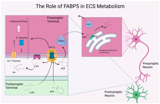
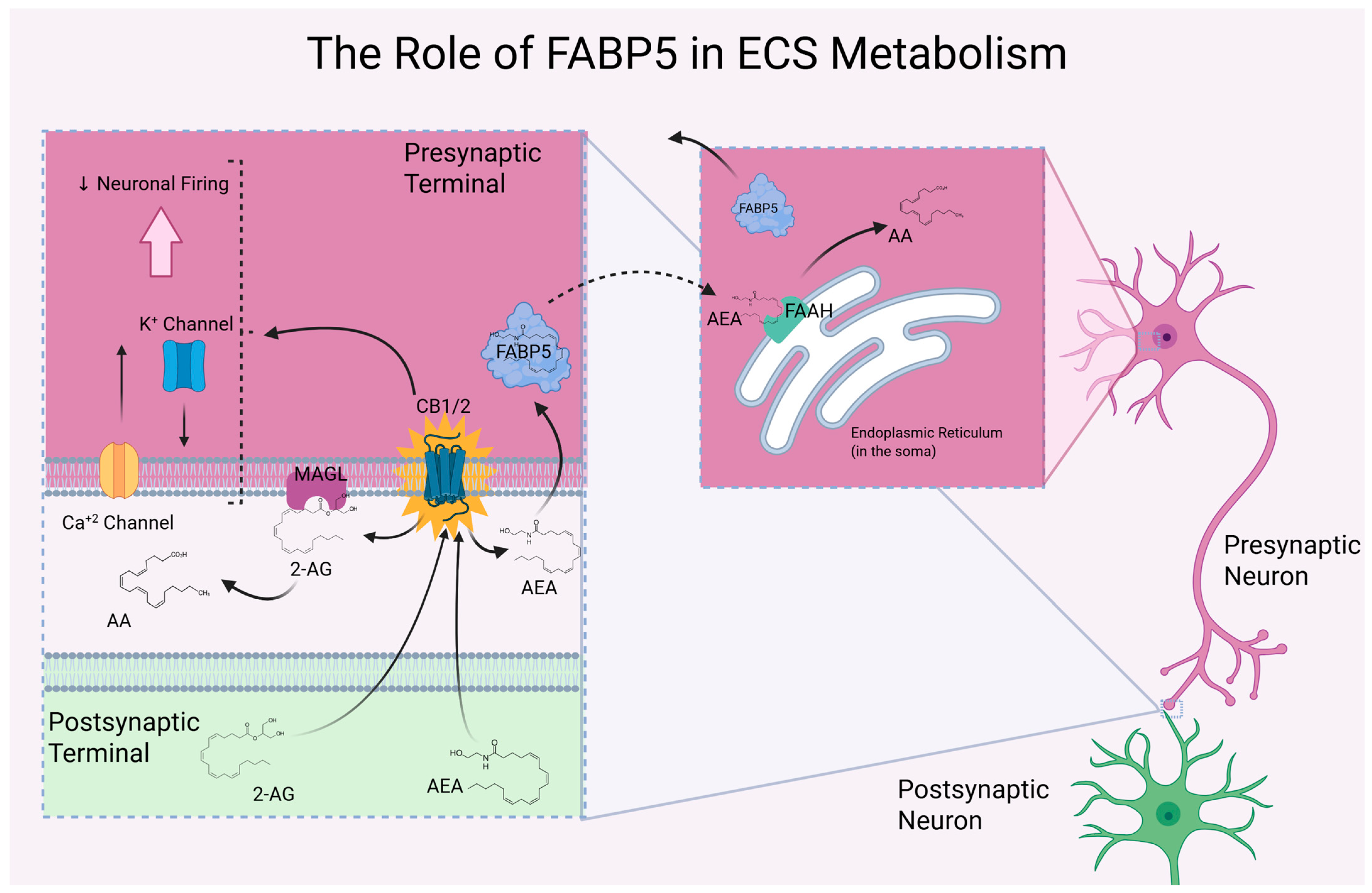
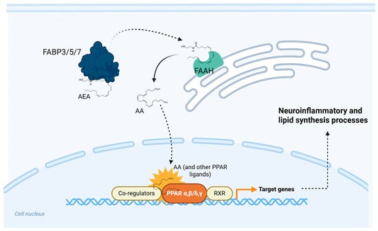
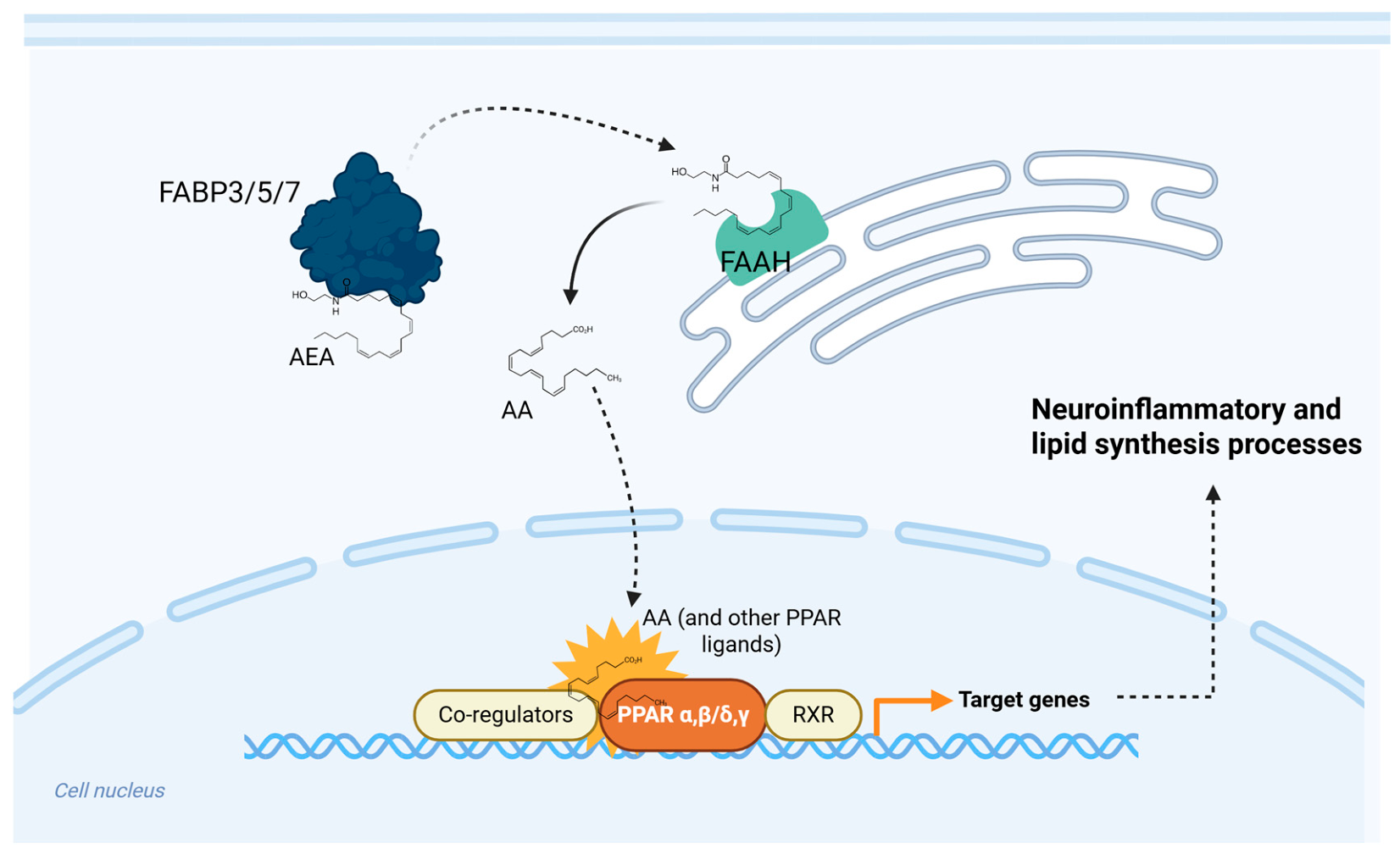
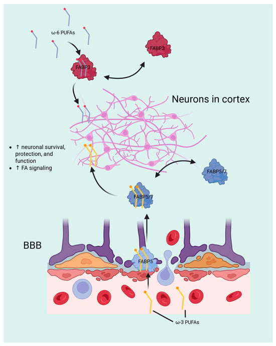
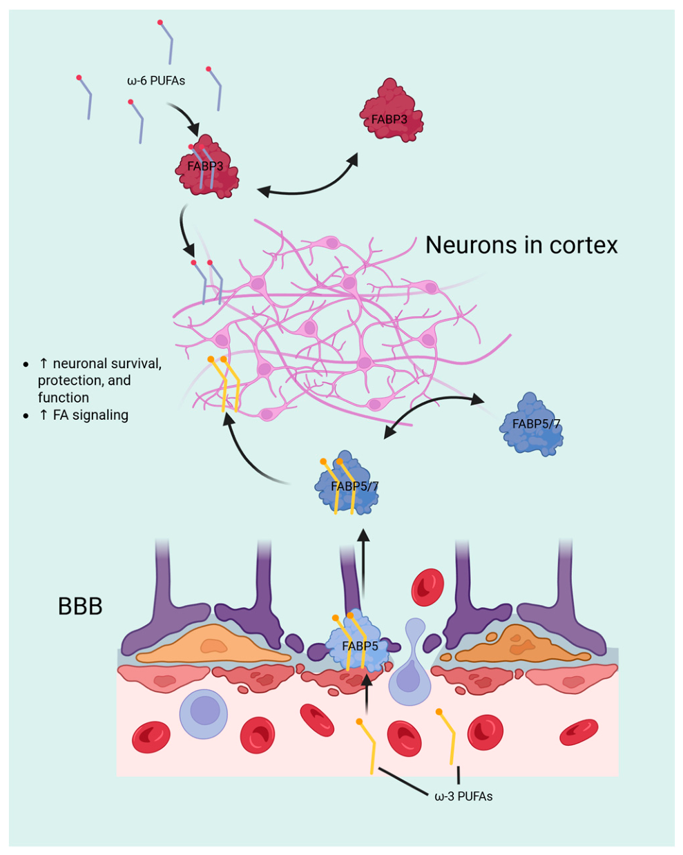
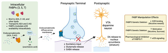
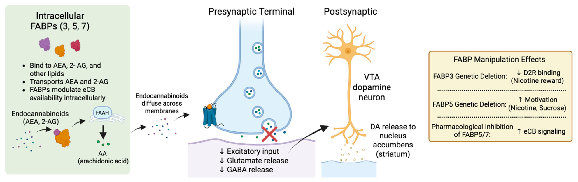
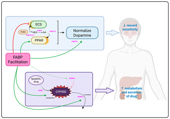
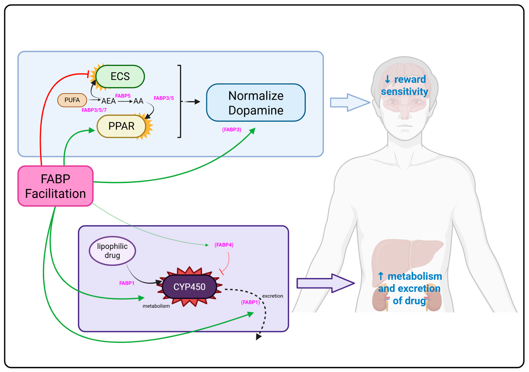

Disclaimer/Publisher’s Note: The statements, opinions and data contained in all publications are solely those of the individual author(s) and contributor(s) and not of MDPI and/or the editor(s). MDPI and/or the editor(s) disclaim responsibility for any injury to people or property resulting from any ideas, methods, instructions or products referred to in the content. |
© 2026 by the authors. Licensee MDPI, Basel, Switzerland. This article is an open access article distributed under the terms and conditions of the Creative Commons Attribution (CC BY) license.