


Abstract
Introduction. Cerebral small vessel disease (SVD) is an early but nonspecific manifestation of Fabry disease. We describe a middle-aged patient with severe SVD in whom recognition of long-standing systemic disease ultimately led to the detection of a likely pathogenic GLA variant and enabled initiation of enzyme replacement therapy before advanced renal involvement developed. Methods. The patient underwent clinical evaluation, brain MRI, whole-genome sequencing, biochemical analysis of dried blood spots, and Sanger sequencing. Results. A 41-year-old man presented with recurrent lacunar strokes, progressive gait impairment, pseudobulbar syndrome, cognitive decline, and bladder dysfunction. Brain MRI revealed severe SVD with confluent white matter hyperintensities, multiple lacunes, and microbleeds. From early adulthood, he had arterial hypertension, hypohidrosis, acroparesthesias, and mild proteinuria. Later, he developed a cataract and an acute dissection of the ascending aorta. Whole-genome sequencing identified a hemizygous GLA frameshift variant (c.1095dup), predicted to truncate alpha-galactosidase A (p.Thr366TyrfsTer9) and classified as likely pathogenic. The diagnosis was confirmed by Sanger sequencing, markedly reduced alpha-galactosidase A activity, and elevated lyso-Gb3 in dried blood spots. Discussion. This case expands the neurologic phenotype of Fabry disease and supports its inclusion in the differential diagnosis of severe early-onset SVD, even when the classic systemic phenotype is incomplete.
1. Introduction
Fabry disease (FD) is an X-linked lysosomal storage disorder caused by pathogenic variants in the GLA gene, which encodes α-galactosidase A (α-Gal A). According to ClinVar, approximately 1000 pathogenic GLA variants have been described. These variants may disrupt the catalytic center or impair protein folding and stability, leading to reduced α-Gal A activity. This results in accumulation of globotriaosylceramide (Gb3) in multiple tissues, predominantly in vascular endothelial cells, but also in podocytes, cardiomyocytes, vascular smooth muscle cells, and neurons, with elevated plasma levels of its deacylated form, lyso-Gb3 [1,2].
The prevalence of FD is estimated at 1 in 40,000–120,000, yet diagnosis remains challenging. Many GLA variants arise de novo and may be confined to individual families. Genotype–phenotype correlation is poor: even relatives with the same variant may differ clinically. Complete loss-of-function GLA variants typically cause classic young-onset FD with acroparesthesias, angiokeratomas, corneal verticillata, proteinuria, cardiomyopathy, and stroke. Variants with residual enzymatic activity often present later with milder systemic involvement [3].
Macrovascular abnormalities such as basilar artery dolichoectasia have long been associated with FD [4]. However, accumulating evidence suggests that recurrent ischemic strokes and white matter hyperintensities due to cerebral small vessel disease (SVD, cerebral microangiopathy) can be a major neurological manifestation of FD, even without classic systemic features [5].
We report a 41-year-old man with severe cerebral SVD and multisystem features. Whole-genome sequencing identified a rare frameshift variant in exon 7 of the GLA gene (c.1095dup), with evidence of marked α-Gal A deficiency.
2. Materials and Methods
The patient was evaluated and treated at the Russian Center of Neurology and Neurosciences. Brain MRI was performed on a 3.0-T Siemens Prisma scanner (Siemens Healthineers, Erlangen, Germany), and imaging markers of cerebral small vessel disease were evaluated in accordance with the STRIVE criteria [6].
Following informed consent for molecular genetic testing, 4 mL of peripheral blood was collected into an EDTA tube and stored at −80 °C until analysis. Genomic DNA was extracted using QIAGEN Mini Spin Columns according to the manufacturer’s instructions (QIAGEN, Hilden, Germany). Sequencing libraries were prepared using the TruSeq DNA PCR-Free Library Prep Kit (Illumina, San Diego, CA, USA) according to the manufacturer’s protocol. Library concentration was determined by quantitative PCR using the KAPA Library Quantification Kit for Illumina platforms (KAPA Biosystems, Wilmington, MA, USA). Whole-genome sequencing was performed on an Illumina NovaSeq 6000 platform (Illumina, San Diego, CA, USA) at Sirius University (Sochi, Russia) in 151 × 2 bp paired-end mode. The mean whole-genome coverage was 58.42×.
Raw sequencing data underwent quality control using FastQC (version 0.11.9). Adapter sequences were removed with AdapterRemoval v2. Filtered reads were aligned to the GRCh38 human reference genome using BWA-MEM (version 0.7) [7]. Variant calling was performed with GATK HaplotypeCaller (version 4) [8], and candidate pathogenic variants were prioritized using OpenCRAVAT (https://opencravat.org, accessed on 26 October 2026) [9]. This workflow identified a hemizygous variant, chrX:g.101398004dup, located in the coding region of the GLA gene (GLA(NM_000169.3):c.1095dup), and predicted to cause a frameshift at codon 366, resulting in a premature stop codon (GLA(NP_000160.1):p.(Thr366TyrfsTer9)).
To confirm the detected variant, Sanger sequencing was performed. PCR primers were designed using Primer3 software (Primer3web, version 4.1.0): forward, GCTGAAGCAAAACAGTGCCT and reverse, ACCAACTTTGTCTTGGGCCT. Amplification was carried out using GenPack PCR Core (Isogen, Moscow, Russia). PCR products were purified using the Cleanup Standard Kit (Evrogen, Moscow, Russia). Cycle sequencing was then performed with the BigDye Terminator v3.1 Cycle Sequencing Kit (Applied Biosystems, Foster City, CA, USA) in a total reaction volume of 10 μL. Sequencing products were purified on DyeEx 2.0 Spin Kit columns (QIAGEN, Hilden, Germany) and analyzed on an ABI 3730xl DNA Analyzer (Applied Biosystems, Foster City, CA, USA).
Variant pathogenicity was assessed according to the ACMG/AMP guidelines [10], with the ClinGen recommendations for the PVS1 criterion [11]. Population frequency (gnomAD v4) and ClinVar records were assessed with OpenCRAVAT. Genes associated with monogenic cerebral SVD (NOTCH3, HTRA1, COL4A1, COL4A2, TREX1, CTSA, ADA2, FOXC1 and COLGALT1), hereditary cerebral amyloid angiopathy (APP, CST3 and ITM2B) and heritable thoracic aortic disease (FBN1, TGFBR1, TGFBR2, SMAD3, COL3A1, ACTA2 and MYH11) were also analyzed.
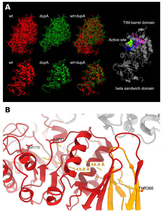
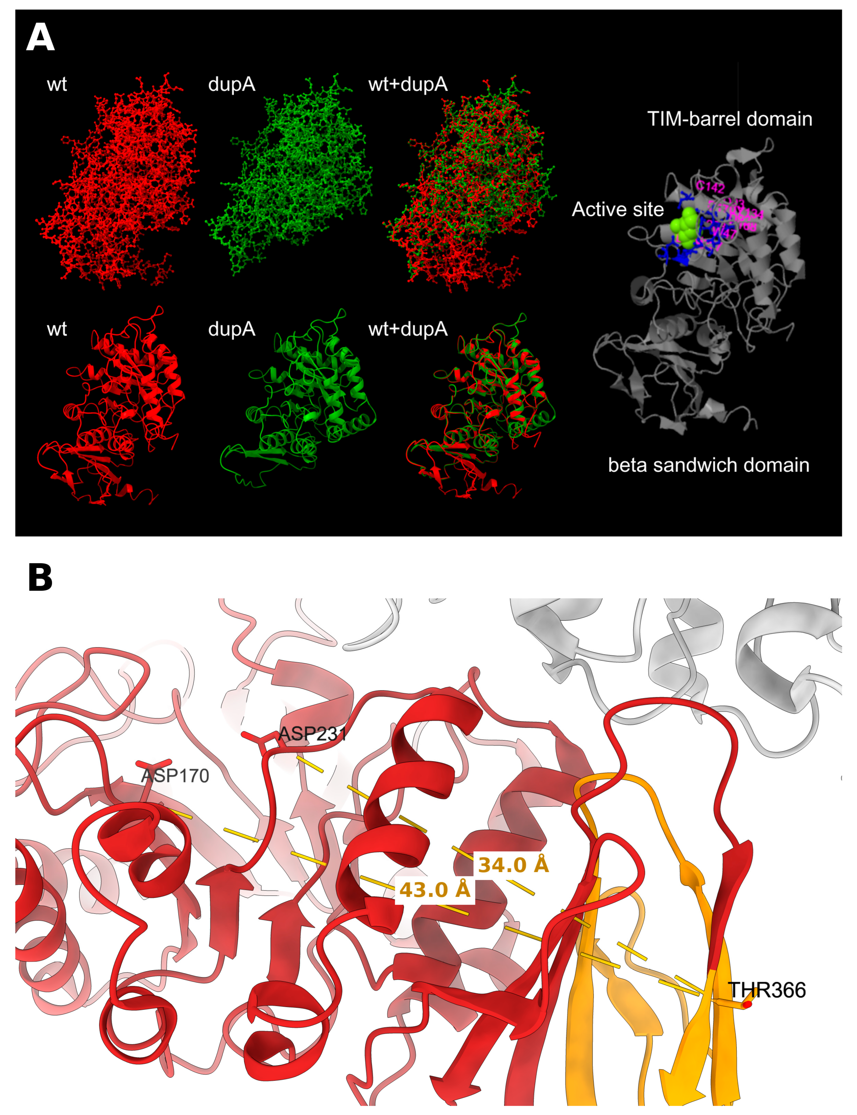
Three-dimensional models of the proteins encoded by wild-type GLA and the identified frameshift variant were generated using the I-TASSER server (https://zhanggroup.org/I-TASSER/, accessed on 16 May 2026) [12] with default parameters. The input sequences were the wild-type precursor (UniProt P06280, 429 amino acids) and the predicted 373-residue mutant protein, including the eight novel residues encoded downstream of the frameshift. Structural models were examined and rendered in UCSF ChimeraX (version 1.12) [13]. The top-ranked models were superimposed on each other and on the crystal structure of human α-Gal A (PDB 1R46) [2] over the catalytic domain (residues 32–330) using Matchmaker, and Cα–Cα distances between the catalytic residues Asp170 and Asp231 and Thr366 were measured on the crystal structure.
Alpha-galactosidase A activity and globotriaosylsphingosine (lyso-Gb3) concentration were measured in dried blood spots (DBS) at the Research Centre for Medical Genetics named after academician N.P. Bochkov by high-performance liquid chromatography–tandem mass spectrometry within multiplex screening panels for lysosomal storage disorders. Before initiation of enzyme replacement therapy (ERT), the patient underwent a multidisciplinary evaluation at the E.M. Tareev Clinic of Rheumatology, Nephrology and Occupational Pathology, including ophthalmological examination, cardiac MRI with gadolinium, electrocardiography, 24-h Holter monitoring, spirometry and renal ultrasound. After eight ERT infusions, he was re-examined by a neurologist at the Russian Center of Neurology and Neurosciences.
3. Results
A 41-year-old man was evaluated at our center in February 2025 for diagnostic clarification of progressive gait dysfunction, bradykinesia, dysarthria, cognitive decline, urinary symptoms, and recurrent lacunar strokes that had developed over three years. The clinical timeline is summarized in Table 1.
Table 1.
Timeline of clinical events.
Since the age of 12 years, he had had heat intolerance, reduced sweating and episodes of severe burning pain in the hands and feet, provoked by heat or fever and relieved by nonsteroidal anti-inflammatory drugs. From the age of 15, he had arterial hypertension with systolic blood pressure of up to 200 mm Hg but received no regular treatment. By the age of 35, the neuropathic pain had decreased markedly. In 2020 (age 36), he developed a cataract in the right eye, and lens replacement was performed in 2021.
At the end of 2022 (age 38), he had an episode of photopsia, marked weakness and nausea, interpreted as a hypertensive crisis. In February 2023 (age 39), he developed acute sensory disturbances in the right half of the body, followed by persistent clumsiness and numbness of the right limbs. About one week later, right-sided hearing loss developed (audiometry later showed bilateral sensorineural hearing loss).
Brain MRI (March 2023) showed multiple white matter hyperintensities and lacunar cysts. Because of multifocal lesions at a young age, an inflammatory disease was suspected, and methylprednisolone (total dose 5 g) was administered ex juvantibus without effect. A spinal cord MRI (March 2023) was normal. Cerebrospinal fluid (CSF) examination (April 2024) showed mildly elevated protein (0.7 g/L), 3 cells/µL and type 4 oligoclonal bands, and repeat testing in August 2024 showed no oligoclonal bands. Tests for Borrelia, lupus anticoagulant and autoantibodies (anti-β2-glycoprotein I, anti-dsDNA, anti-Sm, anti-RNP70) were negative, and visual evoked potentials were normal.
In September 2023, an ascending aortic aneurysm was detected. On 12 December 2023 (age 39), he developed acute chest pain and dyspnea due to dissection of the aneurysm (DeBakey type II) with aortic valve insufficiency and underwent emergency replacement of the aortic valve and ascending aorta with a composite valved conduit (Bentall procedure, mechanical valve). The postoperative course was complicated by hydropericardium requiring drainage. Since then, he has taken antihypertensive drugs and warfarin regularly, with the international normalized ratio within the target range. After surgery, his condition worsened markedly, with increasing gait instability, right-sided weakness, bradykinesia, hypomimia, dysarthria, memory decline and mild urinary incontinence.
At the end of July 2024 (age 40), he developed acute weakness of the left leg and numbness of the left foot. MRI showed a new lacunar infarct in the right thalamus and adjacent internal capsule. Because of gait instability and falls, he began to walk with a cane. In August 2024, hypophonia, choking on liquids, forgetfulness and disinhibition appeared. At his first outpatient visit to our center in December 2024, severe cerebral SVD was diagnosed, and hospitalization was recommended to exclude hereditary causes.
In February 2025 (age 41), the patient was hospitalized at the Russian Center of Neurology and Neurosciences. Family history was unremarkable except for a stroke in a maternal grandmother at age 80. He has no siblings or children, and his parents have not undergone genetic testing. No skin rash was noted. Neurologic examination showed slowed speech, hypomimia, mild facial weakness, greater hearing loss on the right, dysarthria, hypophonia, moderate spastic paraparesis of the legs, bilateral pyramidal signs, mild ataxia, decrement on repetitive hand movements, left-sided sensory loss, and a slow spastic-ataxic gait requiring unilateral support. Neuropsychological testing showed moderate cognitive impairment (Mini-Mental State Examination 20/30, Montreal Cognitive Assessment 22/30).
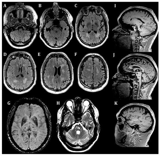
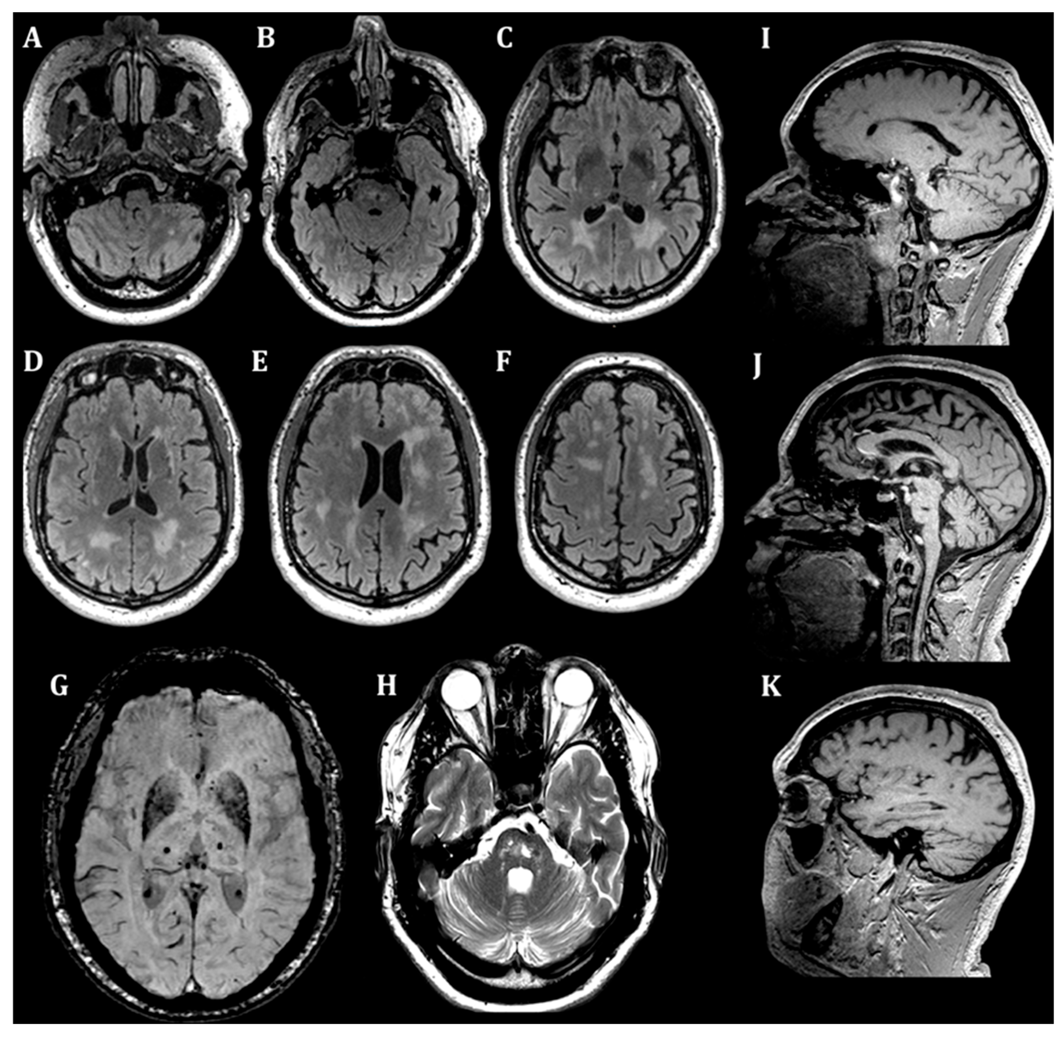
Brain 3T MRI (Figure 1) demonstrated extensive cerebral SVD with numerous lacunes and microbleeds and confluent white matter hyperintensities (Fazekas grade 3) in subcortical and deep white matter, periventricular regions, pons, basal ganglia, and cerebellar hemispheres. Enlarged perivascular spaces and bilateral T2/SWI hypointensity of the lentiform nuclei were also noted. Videonystagmography showed square-wave jerks and downbeat nystagmus, consistent with cerebellar dysfunction.
Figure 1.
Brain MRI. Axial T2-FLAIR images (A–F) show confluent white matter hyperintensities. Susceptibility-weighted imaging (G) demonstrates multiple cerebral microbleeds. Additional T2-weighted (H) and T1-weighted images (I–K) show cystic-gliotic changes with multiple lacunes. Overall, the MRI pattern is consistent with severe cerebral small vessel disease but is not specific for Fabry disease.
Blood pressure monitoring on antihypertensive treatment was normal (mean daytime 124/82 mm Hg, nighttime 111/70 mm Hg). Renal function was preserved (serum creatinine 89 µmol/L, estimated glomerular filtration rate [eGFR] 96 mL/min/1.73 m2), but proteinuria of 0.342 g/L was present, and renal ultrasound showed bilateral renal sinus cysts. Echocardiography showed concentric left ventricular wall thickening, moderate left atrial dilatation and grade II–III mitral regurgitation. N-terminal pro-B-type natriuretic peptide (NT-proBNP) was moderately elevated (232 pg/mL). Duplex ultrasound showed hemodynamically non-significant atherosclerosis of the carotid, subclavian and lower-limb arteries (stenoses ≤35%). Autoimmune and infectious tests in blood and CSF were negative. Serum homocysteine was 13.2 µmol/L, low-density lipoprotein cholesterol was 3.25 mmol/L and HbA1c was 5.2%. Targeted sequencing of the 10 most commonly affected exons of NOTCH3 revealed no pathogenic variants.
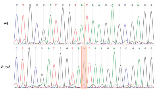
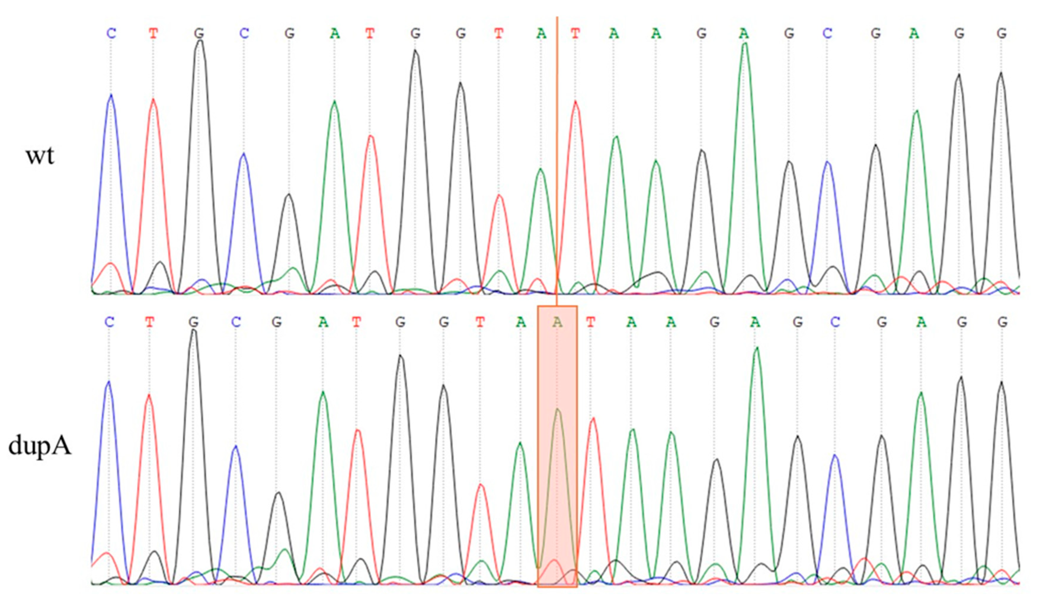
The patient’s young age, together with progressive cerebral SVD and multisystem features, suggested a genetic disorder. FD was suspected because of hypohidrosis and heat intolerance, episodic acral numbness, proteinuria and the aortic aneurysm. Whole-genome sequencing identified a hemizygous GLA frameshift variant in exon 7, GLA(NM_000169.3):c.1095dup, predicted to result in GLA(NP_000160.1):p.(Thr366TyrfsTer9). Sanger sequencing confirmed the variant (Figure 2). No pathogenic or likely pathogenic variants were identified in genes associated with monogenic cerebral SVD, hereditary cerebral amyloid angiopathy or heritable thoracic aortic disease.
Figure 2.
Sanger sequencing confirming the GLA variant NM_000169.3:c.1095dup, predicted to result in NP_000160.1:p.(Thr366TyrfsTer9).
Dried blood spot analysis (March 2026) showed markedly elevated lyso-Gb3 (184.42 ng/mL, reference 0.05–3.0) and markedly reduced α-Gal A activity (0.01 µmol/L/h, reference 0.8–15), whereas the other lysosomal enzymes and lysosphingolipids tested were within normal ranges (Table 2). The diagnosis of FD was established. According to the ACMG/AMP criteria [10] and the ClinGen recommendations for PVS1 [11], the variant was classified as likely pathogenic: PVS1_Strong (frameshift in the last exon removing more than 10% of the protein), PM2_Supporting (absent from gnomAD) and PP4 (enzyme deficiency and elevated lyso-Gb3, highly specific for FD). In September 2026, the variant was listed in ClinVar as pathogenic (VCV004974840, single submitter) on the basis of one previously reported proband without a described clinical phenotype [14].
Table 2.
Laboratory findings before and after initiation of enzyme replacement therapy.
The multidisciplinary evaluation before ERT (April–May 2026) confirmed multisystem involvement. Ophthalmological examination revealed bilateral corneal verticillata, a spoke-like posterior subcapsular cataract in the left eye and tortuous retinal veins. Cardiac MRI showed asymmetric left ventricular hypertrophy (maximal wall thickness 15 mm, normal indexed mass, ejection fraction 76%) with low-normal native T1 values (1116 ms) and a focus of late gadolinium enhancement in the basal inferolateral segment. NT-proBNP was elevated to 657 pg/mL, and chronic heart failure (New York Heart Association class II) was diagnosed. Electrocardiography showed a short PR interval (100 ms) and complete right bundle branch block. Holter monitoring recorded 142 supraventricular and 52 ventricular extrasystoles per 24 h. Chronic kidney disease stage G2 with proteinuria (0.73 g/L) and parapelvic cysts was confirmed. Spirometry revealed moderate airflow obstruction (forced expiratory volume in 1 s [FEV1] 55% of predicted, FEV1/vital capacity 61%). Laboratory tests revealed hyperuricemia and hypercholesterolemia (uric acid 499.8 µmol/L and total cholesterol 6.03 mmol/L, Table 2), as well as hyperhomocysteinemia (homocysteine 13.2 µmol/L).
Enzyme replacement therapy with agalsidase beta (105 mg, approximately 1.2 mg/kg, intravenously every 2 weeks) was initiated on 25 June 2026. Concomitant regular therapy included perindopril, bisoprolol, warfarin, rosuvastatin, febuxostat, B-group vitamins and levodopa/carbidopa. The treatment has been well tolerated. At re-examination at our center after eight infusions (October 2026), proteinuria had decreased from 0.84 to 0.52 g/L (Table 2). Pseudobulbar symptoms had become less pronounced, walking speed had increased, and the patient reported better heat tolerance during the summer months. He continued to walk with a cane, and mild weakness of the right leg persisted. A follow-up brain MRI has been scheduled.
The c.1095dup variant is located in exon 7, the last exon of GLA. Therefore, the premature termination codon is predicted to escape nonsense-mediated mRNA decay [11], and a truncated protein lacking 64 of the 429 native residues, including about two-thirds of the C-terminal β-domain [2], is expected to be synthesized. In the I-TASSER model, the catalytic domain of the mutant protein retained its overall fold, whereas the C-terminal β-domain was disrupted. The truncation site was located 34–43 Å from the catalytic residues (Figure 3).
Figure 3.
Predicted structural effect of the GLA frameshift variant. (A) Top-ranked I-TASSER models of the α-Gal A monomer: wild type (wt, red), mutant p.(Thr366TyrfsTer9) (dupA, green) and their superposition (wt+dupA) in ball-and-stick (top) and ribbon (bottom) representations. The active site is located in the N-terminal (β/α)8 catalytic domain (residues 32–330), which is followed by the C-terminal β-sandwich domain (residues 331–429) [2]. The catalytic domains of the wt and dupA models were nearly identical (Cα RMSD 0.17 Å over 299 residues) and matched the crystal structure (Cα RMSD 0.63 and 0.57 Å, respectively). (B) Crystal structure of human α-Gal A (PDB 1R46, chain A in red, chain B in gray). The region lost in the mutant protein (residues 366–429) is shown in orange. Dashed lines indicate Cα–Cα distances from Thr366 to Asp170 (43.0 Å) and Asp231 (34.0 Å).
4. Discussion
This case illustrates how long-standing, apparently unrelated symptoms may remain unrecognized until severe neurological complications develop, and underlines the importance of a detailed history of systemic abnormalities in young patients with cerebral SVD. Our patient had typical manifestations of FD since childhood—episodic neuropathic pain, heat intolerance and hypohidrosis—followed by early-onset hypertension, proteinuria, renal sinus cysts, cornea verticillata, a spoke-like posterior cataract and sensorineural hearing loss. The attenuation of acroparesthesias in adulthood is consistent with the natural course of FD-related neuropathy. However, the absence of angiokeratomas and gastrointestinal symptoms, as well as the subclinical renal involvement and severe SVD at a young age, delayed the diagnosis, because inflammatory diseases of the central nervous system and other common monogenic causes of SVD were considered first. Dilatation of the ascending aorta and aortic aneurysms have been described in males with FD [15], and the aortic dissection in our patient may reflect FD-related involvement of the aortic wall, although long-standing uncontrolled hypertension, the major risk factor for aortic dissection, probably contributed.
White matter hyperintensities are the most common neuroimaging abnormality in FD, but they are nonspecific and may initially suggest a demyelinating disorder or another hereditary SVD [16,17,18,19,20]. Importantly, stroke may precede overt renal or cardiac involvement [21,22]. In a recent 20-year single-center cohort of 405 adults with FD, 19 cerebrovascular accidents and seven transient ischemic attacks were recorded [23].
Long-standing, poorly controlled hypertension is an important confounder. The patient had a systolic blood pressure of up to 200 mm Hg from the age of 15 years and received no regular treatment until the age of 39. Hypertension is the leading modifiable risk factor for sporadic SVD. In patients with FD, it has been associated with a greater burden of white matter hyperintensities (WMH) and may limit the benefit of ERT on WMH progression [16,24]. On the other hand, hypertension with onset in adolescence may itself reflect early renal or vascular involvement by FD, and the young age at stroke onset and the multisystem manifestations are difficult to attribute to hypertension alone. We therefore consider the severe SVD to result from the combined effects of FD-related vasculopathy and hypertension.
Accumulation of Gb3 in endothelial cells, vascular smooth muscle cells and pericytes is thought to cause endothelial dysfunction, impaired nitric oxide-dependent vasoregulation, oxidative stress and a prothrombotic state [25]. Regional cerebral hyperperfusion associated with dysregulation of the nitric oxide pathway has been demonstrated in FD [26], and lyso-Gb3 promotes the proliferation of vascular smooth muscle cells [27], which may contribute to wall thickening and luminal narrowing of small penetrating arteries. These changes provide a plausible substrate for the lacunar infarcts, white matter hyperintensities and microbleeds observed in our patient.
Because the truncation spares the catalytic residues (Figure 3B), its effect is probably mediated by the loss of most of the C-terminal β-domain, which is closely associated with the catalytic domain [2]. This is expected to compromise folding, stability and possibly dimerization, leading to degradation of the misfolded protein, which is consistent with the near-absent enzyme activity in our patient. However, single-monomer models cannot predict misfolding or residual activity, and the near-identical catalytic domains of the two models largely reflect template-based modeling.
In contrast to GLA variants frequently detected in stroke screening studies, such as p.Ala143Thr, p.Arg118Cys and p.Asp313Tyr, which retain substantial residual enzyme activity and are regarded as variants of uncertain significance or risk modifiers [28], c.1095dup is a null variant. Data on the cerebrovascular phenotype of patients with truncating GLA variants are limited. Tomek et al. described a 40-year-old woman with the exon 7 frameshift variant c.1235_1236delCT who presented with a lacunar mesencephalic infarction [29]. In a cohort of 71 patients with FD, those with truncating variants did not differ from those with non-truncating variants in WMH burden, lacunes or microbleeds, but had more enlarged perivascular spaces [30], which were also present in our patient. Thus, the severity of cerebral SVD does not appear to be determined by the variant type alone. The c.1095dup variant itself has previously been identified in one proband in a long-read sequencing study of 48 probands with FD and classified as likely pathogenic using the same ACMG criteria, but without a clinical description [14]. Our observation shows that it may present with severe cerebral SVD.
This report has limitations. α-Gal A activity and lyso-Gb3 were measured only in DBS, and leukocyte α-Gal A activity and plasma lyso-Gb3 were not determined. However, in males, markedly reduced enzyme activity combined with a null GLA variant is sufficient for diagnosis [1], and normal activities of other lysosomal enzymes in the same sample argue against sample degradation. The parents were not tested, so the inheritance of the variant could not be established, and GLA mRNA was not analyzed. The follow-up after ERT initiation is short (3 months), and the observed improvement may partly reflect rehabilitation, symptomatic therapy and natural fluctuations. Finally, the relative contributions of FD and hypertension to SVD cannot be separated in a single case.
Fabry disease should remain in the differential diagnosis of young or middle-aged adults with cerebral SVD, particularly when accompanied by subtle extracerebral clues: hypohidrosis, neuropathic pain, proteinuria, cataracts, hearing loss, or aortic pathology. Early recognition is clinically important because ERT may be considered before advanced renal or cardiac complications develop. Identification of a pathogenic GLA variant also enables cascade genetic testing of at-risk relatives.
Author Contributions
Conceptualization, P.S.S. and L.A.D.; methodology, L.V.Z., A.D.M. and P.S.S.; formal analysis, L.V.Z. and A.D.M.; validation, L.V.Z., A.D.M. and P.S.S.; investigation, L.V.Z., A.D.M. and P.S.S.; writing—original draft preparation, P.S.S.; writing—review and editing, E.V.G., P.S.S. and M.R.Z.; visualization, P.S.S.; supervision, L.A.D., L.A.K. and A.D.M. All authors have read and agreed to the published version of the manuscript.
Funding
This research received no external funding.
Institutional Review Board Statement
The study was conducted in accordance with the Declaration of Helsinki, and approved by the Local Ethics Committee of the Russian Center of Neurology and Neurosciences (protocol No. 10-4/22 dated 23.11.2022) for studies involving humans.
Informed Consent Statement
Written informed consent has been obtained from the patient to publish this paper.
Data Availability Statement
The data that support the findings of this study are available upon reasonable request.
Acknowledgments
The authors thank the staff of the Research Centre for Medical Genetics named after N.P. Bochkov for performing the biochemical and molecular genetic analyses of dried blood spots, and the staff of the E.M. Tareev Clinic of Rheumatology, Nephrology and Occupational Pathology (Sechenov University) for the detailed instrumental evaluation of the patient before initiation of ERT. A.M. was supported by the Ministry of Science and Higher Education of the Russian Federation (Agreement No. 075-10-2025-017 of 27 February 2025).
Conflicts of Interest
The authors declare no conflicts of interest.
Abbreviations
The following abbreviations are used in this manuscript:
| SVD | Cerebral small vessel disease |
| GLA | Gene encoding α-galactosidase A |
| FD | Fabry disease |
| α-Gal A | Alpha-galactosidase A |
| ACMG/AMP | American College of Medical Genetics and Genomics/Association for Molecular Pathology |
| CSF | Cerebrospinal fluid |
| DBS | Dried blood spot |
| eGFR | Estimated glomerular filtration rate |
| ERT | Enzyme replacement therapy |
| Gb3 | Globotriaosylceramide |
| lyso-Gb3 | Globotriaosylsphingosine |
| NT-proBNP | N-terminal pro-B-type natriuretic peptide |
| WGS | Whole-genome sequencing |
| WMH | White matter hyperintensities |
References
- Gal, A.; Hughes, D.A.; Winchester, B. Toward a consensus in the laboratory diagnostics of Fabry disease: Recommendations of a European expert group. J. Inherit. Metab. Dis. 2011, 34, 509–514. [Google Scholar] [CrossRef] [Scilit]
- Garman, S.C.; Garboczi, D.N. The molecular defect leading to Fabry disease: Structure of human alpha-galactosidase. J. Mol. Biol. 2004, 337, 319–335. [Google Scholar] [CrossRef] [Scilit]
- Zarate, Y.A.; Hopkin, R.J. Fabry’s disease. Lancet 2008, 372, 1427–1435. [Google Scholar] [CrossRef] [Scilit]
- Manara, R.; Carlier, R.Y.; Righetto, S.; Citton, V.; Locatelli, G.; Colas, F.; Ermani, M.; Germain, D.P.; Burlina, A. Basilar artery changes in Fabry disease. AJNR Am. J. Neuroradiol. 2017, 38, 531–536. [Google Scholar] [CrossRef] [Scilit]
- Fellgiebel, A.; Müller, M.J.; Ginsberg, L. CNS manifestations of Fabry’s disease. Lancet Neurol. 2006, 5, 791–795. [Google Scholar] [CrossRef] [Scilit]
- Wardlaw, J.M.; Smith, E.E.; Biessels, G.J.; Cordonnier, C.; Fazekas, F.; Frayne, R.; Lindley, R.I.; O’Brien, J.T.; Barkhof, F.; Benavente, O.R.; et al. Neuroimaging standards for research into small vessel disease and its contribution to ageing and neurodegeneration. Lancet Neurol. 2013, 12, 822–838. [Google Scholar] [CrossRef] [Scilit]
- Li, H. Aligning sequence reads, clone sequences and assembly contigs with BWA-MEM. arXiv 2013, arXiv:1303.3997. [Google Scholar] [CrossRef] [Scilit]
- Van der Auwera, G.A.; Carneiro, M.O.; Hartl, C.; Poplin, R.; del Angel, G.; Levy-Moonshine, A.; Jordan, T.; Shakir, K.; Roazen, D.; Thibault, J.; et al. From FastQ data to high-confidence variant calls: The Genome Analysis Toolkit best practices pipeline. Curr. Protoc. Bioinform. 2013, 43, 11.10.1–11.10.33. [Google Scholar] [CrossRef] [Scilit]
- Pagel, K.A.; Kim, R.; Moad, K.; Busby, B.; Zheng, L.; Tokheim, C.; Ryan, M.; Karchin, R. Integrated informatics analysis of cancer-related variants. JCO Clin. Cancer Inform. 2020, 4, 310–317. [Google Scholar] [CrossRef] [Scilit]
- Richards, S.; Aziz, N.; Bale, S.; Bick, D.; Das, S.; Gastier-Foster, J.; Grody, W.W.; Hegde, M.; Lyon, E.; Spector, E.; et al. Standards and guidelines for the interpretation of sequence variants: A joint consensus recommendation of the American College of Medical Genetics and Genomics and the Association for Molecular Pathology. Genet. Med. 2015, 17, 405–424. [Google Scholar] [CrossRef] [Scilit]
- Abou Tayoun, A.N.; Pesaran, T.; DiStefano, M.T.; Oza, A.; Rehm, H.L.; Biesecker, L.G.; Harrison, S.M. ClinGen Sequence Variant Interpretation Working Group (ClinGen SVI). Recommendations for interpreting the loss of function PVS1 ACMG/AMP variant criterion. Hum. Mutat. 2018, 39, 1517–1524. [Google Scholar] [CrossRef] [Scilit]
- Zhang, Y. I-TASSER server for protein 3D structure prediction. BMC Bioinform. 2008, 9, 40. [Google Scholar] [CrossRef] [Scilit]
- Pettersen, E.F.; Goddard, T.D.; Huang, C.C.; Meng, E.C.; Couch, G.S.; Croll, T.I.; Morris, J.H.; Ferrin, T.E. UCSF ChimeraX: Structure visualization for researchers, educators, and developers. Protein Sci. 2021, 30, 70–82. [Google Scholar] [CrossRef] [Scilit]
- Yao, F.; Hao, N.; Li, D.; Zhang, W.; Zhou, J.; Qiu, Z.; Mao, A.; Meng, W.; Liu, J. Long-read sequencing enables comprehensive molecular genetic diagnosis of Fabry disease. Hum. Genom. 2024, 18, 133. [Google Scholar] [CrossRef] [Scilit]
- Barbey, F.; Qanadli, S.D.; Juli, C.; Brakch, N.; Palaček, T.; Rizzo, E.; Jeanrenaud, X.; Eckhardt, B.; Linhart, A. Aortic remodelling in Fabry disease. Eur. Heart J. 2010, 31, 347–353. [Google Scholar] [CrossRef] [Scilit]
- Stefaniak, J.D.; Parkes, L.M.; Parry-Jones, A.R.; Potter, G.M.; Vail, A.; Jovanovic, A.; Smith, C.J. Enzyme replacement therapy and white matter hyperintensity progression in Fabry disease. Neurology 2018, 91, e1413–e1422. [Google Scholar] [CrossRef] [Scilit]
- Korver, S.; Vergouwe, M.; Hollak, C.E.M.; van Schaik, I.N.; Langeveld, M. Development and clinical consequences of white matter lesions in Fabry disease: A systematic review. Mol. Genet. Metab. 2018, 125, 205–216. [Google Scholar] [CrossRef] [Scilit]
- Johnson, J.W.; Baradaran, H.; Morgan, J.; Odéen, H.; Friel, E.; Bailey, C.; Zielinski, B.A.; Underhill, H.R. The insidious degeneration of white matter and cognitive decline in Fabry disease. PLoS ONE 2025, 20, e0325403. [Google Scholar] [CrossRef] [Scilit]
- Fazekas, F.; Enzinger, C.; Schmidt, R.; Grittner, U.; Giese, A.K.; Hennerici, M.G.; Huber, R.; Jungehulsing, G.J.; Kaps, M.; Kessler, C.; et al. Brain magnetic resonance imaging findings fail to suspect Fabry disease in young patients with an acute cerebrovascular event. Stroke 2015, 46, 1548–1553. [Google Scholar] [CrossRef] [Scilit]
- Broad, R.; Dsouza, M. Fabry disease presenting with lacunar stroke and cerebral white matter lesions, referred as possible demyelination. J. Neurol. Neurosurg. Psychiatry 2024, 95, A29.3. [Google Scholar] [CrossRef] [Scilit]
- Sims, K.; Politei, J.; Banikazemi, M.; Lee, P. Stroke in Fabry disease frequently occurs before diagnosis and in the absence of other clinical events: Natural history data from the Fabry Registry. Stroke 2009, 40, 788–794. [Google Scholar] [CrossRef] [Scilit]
- Moreno-Martinez, D.; Lucas-Del-Pozo, S.; Lavalle, L.; Ramaswami, U.; Ginsberg, L.; Pintos-Morell, G.; Hughes, D.A. Stroke in Fabry disease: Identification of risk factors for stroke in a large single-centre cohort. Eur. J. Neurol. 2025, 32, e70415. [Google Scholar] [CrossRef] [Scilit]
- McCarron, E.P.; Chinnadurai, R.; Meyer, J.; Anderson, T.; Stepien, K.M.; Sharma, R.; Woolfson, P.; Jovanovic, A. Real-world clinical outcomes in adult patients with Fabry disease: A 20-year retrospective observational cohort study from a single centre. Mol. Genet. Metab. Rep. 2025, 43, 101229. [Google Scholar] [CrossRef] [Scilit]
- Rost, N.S.; Cloonan, L.; Kanakis, A.S.; Fitzpatrick, K.M.; Azzariti, D.R.; Clarke, V.; Lourenco, C.M.; Germain, D.P.; Politei, J.M.; Homola, G.A.; et al. Determinants of white matter hyperintensity burden in patients with Fabry disease. Neurology 2016, 86, 1880–1886. [Google Scholar] [CrossRef] [Scilit]
- Kolodny, E.; Fellgiebel, A.; Hilz, M.J.; Sims, K.; Caruso, P.; Phan, T.G.; Politei, J.; Manara, R.; Burlina, A. Cerebrovascular involvement in Fabry disease: Current status of knowledge. Stroke 2015, 46, 302–313. [Google Scholar] [CrossRef] [Scilit]
- Moore, D.F.; Scott, L.T.; Gladwin, M.T.; Altarescu, G.; Kaneski, C.; Suzuki, K.; Pease-Fye, M.; Ferri, R.; Brady, R.O.; Herscovitch, P.; et al. Regional cerebral hyperperfusion and nitric oxide pathway dysregulation in Fabry disease: Reversal by enzyme replacement therapy. Circulation 2001, 104, 1506–1512. [Google Scholar] [CrossRef] [Scilit]
- Aerts, J.M.; Groener, J.E.; Kuiper, S.; Donker-Koopman, W.E.; Strijland, A.; Ottenhoff, R.; van Roomen, C.; Mirzaian, M.; Wijburg, F.A.; Linthorst, G.E.; et al. Elevated globotriaosylsphingosine is a hallmark of Fabry disease. Proc. Natl. Acad. Sci. USA 2008, 105, 2812–2817. [Google Scholar] [CrossRef] [Scilit]
- van der Tol, L.; Smid, B.E.; Poorthuis, B.J.; Biegstraaten, M.; Deprez, R.H.L.; Linthorst, G.E.; Hollak, C.E.M. A systematic review on screening for Fabry disease: Prevalence of individuals with genetic variants of unknown significance. J. Med. Genet. 2014, 51, 1–9. [Google Scholar] [CrossRef] [Scilit]
- Tomek, A.; Reková, P.; Paulasová Schwabová, J.; Olšerová, A.; Škorňa, M.; Nevšímalová, M.; Šimůnek, L.; Herzig, R.; Fafejtová, Š.; Mikulenka, P.; et al. Nationwide screening for Fabry disease in unselected stroke patients. PLoS ONE 2021, 16, e0260601. [Google Scholar] [CrossRef] [Scilit]
- Zhao, Y.; Zhu, Y.; Li, F.; Sun, Y.; Ma, W.; Wu, Y.; Zhang, W.; Wang, Z.; Yuan, Y.; Huang, Y. Brain MRI correlations with disease burden and biomarkers in Fabry disease. J. Neurol. 2023, 270, 4939–4948. [Google Scholar] [CrossRef] [Scilit]

Disclaimer/Publisher’s Note: The statements, opinions and data contained in all publications are solely those of the individual author(s) and contributor(s) and not of MDPI and/or the editor(s). MDPI and/or the editor(s) disclaim responsibility for any injury to people or property resulting from any ideas, methods, instructions or products referred to in the content. |
© 2026 by the authors. Licensee MDPI, Basel, Switzerland. This article is an open access article distributed under the terms and conditions of the Creative Commons Attribution (CC BY) license.