


Abstract
The pathogenesis of colorectal cancer (CRC) is characterized by significant alterations in redox homeostasis, epigenetic regulation and hormone-dependent signaling pathways. Oxidative stress (OS) promotes DNA damage, genomic instability, chronic inflammation, and the activation of several signaling pathways. Furthermore, dysregulation of various microRNAs (miRs) increases proliferation, invasion, metastasis, epithelial–mesenchymal transition, and angiogenesis, and promotes resistance to apoptosis and therapy. Increasing data suggest that there is a two-way interaction between reactive oxygen species (ROS) and miRs, where OS can influence the expression of miRs and, at the same time, miRs can affect intracellular redox state. This interrelationship plays a role in CRC initiation, development, and response to treatment. Estrogen receptor beta (ERβ) represents one of the defensive elements in the development of CRC. Its expression gradually decreases in colorectal cancerogenesis; conversely, its activation suppresses inflammation, proliferation, migration, invasion, and angiogenesis, and enhances apoptosis and DNA repair. In addition, ERβ is capable of modulating the expression of different miRs related to CRC and is involved in the modulation of the intracellular redox state. Limited and often indirect data indicate the role played by miRs and OS in regulating ERβ expression and vice versa. However, disturbing the complex regulatory network involving OS, miRNAs and ERβ may favor CRC development, while restoring it may represent a promising therapeutic strategy.
1. Introduction
Colorectal cancer (CRC) is the third most common cancer among men and the second most common among women. It is also one of the leading causes of cancer-related deaths worldwide, with higher rates observed in high-income countries [1,2]. The current increase in CRC incidence can be attributed to lifestyle changes among younger people under the age of 50 who are not yet covered by screening programs [3]. CRC usually develops from adenomatous polyps through a multifactorial process involving several steps that are influenced by genetic, epigenetic, metabolic and environmental factors [4]. Age, smoking, a sedentary lifestyle, alcohol consumption, obesity, a diet high in saturated fats and low in fiber, gut microbiome, and the presence of chronic inflammatory bowel diseases (IBD) represent the main non-hereditary risk factors for CRC, while familial adenomatous polyposis (FAP) and a genetic predisposition to hereditary non-polyposis colorectal cancer (HNPCC) are the hereditary factors involved in CRC [5,6]. Compared with normal mucosa, many metabolic pathways in CRC are altered. These include the Krebs cycle, nucleotide biosynthesis and glycolysis, as well as those involved in amino acid, lipid and steroid metabolism [7]. The activation of mitogen-activated protein kinases (MAPKs) and the Wnt/β-catenin signaling pathway are involved in the pathogenesis and progression of CRC, to the extent that they are considered to be potential therapeutic targets [8,9]. The primary prevention of CRC aims to avoid risk factors and enhance protective factors. The secondary prevention involves screening programs, early diagnosis and innovative therapies for removing precancerous colorectal neoplastic polyps [10,11]. Given that CRC is more prevalent in males than in females and that the survival rate is higher in young female patients in the same age group (aged 18–44), gender and female hormones play an important role in the onset and survival of CRC [12]. Furthermore, postmenopausal women who do not undergo hormone replacement therapy (HRT) are more likely to develop CRC than those who do [13]. For these reasons, estrogens, and in particular 17β-estradiol (17β-E2), are involved in preventing CRC. This prevention occurs via the predominant estrogen receptor isoform in colon tissue, the estrogen receptor β (ERβ) subtype, which exhibits anti-proliferative, anti-inflammatory and pro-apoptotic properties [14,15]. Studies have shown that low levels of ERβ are present in CRC and that its downregulation is associated with a less favorable prognosis, an increased risk of tumor recurrence, and a more advanced stage of disease [16,17]. In contrast, the expression of the estrogen receptor α subtype (ERα), which is traditionally associated with a proliferative response, is low in normal colon mucosa. However, it is upregulated in CRC, where it activates the Wnt/β-catenin signaling pathway and promotes metastasis [18]. The role of the G protein-coupled estrogen receptor (GPER, also known as GPR30) in CRC is controversial, as it can either promote or suppress tumor growth depending on the context, often modulating rapid non-genomic proliferative pathways [19]. Similarly, ERα signaling is frequently associated with pro-tumorigenic responses. In contrast, the ERβ receptor exerts a protective role in CRC by directly counteracting these oncogenic pathways. Specifically, ERβ inhibits the Wnt/β-catenin pathway to prevent cell proliferation, promotes apoptosis via Bax upregulation, inhibits the inflammatory NF-κB pathway, and regulates redox homeostasis via the regulation of antioxidant enzymes [14,15]. Thus, it is evident that ERβ plays a protective role in CRC, given that its deletion enhances adenoma formation, while its activation has anti-tumorigenic properties [15].
MicroRNAs (miRs), which are single-stranded non-coding RNA molecules constituted by 18–25 nucleotides, can regulate gene expression at the post-transcriptional level by promoting the degradation of specific messenger RNA (mRNA) or inhibiting its translation into protein [20]. Altered miR expression profile and oxidative stress (OS), which is caused by an imbalance between reactive oxygen species (ROS) production and antioxidant levels, are recognized as major cancer promoters and potential therapeutic targets [21]. Dysregulated miR expression contributes to CRC initiation and progression by affecting key processes, including proliferation, apoptosis, invasion, angiogenesis, epithelial–mesenchymal transition (EMT), and treatment response. It may also have diagnostic, prognostic, and therapeutic relevance [22,23,24,25]. The upregulation of ROS levels promotes DNA damage, contributing to tumor-promoting mutations and the inhibition of tumor suppressors [26]. In CRC, mitochondrial impairment and the activation of NADPH oxidases are the main causes of increased ROS levels. These latter play an important role in mutagenesis and the progression of CRC by activating key oncogenic pathways, including the nuclear factor kappa B (NF-κB) pathway, which regulates genes involved in cell survival, proliferation and metastasis [27,28]. Literature data demonstrate the existence of cross-talk between miRs and OS [29,30], as well as a bidirectional interplay between them and ERβ [31,32,33,34]. In particular, miRs can regulate intracellular redox state by affecting signaling pathways that induce or reduce OS, while OS can also alter miR expression [35,36]. Genomic ERβ signaling can reduce OS by increasing antioxidant defenses, such as the expression of antioxidant enzymes like superoxide dismutase and catalase, and by reducing ROS levels [37,38,39]. Additionally, ERβ has been shown to promote apoptosis and prevent malignant transformation in response to OS [40]. However, high ROS levels can alter the conformation and expression of ERβ, which can in certain cases cause an increase in proliferative processes [31,41]. ERβ can upregulate specific miRs through distinct mechanisms. Direct regulation occurs when, upon 17β-E2 binding, this receptor translocates in to the nucleus and binds to the estrogen response elements (EREs), located in the proximity of the promoters of specific miR genes [42,43]. In contrast, indirect regulation involves the transcriptional repression of oncogenes (i.e., Myc, which activates the transcription of several miRs) or through epigenetic mechanisms [44]. This receptor can affect miR levels in human colon cancer cells, thereby contributing to the prevention and treatment of CRC [45]. On the contrary, miRs can also regulate the expression of genes encoding ERα (ESR1) and ERβ (ESR2). Specific miRs decrease expression of ERβ levels or promote the activation of ERβ signal pathways by downregulating specific factors, causing an increase in tumor aggressiveness or delaying cancer development, respectively [46,47]. Several miRNAs have been experimentally validated to target the 3′UTR of ESR2 transcripts via dual-luciferase reporter assays. In vitro functional experiments using miRNA mimics have proven that miR-29b-1-5p and let-7f-1-3p directly repress ESR2 expression, which results in the significant inhibition of downstream ERβ signaling, as observed in mouse hypothalamus under environmental stress [48]. The regulation of the ERβ signaling pathway is usually mediated by epigenetic modifiers or competitive endogenous RNA (ceRNA) network. One such indirect epigenetic regulation is represented by the miR-22-5p/TET2 axis. Unlike other indirect pathways where miRNAs target ESR2 directly, in this case, miR-22-5p regulates the ten-eleven translocation 2 (TET2) enzyme, which is an important epigenetic regulator of DNA demethylation, and thus indirectly modulates chromatin accessibility and represses ESR2 transcription. Importantly, this indirect regulation does not apply only to oncogenesis, but plays a key pathological role in other conditions that are dependent on estrogens; for example, in infertile women with minimal/mild endometriosis during the implantation window, it causes the deregulation of the progesterone response and infertility associated with implantation failure [49]. Figure 1 shows the intricate OS–miR–ERβ network that is balanced in normal cells, but which, when disrupted, could contribute to CRC initiation and progression.
Figure 1.
An emerging complex network involving OS–miR–ERβ that controls cellular homeostasis and cancer development.
This review aims to highlight the key molecular mechanisms involved in the roles of OS and miRs and the protective role of ERβ-mediated estrogen in CRC. Despite the fact that extensive studies have been conducted on OS, miR dysregulation and ERβ signaling pathways as factors involved in CRC, their interconnections have never been examined in a comprehensive manner. Our review clarifies the potential connections between these factors that could lead to shared mechanisms regulating inflammation, proliferation, apoptosis, EMT, invasion, metastasis and therapeutic responses. It should be emphasized that distinguishing well-established molecular mechanisms from indirect evidence and mechanistic hypotheses has highlighted a lack of clear information regarding the presence and role of an integrated OS–miR–ERβ axis in CRC development. However, the information contained in this review could facilitate the identification of new potential biomarkers and therapeutic approaches targeting the interactions among these three regulatory components, with the aim of preventing and treating CRC.
2. Oxidative Stress and CRC
Under physiological conditions, ROS are produced during redox reactions that form part of the normal metabolism and during the inflammatory response [50]. Their levels are highly regulated by the body’s antioxidant systems with the aim to maintain redox homeostasis [51], and ROS are involved in numerous cellular processes by regulating several signaling pathways [52]. These include those mediated by the transcription factor NF-κB, mitogen-activated protein kinases (MAPKs), phosphoinositide-3-kinase (PI3K)/protein kinase B (Akt), nuclear factor erythroid 2-related factor 2 (Nrf2), Kelch-like ECH-associated protein 1 (Keap1), antioxidant response elements (AREs), toll-like receptor 4 (TLR4) and signal transducer and activator of transcription 3 (STAT3) [53,54,55]. At intestinal level, the physiological redox state regulates signaling pathways, including Wnt/β-catenin and Notch, involved in proliferation, crypt-to-villus migration and stem cell differentiation [56]. However, if ROS production becomes excessive and is not offset by antioxidant systems, OS occurs. This causes cellular damage and serious metabolic disorders by reacting with biological molecules, such as nucleic acids, lipids, and proteins [57]. It also alters cell signaling pathways, which can lead to the development of cancer and resistance to treatment [27]. However, the OS is involved in processes ranging from tumor formation to tumor cell death via mechanisms involving mitochondrial and endoplasmic reticulum stress [58]. In addition, the accumulation of ROS and disorders of iron metabolism together with lipid peroxidation cause ferroptosis, a form of iron- and ROS-dependent cell death [59]. Thus, the role of OS in cancer is dual and complex. On the one hand, it causes the onset of cancer; on the other hand, if OS levels are high, it can also induce cell death [60]. Notably, the biological effect of ROS on CRC progression is highly dictated by a fine homeostatic regulation; although physiological levels of ROS act as necessary second messengers in signaling pathways, excessive pathological ROS cause genomic instability and promote aberrant oncogenic pathways. In CRC progression, chronic OS is mainly caused by mitochondrial malfunction and upregulation of NOXs, resulting in an abnormal increase in ROS production. This leads to extensive DNA damage and mutations and chromosomal instability, thus promoting malignancy [61]. On the other hand, ROS may change the expression, biogenesis, stability, and functions of miRs in different cancer types, including CRC, through several mechanisms [62]. Among these are the activation of redox-sensitive transcription factors such as NF-κB and HIF-1α, directly responsible for inflammatory and hypoxic responses. Moreover, OS negatively affects the stability and activity of miRs by controlling the expression of proteins participating in miR biogenesis and maturation, such as the RNA-binding protein DiGeorge critical region 8 (DGCR8) and Dicer, and by inducing epigenetic changes, like DNA methylation, histone acetylation, and phosphorylation [62,63].
Ferroptosis, an iron- and ROS-dependent form of cell death, may suppress or promote tumor development depending on the cellular context [64,65,66]. Lipid peroxidation through ROS pathway is an essential mediator in ferroptosis in CRCs. The iron-dependent oxidative metabolism of lipid leads to the oxidation of polyunsaturated fatty acids (PUFAs) in the cell and mitochondrial membrane phospholipids, causing damage to membrane and ferroptotic cell death. In a healthy state, the GSH–GPX4 pathway acts as an antioxidant against lipid peroxidation; however, in the absence of GSH and GPX4, the accumulation of lipid peroxides causes ferroptosis. In CRC, this process is important because of the possibility of using ferroptosis induction as a tool of tumor cell elimination, including treatment-resistant populations [67].
Chronic inflammation associated with IBD can significantly promote the development, progression and onset of CRC. In fact, IBDs are characterized by an overproduction of ROS, which is exacerbated by high levels of inflammatory mediators, such as cytokines and chemokines. These, in turn, induce ROS generation enhancing cancer progression [68,69]. Furthermore, CRC development can be triggered by certain carcinogenic compounds that cause OS [70,71].
Analysis of OS markers shows decreased levels of reduced glutathione (GSH), glutathione reductase, glutathione peroxidase (GPX) and catalase (CAT), and increased levels of oxidized glutathione (GSSG) and superoxide dismutase (SOD) in CRC patients [72,73,74]. In fact, serum OS parameters, such as total oxidant status, total antioxidant status, and the oxidative stress index, are related to CRC status and may be associated with CRC severity [75]. Levels of NADPH oxidase (NOX1), cyclooxygenase-2 (COX-2), and tumor necrosis factor-α (TNFα), all of which are related to OS, are also high in the serum of patients with CRC compared with healthy patients [76]. Furthermore, the presence of elevated levels of 8-oxo-2′-deoxy-7,8-dihydroguanosine (8-oxodG), one of the main products of DNA oxidation, in the plasma, urine, and serum also suggests elevated levels of ROS in CRC [77,78]. It has been demonstrated that OS stimulates intestinal carcinogenesis by increasing the levels of 8-oxodG in MUTYH-deficient mice, a model characterized by impaired base excision repair and increased susceptibility to CRC [79]. 4-hydroxynonenal (4-HNE), a very reactive aldehyde that is generated by lipid peroxidation resulting from OS, causes DNA cross-links and mutations, as well as altering protein function [80,81]. High levels of 4-HNE are found in CRC cells and the tumor environment, where it activates the non-canonical Hedgehog (HH) signaling pathway in cancer stem cells, increasing CRC progression [82]. Moreover, 4-HNE can also activate COX-2, a key molecule in the development and progression of CRC [83,84], and promote the activation of Nrf2 and the expression of heme oxygenase-1 (HO-1) [85]. Following the surgical removal of the tumor, a reduction in biomarkers of OS and an increase in antioxidant defenses were observed. These changes may slow the progression of CRC by improving redox homeostasis [86].
2.1. Nrf2 Signaling Pathways
Under OS, Nrf2 dissociates from Keap1, translocates into the nucleus, binds to DNA sequences called antioxidant response elements (AREs) and induces the transcription of important antioxidant genes that protect cells. For this reason, Nrf2 is often considered essential for cancer prevention [87]. However, although Nrf2 provides protection against carcinogenesis in the early stages of the disease [88,89], numerous studies have shown that high levels of Nrf2 are present in various tumors, including CRC, and are associated with tumor size and poor prognosis [90,91]. In particular, transient Nrf2 activation protects normal colonocytes from oxidative DNA damage and malignant transformation and plays a protective role in the early stages of tumorigenesis. However, persistent activation of Nrf2 plays a disruptive role in CRC cells in later stages. In fact, ROS and increased DNA damage can cause hyperactivation of Nrf2, resulting in increased cancer cell survival and reduced apoptosis [92].
The activation of the Nrf2/Keap1 and Nrf2/ARE pathways protects against the cytotoxicity that arises due to the anticancer drugs and radiation-induced damage, and Nrf2 could play an important role in chemoresistance to CRC [93,94,95]. In CRC, activation of the Nrf2/Keap1 signaling pathway by mitochondrial ROS affects glycolysis, oxidative phosphorylation, mitochondrial biogenesis, and mitophagy [96]. Adenylosuccinate lyase (ADSL), an essential enzyme for de novo purine biosynthesis, is considered an oncogene for CRC, given that its expression is elevated in this tumor and promotes OS and activation of the Nrf2/Keap1 pathway, resulting in increased proliferation and migration of CRC cell lines [97]. In CRC, the transcription factor forkhead box A2 (FOXA2), which is involved in tumor cell proliferation, is overexpressed and positively correlates with the activation of Nrf2/GPX4, which is a ferroptosis inhibitor. In fact, inhibiting FOXA2 causes the inactivation of the Nrf2/GPX4 signaling pathway, resulting in the induction of ferroptosis and the suppression of CRC [98]. The activation of Nrf2 by OS promotes the expression of HO-1, a stress-responsive enzyme, which is highly expressed in CRC tissue and is mainly located in cell nuclei [99,100]. It seems that the expression of nuclear HO-1 is associated with the development of CRC, and that HO-1 upregulation is involved in progression and metastasis by promoting CRC cell invasion and angiogenesis, while reducing apoptosis and ferroptosis [100,101,102,103,104]. However, HO-1 can act as both an anti- and pro-tumorigenic agent, as HO-1 activation has been shown to increase survival rates in patients with CRC and suppress colorectal carcinogenesis [103,105]. In fact, activation of the endoplasmic reticulum stress-related kinase (PERK)/Nrf2/HO-1 signaling pathway by OS leads to ferroptosis, alongside decreased GSH levels and increased lipid peroxidation [106].
2.2. Wnt/β-Catenin Signaling Pathway
The Wnt/β-catenin signaling pathway is upregulated by OS and stimulates colon cancer development by acting on its cancer-related downstream targets, as well as by interacting with the tumor microenvironment and cancer stem cells [107,108]. It is activated in CRC alongside a reduction in the function of adenomatous polyposis coli (APC), a tumor suppressor protein that prevents carcinogenesis by degrading β-catenin, thereby avoiding uncontrolled cell growth and proliferation [109,110,111]. However, under conditions of moderate OS, β-catenin and APC expression increases in primary CRC cells and decreases in metastatic cells [112]. Furthermore, the inhibition of Jun N-terminal kinase (JNK), which is also activated by mitochondrial ROS [113], restores β-catenin and APC to control values in primary CRC cells but not in metastatic cells, indicating that moderate OS differentially modulates the interplay between Wnt/β-catenin and JNK in these cells [112]. In CRC cells, treatment with exogenous H2O2 increases the expression of β-catenin and leucine-rich repeat G protein-coupled receptor 5 (LGR5), which is also involved in cancer development and progression [114,115]. However, although the LGR5 gene is a target of Wnt signaling, the overexpression of LGR5 is associated with JNK signaling activation by ROS [114]. Levels of solute carrier family 7 member 11 (SLC7A11), a transmembrane cystine/glutamate transporter protein which plays a key role in controlling ferroptosis and glutathione homeostasis, are elevated in tissue samples from CRC patients and are associated with tumor stage and lymph node metastasis due to reduced ferroptosis [116]. The primary metabolite of the commonly used pesticide p-dichlorodiphenyltrichloroethane (DDT) stimulates the growth of human colorectal adenocarcinoma cell lines by activating NADPH oxidase and decreasing GSH levels, as well as the activities of SOD and CAT. This subsequently increases ROS production and activates the Wnt/β-catenin and Hedgehog/Gli1 signaling pathways [117]. Increased proliferation of cells in CRC due to activation of the Wnt/β-catenin pathway under OS can be mediated by STAT3 activation [118]. Sirtuin 1 (Sirt1), an NAD+-dependent deacetylase, which deacetylases histone and non-histone proteins, is overexpressed in CRC cells in response to OS and plays a role in CRC development and progression by promoting migration, proliferation, and metastasis, while reducing apoptosis [119,120,121]. In particular, Sirt1 in CRC contributes to fatty acid oxidation by deacetylating β-catenin, and to increased proliferation by inhibiting apoptosis through deacetylating p53 tumor suppressor protein [122,123].
2.3. PI3K/Akt and NF-κB Signaling Pathways
The activation of the phosphatidylinositol-3-kinase/protein kinase B (PI3K/Akt) signaling pathway plays a key role in increasing cell proliferation and invasion, and reducing apoptosis in CRC [124,125,126]. There is cross-talk between this signaling pathway and ROS which activate the PI3K cascade by inhibiting phosphatase and tensin homolog (PTEN), a negative regulator of the pathway and tumor suppressor. OS also activates AKT and stimulates receptor tyrosine kinases (RTKs). Conversely, PI3K/Akt contributes to increased ROS levels by activating NADPH oxidases or influencing mitochondrial bioenergetics [127]. COX-2, which is involved in inflammation via prostaglandin production, is induced by ROS and promotes CRC cell proliferation and invasion, while it inhibits apoptosis by downregulating the expression of PTEN and activating the PI3K/Akt pathway [128,129]. Tumorigenesis is closely associated with NF-κB, which influences a range of processes, including infiltration, metastasis, apoptosis, cell cycle regulation and differentiation [130]. NF-κB activation by OS is particularly involved in carcinogenesis due to inflammation, and an increase in this transcription factor is present in inflammation-related CRC [131,132,133]. Additionally, NF-κB can be activated by signaling pathways such as TLR4, MAPK, STAT and PI3K, which are affected by OS. This involves a complex network that can influence CRC development and progression [134]. In fact, NF-κB plays an anti-apoptotic role by interacting with genes and signaling pathways in CRC [28,135,136]. It has been observed that NF-κB activation occurs in the tissues of CRC patients alongside an increase in metalloproteinase (MMP) 9 [137]. Indeed, OS is also able to activate MMPs, and elevated levels of MMP2 and MMP9, which play a role in ECM degradation responsible for cell migration and metastatic and invasive growth, have been found in the serum of patients with CRC and are associated with tumor stage [138,139,140].
Figure 2 schematizes the principal redox-regulated pathways activated by OS in CRC.
Figure 2.
Principal redox-sensitive signaling pathways activated by increased levels of ROS in CRC. ROS, which increase under conditions of oxidative stress, activate several redox-sensitive signaling pathways, including the Nrf2/Keap1, Wnt/β-catenin, PI3K/Akt and NF-κB pathways. These affect cell proliferation, survival, apoptosis, invasion, angiogenesis, stemness, metabolic reprogramming and therapy resistance. Although transient Nrf2 activation protects cells by maintaining redox homeostasis, sustained activation in cancer cells may promote tumor progression. Combined activation of these signaling networks ultimately promotes the initiation, progression and metastatic dissemination of CRC. This image was created with Created in BioRender software. Gaia Palmini. (2026) (https://www.biorender.com/). Abbreviations: ROS, reactive oxygen species; Nrf2, nuclear factor erythroid 2-related factor 2; Keap1, Kelch-like ECH-associated protein 1; ARE, antioxidant response element; CAT, catalase; SOD, superoxide dismutase; HO-1, heme oxygenase-1; GPX, glutathione peroxidase; LGR5, leucine-rich repeat-containing G protein-coupled receptor 5; APC, adenomatous polyposis coli; TCF/LEF, T-cell factor/lymphoid enhancer factor; SLC7A11, solute carrier family 7 member 11; PI3K/Akt, phosphoinositide-3-kinase/protein kinase B; PTEN, phosphatase and tensin homolog; NF-κB, nuclear factor kappa B; COX-2, cyclooxygenase 2; MMPs, metalloproteinases; IL-6, interleukin-6; TNFα, tumor necrosis factor-α.
3. miRs and CRC
miRs act by binding to complementary sequences in the 3′-untranslated region (3′-UTR) of target mRNAs, which can then be degraded or repressed [141]. Dysregulation of miRs alters the expression of their target mRNAs, thereby increasing or decreasing the levels of related proteins. The onset and progression of CRC are affected by epigenetic factors, with miRs playing a significant role by modulating the expression of genes involved in cell proliferation, apoptosis, angiogenesis, and metastasis. However, whether miRs act as oncogenes (oncomiRs) or tumor suppressors depends on their target gene [142,143]. The expression of various miRs is altered in CRC, so detecting these can help with diagnosis, prognosis, and therapeutic approaches [144]. For instance, the upregulation of miR-141-3p and the downregulation of miR-10b-5p in tissue samples from CRC patients have diagnostic value in identifying the disease. This suggests that colonoscopy, alongside determining the level of miR expression in colon mucosa, can provide more accurate information for early CRC diagnosis [145]. Overexpression of miR-549a, miR-552, and miR-592 in peripheral blood and tissue samples from CRC patients can be considered as a useful biomarker for diagnosing CRC and differentiating it from other tumors [146].
3.1. Principal miRs Upregulated in CRC and Their Role
3.1.1. miR-21
The oncomiR miR-21 is highly expressed in CRC and is one of the key miRs involved in CRC progression, contributing to cell proliferation, invasion, and extracellular matrix construction [147,148], and promoting epithelial–mesenchymal transition (EMT), which is involved in cancer progression and metastasis, by inhibiting the expression of mothers against decapentaplegic homolog 7 (SMAD7), programmed cell death 4 (PDCD4), and PTEN [147,149,150,151]. PTEN acts as a negative regulator of the PI3K/Akt signaling pathway by reducing Akt phosphorylation. Thus, high levels of miR-21 in CRC cells result in reduced PTEN levels, thereby increasing Akt phosphorylation and promoting the activation of the PI3K/Akt signaling pathway. The latter lead to increased cell invasion, migration and proliferation, and reduced apoptosis [152]. High circulating levels of miR-21, alongside increased levels of miR-29a and miR-92a, could help to diagnose CRC at an early stage and improve the effectiveness of diagnosis [153,154]. The combined upregulation of miR-21 and downregulation of miR-145, involving the Kirsten rat sarcoma viral oncogene homolog (K-Ras) protein, creates a modified gene network that plays a key role in progression, metastasis, invasion, and the growth of cancer stem cells (CSCs) [153,155,156].
3.1.2. miR-223
High levels of miR-223 in CRC tissues are linked to an increased risk of disease progression, as well as being associated with the tumor-node-metastasis (TNM) stage, lymph node involvement, distant metastasis, and poorer survival [157,158,159,160,161]. In particular, miR-223 induces cell proliferation, migration and invasion and reduces apoptosis by downregulating tumor suppressors such as PR/SET domain 1 (PRDM1), BCL2-like protein 11 (BCL2L11), forkhead transcription factor O3 (FOXO3), Ras homolog family member B (RhoB), F-box and WD repeat domain-containing 7 (FBXW7), RAS p21 protein activator 1 (RASA1), and F-box only protein 8 (FBX8) [162,163,164,165,166,167,168]. In addition, miR-223 promotes drug resistance by reducing FBXW7 and inducing EMT [169]. Together with upregulation of miR-106a-5p, miR-223 is involved in the downregulation of solute carrier family 4 member 4 (SLC4A4), which has tumor-suppressive properties and is expressed at low levels in CRC [170]. However, literature data show that miR-223 may also inhibit NLR family pyrin domain-containing 3 (NLRP3), which is involved in CRC due to inflammation, but this alone is insufficient to prevent CRC metastasis [171].
3.1.3. miR-31
Increased levels of miR-31 in CRC tissue samples are associated with advanced stages and poor tumor differentiation [172]. They contribute to the development and progression of CRC by promoting cell migration and invasion, and by suppressing the expression of T-cell lymphoma invasion and metastasis 1 (TIAM1) [173]. They also activate the RAS, MAPK kinase 5/extracellular signal-regulated kinase 5 (MEK5/ERK5) and hypoxia-inducible transcription factor 1α (HIF-1α) signaling pathways [174,175,176,177]. In CRC cell lines, miR-31 reduces sensitivity to 5-fluorouracil (5-FU) and increases migration and invasion [178]. However, it also induces radiosensitivity by suppressing serine/threonine-protein kinase 40 (STK40), suggesting that miR-31 could eventually be considered a biomarker of the radiation response [179].
3.1.4. miR-155
Compared with adjacent normal tissue, miR-155 is overexpressed in tumor tissues from CRC patients and plays an important role in promoting or inhibiting tumor progression [180,181]. In human CRC samples, the targets of miR-155 include several tumor suppressors, such as suppressor of cytokine signaling 1 (SOCS1), protein tyrosine phosphatase receptor type J (PTPRJ) and PTEN, resulting in the promotion of tumor growth, invasion, and migration [182,183,184]. In addition, miR-155 activates cancer-associated fibroblasts, thereby promoting metastasis and invasion in CRC by targeting SOCS-1 and subsequently promoting the activation of Janus kinase 2 (JAK2)/STAT3 signaling pathway and the transcriptional upregulation of NF-κB/p65 [185]. Elevated levels of miR-155 increase claudin-1 and β-catenin expression, downregulate E-cadherin, upregulate zinc finger E-box binding homeobox 1 (ZEB1), and activate Wnt/β-catenin signaling pathway in human CRC cell lines. This promotes an EMT-like transformation, increases cell migration, proliferation and invasion, and reduces cell-to-cell adhesion [180,186]. They also increase colon cancer resistance to cisplatin by decreasing FOXO3 levels [187]. However, literature data have shown that the overexpression of miR-155 reduces the proliferation of CRC cells, causes cell cycle arrest, induces apoptosis, and regulates cell migration by inhibiting the expression of casitas B-lineage lymphoma (CBL) and collagen triple helix repeat containing 1 (CTHRC1) [181,188].
3.1.5. miR-135b
miR-135b is also overexpressed in CRC tissue, and its levels in stool samples from CRC patients are higher than those detected in healthy patients. Therefore, miR-135b can be considered a possible non-invasive biomarker for CRC detection [189]. Upregulation of miR-135b causes proliferative, anti-apoptotic, invasive, and metastatic effects by reducing the expression of its targets, including APC, transforming growth factor β receptor 2 (TGFβR2), death-associated protein kinase 1 (DAPK1), factor inhibiting hypoxia-inducible factor 1-alpha (HIF-1α) (FIH-1), metastasis suppressor 1 (MTSS1), and tumor suppressor kinase 2 (LATS2) [190,191,192]. Furthermore, it reduces the expression of ubiquitin-specific peptidase 13 (USP13), resulting in the ubiquitination and degradation of PTEN and the activation of the PI3K/Akt pathway [193]. In CRC cells, upregulation of miR-135b increases resistance to cisplatin by targeting Krüppel-like factor 13 (KLF13) [194], while it reduces apoptosis induced by oxaliplatin (Oxa) by decreasing the expression of FOXO1 and B-cell lymphoma 2 (BCL2) family members, BCL2L11 and Noxa [195]. Indeed, the expression of miR-135b-5p was found to be significantly increased in Oxa-resistant CRC cells and in the serum of CRC patients. It promotes Oxa resistance by inducing autophagy through the suppression of mitochondrial E3 ubiquitin protein ligase 1 (MUL1), which normally degrades the key autophagy molecule, Unc-51-like kinase 1 (ULK1), via ubiquitination [196]. Furthermore, miR-135b-5p promotes the proliferation, invasion and cetuximab resistance in CRC by negatively regulating the transcription factor forkhead box N3 (FOXN3), thereby activating the EMT and the Wnt/β-catenin signaling pathway [197].
3.1.6. miR-27a-3p
Upregulation of miR-27a-3p levels in CRC tissues and cell lines causes apoptosis inhibition and increases proliferation, migration, and invasion by reducing the expression of B-cell translocation 1 (BTG1), calcium voltage-gated channel auxiliary subunit alpha-2/delta 3 (CACNA2D3), transcription factor 7-like 2 (TCF7L2) and TGFβR2 [198,199,200]. Moreover, miR-27a-3p promotes CRC progression through the inhibition of retinoid X receptor alpha and the activation of the Wnt/β-catenin signaling pathway [201].
3.1.7. miR-17-92 Cluster
Six distinct miRs, namely miR-17, miR-18a, miR-19a, miR-20a, and miR-92a, are derived from the transcription of the polycistronic miR-17-92 cluster, which is often amplified in various cancer types, including CRC [202]. In CRC, several members of the miR-17-92 cluster are mainly active in promoting tumor cell growth, survival, migration and invasion and inhibiting apoptosis by targeting tumor suppressor genes, such as PTEN, BCL2L11, Krüppel-like factor 4 (KLF4), forkhead box F2 (FOXF2), and FBXW7 [203,204,205,206,207]. One key mechanism underlying the dysregulation of the miR-17-92 cluster in CRC involves the activation of the Wnt/β-catenin signaling pathway [208]. Conversely, β-catenin directly enhances transcription of the miR-17-92 cluster, whose members then reinforce Wnt signaling by inhibiting their negative regulators, creating a feedback loop that sustains tumor growth and EMT [203,209]. The miR-17-92 cluster also interacts with other oncogenic pathways, including the epidermal growth factor receptor (EGFR)/PI3K/Akt and TGFβ/Smad pathways. This contributes to angiogenesis, extracellular matrix remodeling, and metastasis [210,211]. The miR-17-92 cluster promotes angiogenesis primarily by suppressing anti-angiogenic factors, such as thrombospondin-1 (TSP-1) and connective tissue growth factor (CTGF) [212]. It also induces chemoresistance, especially against 5-FU, via mechanisms linked to PTEN repression and altered mitochondrial homeostasis [213,214].
Table 1 summarizes the main upregulated miRs and their underlying molecular mechanisms and effects in CRC.
Table 1.
Principal miRs upregulated in CRC.
3.2. Principal miRs Downregulated in CRC and Their Role
3.2.1. miR-143/145 Cluster
Downregulation of miR-143/145 cluster in CRC tissue samples and human colon cancer cell lines is associated with tumor initiation and contributes to CRC progression and differentiation by regulating epigenetic, proliferative, and survival pathways [215,216,217,218,219,220,221,222,223]. In addition, it increases the expression of the insulin-like growth factor 1 receptor (IGF1R), Krüppel-like factor 5 (KLF5), and cluster of differentiation 44 (CD44) proteins, promoting cell proliferation, angiogenesis, migration, EMT, and metastasis [224,225,226]. Despite their coordinated action, accumulating evidence shows that miR-143 and miR-145 also have distinct targets but complementary roles in CRC. In particular, low levels of miR-143 are associated with high levels of Kirsten rat sarcoma virus (KRAS) protein in CRC, subsequently activating the MAPK/ERK1/2 pathway [227]. Additionally, transcription of the Ras-responsive element-binding protein (RREB1), which is activated downstream of KRAS, inhibits expression of the miR-143/145 cluster, creating a negative feedback loop that increases KRAS signaling [228]. In fact, the carotenoid crocin suppresses proliferation and promotes apoptosis in CRC by upregulating the miR-143/145 cluster, which leads to the downregulation of KRAS, RREB1 and the inhibition of PI3K/Akt signaling [229]. Reduced levels of the miR-143/145 cluster increase CRC cell proliferation by increasing the expression of cell cycle regulators, such as cyclin-dependent kinase 6 (Cdk6), and ERK5 via the negative regulation of miR-145 or miR-143, respectively [230]. Low levels of miR-143 promote cell proliferation, metastasis, and angiogenesis by increasing DNA methyltransferase 3A (DNMT3A) and metastasis-associated protein 1 (MACC1), and activating the PI3K/AKT/HIF-1/vascular endothelial growth factor (VEGF) pathway [217,219,231]. In addition to its effect on proliferation in CRC cells, deregulated miR-145 enhances EMT, migration, and invasion by increasing E-cadherin, vimentin, Snail 1, zinc finger e-box binding homeobox (ZEB1), LIM and SH3 protein 1 (LASP1), and fascin 1 (FSCN1) [232,233]. However, it has been shown that sulfiredoxin (Srx), a specific oxidoreductase that also plays a role in oncogenic signaling, promotes the invasion of CRC cells by downregulating miR-143 and consequently increasing FSCN1 [234]. These data suggest that the FSCN1 mRNA is targeted by both miR-145 and miR-143, which, when downregulated, are also involved in drug resistance in CRC by increasing hexokinase 2 (HK2), IGF1R, and RAD18 expression [219,235,236]. A meta-analysis study of an elevated number of CRC patients has reported that low miR-143 expression is particularly related to disease progression. In contrast, low miR-145 expression correlates with a poorer prognosis and shorter overall survival, identifying miR-145 as a more accurate prognostic factor in CRC [237].
3.2.2. miR-34 Family
The miR-34 family (comprising miR-34a, miR-34b and miR-34c) is downregulated in CRC, and its reduced levels upregulate the expression of proteins associated with lymph node metastasis and poor patient survival, such as the inhibin subunit beta B (INHBB), the tyrosine kinase membrane receptor AXL, the fibroblast growth factor receptor 1 (FGFR1), and the platelet-derived growth factor receptor beta (PDGFRB) [238]. The simultaneous reduction of all members of the miR-34 family upregulates the DREAM complex in the CRC cell line HCT116, leading to increased migration, invasion, autophagy, chemoresistance, and EMT [239]. In particular, downregulation of miR-34a in CRC promotes proliferation, by upregulating the expression of formin-like 2 (FMNL2), E2F transcription factor 5 (E2F5) and high mobility group box 1 (HMGB1) proteins, and reduces apoptosis, by upregulating anti-apoptotic genes (BCL2) [240,241,242]. In addition, it increases the migration of CRC cells by upregulating the expression of Notch1 and Jagged1 proteins and the Sirt1/p53 signaling pathway [243,244]. Low levels of miR-34a induce an increase in the expression of interleukin (IL)-6 receptor (IL-6R), which, after binding with IL-6, activates STAT3, which in turn reduces levels of miR-34a, resulting in EMT, migration, and invasion of CRC cells [245]. In CRC cell lines, the levels of miR-34c are inversely related to those of the KIT proto-oncogene ligand (KITLG) and the transcription factor Myc. This results in increased proliferation, cellular transformation, migration and invasion, and decreased apoptosis [246,247].
3.2.3. miR-126
There is accumulating evidence that miR-126 is significantly downregulated in CRC tissue compared with adjacent normal mucosa, and that it plays a pivotal role in CRC development and progression [248,249]. The downregulation of miR-126 activates the RhoA, Akt and ERK1/2 signaling pathways, thereby increasing cell proliferation and invasion [249,250]. Hypermethylation of the epidermal growth factor-like domain 7 (EGFL7) gene/miR-126 locus leads to the silencing of miR-126 transcription in CRC, resulting in the upregulation of pro-angiogenic factors, such as VEGF, and causing tumor vascularization and invasive growth [251]. In models of colitis-associated CRC, reduced miR-126 expression enhances tumor development by increasing the expression of C-X-C motif chemokine ligand 12 (CXCL12), which promotes the recruitment of macrophages and the activation of inflammatory pathways, including IL-6 signaling. This suggests that miR-126 is implicated in inflammation-associated colorectal (IAC) tumorigenesis [252]. From a clinical perspective, miR-126 has proven to be a promising prognostic and predictive biomarker. Higher levels of miR-126 expression in tumors have been linked to better overall survival and a more favorable prognosis in patients with CRC [253].
3.2.4. miR-138
In human CRC tissues and cell lines, it was found that the expression of miR-138 is reduced, and this correlates inversely with the levels of the twist family bHLH transcription factor 2 (TWIST2), Sirt1, podocalyxin-like protein 1 (PODXL), programmed death ligand 1 (PD-L1), human telomESR2ase reverse transcriptase (hTERT), and Myc proteins. In addition, downregulation of miR-138 activates the nuclear factor I B (NFIB)/Snail 1 and specificity protein 1 (SP1)/leucine-rich repeat-containing G protein-coupled receptor 5 (LGR5) pathways. All of this promotes cell proliferation, viability, invasion, migration, metastasis, and EMT, and reduces apoptosis [254,255,256,257,258,259,260,261,262]. Low levels of miR-138 are also involved in drug resistance by increasing the expression of 3-phosphoinositide-dependent protein kinase 1 (PDK1), which is responsible for resistance to Oxa in CRC cell lines [263].
3.2.5. miR-133a and miR-133b
The reduction of miR-133a and miR-133b expression in CRC tissue has been correlated with advanced tumor stage and lymph node metastasis, suggesting that these miRs can be useful as prognostic biomarkers [264,265]. In particular, reduced levels of miR-133a have been linked to an increase in the expression of the ring finger and FYVE-like domain-containing E3 ubiquitin protein ligase (RFFL), which in turn inhibits the expression of the p53 tumor suppressor protein and its downstream target, the p21 protein, thereby inducing CRC cell growth. As p53 correlates with cell responses to chemotherapeutic agents, low levels of miR-133a may be implicated in the reduced sensitivity of CRC cells to drug treatment. Increased levels of miR-133a have been shown to sensitize HCT116 cells to doxorubicin and Oxa by triggering early apoptosis [266]. Low levels of miR-133a are related to increased expression of ubiquitin-like modifier-activating enzyme 2 (UBA2), eukaryotic initiation factor 4A1 (eIF4A1), LASP1, CXCR4, TATA-box binding protein-like 1 (TBPL1), homeobox A9 (HOXA9), ZEB1, and E-cadherin, as well as MAPK signaling activation. This results in enhanced proliferation, invasion, migration, metastasis and apoptosis inhibition in CRC tissue and cell lines [267,268,269,270,271,272]. The downregulation of miR-133b upregulates disruptor of telomeric silencing 1-like (DOT1L), a histone H3K79 methyltransferase, which is involved in maintaining stem cell-like properties and chemoresistance, leading to CSC differentiation and decreased sensitivity to 5-FU and Oxa [273]. Furthermore, loss of miR-133b by increased expression of SRY-box transcription factor 13 (SOX13) and ATP-binding cassette subfamily C member 5 (ABCC5) induces resistance to Oxa and 5-FU, respectively, in CRC cells [274,275].
3.2.6. miR-497
In CRC, miR-497 loss upregulates the expression of kinase suppressor of Ras1 (KSR1), which is associated with metastasis and chemoresistance, while it activates the expression of IGF1R, insulin receptor substrate 1 (IRS1), VEGF, MMP-9, as well as the ERK1/2 and PI3K/Akt signaling pathways, resulting in cell proliferation, invasion, metastasis, and resistance to apoptosis [276,277,278,279,280]. In addition, downregulation of miR-497 is related to upregulation of Fos-related antigen 1 (Fra-1) resulting in the induction of EMT in HT29 cells and in vivo experiments [281].
3.2.7. Let-7 Family
The let-7 family is significantly downregulated in CRC tissue, and the levels of its members are associated with major tumor aggressiveness and poor differentiation [282,283]. The downregulation of some members of the Let-7 family is related to perineural invasion and advanced tumor stage through activation of the IGF signaling pathway [282], while the downregulation of Let-7g promotes proliferation, migration and invasion in CRC by increasing extracellular calcium influx through store-operated calcium channels [283]. Furthermore, Let-7e expression in CRC cells increases the levels of the ST8 alpha-N-acetyl-neuraminide alpha-2,8-sialyltransferase 1 (ST8SIA1) and IGF1R proteins, which are associated with tumor progression, chemoresistance and radioresistance [284,285,286].
Table 2 summarizes the main downregulated miRs and their underlying molecular mechanisms and effects in CRC.
Table 2.
Principal miRs downregulated in CRC.
4. Interplay Between miRs and OS in CRC
There is a two-way relationship between miRs and ROS: ROS can affect the processing and expression of miRs, while miRs can influence ROS-related signaling pathways. In fact, miRs can modulate intracellular redox homeostasis and carcinogenesis by affecting the production of ROS and regulating several signaling pathways, including the Nrf2/Keap1, SOD/catalase and mitochondrial regulatory pathways, as well as the activity of various essential enzymes. Conversely, ROS can alter the expression, biogenesis and function of miRs in various cancer types through various mechanisms. These include activating transcription factors such as NF-κB and HIF-1α, regulating the expression of proteins involved in miR synthesis and maturation, such as the RNA-binding protein DiGeorge critical region 8 (DGCR8) and Dicer, and inducing epigenetic modifications including DNA methylation, histone acetylation and phosphorylation [62,63].
In particular, OS alters the epigenetic profile by promoting the hypermethylation of promoter regions of miR genes, thereby leading to their transcriptional silencing [62]. In addition, the OS may influence the stability and regulatory activity of miRs by expressing genes involved in antioxidant defense, mitochondrial function, inflammation, cell proliferation and apoptosis. This contributes to the development and progression of CRC [288].
In colitis-associated carcinoma (CAC), high levels of miR-222-3p are associated with OS and increased cytokine production causing the downregulation of Brahma-related gene 1 (BRG1), a chromatin remodeling factor, and the inhibition of the Nrf2/HO-1 antioxidant pathway [289]. Conversely, inhibiting miR-222-3p reduces OS and inflammation in mice with ulcerative colitis (UC) by activating the BRG1/Nrf2/HO-1 signaling pathway. This results in the mitigation of UC injuries and the prevention of transition to CAC [290]. The simultaneous treatment of HCT116 cells with mimics of miR-186, miR-2216b, miR-337-3p, and miR-760 stimulates senescence by decreasing the expression of protein kinase CKII, resulting in an enhancement of ROS and upregulation of p53 and p21Cip1/WAF1 protein expression [291]. In CRC cell lines, the overexpression of miR-210 increases ROS production, induces cell cycle arrest at the G2/M phase and promotes apoptosis by upregulating the expression of the pro-apoptotic protein Bim and activating caspase-2. Interestingly, inhibiting ROS production prevents cells from undergoing apoptosis in response to miR-210 overexpression, thus establishing a direct link between this miR and ROS in apoptotic pathways [292]. Furthermore, it has been demonstrated that hypoxia increases the expression of miR-210 in CRC cell lines, resulting in an increase in glycolysis and a decrease in mitochondrial function. Thus, miR-210 increases the sensitivity of cancer cells to glycolysis suppressor and represents a link between microenvironmental stress, oxidative phosphorylation, ROS and iron homeostasis, as it reduces the expression of two important factors of the mitochondrial electron transport chain and the tricarboxylic acid cycle: iron-sulfur cluster scaffold homolog (ISCU) and cytochrome c oxidase assembly protein (COX10) [293]. High levels of miR-27a-3p, which are observed in CRC cell lines and human CRC tissue, are associated with reduced expression of the calcium voltage-gated channel auxiliary subunit alpha2delta-3 (CACNA2D3). This leads to cell proliferation and reduced apoptosis, due to the downregulation of intracellular Ca2+ and ROS levels, as well as the inactivation of the mitochondrial apoptotic pathway [199]. Additionally, miR-20a is upregulated in CRC cell lines, and its knockdown enhances the sensitivity of these cells to cisplatin via a pathway involving ROS, ASK1 overexpression and phosphorylation, and JNK activation [294]. The induced overexpression of certain miRs, including miR-124, miR-1 and miR-133b, in CRC cell lines leads to OS which promotes apoptosis and inhibits cell growth [295,296]. In particular, the overexpression of miR-143 downregulates SOD1, peroxiredoxin 2 (PRDX2) and PRDX6, and, in addition to reducing cell proliferation, increases sensitivity to oxaliplatin-induced apoptosis [297]. Additionally, the upregulation of miR-22-3p induces CRC cell apoptosis by increasing ROS levels and decreasing Bcl-xL and mitochondrial membrane potential [298]. On the contrary, miR-1225-5p overexpression inhibits migration and invasion in HT-29 cells and reduces ROS production by activating the Keap1/Nrf2/HO-1 signaling pathway [299]. In CRC tissue and cell lines, the downregulation of miR-369-3p expression is associated with the overexpression of the transcription factor 4 (TGF4) gene. This results in increased cell proliferation and invasion, as well as increased OS, by upregulating E-cadherin and VEGF proteins and reducing SOD activity, while also raising lactate dehydrogenase (LDH) activity and malondialdehyde (MDA) levels [300]. Additionally, downregulation of miR-138-5p in CRC promotes cell growth by increasing the mitochondrial calcium uniporter (MCU) protein levels, thereby enhancing mitochondrial biogenesis and ROS production [301]. Low levels of miR-212 and miR-128 were detected in CRC tissue and cell lines [302,303]. However, when upregulated, miR-212 reduces the expression of the antioxidant enzyme manganese superoxide dismutase (MnSOD), thereby suppressing invasion, migration, EMT and metastasis [302]. In contrast, high levels of miR-128 promote tumor necrosis factor-related apoptosis-inducing ligand (TRAIL)-induced apoptosis by decreasing Sirt-1 expression, resulting in increased ROS and decreased death receptor 5 (DR5) expression [303].
miR-335-5p, miR-26b-5p, and miR-34a-5p are shear-sensitive and ROS-mediated miRs [304]. In HCT116 cells, shear stress induces specific intracellular ROS production, the upregulation of miR-355-5p and downregulation of miR-34a-5p. However, it has been shown that the expression of all three miRs is inversely related to hydroxyl radical levels, meaning that high levels of miR-335-5p, miR-26b-5p, and miR-34a-5p in the presence of low levels of hydroxyl radicals increase apoptotic processes and inhibit migration, invasion, EMT, proliferation and metastasis [304]. Treating the CRC cell line CT26 with linoleic acid (LA) suppresses cell proliferation by inducing mitochondrial OS, which is involved in the upregulation of miR-494. This, in turn, reduces the expression of Myc and peroxisome proliferator-activated receptor gamma coactivator 1 alpha (PGC-1α), which are involved in the regulation of glycolysis and oxidative phosphorylation, respectively [305].
Curcumin’s anticancer role in colon cancer cells is due to its ability to induce ROS production, resulting in reduced expression of miR-27a, miR-20a and miR-17-5p. This leads to the subsequent upregulation of ZBTB10 and ZBTB4, which are zinc finger and BTB domain-containing proteins that repress genes involved in cell growth and division [306]. In HCT116 cells treated with various concentrations of H2O2, OS regulates the interaction between miR-138 and the HIF1α gene. In particular, under low OS, the levels of the long non-coding RNA RP11-431M3.1 are high and it sequesters miR-138, which is unable to bind HIF-1α, resulting in cell migration and proliferation. Conversely, under high OS, RP11-431M3.1 levels are low and miR-138 can interact with the HIF1α gene, inducing cell apoptosis. In both conditions, an increase in Nrf2 occurs [307]. In cancer cells, H2O2 promotes an upregulation of miR-21 levels, which is blocked by antioxidants, thus suggesting that OS is principally involved in the upregulation of this miR. These observations have actually been described in various cancer models, and could also occur in CRC, given that miR-21 is upregulated in this tumor and enhances proliferation, invasiveness, EMT and chemoresistance [308]. However, it has been shown that the inhibition of aldose reductase reduces cell growth by downregulating miR-21 and increasing its target PTEN and FOXO3 through the inhibition of ROS production and consequent inactivation of the transcription factor activator protein 1 (AP1) in human CRC cell lines [309]. Although several miRs have been reported to respond to OS in different cancer types, direct experimental evidence demonstrating ROS-mediated regulation of their expression specifically in CRC is limited. Current knowledge is therefore largely based on indirect evidence or extrapolated from studies performed in non-CRC models [308]. It is important to note that evidence supporting one direction of this interaction does not necessarily prove the existence of a complete bidirectional feedback mechanism. Although altering ROS levels or miR expression may reveal a specific regulatory relationship, establishing a reciprocal feedback loop requires evidence that ROS regulate the miR expression, which in turn can modulate ROS levels through a defined molecular mechanism. Therefore, the above interactions should be interpreted according to the type and strength of the available experimental evidence.
Figure 3 summarizes the relationship between OS and the principal miRs in CRC.
Figure 3.
Bidirectional relationship between oxidative stress and microRNA dysregulation in CRC. Oxidative stress (OS) affects the expression of various microRNAs (miRs); conversely, dysregulated miRs can modulate intracellular reactive oxygen species (ROS) levels. The top panel shows upregulated or downregulated miRs that promote OS by affecting ROS generation and/or antioxidant mechanisms. The bottom panel summarizes miRs that have been reported to be either upregulated or downregulated in response to OS. The solid red arrows connecting these panels to the OS are directional. However, these findings emphasize the existence of a regulatory loop between OS and miRs that contributes to the development and progression of CRC. Created in BioRender software. Gaia Palmini. (2026) (https://www.biorender.com/).
5. Protective Role of ER-β in CRC
The incidence and prognosis of CRC differ between sexes. 17β-E2 plays a protective role through ERβ, contributing to the maintenance of epithelial architecture, gastrointestinal physiology, and immune responses in normal colon tissue [310,311]. Its loss has been linked to increased gut permeability, colitis and CRC risk [312]. Both ERβ and ERα are expressed in normal colorectal tissue, but ERβ is more prevalent. In CRC an increased ERα/ERβ ratio is associated with tumor stage and grade, particularly in men [18,313]. ERβ-mediated protection against CAC involves the inhibition of TNFα and NF-κB signaling pathway and macrophage infiltration, as well as the modulation of gut microbiota composition [314,315]. Additionally, 17β-E2 can reduce DNA damage via ERβ by decreasing ERα and pro-inflammatory mediators, while increasing antioxidant enzymes and ERβ levels. Estrogen also stimulates ERβ expression in colon cancer cells, partly via p38 MAPK signaling [316,317,318,319]. Upon activation, ERβ interacts with chromatin at ERE and regulatory motifs, including those associated with cell adhesion, apoptosis and the Wnt signaling pathway, all of which play a central role in CRC [320]. Its antitumoral roles include the promotion of DNA repair, the suppression of oncogenes, the inhibition of cell proliferation, the induction of apoptosis, and the inhibition of metastasis [15,321]. The loss of the mismatch repair (MMR) system, which includes MutL Homolog 1 (MLH1), is often the cause of mutations in CRC, and 17β-E2 is able to enhance MLH1 in colon cancer cells overexpressing ERβ [322,323]. ERβ also reduces the expression of oncogenes, such as c-Myb, c-Myc and PROX1, and this could be due to the role of this receptor in changing the pool of miRs in colon cancer cells [45,324,325].
Activation of ERβ in CRC cells reduces proliferative and anti-apoptotic capacity by modulating genes involved in cell cycle control and apoptosis [317,321,326,327]. In particular, the overexpression of ERβ in CRC cells downregulates cyclin D1, promoting its degradation and activating autophagy by inhibiting the mammalian target of rapamycin (mTOR) or activating Bcl-2/adenovirus E1B 19-kDa-interacting protein 3 (BNIP3) [317,321]. It also downregulates the expression of cyclin E, polo-like kinase 1 (PLK1) and cMyc and upregulates the expression of the cyclin-dependent kinase inhibitor p21 (CDKN1A), blocking the cell cycle in the G1-S phase [317,326,327]. ERβ activation inhibits the proliferation, migration and survival of CRC cells by reducing nuclear β-catenin levels, thereby inhibiting the Wnt/β-catenin signaling pathway, and increasing the expression of anti-tumorigenic molecules, such as 15-hydroxyprostaglandin dehydrogenase (15-PGDH) [328]. It promotes the apoptosis of colon cancer cells by upregulating p53 expression, activating caspases 8 and 9, and increasing the expression of pro-apoptotic genes such as Bax, Noxa and BCL2L11 [329]. It also reduces c-Myb protein levels, which consequently leads to decreased Bcl-2 expression [330,331,332]. Furthermore, ERβ can induce apoptosis via the p38 MAPK pathway, promoting caspase-3 activation and poly(ADP-ribose) polymerase cleavage [332,333]. The inhibitory role of 17β-E2 on the migration of LoVo colon cancer cells is due to the reduced expression of migration-related factors such as urokinase plasminogen activator (uPA), tissue plasminogen activator (tPA), MMP-2, and MMP-9 without altering the expression of plasminogen activator inhibitor-1 (PAI-1) and tissue inhibitor of MMPs (TIMPs). This effect is mediated by Erβ, p38 MAPK and the p53 signaling pathway [334,335]. In CRC cells, ERβ downregulates genes related to angiogenesis and invasion, such as platelet-derived growth factor subunit A (PDGFA) and angiopoietin-like 4 (ANGPTL4). It also suppresses the hypoxia-responsive pathway and the consequent activation of HIF, which play a role in tumor resistance to chemotherapy and radiotherapy [336].
A variety of natural and synthetic compounds that are structural analogues of estrogen, such as phytoestrogens and xenoestrogens, bind to ERβ and have different effects depending on their structure. Xenoestrogens, for example, are synthetic compounds that can disrupt estrogen signaling and adversely affect human physiology, increasing susceptibility to diseases such as CRC [337,338]. For example, the synthetic plasticizer bisphenol A interacts with ERβ and, as a 17β-E2 antagonist, blocks the activity of the receptor, thereby promoting CRC progression [339]. Conversely, phytoestrogens, which are found in fruit and vegetables, exert anticancer effects in the colon by binding to ERβ [15]. In particular, in colon cancer cells, the inhibition of cell growth and the upregulation of ERβ expression due to genistein are mediated by its binding to the receptor [340,341]. It has been shown that genistein treatment in ovariectomized rats induces the apoptosis and reduces the proliferation of colonocytes [342]. Quercitin and the kaempferide triglycoside, a glycosylated flavonol purified from a Dianthus caryophyllus cultivar, show anti-proliferative properties mediated by ERβ [340,343]. Calycosin inhibits proliferation and invasion, and activates apoptosis, by downregulating IGF-1R, increasing PTEN, and inhibiting the activation of PI3K/Akt signaling pathways in CRC cells, via ERβ [344,345,346]. Recently, it has been shown that natural δ-tocopherol and two of its semi-synthetic derivatives, bis-δ-tocopheryl sulfide and bis-δ-tocopheryl disulfide, reduce proliferation and upregulate the expression of the ERβ receptor in HCT8 cells overexpressing this receptor, in a manner similar to that of estrogen [317]. Figure 4 provides a summary of the principal mechanisms involved in the protective role of ERβ in CRC.
Figure 4.
The principal molecular mechanisms involved in the ERβ-mediated anti-tumorigenic role in CRC. 17β-E2, phytoestrogens and ERβ-selective agonists bind to ERβ and activate the receptor, which then translocates to the nucleus and promotes the transcription of genes that contribute to the prevention and treatment of CRC. ERβ regulates the tumor environment by inhibiting pro-inflammatory signaling pathways, enhancing immune response and regulating microbiota composition. At the cellular level, ERβ suppresses tumor cell proliferation, promotes apoptosis, enhances genomic stability and limits tumor progression by inhibiting invasion and metastasis. Created in BioRender software. Gaia Palmini. (2026) (https://www.biorender.com/). Abbreviations: NF-κB, nuclear factor kappa B; CDKN1A, cyclin-dependent kinase inhibitor p21; PLK1, polo-like kinase 1; mTOR, mammalian target of rapamycin; 15-PGDH, 15-hydroxyprostaglandin dehydrogenase; MLH1, MutL Homolog 1; BCL2L11, BCL-2-like protein 11; ANGPTL4, angiopoietin-like 4; uPA, urokinase plasminogen activator; tPA, tissue plasminogen activator; HIF, hypoxia-inducible factor; MMPs, matrix metalloproteinases; PDGFA, platelet-derived growth factor subunit A.
6. Relationship Between ERβ and Oxidative Stress
Although there is a lack of direct evidence linking ERβ to intracellular redox regulation in CRC, several studies suggest that reducing ERβ creates an environment that promotes CRC development by inducing inflammation that is known to promote OS during colorectal carcinogenesis [15,347]. Conversely, in CRC, ERβ inhibits pro-inflammatory signaling pathways such as NF-κB and PI3K/AKT, which are important in the development of OS and tumor progression [347]. Furthermore, some experimental evidence from other organs indicates that ERβ may act as an antioxidant by altering mitochondrial processes and OS-related pathways [348,349]. Consequently, this receptor may contribute to maintaining redox balance in colonocytes. The role of ERβ in the regulation of intracellular redox state is related to cell type and experimental conditions. This is demonstrated by the way in which it mediates ROS production in TCam-2 seminoma cells [350] while leaving unaffected ROS levels in starved human colon adenocarcinoma HCT8 cells overexpressing ERβ protein [317]. In the human colorectal adenocarcinoma cell line DLD-1, 17β-E2-mediated ERβ activation induces neuroglobin (NGB) expression. NGB is an oxygen sensor principally present in neurons to protect them from OS and to impair apoptosis induced by estrogen. Thus, the upregulation of NGB protects colon cancer cells against damage due to OS, but it also reverses the pro-apoptotic role of 17β-E2, reducing the anti-tumorigenic role of estrogen [351]. The anti-proliferative effect of the ethanolic extract of Fagonia cretica, which is rich in polyphenols, is due to decreased expression of DNA methyltransferase 1 (DNMT1) and increased ROS production, as well as increased ERβ expression [352]. To the best of our knowledge, there is no direct evidence in the literature demonstrating that OS induces ERβ downregulation in CRC. However, OS is a recognized driver of epigenetic instability, promoting alterations in DNA methylation, histone modifications, and non-coding RNA expression in cancer and may therefore indirectly contribute to the suppression of ERβ expression that can be regulated by epigenetic mechanism [353,354], but this relationship has not been characterized in CRC so far. This could cause a feedback loop in which higher ROS leads to downregulation of ERβ, thus making cells more susceptible to OS and inflammation. Therefore, it is possible to assume that ERβ helps to maintain redox homeostasis in the colon, and that its loss may contribute to a redox imbalance in CRC. However, the potential links between OS and ERβ have not been specifically characterized in CRC and can thus far only be suggested by indirect studies. Much more research is needed to explore how OS affects ERβ function in CRC and vice versa. Such studies will help to identify new treatment targets and develop strategies to regulate the redox balance and ERβ expression in CRC.
7. Relationship Between ERβ and miRs
It has been shown that ER activity is closely interconnected with miRs via regulatory networks, promoting a reciprocal circuit that influences cell growth, cell death, inflammation, EMT and metastasis [355]. ERβ can modulate the expression of specific miRs linked to cell growth, death, EMT, and metastasis [356], while miRs can directly regulate ERβ expression or that of components related to ERβ-dependent signaling pathways [357]. Indeed, the upregulation of ERβ expression in colon cancer cells can significantly alter the CRC-related miRs, suggesting that the anticancer role attributed to ERβ may be partly due to miR-dependent mechanisms [45]. The expression of ERβ in CRC cells regulates the expression of 27 miRs, with miR-205 and miR-10a/b being upregulated and the rest being downregulated. Some miRs regulated by Myc, including miR-200a/b, miR-9, miR-106a and miR-135a-1, can be regulated directly by both ER-beta and MYC [45]. However, it has been shown that, in CRC, ERβ-mediated repression of the miR-17-92 cluster and miR-106 is due only to Myc downregulation, while Myc is not involved in the reduced levels of miR200a/b and miR-221. In particular, ERβ-mediated repression of the miR-17-92 cluster is due to the downregulation of Myc and promotes an increase in clusterin (CLU) and nuclear receptor coactivator 3 (NCOA3) expression, resulting in the apoptotic capacity enhancement of CRC cells, as well as prolonged survival and a decreased probability of death in colon cancer [45]. It is possible that mechanisms involved in the increase in apoptosis in CRC due to downregulation of Myc and miR 17-92 are due to the increase in DNA repair and pro-apoptotic p53, which are effectively increased by ERβ activation and Myc downregulation [358,359]. ERβ also causes low expression of miR-200a/b, which is linked to reduced migration in the human colorectal adenocarcinoma cell line SW480 [45]. Furthermore, ERβ-induced downregulation of miR-196a-1 results in increased annexin 1 expression, which in turn inhibits NF-κB and inflammation, thus contributing to an anti-tumorigenic effect [45,360]. The treatment of CRC cell lines with 17β-E2 promotes the downregulation of oncogenic miRs, such as miR-135b, miR-31 and miR-155, via ERβ, and this effect is reversed by the estrogen receptor antagonist ICI182,780 [361]. In the HT-29 colon cancer cell line, the fermented extract of naringenin promotes an increase in ERβ expression, inducing a reduction in the levels of the miR-200c and miR-141, even in the presence of bisphenol A, which is related to cancer progression. This results in an increase in PTEN, which downregulates proliferation and migration [362]. Calycosin, a natural isoflavone extracted from Radix Astragali, promotes apoptosis and inhibits the growth of SW480 cells by inducing ERβ expression, inhibiting the PI3K/Akt signaling pathway, and downregulating the expression of IGF-1R and miR-95. Reduction in miR-95 levels or an increase in apoptosis when ERβ is inhibited do not occur, suggesting a relationship between this microRNA and the estrogen receptor in the pro-apoptotic and anti-proliferative role of calycosin [344]. In addition, calycosin has been shown to exhibit anti-tumor effects by decreasing miR-17 and upregulating PTEN in another human colorectal carcinoma cell line (HCT116) [346].
Emerging literature evidence shows that miRs may also regulate ERβ expression. This has been demonstrated in other hormone-responsive tissues, such as breast cancer, where the downregulation of miR-92 in MCF-7 cells induces ERβ1 expression, and in endometriosis, where some miRs can regulate the expression of ERα and ERβ, contributing to progesterone resistance [34,46]. To our knowledge, there is little data in the literature regarding the regulation of ERβ expression by miRs. It has been demonstrated that miR-129, which is highly expressed in tissue and serum samples from CRC patients, downregulates the expression of ERβ, acting as an oncogene involved in CRC progression [363]. Conversely, the downregulation of this miR increases ERβ levels, indicating that this receptor is a target of miR-129 [363].
Overall, the existing data suggest that ERβ influences the expression and function of miRs in patients with CRC; however, few studies exist regarding the impact of ERβ downregulation via miRs. Hence, the downregulation of ERβ during the onset of colorectal cancer may be influenced more by other regulatory processes than by miRs. It will be useful in the future to investigate whether some deregulated miRs can reduce ERβ levels by targeting proteins that positively regulate signaling pathways involved in ERβ expression and function. Understanding the complex regulatory circuit between ERβ and miRs may allow one to identify ERβ agonists or factors that can modulate individual miRs, which could be useful in developing a promising therapeutic approach for CRC.
8. Conclusions and Future Perspectives
The present data suggest that the OS, miRs, and ERβ take part in CRC pathogenesis and development. They function independently by regulating biological processes associated with tumor development. In particular, OS plays a dual role in CRC. It is involved in the onset of tumor and, at the same time, it reduces CRC development and progression. miRs promote CRC development and progression by regulating the genes involved in key cellular processes, including proliferation, apoptosis, angiogenesis, and metastasis. Their dysregulation can contribute to tumor formation, and the expression profile of specific miRs shows promise as a biomarker for the early detection and monitoring of CRC. ERβ mainly has protective and anti-tumorigenic effects in CRC. It inhibits inflammation, proliferation, invasion and angiogenesis, while promoting DNA repair and apoptosis. Consequently, loss or reduced expression of ERβ may contribute to CRC development and progression, establishing ERβ as a potential therapeutic and preventive target. The growing body of literature proves that they act as components of a complicated regulatory network, where ROS regulate the expression of miRs, while the latter regulate redox-sensitive pathways, and ERβ regulates inflammatory and tumor-related signaling. Depending on the genes they target, miRs can either promote or inhibit CRC progression by regulating OS, cell proliferation, apoptosis and response to treatment. Conversely, OS regulates miRNA expression, thereby altering their downstream effects on cellular signaling and tumor progression. A potential bidirectional relationship between ERβ and OS may exist in CRC, whereby ERβ could contribute to redox homeostasis by limiting inflammation and oxidative stress, while its loss could promote a pro-oxidative and pro-inflammatory environment. However, there is limited direct evidence for OS-mediated regulation of ERβ in CRC, highlighting the need for further investigation. ERβ acts as a CRC suppressor by maintaining redox homeostasis, limiting inflammation by inhibiting the NF-κB and PI3K/AKT pathways, and regulating the expression of several miRs involved in apoptosis, proliferation, epithelial–mesenchymal transition, and DNA mismatch repair. It has been suggested that ERβ and miRs form a complex bidirectional regulatory circuit that contributes to the control of CRC progression. ERβ can modulate miR expression to promote anti-tumorigenic effects, while the alteration of specific miRs, such as the upregulation of miR-129, may negatively regulate ERβ expression in CRC. Specifically, miR-129 directly binds to the ESR2 3′UTR, leading to ERβ repression, and contributes to the regulation of tumorigenesis in CRC through the control of cell behavior using various molecular targets, such as ERβ and SOX4. The role of miR-129 in the pathogenesis of CRC has been shown to control the malignant properties of cells, such as proliferation, migration, invasion, and EMT [363,364]. As in previous cases, the evidence regarding the regulation of ERβ by miRs in CRC is limited. This suggests that further studies are needed to clarify this interaction and its potential therapeutic relevance. Another open problem is whether OS, miRs, and ERβ form a consistent regulatory pathway in CRC. While individual links between the components of the pathway have been described in the literature, there are currently no studies examining the entire pathway. Several proposed interactions, in particular, are supported by indirect observations or studies conducted in different experimental models. Future research should investigate these relationships within the same CRC models to clarify their causal and functional relevance. It should also determine whether modulating them together might offer therapeutic advantages over targeting each component individually. This integrated approach provides a framework for identifying important knowledge gaps and generating testable hypotheses for future mechanistic and translational studies. Future studies should aim to clarify the relationships in the OS–miR–ERβ axis and determine whether targeting just one of them could restore the pathway. Consequently, elucidating this novel regulatory pathway will shed light on new potential biomarkers and pave the way for the development of tailored CRC prevention and therapy approaches. Table 3 summarizes the principal miRs associated with OS and ERβ in CRC.
Table 3.
Principal miRs associated with OS and ERβ in CRC.
Author Contributions
Conceptualization, T.I., F.T. and M.L.B.; writing—original draft preparation, T.I.; writing—review and editing, T.I., F.T. and S.D.; critical revision, P.P., F.R. and F.T.; data collection, R.Z., R.C., G.P., S.D. and I.F.; supervision, F.T. and M.L.B. All authors have read and agreed to the published version of the manuscript.
Funding
This research received no external funding.
Institutional Review Board Statement
Not applicable.
Informed Consent Statement
Not applicable.
Data Availability Statement
No new data were created or analyzed in this study. Data sharing is not applicable to this article.
Acknowledgments
This research was supported by Fondazione Italiana Ricerca sulle Malattie dell’Osso (F.I.R.M.O. Onlus).
Conflicts of Interest
The authors declare no conflicts of interest.
Abbreviations
The following abbreviations are used in this manuscript:
| 15-PGDH | 15-Hydroxyprostaglandin dehydrogenase |
| 17β-E2 | 17β-Estradiol |
| 4-HNE | 4-Hydroxynonenal |
| 5-FU | 5-Fluorouracil |
| 8-oxodG | 8-Oxo-2′-deoxy-7,8-dihydroguanosine |
| ABCC5 | ATP-binding cassette subfamily C member 5 |
| ACSL5 | Acyl-CoA synthetase long-chain family member 5 |
| Akt | Protein kinase B |
| ANGPTL4 | Angiopoietin-like 4 |
| AP-1 | Activator protein 1 |
| APC | Adenomatous polyposis coli |
| ARE | Antioxidant response element(s) |
| AXL | AXL receptor tyrosine kinase |
| Bax | BCL2-associated X protein |
| BCL2 | B-cell lymphoma 2 |
| BCL2L11 | B-cell lymphoma 2-like 11 |
| BCL2L12 | B-cell leukemia 2-like 12 |
| Bim | BCL2-like protein 11 |
| BNIP3 | BCL-2/adenovirus E1B 19-kDa-interacting protein 3 |
| BPA | Bisphenol A |
| BTG1 | B-cell translocation gene 1 |
| CAC | Colitis-associated carcinoma |
| CACNA2D3 | Calcium voltage-gated channel auxiliary subunit alpha-2/delta 3 |
| Cdk6 | Cyclin-dependent kinase 6 |
| CDKN1A | Cyclin-dependent kinase inhibitor 1A |
| CLU | Clusterin |
| c-Myb | c-Myb proto-oncogene |
| COX10 | Cytochrome c oxidase assembly protein 10 |
| COX-2 | Cyclooxygenase-2 |
| CRC | Colorectal cancer |
| CSC | Cancer stem cell |
| CTGF | Connective tissue growth factor |
| CXCL12 | C-X-C motif chemokine ligand 12 |
| CXCR4 | C-X-C motif chemokine receptor 4 |
| DGCR8 | DiGeorge critical region 8 |
| DNMT | DNA methyltransferase |
| DOT1L | Disruptor of telomeric silencing 1-like |
| DR5 | Death receptor 5 |
| DREAM | Dimerization partner, RB-like, E2F and Myb |
| E2F5 | E2F transcription factor 5 |
| ECM | Extracellular matrix |
| EGFR | Epidermal growth factor receptor |
| eIF4A1 | Eukaryotic initiation factor 4A1 |
| EMT | Epithelial–mesenchymal transition |
| ER | Estrogen receptor |
| ERE | Estrogen response element |
| ERK | Extracellular signal-regulated kinase |
| ESR | Estrogen receptor |
| FAP | Familial adenomatous polyposis |
| FBX8 | F-box only protein 8 |
| FBXW7 | F-box and WD repeat domain-containing 7 |
| FGFR1 | Fibroblast growth factor receptor 1 |
| FOX | Forkhead box |
| Fra-1 | Fos-related antigen 1 |
| FSCN1 | Fascin 1 |
| Gli1 | GLI family zinc finger 1 |
| GPER | G protein-coupled estrogen receptor |
| GPR30 | G protein-coupled receptor 30 |
| GPX | Glutathione peroxidase |
| HH | Hedgehog |
| HIF | Hypoxia-inducible factor |
| HK2 | Hexokinase 2 |
| HMGB1 | High mobility group box 1 |
| HNPCC | Hereditary non-polyposis colorectal cancer |
| HO-1 | Heme oxygenase-1 |
| HOXA9 | Homeobox A9 |
| HRT | Hormone replacement therapy |
| hTERT | Human telomerase reverse transcriptase |
| IAC | Inflammation-associated colorectal |
| IBD | Inflammatory bowel disease |
| ICI182,78 | Fulvestrant |
| IGF | Insulin-like growth factor |
| IL-6 | Interleukin-6 |
| INHBB | Inhibin subunit beta B |
| IRS1 | Insulin receptor substrate 1 |
| ISCU | Iron-sulfur cluster scaffold homolog |
| JAK2 | Janus kinase 2 |
| JNK | Jun N-terminal kinase |
| Keap1 | Kelch-like ECH-associated protein 1 |
| KITLG | KIT proto-oncogene ligand |
| KLF | Krüppel-like factor |
| K-Ras | Kirsten rat sarcoma virus |
| KSR1 | Kinase suppressor of Ras 1 |
| LA | Linoleic acid |
| LASP1 | LIM and SH3 protein 1 |
| LATS2 | Large tumor suppressor kinase 2 |
| LGR5 | Leucine-rich repeat-containing G protein-coupled receptor 5 |
| MAPK | Mitogen-activated protein kinase |
| MCU | Mitochondrial calcium uniporter |
| MEK5 | MAP kinase kinase 5 |
| miR | MicroRNA |
| MLH1 | MutL Homolog 1 |
| MMPs | Matrix metalloproteinases |
| MnSOD | Manganese superoxide dismutase |
| mRNA | Messenger RNA |
| mTOR | Mammalian target of rapamycin |
| MTSS1 | Metastasis suppressor 1 |
| MUL1 | Mitochondrial E3 ubiquitin protein ligase 1 |
| MUTYH | MutY DNA glycosylase homolog |
| Myc | MYC proto-oncogene |
| NCOA3 | Nuclear receptor coactivator 3 |
| NFIB | Nuclear factor I B |
| NF-κB | Nuclear factor kappa B |
| NGB | Neuroglobin |
| NLRP3 | NLR family pyrin domain-containing 3 |
| NOX1 | NADPH oxidase 1 |
| Nrf2 | Nuclear factor erythroid 2-related factor 2 |
| OS | Oxidative stress |
| Oxa | Oxaliplatin |
| p21 | Cyclin-dependent kinase inhibitor p21 |
| p21Cip1/WAF1 | Cyclin-dependent kinase inhibitor 1A |
| p53 | Tumor protein p53 |
| PA | Plasminogen activator |
| PAI-1 | Plasminogen activator inhibitor-1 |
| PDCD4 | Programmed cell death 4 |
| PDGFA | Platelet-derived growth factor subunit A |
| PDGFRB | Platelet-derived growth factor receptor beta |
| PERK | Protein kinase R-like endoplasmic reticulum kinase |
| PGC-1α | Peroxisome proliferator-activated receptor gamma coactivator 1-alpha |
| PI3K | Phosphoinositide 3-kinase |
| PLK1 | Polo-like kinase 1 |
| PRDM1 | PR/SET domain 1 |
| PRDX | Peroxiredoxin |
| PROX1 | Prospero homeobox 1 |
| PTEN | Phosphatase and tensin homolog |
| PTPRJ | Protein tyrosine phosphatase receptor type J |
| RAD18 | RAD18 E3 ubiquitin ligase |
| RAS | Rat sarcoma |
| RASA1 | RAS p21 protein activator 1 |
| RFFL | Ring finger and FYVE-like domain-containing E3 ubiquitin protein ligase |
| Rho | Ras homolog family member |
| ROS | Reactive oxygen species |
| RREB1 | Ras-responsive element-binding protein 1 |
| RTK | Receptor tyrosine kinase |
| SATB2 | Special AT-rich sequence-binding protein 2 |
| Sirt1 | Sirtuin 1 |
| SLC4A | Solute carrier family 4 member |
| SMAD7 | Mothers against decapentaplegic homolog 7 |
| SOCS1 | Suppressor of cytokine signaling 1 |
| SOD | Superoxide dismutase |
| SOX13 | SRY-box transcription factor 13 |
| SP1 | Specificity protein 1 |
| Srx | Sulfiredoxin |
| ST8SIA1 | ST8 alpha-N-acetyl-neuraminide alpha-2,8-sialyltransferase 1 |
| STAT3 | Signal transducer and activator of transcription 3 |
| TBPL1 | TATA-box binding protein-like 1 |
| TCF/LEF | T-cell factor/lymphoid enhancer factor |
| TCF7L2 | Transcription factor 7-like 2 |
| TGF | Transforming growth factor |
| TIAM1 | T-cell lymphoma invasion and metastasis 1 |
| TIMPs | Tissue inhibitors of metalloproteinases |
| TLR4 | Toll-like receptor 4 |
| TNFα | Tumor necrosis factor-alpha |
| TNM | Tumor-node metastasis |
| TRAIL | Tumor necrosis factor-related apoptosis-inducing ligand |
| TSP-1 | Thrombospondin-1 |
| UBA2 | Ubiquitin-like modifier-activating enzyme 2 |
| UC | Ulcerative colitis |
| ULK1 | Unc-51-like kinase 1 |
| VEGF | Vascular endothelial growth factor |
| Wnt | Wingless-related integration site |
| ZBTB | Zinc finger and BTB domain-containing |
| ZEB1 | Zinc finger E-box binding homeobox 1 |
References
- Furtak-Niczyporuk, M.; Zardzewiały, W.; Balicki, D.; Bernacki, R.; Jaworska, G.; Kozłowska, M.; Drop, B. Colorectal Cancer-The Worst Enemy Is the One We Do Not Know. Int. J. Environ. Res. Public Health 2023, 20, 1866. [Google Scholar] [CrossRef] [Scilit] [PubMed]
- Chang, S.; Chang, Y.; Li, F.; Xu, Z.; Han, X.; Liu, Y.; Li, H.; Hu, S.; Tang, T. Global, Regional, National Burden of Colorectal Cancer from 1990 to 2021, with Projections of Incidence to 2050: A Systematic Analysis of the Global Burden of Disease Study 2021. Front. Oncol. 2025, 15, 1597847. [Google Scholar] [CrossRef] [Scilit] [PubMed]
- Sung, H.; Siegel, R.L.; Laversanne, M.; Jiang, C.; Morgan, E.; Zahwe, M.; Cao, Y.; Bray, F.; Jemal, A. Colorectal Cancer Incidence Trends in Younger Versus Older Adults: An Analysis of Population-Based Cancer Registry Data. Lancet Oncol. 2025, 26, 51–63. [Google Scholar] [CrossRef] [Scilit] [PubMed]
- Kanth, P.; Inadomi, J.M. Screening and Prevention of Colorectal Cancer. BMJ 2021, 374, n1855. [Google Scholar] [CrossRef] [Scilit] [PubMed]
- Dalamaga, M.; Rozani, S.; Petropoulou, D. Why Is Colorectal Cancer Occurring Earlier? Metabolic Dysfunction, Underrecognized Carcinogens, and Emerging Controversies. Curr. Obes. Rep. 2026, 15, 24. [Google Scholar] [CrossRef] [Scilit] [PubMed]
- Goosenberg, E.; Kaur, A.; Babiker, H.M. A Review of Hereditary Colorectal Cancers. In StatPearls; StatPearls Publishing: Treasure Island, FL, USA, 2026. [Google Scholar]
- La Vecchia, S.; Sebastián, C. Metabolic Pathways Regulating Colorectal Cancer Initiation and Progression. Semin. Cell Dev. Biol. 2020, 98, 63–70. [Google Scholar] [CrossRef] [Scilit] [PubMed]
- Fang, J.Y.; Richardson, B.C. The MAPK Signalling Pathways and Colorectal Cancer. Lancet Oncol. 2005, 6, 322–327. [Google Scholar] [CrossRef] [Scilit] [PubMed]
- Zhao, H.; Ming, T.; Tang, S.; Ren, S.; Yang, H.; Liu, M.; Tao, Q.; Xu, H. Wnt Signaling in Colorectal Cancer: Pathogenic Role and Therapeutic Target. Mol. Cancer 2022, 21, 144. [Google Scholar] [CrossRef] [Scilit] [PubMed]
- Roshandel, G.; Ghasemi-Kebria, F.; Malekzadeh, R. Colorectal Cancer: Epidemiology, Risk Factors, and Prevention. Cancers 2024, 16, 1530. [Google Scholar] [CrossRef] [Scilit] [PubMed]
- Brenner, H.; Chang-Claude, J.; Seiler, C.M.; Rickert, A.; Hoffmeister, M. Protection from Colorectal Cancer after Colonoscopy: A Population-Based, Case-Control Study. Ann. Intern. Med. 2011, 154, 22–30. [Google Scholar] [CrossRef] [Scilit] [PubMed]
- Abancens, M.; Bustos, V.; Harvey, H.; McBryan, J.; Harvey, B.J. Sexual Dimorphism in Colon Cancer. Front. Oncol. 2020, 10, 607909. [Google Scholar] [CrossRef] [Scilit] [PubMed]
- Xu, L.; Li, L.; Xu, D.; Qiu, J.; Feng, Q.; Wen, T.; Lu, S.; Meng, F.; Shu, X. Hormone Replacement Therapy in Relation to the Risk of Colorectal Cancer in Women by BMI: A Multicentre Study with Propensity Score Matching. Int. J. Clin. Oncol. 2022, 27, 765–773. [Google Scholar] [CrossRef] [Scilit] [PubMed]
- Das, P.K.; Saha, J.; Pillai, S.; Lam, A.K.-Y.; Gopalan, V.; Islam, F. Implications of Estrogen and Its Receptors in Colorectal Carcinoma. Cancer Med. 2023, 12, 4367–4379. [Google Scholar] [CrossRef] [Scilit] [PubMed]
- Williams, C.; DiLeo, A.; Niv, Y.; Gustafsson, J.-Å. Estrogen Receptor Beta as Target for Colorectal Cancer Prevention. Cancer Lett. 2016, 372, 48–56. [Google Scholar] [CrossRef] [Scilit] [PubMed]
- Jassam, N.; Bell, S.M.; Speirs, V.; Quirke, P. Loss of Expression of Oestrogen Receptor Beta in Colon Cancer and Its Association with Dukes’ Staging. Oncol. Rep. 2005, 14, 17–21. [Google Scholar] [PubMed]
- Konstantinopoulos, P.A.; Kominea, A.; Vandoros, G.; Sykiotis, G.P.; Andricopoulos, P.; Varakis, I.; Sotiropoulou-Bonikou, G.; Papavassiliou, A.G. Oestrogen Receptor Beta (ERbeta) Is Abundantly Expressed in Normal Colonic Mucosa, but Declines in Colon Adenocarcinoma Paralleling the Tumour’s Dedifferentiation. Eur. J. Cancer 2003, 39, 1251–1258. [Google Scholar] [CrossRef] [Scilit] [PubMed]
- Topi, G.; Satapathy, S.R.; Ghatak, S.; Hellman, K.; Ek, F.; Olsson, R.; Ehrnström, R.; Lydrup, M.-L.; Sjölander, A. High Oestrogen Receptor Alpha Expression Correlates with Adverse Prognosis and Promotes Metastasis in Colorectal Cancer. Cell Commun. Signal. CCS 2024, 22, 198. [Google Scholar] [CrossRef] [Scilit] [PubMed]
- Jacenik, D.; Beswick, E.J.; Krajewska, W.M.; Prossnitz, E.R. G Protein-Coupled Estrogen Receptor in Colon Function, Immune Regulation and Carcinogenesis. World J. Gastroenterol. 2019, 25, 4092–4104. [Google Scholar] [CrossRef] [Scilit] [PubMed]
- Jonas, S.; Izaurralde, E. Towards a Molecular Understanding of microRNA-Mediated Gene Silencing. Nat. Rev. Genet. 2015, 16, 421–433. [Google Scholar] [CrossRef] [Scilit] [PubMed]
- Vahidi, S.; Agah, S.; Mirzajani, E.; Asghari Gharakhyli, E.; Norollahi, S.E.; Rahbar Taramsari, M.; Babaei, K.; Samadani, A.A. microRNAs, Oxidative Stress, and Genotoxicity as the Main Inducers in the Pathobiology of Cancer Development. Horm. Mol. Biol. Clin. Investig. 2024, 45, 55–73. [Google Scholar] [CrossRef] [Scilit] [PubMed]
- Hammouz, R.Y.; Kołat, D.; Kałuzińska, Ż.; Płuciennik, E.; Bednarek, A.K. MicroRNAs: Their Role in Metastasis, Angiogenesis, and the Potential for Biomarker Utility in Bladder Carcinomas. Cancers 2021, 13, 891. [Google Scholar] [CrossRef] [Scilit] [PubMed]
- Perše, M. Oxidative Stress in the Pathogenesis of Colorectal Cancer: Cause or Consequence? BioMed Res. Int. 2013, 2013, 725710. [Google Scholar] [CrossRef] [Scilit] [PubMed]
- Shirzad, S.; Eterafi, M.; Karimi, Z.; Barazesh, M. MicroRNAs Involved in Colorectal Cancer, a Rapid Mini-Systematic Review. BMC Cancer 2025, 25, 934. [Google Scholar] [CrossRef] [Scilit] [PubMed]
- Pająk, W.; Kleinrok, J.; Pec, J.; Michno, K.; Wojtas, J.; Badach, M.; Teresińska, B.; Baj, J. Micro RNA in Colorectal Cancer-Potential Diagnostic and Prognostic Markers—An Updated Review. Int. J. Mol. Sci. 2025, 26, 8615. [Google Scholar] [CrossRef] [Scilit] [PubMed]
- Attique, I.; Haider, Z.; Khan, M.; Hassan, S.; Soliman, M.M.; Ibrahim, W.N.; Anjum, S. Reactive Oxygen Species: From Tumorigenesis to Therapeutic Strategies in Cancer. Cancer Med. 2025, 14, e70947. [Google Scholar] [CrossRef] [Scilit] [PubMed]
- Liu, H.; Liu, X.; Zhang, C.; Zhu, H.; Xu, Q.; Bu, Y.; Lei, Y. Redox Imbalance in the Development of Colorectal Cancer. J. Cancer 2017, 8, 1586–1597. [Google Scholar] [CrossRef] [Scilit] [PubMed]
- Veljkovic, A.; Stanojevic, G.; Brankovic, B.; Roumeliotis, S.; Leivaditis, K.; Djordjevic, B.; Li, X.; Klisic, A.; Hadzi-Djokic, J.; Kocic, G. MMP-9 Activation via ROS/NF-κB Signaling in Colorectal Cancer Progression: Molecular Insights and Prognostic–Therapeutic Perspectives. Curr. Issues Mol. Biol. 2025, 47, 557. [Google Scholar] [CrossRef] [Scilit] [PubMed]
- Iantomasi, T.; Romagnoli, C.; Palmini, G.; Donati, S.; Falsetti, I.; Miglietta, F.; Aurilia, C.; Marini, F.; Giusti, F.; Brandi, M.L. Oxidative Stress and Inflammation in Osteoporosis: Molecular Mechanisms Involved and the Relationship with microRNAs. Int. J. Mol. Sci. 2023, 24, 3772. [Google Scholar] [CrossRef] [Scilit] [PubMed]
- Iantomasi, T.; Aurilia, C.; Donati, S.; Falsetti, I.; Palmini, G.; Carossino, R.; Zonefrati, R.; Ranaldi, F.; Brandi, M.L. Oxidative Stress, MicroRNAs, and Long Non-Coding RNAs in Osteoarthritis Pathogenesis: Cross-Talk and Molecular Mechanisms Involved. Int. J. Mol. Sci. 2025, 26, 6428. [Google Scholar] [CrossRef] [Scilit] [PubMed]
- Tamir, S.; Izrael, S.; Vaya, J. The Effect of Oxidative Stress on ERalpha and ERbeta Expression. J. Steroid Biochem. Mol. Biol. 2002, 81, 327–332. [Google Scholar] [CrossRef] [Scilit] [PubMed]
- Nadal-Serrano, M.; Sastre-Serra, J.; Pons, D.G.; Miró, A.M.; Oliver, J.; Roca, P. The ERalpha/ERbeta Ratio Determines Oxidative Stress in Breast Cancer Cell Lines in Response to 17beta-Estradiol. J. Cell. Biochem. 2012, 113, 3178–3185. [Google Scholar] [CrossRef] [Scilit] [PubMed]
- Han, Z.; Zhang, Y.; Sun, Y.; Chen, J.; Chang, C.; Wang, X.; Yeh, S. ERβ-Mediated Alteration of circATP2B1 and miR-204-3p Signaling Promotes Invasion of Clear Cell Renal Cell Carcinoma. Cancer Res. 2018, 78, 2550–2563. [Google Scholar] [CrossRef] [Scilit] [PubMed]
- Hon, J.-X.; Wahab, N.A.; Karim, A.K.A.; Mokhtar, N.M.; Mokhtar, M.H. Exploring the Role of MicroRNAs in Progesterone and Estrogen Receptor Expression in Endometriosis. Biomedicines 2024, 12, 2218. [Google Scholar] [CrossRef] [Scilit] [PubMed]
- Banerjee, J.; Khanna, S.; Bhattacharya, A. MicroRNA Regulation of Oxidative Stress. Oxid. Med. Cell. Longev. 2017, 2017, 2872156. [Google Scholar] [CrossRef] [Scilit] [PubMed]
- Akbari, A.; Majd, H.M.; Rahnama, R.; Heshmati, J.; Morvaridzadeh, M.; Agah, S.; Amini, S.M.; Masoodi, M. Cross-Talk between Oxidative Stress Signaling and microRNA Regulatory Systems in Carcinogenesis: Focused on Gastrointestinal Cancers. Biomed. Pharmacother. Biomed. Pharmacother. 2020, 131, 110729. [Google Scholar] [CrossRef] [Scilit] [PubMed]
- Martinez-Bernabe, T.; Sastre-Serra, J.; Ciobu, N.; Oliver, J.; Pons, D.G.; Roca, P. Estrogen Receptor Beta (ERβ) Maintains Mitochondrial Network Regulating Invasiveness in an Obesity-Related Inflammation Condition in Breast Cancer. Antioxidants 2021, 10, 1371. [Google Scholar] [CrossRef] [Scilit] [PubMed]
- Hattori, S. Silibinin Protects Rat Pancreatic B-cell through Up-regulation of Estrogen Receptors’ Signaling Against Amylin- or Aβ1–42-induced Reactive Oxygen Species/Reactive Nitrogen Species Generation. Phytother. Res. 2019, 33, 998–1009. [Google Scholar] [CrossRef] [Scilit] [PubMed]
- Pons, D.G.; Torrens-Mas, M.; Nadal-Serrano, M.; Sastre-Serra, J.; Roca, P.; Oliver, J. The Presence of Estrogen Receptor β Modulates the Response of Breast Cancer Cells to Therapeutic Agents. Int. J. Biochem. Cell Biol. 2015, 66, 85–94. [Google Scholar] [CrossRef] [Scilit] [PubMed]
- Guo, Y.; Cai, X.; Lu, H.; Li, Q.; Zheng, Y.; Lin, Z.; Cheng, Z.; Yang, M.; Zhang, L.; Xiang, L.; et al. 17β-Estradiol Promotes Apoptosis of HepG2 Cells Caused by Oxidative Stress by Increasing Foxo3a Phosphorylation. Front. Pharmacol. 2021, 12, 607379. [Google Scholar] [CrossRef] [Scilit] [PubMed]
- Christoforou, P.; Christopoulos, P.F.; Koutsilieris, M. The Role of Estrogen Receptor β in Prostate Cancer. Mol. Med. 2014, 20, 427–434. [Google Scholar] [CrossRef] [Scilit] [PubMed]
- Wickramasinghe, N.S.; Manavalan, T.T.; Dougherty, S.M.; Riggs, K.A.; Li, Y.; Klinge, C.M. Estradiol Downregulates miR-21 Expression and Increases miR-21 Target Gene Expression in MCF-7 Breast Cancer Cells. Nucleic Acids Res. 2009, 37, 2584–2595. [Google Scholar] [CrossRef] [Scilit] [PubMed]
- Ding, J.; Yeh, C.-R.; Sun, Y.; Lin, C.; Chou, J.; Ou, Z.; Chang, C.; Qi, J.; Yeh, S. Estrogen Receptor β Promotes Renal Cell Carcinoma Progression via Regulating LncRNA HOTAIR-miR-138/200c/204/217 Associated CeRNA Network. Oncogene 2018, 37, 5037–5053. [Google Scholar] [CrossRef] [Scilit] [PubMed]
- Ou, Z.; Wang, Y.; Chen, J.; Tao, L.; Zuo, L.; Sahasrabudhe, D.; Joseph, J.; Wang, L.; Yeh, S. Estrogen Receptor β Promotes Bladder Cancer Growth and Invasion via Alteration of miR-92a/DAB2IP Signals. Exp. Mol. Med. 2018, 50, 1–11. [Google Scholar] [CrossRef] [Scilit] [PubMed]
- Edvardsson, K.; Nguyen-Vu, T.; Kalasekar, S.M.; Pontén, F.; Gustafsson, J.-Å.; Williams, C. Estrogen Receptor β Expression Induces Changes in the microRNA Pool in Human Colon Cancer Cells. Carcinogenesis 2013, 34, 1431–1441. [Google Scholar] [CrossRef] [Scilit] [PubMed]
- Al-Nakhle, H.; Burns, P.A.; Cummings, M.; Hanby, A.M.; Hughes, T.A.; Satheesha, S.; Shaaban, A.M.; Smith, L.; Speirs, V. Estrogen Receptor β1 Expression Is Regulated by miR-92 in Breast Cancer. Cancer Res. 2010, 70, 4778–4784. [Google Scholar] [CrossRef] [Scilit] [PubMed]
- Jiang, Z.; Zhang, Y.; Chen, X.; Wang, Y.; Wu, P.; Wu, C.; Chen, D. microRNA-1271 Impedes the Development of Prostate Cancer by Downregulating PES1 and Upregulating ERβ. J. Transl. Med. 2020, 18, 209. [Google Scholar] [CrossRef] [Scilit] [PubMed]
- Mak, K.W.Y.; He, W.; Loganathan, N.; Belsham, D.D. Bisphenol A Alters the Levels of miRNAs That Directly and/or Indirectly Target Neuropeptide Y in Murine Hypothalamic Neurons. Genes 2023, 14, 1773. [Google Scholar] [CrossRef] [Scilit] [PubMed]
- Xiao, L.; Pei, T.; Huang, W.; Zhou, M.; Fu, J.; Tan, J.; Liu, T.; Song, Y.; Yang, S. MicroRNA22-5p Targets Ten-Eleven Translocation and Regulates Estrogen Receptor 2 Expression in Infertile Women with Minimal/Mild Endometriosis during Implantation Window. PLoS ONE 2020, 15, e0234086. [Google Scholar] [CrossRef] [Scilit] [PubMed]
- Di Meo, S.; Reed, T.T.; Venditti, P.; Victor, V.M. Role of ROS and RNS Sources in Physiological and Pathological Conditions. Oxid. Med. Cell. Longev. 2016, 2016, 1245049. [Google Scholar] [CrossRef] [Scilit] [PubMed]
- Chaudhary, P.; Janmeda, P.; Docea, A.O.; Yeskaliyeva, B.; Abdull Razis, A.F.; Modu, B.; Calina, D.; Sharifi-Rad, J. Oxidative Stress, Free Radicals and Antioxidants: Potential Crosstalk in the Pathophysiology of Human Diseases. Front. Chem. 2023, 11, 1158198. [Google Scholar] [CrossRef] [Scilit] [PubMed]
- Zuo, L.; Zhou, T.; Pannell, B.K.; Ziegler, A.C.; Best, T.M. Biological and Physiological Role of Reactive Oxygen Species—The Good, the Bad and the Ugly. Acta Physiol. 2015, 214, 329–348. [Google Scholar] [CrossRef] [Scilit] [PubMed]
- Zhang, J.; Wang, X.; Vikash, V.; Ye, Q.; Wu, D.; Liu, Y.; Dong, W. ROS and ROS-Mediated Cellular Signaling. Oxid. Med. Cell. Longev. 2016, 2016, 4350965. [Google Scholar] [CrossRef] [Scilit] [PubMed]
- Lucas, K.; Maes, M. Role of the Toll Like Receptor (TLR) Radical Cycle in Chronic Inflammation: Possible Treatments Targeting the TLR4 Pathway. Mol. Neurobiol. 2013, 48, 190–204. [Google Scholar] [CrossRef] [Scilit] [PubMed]
- Carballo, M.; Conde, M.; El Bekay, R.; Martín-Nieto, J.; Camacho, M.J.; Monteseirín, J.; Conde, J.; Bedoya, F.J.; Sobrino, F. Oxidative Stress Triggers STAT3 Tyrosine Phosphorylation and Nuclear Translocation in Human Lymphocytes. J. Biol. Chem. 1999, 274, 17580–17586. [Google Scholar] [CrossRef] [Scilit] [PubMed]
- Pérez, S.; Taléns-Visconti, R.; Rius-Pérez, S.; Finamor, I.; Sastre, J. Redox Signaling in the Gastrointestinal Tract. Free Radic. Biol. Med. 2017, 104, 75–103. [Google Scholar] [CrossRef] [Scilit] [PubMed]
- Sreevalsan, S.; Safe, S. Reactive Oxygen Species and Colorectal Cancer. Curr. Colorectal Cancer Rep. 2013, 9, 350–357. [Google Scholar] [CrossRef] [Scilit] [PubMed]
- Li, K.; Deng, Z.; Lei, C.; Ding, X.; Li, J.; Wang, C. The Role of Oxidative Stress in Tumorigenesis and Progression. Cells 2024, 13, 441. [Google Scholar] [CrossRef] [Scilit] [PubMed]
- Dixon, S.J.; Lemberg, K.M.; Lamprecht, M.R.; Skouta, R.; Zaitsev, E.M.; Gleason, C.E.; Patel, D.N.; Bauer, A.J.; Cantley, A.M.; Yang, W.S.; et al. Ferroptosis: An Iron-Dependent Form of Nonapoptotic Cell Death. Cell 2012, 149, 1060–1072. [Google Scholar] [CrossRef] [Scilit] [PubMed]
- Gorrini, C.; Harris, I.S.; Mak, T.W. Modulation of Oxidative Stress as an Anticancer Strategy. Nat. Rev. Drug Discov. 2013, 12, 931–947. [Google Scholar] [CrossRef] [Scilit] [PubMed]
- Meira, L.B.; Bugni, J.M.; Green, S.L.; Lee, C.-W.; Pang, B.; Borenshtein, D.; Rickman, B.H.; Rogers, A.B.; Moroski-Erkul, C.A.; McFaline, J.L.; et al. DNA Damage Induced by Chronic Inflammation Contributes to Colon Carcinogenesis in Mice. J. Clin. Investig. 2008, 118, 2516–2525. [Google Scholar] [CrossRef] [Scilit] [PubMed]
- Lu, C.; Zhou, D.; Wang, Q.; Liu, W.; Yu, F.; Wu, F.; Chen, C. Crosstalk of MicroRNAs and Oxidative Stress in the Pathogenesis of Cancer. Oxid. Med. Cell. Longev. 2020, 2020, 2415324. [Google Scholar] [CrossRef] [Scilit] [PubMed]
- Lin, Y.-H. MicroRNA Networks Modulate Oxidative Stress in Cancer. Int. J. Mol. Sci. 2019, 20, 4497. [Google Scholar] [CrossRef] [Scilit] [PubMed]
- Lei, G.; Zhang, Y.; Koppula, P.; Liu, X.; Zhang, J.; Lin, S.H.; Ajani, J.A.; Xiao, Q.; Liao, Z.; Wang, H.; et al. The Role of Ferroptosis in Ionizing Radiation-Induced Cell Death and Tumor Suppression. Cell Res. 2020, 30, 146–162. [Google Scholar] [CrossRef] [Scilit] [PubMed]
- Dai, E.; Han, L.; Liu, J.; Xie, Y.; Kroemer, G.; Klionsky, D.J.; Zeh, H.J.; Kang, R.; Wang, J.; Tang, D. Autophagy-Dependent Ferroptosis Drives Tumor-Associated Macrophage Polarization via Release and Uptake of Oncogenic KRAS Protein. Autophagy 2020, 16, 2069–2083. [Google Scholar] [CrossRef] [Scilit] [PubMed]
- Zhong, Y.; Zhang, W.; Yu, H.; Lin, L.; Gao, X.; He, J.; Li, D.; Chen, Y.; Zeng, Z.; Xu, Y.; et al. Multi-Platform-Based Characterization of Ferroptosis in Human Colorectal Cancer. iScience 2022, 25, 104750. [Google Scholar] [CrossRef] [Scilit] [PubMed]
- Li, Y.; Cheng, J.; Li, C.; Miao, J.; Yang, J.; Zhang, L.; Yang, C.; Zhang, H. Unveiling the Hidden Power of Ferroptosis: A Promising Strategy for Treating Colorectal Cancer. J. Biomed. Sci. Eng. 2025, 18, 351–371. [Google Scholar] [CrossRef]
- Muro, P.; Zhang, L.; Li, S.; Zhao, Z.; Jin, T.; Mao, F.; Mao, Z. The Emerging Role of Oxidative Stress in Inflammatory Bowel Disease. Front. Endocrinol. 2024, 15, 1390351. [Google Scholar] [CrossRef] [Scilit] [PubMed]
- Raman, D.; Baugher, P.J.; Thu, Y.M.; Richmond, A. Role of Chemokines in Tumor Growth. Cancer Lett. 2007, 256, 137–165. [Google Scholar] [CrossRef] [Scilit] [PubMed]
- Yang, J.; Li, T.Z.; Xu, G.H.; Luo, B.B.; Chen, Y.X.; Zhang, T. Low-Concentration Capsaicin Promotes Colorectal Cancer Metastasis by Triggering ROS Production and Modulating Akt/mTOR and STAT-3 Pathways. Neoplasma 2013, 60, 364–372. [Google Scholar] [CrossRef] [Scilit] [PubMed]
- Wang, X.; Mandal, A.K.; Saito, H.; Pulliam, J.F.; Lee, E.Y.; Ke, Z.-J.; Lu, J.; Ding, S.; Li, L.; Shelton, B.J.; et al. Arsenic and Chromium in Drinking Water Promote Tumorigenesis in a Mouse Colitis-Associated Colorectal Cancer Model and the Potential Mechanism Is ROS-Mediated Wnt/β-Catenin Signaling Pathway. Toxicol. Appl. Pharmacol. 2012, 262, 11–21. [Google Scholar] [CrossRef] [Scilit] [PubMed]
- Acevedo-León, D.; Monzó-Beltrán, L.; Pérez-Sánchez, L.; Naranjo-Morillo, E.; Gómez-Abril, S.Á.; Estañ-Capell, N.; Bañuls, C.; Sáez, G. Oxidative Stress and DNA Damage Markers in Colorectal Cancer. Int. J. Mol. Sci. 2022, 23, 11664. [Google Scholar] [CrossRef] [Scilit] [PubMed]
- Cecerska-Heryć, E.; Surowska, O.; Heryć, R.; Serwin, N.; Napiontek-Balińska, S.; Dołęgowska, B. Are Antioxidant Enzymes Essential Markers in the Diagnosis and Monitoring of Cancer Patients—A Review. Clin. Biochem. 2021, 93, 1–8. [Google Scholar] [CrossRef] [Scilit] [PubMed]
- Acevedo-León, D.; Monzó-Beltrán, L.; Gómez-Abril, S.Á.; Estañ-Capell, N.; Camarasa-Lillo, N.; Pérez-Ebri, M.L.; Escandón-Álvarez, J.; Alonso-Iglesias, E.; Santaolaria-Ayora, M.L.; Carbonell-Moncho, A.; et al. The Effectiveness of Glutathione Redox Status as a Possible Tumor Marker in Colorectal Cancer. Int. J. Mol. Sci. 2021, 22, 6183. [Google Scholar] [CrossRef] [Scilit] [PubMed]
- Wu, R.; Feng, J.; Yang, Y.; Dai, C.; Lu, A.; Li, J.; Liao, Y.; Xiang, M.; Huang, Q.; Wang, D.; et al. Significance of Serum Total Oxidant/Antioxidant Status in Patients with Colorectal Cancer. PLoS ONE 2017, 12, e0170003. [Google Scholar] [CrossRef] [Scilit] [PubMed]
- Mowat, A.N.; Al-Abady, Z.N. Study the Impact of NADPH Oxidase1,TNF-Alpha and Cyclooxygenase-2 On Colon Cancer Patients. Asian Pac. J. Cancer Prev. APJCP 2025, 26, 877–887. [Google Scholar] [CrossRef] [Scilit] [PubMed]
- Skalska-Bugala, A.; Siomek-Gorecka, A.; Banaszkiewicz, Z.; Olinski, R.; Rozalski, R. Urinary Measurement of Epigenetic DNA Modifications and 8-oxodG as Possible Noninvasive Markers of Colon Cancer Evolution. Int. J. Mol. Sci. 2022, 23, 13826. [Google Scholar] [CrossRef] [Scilit] [PubMed]
- Roszkowski, K.; Jozwicki, W.; Blaszczyk, P.; Mucha-Malecka, A.; Siomek, A. Oxidative Damage DNA: 8-oxoGua and 8-oxodG as Molecular Markers of Cancer. Med. Sci. Monit. Int. Med. J. Exp. Clin. Res. 2011, 17, CR329–CR333. [Google Scholar] [CrossRef] [Scilit] [PubMed]
- Ohno, M.; Takano, N.; Hidaka, K.; Sasaki, F.; Yamauchi, K.; Aoki, Y.; Nohmi, T.; Nakabeppu, Y.; Nakatsu, Y.; Tsuzuki, T. Oxidative Stress Accelerates Intestinal Tumorigenesis by Enhancing 8-Oxoguanine-Mediated Mutagenesis in MUTYH-Deficient Mice. Genome Res. 2024, 34, 47–56. [Google Scholar] [CrossRef] [Scilit] [PubMed]
- Feng, Z.; Hu, W.; Amin, S.; Tang, M. Mutational Spectrum and Genotoxicity of the Major Lipid Peroxidation Product, Trans-4-Hydroxy-2-Nonenal, Induced DNA Adducts in Nucleotide Excision Repair-Proficient and -Deficient Human Cells. Biochemistry 2003, 42, 7848–7854. [Google Scholar] [CrossRef] [Scilit] [PubMed]
- Huang, H.; Kozekov, I.D.; Kozekova, A.; Wang, H.; Lloyd, R.S.; Rizzo, C.J.; Stone, M.P. DNA Cross-Link Induced by Trans-4-Hydroxynonenal. Environ. Mol. Mutagen. 2010, 51, 625–634. [Google Scholar] [CrossRef] [Scilit] [PubMed]
- Huang, X.; Huang, L.; Ma, C.; Hong, M.; Xu, L.; Ju, Y.; Li, H.; Wang, Y.; Wang, X. 4-Hydroxynonenal Promotes Colorectal Cancer Progression Through Regulating Cancer Stem Cell Fate. Antioxid. Redox Signal. 2025, 42, 265–279. [Google Scholar] [CrossRef] [Scilit] [PubMed]
- Uchida, K. HNE as an Inducer of COX-2. Free Radic. Biol. Med. 2017, 111, 169–172. [Google Scholar] [CrossRef] [Scilit] [PubMed]
- Negi, R.R.; Rana, S.V.; Gupta, V.; Gupta, R.; Chadha, V.D.; Prasad, K.K.; Dhawan, D.K. Over-Expression of Cyclooxygenase-2 in Colorectal Cancer Patients. Asian Pac. J. Cancer Prev. APJCP 2019, 20, 1675–1681. [Google Scholar] [CrossRef] [Scilit] [PubMed]
- Zhang, H.; Forman, H.J. Signaling Pathways Involved in Phase II Gene Induction by Alpha, Beta-Unsaturated Aldehydes. Toxicol. Ind. Health 2009, 25, 269–278. [Google Scholar] [CrossRef] [Scilit] [PubMed]
- Salehi, S.S.; Mirmiranpour, H.; Rabizadeh, S.; Esteghamati, A.; Tomasello, G.; Alibakhshi, A.; Najafi, N.; Rajab, A.; Nakhjavani, M. Improvement in Redox Homeostasis after Cytoreductive Surgery in Colorectal Adenocarcinoma. Oxid. Med. Cell. Longev. 2021, 2021, 8864905. [Google Scholar] [CrossRef] [Scilit] [PubMed]
- Lu, C.; Xue, L.; Luo, K.; Liu, Y.; Lai, J.; Yao, X.; Xue, Y.; Huo, W.; Meng, C.; Xia, D.; et al. Colon-Accumulated Gold Nanoclusters Alleviate Intestinal Inflammation and Prevent Secondary Colorectal Carcinogenesis via Nrf2-Dependent Macrophage Reprogramming. ACS Nano 2023, 17, 18421–18432. [Google Scholar] [CrossRef] [Scilit] [PubMed]
- Iida, K.; Itoh, K.; Kumagai, Y.; Oyasu, R.; Hattori, K.; Kawai, K.; Shimazui, T.; Akaza, H.; Yamamoto, M. Nrf2 Is Essential for the Chemopreventive Efficacy of Oltipraz Against Urinary Bladder Carcinogenesis. Cancer Res. 2004, 64, 6424–6431. [Google Scholar] [CrossRef] [Scilit] [PubMed]
- Osburn, W.O.; Karim, B.; Dolan, P.M.; Liu, G.; Yamamoto, M.; Huso, D.L.; Kensler, T.W. Increased Colonic Inflammatory Injury and Formation of Aberrant Crypt Foci in Nrf2-Deficient Mice upon Dextran Sulfate Treatment. Int. J. Cancer 2007, 121, 1883–1891. [Google Scholar] [CrossRef] [Scilit] [PubMed]
- Kitamura, H.; Motohashi, H. NRF2 Addiction in Cancer Cells. Cancer Sci. 2018, 109, 900–911. [Google Scholar] [CrossRef] [Scilit] [PubMed]
- Sadeghi, M.R.; Jeddi, F.; Soozangar, N.; Somi, M.H.; Shirmohamadi, M.; Khaze, V.; Samadi, N. Nrf2/P-Glycoprotein Axis Is Associated with Clinicopathological Characteristics in Colorectal Cancer. Biomed. Pharmacother. Biomed. Pharmacother. 2018, 104, 458–464. [Google Scholar] [CrossRef] [Scilit] [PubMed]
- Sadeghi, M.R.; Jeddi, F.; Soozangar, N.; Somi, M.H.; Samadi, N. The Role of Nrf2-Keap1 Axis in Colorectal Cancer, Progression, and Chemoresistance. Tumor Biol. 2017, 39, 1010428317705510. [Google Scholar] [CrossRef] [Scilit] [PubMed]
- Wang, X.J.; Li, Y.; Luo, L.; Wang, H.; Chi, Z.; Xin, A.; Li, X.; Wu, J.; Tang, X. Oxaliplatin Activates the Keap1/Nrf2 Antioxidant System Conferring Protection Against the Cytotoxicity of Anticancer Drugs. Free Radic. Biol. Med. 2014, 70, 68–77. [Google Scholar] [CrossRef] [Scilit] [PubMed]
- Park, J.M.; Kim, S.; Bae, S.U.; Byun, S.J.; Seo, I.; Lee, H.W. Nuclear Factor Erythroid 2-Related Factor 2/Kelch-Like ECH-Associated Protein 1 as a Predictor of Prognosis and Radiotherapy Resistance in Patients with Locally Advanced Rectal Cancer: A Prospective Analysis. J. Korean Med. Sci. 2023, 38, e200. [Google Scholar] [CrossRef] [Scilit] [PubMed]
- Zhu, X.; Tian, X.; Yang, M.; Yu, Y.; Zhou, Y.; Gao, Y.; Zhang, L.; Li, Z.; Xiao, Y.; Moses, R.E.; et al. Procyanidin B2 Promotes Intestinal Injury Repair and Attenuates Colitis-Associated Tumorigenesis via Suppression of Oxidative Stress in Mice. Antioxid. Redox Signal. 2021, 35, 75–92. [Google Scholar] [CrossRef] [Scilit] [PubMed]
- Chang, L.-C.; Fan, C.-W.; Tseng, W.-K.; Hua, C.-C. Associations between the Nrf2/Keap1 Pathway and Mitochondrial Functions in Colorectal Cancer Are Affected by Metastasis. Cancer Biomark. Sect. Dis. Markers 2020, 27, 163–171. [Google Scholar] [CrossRef] [Scilit] [PubMed]
- Taha-Mehlitz, S.; Bianco, G.; Coto-Llerena, M.; Kancherla, V.; Bantug, G.R.; Gallon, J.; Ercan, C.; Panebianco, F.; Eppenberger-Castori, S.; von Strauss, M.; et al. Adenylosuccinate Lyase Is Oncogenic in Colorectal Cancer by Causing Mitochondrial Dysfunction and Independent Activation of NRF2 and mTOR-MYC-Axis. Theranostics 2021, 11, 4011–4029. [Google Scholar] [CrossRef] [Scilit] [PubMed]
- Liu, X.; Yan, C.; Chang, C.; Meng, F.; Shen, W.; Wang, S.; Zhang, Y. FOXA2 Suppression by TRIM36 Exerts Anti-Tumor Role in Colorectal Cancer via Inducing NRF2/GPX4-Regulated Ferroptosis. Adv. Sci. 2023, 10, e2304521. [Google Scholar] [CrossRef] [Scilit] [PubMed]
- Yin, H.; Fang, J.; Liao, L.; Maeda, H.; Su, Q. Upregulation of Heme Oxygenase-1 in Colorectal Cancer Patients with Increased Circulation Carbon Monoxide Levels, Potentially Affects Chemotherapeutic Sensitivity. BMC Cancer 2014, 14, 436. [Google Scholar] [CrossRef] [Scilit] [PubMed]
- Andrés, N.C.; Fermento, M.E.; Gandini, N.A.; Romero, A.L.; Ferro, A.; Donna, L.G.; Curino, A.C.; Facchinetti, M.M. Heme Oxygenase-1 Has Antitumoral Effects in Colorectal Cancer: Involvement of P53. Exp. Mol. Pathol. 2014, 97, 321–331. [Google Scholar] [CrossRef] [Scilit] [PubMed]
- Fahrer, J.; Wittmann, S.; Wolf, A.-C.; Kostka, T. Heme Oxygenase-1 and Its Role in Colorectal Cancer. Antioxidants 2023, 12, 1989. [Google Scholar] [CrossRef] [Scilit] [PubMed]
- Seo, G.S.; Jiang, W.-Y.; Chi, J.H.; Jin, H.; Park, W.-C.; Sohn, D.H.; Park, P.-H.; Lee, S.H. Heme Oxygenase-1 Promotes Tumor Progression and Metastasis of Colorectal Carcinoma Cells by Inhibiting Antitumor Immunity. Oncotarget 2015, 6, 19792–19806. [Google Scholar] [CrossRef] [Scilit] [PubMed]
- Singhabahu, R.; Kodagoda Gamage, S.M.; Gopalan, V. Pathological Significance of Heme Oxygenase-1 as a Potential Tumor Promoter in Heme-Induced Colorectal Carcinogenesis. Cancer Pathog. Ther. 2024, 2, 65–73. [Google Scholar] [CrossRef] [Scilit] [PubMed]
- Callahan, R.C.; Curry, J.; Bhagavatula, G.; Staley, A.W.; Schaefer, R.E.M.; Minhajuddin, F.; Zhou, L.; Neuhart, R.; Atif, S.M.; Orlicky, D.J.; et al. Epithelial HO-1 regulates iron availability and promotes colonic tumorigenesis in a context-dependent manner. JCI Insight 2025, 11, e181032. [Google Scholar] [CrossRef] [Scilit] [PubMed]
- Becker, J.C.; Fukui, H.; Imai, Y.; Sekikawa, A.; Kimura, T.; Yamagishi, H.; Yoshitake, N.; Pohle, T.; Domschke, W.; Fujimori, T. Colonic Expression of Heme Oxygenase-1 Is Associated with a Better Long-Term Survival in Patients with Colorectal Cancer. Scand. J. Gastroenterol. 2007, 42, 852–858. [Google Scholar] [CrossRef] [Scilit] [PubMed]
- Wei, R.; Zhao, Y.; Wang, J.; Yang, X.; Li, S.; Wang, Y.; Yang, X.; Fei, J.; Hao, X.; Zhao, Y.; et al. Tagitinin C Induces Ferroptosis through PERK-Nrf2-HO-1 Signaling Pathway in Colorectal Cancer Cells. Int. J. Biol. Sci. 2021, 17, 2703–2717. [Google Scholar] [CrossRef] [Scilit] [PubMed]
- Polakis, P. Wnt Signaling in Cancer. Cold Spring Harb. Perspect. Biol. 2012, 4, a008052. [Google Scholar] [CrossRef] [Scilit] [PubMed]
- Rasola, A.; Fassetta, M.; De Bacco, F.; D’Alessandro, L.; Gramaglia, D.; Di Renzo, M.F.; Comoglio, P.M. A Positive Feedback Loop between Hepatocyte Growth Factor Receptor and Beta-Catenin Sustains Colorectal Cancer Cell Invasive Growth. Oncogene 2007, 26, 1078–1087. [Google Scholar] [CrossRef] [Scilit] [PubMed]
- Krishnamurthy, N.; Kurzrock, R. Targeting the Wnt/Beta-Catenin Pathway in Cancer: Update on Effectors and Inhibitors. Cancer Treat. Rev. 2018, 62, 50–60. [Google Scholar] [CrossRef] [Scilit] [PubMed]
- Roberts, D.M.; Pronobis, M.I.; Poulton, J.S.; Kane, E.G.; Peifer, M. Regulation of Wnt Signaling by the Tumor Suppressor Adenomatous Polyposis Coli Does Not Require the Ability to Enter the Nucleus or a Particular Cytoplasmic Localization. Mol. Biol. Cell 2012, 23, 2041–2056. [Google Scholar] [CrossRef] [Scilit] [PubMed]
- Catalano, T.; D’Amico, E.; Moscatello, C.; Di Marcantonio, M.C.; Ferrone, A.; Bologna, G.; Selvaggi, F.; Lanuti, P.; Cotellese, R.; Curia, M.C.; et al. Oxidative Distress Induces Wnt/β-Catenin Pathway Modulation in Colorectal Cancer Cells: Perspectives on APC Retained Functions. Cancers 2021, 13, 6045. [Google Scholar] [CrossRef] [Scilit] [PubMed]
- Aceto, G.M.; Pagotto, S.; Del Pizzo, F.D.; Saoca, C.; Selvaggi, F.; Visone, R.; Cotellese, R.; Aguennouz, M.; Lattanzio, R.; Catalano, T. Differential Regulation of Wingless-Wnt/c-Jun N-terminal Kinase Crosstalk via Oxidative Eustress in Primary and Metastatic Colorectal Cancer Cells. Biomedicines 2024, 12, 1816. [Google Scholar] [CrossRef] [Scilit] [PubMed]
- Papa, S.; Choy, P.M.; Bubici, C. The ERK and JNK Pathways in the Regulation of Metabolic Reprogramming. Oncogene 2019, 38, 2223–2240. [Google Scholar] [CrossRef] [Scilit] [PubMed]
- Morsy, H.; Gaballah, A.; Samir, M.; Nakundi, V.; Shamseya, M.; Mahrous, H.; Ghazal, A.; Hashish, M.; Arafat, W. LGR5, HES1 and ATOH1 in Young Rectal Cancer Patients in Egyptian. Asian Pac. J. Cancer Prev. APJCP 2021, 22, 2819–2830. [Google Scholar] [CrossRef] [Scilit] [PubMed]
- Kim, S.-H.; Kim, K.-H.; Yoo, B.-C.; Ku, J.-L. Induction of LGR5 by H2O2 Treatment Is Associated with Cell Proliferation via the JNK Signaling Pathway in Colon Cancer Cells. Int. J. Oncol. 2012, 41, 1744–1750. [Google Scholar] [CrossRef] [Scilit] [PubMed][Green Version]
- Ou, Y.; Wu, N.; Shu, L.; Zhao, Y.; Bao, Y.; Wu, Q. The High Expression of SLC7A11 and GPX4 Are Significantly Correlated with β-Catenin in Colorectal Cancer. Cancer Manag. Res. 2024, 16, 1639–1648. [Google Scholar] [CrossRef] [Scilit] [PubMed]
- Song, L.; Liu, J.; Jin, X.; Li, Z.; Zhao, M.; Liu, W. P, p’-Dichlorodiphenyldichloroethylene Induces Colorectal Adenocarcinoma Cell Proliferation through Oxidative Stress. PLoS ONE 2014, 9, e112700. [Google Scholar] [CrossRef] [Scilit] [PubMed]
- Kawada, M.; Seno, H.; Uenoyama, Y.; Sawabu, T.; Kanda, N.; Fukui, H.; Shimahara, Y.; Chiba, T. Signal Transducers and Activators of Transcription 3 Activation Is Involved in Nuclear Accumulation of Beta-Catenin in Colorectal Cancer. Cancer Res. 2006, 66, 2913–2917. [Google Scholar] [CrossRef] [Scilit] [PubMed]
- Yu, L.; Dong, L.; Li, H.; Liu, Z.; Luo, Z.; Duan, G.; Dai, X.; Lin, Z. Ubiquitination-Mediated Degradation of SIRT1 by SMURF2 Suppresses CRC Cell Proliferation and Tumorigenesis. Oncogene 2020, 39, 4450–4464. [Google Scholar] [CrossRef] [Scilit] [PubMed]
- Lee, G.J.; Jung, Y.H.; Kim, T.-J.; Chong, Y.; Jeong, S.-W.; Lee, I.K.; Woo, I.S. Surtuin 1 as a Potential Prognostic Biomarker in Very Elderly Patients with Colorectal Cancer. Korean J. Intern. Med. 2021, 36, S235–S244. [Google Scholar] [CrossRef] [Scilit] [PubMed]
- Pan, J.-H.; Zhou, H.; Zhu, S.-B.; Huang, J.-L.; Zhao, X.-X.; Ding, H.; Qin, L.; Pan, Y.-L. Nicotinamide Phosphoribosyl Transferase Regulates Cell Growth via the Sirt1/P53 Signaling Pathway and Is a Prognosis Marker in Colorectal Cancer. J. Cell. Physiol. 2019, 234, 4385–4395. [Google Scholar] [CrossRef] [Scilit] [PubMed]
- Wei, Z.; Xia, J.; Li, J.; Cai, J.; Shan, J.; Zhang, C.; Zhang, L.; Wang, T.; Qian, C.; Liu, L. SIRT1 Promotes Glucolipid Metabolic Conversion to Facilitate Tumor Development in Colorectal Carcinoma. Int. J. Biol. Sci. 2023, 19, 1925–1940. [Google Scholar] [CrossRef] [Scilit] [PubMed]
- Wang, X.-W.; Jiang, Y.-H.; Ye, W.; Shao, C.-F.; Xie, J.-J.; Li, X. SIRT1 Promotes the Progression and Chemoresistance of Colorectal Cancer through the P53/miR-101/KPNA3 Axis. Cancer Biol. Ther. 2023, 24, 2235770. [Google Scholar] [CrossRef] [Scilit] [PubMed]
- Ruan, H.-X.; Qin, X.-N.; Huang, W.; Lin, L. IL-22 Activates the PI3K-AKT Pathway to Promote Colorectal Cancer Cell Proliferation and Metastasis. Discov. Oncol. 2024, 15, 317. [Google Scholar] [CrossRef] [Scilit] [PubMed]
- Chen, M.; Tan, A.-H.; Li, J. Curcumin Represses Colorectal Cancer Cell Proliferation by Triggering Ferroptosis via PI3K/Akt/mTOR Signaling. Nutr. Cancer 2023, 75, 726–733. [Google Scholar] [CrossRef] [Scilit] [PubMed]
- Chang, W.-L.; Peng, J.-Y.; Hong, C.-L.; Li, P.-C.; Chye, S.M.; Lu, F.-J.; Lin, H.-Y.; Chen, C.-H. Piperine Induces Apoptosis and Cell Cycle Arrest via Multiple Oxidative Stress Mechanisms and Regulation of PI3K/Akt and MAPK Signaling in Colorectal Cancer Cells. Antioxidants 2025, 14, 892. [Google Scholar] [CrossRef] [Scilit] [PubMed]
- Koundouros, N.; Poulogiannis, G. Phosphoinositide 3-Kinase/Akt Signaling and Redox Metabolism in Cancer. Front. Oncol. 2018, 8, 160. [Google Scholar] [CrossRef] [Scilit] [PubMed]
- Wong, W.T.; Tian, X.Y.; Chen, Y.; Leung, F.P.; Liu, L.; Lee, H.K.; Ng, C.F.; Xu, A.; Yao, X.; Vanhoutte, P.M.; et al. Bone Morphogenic Protein-4 Impairs Endothelial Function through Oxidative Stress-Dependent Cyclooxygenase-2 Upregulation: Implications on Hypertension. Circ. Res. 2010, 107, 984–991. [Google Scholar] [CrossRef] [Scilit] [PubMed]
- Chang, J.; Tang, N.; Fang, Q.; Zhu, K.; Liu, L.; Xiong, X.; Zhu, Z.; Zhang, B.; Zhang, M.; Tao, J. Inhibition of COX-2 and 5-LOX Regulates the Progression of Colorectal Cancer by Promoting PTEN and Suppressing PI3K/AKT Pathway. Biochem. Biophys. Res. Commun. 2019, 517, 1–7. [Google Scholar] [CrossRef] [Scilit] [PubMed]
- Lv, Z.; Li, C.; Zhang, W.; Jin, C.; Shao, Y.; Xuemei, D.; Qingxi, H. Nemo like Kinase Negatively Regulates NF-κB Activation and Coelomocytes Apoptosis in Apostichopus japonicus. Dev. Comp. Immunol. 2016, 54, 109–115. [Google Scholar] [CrossRef] [Scilit] [PubMed]
- Berardi, R.; Maccaroni, E.; Mandolesi, A.; Mantello, G.; Onofri, A.; Biscotti, T.; Pierantoni, C.; Siquini, W.; Marmorale, C.; Guerrieri, M.; et al. Nuclear Factor-κB Predicts Outcome in Locally Advanced Rectal Cancer Patients Receiving Neoadjuvant Radio-Chemotherapy. Dig. Liver Dis. Off. J. Ital. Soc. Gastroenterol. Ital. Assoc. Study Liver 2012, 44, 617–622. [Google Scholar] [CrossRef] [Scilit] [PubMed]
- Xu, F.; Li, W.; Zheng, X.-J.; Hao, Y.; Yang, Y.-H.; Yang, H.; Zhang, S.; Cao, W.-X.; Li, X.-X.; Zhang, X.; et al. 3-O-Acetyl-11-Keto-β-Boswellic Acid Suppresses Colitis-Associated Colorectal Cancer by Inhibiting the NF-Kb Signaling Pathway and Remodeling Gut Microbiota. Oncol. Res. 2025, 33, 1969–1989. [Google Scholar] [CrossRef] [Scilit] [PubMed]
- Ma, Q.; Hao, S.; Hong, W.; Tergaonkar, V.; Sethi, G.; Tian, Y.; Duan, C. Versatile Function of NF-ĸB in Inflammation and Cancer. Exp. Hematol. Oncol. 2024, 13, 68. [Google Scholar] [CrossRef] [Scilit] [PubMed]
- Zhang, Y.-Y.; Wang, H.; Wang, S.-L.; Zhou, C.-H.; Song, N.-N.; Cheng, L.-M. Mechanism of traditional Chinese medicine in treating ulcerative colitis-related colorectal cancer by regulating NF-κB-related signaling pathways: A review. Zhongguo Zhong Yao Za Zhi Zhongguo Zhongyao Zazhi China J. Chin. Mater. Medica 2024, 49, 1455–1466. [Google Scholar] [CrossRef] [Scilit] [PubMed]
- Li, J.; He, J.; Wang, Y.; Shu, Y.; Zhou, J. SMC1 Promotes Proliferation and Inhibits Apoptosis through the NF-κB Signaling Pathway in Colorectal Cancer. Oncol. Rep. 2019, 42, 1329–1342. [Google Scholar] [CrossRef] [Scilit] [PubMed]
- Zhao, C.; Zhao, Q.; Zhang, C.; Wang, G.; Yao, Y.; Huang, X.; Zhan, F.; Zhu, Y.; Shi, J.; Chen, J.; et al. miR-15b-5p Resensitizes Colon Cancer Cells to 5-Fluorouracil by Promoting Apoptosis via the NF-κB/XIAP Axis. Sci. Rep. 2017, 7, 4194. [Google Scholar] [CrossRef] [Scilit] [PubMed]
- Yuan, Z.; Liang, X.; Zhan, Y.; Wang, Z.; Xu, J.; Qiu, Y.; Wang, J.; Cao, Y.; Le, V.-M.; Ly, H.-T.; et al. Targeting CD133 Reverses Drug-Resistance via the AKT/NF-κB/MDR1 Pathway in Colorectal Cancer. Br. J. Cancer 2020, 122, 1342–1353. [Google Scholar] [CrossRef] [Scilit] [PubMed]
- Valentin, F.; Bueb, J.-L.; Kieffer, P.; Tschirhart, E.; Atkinson, J. Oxidative Stress Activates MMP-2 in Cultured Human Coronary Smooth Muscle Cells. Fundam. Clin. Pharmacol. 2005, 19, 661–667. [Google Scholar] [CrossRef] [Scilit] [PubMed]
- Fontani, F.; Marcucci, T.; Picariello, L.; Tonelli, F.; Vincenzini, M.T.; Iantomasi, T. Redox Regulation of MMP-3/TIMP-1 Ratio in Intestinal Myofibroblasts: Effect of N-Acetylcysteine and Curcumin. Exp. Cell Res. 2014, 323, 77–86. [Google Scholar] [CrossRef] [Scilit] [PubMed]
- Gopcevic, K.; Rovcanin, B.; Kekic, D.; Krivokapic, Z.; Dragutinovic, V. Matrix Metalloproteinase-2 and -9, Lactate, and Malate Dehydrogenase and Lipid Peroxides in Sera of Patients with Colorectal Carcinoma. Folia Biol. 2017, 63, 190–196. [Google Scholar] [CrossRef] [Scilit] [PubMed]
- O’Brien, J.; Hayder, H.; Zayed, Y.; Peng, C. Overview of MicroRNA Biogenesis, Mechanisms of Actions, and Circulation. Front. Endocrinol. 2018, 9, 402. [Google Scholar] [CrossRef] [Scilit] [PubMed]
- Jung, G.; Hernández-Illán, E.; Moreira, L.; Balaguer, F.; Goel, A. Epigenetics of Colorectal Cancer: Biomarker and Therapeutic Potential. Nat. Rev. Gastroenterol. Hepatol. 2020, 17, 111–130. [Google Scholar] [CrossRef] [Scilit] [PubMed]
- Sado, A.I.; Batool, W.; Ahmed, A.; Zafar, S.; Patel, S.K.; Mohan, A.; Zia, U.; Aminpoor, H.; Kumar, V.; Tejwaney, U. Role of microRNA in Colorectal Carcinoma (CRC): A Narrative Review. Ann. Med. Surg. 2024, 86, 308–318. [Google Scholar] [CrossRef] [Scilit] [PubMed]
- Yaghoubi, N.; Zahedi Avval, F.; Khazaei, M.; Aghaee-Bakhtiari, S.H. MicroRNAs as Potential Investigative and Predictive Biomarkers in Colorectal Cancer. Cell. Signal. 2021, 80, 109910. [Google Scholar] [CrossRef] [Scilit] [PubMed]
- Bagryantsev, M.V.; Yanyshev, A.A.; Ryabkov, M.G.; Abelevich, A.I.; Dezortsev, I.L.; Bazayev, A.V. Diagnosis of Colorectal Cancer and Adenomatous Polyps of the Colon Based on the Level of MicroRNA Expression in the Mucous Membrane (Pilot Clinical Study). Sovrem. Tekhnologii Meditsine 2024, 16, 45–51. [Google Scholar] [CrossRef] [Scilit] [PubMed]
- Akbar, S.; Mashreghi, S.; Kalani, M.R.; Valanik, A.; Ahmadi, F.; Aalikhani, M.; Bazi, Z. Blood miRNAs miR-549a, miR-552, and miR-592 Serve as Potential Disease-Specific Panels to Diagnose Colorectal Cancer. Heliyon 2024, 10, e28492. [Google Scholar] [CrossRef] [Scilit] [PubMed]
- Sun, L.-H.; Tian, D.; Yang, Z.-C.; Li, J.-L. Exosomal miR-21 Promotes Proliferation, Invasion and Therapy Resistance of Colon Adenocarcinoma Cells Through Its Target PDCD4. Sci. Rep. 2020, 10, 8271. [Google Scholar] [CrossRef] [Scilit] [PubMed]
- Zhao, J.; Zhang, Y.; Zhao, G. Emerging Role of microRNA-21 in Colorectal Cancer. Cancer Biomark. Sect. Dis. Markers 2015, 15, 219–226. [Google Scholar] [CrossRef] [Scilit] [PubMed]
- Wang, H.; Nie, L.; Wu, L.; Liu, Q.; Guo, X. NR2F2 Inhibits Smad7 Expression and Promotes TGF-β-Dependent Epithelial-Mesenchymal Transition of CRC via Transactivation of miR-21. Biochem. Biophys. Res. Commun. 2017, 485, 181–188. [Google Scholar] [CrossRef] [Scilit] [PubMed]
- Asangani, I.A.; Rasheed, S.A.K.; Nikolova, D.A.; Leupold, J.H.; Colburn, N.H.; Post, S.; Allgayer, H. MicroRNA-21 (miR-21) Post-Transcriptionally Downregulates Tumor Suppressor Pdcd4 and Stimulates Invasion, Intravasation and Metastasis in Colorectal Cancer. Oncogene 2008, 27, 2128–2136. [Google Scholar] [CrossRef] [Scilit] [PubMed]
- Xiong, Y.; Zhang, Y.-Y.; Wu, Y.-Y.; Wang, X.-D.; Wan, L.-H.; Li, L.; Zhou, L.-M. Correlation of Over-Expressions of miR-21 and Notch-1 in Human Colorectal Cancer with Clinical Stages. Life Sci. 2014, 106, 19–24. [Google Scholar] [CrossRef] [Scilit] [PubMed]
- Xiong, B.; Cheng, Y.; Ma, L.; Zhang, C. MiR-21 Regulates Biological Behavior through the PTEN/PI-3 K/Akt Signaling Pathway in Human Colorectal Cancer Cells. Int. J. Oncol. 2013, 42, 219–228. [Google Scholar] [CrossRef] [Scilit] [PubMed]
- Rattan Negi, R.; Rana, S.V.; Gupta, V.; Gupta, R.; Dhawan, D.K. Evaluation of the Plasma Expression Levels of miR-21 and miR-145 as Potential Non-Invasive Biomarkers for Early Detection of Colorectal Cancer. Asian Pac. J. Cancer Prev. APJCP 2024, 25, 2797–2804. [Google Scholar] [CrossRef] [Scilit] [PubMed]
- Fathi, S.; Aazzane, O.; Guendaoui, S.; Tawfiq, N.; Sahraoui, S.; Guessous, F.; Karkouri, M. A miRNA Signature for Non-Invasive Colorectal Cancer Diagnosis in Morocco: miR-21, miR-29a and miR-92a. Non-Coding RNA 2025, 11, 26. [Google Scholar] [CrossRef] [Scilit] [PubMed]
- Yu, Y.; Nangia-Makker, P.; Farhana, L.; G Rajendra, S.; Levi, E.; Majumdar, A.P.N. miR-21 and miR-145 Cooperation in Regulation of Colon Cancer Stem Cells. Mol. Cancer 2015, 14, 98. [Google Scholar] [CrossRef] [Scilit] [PubMed]
- Liu, Q.; Yang, W.; Luo, Y.; Hu, S.; Zhu, L. Correlation between miR-21 and miR-145 and the Incidence and Prognosis of Colorectal Cancer. J. BUON Off. J. Balk. Union Oncol. 2018, 23, 29–35. [Google Scholar]
- Zhang, J.; Luo, X.; Li, H.; Yue, X.; Deng, L.; Cui, Y.; Lu, Y. MicroRNA-223 Functions as an Oncogene in Human Colorectal Cancer Cells. Oncol. Rep. 2014, 32, 115–120. [Google Scholar] [CrossRef] [Scilit] [PubMed]
- Earle, J.S.L.; Luthra, R.; Romans, A.; Abraham, R.; Ensor, J.; Yao, H.; Hamilton, S.R. Association of microRNA Expression with Microsatellite Instability Status in Colorectal Adenocarcinoma. J. Mol. Diagn. JMD 2010, 12, 433–440. [Google Scholar] [CrossRef] [Scilit] [PubMed]
- Fu, J.; Tang, W.; Du, P.; Wang, G.; Chen, W.; Li, J.; Zhu, Y.; Gao, J.; Cui, L. Identifying microRNA-mRNA Regulatory Network in Colorectal Cancer by a Combination of Expression Profile and Bioinformatics Analysis. BMC Syst. Biol. 2012, 6, 68. [Google Scholar] [CrossRef] [Scilit] [PubMed]
- Li, Z.; Yang, Y.; Du, L.; Dong, Z.; Wang, L.; Zhang, X.; Zhou, X.; Zheng, G.; Qu, A.; Wang, C. Overexpression of miR-223 Correlates with Tumor Metastasis and Poor Prognosis in Patients with Colorectal Cancer. Med. Oncol. 2014, 31, 256. [Google Scholar] [CrossRef] [Scilit] [PubMed]
- Mahmoud, H.A.; El Amin, H.A.; Ahmed, E.S.M.; Kenawy, A.G.; El-Ebidi, A.M.; ElNakeeb, I.; Kholef, E.F.M.; Elsewify, W.A.E. Role of MicroRNA-223 and MicroRNA-182 as Novel Biomarkers in Early Detection of Colorectal Cancer. Int. J. Gen. Med. 2022, 15, 3281–3291. [Google Scholar] [CrossRef] [Scilit] [PubMed]
- Chai, B.; Guo, Y.; Cui, X.; Liu, J.; Suo, Y.; Dou, Z.; Li, N. MiR-223-3p Promotes the Proliferation, Invasion and Migration of Colon Cancer Cells by Negative Regulating PRDM1. Am. J. Transl. Res. 2019, 11, 4516–4523. [Google Scholar] [PubMed]
- Cheng, Z.; Cao, Y.; Ni, Q.; Qin, J. miR-223 Promotes Proliferation of Colon Cancer Cells by Down-Regulating Bcl-2-like Protein 11 (BIM) Expression. J. Biomater. Tissue Eng. 2019, 9, 1424–1428. [Google Scholar] [CrossRef] [Scilit]
- Ju, H.; Tan, J.-Y.; Cao, B.; Song, M.-Q.; Tian, Z.-B. Effects of miR-223 on Colorectal Cancer Cell Proliferation and Apoptosis Through Regulating FoxO3a/BIM. Eur. Rev. Med. Pharmacol. Sci. 2018, 22, 3771–3778. [Google Scholar] [CrossRef] [PubMed]
- Wei, L.-J.; Li, J.-A.; Bai, D.-M.; Song, Y. miR-223-RhoB Signaling Pathway Regulates the Proliferation and Apoptosis of Colon Adenocarcinoma. Chem. Biol. Interact. 2018, 289, 9–14. [Google Scholar] [CrossRef] [Scilit] [PubMed]
- Liu, Z.; Ma, T.; Duan, J.; Liu, X.; Liu, L. MicroRNA-223-induced Inhibition of the FBXW7 Gene Affects the Proliferation and Apoptosis of Colorectal Cancer Cells via the Notch and Akt/mTOR Pathways. Mol. Med. Rep. 2021, 23, 154. [Google Scholar] [CrossRef] [Scilit] [PubMed]
- Sun, D.; Wang, C.; Long, S.; Ma, Y.; Guo, Y.; Huang, Z.; Chen, X.; Zhang, C.; Chen, J.; Zhang, J. C/EBP-β-Activated microRNA-223 Promotes Tumour Growth Through Targeting RASA1 in Human Colorectal Cancer. Br. J. Cancer 2015, 112, 1491–1500. [Google Scholar] [CrossRef] [Scilit] [PubMed]
- Wang, F.F.; Zhang, X.J.; Yan, Y.R.; Zhu, X.H.; Yu, J.; Ding, Y.; Hu, J.L.; Zhou, W.J.; Zeng, Z.C.; Liao, W.T.; et al. FBX8 Is a Metastasis Suppressor Downstream of miR-223 and Targeting mTOR for Degradation in Colorectal Carcinoma. Cancer Lett. 2017, 388, 85–95. [Google Scholar] [CrossRef] [Scilit] [PubMed]
- Ding, J.; Zhao, Z.; Song, J.; Luo, B.; Huang, L. MiR-223 Promotes the Doxorubicin Resistance of Colorectal Cancer Cells via Regulating Epithelial-Mesenchymal Transition by Targeting FBXW7. Acta Biochim. Biophys. Sin. 2018, 50, 597–604. [Google Scholar] [CrossRef] [Scilit] [PubMed]
- Ranjbaran, J.; Safarpour, H.; Nomiri, S.; Tavakoli, T.; Rezaei, Z.; Salmani, F.; Larki, P.; Chamani, E. Experimental Validation of in Silico Analysis Estimated the Reverse Effect of Upregulated Hsa-miR-106a-5p and Hsa-miR-223-3p on SLC4A4 Gene Expression in Iranian Patients with Colorectal Adenocarcinoma by RT-qPCR. Cancer Med. 2023, 12, 7005–7018. [Google Scholar] [CrossRef] [Scilit] [PubMed]
- Hamza, S.; Garanina, E.E.; Shkair, L.; Alsaadi, M.; Khaiboullina, S.F.; Tezcan, G. Implications of NLRP3 Suppression Using Glibenclamide and miR-223 Against Colorectal Cancer. Pharmaceuticals 2024, 17, 299. [Google Scholar] [CrossRef] [Scilit] [PubMed]
- Schee, K.; Boye, K.; Abrahamsen, T.W.; Fodstad, Ø.; Flatmark, K. Clinical Relevance of microRNA miR-21, miR-31, miR-92a, miR-101, miR-106a and miR-145 in Colorectal Cancer. BMC Cancer 2012, 12, 505. [Google Scholar] [CrossRef] [Scilit] [PubMed]
- Cottonham, C.L.; Kaneko, S.; Xu, L. miR-21 and miR-31 Converge on TIAM1 to Regulate Migration and Invasion of Colon Carcinoma Cells. J. Biol. Chem. 2010, 285, 35293–35302. [Google Scholar] [CrossRef] [Scilit] [PubMed]
- Sun, D.; Yu, F.; Ma, Y.; Zhao, R.; Chen, X.; Zhu, J.; Zhang, C.-Y.; Chen, J.; Zhang, J. MicroRNA-31 Activates the RAS Pathway and Functions as an Oncogenic MicroRNA in Human Colorectal Cancer by Repressing RAS P21 GTPase Activating Protein 1 (RASA1). J. Biol. Chem. 2013, 288, 9508–9518. [Google Scholar] [CrossRef] [Scilit] [PubMed]
- Yang, M.-H.; Yu, J.; Chen, N.; Wang, X.-Y.; Liu, X.-Y.; Wang, S.; Ding, Y.-Q. Elevated microRNA-31 Expression Regulates Colorectal Cancer Progression by Repressing Its Target Gene SATB2. PLoS ONE 2013, 8, e85353. [Google Scholar] [CrossRef] [Scilit] [PubMed]
- Mansour, M.A.; Hyodo, T.; Ito, S.; Kurita, K.; Kokuryo, T.; Uehara, K.; Nagino, M.; Takahashi, M.; Hamaguchi, M.; Senga, T. SATB2 Suppresses the Progression of Colorectal Cancer Cells via Inactivation of MEK5/ERK5 Signaling. FEBS J. 2015, 282, 1394–1405. [Google Scholar] [CrossRef] [Scilit] [PubMed]
- Chen, T.; Yao, L.-Q.; Shi, Q.; Ren, Z.; Ye, L.-C.; Xu, J.-M.; Zhou, P.-H.; Zhong, Y.-S. MicroRNA-31 Contributes to Colorectal Cancer Development by Targeting Factor Inhibiting HIF-1α (FIH-1). Cancer Biol. Ther. 2014, 15, 516–523. [Google Scholar] [CrossRef] [Scilit] [PubMed]
- Wang, C.-J.; Stratmann, J.; Zhou, Z.-G.; Sun, X.-F. Suppression of microRNA-31 Increases Sensitivity to 5-FU at an Early Stage, and Affects Cell Migration and Invasion in HCT-116 Colon Cancer Cells. BMC Cancer 2010, 10, 616. [Google Scholar] [CrossRef] [Scilit] [PubMed]
- Zhang, W.; Zhu, Y.; Zhou, Y.; Wang, J.; Jiang, P.; Xue, L. miRNA-31 Increases Radiosensitivity through Targeting STK40 in Colorectal Cancer Cells. Asia Pac. J. Clin. Oncol. 2022, 18, 267–278. [Google Scholar] [CrossRef] [Scilit] [PubMed]
- Zhang, G.-J.; Xiao, H.-X.; Tian, H.-P.; Liu, Z.-L.; Xia, S.-S.; Zhou, T. Upregulation of microRNA-155 Promotes the Migration and Invasion of Colorectal Cancer Cells through the Regulation of Claudin-1 Expression. Int. J. Mol. Med. 2013, 31, 1375–1380. [Google Scholar] [CrossRef] [Scilit] [PubMed]
- Liu, J.; Chen, Z.; Xiang, J.; Gu, X. MicroRNA-155 Acts as a Tumor Suppressor in Colorectal Cancer by Targeting CTHRC1 in Vitro. Oncol. Lett. 2018, 15, 5561–5568. [Google Scholar] [CrossRef] [Scilit] [PubMed]
- Ling, Q.; Fang, J.; Zhai, C.; Huang, W.; Chen, Y.; Zhou, T.; Liu, Y.; Fang, X. Berberine Induces SOCS1 Pathway to Reprogram the M1 Polarization of Macrophages via miR-155-5p in Colitis-Associated Colorectal Cancer. Eur. J. Pharmacol. 2023, 949, 175724. [Google Scholar] [CrossRef] [Scilit] [PubMed]
- Zhang, X.-F.; Tu, R.; Li, K.; Ye, P.; Cui, X. Tumor Suppressor PTPRJ Is a Target of miR-155 in Colorectal Cancer. J. Cell. Biochem. 2017, 118, 3391–3400. [Google Scholar] [CrossRef] [Scilit] [PubMed]
- Khoshinani, H.M.; Afshar, S.; Pashaki, A.S.; Mahdavinezhad, A.; Nikzad, S.; Najafi, R.; Amini, R.; Gholami, M.H.; Khoshghadam, A.; Saidijam, M. Involvement of miR-155/FOXO3a and miR-222/PTEN in Acquired Radioresistance of Colorectal Cancer Cell Line. Jpn. J. Radiol. 2017, 35, 664–672. [Google Scholar] [CrossRef] [Scilit] [PubMed]
- Wang, D.; Wang, X.; Song, Y.; Si, M.; Sun, Y.; Liu, X.; Cui, S.; Qu, X.; Yu, X. Exosomal miR-146a-5p and miR-155-5p Promote CXCL12/CXCR7-Induced Metastasis of Colorectal Cancer by Crosstalk with Cancer-Associated Fibroblasts. Cell Death Dis. 2022, 13, 380. [Google Scholar] [CrossRef] [Scilit] [PubMed]
- Liu, N.; Jiang, F.; Han, X.-Y.; Li, M.; Chen, W.-J.; Liu, Q.-C.; Liao, C.-X.; Lv, Y.-F. MiRNA-155 Promotes the Invasion of Colorectal Cancer SW-480 Cells through Regulating the Wnt/β-Catenin. Eur. Rev. Med. Pharmacol. Sci. 2018, 22, 101–109. [Google Scholar] [CrossRef] [Scilit] [PubMed]
- Gao, Y.; Liu, Z.; Ding, Z.; Hou, S.; Li, J.; Jiang, K. MicroRNA-155 Increases Colon Cancer Chemoresistance to Cisplatin by Targeting Forkhead Box O3. Oncol. Lett. 2018, 15, 4781–4788. [Google Scholar] [CrossRef] [Scilit] [PubMed]
- Yu, H.; Xu, W.; Gong, F.; Chi, B.; Chen, J.; Zhou, L. MicroRNA-155 Regulates the Proliferation, Cell Cycle, Apoptosis and Migration of Colon Cancer Cells and Targets CBL. Exp. Ther. Med. 2017, 14, 4053–4060. [Google Scholar] [CrossRef] [Scilit] [PubMed]
- Diosdado, B.; van de Wiel, M.A.; Terhaar Sive Droste, J.S.; Mongera, S.; Postma, C.; Meijerink, W.J.H.J.; Carvalho, B.; Meijer, G.A. MiR-17-92 Cluster Is Associated with 13q Gain and c-Myc Expression during Colorectal Adenoma to Adenocarcinoma Progression. Br. J. Cancer 2009, 101, 707–714. [Google Scholar] [CrossRef] [Scilit] [PubMed]
- Valeri, N.; Braconi, C.; Gasparini, P.; Murgia, C.; Lampis, A.; Paulus-Hock, V.; Hart, J.R.; Ueno, L.; Grivennikov, S.I.; Lovat, F.; et al. MicroRNA-135b Promotes Cancer Progression by Acting as a Downstream Effector of Oncogenic Pathways in Colon Cancer. Cancer Cell 2014, 25, 469–483. [Google Scholar] [CrossRef] [Scilit] [PubMed]
- Wu, W.; Wang, Z.; Yang, P.; Yang, J.; Liang, J.; Chen, Y.; Wang, H.; Wei, G.; Ye, S.; Zhou, Y. MicroRNA-135b Regulates Metastasis Suppressor 1 Expression and Promotes Migration and Invasion in Colorectal Cancer. Mol. Cell. Biochem. 2014, 388, 249–259. [Google Scholar] [CrossRef] [Scilit] [PubMed]
- He, Y.; Wang, J.; Wang, J.; Yung, V.Y.-W.; Hsu, E.; Li, A.; Kang, Q.; Ma, J.; Han, Q.; Jin, P.; et al. MicroRNA-135b Regulates Apoptosis and Chemoresistance in Colorectal Cancer by Targeting Large Tumor Suppressor Kinase 2. Am. J. Cancer Res. 2015, 5, 1382–1395. [Google Scholar] [PubMed]
- Xiang, S.; Fang, J.; Wang, S.; Deng, B.; Zhu, L. MicroRNA-135b Regulates the Stability of PTEN and Promotes Glycolysis by Targeting USP13 in Human Colorectal Cancers. Oncol. Rep. 2015, 33, 1342–1348. [Google Scholar] [CrossRef] [Scilit] [PubMed]
- Zeng, W.; Pan, J.; Ye, G. miR-135b Aggravates Fusobacterium Nucleatum-Induced Cisplatin Resistance in Colorectal Cancer by Targeting KLF13. J. Microbiol. 2024, 62, 63–73. [Google Scholar] [CrossRef] [Scilit] [PubMed]
- Qin, Y.; Li, L.; Wang, F.; Zhou, X.; Liu, Y.; Yin, Y.; Qi, X. Knockdown of Mir-135b Sensitizes Colorectal Cancer Cells to Oxaliplatin-Induced Apoptosis Through Increase of FOXO1. Cell. Physiol. Biochem. Int. J. Exp. Cell. Physiol. Biochem. Pharmacol. 2018, 48, 1628–1637. [Google Scholar] [CrossRef] [Scilit] [PubMed]
- Wang, H.; Wang, X.; Zhang, H.; Deng, T.; Liu, R.; Liu, Y.; Li, H.; Bai, M.; Ning, T.; Wang, J.; et al. The HSF1/miR-135b-5p Axis Induces Protective Autophagy to Promote Oxaliplatin Resistance through the MUL1/ULK1 Pathway in Colorectal Cancer. Oncogene 2021, 40, 4695–4708. [Google Scholar] [CrossRef] [Scilit] [PubMed]
- Peng, C.; Li, X.; Yao, Y.; Nie, Y.; Fan, L.; Zhu, C. MiR-135b-5p Promotes Cetuximab Resistance in Colorectal Cancer by Regulating FOXN3. Cancer Biol. Ther. 2024, 25, 2373497. [Google Scholar] [CrossRef] [Scilit] [PubMed]
- Su, C.; Huang, D.-P.; Liu, J.-W.; Liu, W.-Y.; Cao, Y.-O. miR-27a-3p Regulates Proliferation and Apoptosis of Colon Cancer Cells by Potentially Targeting BTG1. Oncol. Lett. 2019, 18, 2825–2834. [Google Scholar] [CrossRef] [Scilit] [PubMed]
- Geng, C.; Zhen, J.; Zhu, N.; Wang, F.; Ji, R.; Sun, L.; Du, H.; Yan, S.; Zhong, Z.; Zang, Y.; et al. miR-27a-3p Targets CACNA2D3 to Promote Colorectal Cancer Progression via the Ca2+/ROS/Mitochondrial Apoptotic Pathway. Biochim. Biophys. Acta Mol. Basis Dis. 2026, 1872, 168105. [Google Scholar] [CrossRef] [Scilit] [PubMed]
- Bi, L.; Zhou, Y.; Zhang, Y.; Zhang, X. MiR-27a-3p Exacerbates Cell Migration and Invasion in Right-Sided/Left-Sided Colorectal Cancer by Targeting TGFBR2/TCF7L2. Cell. Mol. Biol. 2024, 70, 148–154. [Google Scholar] [CrossRef] [Scilit] [PubMed]
- Liang, J.; Tang, J.; Shi, H.; Li, H.; Zhen, T.; Duan, J.; Kang, L.; Zhang, F.; Dong, Y.; Han, A. miR-27a-3p Targeting RXRα Promotes Colorectal Cancer Progression by Activating Wnt/β-Catenin Pathway. Oncotarget 2017, 8, 82991–83008. [Google Scholar] [CrossRef] [Scilit] [PubMed]
- Wu, C.W.; Ng, S.C.; Dong, Y.; Tian, L.; Ng, S.S.M.; Leung, W.W.; Law, W.T.; Yau, T.O.; Chan, F.K.L.; Sung, J.J.Y.; et al. Identification of microRNA-135b in Stool as a Potential Noninvasive Biomarker for Colorectal Cancer and Adenoma. Clin. Cancer Res. Off. J. Am. Assoc. Cancer Res. 2014, 20, 2994–3002. [Google Scholar] [CrossRef] [Scilit] [PubMed]
- Jiang, H.; Wang, P.; Wang, Q.; Wang, B.; Mu, J.; Zhuang, X.; Zhang, L.; Yan, J.; Miller, D.; Zhang, H.-G. Quantitatively Controlling Expression of miR-17~92 Determines Colon Tumor Progression in a Mouse Tumor Model. Am. J. Pathol. 2014, 184, 1355–1368. [Google Scholar] [CrossRef] [Scilit] [PubMed]
- Tsuchida, A.; Ohno, S.; Wu, W.; Borjigin, N.; Fujita, K.; Aoki, T.; Ueda, S.; Takanashi, M.; Kuroda, M. miR-92 Is a Key Oncogenic Component of the miR-17-92 Cluster in Colon Cancer. Cancer Sci. 2011, 102, 2264–2271. [Google Scholar] [CrossRef] [Scilit] [PubMed]
- Zhang, G.-J.; Li, L.-F.; Yang, G.-D.; Xia, S.-S.; Wang, R.; Leng, Z.-W.; Liu, Z.-L.; Tian, H.-P.; He, Y.; Meng, C.-Y.; et al. MiR-92a Promotes Stem Cell-like Properties by Activating Wnt/β-Catenin Signaling in Colorectal Cancer. Oncotarget 2017, 8, 101760–101770. [Google Scholar] [CrossRef] [Scilit] [PubMed]
- Yu, F.-B.; Sheng, J.; Yu, J.-M.; Liu, J.-H.; Qin, X.-X.; Mou, B. MiR-19a-3p Regulates the Forkhead Box F2-Mediated Wnt/β-Catenin Signaling Pathway and Affects the Biological Functions of Colorectal Cancer Cells. World J. Gastroenterol. 2020, 26, 627–644. [Google Scholar] [CrossRef] [Scilit] [PubMed]
- Sun, T.; Yin, Y.-F.; Jin, H.-G.; Liu, H.-R.; Tian, W.-C. Exosomal microRNA-19b Targets FBXW7 to Promote Colorectal Cancer Stem Cell Stemness and Induce Resistance to Radiotherapy. Kaohsiung J. Med. Sci. 2022, 38, 108–119. [Google Scholar] [CrossRef] [Scilit] [PubMed]
- Li, Y.; Lauriola, M.; Kim, D.; Francesconi, M.; D’Uva, G.; Shibata, D.; Malafa, M.P.; Yeatman, T.J.; Coppola, D.; Solmi, R.; et al. Adenomatous Polyposis Coli (APC) Regulates miR17-92 Cluster through β-Catenin Pathway in Colorectal Cancer. Oncogene 2016, 35, 4558–4568. [Google Scholar] [CrossRef] [Scilit] [PubMed]
- Yuan, G.; Liu, B.; Han, W.; Zhao, D. LncRNA-MIR17HG Mediated Upregulation of miR-17 and miR-18a Promotes Colon Cancer Progression via Activating Wnt/β-Catenin Signaling. Transl. Cancer Res. 2019, 8, 1097–1108. [Google Scholar] [CrossRef] [Scilit] [PubMed]
- Danielsen, S.A.; Eide, P.W.; Nesbakken, A.; Guren, T.; Leithe, E.; Lothe, R.A. Portrait of the PI3K/AKT Pathway in Colorectal Cancer. Biochim. Biophys. Acta 2015, 1855, 104–121. [Google Scholar] [CrossRef] [Scilit] [PubMed]
- Lin, E.; Kuo, P.-H.; Liu, Y.-L.; Yang, A.C.; Tsai, S.-J. Transforming Growth Factor-β Signaling Pathway-Associated Genes SMAD2 and TGFBR2 Are Implicated in Metabolic Syndrome in a Taiwanese Population. Sci. Rep. 2017, 7, 13589. [Google Scholar] [CrossRef] [Scilit] [PubMed]
- Dews, M.; Homayouni, A.; Yu, D.; Murphy, D.; Sevignani, C.; Wentzel, E.; Furth, E.E.; Lee, W.M.; Enders, G.H.; Mendell, J.T.; et al. Augmentation of Tumor Angiogenesis by a Myc-Activated microRNA Cluster. Nat. Genet. 2006, 38, 1060–1065. [Google Scholar] [CrossRef] [Scilit] [PubMed]
- Fang, L.; Li, H.; Wang, L.; Hu, J.; Jin, T.; Wang, J.; Yang, B.B. MicroRNA-17-5p Promotes Chemotherapeutic Drug Resistance and Tumour Metastasis of Colorectal Cancer by Repressing PTEN Expression. Oncotarget 2014, 5, 2974–2987. [Google Scholar] [CrossRef] [Scilit] [PubMed]
- Sun, K.; Chen, L.; Li, Y.; Huang, B.; Yan, Q.; Wu, C.; Lai, Q.; Fang, Y.; Cai, J.; Liu, Y.; et al. METTL14-Dependent Maturation of Pri-miR-17 Regulates Mitochondrial Homeostasis and Induces Chemoresistance in Colorectal Cancer. Cell Death Dis. 2023, 14, 148. [Google Scholar] [CrossRef] [Scilit] [PubMed]
- Wiik, E.N.; Pettersen, H.S.; Skogseth, H.; Halgunset, J.; Wibe, A. miR-143 and miR-145 in Colorectal Cancer: A Digital Pathology Approach on Expressions and Protein Correlations. APMIS Acta Pathol. Microbiol. Immunol. Scand. 2025, 133, e70051. [Google Scholar] [CrossRef] [Scilit] [PubMed]
- Pagliuca, A.; Valvo, C.; Fabrizi, E.; di Martino, S.; Biffoni, M.; Runci, D.; Forte, S.; De Maria, R.; Ricci-Vitiani, L. Analysis of the Combined Action of miR-143 and miR-145 on Oncogenic Pathways in Colorectal Cancer Cells Reveals a Coordinate Program of Gene Repression. Oncogene 2013, 32, 4806–4813. [Google Scholar] [CrossRef] [Scilit] [PubMed]
- Ng, E.K.O.; Tsang, W.P.; Ng, S.S.M.; Jin, H.C.; Yu, J.; Li, J.J.; Röcken, C.; Ebert, M.P.A.; Kwok, T.T.; Sung, J.J.Y. MicroRNA-143 Targets DNA Methyltransferases 3A in Colorectal Cancer. Br. J. Cancer 2009, 101, 699–706. [Google Scholar] [CrossRef] [Scilit] [PubMed]
- Borralho, P.M.; Kren, B.T.; Castro, R.E.; da Silva, I.B.M.; Steer, C.J.; Rodrigues, C.M.P. MicroRNA-143 Reduces Viability and Increases Sensitivity to 5-Fluorouracil in HCT116 Human Colorectal Cancer Cells. FEBS J. 2009, 276, 6689–6700. [Google Scholar] [CrossRef] [Scilit] [PubMed]
- Qian, X.; Yu, J.; Yin, Y.; He, J.; Wang, L.; Li, Q.; Zhang, L.-Q.; Li, C.-Y.; Shi, Z.-M.; Xu, Q.; et al. MicroRNA-143 Inhibits Tumor Growth and Angiogenesis and Sensitizes Chemosensitivity to Oxaliplatin in Colorectal Cancers. Cell Cycle 2013, 12, 1385–1394. [Google Scholar] [CrossRef] [Scilit] [PubMed]
- Romanowicz, H.; Hogendorf, P.; Majos, A.; Durczyński, A.; Wojtasik, D.; Smolarz, B. Analysis of miR-143, miR-1, miR-210 and Let-7e Expression in Colorectal Cancer in Relation to Histopathological Features. Genes 2022, 13, 875. [Google Scholar] [CrossRef] [Scilit] [PubMed]
- Qin, J.; Wang, F.; Jiang, H.; Xu, J.; Jiang, Y.; Wang, Z. MicroRNA-145 Suppresses Cell Migration and Invasion by Targeting Paxillin in Human Colorectal Cancer Cells. Int. J. Clin. Exp. Pathol. 2015, 8, 1328–1340. [Google Scholar] [PubMed]
- Xu, Q.; Liu, L.-Z.; Qian, X.; Chen, Q.; Jiang, Y.; Li, D.; Lai, L.; Jiang, B.-H. MiR-145 Directly Targets p70S6K1 in Cancer Cells to Inhibit Tumor Growth and Angiogenesis. Nucleic Acids Res. 2012, 40, 761–774. [Google Scholar] [CrossRef] [Scilit] [PubMed]
- Sachdeva, M.; Mo, Y.-Y. miR-145-Mediated Suppression of Cell Growth, Invasion and Metastasis. Am. J. Transl. Res. 2010, 2, 170–180. [Google Scholar] [PubMed]
- Su, J.; Liang, H.; Yao, W.; Wang, N.; Zhang, S.; Yan, X.; Feng, H.; Pang, W.; Wang, Y.; Wang, X.; et al. MiR-143 and MiR-145 Regulate IGF1R to Suppress Cell Proliferation in Colorectal Cancer. PLoS ONE 2014, 9, e114420. [Google Scholar] [CrossRef] [Scilit] [PubMed]
- Takagi, Y.; Sakai, N.; Yoshitomi, H.; Furukawa, K.; Takayashiki, T.; Kuboki, S.; Takano, S.; Suzuki, D.; Kagawa, S.; Mishima, T.; et al. High Expression of Krüppel-like Factor 5 Is Associated with Poor Prognosis in Patients with Colorectal Cancer. Cancer Sci. 2020, 111, 2078–2092. [Google Scholar] [CrossRef] [Scilit] [PubMed]
- Ziranu, P.; Pretta, A.; Aimola, V.; Cau, F.; Mariani, S.; D’Agata, A.P.; Codipietro, C.; Rizzo, D.; Dell’Utri, V.; Sanna, G.; et al. CD44: A New Prognostic Marker in Colorectal Cancer? Cancers 2024, 16, 1569. [Google Scholar] [CrossRef] [Scilit] [PubMed]
- Chen, X.; Guo, X.; Zhang, H.; Xiang, Y.; Chen, J.; Yin, Y.; Cai, X.; Wang, K.; Wang, G.; Ba, Y.; et al. Role of miR-143 Targeting KRAS in Colorectal Tumorigenesis. Oncogene 2009, 28, 1385–1392. [Google Scholar] [CrossRef] [Scilit] [PubMed]
- Kent, O.A.; Fox-Talbot, K.; Halushka, M.K. RREB1 Repressed miR-143/145 Modulates KRAS Signaling through Downregulation of Multiple Targets. Oncogene 2013, 32, 2576–2585. [Google Scholar] [CrossRef] [Scilit] [PubMed]
- Hosseini, S.S.; Nazifi, P.; Amini, M.; Zargari, F.; Yari, A.H.; Baradaran, B.; Mahboob, S.; Mokhtarzadeh, A. Crocin Suppresses Colorectal Cancer Cell Proliferation by Regulating miR-143/145 and KRAS/RREB1 Pathways. Anticancer Agents Med. Chem. 2023, 23, 1916–1923. [Google Scholar] [CrossRef] [Scilit] [PubMed]
- Zhu, H.; Dougherty, U.; Robinson, V.; Mustafi, R.; Pekow, J.; Kupfer, S.; Li, Y.C.; Hart, J.; Goss, K.; Fichera, A.; et al. EGFR Signals Downregulate Tumor Suppressors miR-143 and miR-145 in Western Diet-Promoted Murine Colon Cancer: Role of G1 Regulators. Mol. Cancer Res. MCR 2011, 9, 960–975. [Google Scholar] [CrossRef] [Scilit] [PubMed]
- Zhang, Y.; Wang, Z.; Chen, M.; Peng, L.; Wang, X.; Ma, Q.; Ma, F.; Jiang, B. MicroRNA-143 Targets MACC1 to Inhibit Cell Invasion and Migration in Colorectal Cancer. Mol. Cancer 2012, 11, 23. [Google Scholar] [CrossRef] [Scilit] [PubMed]
- Wang, W.; Ji, G.; Xiao, X.; Chen, X.; Qin, W.-W.; Yang, F.; Li, Y.-F.; Fan, L.-N.; Xi, W.-J.; Huo, Y.; et al. Epigenetically Regulated miR-145 Suppresses Colon Cancer Invasion and Metastasis by Targeting LASP1. Oncotarget 2016, 7, 68674–68687. [Google Scholar] [CrossRef] [Scilit] [PubMed]
- Chen, Y.; He, M.; Cui, L.; Zhang, J.; Huang, H.; Tao, Z. MiR-145 Encapsulated Small Extracellular Vesicles Inhibit Colorectal Cancer Progression by Downregulating Fascin Actin-Bundling Protein 1 Expression. Stem Cell Res. Ther. 2025, 16, 343. [Google Scholar] [CrossRef] [Scilit] [PubMed]
- Jiang, H.; Thapa, P.; Ding, N.; Hao, Y.; Alshahrani, A.; Wang, C.; Evers, B.M.; Wei, Q. Sulfiredoxin Promotes Cancer Cell Invasion through Regulation of the miR143-Fascin Axis. Mol. Cell. Biol. 2022, 42, e0005122. [Google Scholar] [CrossRef] [Scilit] [PubMed]
- Chen, W.; Chen, Y.; Hui, T. microRNA-143 Interferes the EGFR-Stimulated Glucose Metabolism to Re-Sensitize 5-FU Resistant Colon Cancer Cells via Targeting Hexokinase 2. J. Chemother. 2023, 35, 539–549. [Google Scholar] [CrossRef] [Scilit] [PubMed]
- Liu, R.-L.; Dong, Y.; Deng, Y.-Z.; Wang, W.-J.; Li, W.-D. Tumor Suppressor miR-145 Reverses Drug Resistance by Directly Targeting DNA Damage-Related Gene RAD18 in Colorectal Cancer. Tumour Biol. 2015, 36, 5011–5019. [Google Scholar] [CrossRef] [Scilit] [PubMed]
- Li, C.; Yan, G.; Yin, L.; Liu, T.; Li, C.; Wang, L. Prognostic Roles of microRNA 143 and microRNA 145 in Colorectal Cancer: A Meta-Analysis. Int. J. Biol. Markers 2019, 34, 6–14. [Google Scholar] [CrossRef] [Scilit] [PubMed]
- Jiang, L.; Hermeking, H. miR-34a and miR-34b/c Suppress Intestinal Tumorigenesis. Cancer Res. 2017, 77, 2746–2758. [Google Scholar] [CrossRef] [Scilit] [PubMed]
- Huang, Z.; Kaller, M.; Hermeking, H. CRISPR/Cas9-Mediated Inactivation of miR-34a and miR-34b/c in HCT116 Colorectal Cancer Cells: Comprehensive Characterization after Exposure to 5-FU Reveals EMT and Autophagy as Key Processes Regulated by miR-34. Cell Death Differ. 2023, 30, 2017–2034. [Google Scholar] [CrossRef] [Scilit] [PubMed]
- Lu, G.; Sun, Y.; An, S.; Xin, S.; Ren, X.; Zhang, D.; Wu, P.; Liao, W.; Ding, Y.; Liang, L. MicroRNA-34a Targets FMNL2 and E2F5 and Suppresses the Progression of Colorectal Cancer. Exp. Mol. Pathol. 2015, 99, 173–179. [Google Scholar] [CrossRef] [Scilit] [PubMed]
- Chandrasekaran, K.S.; Sathyanarayanan, A.; Karunagaran, D. Downregulation of HMGB1 by miR-34a Is Sufficient to Suppress Proliferation, Migration and Invasion of Human Cervical and Colorectal Cancer Cells. Tumour Biol. 2016, 37, 13155–13166. [Google Scholar] [CrossRef] [Scilit] [PubMed]
- Agostini, M.; Knight, R.A. miR-34: From Bench to Bedside. Oncotarget 2014, 5, 872–881. [Google Scholar] [CrossRef] [Scilit] [PubMed]
- Zhang, X.; Ai, F.; Li, X.; Tian, L.; Wang, X.; Shen, S.; Liu, F. MicroRNA-34a Suppresses Colorectal Cancer Metastasis by Regulating Notch Signaling. Oncol. Lett. 2017, 14, 2325–2333. [Google Scholar] [CrossRef] [Scilit] [PubMed]
- Lai, M.; Du, G.; Shi, R.; Yao, J.; Yang, G.; Wei, Y.; Zhang, D.; Xu, Z.; Zhang, R.; Li, Y.; et al. MiR-34a Inhibits Migration and Invasion by Regulating the SIRT1/P53 Pathway in Human SW480 Cells. Mol. Med. Rep. 2015, 11, 3301–3307. [Google Scholar] [CrossRef] [Scilit] [PubMed][Green Version]
- Rokavec, M.; Öner, M.G.; Li, H.; Jackstadt, R.; Jiang, L.; Lodygin, D.; Kaller, M.; Horst, D.; Ziegler, P.K.; Schwitalla, S.; et al. IL-6R/STAT3/miR-34a Feedback Loop Promotes EMT-Mediated Colorectal Cancer Invasion and Metastasis. J. Clin. Investig. 2014, 124, 1853–1867. [Google Scholar] [CrossRef] [Scilit] [PubMed]
- Yang, S.; Li, W.; Dong, F.; Sun, H.; Wu, B.; Tan, J.; Zou, W.; Zhou, D. KITLG Is a Novel Target of miR-34c That Is Associated with the Inhibition of Growth and Invasion in Colorectal Cancer Cells. J. Cell. Mol. Med. 2014, 18, 2092–2102. [Google Scholar] [CrossRef] [Scilit] [PubMed]
- Hirose, K.; Masuda, T.; Otsu, H.; Tobo, T.; Hosoda, K.; Abe, T.; Nakano, Y.; Hashimoto, M.; Tatsumi, T.; Ikehara, T.; et al. Polarity Gene PARD6B Promotes Tumor Growth of Colorectal Cancer via Increasing MYC Expression. Cancer Sci. 2025, 116, 2507–2522. [Google Scholar] [CrossRef] [Scilit] [PubMed]
- Li, X.-M.; Wang, A.-M.; Zhang, J.; Yi, H. Down-Regulation of miR-126 Expression in Colorectal Cancer and Its Clinical Significance. Med. Oncol. 2011, 28, 1054–1057. [Google Scholar] [CrossRef] [Scilit] [PubMed]
- Yuan, W.; Guo, Y.-Q.; Li, X.-Y.; Deng, M.-Z.; Shen, Z.-H.; Bo, C.-B.; Dai, Y.-F.; Huang, M.-Y.; Yang, Z.-Y.; Quan, Y.-S.; et al. MicroRNA-126 Inhibits Colon Cancer Cell Proliferation and Invasion by Targeting the Chemokine (C-X-C Motif) Receptor 4 and Ras Homolog Gene Family, Member A, Signaling Pathway. Oncotarget 2016, 7, 60230–60244. [Google Scholar] [CrossRef] [Scilit] [PubMed]
- Liu, Y.; Zhou, Y.; Feng, X.; An, P.; Quan, X.; Wang, H.; Ye, S.; Yu, C.; He, Y.; Luo, H. MicroRNA-126 Functions as a Tumor Suppressor in Colorectal Cancer Cells by Targeting CXCR4 via the AKT and ERK1/2 Signaling Pathways. Int. J. Oncol. 2014, 44, 203–210. [Google Scholar] [CrossRef] [Scilit] [PubMed]
- Zhang, Y.; Wang, X.; Xu, B.; Wang, B.; Wang, Z.; Liang, Y.; Zhou, J.; Hu, J.; Jiang, B. Epigenetic Silencing of miR-126 Contributes to Tumor Invasion and Angiogenesis in Colorectal Cancer. Oncol. Rep. 2013, 30, 1976–1984. [Google Scholar] [CrossRef] [Scilit] [PubMed]
- Wu, S.; Yuan, W.; Luo, W.; Nie, K.; Wu, X.; Meng, X.; Shen, Z.; Wang, X. miR-126 Downregulates CXCL12 Expression in Intestinal Epithelial Cells to Suppress the Recruitment and Function of Macrophages and Tumorigenesis in a Murine Model of Colitis-Associated Colorectal Cancer. Mol. Oncol. 2022, 16, 3465–3489. [Google Scholar] [CrossRef] [Scilit] [PubMed]
- Selven, H.; Busund, L.-T.R.; Andersen, S.; Bremnes, R.M.; Kilvær, T.K. High Expression of microRNA-126 Relates to Favorable Prognosis for Colon Cancer Patients. Sci. Rep. 2021, 11, 9592. [Google Scholar] [CrossRef] [Scilit] [PubMed]
- Long, L.; Huang, G.; Zhu, H.; Guo, Y.; Liu, Y.; Huo, J. Down-Regulation of miR-138 Promotes Colorectal Cancer Metastasis via Directly Targeting TWIST2. J. Transl. Med. 2013, 11, 275. [Google Scholar] [CrossRef] [Scilit] [PubMed]
- Kang, X.; Kong, B.; Chen, Q.; Zhao, S. Low Expression of miR-138 Inhibit the Proliferation, Migration and Invasion of Colorectal Cancer and Affect Patient Survival by Targeting SIRT1. Transl. Cancer Res. 2021, 10, 3548–3559. [Google Scholar] [CrossRef] [Scilit] [PubMed]
- Xu, Y.; Pan, Z.-G.; Shu, L.; Li, Q.-J. Podocalyxin-like, Targeted by miR-138, Promotes Colorectal Cancer Cell Proliferation, Migration, Invasion and EMT. Eur. Rev. Med. Pharmacol. Sci. 2018, 22, 8664–8674. [Google Scholar] [CrossRef] [Scilit] [PubMed]
- You, C.; Jin, L.; Xu, Q.; Shen, B.; Jiao, X.; Huang, X. Expression of miR-21 and miR-138 in Colon Cancer and Its Effect on Cell Proliferation and Prognosis. Oncol. Lett. 2019, 17, 2271–2277. [Google Scholar] [CrossRef] [Scilit] [PubMed]
- Desi, N.; Teh, V.; Tong, Q.Y.; Lim, C.Y.; Tabatabaeian, H.; Chew, X.H.; Sanchez-Mejias, A.; Chan, J.J.; Zhang, B.; Pitcheshwar, P.; et al. MiR-138 Is a Potent Regulator of the Heterogenous MYC Transcript Population in Cancers. Oncogene 2022, 41, 1178–1189. [Google Scholar] [CrossRef] [Scilit] [PubMed]
- Zhao, L.; Yu, H.; Yi, S.; Peng, X.; Su, P.; Xiao, Z.; Liu, R.; Tang, A.; Li, X.; Liu, F.; et al. The Tumor Suppressor miR-138-5p Targets PD-L1 in Colorectal Cancer. Oncotarget 2016, 7, 45370–45384. [Google Scholar] [CrossRef] [Scilit] [PubMed]
- Xu, W.; Chen, B.; Ke, D.; Chen, X. MicroRNA-138-5p Targets the NFIB-Snail1 Axis to Inhibit Colorectal Cancer Cell Migration and Chemoresistance. Cancer Cell Int. 2020, 20, 533. [Google Scholar] [CrossRef] [Scilit] [PubMed]
- Zou, Q.-T.; Lin, Y.; Luo, Q.-Y. miR-138-5p Inhibits the Progression of Colorectal Cancer via Regulating SP1/LGR5 Axis. Cell Biol. Int. 2023, 47, 273–282. [Google Scholar] [CrossRef] [Scilit] [PubMed]
- Wang, X.; Zhao, Y.; Cao, W.; Wang, C.; Sun, B.; Chen, J.; Li, S.; Chen, J.; Cui, M.; Zhang, B.; et al. miR-138-5p Acts as a Tumor Suppressor by Targeting hTERT in Human Colorectal Cancer. Int. J. Clin. Exp. Pathol. 2017, 10, 11516–11525. [Google Scholar] [PubMed]
- Wang, Y.; Zhang, D.; Li, Y.; Fang, F. MiR-138 Suppresses the PDK1 Expression to Decrease the Oxaliplatin Resistance of Colorectal Cancer. OncoTargets Ther. 2020, 13, 3607–3618. [Google Scholar] [CrossRef] [Scilit] [PubMed]
- Wang, L.-L.; Du, L.-T.; Li, J.; Liu, Y.-M.; Qu, A.-L.; Yang, Y.-M.; Zhang, X.; Zheng, G.-X.; Wang, C.-X. Decreased Expression of miR-133a Correlates with Poor Prognosis in Colorectal Cancer Patients. World J. Gastroenterol. 2014, 20, 11340–11346. [Google Scholar] [CrossRef] [Scilit] [PubMed]
- Yu, J.; Xu, J.; Zhao, J.; Zhang, R. Serum miR-133b Is a Potential Novel Prognostic Biomarker for Colorectal Cancer. Int. J. Clin. Exp. Pathol. 2017, 10, 11673–11678. [Google Scholar] [PubMed]
- Dong, Y.; Zhao, J.; Wu, C.-W.; Zhang, L.; Liu, X.; Kang, W.; Leung, W.-W.; Zhang, N.; Chan, F.K.L.; Sung, J.J.Y.; et al. Tumor Suppressor Functions of miR-133a in Colorectal Cancer. Mol. Cancer Res. MCR 2013, 11, 1051–1060. [Google Scholar] [CrossRef] [Scilit] [PubMed]
- Cheng, Y.; Wang, Y.; Cheng, Y.; Yang, Q.; Zhang, L.; Li, Z.; Cheng, J. FOXD3-Induced miR-133a Blocks Progression and Metastasis of Colorectal Cancer through Regulating UBA2. J. Cancer 2021, 12, 6145–6154. [Google Scholar] [CrossRef] [Scilit] [PubMed]
- Li, W.; Chen, A.; Xiong, L.; Chen, T.; Tao, F.; Lu, Y.; He, Q.; Zhao, L.; Ou, R.; Xu, Y. miR-133a Acts as a Tumor Suppressor in Colorectal Cancer by Targeting eIF4A1. Tumour Biol. 2017, 39, 1010428317698389. [Google Scholar] [CrossRef] [Scilit] [PubMed]
- Wang, H.; An, H.; Wang, B.; Liao, Q.; Li, W.; Jin, X.; Cui, S.; Zhang, Y.; Ding, Y.; Zhao, L. miR-133a Represses Tumour Growth and Metastasis in Colorectal Cancer by Targeting LIM and SH3 Protein 1 and Inhibiting the MAPK Pathway. Eur. J. Cancer 2013, 49, 3924–3935. [Google Scholar] [CrossRef] [Scilit] [PubMed]
- Duan, F.-T.; Qian, F.; Fang, K.; Lin, K.-Y.; Wang, W.-T.; Chen, Y.-Q. miR-133b, a Muscle-Specific microRNA, Is a Novel Prognostic Marker That Participates in the Progression of Human Colorectal Cancer via Regulation of CXCR4 Expression. Mol. Cancer 2013, 12, 164. [Google Scholar] [CrossRef] [Scilit] [PubMed]
- Xiang, K.-M.; Li, X.-R. MiR-133b Acts as a Tumor Suppressor and Negatively Regulates TBPL1 in Colorectal Cancer Cells. Asian Pac. J. Cancer Prev. APJCP 2014, 15, 3767–3772. [Google Scholar] [CrossRef] [Scilit] [PubMed]
- Wang, X.; Bu, J.; Liu, X.; Wang, W.; Mai, W.; Lv, B.; Zou, J.; Mo, X.; Li, X.; Wang, J.; et al. miR-133b Suppresses Metastasis by Targeting HOXA9 in Human Colorectal Cancer. Oncotarget 2017, 8, 63935–63948. [Google Scholar] [CrossRef] [Scilit] [PubMed]
- Lv, L.; Li, Q.; Chen, S.; Zhang, X.; Tao, X.; Tang, X.; Wang, S.; Che, G.; Yu, Y.; He, L. miR-133b Suppresses Colorectal Cancer Cell Stemness and Chemoresistance by Targeting Methyltransferase DOT1L. Exp. Cell Res. 2019, 385, 111597. [Google Scholar] [CrossRef] [Scilit] [PubMed]
- Lv, L.; Yi, L.; Huang, B.; Zhou, C.; Zhang, L. Hsa_circ_0071589 Aggravates Stemness and Oxaliplatin Resistance in Colorectal Cancer through Sponging miR-133b to Upregulate SOX13 Expression. Mol. Cell. Biochem. 2024, 479, 2055–2068. [Google Scholar] [CrossRef] [Scilit] [PubMed]
- He, X.; Ma, J.; Zhang, M.; Cui, J.; Yang, H. Circ_0007031 Enhances Tumor Progression and Promotes 5-Fluorouracil Resistance in Colorectal Cancer Through Regulating miR-133b/ABCC5 Axis. Cancer Biomark. Sect. Dis. Markers 2020, 29, 531–542. [Google Scholar] [CrossRef] [Scilit] [PubMed]
- Wang, L.; Jiang, C.; Li, D.; Ge, X.; Shi, Z.; Li, C.; Liu, X.; Yin, Y.; Zhen, L.; Liu, L.-Z.; et al. MicroRNA-497 Inhibits Tumor Growth and Increases Chemosensitivity to 5-Fluorouracil Treatment by Targeting KSR1. Oncotarget 2016, 7, 2660–2671. [Google Scholar] [CrossRef] [Scilit] [PubMed]
- Guo, S.T.; Jiang, C.C.; Wang, G.P.; Li, Y.P.; Wang, C.Y.; Guo, X.Y.; Yang, R.H.; Feng, Y.; Wang, F.H.; Tseng, H.-Y.; et al. MicroRNA-497 Targets Insulin-like Growth Factor 1 Receptor and Has a Tumour Suppressive Role in Human Colorectal Cancer. Oncogene 2025, 44, 2008–2009. [Google Scholar] [CrossRef] [Scilit] [PubMed]
- Cui, Z.; Han, B.; Wang, X.; Li, Z.; Wang, J.; Lv, Y. Long Non-Coding RNA TTN-AS1 Promotes the Proliferation and Invasion of Colorectal Cancer Cells by Activating miR-497-Mediated PI3K/Akt/mTOR Signaling. OncoTargets Ther. 2019, 12, 11531–11539. [Google Scholar] [CrossRef] [Scilit] [PubMed]
- Xu, Y.; Chen, J.; Gao, C.; Zhu, D.; Xu, X.; Wu, C.; Jiang, J. MicroRNA-497 Inhibits Tumor Growth through Targeting Insulin Receptor Substrate 1 in Colorectal Cancer. Oncol. Lett. 2017, 14, 6379–6386. [Google Scholar] [CrossRef] [Scilit] [PubMed][Green Version]
- Qiu, Y.; Yu, H.; Shi, X.; Xu, K.; Tang, Q.; Liang, B.; Hu, S.; Bao, Y.; Xu, J.; Cai, J.; et al. microRNA-497 Inhibits Invasion and Metastasis of Colorectal Cancer Cells by Targeting Vascular Endothelial Growth Factor-A. Cell Prolif. 2016, 49, 69–78. [Google Scholar] [CrossRef] [Scilit] [PubMed]
- Zhang, N.; Shen, Q.; Zhang, P. miR-497 Suppresses Epithelial-Mesenchymal Transition and Metastasis in Colorectal Cancer Cells by Targeting Fos-Related Antigen-1. OncoTargets Ther. 2016, 9, 6597–6604. [Google Scholar] [CrossRef] [Scilit] [PubMed]
- Niculae, A.M.; Dobre, M.; Herlea, V.; Manuc, T.E.; Trandafir, B.; Milanesi, E.; Hinescu, M.E. Let-7 microRNAs Are Possibly Associated with Perineural Invasion in Colorectal Cancer by Targeting IGF Axis. Life 2022, 12, 1638. [Google Scholar] [CrossRef] [Scilit] [PubMed]
- Chang, C.-M.; Wong, H.S.-C.; Huang, C.-Y.; Hsu, W.-L.; Maio, Z.-F.; Chiu, S.-J.; Tsai, Y.-T.; Chen, B.-K.; Wan, Y.-J.Y.; Wang, J.-Y.; et al. Functional Effects of Let-7g Expression in Colon Cancer Metastasis. Cancers 2019, 11, 489. [Google Scholar] [CrossRef] [Scilit] [PubMed]
- Shan, Y.; Liu, Y.; Zhao, L.; Liu, B.; Li, Y.; Jia, L. MicroRNA-33a and Let-7e Inhibit Human Colorectal Cancer Progression by Targeting ST8SIA1. Int. J. Biochem. Cell Biol. 2017, 90, 48–58. [Google Scholar] [CrossRef] [Scilit] [PubMed]
- Samadi, P.; Afshar, S.; Amini, R.; Najafi, R.; Mahdavinezhad, A.; Sedighi Pashaki, A.; Gholami, M.H.; Saidijam, M. Let-7e Enhances the Radiosensitivity of Colorectal Cancer Cells by Directly Targeting Insulin-like Growth Factor 1 Receptor. J. Cell. Physiol. 2019, 234, 10718–10725. [Google Scholar] [CrossRef] [Scilit] [PubMed]
- Li, Z.; Pan, W.; Shen, Y.; Chen, Z.; Zhang, L.; Zhang, Y.; Luo, Q.; Ying, X. IGF1/IGF1R and microRNA Let-7e down-Regulate Each Other and Modulate Proliferation and Migration of Colorectal Cancer Cells. Cell Cycle 2018, 17, 1212–1219. [Google Scholar] [CrossRef] [Scilit] [PubMed]
- Zou, G.; Wang, R.; Wang, M. Clinical Response and Prognostic Significance of Serum miR-497 Expression in Colorectal Cancer. Cancer Biomark. Sect. Dis. Markers 2019, 25, 11–18. [Google Scholar] [CrossRef] [Scilit] [PubMed]
- Gharib, E.; Nasri Nasrabadi, P.; Reza Zali, M. miR-497-5p Mediates Starvation-Induced Death in Colon Cancer Cells by Targeting Acyl-CoA Synthetase-5 and Modulation of Lipid Metabolism. J. Cell. Physiol. 2020, 235, 5570–5589. [Google Scholar] [CrossRef] [Scilit] [PubMed]
- Wang, X.-J.; Zhang, D.; Yang, Y.-T.; Li, X.-Y.; Li, H.-N.; Zhang, X.-P.; Long, J.-Y.; Lu, Y.-Q.; Liu, L.; Yang, G.; et al. Suppression of microRNA-222-3p Ameliorates Ulcerative Colitis and Colitis-Associated Colorectal Cancer to Protect Against Oxidative Stress via Targeting BRG1 to Activate Nrf2/HO-1 Signaling Pathway. Front. Immunol. 2023, 14, 1089809. [Google Scholar] [CrossRef] [Scilit] [PubMed]
- Wang, X.; Ji, H.; Yang, Y.; Zhang, D.; Kong, X.; Li, X.; Li, H.; Lu, Y.; Yang, G.; Liu, J.; et al. Moxibustion Regulates the BRG1/Nrf2/HO-1 Pathway by Inhibiting MicroRNA-222-3p to Prevent Oxidative Stress in Intestinal Epithelial Cells in Ulcerative Colitis and Colitis-Associated Colorectal Cancer. J. Immunol. Res. 2024, 2024, 8273732. [Google Scholar] [CrossRef] [Scilit] [PubMed]
- Kim, S.Y.; Lee, Y.-H.; Bae, Y.-S. MiR-186, miR-216b, miR-337-3p, and miR-760 Cooperatively Induce Cellular Senescence by Targeting α Subunit of Protein Kinase CKII in Human Colorectal Cancer Cells. Biochem. Biophys. Res. Commun. 2012, 429, 173–179. [Google Scholar] [CrossRef] [Scilit] [PubMed]
- Tagscherer, K.E.; Fassl, A.; Sinkovic, T.; Richter, J.; Schecher, S.; Macher-Goeppinger, S.; Roth, W. MicroRNA-210 Induces Apoptosis in Colorectal Cancer via Induction of Reactive Oxygen. Cancer Cell Int. 2016, 16, 42. [Google Scholar] [CrossRef] [Scilit] [PubMed]
- Chen, Z.; Li, Y.; Zhang, H.; Huang, P.; Luthra, R. Hypoxia-Regulated microRNA-210 Modulates Mitochondrial Function and Decreases ISCU and COX10 Expression. Oncogene 2010, 29, 4362–4368. [Google Scholar] [CrossRef] [Scilit] [PubMed]
- Zhang, L.; He, L.; Zhang, H.; Chen, Y. Knockdown of MiR-20a Enhances Sensitivity of Colorectal Cancer Cells to Cisplatin by Increasing ASK1 Expression. Cell. Physiol. Biochem. 2018, 47, 1432–1441. [Google Scholar] [CrossRef] [Scilit] [PubMed]
- Taniguchi, K.; Sugito, N.; Kumazaki, M.; Shinohara, H.; Yamada, N.; Nakagawa, Y.; Ito, Y.; Otsuki, Y.; Uno, B.; Uchiyama, K.; et al. MicroRNA-124 Inhibits Cancer Cell Growth through PTB1/PKM1/PKM2 Feedback Cascade in Colorectal Cancer. Cancer Lett. 2015, 363, 17–27. [Google Scholar] [CrossRef] [Scilit] [PubMed]
- Taniguchi, K.; Sakai, M.; Sugito, N.; Kumazaki, M.; Shinohara, H.; Yamada, N.; Nakayama, T.; Ueda, H.; Nakagawa, Y.; Ito, Y.; et al. PTBP1-Associated microRNA-1 and -133b Suppress the Warburg Effect in Colorectal Tumors. Oncotarget 2016, 7, 18940–18952. [Google Scholar] [CrossRef] [Scilit] [PubMed]
- Gomes, S.E.; Pereira, D.M.; Roma-Rodrigues, C.; Fernandes, A.R.; Borralho, P.M.; Rodrigues, C.M.P. Convergence of miR-143 Overexpression, Oxidative Stress and Cell Death in HCT116 Human Colon Cancer Cells. PLoS ONE 2018, 13, e0191607. [Google Scholar] [CrossRef] [Scilit] [PubMed]
- Yadav, P.; Sharma, P.; Chetlangia, N.; Mayalagu, P.; Karunagaran, D. Upregulation of miR-22-3p Contributes to Plumbagin-Mediated Inhibition of Wnt Signaling in Human Colorectal Cancer Cells. Chem. Biol. Interact. 2022, 368, 110224. [Google Scholar] [CrossRef] [Scilit] [PubMed]
- Yang, K.; Shen, Z.; Zou, Y.; Gao, K. Rosmarinic Acid Inhibits Migration, Invasion, and P38/AP-1 Signaling via miR-1225-5p in Colorectal Cancer Cells. J. Recept. Signal Transduct. Res. 2021, 41, 284–293. [Google Scholar] [CrossRef] [Scilit] [PubMed]
- Li, Y.; Sun, J.; Granados-López, A.J.; Chu, Z.; Zhang, H. In Vitro Study of miRNA-369-3p Targeting TCF4 Regulating the Malignant Biological Behavior of Colon Cancer Cells. J. Gastrointest. Oncol. 2023, 14, 2124–2133. [Google Scholar] [CrossRef] [Scilit] [PubMed]
- Zhu, J.; Zhang, C.; Wang, Z.; Shi, L.; Li, L.; Wu, H.; Liu, M. miR-138-5p Targets MCU to Inhibit Mitochondrial Biogenesis and Colorectal Cancer Growth. J. Cell. Mol. Med. 2023, 27, 2112–2122. [Google Scholar] [CrossRef] [Scilit] [PubMed]
- Meng, X.; Wu, J.; Pan, C.; Wang, H.; Ying, X.; Zhou, Y.; Yu, H.; Zuo, Y.; Pan, Z.; Liu, R.-Y.; et al. Genetic and Epigenetic Down-Regulation of microRNA-212 Promotes Colorectal Tumor Metastasis via Dysregulation of MnSOD. Gastroenterology 2013, 145, 426–436.e1-6. [Google Scholar] [CrossRef] [Scilit] [PubMed]
- Lian, B.; Yang, D.; Liu, Y.; Shi, G.; Li, J.; Yan, X.; Jin, K.; Liu, X.; Zhao, J.; Shang, W.; et al. miR-128 Targets the SIRT1/ROS/DR5 Pathway to Sensitize Colorectal Cancer to TRAIL-Induced Apoptosis. Cell. Physiol. Biochem. 2018, 49, 2151–2162. [Google Scholar] [CrossRef] [Scilit] [PubMed]
- KrishnaPriya, S.; Nair, P.S.; Bhalla, P.; Karunagaran, D.; Suraishkumar, G.K. Shear Stress and microRNAs for Better Metastatic Cancer Management. Biotechnol. Prog. 2024, 40, e3396. [Google Scholar] [CrossRef] [Scilit] [PubMed]
- Ogata, R.; Mori, S.; Kishi, S.; Sasaki, R.; Iwata, N.; Ohmori, H.; Sasaki, T.; Nishiguchi, Y.; Nakashima, C.; Goto, K.; et al. Linoleic Acid Upregulates Microrna-494 to Induce Quiescence in Colorectal Cancer. Int. J. Mol. Sci. 2021, 23, 225. [Google Scholar] [CrossRef] [Scilit] [PubMed]
- Gandhy, S.U.; Kim, K.; Larsen, L.; Rosengren, R.J.; Safe, S. Curcumin and Synthetic Analogs Induce Reactive Oxygen Species and Decreases Specificity Protein (Sp) Transcription Factors by Targeting microRNAs. BMC Cancer 2012, 12, 564. [Google Scholar] [CrossRef] [Scilit] [PubMed]
- Bose, G.S.; Jindal, S.; Landage, K.G.; Jindal, A.; Mahale, M.P.; Kulkarni, A.P.; Mittal, S. SMAR1 and P53-Regulated lncRNA RP11-431M3.1 Enhances HIF1A Translation via miR-138 in Colorectal Cancer Cells under Oxidative Stress. FEBS J. 2024, 291, 4696–4713. [Google Scholar] [CrossRef] [Scilit] [PubMed]
- Lin, J.; Chuang, C.-C.; Zuo, L. Potential Roles of microRNAs and ROS in Colorectal Cancer: Diagnostic Biomarkers and Therapeutic Targets. Oncotarget 2017, 8, 17328–17346. [Google Scholar] [CrossRef] [Scilit] [PubMed]
- Saxena, A.; Tammali, R.; Ramana, K.V.; Srivastava, S.K. Aldose Reductase Inhibition Prevents Colon Cancer Growth by Restoring Phosphatase and Tensin Homolog through Modulation of miR-21 and FOXO3a. Antioxid. Redox Signal. 2013, 18, 1249–1262. [Google Scholar] [CrossRef] [Scilit] [PubMed]
- Looijer-van Langen, M.; Hotte, N.; Dieleman, L.A.; Albert, E.; Mulder, C.; Madsen, K.L. Estrogen Receptor-β Signaling Modulates Epithelial Barrier Function. Am. J. Physiol. Gastrointest. Liver Physiol. 2011, 300, G621–G626. [Google Scholar] [CrossRef] [Scilit] [PubMed]
- González-Flores, E.; Garcia-Carbonero, R.; Élez, E.; Redondo-Cerezo, E.; Safont, M.J.; Vera García, R. Gender and Sex Differences in Colorectal Cancer Screening, Diagnosis and Treatment. Clin. Transl. Oncol. 2025, 27, 2825–2837. [Google Scholar] [CrossRef] [PubMed]
- Pierdominici, M.; Maselli, A.; Varano, B.; Barbati, C.; Cesaro, P.; Spada, C.; Zullo, A.; Lorenzetti, R.; Rosati, M.; Rainaldi, G.; et al. Linking Estrogen Receptor β Expression with Inflammatory Bowel Disease Activity. Oncotarget 2015, 6, 40443–40451. [Google Scholar] [CrossRef] [Scilit] [PubMed]
- Nüssler, N.C.; Reinbacher, K.; Shanny, N.; Schirmeier, A.; Glanemann, M.; Neuhaus, P.; Nussler, A.K.; Kirschner, M. Sex-Specific Differences in the Expression Levels of Estrogen Receptor Subtypes in Colorectal Cancer. Gend. Med. 2008, 5, 209–217. [Google Scholar] [CrossRef] [Scilit] [PubMed]
- Hases, L.; Indukuri, R.; Birgersson, M.; Nguyen-Vu, T.; Lozano, R.; Saxena, A.; Hartman, J.; Frasor, J.; Gustafsson, J.-Å.; Katajisto, P.; et al. Intestinal Estrogen Receptor Beta Suppresses Colon Inflammation and Tumorigenesis in Both Sexes. Cancer Lett. 2020, 492, 54–62. [Google Scholar] [CrossRef] [Scilit] [PubMed]
- Ibrahim, A.; Hugerth, L.W.; Hases, L.; Saxena, A.; Seifert, M.; Thomas, Q.; Gustafsson, J.-Å.; Engstrand, L.; Williams, C. Colitis-Induced Colorectal Cancer and Intestinal Epithelial Estrogen Receptor Beta Impact Gut Microbiota Diversity. Int. J. Cancer 2019, 144, 3086–3098. [Google Scholar] [CrossRef] [Scilit] [PubMed]
- Saleiro, D.; Murillo, G.; Benya, R.V.; Bissonnette, M.; Hart, J.; Mehta, R.G. Estrogen Receptor-β Protects Against Colitis-Associated Neoplasia in Mice. Int. J. Cancer 2012, 131, 2553–2561. [Google Scholar] [CrossRef] [Scilit] [PubMed]
- Falsetti, I.; Palmini, G.; Zonefrati, R.; Vasa, K.; Donati, S.; Aurilia, C.; Baroncelli, A.; Viglianisi, C.; Ranaldi, F.; Iantomasi, T.; et al. Antiproliferative Role of Natural and Semi-Synthetic Tocopherols on Colorectal Cancer Cells Overexpressing the Estrogen Receptor β. Int. J. Mol. Sci. 2025, 26, 2305. [Google Scholar] [CrossRef] [Scilit] [PubMed]
- Mahbub, A.A.; Aslam, A.; Elzubier, M.E.; El-Boshy, M.; Abdelghany, A.H.; Ahmad, J.; Idris, S.; Almaimani, R.; Alsaegh, A.; El-Readi, M.Z.; et al. Enhanced Anti-Cancer Effects of Oestrogen and Progesterone Co-Therapy Against Colorectal Cancer in Males. Front. Endocrinol. 2022, 13, 941834. [Google Scholar] [CrossRef] [Scilit] [PubMed]
- Song, C.-H.; Kim, N.; Lee, S.M.; Nam, R.H.; Choi, S.I.; Kang, S.R.; Shin, E.; Lee, D.H.; Lee, H.-N.; Surh, Y.-J. Effects of 17β-Estradiol on Colorectal Cancer Development after Azoxymethane/Dextran Sulfate Sodium Treatment of Ovariectomized Mice. Biochem. Pharmacol. 2019, 164, 139–151. [Google Scholar] [CrossRef] [Scilit] [PubMed]
- Indukuri, R.; Jafferali, M.H.; Song, D.; Damdimopoulos, A.; Hases, L.; Zhao, C.; Archer, A.; Williams, C. Genome-Wide Estrogen Receptor β Chromatin Binding in Human Colon Cancer Cells Reveals Its Tumor Suppressor Activity. Int. J. Cancer 2021, 149, 692–706. [Google Scholar] [CrossRef] [Scilit] [PubMed]
- Wei, Y.; Huang, C.; Wu, H.; Huang, J. Estrogen Receptor Beta (ERβ) Mediated-CyclinD1 Degradation via Autophagy Plays an Anti-Proliferation Role in Colon Cells. Int. J. Biol. Sci. 2019, 15, 942–952. [Google Scholar] [CrossRef] [Scilit] [PubMed]
- Lizardo, D.Y.; Kuang, C.; Hao, S.; Yu, J.; Huang, Y.; Zhang, L. Immunotherapy Efficacy on Mismatch Repair-Deficient Colorectal Cancer: From Bench to Bedside. Biochim. Biophys. Acta Rev. Cancer 2020, 1874, 188447. [Google Scholar] [CrossRef] [Scilit] [PubMed]
- Lu, J.-Y.; Jin, P.; Gao, W.; Wang, D.-Z.; Sheng, J.-Q. Estrogen Enhances Mismatch Repair by Induction of MLH1 Expression via Estrogen Receptor-β. Oncotarget 2017, 8, 38767–38779. [Google Scholar] [CrossRef] [Scilit] [PubMed]
- Edvardsson, K.; Ström, A.; Jonsson, P.; Gustafsson, J.-Å.; Williams, C. Estrogen Receptor β Induces Antiinflammatory and Antitumorigenic Networks in Colon Cancer Cells. Mol. Endocrinol. 2011, 25, 969–979. [Google Scholar] [CrossRef] [Scilit] [PubMed]
- Nguyen-Vu, T.; Wang, J.; Mesmar, F.; Mukhopadhyay, S.; Saxena, A.; McCollum, C.W.; Gustafsson, J.-Å.; Bondesson, M.; Williams, C. Estrogen Receptor Beta Reduces Colon Cancer Metastasis Through a Novel miR-205–PROX1 Mechanism. Oncotarget 2016, 7, 42159–42171. [Google Scholar] [CrossRef] [Scilit] [PubMed]
- Martineti, V.; Picariello, L.; Tognarini, I.; Carbonell Sala, S.; Gozzini, A.; Azzari, C.; Mavilia, C.; Tanini, A.; Falchetti, A.; Fiorelli, G.; et al. ERbeta Is a Potent Inhibitor of Cell Proliferation in the HCT8 Human Colon Cancer Cell Line through Regulation of Cell Cycle Components. Endocr. Relat. Cancer 2005, 12, 455–469. [Google Scholar] [CrossRef] [Scilit] [PubMed]
- Hartman, J.; Edvardsson, K.; Lindberg, K.; Zhao, C.; Williams, C.; Ström, A.; Gustafsson, J.-A. Tumor Repressive Functions of Estrogen Receptor Beta in SW480 Colon Cancer Cells. Cancer Res. 2009, 69, 6100–6106. [Google Scholar] [CrossRef] [Scilit] [PubMed]
- Topi, G.; Satapathy, S.R.; Dash, P.; Fred Mehrabi, S.; Ehrnström, R.; Olsson, R.; Lydrup, M.-L.; Sjölander, A. Tumour-Suppressive Effect of Oestrogen Receptor β in Colorectal Cancer Patients, Colon Cancer Cells, and a Zebrafish Model. J. Pathol. 2020, 251, 297–309. [Google Scholar] [CrossRef] [Scilit] [PubMed]
- Hsu, H.-H.; Cheng, S.-F.; Wu, C.-C.; Chu, C.-H.; Weng, Y.-J.; Lin, C.-S.; Lee, S.-D.; Wu, H.-C.; Huang, C.-Y.; Kuo, W.-W. Apoptotic Effects of Over-Expressed Estrogen Receptor-Beta on LoVo Colon Cancer Cell Is Mediated by P53 Signalings in a Ligand-Dependent Manner. Chin. J. Physiol. 2006, 49, 110–116. [Google Scholar] [PubMed]
- Weige, C.C.; Allred, K.F.; Armstrong, C.M.; Allred, C.D. P53 Mediates Estradiol Induced Activation of Apoptosis and DNA Repair in Non-Malignant Colonocytes. J. Steroid Biochem. Mol. Biol. 2012, 128, 113–120. [Google Scholar] [CrossRef] [Scilit] [PubMed]
- Wilkins, H.R.; Doucet, K.; Duke, V.; Morra, A.; Johnson, N. Estrogen Prevents Sustained COLO-205 Human Colon Cancer Cell Growth by Inducing Apoptosis, Decreasing c-Myb Protein, and Decreasing Transcription of the Anti-Apoptotic Protein Bcl-2. Tumour Biol. 2010, 31, 16–22. [Google Scholar] [CrossRef] [Scilit] [PubMed]
- Caiazza, F.; Galluzzo, P.; Lorenzetti, S.; Marino, M. 17Beta-Estradiol Induces ERbeta up-Regulation via P38/MAPK Activation in Colon Cancer Cells. Biochem. Biophys. Res. Commun. 2007, 359, 102–107. [Google Scholar] [CrossRef] [Scilit] [PubMed]
- Acconcia, F.; Totta, P.; Ogawa, S.; Cardillo, I.; Inoue, S.; Leone, S.; Trentalance, A.; Muramatsu, M.; Marino, M. Survival versus Apoptotic 17beta-Estradiol Effect: Role of ER Alpha and ER Beta Activated Non-Genomic Signaling. J. Cell. Physiol. 2005, 203, 193–201. [Google Scholar] [CrossRef] [Scilit] [PubMed]
- Hsu, H.-H.; Liu, C.-J.; Shen, C.-Y.; Chen, Y.-J.; Chen, L.-M.; Kuo, W.-H.; Lin, Y.-M.; Chen, R.-J.; Tsai, C.-H.; Tsai, F.-J.; et al. P38α MAPK Mediates 17β-Estradiol Inhibition of MMP-2 and -9 Expression and Cell Migration in Human Lovo Colon Cancer Cells. J. Cell. Physiol. 2012, 227, 3648–3660. [Google Scholar] [CrossRef] [Scilit] [PubMed]
- Hsu, H.-H.; Kuo, W.-W.; Ju, D.-T.; Yeh, Y.-L.; Tu, C.-C.; Tsai, Y.-L.; Shen, C.-Y.; Chang, S.-H.; Chung, L.-C.; Huang, C.-Y. Estradiol Agonists Inhibit Human LoVo Colorectal-Cancer Cell Proliferation and Migration Through P53. World J. Gastroenterol. 2014, 20, 16665–16673. [Google Scholar] [CrossRef] [Scilit] [PubMed]
- Rawłuszko-Wieczorek, A.A.; Lipowicz, J.; Nowacka, M.; Ostrowska, K.; Pietras, P.; Blatkiewicz, M.; Ruciński, M.; Jagodziński, P.P.; Nowicki, M. Estrogen Receptor β Affects Hypoxia Response in Colorectal Cancer Cells. Biochim. Biophys. Acta Mol. Basis Dis. 2024, 1870, 166894. [Google Scholar] [CrossRef] [Scilit] [PubMed]
- Bolli, A.; Bulzomi, P.; Galluzzo, P.; Acconcia, F.; Marino, M. Bisphenol A Impairs Estradiol-Induced Protective Effects Against DLD-1 Colon Cancer Cell Growth. IUBMB Life 2010, 62, 684–687. [Google Scholar] [CrossRef] [Scilit] [PubMed]
- Marino, M. Xenoestrogens Challenge 17β-Estradiol Protective Effects in Colon Cancer. World J. Gastrointest. Oncol. 2014, 6, 67–73. [Google Scholar] [CrossRef] [Scilit] [PubMed][Green Version]
- Iessi, E.; Cittadini, C.; Maranghi, F.; Tassinari, R.; Iorio, E.; Puglisi, R.; Mattia, G.; Frustagli, G.; Coppola, L.; Lori, G.; et al. Role of Bisphenol A in the Development and Progression of Colorectal Cancer: Possible Sex-Specific Effects of Endogenous and Exogenous Estrogens. Biomedicines 2025, 13, 2717. [Google Scholar] [CrossRef] [Scilit] [PubMed]
- Pampaloni, B.; Palmini, G.; Mavilia, C.; Zonefrati, R.; Tanini, A.; Brandi, M.L. In Vitro Effects of Polyphenols on Colorectal Cancer Cells. World J. Gastrointest. Oncol. 2014, 6, 289–300. [Google Scholar] [CrossRef] [Scilit] [PubMed]
- Arai, N.; Ström, A.; Rafter, J.J.; Gustafsson, J.A. Estrogen Receptor Beta mRNA in Colon Cancer Cells: Growth Effects of Estrogen and Genistein. Biochem. Biophys. Res. Commun. 2000, 270, 425–431. [Google Scholar] [CrossRef] [Scilit] [PubMed]
- Schleipen, B.; Hertrampf, T.; Fritzemeier, K.H.; Kluxen, F.M.; Lorenz, A.; Molzberger, A.; Velders, M.; Diel, P. ERβ-Specific Agonists and Genistein Inhibit Proliferation and Induce Apoptosis in the Large and Small Intestine. Carcinogenesis 2011, 32, 1675–1683. [Google Scholar] [CrossRef] [Scilit] [PubMed]
- Martineti, V.; Tognarini, I.; Azzari, C.; Carbonell Sala, S.; Clematis, F.; Dolci, M.; Lanzotti, V.; Tonelli, F.; Brandi, M.L.; Curir, P. Inhibition of In Vitro Growth and Arrest in the G0/G1 Phase of HCT8 Line Human Colon Cancer Cells by Kaempferide Triglycoside from Dianthus caryophyllus. Phytother. Res. 2010, 24, 1302–1308. [Google Scholar] [CrossRef] [Scilit] [PubMed]
- Zhao, X.; Li, X.; Ren, Q.; Tian, J.; Chen, J. Calycosin Induces Apoptosis in Colorectal Cancer Cells, Through Modulating the ERβ/MiR-95 and IGF-1R, PI3K/Akt Signaling Pathways. Gene 2016, 591, 123–128. [Google Scholar] [CrossRef] [Scilit] [PubMed]
- Zhu, L.; Liu, S.; Liao, Y.-F.; Sheng, Y.-M.; He, J.-C.; Cai, Z.-X.; Man, Q.; Wu, Y.-Y. Calycosin Suppresses Colorectal Cancer Progression by Targeting ERβ, Upregulating PTEN, and Inhibiting PI3K/Akt Signal Pathway. Cell Biol. Int. 2022, 46, 1367–1377. [Google Scholar] [CrossRef] [Scilit] [PubMed]
- Chen, J.; Zhao, X.; Li, X.; Wu, Y. Calycosin Induces Apoptosis by the Regulation of ERβ/miR-17 Signaling Pathway in Human Colorectal Cancer Cells. Food Funct. 2015, 6, 3091–3097. [Google Scholar] [CrossRef] [Scilit] [PubMed]
- Barzi, A.; Lenz, A.M.; Labonte, M.J.; Lenz, H.-J. Molecular Pathways: Estrogen Pathway in Colorectal Cancer. Clin. Cancer Res. Off. J. Am. Assoc. Cancer Res. 2013, 19, 5842–5848. [Google Scholar] [CrossRef] [Scilit] [PubMed]
- Pedram, A.; Razandi, M.; Wallace, D.C.; Levin, E.R. Functional Estrogen Receptors in the Mitochondria of Breast Cancer Cells. Mol. Biol. Cell 2006, 17, 2125–2137. [Google Scholar] [CrossRef] [Scilit] [PubMed]
- Tsialtas, I.; Georgantopoulos, A.; Karipidou, M.E.; Kalousi, F.D.; Karra, A.G.; Leonidas, D.D.; Psarra, A.-M.G. Anti-Apoptotic and Antioxidant Activities of the Mitochondrial Estrogen Receptor Beta in N2A Neuroblastoma Cells. Int. J. Mol. Sci. 2021, 22, 7620. [Google Scholar] [CrossRef] [Scilit] [PubMed]
- Panza, S.; Santoro, M.; De Amicis, F.; Morelli, C.; Passarelli, V.; D’Aquila, P.; Giordano, F.; Cione, E.; Passarino, G.; Bellizzi, D.; et al. Estradiol via Estrogen Receptor Beta Influences ROS Levels through the Transcriptional Regulation of SIRT3 in Human Seminoma TCam-2 Cells. Tumour Biol. 2017, 39, 1010428317701642. [Google Scholar] [CrossRef] [Scilit] [PubMed]
- Fiocchetti, M.; Camilli, G.; Acconcia, F.; Leone, S.; Ascenzi, P.; Marino, M. ERβ-Dependent Neuroglobin up-Regulation Impairs 17β-Estradiol-Induced Apoptosis in DLD-1 Colon Cancer Cells upon Oxidative Stress Injury. J. Steroid Biochem. Mol. Biol. 2015, 149, 128–137. [Google Scholar] [CrossRef] [Scilit] [PubMed]
- Kirti, A.; Tiwari, M. Fagonia cretica Inhibits Proliferation of Colorectal and Prostate Cancer Cells through DNMT1 Downregulation, Oxidative Stress Induction, and ER-Beta Activation: An In Vitro and In Silico Study. J. Ethnopharmacol. 2026, 355, 120741. [Google Scholar] [CrossRef] [Scilit] [PubMed]
- García-Guede, Á.; Vera, O.; Ibáñez-de-Caceres, I. When Oxidative Stress Meets Epigenetics: Implications in Cancer Development. Antioxidants 2020, 9, 468. [Google Scholar] [CrossRef] [Scilit] [PubMed]
- Hua, H.; Zhang, H.; Kong, Q.; Jiang, Y. Mechanisms for Estrogen Receptor Expression in Human Cancer. Exp. Hematol. Oncol. 2018, 7, 24. [Google Scholar] [CrossRef] [Scilit] [PubMed]
- Cochrane, D.R.; Cittelly, D.M.; Richer, J.K. Steroid Receptors and microRNAs: Relationships Revealed. Steroids 2011, 76, 1–10. [Google Scholar] [CrossRef] [Scilit] [PubMed]
- Paris, O.; Ferraro, L.; Grober, O.M.V.; Ravo, M.; De Filippo, M.R.; Giurato, G.; Nassa, G.; Tarallo, R.; Cantarella, C.; Rizzo, F.; et al. Direct Regulation of microRNA Biogenesis and Expression by Estrogen Receptor Beta in Hormone-Responsive Breast Cancer. Oncogene 2012, 31, 4196–4206. [Google Scholar] [CrossRef] [Scilit] [PubMed]
- Zhou, Y.; Liu, X. The Role of Estrogen Receptor Beta in Breast Cancer. Biomark. Res. 2020, 8, 39. [Google Scholar] [CrossRef] [Scilit] [PubMed]
- Park, J.E.; Ahn, C.-H.; Sim, D.Y.; Park, S.-Y.; Cha, H.J.; Shim, B.-S.; Kim, B.; Kim, S.-H. NOP56 Drives Colorectal Cancer Progression by Modulating P53 Acetylation Through SIRT1/P300. Int. J. Biol. Sci. 2026, 22, 4537–4554. [Google Scholar] [CrossRef] [Scilit] [PubMed]
- Ng, H.W.; Perkins, R.; Tong, W.; Hong, H. Versatility or Promiscuity: The Estrogen Receptors, Control of Ligand Selectivity and an Update on Subtype Selective Ligands. Int. J. Environ. Res. Public Health 2014, 11, 8709–8742. [Google Scholar] [CrossRef] [Scilit] [PubMed]
- Zhang, Z.; Huang, L.; Zhao, W.; Rigas, B. Annexin 1 Induced by Anti-Inflammatory Drugs Binds to NF-κB and Inhibits Its Activation: Anticancer Effects In Vitro and In Vivo. Cancer Res. 2010, 70, 2379–2388. [Google Scholar] [CrossRef] [Scilit] [PubMed]
- He, Y.; Sheng, J.; Ling, X.; Fu, L.; Jin, P.; Yen, L.; Rao, J. Estradiol Regulates miR-135b and Mismatch Repair Gene Expressions via Estrogen Receptor-β in Colorectal Cells. Exp. Mol. Med. 2012, 44, 723–732. [Google Scholar] [CrossRef] [Scilit] [PubMed]
- Lozano-Herrera, S.J.; Luna-Bárcenas, G.; Guevara-González, R.G.; Campos-Vega, R.; Solís-Sáinz, J.C.; Hernández-Puga, A.G.; Vergara-Castañeda, H.A. Fermentation Extract of Naringenin Increases the Expression of Estrogenic Receptor β and Modulates Genes Related to the P53 Signalling Pathway, miR-200c and miR-141 in Human Colon Cancer Cells Exposed to BPA. Molecules 2022, 27, 6588. [Google Scholar] [CrossRef] [Scilit] [PubMed]
- Ya, G.; Wang, H.; Ma, Y.; Hu, A.; Ma, Y.; Hu, J.; Yu, Y. Serum miR-129 Functions as a Biomarker for Colorectal Cancer by Targeting Estrogen Receptor (ER) β. Pharmazie 2017, 72, 107–112. [Google Scholar] [CrossRef] [Scilit] [PubMed]
- Chen, Z.; Zhong, T.; Zhong, J.; Tang, Y.; Ling, B.; Wang, L. MicroRNA-129 Inhibits Colorectal Cancer Cell Proliferation, Invasion and Epithelial-to-mesenchymal Transition by Targeting SOX4. Oncol. Rep. 2021, 45, 61. [Google Scholar] [CrossRef] [Scilit] [PubMed]
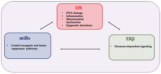
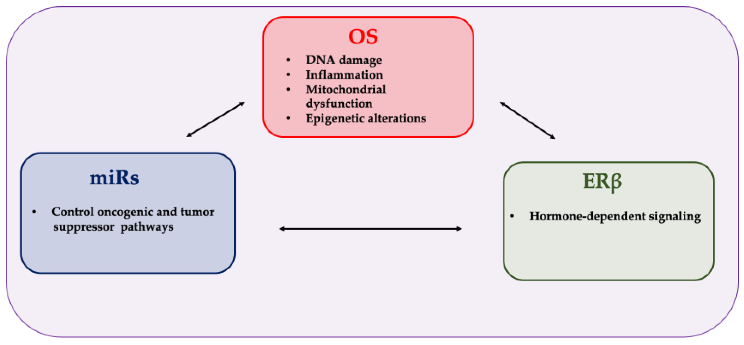
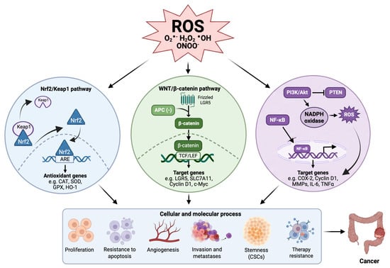
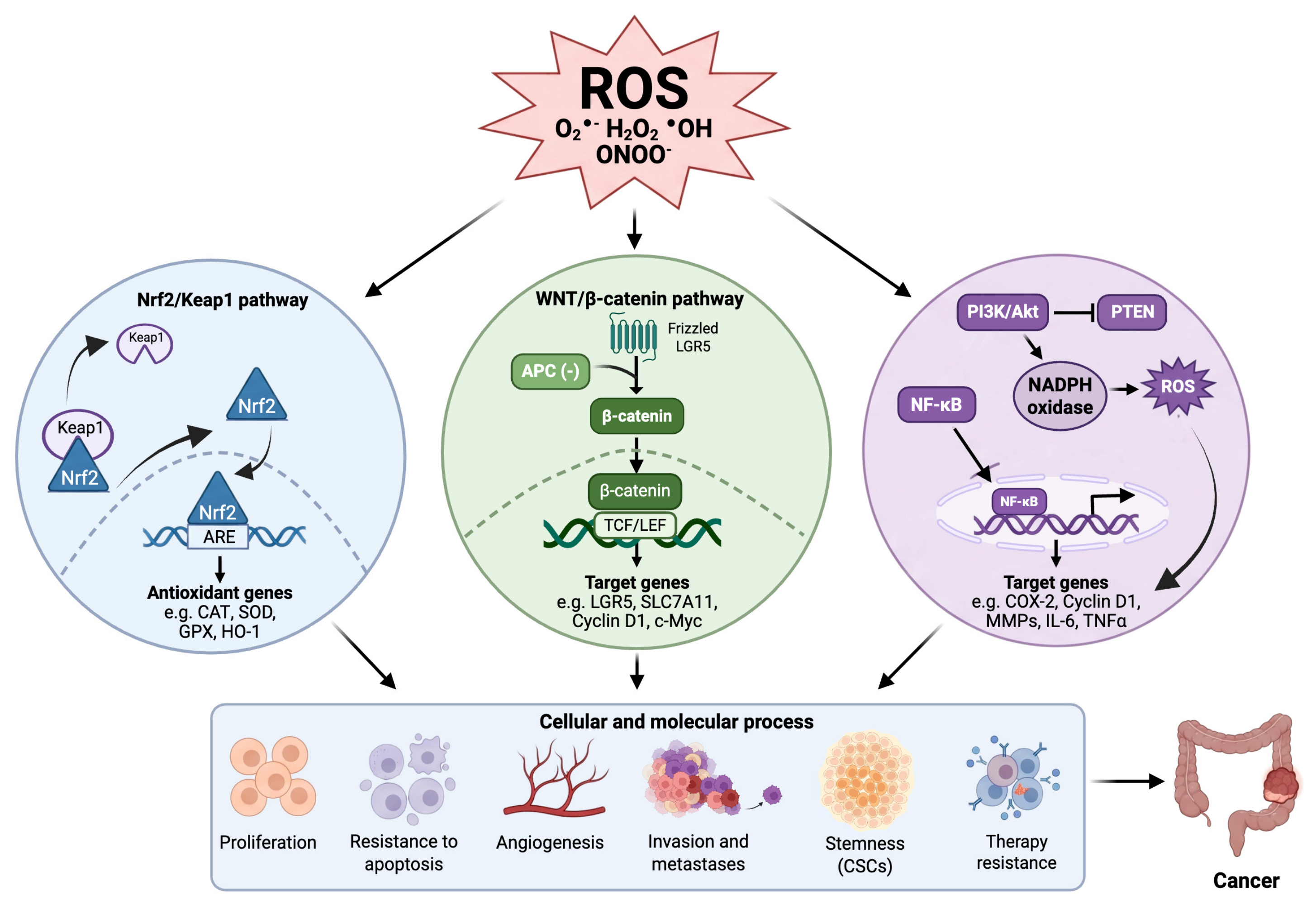
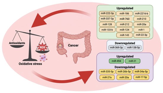
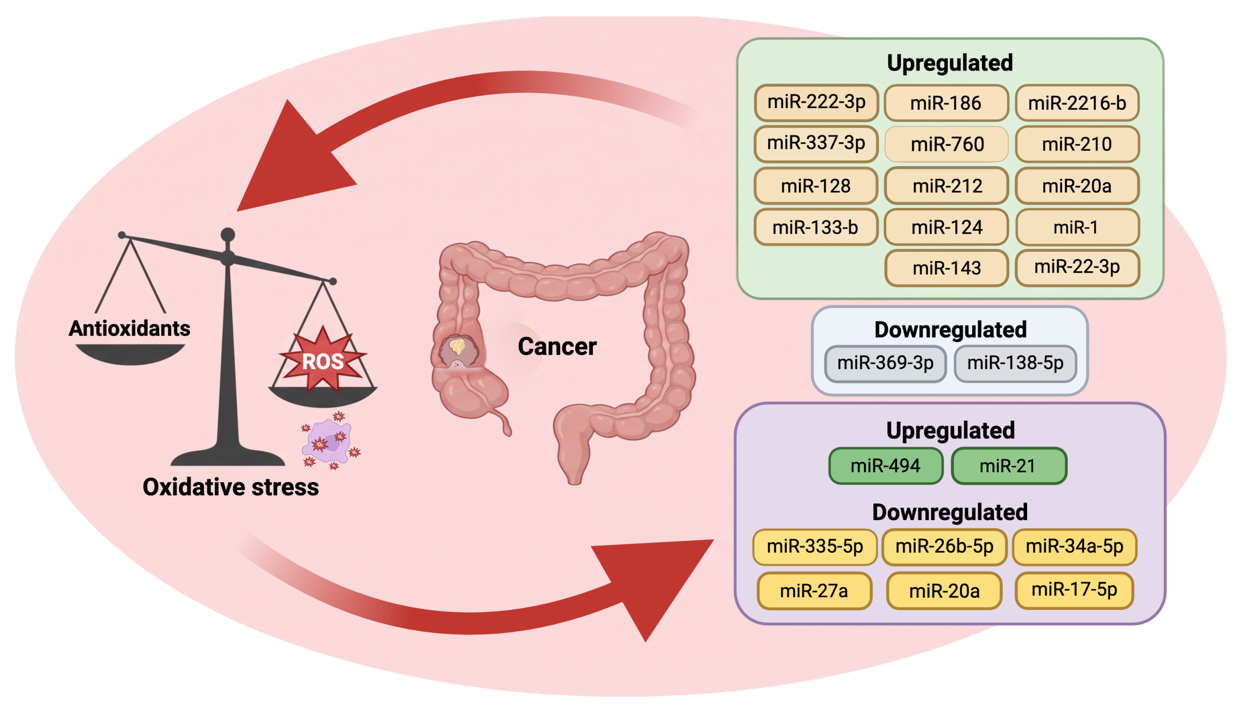
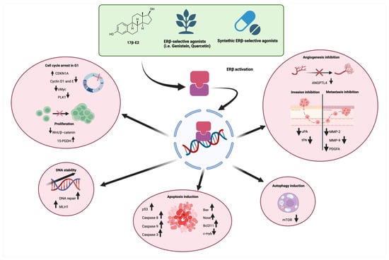
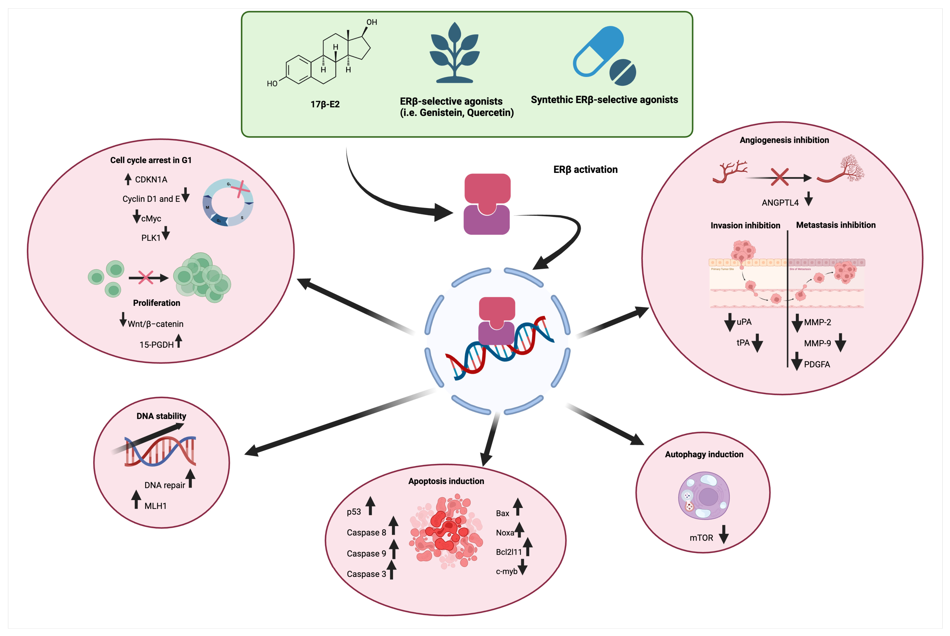

Disclaimer/Publisher’s Note: The statements, opinions and data contained in all publications are solely those of the individual author(s) and contributor(s) and not of MDPI and/or the editor(s). MDPI and/or the editor(s) disclaim responsibility for any injury to people or property resulting from any ideas, methods, instructions or products referred to in the content. |
© 2026 by the authors. Licensee MDPI, Basel, Switzerland. This article is an open access article distributed under the terms and conditions of the Creative Commons Attribution (CC BY) license.