


Abstract
Saffron (Crocus sativus L.) is a high-value crop vulnerable to potyvirus infections threatening its yield and quality. In this study, we combined Oxford Nanopore long-read sequencing with exploratory transcriptomic profiling to characterize the saffron virome and to describe expression profiles associated with two distinct infection histories: (i) saffron plants experimentally inoculated with cucumber mosaic virus (CMV; Cucumovirus CMV) and turnip mosaic virus (TuMV; Potyvirus rapae) under controlled greenhouse conditions, and (ii) saffron plants naturally infected by diverse viruses. We identified six plant-infecting viral families in both conditions, including Potyviridae, Geminiviridae, Caulimoviridae, Tymoviridae, Aspiviridae, and Partitiviridae. Transcriptomic profiling revealed distinct expression profiles associated with each infection background. Given the limitations of the experimental design, gene expression differences are interpreted descriptively. We describe pathway enrichments associated with antiviral responses. Naturally infected plants exhibited a broad-spectrum, tolerance-based response characterized by the upregulation of photosynthesis-related genes, calcium-mediated signaling components, and stress-responsive transcription factors. In contrast, virus-inoculated plants activated a targeted antiviral program involving RNA silencing, autophagy, ubiquitin-mediated proteolysis, and hormonal regulation. Both GO and KEGG enrichment analyses supported these findings, highlighting photosynthesis and metabolic flexibility in naturally infected plants versus hypersensitive response, RNA surveillance, and lignin biosynthesis in virus-inoculated plants. This work provides a comprehensive view of the saffron virome and offers a hypothesis-generating overview of transcriptional responses associated with natural versus experimental virus infections. These findings advance the understanding of the saffron virome and provide a valuable resource for breeding virus-resistant cultivars.
1. Introduction
Viral diseases represent a major challenge to global agriculture. Globalization and the intensification of international trade in agricultural products play a pivotal role in enabling the transmission of plant viruses and viroids to previously unaffected regions, consequently impacting food production systems and ecosystems in unpredictable ways [1,2].
The genus Potyvirus, within the Potyviridae family, constitutes the largest group of plant-infecting viruses transmitted by aphids, currently comprising 201 officially recognized species [3]. Potyviruses are frequently involved in mixed infections, where they can induce synergistic interactions that intensify symptom severity and contribute to significant yield losses, posing a threat to global food security [1].
Symptom-based diagnosis of plant viral infections is unreliable due to the overlap between viral symptoms and those caused by abiotic stresses such as nutrient deficiencies [4]. Therefore, there is an urgent need for rapid, sensitive, versatile and field-deployable diagnostic tools for the detection of plant viruses and viroids. Traditional detection methods, such as PCR and immunological assays, are often limited by their time-consuming protocols and relatively low efficiency [4]. High-throughput sequencing (HTS) has transformed plant virus diagnostics, enabling rapid and generic detection without prior knowledge of the pathogen [2].
Plants deploy a sophisticated array of genetic defense mechanisms to combat viral infections, with RNA silencing, and particularly RNA interference (RNAi), serving as a primary antiviral strategy through the regulation of gene expression by small RNAs such as miRNAs and siRNAs [5,6]. Upon pathogen detection, plants activate complex signaling pathways involving recognition receptors that trigger both localized and systemic defense responses, including structural barriers, chemical defenses, systemic acquired resistance (SAR), and induced systemic resistance (ISR) [7,8]. Viruses counteract these responses by producing suppressor proteins that inhibit the host RNA silencing machinery [6,9]. Recent advances in genetic engineering, including CRISPR/Cas9 and RNAi-based technologies, hold considerable promise for enhancing crop resistance by fine-tuning these molecular defenses [7,8]. This interplay between plant antiviral immunity and viral strategies is central to current efforts aimed at developing durable, broad-spectrum resistance in crops [10]. Oxford Nanopore Technology (ONT) platforms such as MinION, GridION, and PromethION, provide real-time long-read sequencing. This technology is ideal for the characterization of complex viral genomes, the resolution of mixed infections, and rapid detection workflows [2]. Nanopore-based diagnostics have already been successfully applied to detect known and novel potyviruses [2,11]. HTS has also expanded the understanding of viral infections in numerous plant species, including the detection of plum pox virus (PPV) and several cassava mosaic viruses (ACMV, EACMV, and SLCMV) in different hosts [12,13,14]. Additionally, yam-infecting viruses such as Dioscorea bacilliform virus (DBV) and Yam mild mosaic virus (YMMV) have been detected by HTS approaches [15]. The discovery of the novel bipartite begomovirus Cowpea bright yellow mosaic virus (CoBYMV) further highlights the broad applicability of HTS in detecting viral pathogens across crops [16].
Saffron (Crocus sativus L.), a member of the Iridaceae family, is a medicinal and aromatic plant of high economic value. Its dried stigmas, prized for their flavor, aroma and color, constitute the most expensive spice worldwide. Saffron is cultivated across Europe, Asia, and America [17]. Iran comprises approximately 90% of total global saffron production, followed by Spain (10–12%), India (3.3%), Greece (2%), and Morocco (3%) [17].
Several plant viruses have been reported to be infecting saffron worldwide, including in Iran [18]. The long-standing cultivation of saffron, its clonal propagation through corm, and its global economic relevance underscore the importance of characterizing its virome [17]. This is further supported by growing evidence of latent viral infections in various crops [11]. Viral infections in saffron have been associated with reduced yield and compromised quality of secondary metabolites [18,19]. The emergence of HTS technologies now enables comprehensive profiling of virus communities in plants. In this study, we report for the first time the identification of six plant-infecting viral families, Potyviridae, Geminiviridae, Caulimoviridae, Tymoviridae, Aspiviridae, and Partitiviridae, in Iranian saffron using Oxford Nanopore sequencing.
2. Materials and Methods
2.1. Plant Material
Saffron (Crocus sativus L.) samples, all originating from commercial field sources, were maintained under strictly isolated greenhouse conditions at Tarbiat Modares University, Tehran, Iran. A total of 30 samples were analyzed, including experimentally inoculated plants and non-inoculated plants exhibiting symptoms suggestive of viral infection (naturally infected).
The virus-inoculated group was derived from a batch of high-quality, sorted corms, obtained as apparently healthy material, from a single farm. These corms were planted in the greenhouse in 2019 and maintained until 2022. Prior to inoculation, targeted screening for viral infections was performed by back-inoculation on indicator plants and RT-PCR using degenerate and virus-specific primers against some known saffron-associated viruses. This analysis yielded negative results at that time. Plants were subsequently mechanically inoculated with cucumber mosaic virus (CMV; Cucumovirus CMV) and turnip mosaic virus (TuMV; Potyvirus rapae) as described by Shamshiri et al. (2025) [18]. These plants served as the experimentally inoculated treatment group.
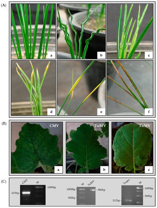
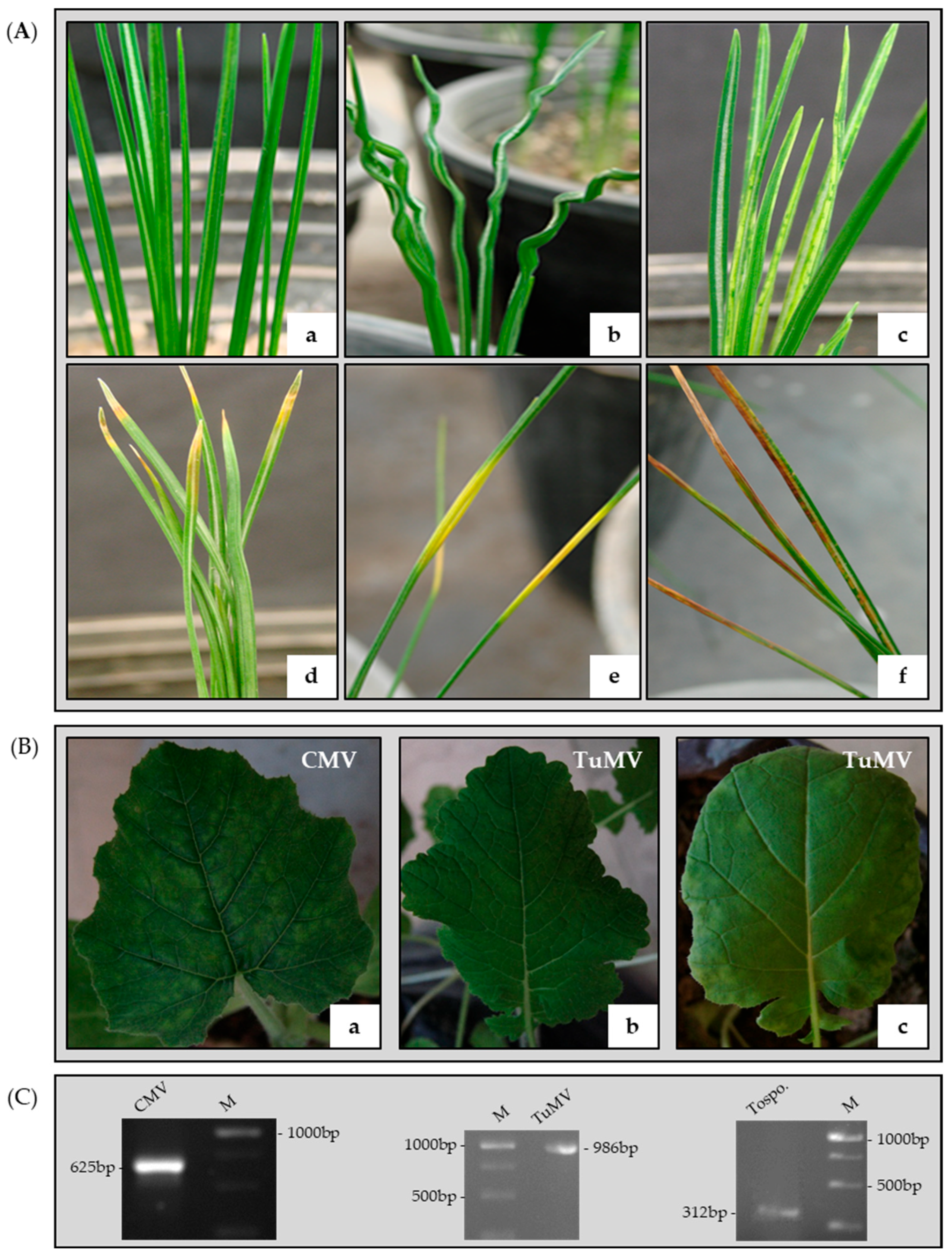
For both groups, symptoms were visually classified into six phenotypic categories: asymptomatic, leaf curling, leaf mosaic, leaf tip yellowing, mid-leaf yellowing and leaf tip browning (Figure 1A).
Figure 1.
(A) Symptomatic and asymptomatic saffron samples: (a) without symptom; (b) leaf curling; (c) leaf mosaic; (d) leaf tip yellowing; (e) mid-leaf yellowing; (f) leaf tip browning. (B) Detection of CMV and TuMV by back-inoculation in indicator plants including: (a) pumpkin, (b) turnip, and (c) rapeseed. (C) Detection of CMV, TuMV, and Tospovirus by RT-PCR in naturally infected saffron samples. Lane M: GeneRuler 1 kb DNA ladder (Fermentas, Vilnius, Lithuania).
The naturally infected group consisted of whole plants (corms + leaves) collected annually from 2019 to 2022 in four distinct regions of Torbat-e Heydarieh County, Razavi Khorasan Province, Iran. Samples were transferred into isolated greenhouse conditions and maintained separately. The presence of viral infection was confirmed by back-inoculation on indicator hosts (Figure 1B) and RT-PCR assays using both degenerate and virus-specific primers targeting the Tospovirus genus, CMV and TuMV (Figure 1C). Total RNA was extracted using the RNX-Plus kit (SinaClon, Iran) and cDNA was synthesized with the Easy™ cDNA Synthesis kit (Parstous, Iran). For Tospovirus detection, a 312 bp fragment of the L RNA segment was amplified using consensus primers [20]. CMV detection targeted a 625 bp fragment of the coat protein (CP) gene [21], and TuMV detection amplified a 986 bp fragment including 54 bp of the 3′ end of the NIb gene, the complete CP gene, and 65 bp of the 3′ untranslated region (UTR) following Sánchez et al. [22]. Primer sequences are listed in Supplementary Table S1. Both experimentally inoculated and naturally infected plants were used for comparative virome profiling and transcriptomic analysis to identify known and potentially novel viruses associated with saffron.
2.2. RNA Extraction and Quality Assessment
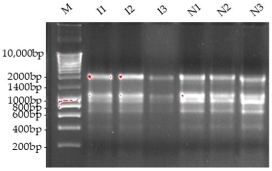
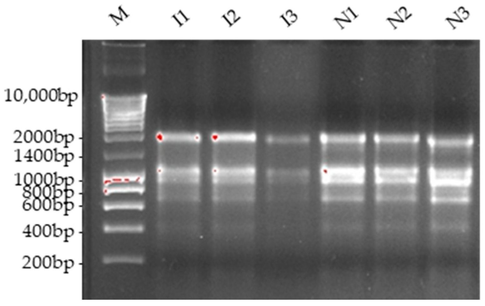
For transcriptomic analysis, total RNA was isolated from saffron leaves collected from virus-inoculated and naturally infected plants presenting a pool of distinctive viral infection symptoms (leaf curling, leaf mosaic, leaf tip yellowing, mid-leaf yellowing and leaf tip browning). RNA extraction was performed using the FavorPrepTM Plant total RNA Purification Mini Kit (Favorgen Biotech Corp., PingTung, Taiwan). RNA was treated with DNase I (Promega Corp., Madison, WI, USA) and purified by phenol–chloroform extraction. RNA quality and quantity were determined using a NanoDrop 2000c spectrophotometer (Thermo Fisher Scientific, Wilmington, DE, USA) and integrity was verified by electrophoresis on 1.2% agarose gel (Figure 2). Three biological replicates were collected per sample. Only samples meeting quality criteria (260/280 ratio of approximately 2.0 and 260/230 ratio between 2.0 and 2.2) were used for ribodepletion and transcriptome library construction. Equal amounts of high-quality RNA from each individual within the same experimental group were pooled prior to subsequent steps.
Figure 2.
Evaluation of the quality of total RNA samples after treatment with RQ1 RNase-Free DNase I using agarose gel analysis. I1: virus-inoculated saffron sample 1; I2: virus-inoculated saffron sample 2; I3: virus-inoculated saffron sample 3; N1: naturally infected saffron sample 1; N2: naturally infected saffron sample 2; N3: naturally infected saffron sample 3; M: NZYDNA Ladder III (NZYTech, Portugal).
2.3. Host Ribodepletion and cDNA Synthesis
Ribodepletion of host rRNA was performed using the RiboMinus™ Plant Kit for RNA-Seq (Invitrogen, Carlsbad, CA, USA) on pooled RNA from three biological replicates per condition, following the manufacturer’s instructions. mRNA concentration was measured using both a Nanodrop spectrophotometer and a Qubit fluorometer with the RNA HS Assay Kit (Thermo Fisher Scientific, Waltham, MA, USA). Efficiency of ribodepletion was verified by electrophoresis on 1.2% agarose gel (Supplementary Figure S1).
Double-stranded cDNA was synthesized using random primers and the Maxima H Minus Double-Stranded cDNA Synthesis Kit (Thermo Fisher Scientific, Waltham, MA, USA), followed by purification using the GeneJET PCR Purification Kit (Thermo Fisher Scientific, Waltham, MA, USA). cDNA concentration was determined using the Qubit 1X dsDNA HS Assay Kit (Thermo Fisher Scientific, Waltham, MA, USA) [23].
2.4. Library Preparation and Sequencing
Transcriptomic libraries were constructed from double-stranded cDNA of each condition using the Direct cDNA Sequencing kit (SQK-DCS109, ONT, Oxford, UK). End repair and dA-tailing were performed according to the kit protocol, followed by adapter ligation. Libraries were loaded onto a MinION device with R9.4.1 flow cells (ONT, Oxford, UK) and sequenced for 40 h [23].
2.5. Transcriptomic Analysis of Nanopore Sequencing Data
Base-calling was performed using Guppy (v3.3.1). Read quality was assessed with NanoPlot (v1.33.0) [24] and Porechop (v0.2.4) was used for trimming raw reads to obtain a clean set of reads for subsequent analyses. Clean reads were mapped to the reference genome using minimap2 (v2.28) with the “-ax splice” option [25] and processed with SAMtools (v1.19). Due to the lack of a well-annotated genome for Crocus sativus, the genome of Asparagus officinalis was used as reference. Sorted alignment files and the GFF annotation file for A. officinalis were used to generate the count table in OmicsBox (Biobam, Valencia, Spain, v3.1.11) using the Gene Level Quantification mode.
Because biological replicates were pooled before ribodepletion, pairwise differential expression analysis was performed using NOISeq-sim, a method designed for datasets lacking biological replicates [26]. This algorithm generates simulated technical replicates from the empirical distribution of the data to estimate noise and variability, enabling robust probability-based differential expression detections in non-replicated designs. Simulated replicates were generated using the parameters nss = 5, pnr = 0.2, v = 0.02, and k = 0.5, with RPKM normalization and length correction. Genes with probability (q) > 0.9 were considered differentially expressed. Transcripts with M > 0 were considered upregulated in naturally infected samples, whereas those with M < 0 were considered upregulated in virus-inoculated samples.
Gene Ontology (GO) enrichment analysis was performed independently for each dataset using ShinyGO (v0.82) [27]. Treemaps were generated using Revigo (http://revigo.irb.hr, accessed on 18 June 2024) [28]. Protein sequences of the DEGs were used for KEGG pathway analysis (Kyoto Encyclopedia of Genes and Genomes) with KOBAS (http://bioinfo.org/kobas, accessed on 17 June 2024) [29], with statistical enrichment assessed via hypergeometric and the Fishers’ exact test (p < 0.05, corrected p < 0.05).
2.6. qRT-PCR Validation of Differential Genes Expression
To validate the expression profiles of selected DEGs identified by RNA-seq, qRT-PCR was performed using RNA from individual biological replicates (not pooled plant material) of virus-inoculated and naturally infected samples. cDNA was synthesized from 1 µg of total RNA using the High-Capacity cDNA Reverse Transcription Kit (Applied Biosystems, Waltham, MA, USA).
qPCR was performed in 48-well optical plates using the Step One Real-Time PCR System (Applied Biosystems, Waltham, MA, USA) and NZYSupreme qPCR Green Master Mix (NZYtech, Lisbon, Portugal) to monitor dsDNA synthesis. Each reaction contained 400 µM of each primer, 1.5 µL of cDNA (12.5 ng RNA input) and master mix in a final volume of 20 µL. Thermal cycling conditions were: 95 °C for 2 min, followed by 40 cycles of 95 °C for 5 s and 60 °C for 30 s. Melt curve analysis was performed to confirm product uniformity. Target genes serine/threonine-protein kinase PBS1-like (PBS1) and Suppressor of Gene Silencing 3 homolog (Supr), Ubiquitin (UBI), and Tubulin (TUB) were used as internal controls for data normalization. Primers were designed using Primer-BLAST (https://www.ncbi.nlm.nih.gov/tools/primer-blast, accessed on 27 June 2024) and are listed in Supplementary Table S2. Three biological and three technical replicates were analyzed per sample. Relative expression was calculated using the comparative Ct method [30] and normalized to virus-inoculated samples. Results are presented as mean ± standard error from three biological replicates. Data were analyzed using DataAssist (Applied Biosystems, Waltham, MA, USA, v3.0).
2.7. Plant Virus Detection with the Core DIAMOND + MEGAN Pipeline
To identify plant viruses present in saffron samples, a long-read metagenomic approach was applied. Reads that did not align to the Crocus reference genome were retained and subjected to taxonomic classification. These reads were aligned at the protein-level against the NCBI non-redundant (nr) virus database using DIAMOND (v2.0.8) in frameshift-aware mode, which is specifically designed to handle the higher error rates of ONT long reads. The alignment was performed with the parameters -F 15 -f 100—range-culling—top 10, which optimize sensitivity and specificity for long-read data. The resulting DIAMOND alignment archive (DAA) files were processed using MEGANIZER (part of the MEGAN 6 package, University of Tübingen, Tübingen, Germany, v6.25.10) to perform taxonomic binning. The interval-union Lowest Common Ancestor (LCA) algorithm, specifically designed for long-reads, was applied with long-read settings (percent to cover = 51%). This algorithm divides each read into segments based on alignment boundaries and assigns the read to the lowest taxonomic node whose associated alignments cover at least 51% of the aligned portion of the read. This approach significantly improves taxonomic resolution for long, error-prone sequences that may span multiple genes. Taxonomic classification was based on the NCBI taxonomy.
Meganized DAA files were visualized and analyzed in MEGAN 6 to generate taxonomic trees, charts, and family- and species-level summaries. Comparative analysis between virus-inoculated and naturally infected conditions was performed using normalized aligned base counts to correct for differences in library size (sequencing depth) between samples, ensuring that observed variations reflect relative abundance rather than dataset size. From these values, we derived the relative viral abundance, defined as the fraction of aligned bases assigned to that virus within the set of plant-infecting viruses detected in each sample (i.e., excluding non-plant viruses). All steps followed the DIAMOND + MEGAN long-read protocol described by Bağcı et al. [31], with adaptations for plant virus detection [32]. To validate these metagenomic detections, we performed confirmatory read mapping using minimap2 with parameters optimized for high sensitivity (-k11 -w1). For each virus, reads assigned by MEGAN were extracted and mapped against the corresponding reference genome. The resulting SAM files were converted to BAM, sorted and indexed using samtools. Coverage and depth statistics were calculated with samtools depth -a, and the percentage of the genome covered at ≥1× and ≥5× was determined. This approach allowed us to distinguish between viruses present as complete genomes and those detected only through conserved protein domains.
2.8. Confirmatory Test of the HTS Results Using RT-PCR
RT-PCR was performed to validate the presence of viruses identified through nanopore sequencing. We used the same RNA samples as used in the sequence experiment and cDNA was synthesized as described in Section 2.6. Primers were designed using Primer-BLAST and sequences are listed in Supplementary Table S3. PCR reaction was performed in a standard VWR thermal cycler (VWR International, Radnor, PA, USA) using virus-specific primers. Reactions contained 0.5 µM of each primer, 1.5 µL of cDNA (12.5 ng RNA input), 200 µM of dNTPs 5× Buffer and Phire Hot Start II DNA Polymerase (Thermo Fisher Scientific, Waltham, MA, USA). Thermal cycling conditions are listed in Supplementary Table S4. Amplification products were visualized by 1.2% agarose gel electrophoresis to confirm the presence of expected amplicon sizes.
3. Results
The objective of this research was to analyze the transcriptional response of saffron to different viral infections and to identify viruses infecting saffron in Iran through Oxford Nanopore sequencing. The cDNA libraries of saffron samples were generated from both naturally infected and virus-inoculated samples using random hexamer primers and sequenced on the MinION device. We compared the expression profiles associated with two infection conditions: experimentally inoculated CMV/TuMV plants and naturally infected plants.
3.1. Transcriptomic Analysis (RNA-Seq)
Sequencing generated 15 GB of data from virus-inoculated libraries and 8 GB from naturally infected (non-inoculated) libraries. After adapter trimming and quality filtering, a total of 64.2 and 39.9 million clean reads were obtained from virus-inoculated and naturally infected samples, respectively. Mapping rates to the A. officinalis reference genome were high in both datasets (95.28% in virus-inoculated plants and 97.73% in naturally infected plants), with primary alignments of 82.34% and 90.80%, respectively. Differential expression analysis using NOISeq-sim identified a total of 612 differentially expressed genes (DEGs) between virus-inoculated and naturally infected plants. Among these, 348 genes appeared to be upregulated in virus-inoculated plants and 264 in naturally infected plants. Functional annotation and enrichment analysis of DEGs were performed using Gene Ontology (GO) and Kyoto Encyclopedia of Genes and Genomes (KEGG) pathways to explore the biological processes and pathways potentially associated with each condition. As biological replicates were not available, the DEGs identified here should be interpreted with caution and considered as candidate genes for further validation.
Overall, the results suggested that virus-inoculated plants may show enrichment in more specialized immune and antiviral processes, whereas naturally infected plants appeared to exhibit enrichment in patterns associated with photosynthesis and broader stress adaptation pathways.
3.1.1. Naturally Infected Plants
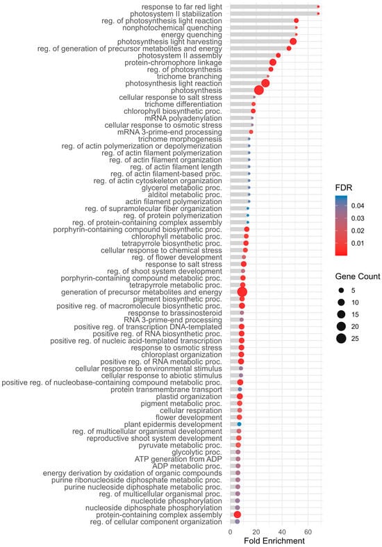
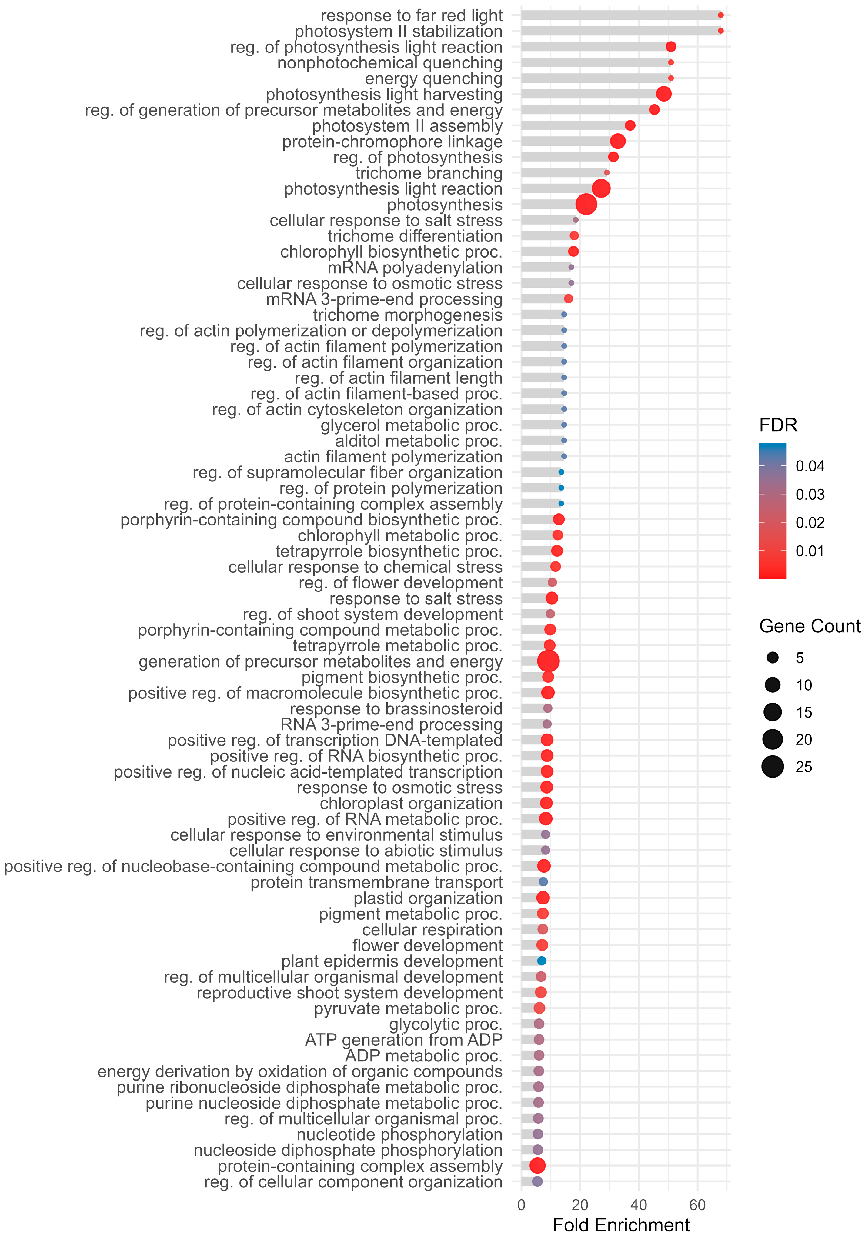
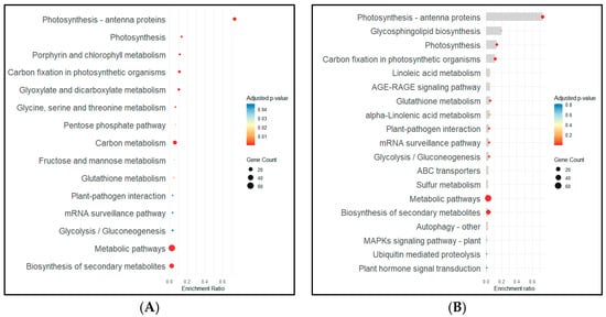
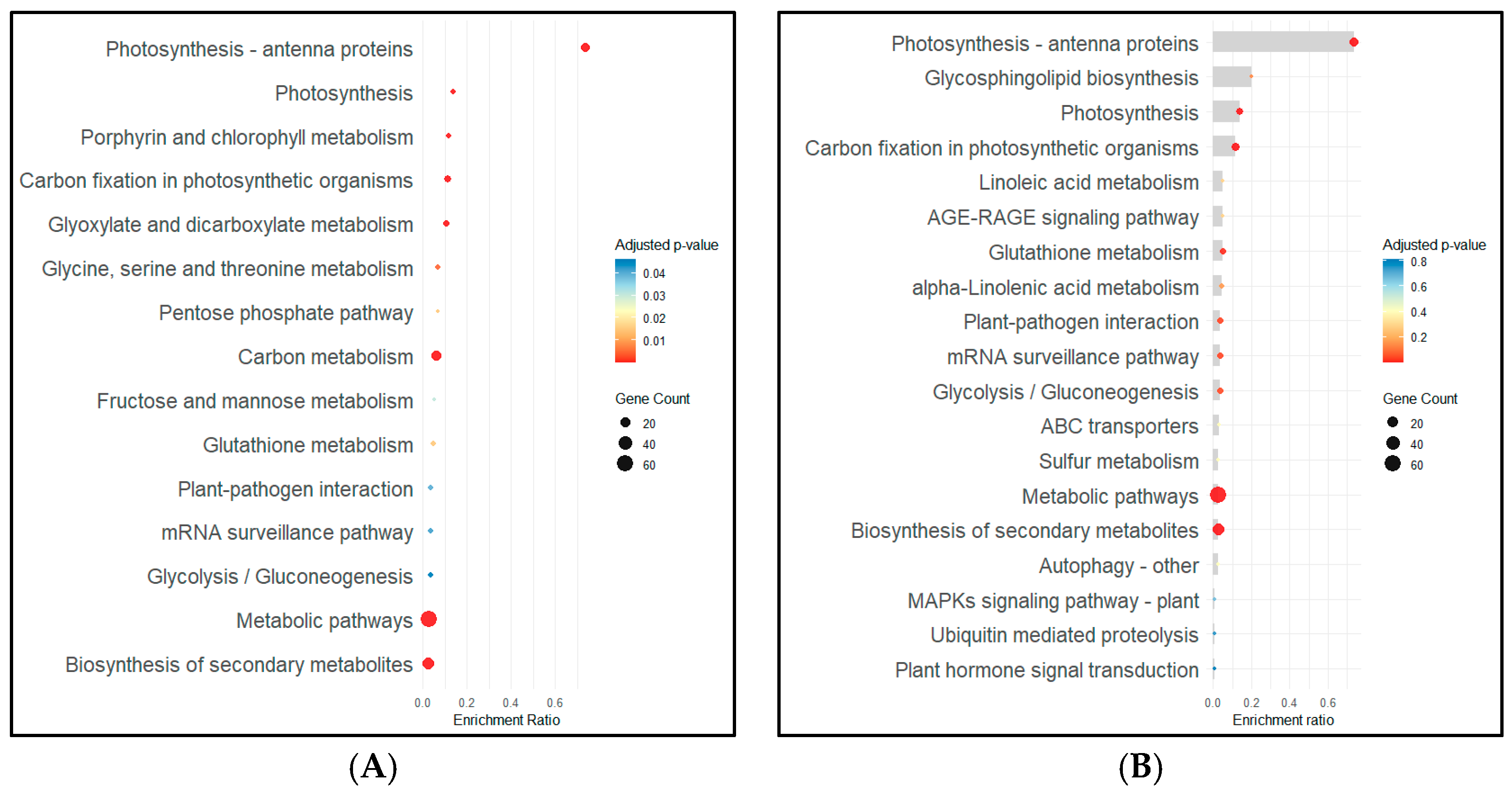
In naturally infected (non-inoculated) samples, the transcriptional profile appeared to be predominantly associated with photosynthesis-related processes and metabolic flexibility. Upregulated genes included ribulose bisphosphate carboxylase (RuBisCO), phosphoglycerate kinase, ferredoxin-NADP reductase, oxygen-evolving enhancer protein 1, and multiple chlorophyll a–b binding proteins. GO enrichment suggested representation of photosynthetic processes, such as photosystem II stabilization (GO:0042549), regulation of photosynthesis light reaction (GO:0042548), photosynthesis light harvesting (GO:0009765), regulation of photosynthesis (GO:0010109), as well as photoprotection-related terms such as response to far red light (GO:0010218), energy quenching (GO:1990066), and nonphotochemical quenching (GO:0010196) (Figure 3). KEGG enrichment analysis was consistent with these observations, showing enrichment in pathways such as photosynthesis, photosynthesis-antenna proteins, porphyrin and chlorophyll metabolism, glyoxylate and dicarboxylate metabolism, and carbon fixation (adjusted p value < 0.05), suggesting increased activation of photosynthetic machinery (Figure 4A).
Figure 3.
Top Gene Ontology (GO) Biological Process terms enriched in naturally infected plants (FDR < 0.05). The x-axis shows Fold Enrichment (magnitude of enrichment), point size indicates gene count, and color represents FDR (False Discovery Rate).
Figure 4.
KEGG pathway enrichment analysis in naturally infected plants. (A) Dot plot of pathways identified using KOBAS (adjusted p-value < 0.05). Pathways are ranked by enrichment ratio, point size represents gene count, and color indicates adjusted p-value. (B) Bar and dot plot of pathways identified using KOBAS, including some important terms with adjusted p-value > 0.05.
In addition to photosynthesis-related categories, naturally infected plants showed the upregulation of several signal transduction components, including serine/threonine-protein kinases (PBL9, HT1, STN7 and STN8, BSK3, OXI1-like, and PBS1-like), calcium-dependent protein kinases (CDPK26, CDPK20-like), and calcium-binding proteins (CML49). Transcriptional regulators such as MYB3R-1, NF-Y subunit B-11-like, and heat stress TF A-4b-like were also induced.
Defense-related genes were present, but they appeared less prominent than in virus-inoculated plants. These included E3 ubiquitin ligases (HERC2, SP1), RNA helicases (DEAD-box 53), and chaperones (T-complex protein 1 subunit alpha). Additionally, genes involved in RNA processing and protein degradation, such as protein TPLATE, proteasome subunit beta type-5-B-like, and components of the RNA-directed gene silencing machinery, including DNA-directed RNA polymerases II, IV, and V subunit 6A-like showed increased expression.
KEGG analysis identified enriched categories related to metabolic pathways and biosynthesis of secondary metabolites, along with defense-related pathways like glutathione metabolism and plant–pathogen interaction (Figure 4A). Other pathways potentially linked with defense, like linoleic acid metabolism, alpha-linolenic acid metabolism, glycosphingolipid biosynthesis, sulfur metabolism, MAPK signaling, autophagy, ubiquitin-mediated proteolysis, ABC transporters, and plant hormone signal transduction were detected but did not reach significant adjusted p-value thresholds (adjusted p value > 0.05) (Figure 4B). GO terms associated with stress responses were also overrepresented, including cellular response to abiotic stimulus (GO:0071214), cellular response to salt stress (GO:0071472), cellular response to osmotic stress (GO:0071470) and cellular response to chemical stress (GO:0062197) (Figure 3).
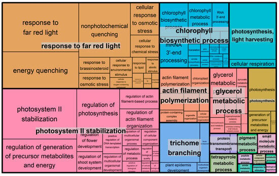
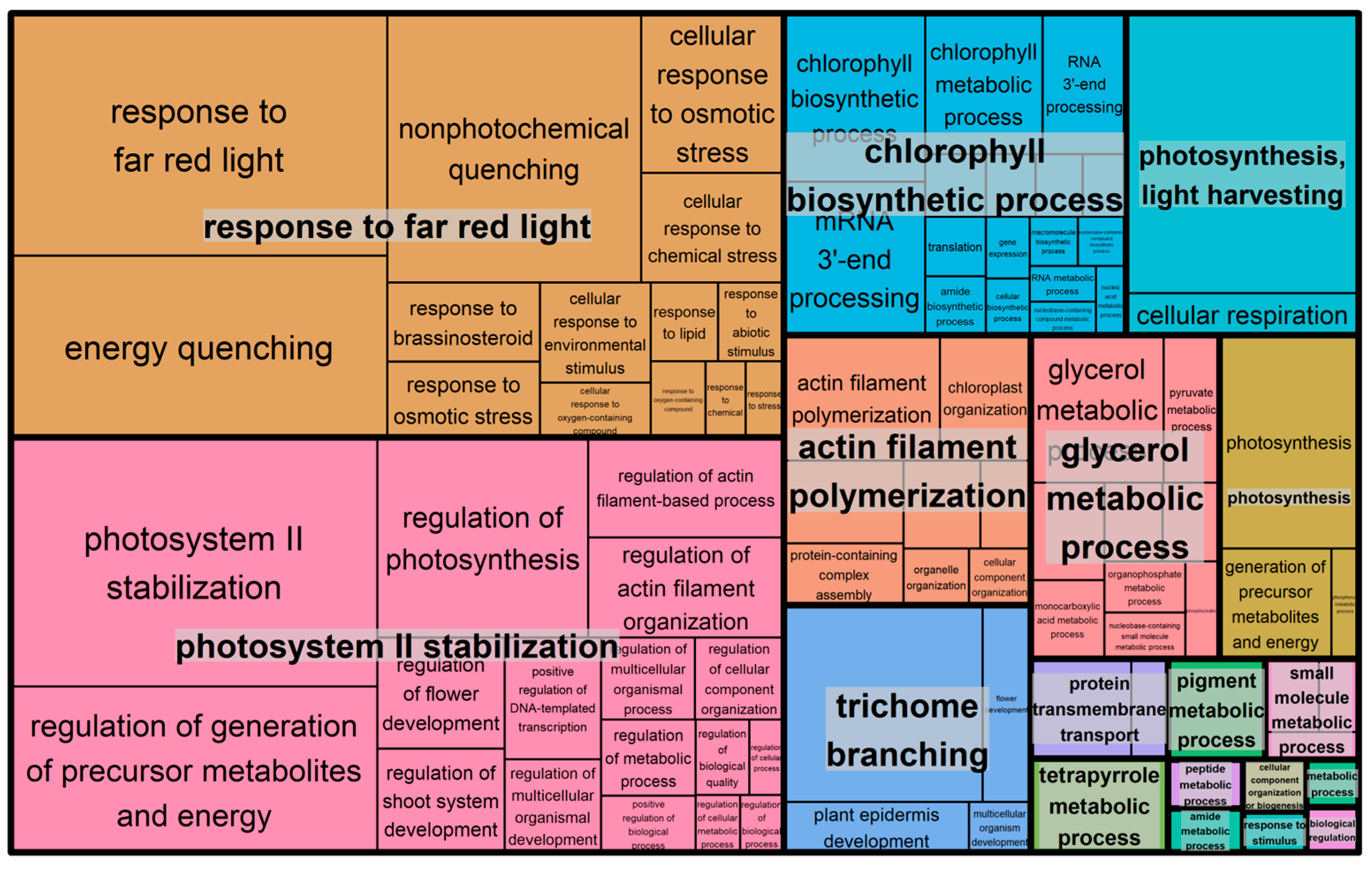
The Revigo treemap for naturally infected plants (Figure 5) grouped enriched GO terms into major functional clusters. These include response to far red light, photosystem II stabilization, chlorophyll biosynthetic process, photosynthesis and light harvesting, as well as structural or stress-related processes such as trichome branching, actin filament polymerization, and glycerol metabolic process. Summarized GO enrichment results can also be found in Figure S2. This organization appeared consistent with a transcriptional profile centered on photosynthetic activity, photoprotection, and broader adaptive metabolic adjustments under naturally infected conditions.
Figure 5.
Revigo treemap summarizing GO enrichment results for naturally infected plants. The treemap displays representative GO terms after semantic clustering and redundancy reduction. Larger boxes indicate more significant terms.
3.1.2. Virus-Inoculated Plants
In plants inoculated with TuMV and CMV, the transcriptional profile suggested a more specialized antiviral response, with indications of enrichment in genes associated with vesicle trafficking, autophagy, and RNA silencing. Upregulated genes included Suppressor of Gene Silencing 3 (SGS3) and multiple DExH and DEAD-box ATP-dependent RNA helicases (e.g., DExH11, DExH3, DEAD-box ATP-dependent RNA helicase 7, 18, 28, 29, and 56), together with autophagy-related protein (ATG2, ATG9 and ATG11) and aspartic proteinase CDR1. Additional components of the protein degradation machinery also appeared induced, including E3 ubiquitin-protein ligase (KEG, UPL3, and UPL4) and chaperones (T-complex protein 1 subunit epsilon). Signaling elements such as mitogen-activated protein kinases (MAPKs), cyclic nucleotide-gated channels (CNGC2 and CNGC20), and serine/threonine-protein kinases (D6PKL1 and PBL19) were detected along with delta-1-pyrroline-5-carboxylate synthase-like. Hormonal and epigenetic regulators showed increased expression, including ethylene-responsive TF 1, auxin transport protein BIG, auxin response factor 17, homeobox-DDT domain protein RLT2, and polycomb group protein Embryonic Flower 2. Genes involved in RNA processing, such as pre-mRNA-processing protein 40A and DNA-directed RNA polymerases II, IV, and V subunit 3-like were also upregulated. DEGs additionally included leucine-rich repeat (LRR) proteins, callose synthases, and antioxidant enzymes (such as glutathione peroxidases).
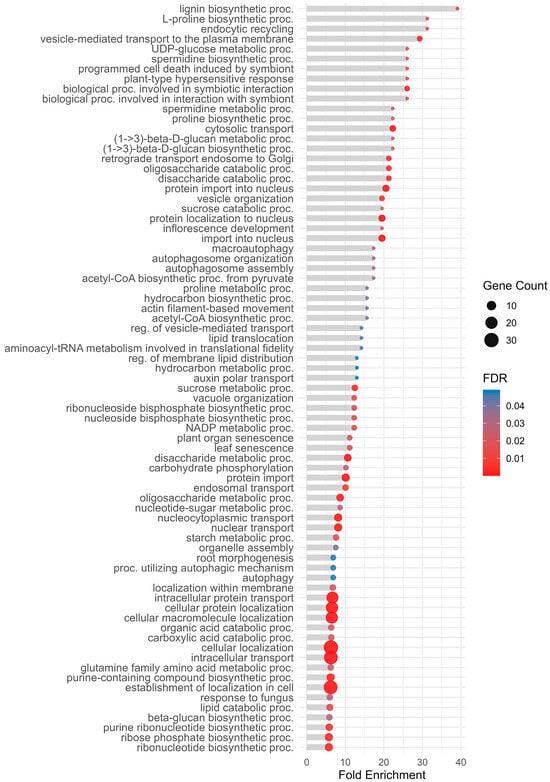
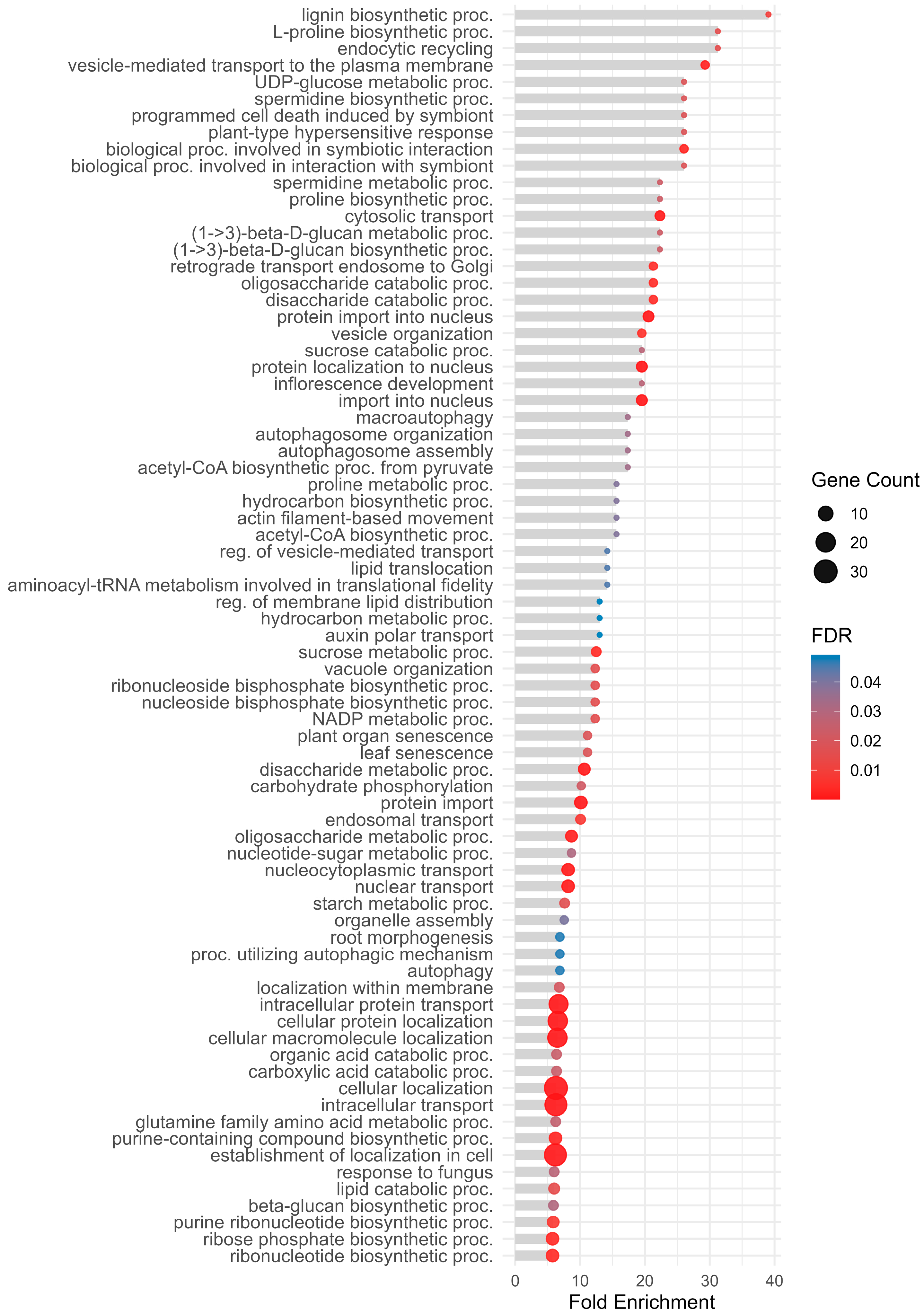
GO enrichment analysis in these plants (Figure 6) suggested the involvement of processes potentially associated with immune defense and cell wall fortification like lignin biosynthetic process (GO:0009809), programmed cell death induced by symbiont (GO:0034050), and plant-type hypersensitive response (GO:0009626). Additional enriched categories included biological processes involved in symbiotic interaction (GO:0044403), interaction with symbiont (GO:0051702), and response to fungus (GO:0009620). Autophagy-related terms were strongly represented, including autophagy (GO:0006914), macroautophagy (GO:0016236), autophagosome assembly (GO:0000045), and autophagosome organization (GO:1905037). Metabolic processes were indicated by terms like UDP-glucose metabolic process (GO:0006011), beta-glucan biosynthetic process (GO:0051274), proline biosynthetic process (GO:0006561), spermidine biosynthetic and metabolic processes (GO:0008295, GO:0008216), and carbohydrate-related categories like sucrose metabolic process (GO:0005985) and disaccharide catabolic process (GO:0046352). Other terms include endocytic recycling (GO:0032456) and L-proline biosynthetic process (GO:0055129).
Figure 6.
Top Gene Ontology (GO) Biological Process terms enriched in virus-inoculated plants (FDR < 0.05). The x-axis shows Fold Enrichment (magnitude of enrichment), point size indicates gene count, and color represents FDR (False Discovery Rate).
Additional GO terms such as defense response (GO:0006952, GO:0098542), response to external biotic stimulus (GO:0043207), response to stress (GO:0006950), response to external stimulus (GO:0009605), intracellular signal transduction (GO:0035556), and ion transport (GO:0006811) were also detected but with lower enrichment magnitude compared to those represented in Figure 6.
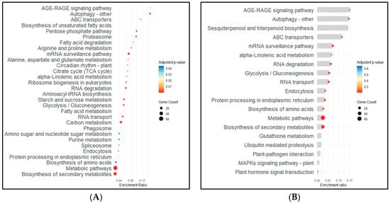
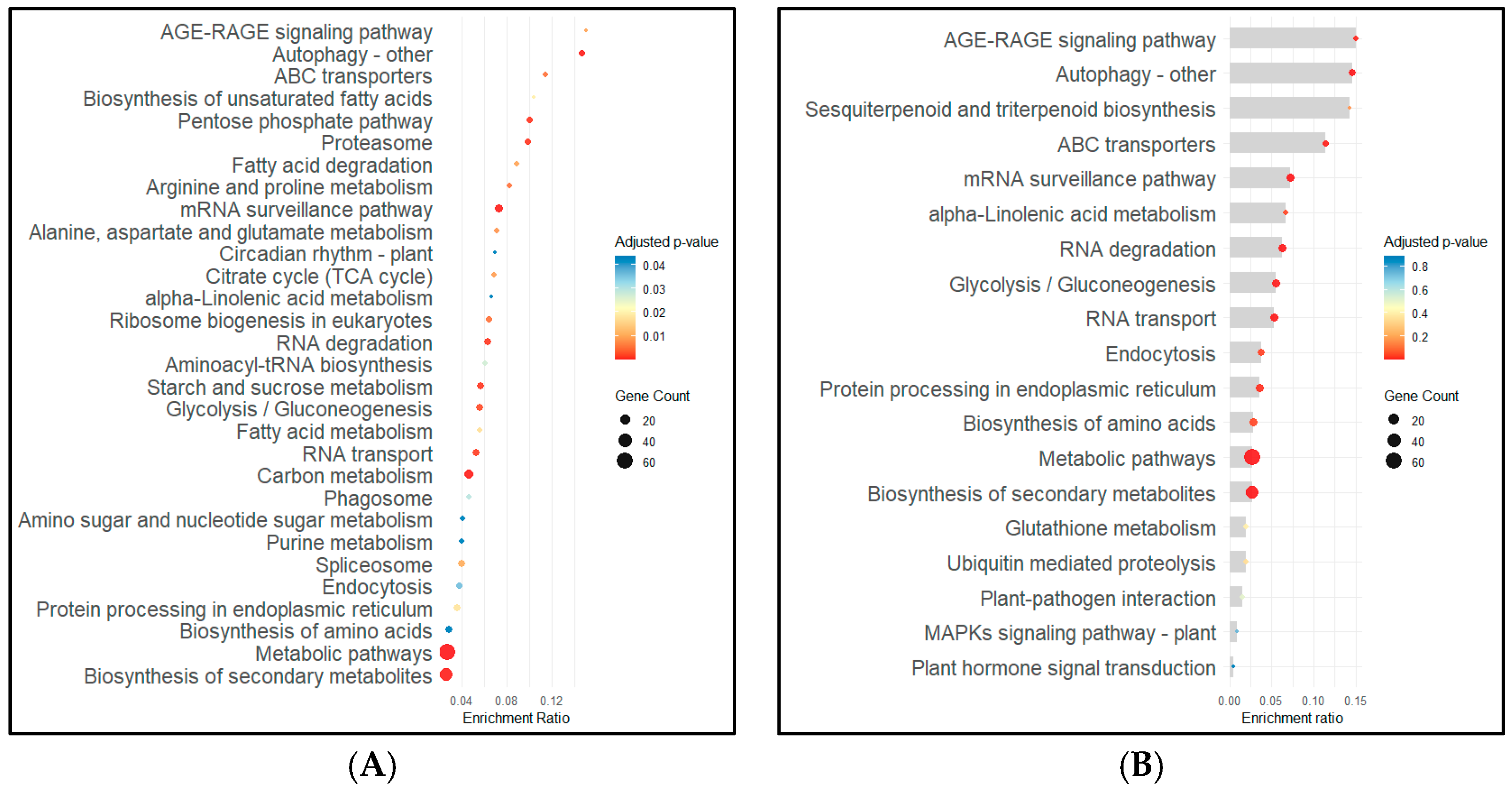
KEGG enrichment analysis in virus-inoculated plants appeared consistent with activation of pathways often associated with antiviral defense. Enriched pathways included autophagy, AGE-RAGE signaling pathway, mRNA surveillance pathway, RNA degradation, RNA transport, proteasome, and protein processing in the endoplasmic reticulum, suggesting activation of degradation systems and stress signaling. Additional pathways such as ABC transporters, alpha-Linolenic acid metabolism, glycolysis/gluconeogenesis, biosynthesis of secondary metabolites or endocytosis were also detected (Figure 7A). Other pathways identified in the analysis included plant-pathogen interaction, MAPKs signaling, glutathione metabolism, ubiquitin-mediated proteolysis, and sesquiterpenoid and triterpenoid biosynthesis did not reach significant adjusted p-values (Figure 7B).
Figure 7.
KEGG pathway enrichment analysis in virus-inoculated plants. (A) Dot plot of pathways identified using KOBAS (adjusted p-value < 0.05). Pathways are ranked by enrichment ratio, point size represents gene count, and color indicates adjusted p-value. (B) Bar and dot plot of pathways identified using KOBAS, including some important terms with adjusted p-value > 0.05.
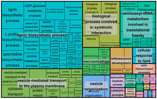
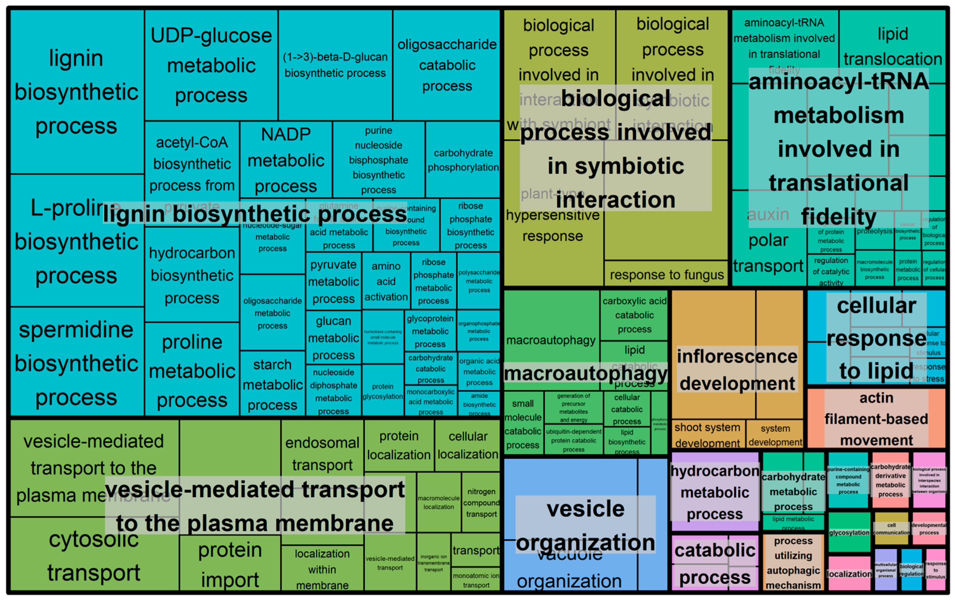
The Revigo treemap for virus-inoculated plants (Figure 8) grouped enriched GO terms into clusters including lignin biosynthetic processes, vesicle-mediated transport to the plasma membrane, biological process involved in symbiotic interaction, macroautophagy, vesicle organization, and aminoacyl-tRNA metabolism involved in translational fidelity. Summarized GO enrichment results can also be found in Figure S3. Overall, these clusters appeared consistent with a coordinated transcriptional response involving defense-related, degradation, and vesicle trafficking pathways.
Figure 8.
Revigo treemap summarizing GO enrichment results for virus-inoculated plants. The treemap displays representative GO terms after semantic clustering and redundancy reduction. Larger boxes indicate more significant terms.
3.2. Validation of Key Defense-Related Genes Through qRT-PCR
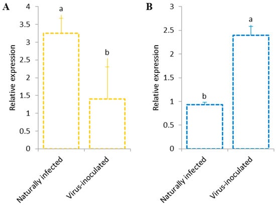
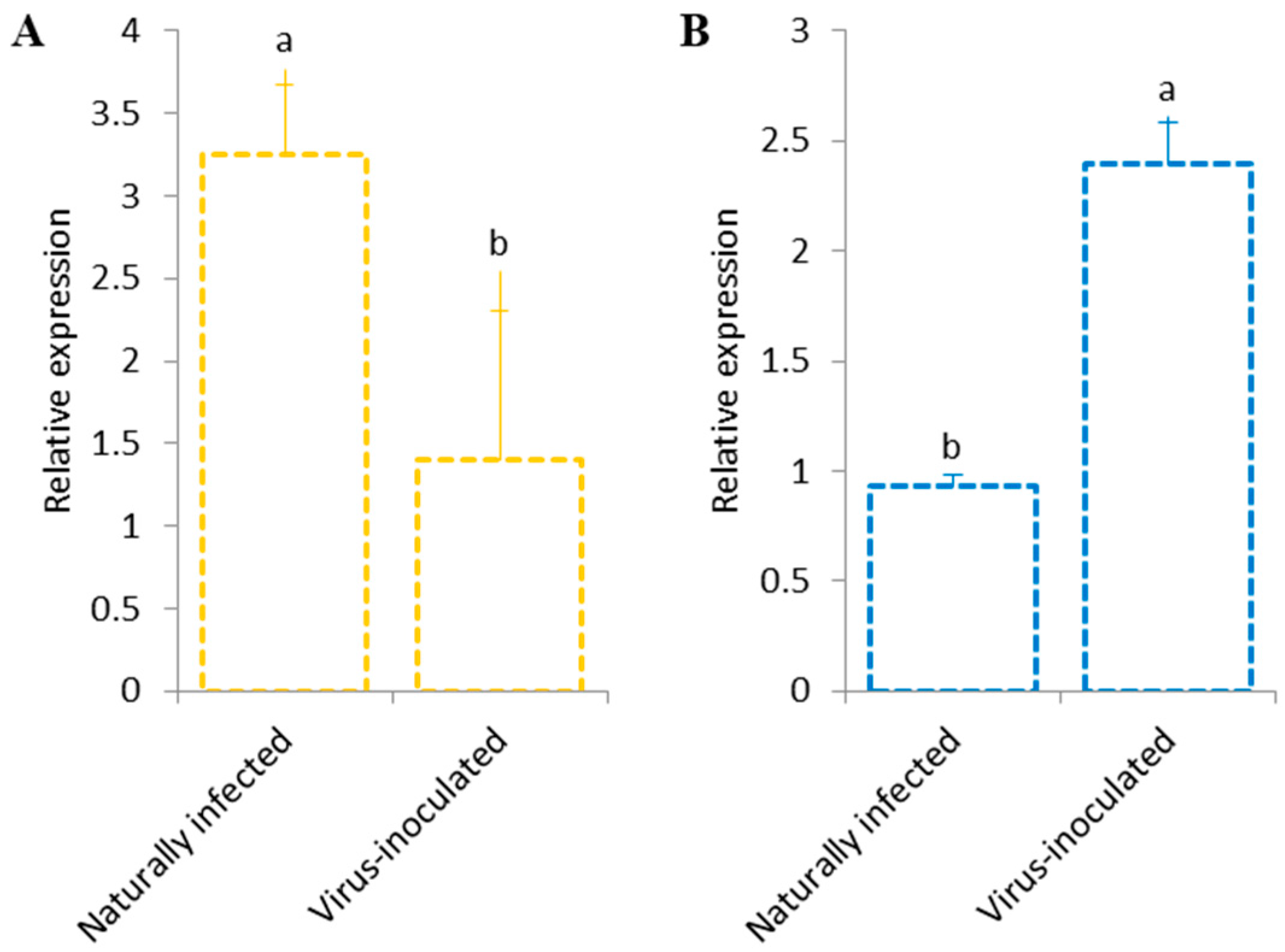
To validate the transcriptomic findings, qRT-PCR was performed on selected defense-related genes using independent biological replicates. The results confirmed that serine/threonine-protein kinase PBS1-like (PBS1) was significantly up-regulated in naturally infected samples, while suppressor of gene silencing 3 homolog (Supr) showed predominant up-regulation in samples that have been additionally inoculated with CMV and TuMV (Figure 9). These results were consistent with the expression observed in the HTS dataset, providing independent validation of these transcriptional patterns.
Figure 9.
qRT-PCR validation of defense-related gene expression. (A) Serine/threonine-protein kinase PBS1-like; (B) protein suppressor of gene silencing 3 homolog. Data correspond to mean ± standard error. Different letters indicate statistical differences at p ≤ 0.05.
3.3. Detection of Viruses
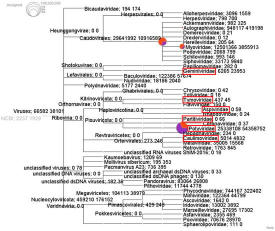
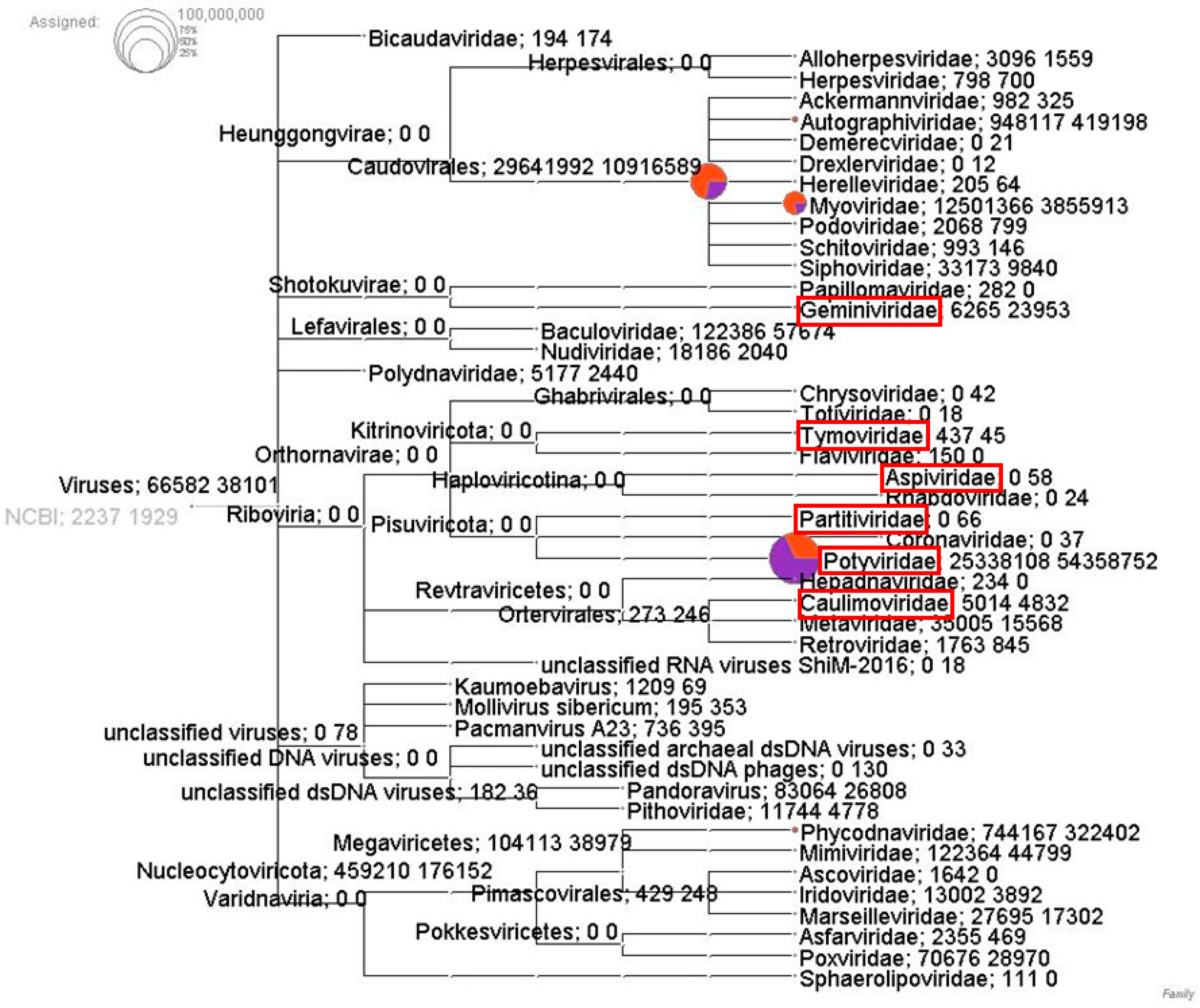
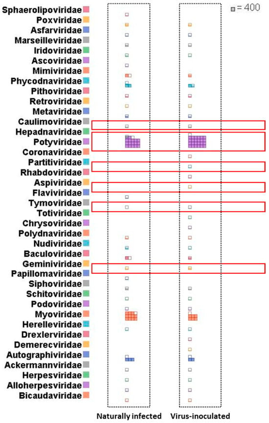
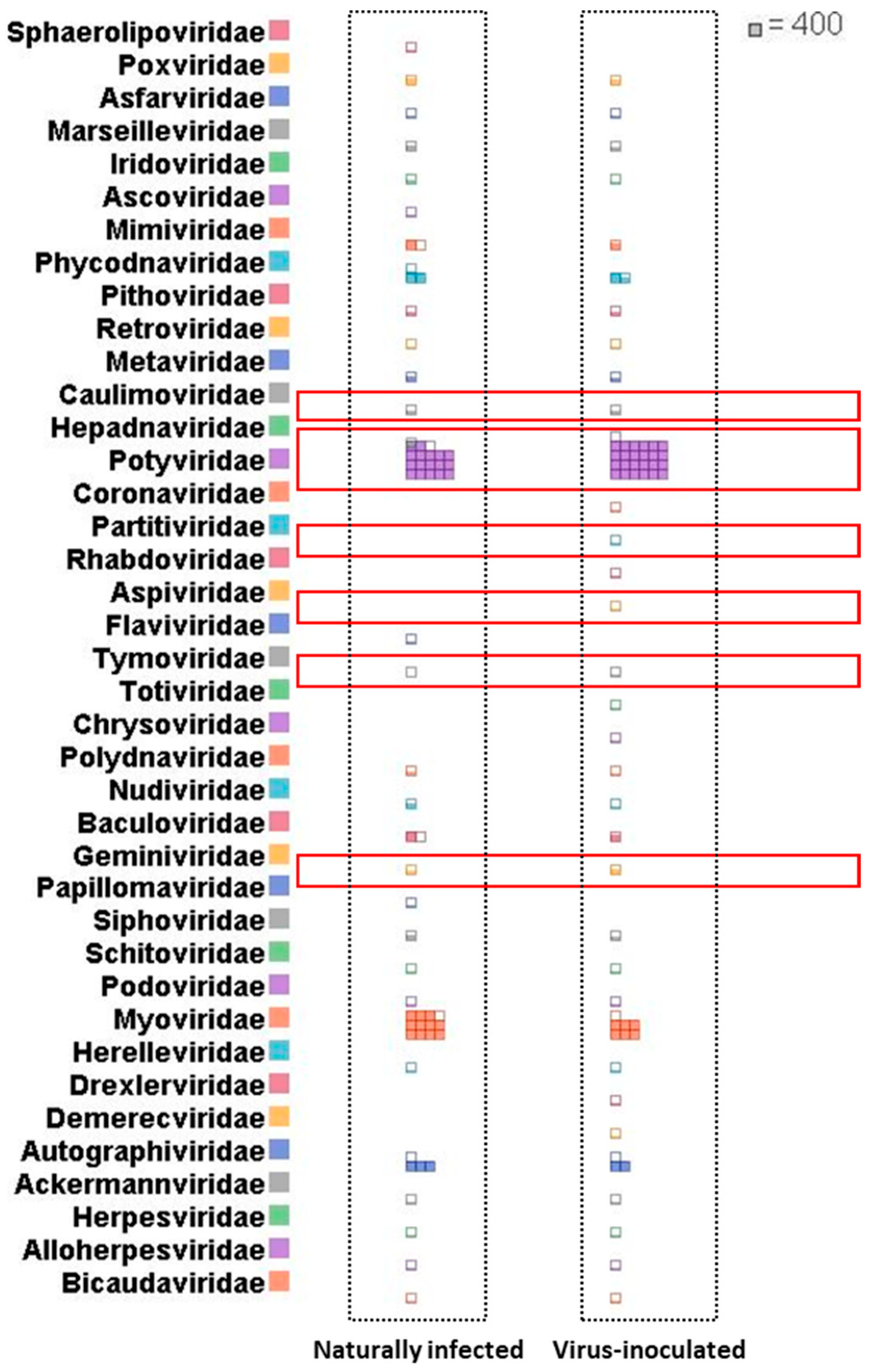
HTS combined with the DIAMOND + MEGAN long-read pipeline, enabled the detection and taxonomic classification of viral sequences in both virus-inoculated and naturally infected saffron samples. The use of frameshift-aware alignment and the interval-union LCA algorithm, both specifically designed for long-read metagenomic data, facilitated taxonomic assignment, even considering the higher error rates characteristic of ONT sequencing [31,32]. Taxonomic binning indicated the presence of multiple viral families, including six families known to infect plants: Potyviridae, Geminiviridae, Caulimoviridae, Tymoviridae, Aspiviridae, and Partitiviridae (Figure 10 and Figure 11). These families include well-characterized plant-infecting viruses, some of which appeared notably abundant in the analyzed libraries. A complete list of plant-infecting viruses detected in the dataset is provided in Supplementary Table S5. For each virus, the table reports the normalized aligned base counts in naturally infected and virus-inoculated conditions and the derived relative viral abundance, allowing depth-independent comparison of virome composition across conditions. In total, 16.6 million and 30.1 million bases were assigned to plant viruses in the naturally infected and virus-inoculated samples, respectively.
Figure 10.
Comparative taxonomic distribution of viral sequences between naturally infected (orange) and virus-inoculated (purple) conditions using DIAMOND + MEGAN long-read pipeline. Long reads were aligned against the NCBI-nr virus protein database using DIAMOND (frame-shift-aware mode), and taxonomic binning was performed in MEGAN using the interval-union LCA algorithm (percent to cover = 51%). Circle size is proportional to the number of aligned bases assigned to each taxon, and values next to nodes indicate counts for assigned bases of naturally infected and virus-inoculated plants, respectively. Comparative analysis was performed using normalized counts to correct differences between samples. Plant-infecting viral families of interest (Geminiviridae, Tymoviridae, Aspiviridae, Partitiviridae, Potyviridae, Caulimoviridae) are highlighted in red boxes. Visualization was generated from meganized DAA files in MEGAN 6.
Figure 11.
Brick chart comparing the relative abundance of viral families between naturally infected and virus-inoculated conditions using the DIAMOND + MEGAN long-read pipeline. Each row corresponds to a viral family classified according to the NCBI taxonomy. Columns group samples by condition: left, naturally infected; right, virus-inoculated. Colored tiles represent normalized aligned base counts (aligned bases normalized to sequencing depth) contributed by individual samples to each family. Larger, denser blocks indicate higher and more consistent representation within a condition. Data were obtained from meganized DAA files after DIAMOND frameshift-aware alignment and MEGAN interval-union LCA binning (percent to cover = 51%). Viral families highlighted in red boxes (Geminiviridae, Tymoviridae, Aspiviridae, Partitiviridae, Potyviridae, Caulimoviridae) correspond to plant-infecting viruses of interest. Visualization was generated from meganized DAA files using MEGAN 6.
The taxonomic tree (Figure 10) shows the hierarchical distribution of viral families, with values next to each node indicating the number of assigned bases for naturally infected and virus-inoculated samples, respectively. The brick chart (Figure 11) summarizes each sample’s normalized contribution to each family, showing differences in relative abundance across conditions. Overall, Potyviridae appeared as the most represented family in both datasets, with virus-inoculated plants exhibiting higher abundance for several potyviruses. Geminiviridae was also detected in both conditions with prominent signals such as wheat dwarf virus (WDV; Mastrevirus hordei), while virus-inoculated plants showed higher representation of several mastreviruses. Tymoviridae appeared more abundant in naturally infected plants but was present in both conditions, as was Caulimoviridae, which did not show a clear enrichment pattern though it displayed greater taxonomic diversity in the virus-inoculated sample. In contrast, Partitiviridae and Aspiviridae were detected exclusively in the virus-inoculated dataset.
In addition to saffron latent virus (SaLV; Potyvirus croci) and turnip mosaic virus (TuMV; Potyvirus rapae), which have been previously reported as saffron pathogens in Iran, metagenomic analysis indicated the presence of several additional potyviruses not previously documented infecting saffron in both virus-inoculated and naturally infected samples. These newly detected viruses included bean yellow mosaic virus (BYMV; Potyvirus phaseoluteum), mediterranean ruda virus (MeRV; Potyvirus rutae), clover yellow vein virus (CYVV; Potyvirus trifolii), lily mottle virus (LMoV; Potyvirus lilimaculae), paris virus 1 (ParV1; Potyvirus paris), tulip breaking virus (TBV; Potyvirus tulipadefractum) and Hubei Poty-like virus 1 (unclassified Potyviridae). These detections expand the currently known virome associated with saffron and suggest a more complex viral diversity than previously reported in saffron plants.
In virus-inoculated samples, several high-abundance plant viruses were detected as potentially playing a major role in viral dynamics. These included wheat dwarf virus (WDV; Mastrevirus hordei) that belonged to the Geminiviridae family, and a wide range of potyviruses typically associated with crops such as amaranth, asparagus, carrot, cucurbits, lettuce, lily, papaya, plum, potato, sweet potato, yam, and zucchini.
To complement protein-level taxonomic assignments with genome-level evidence, confirmatory read mapping was performed (Table S6). This approach supported the presence of SaLV which showed complete genome coverage (100%) and high depth (8628×) in the virus-inoculated sample, and similarly high coverage (100%, 1406×) in the naturally infected sample. Three additional potyviruses were also supported with high coverage: BYMV (93.8%, 767×), CYVV (93.5%, 378×), and MeRV (82.2%, 169×). Other viruses detected by DIAMOND + MEGAN, including Hubei Poty-like virus 1 and Paris virus 1, showed partial coverage (21.7% and 25.5%, respectively). In contrast, wheat dwarf virus, turnip mosaic virus, tulip breaking virus, and lily mottle virus showed minimal or no genome coverage, suggesting that they may represent detections based on conserved regions of virus proteins.
Additionally, a notable number of reads corresponded to bacteriophages, most prominently Prochlorococcus phage P-TIM68 and Cyanophage S-RIM32. Although these are not plant pathogens, they may reflect the associated bacterial microbiota. As they are not part of the saffron virome, they were excluded from further analysis (Figure S4).
Despite the negative pre-screening of plants prior to virus inoculation, the combined HTS and mapping approach indicated the presence of multiple co-infections in both conditions, underscoring the complexity of the saffron-associated virome.
3.4. Validation of Viruses Detected by HTS
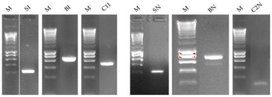
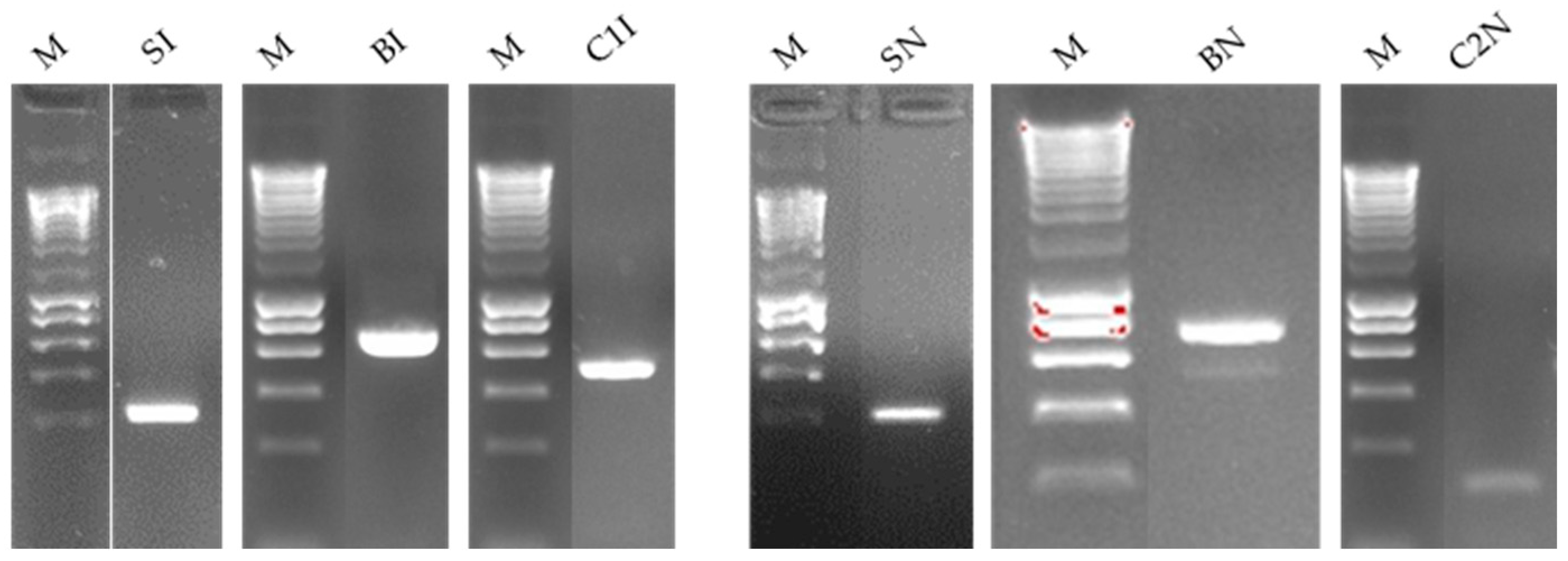
To confirm the presence of viruses detected through HTS, RT-PCR were performed using virus-specific primers. The viruses successfully validated were saffron latent virus (SaLV; Potyvirus croci), bean yellow mosaic virus (BYMV; Potyvirus phaseoluteum), and clover yellow vein virus (CYVV; Potyvirus trifolii). Amplification of expected fragments for each virus confirmed their presence in the biological samples (Figure 12), reinforcing the accuracy of the HTS-based identifications.
Figure 12.
RT-PCR validation of viruses identified through metagenomic analysis. Specific primer pairs were used to amplify fragments of SaLV (S: 233bp), BYMV (B: 614 bp), and CYVV (C1: 491 bp and C2: 177 bp). Sample origin: I, virus-inoculated; N, naturally infected; SI, SaLV detected in virus-inoculated sample; BI, BYMV detected in virus-inoculated sample; C1I, CYVV detected in virus-inoculated samples; SN, SaLV detected in naturally infected sample; BN, BYMV detected in naturally infected sample; C2N, CYVV detected in naturally infected samples; M, NZYDNA Ladder III (NZYTech, Portugal).
4. Discussion
This study provides a comprehensive characterization of the saffron virome using Oxford Nanopore sequencing, complemented by an exploratory transcriptomic overview of two distinct infection backgrounds: experimental CMV/TuMV inoculation and naturally occurring mixed infections. The metagenomic analysis suggested the presence of multiple plant-infecting viral families in saffron, indicating that mixed infections represent a common scenario in this crop. The RNA-seq analysis pointed to enriched pathways in each dataset that appear to be consistent with known antiviral responses to CMV and TuMV infections, as well as those described under naturally occurring mixed viral infections.
Several methodological aspects should be considered when interpreting the transcriptomic results. First, the study design lacked biological replicates, as samples from each condition were pooled prior to library preparation. Second, the absence of a virus-free healthy control must be acknowledged, as initial pre-inoculation screening in the virus-inoculated group did not exclude latent or background infections that were later detected through metagenomic analysis. Third, because the virome composition differed between the two groups, the observed expression differences cannot be attributed to individual viruses, but, rather, reflect broader infection backgrounds.
While the absence of biological replicates is not ideal for differential expression analysis and results must be interpreted with caution, pooling strategies can provide reasonable estimates of gene expression, particularly for genes with moderate-to-high expression levels [33]. Although this approach prevents the estimation of within-group biological variability, each sample represented a pool of three independent RNA extractions obtained from leaves of different plants, thus incorporating some degree of biological diversity within each condition. To mitigate analytical limitations, differential expression analysis was performed using the NOISeq-sim algorithm, which is specifically designed for datasets lacking replicates. Although NOISeq-sim offers an approximation rather than a substitute for true biological replication, the genes identified as differentially expressed likely reflect those exhibiting the most pronounced expression differences between conditions [26]. Similar strategies have been applied in other transcriptomic studies facing comparable limitations [34,35,36,37,38]. Furthermore, validation of selected DEGs by qRT-PCR on individual biological replicates (non-pooled material), provided independent indications consistent with the expression patterns observed. Overall, although the transcriptomic findings should be viewed as descriptive and exploratory rather than definitive mechanistic conclusions, the analysis provides candidate genes and pathways which can be used for future functional studies.
4.1. Different Defense Strategies in Virus-Inoculated and Naturally Infected Plants
The transcriptomic analysis of saffron plants suggests that both virus-inoculated and naturally infected groups display transcriptional changes compatible with defense-related responses, although showing patterns differing between conditions. In virus-inoculated plants, the expression profiles appear to be consistent with a more targeted antiviral response. Upregulated genes included RNA silencing components such as RNA helicases and SGS3, which are known to play a central role in targeting and degrading viral RNA to restrict infection [39]. Transcripts associated with autophagy and ubiquitin-mediated proteolysis pathways, including E3 ubiquitin ligases KEG and UPL3, also showed increased abundance, potentially indicating the activation of degradation routes for viral components [40,41]. The modulation of systemic defense markers such as CDR1-like may further suggest the involvement of regulated immune signaling pathways in response to viral challenge [42]. In contrast, the patterns observed in naturally infected saffron plants may reflect a broader and less virus-specific response. Rather than showing activation of canonical antiviral pathways, these plants exhibited increased expression of genes involved in photosynthesis, carbon fixation, and primary metabolism, processes that can support energy production and maintain physiological functions under multifactorial stress [43]. These metabolic adjustments may represent a more generalized adaptive strategy, rather than a response oriented toward a single pathogen.
Differences between the two plant types were also evident at the signaling level. In virus-inoculated plants, upregulated genes included ion channels regulated by cyclic nucleotides and serine/threonine kinases, such as D6PKL1 and PBL19, which have roles in calcium-dependent signaling and downstream defense activation [44,45]. These signaling cascades could contribute to more localized or directed immune responses, including forms of programmed cell death associated with antiviral defense [46]. In contrast, naturally infected plants appeared to modulate more general routes such as MAPK signaling and glutathione metabolism, which are commonly linked to stress tolerance and protein homeostasis [47]. The absence of transcriptional markers typically associated with hypersensitive response in this group is in line with a response that may prioritize tolerance over rapid pathogen elimination [48].
From a metabolic perspective, virus-inoculated plants showed expression patterns that might indicate increased activity in carbohydrate-related pathways, including glycolysis and starch/sucrose metabolism, possibly supporting the energetic demands associated with defense activation [19]. Naturally infected plants, by contrast, maintained expression profiles more consistent with sustaining primary metabolic processes.
Overall, these observations point to the possibility of distinct molecular strategies in saffron plants facing viral infection. Virus-inoculated plants exhibited transcriptional patterns compatible with a targeted and highly coordinated antiviral program integrating specialized defense mechanisms, whereas naturally infected plants showed broader, more generalized strategies that may reflect metabolic adjustment and adaptation to a complex infection context. While these interpretations remain exploratory, they provide a useful framework for guiding future functional studies aimed at improving stress resilience in saffron.
4.2. Virus Identification
Saffron production faces significant threats from potyviruses, which have been reported as major viral pathogens affecting global cultivation [49]. Due to saffron’s sterile, triploid nature, its exclusive vegetative propagation through corms produces only a limited number of new planting materials per season [49]. This reliance on corm multiplication complicates virus management because viruses can persist in dormant corm tissues, becoming a key source of infection in new crops [50]. The high prevalence of viral infections negatively impacts saffron’s yield, quality, and plant physiology [18,19].
To address these challenges and enhance sustainability, modern agricultural practices must prioritize the production of virus-free corms. Tissue culture techniques, including meristem and shoot-tip culture combined with thermotherapy or chemotherapy, offer effective means for mass-propagating pathogen-free corms and improving genetic traits [50]. This approach is particularly vital in regions like Iran, where mixed viral infections are common in saffron corms, making virus-free plant production essential for maintaining crop health and quality.
In this study, we conducted multiple field visits and collected saffron samples from Torbat-e Heydarieh County, recognized as Iran’s largest saffron cultivation region and one of its oldest major growing areas. Torbat-e Heydarieh County represents Iran’s largest saffron cultivation region, making these findings particularly significant for national production. Samples displaying diverse symptoms were obtained from concentrated locations within this county for virome analysis using MinION Nanopore sequencing. The sequencing indicated the presence of six plant-infecting viral families in the analyzed saffron samples: Potyviridae, Geminiviridae, Caulimoviridae, Tymoviridae, Aspiviridae, and Partitiviridae. Potyviruses appeared to be the most prevalent detected group, including SaLV, TuMV, BYMV, MeRV, and CYVV. These observations are consistent with previous reports identifying potyviruses as major viral agents affecting saffron cultivation in Iran and other growing regions [51,52,53].
The identification and characterization of saffron viruses have been previously approached using diverse methodologies [51,54,55,56]. Several potyviruses have been identified in Iran, including saffron latent virus (SaLV; Potyvirus croci), turnip mosaic virus (TuMV; Potyvirus rapae), and saffron yellow mosaic virus (SYMV; Potyvirus crocitessellati), highlighting the prevalence of this viral genus within the Potyviridae family, the largest group of plant-infecting RNA viruses [51,54,55]. A recent study in Spain identified 17 viruses, including seven novel ones, in Crocus sativus cultivars using RNA-sequencing [57], highlighting the relevance of RNA-seq and bioinformatics tools for virome characterization.
In this study, Nanopore sequencing did not detect CMV and Tospovirus in naturally infected samples, although these viruses were confirmed by RT-PCR. This outcome aligns with previous reports indicating that some sequencing approaches, including cDNA-PCR from rRNA-depleted RNA, may fail to detect certain viruses despite showing good overall performance [2]. The persistence of viruses in dormant corms compromises yield, morphological traits, physiological functions, and spice quality, emphasizing the need for the refinement of phytosanitary practices [18,19]. These results suggest the importance of refining methodological approaches and continuing virome research to better assess how viral infections influence saffron quality, yield, and overall crop performance. Such efforts can contribute to the development of more sustainable cultivation systems and improved phytosanitary practices. Ultimately, a thorough understanding of the saffron virome is essential for improving phytosanitary management and ensuring the long-term sustainability of saffron cultivation against viral challenges.
5. Conclusions
This study employing Oxford Nanopore Technologies (ONT) sequencing provides an initial framework for understanding the virome of Crocus sativus (saffron) and offers insight into complex host–pathogen interactions. The detection of six plant-infecting viral families in both conditions, including viruses not previously reported in Iranian saffron, underscores the viral diversity affecting this crop. The exploratory differential expression analysis generated an overview of transcriptional responses that may be associated with antiviral defense processes. Pathway-level patterns, including those involving serine/threonine-protein kinases and transcription factors, appear to be consistent with the activation of key signaling networks potentially engaged during viral infection. Although the interpretation presented here remains preliminary due to the study’s design constraints, the results contribute to the growing body of molecular knowledge of viral diversity in agricultural ecosystems and offer initial insights that may support the development of targeted viral-resistance strategies in future saffron crop improvement initiatives. Future work should implement factorial inoculation designs with virus-free controls to enable mechanistic interpretation.
Supplementary Materials
The following supporting information can be downloaded at: https://www.mdpi.com/article/10.3390/agronomy16080803/s1, Figure S1: Evaluation of the quality of total RNA samples using agarose gel analysis, after removal of plant host ribosomal RNA. I: Virus-inoculated saffron sample; N: Naturally infected saffron sample; M: NZYDNA Ladder III (NZYTech, Portugal); Figure S2: Summarized GO Enrichment results in naturally infected plants (Revigo); Figure S3: Summarized GO Enrichment results in virus-inoculated plants (Revigo); Figure S4: Bacteriophages clade; Table S1: Primer sequences utilized for the detection of the virus within saffron samples and for internal control; Table S2: Primer sequences for target and endogenous genes used in RT-qPCR for validate RNA-seq results; Table S3: Primer sequences to confirmatory test of the HTS results using RT-PCR; Table S4: Thermal cycling conditions to confirmatory test of the HTS results using RT-PCR; Table S5: Plant-infecting viruses detected by DIAMOND + MEGAN (long-read mode) in virus-inoculated (I) and naturally infected (NI) samples. NABC: normalized aligned-base counts; RA = relative abundance within the set of plant-infecting viruses detected in each sample (excludes non-plant viruses). Totals refer to the sum of NABC assigned to plant-infecting viruses in NI and I samples; Table S6: Genome-level validation by read mapping (minimap2; settings -k11 -w1) in virus-inoculated (I) and naturally infected (NI) samples of Saffron latent virus (SaLV), Bean yellow mosaic virus (BYMV), Clover yellow vein virus (CYVV), Mediterranean ruda virus (MeRV), Paris virus 1 (ParV1), Hubei Poty-like virus 1 (HPLV), Wheat dwarf virus (WDV), Turnip mosaic virus (TuMV), Tulip breaking virus (TBV) and Lily mottle virus (LMoV). Candidate reads correspond to the number of long reads exported after DIAMOND + MEGAN assignment for each virus. Mean depth is the average per-nucleotide depth across the viral reference, and Genome ≥1×/≥5× as the percentage of reference positions covered by at least 1 or 5 reads, respectively.
Author Contributions
Conceptualization, C.S., S.R., J.M.V. and M.S.; methodology, S.R. and M.S.; software, S.R. and J.M.V.; validation, S.R. and J.M.V.; formal analysis, S.R.; investigation, M.S. and S.R.; resources, C.S. and M.S.-B.; data curation, S.R. and M.S.; writing—original draft preparation, M.S.; writing—review and editing, C.S. and S.R.; visualization, S.R. and M.S.; supervision, C.S. and M.S.-B.; funding acquisition, C.S. and N.V. All authors have read and agreed to the published version of the manuscript.
Funding
This research was partially funded by the Xunta de Galicia through project number IN607A 2021/06 and Tarbiat Modares University, Tehran, Iran.
Data Availability Statement
The raw data supporting the conclusions of this article will be made available by the authors on request.
Acknowledgments
The authors want to thank Carmen Fuentes González and Bruno Fernández Moreira for their valuable assistance in the validation of the HTS results during their internship at MBG-CSIC. We gratefully acknowledge the computational support provided by CESGA (Galician Supercomputing Center).
Conflicts of Interest
The authors declare no conflicts of interest.
References
- Jones, R.A.C.; Naidu, R.A. Global dimensions of plant virus diseases: Current status and future perspectives. Annu. Rev. Virol. 2019, 6, 387–409. [Google Scholar] [CrossRef] [Scilit] [PubMed]
- Pecman, A.; Adams, I.; Gutiérrez-Aguirre, I.; Fox, A.; Boonham, N.; Ravnikar, M.; Kutnjak, D. Systematic Comparison of Nanopore and Illumina Sequencing for the Detection of Plant Viruses and Viroids Using Total RNA Sequencing Approach. Front. Microbiol. 2022, 13, 883921. [Google Scholar] [CrossRef] [Scilit]
- Inoue-Nagata, A.K.; Jordan, R.; Kreuze, J.; Li, F.; Lopez-Moya, J.J.; Makinen, K.; Ohshima, K.; Wylie, S.J.; Ictv Report Consortium. ICTV virus taxonomy profile: Potyviridae 2022. J. Gen. Virol. 2022, 103, 001738. [Google Scholar] [CrossRef] [Scilit] [PubMed]
- Sun, K.; Liu, Y.; Zhou, X.; Yin, C.; Zhang, P.; Yang, Q.; Mao, L.; Shentu, X.; Yu, X. Nanopore sequencing technology and its application in plant virus diagnostics. Front. Microbiol. 2022, 13, 939666. [Google Scholar] [CrossRef] [Scilit]
- Pretorius, L.S.; Hussein, N.; Al-Amery, A.; Moyle, R.L.; Schenk, P.M. Natural and engineered defenses against plant viruses. Curr. Biotechnol. 2017, 6, 339–348. [Google Scholar] [CrossRef] [Scilit]
- Prasad, A.; Sharma, N.; Muthamilarasan, M.; Rana, S.; Prasad, M. Recent advances in small RNA mediated plant-virus interactions. Crit. Rev. Biotechnol. 2019, 39, 587–601. [Google Scholar] [CrossRef] [Scilit] [PubMed]
- Ijaz, U.; Zhao, C.; Shabala, S.; Zhou, M. Molecular basis of plant–pathogen interactions in the agricultural context. Biology 2024, 13, 421. [Google Scholar] [CrossRef] [Scilit]
- Rithesh, L. Defense mechanisms and disease resistance in plant-pathogen interactions. Plant Sci. Arch. 2020, 5, 1–4. [Google Scholar] [CrossRef] [Scilit]
- Deng, Z.; Ma, L.; Zhang, P.; Zhu, H. Small RNAs participate in plant–virus interaction and their application in plant viral defense. Int. J. Mol. Sci. 2022, 23, 696. [Google Scholar] [CrossRef] [Scilit]
- Liu, J.Z.; Li, F.; Liu, Y. Editorial: Plant immunity against viruses. Front. Microbiol. 2017, 8, 520. [Google Scholar] [CrossRef] [Scilit]
- Choi, D.; Rai, M.; Rai, A.; Yamazaki, M.; Hahn, Y. Discovery of two novel potyvirus genome sequences by high-throughput RNA sequencing in Aconitum carmichaelii tissue samples. Acta Virol. 2023, 67, 11782. [Google Scholar] [CrossRef] [Scilit]
- Bronzato Badial, A.; Sherman, D.; Stone, A.; Gopakumar, A.; Wilson, V.; Schneider, W.; King, J. Nanopore sequencing as a surveillance tool for plant pathogens in plant and insect tissues. Plant Dis. 2018, 102, 1648–1652. [Google Scholar] [CrossRef] [Scilit] [PubMed]
- Boykin, L.M.; Sseruwagi, P.; Alicai, T.; Ateka, E.; Mohammed, I.U.; Stanton, J.-A.L.; Kayuki, C.; Mark, D.; Fute, T.; Erasto, J.; et al. Tree lab: Portable genomics for early detection of plant viruses and pests in sub-Saharan Africa. Genes 2019, 10, 632. [Google Scholar] [CrossRef] [Scilit] [PubMed]
- Leiva Ana, M.; Siriwan, W.; Lopez-Alvarez, D.; Barrantes, I.; Hemniam, N.; Saokham, K.; Cuellar, W.J. Nanopore-based complete genome sequence of a Sri Lankan cassava mosaic virus (Geminivirus) strain from Thailand. Microbiol. Resour. Announc. 2020, 9, e01274-19. [Google Scholar] [CrossRef] [Scilit]
- Filloux, D.; Fernandez, E.; Loire, E.; Claude, L.; Galzi, S.; Candresse, T.; Winter, S.; Jeeva, M.L.T.; Makeshkumar, T.; Martin, D.P.; et al. Nanopore-based detection and characterization of yam viruses. Sci. Rep. 2018, 8, 17879. [Google Scholar] [CrossRef] [Scilit]
- Naito, F.Y.B.; Melo, F.L.; Fonseca, M.E.N.; Santos, C.A.F.; Chanes, C.R.; Ribeiro, B.M.; Gilbertson, R.L.; Boiteux, L.S.; Pereira-Carvalho, R.C. Nanopore sequencing of a novel bipartite New World begomovirus infecting cowpea. Arch. Virol. 2019, 164, 1907–1910. [Google Scholar] [CrossRef] [Scilit]
- Pandita, D. Saffron (Crocus sativus L.): Phytochemistry, therapeutic significance and omics-based biology. In Medicinal and Aromatic Plants: Expanding Their Horizons Through Omics; Elsevier Inc.: Amsterdam, The Netherlands, 2021; pp. 325–396. [Google Scholar] [CrossRef] [Scilit]
- Shamshiri, M.; Sánchez, C.; Rico, S.; Mokhtassi-Bidgoli, A.; Ayyari, M.; Rezadoost, H.; Shams-Bakhsh, M. Molecular, metabolic, and physiological responses to progressive biotic stress caused by cucumber mosaic virus and turnip mosaic virus in saffron. Horticulturae 2025, 11, 96. [Google Scholar] [CrossRef] [Scilit]
- Parizad, S.; Dizadji, A.; Habibi, M.K.; Winter, S.; Kalantari, S.; Movi, S.; Lorenzo Tendero, C.; Alonso, G.L.; Moratalla-Lopez, N. The effects of geographical origin and virus infection on the saffron (Crocus sativus L.) quality. Food Chem. 2019, 295, 387–394. [Google Scholar] [CrossRef] [Scilit]
- Huang, K.S.; Li, S.L.; Sun, J.H.; Wang, Y.C.; Jan, F.J.; Chen, T.C. Development of a generic method for inspection of Tospovirus es. Eur. J. Plant Pathol. 2018, 150, 457–469. [Google Scholar] [CrossRef] [Scilit]
- Asghari, N.; Koolivand, D.; Eini, O. Differential expression of HSP90 and AGO genes in tomato infected by Cucumber mosaic virus. Crop Biotechnol. 2020, 10, 31–42. [Google Scholar] [CrossRef]
- Sánchez, F.; Wang, X.; Jenner, C.E.; Walsh, J.A.; Ponz, F. Strains of Turnip mosaic potyvirus as defined by the molecular analysis of the coat protein gene of the virus. Virus Res. 2003, 94, 33–43. [Google Scholar] [CrossRef] [Scilit] [PubMed]
- Liefting, L.W.; Waite, D.W.; Thompson, J.R. Application of Oxford Nanopore Technology to Plant Virus Detection. Viruses 2021, 13, 1424. [Google Scholar] [CrossRef] [Scilit]
- De Coster, W.; Rademakers, R. NanoPack2: Population-scale evaluation of long-read sequencing data. Bioinformatics 2023, 39, btad311. [Google Scholar] [CrossRef] [Scilit]
- Li, H. Minimap2: Pairwise alignment for nucleotide sequences. Bioinformatics 2018, 34, 3094–3100. [Google Scholar] [CrossRef] [Scilit]
- Tarazona, S.; García-Alcalde, F.; Dopazo, J.; Ferrer, A.; Conesa, A. Differential expression in RNA-seq: A matter of depth. Genome Res. 2011, 21, 2213–2223. [Google Scholar] [CrossRef] [Scilit] [PubMed] [PubMed Central]
- Ge, S.X.; Jung, D.; Yao, R. ShinyGO: A graphical gene-set enrichment tool for animals and plants. Bioinformatics 2020, 36, 2628–2629. [Google Scholar] [CrossRef] [Scilit]
- Supek, F.; Bošnjak, M.; Škunca, N.; Šmuc, T. REVIGO summarizes and visualizes long lists of Gene Ontology terms. PLoS ONE 2011, 6, e21800. [Google Scholar] [CrossRef] [Scilit]
- Xie, C.; Mao, X.; Huang, J.; Ding, Y.; Wu, J.; Dong, S.; Kong, L.; Gao, G.; Li, C.-Y.; Wei, L. KOBAS 2.0: A web server for annotation and identification of enriched pathways and diseases. Nucleic Acids Res. 2011, 39, 316–322. [Google Scholar] [CrossRef] [Scilit] [PubMed]
- Schmittgen, T.D.; Livak, K.J. Analyzing real-time PCR data by the comparative CT method. Nat. Protoc. 2008, 3, 1101–1108. [Google Scholar] [CrossRef] [Scilit]
- Bağcı, C.; Patz, S.; Huson, D.H. DIAMOND+MEGAN: Fast and Easy Taxonomic and Functional Analysis of Short and Long Microbiome Sequences. Curr. Protoc. 2021, 1, e59. [Google Scholar] [CrossRef] [Scilit] [PubMed]
- Gautam, A.; Felderhoff, H.; Bağci, C.; Huson Daniel, H. Using AnnoTree to get more assignments, faster, in DIAMOND+MEGAN microbiome analysis. mSystems 2022, 7, e01408–e01421. [Google Scholar] [CrossRef] [Scilit]
- Konczal, M.; Koteja, P.; Stuglik, M.T.; Radwan, J.; Babik, W. Accuracy of allele frequency estimation using pooled RNA-Seq. Mol. Ecol. Resour. 2014, 14, 381–392. [Google Scholar] [CrossRef] [Scilit] [PubMed]
- Pauletto, M.; Milan, M.; Moreira, R.; Novoa, B.; Figueras, A.; Babbucci, M.; Patarnello, T.; Bargelloni, L. Deep transcriptome sequencing of Pecten maximus hemocytes: A genomic resource for bivalve immunology. Fish Shellfish Immunol. 2014, 37, 154–165. [Google Scholar] [CrossRef] [Scilit] [PubMed]
- Behringer, D.; Zimmermann, H.; Ziegenhagen, B.; Liepelt, S. Differential Gene Expression Reveals Candidate Genes for Drought Stress Response in Abies alba (Pinaceae). PLoS ONE 2015, 10, e0124564. [Google Scholar] [CrossRef] [Scilit] [PubMed] [PubMed Central]
- Pinosio, S.; González-Martínez, S.C.; Bagnoli, F.; Cattonaro, F.; Grivet, D.; Marroni, F.; Lorenzo, Z.; Pausas, J.G.; Verdú, M.; Vendramin, G.G. First insights into the transcriptome and development of new genomic tools of a widespread circum-Mediterranean tree species, Pinus halepensis Mill. Mol. Ecol. Resour. 2014, 14, 846–856. [Google Scholar] [CrossRef] [Scilit] [PubMed]
- Asmus, J.J.; Naidoo-Blassopels, R.K.; Pérez-Torrado, R.; Bauer, F.F. Investigating the transcriptional fingerprints of cocultured Saccharomyces cerevisiae and Lachancea thermotolerans in a model wine environment. Front. Microbiol. 2026, 16, 1720597. [Google Scholar] [CrossRef] [Scilit] [PubMed]
- Zhao, X.; Yu, H.; Kong, L.; Liu, S.; Li, Q. Comparative transcriptome analysis of two oysters, Crassostrea gigas and Crassostrea hongkongensis provides insights into adaptation to hypo-osmotic conditions. PLoS ONE 2014, 9, e111915, Erratum in PLoS ONE 2015, 10, e0118665. https://doi.org/10.1371/journal.pone.0118665. [Google Scholar] [CrossRef] [Scilit] [PubMed] [PubMed Central]
- Li, F.; Zhang, C.; Tang, Z.; Zhang, L.; Dai, Z.; Lyu, S.; Li, Y.; Hou, X.; Bernards, M.; Wang, A. A plant RNA virus activates selective autophagy in a UPR-dependent manner to promote virus infection. New Phytol. 2020, 227, 806–822. [Google Scholar] [CrossRef] [Scilit]
- Hafrén, A.; Üstün, S.; Hochmuth, A.; Svenning, S.; Johansen, T.; Hofius, D. Turnip mosaic virus counteracts selective autophagy of the viral silencing suppressor HCpro. Plant Physiol. 2017, 176, 649–662. [Google Scholar] [CrossRef] [Scilit]
- Yin, Z.; Popelka, H.; Lei, Y.; Yang, Y.; Klionsky, D.J. The Roles of Ubiquitin in Mediating Autophagy. Cells 2020, 9, 2025. [Google Scholar] [CrossRef] [Scilit]
- Dietrich, P.; Moeder, W.; Yoshiokab, K. Plant Cyclic Nucleotide-Gated Channels: New Insights on Their Functions and Regulation. Plant Physiol. 2020, 184, 27–38. [Google Scholar] [CrossRef] [Scilit]
- Zhang, H.; Zhao, Y.; Zhu, J.-K. Thriving under Stress: How Plants Balance Growth and the Stress Response 2020, Thriving under Stress: How Plants Balance. Dev. Cell 2020, 55, 529. [Google Scholar] [CrossRef] [Scilit]
- Xu, T.; Niu, J.; Jiang, Z. Sensing Mechanisms: Calcium Signaling Mediated Abiotic Stress in Plants. Front. Plant Sci. 2022, 13, 925863. [Google Scholar] [CrossRef] [Scilit]
- Li, Y.; Huang, X.; Gong, B.Q.; Yu, Y.; Li, J.F.; Xue, J. CERK1-mediated phosphorylation of PBL19 is crucial for antifungaldefense in plants. Planta 2025, 261, 56. [Google Scholar] [CrossRef] [Scilit] [PubMed]
- Piau, M.; Schmitt-Keichinger, C. The Hypersensitive Response to Plant Viruses. Viruses 2023, 15, 2000. [Google Scholar] [CrossRef] [Scilit] [PubMed]
- Boro, P.; Chattopadhyay, S. Crosstalk between MAPKs and GSH under stress: A critical review. J. Biosci. 2022, 47, 71. [Google Scholar] [CrossRef] [Scilit]
- Salguero-Linares, J.; Coll, N.S. Cell death as a defense strategy against pathogens in plants and animals. PLoS Pathog. 2023, 19, e1011253. [Google Scholar] [CrossRef] [Scilit] [PubMed]
- Schmidt, T.; Heitkam, T.; Liedtke, S.; Schubert, V.; Menzel, G. Adding color to a century-old enigma: Multi-color chromosome identification unravels the autotriploid nature of saffron (Crocus sativus) as a hybrid of wild Crocus cartwrightianus cytotypes. New Phytol. 2019, 222, 1965–1980. [Google Scholar] [CrossRef] [Scilit]
- Moshtaghi, N. Tissue and cell culture of saffron. In Saffron; Elsevier: Amsterdam, The Netherlands, 2020; pp. 229–246. [Google Scholar] [CrossRef] [Scilit]
- Tavoosi, M.; Moradi, Z.; Mehrvar, M. First report of Turnip mosaic virus infecting saffron in Iran. Virus Dis. 2022, 33, 489–491. [Google Scholar] [CrossRef] [Scilit]
- Valouzi, H.; Dizadji, A.; Golnaraghi, A.; Salami, S.A.; Fontdevila Pareta, N.; Önder, S.; Selmi, I.; Rollin, J.; Berhal, C.; Tamisier, L.; et al. A Virome Scanning of Saffron (Crocus sativus L.) at the National Scale in Iran Using High-Throughput Sequencing Technologies. Viruses 2025, 17, 1079. [Google Scholar] [CrossRef] [Scilit]
- Shanmugam, K.; Perumal, R.; Oliva, R.; Cheng, H.W.; Iruthayasamy, J.; Angappan, S.; Chandrasekaran, I.R.; Nallusamy, S. Seed transmission of potyviruses: A threat to crop health. Not. Bot. Horti Agrobot. 2024, 52, 13698. [Google Scholar] [CrossRef] [Scilit]
- Parizad, S.; Dizadji, A.; Koohi Habibi, M.; García-Arenal, F.; Winter, S.; Ayllón, M.A.; Kalantari, S. Prevalence of saffron latent virus (SaLV), a new potyvirus species, in saffron fields of Iran. J. Plant Pathol. 2017, 99, 802. [Google Scholar] [CrossRef] [Scilit]
- Tavoosi, M.; Moradi, Z.; Mehrvar, M. Virome analysis of potyvirus populations infecting saffron in Iran: The discovery of a novel potyvirus. Eur. J. Plant Pathol. 2024, 168, 453–466. [Google Scholar] [CrossRef] [Scilit]
- Roumi, V.; Rastgou, M. In silico identification and phylogenetic analysis of two saffron infecting viruses in Iran using high throughput sequencing. J. Appl. Res. Plant Prot. 2024, 13, 259–267. [Google Scholar] [CrossRef]
- Martínez-Fajardo, C.; Navarro-Simarro, P.; Morote, L.; Rubio-Moraga, A.; Mondéjar-López, M.; Niza, E.; Argandona, J.; Ahrazem, O.; Gómez-Gómez, L.; López-Jiménez, A.J. Exploring the viral landscape of saffron through metatranscriptomic analysis. Virus Res. 2024, 345, 199389. [Google Scholar] [CrossRef] [Scilit]
Disclaimer/Publisher’s Note: The statements, opinions and data contained in all publications are solely those of the individual author(s) and contributor(s) and not of MDPI and/or the editor(s). MDPI and/or the editor(s) disclaim responsibility for any injury to people or property resulting from any ideas, methods, instructions or products referred to in the content. |
© 2026 by the authors. Licensee MDPI, Basel, Switzerland. This article is an open access article distributed under the terms and conditions of the Creative Commons Attribution (CC BY) license.