


Abstract
The antimicrobial activity of multicomponent, magnetron-sputtered glass coatings was evaluated against phytopathogenic fungi (Botrytis cinerea, Fusarium oxysporum, Cladosporium fulvum, Alternaria solani) and the chromista Phytophthora infestans, with Aspergillus fumigatus included as a model opportunistic pathogen. Fourteen Cu-based multicomponent coatings were deposited on glass using multi-alloy targets composed of Sn, Zn, Al, Ni, Fe, Ti, Mn, Nb, or Co in two high-transmittance variants (≥85% and ≥88%). Antimicrobial activity was assessed in two assays: (A) spore survival after 24–72 h contact, and (B) hyphal growth over 7 days following coating exposure under light and dark conditions. Spore viability decreased after incubation on high-Cu coatings, which showed inhibition for most strains, particularly B. cinerea, F. oxysporum, and P. infestans. The effects on spore germination were independent of the direct transmittance value of the coated glass. Hyphal growth was generally less affected by a high Cu content for most strains. Hyphal growth of F. oxysporum, C. fulvum, A. solani and B. cinerea was reduced by up to 30% on selected multicomponent coatings. For most strains, hyphal growth showed no inhibition after light incubation on coatings. However, light-dependent effects were observed for A. solani, A. fumigatus and P. infestans, while B. cinerea and C. fulvum showed reduced sensitivity during the first two days. High-Cu coatings were most effective at inhibiting spore germination, whereas hyphal growth on multicomponent coatings may respond to different ions. Therefore, high-Cu, two-component coatings may be recommended for practical greenhouse applications.
1. Introduction
Microorganisms, including filamentous fungi, cause plant diseases and are the main reason for crop losses worldwide. This has stimulated the development of innovative coatings to prevent their growth and spread. Pathogenic microorganisms are especially common in greenhouses due to the warm and humid conditions. They spread through airborne spores and persist in plant debris and soil [1]. Botrytis cinerea, the fungus that causes gray mold disease, attacks a wide range of greenhouse crops, including vegetables and fruits such as tomatoes, cucumbers, peppers, strawberries and basil, as well as most potted plants [2]. This phytopathogenic microorganism induces symptoms such as fruit rot, stem blight, and post-harvest decay. Similarly, Fusarium species are often detected in greenhouse crops, including tomatoes, cucumbers, peppers, melons, and others [3,4]. In tomatoes, the main causes of wilting and root/crown rot are Fusarium oxysporum f. sp. lycopersici and F. oxysporum f. sp. radicis-lycopersici, and outbreaks of the disease have been reported in greenhouse production systems around the world [5]. Its pathogenicity is driven by diverse virulence factors, including cell wall-degrading enzymes, effectors, and mycotoxins such trichothecenes and fumonisins, which can contaminate cereals, fruits, and vegetables. Chronic exposure to these toxins through food is a recognized as a health risk, potentially causing immunosuppression, gut microbiome imbalance, and other toxic effects in humans and animals [6]. Fusarium oxysporum is both a devastating plant pathogen and a source of foodborne risk due to its mycotoxin production [7].
Cladosporium fulvum is a specialized fungal pathogen responsible for tomato leaf mold, a disease that affects greenhouse tomato crops worldwide. It attacks leaves, which can lead to curling, wilting, and defoliation, reducing fruit yield [8,9,10]. Alternaria solani is a necrotrophic fungus that primarily attacks tomatoes and potatoes, as well as eggplants and other plants belonging to the Solanaceae family [11]. Infection manifests as characteristic dark brown or black spots with concentric rings on the leaves, leading to reduced photosynthesis and significant crop losses of up to 78% for tomatoes and 50% for potatoes [1,12]. Phytophthora infestans belongs to the Chromista kingdom (Oomycota phylum/Oomycetes class) and is a phytopathogen that attacks tomatoes and other greenhouse-grown plants. It causes late blight in greenhouse tomato crops and attacks other nightshade plants, including some ornamental varieties. The sporangia of this pathogen are highly susceptible to disintegration in response to changes in relative humidity, and can disperse through the air to other plant tissues [13,14,15].
In greenhouse and garden soils, Aspergillus fumigatus in some cases represented 35–70% of colony-forming units (CFUs) [16,17]. This fungus, which is found worldwide, is known to be both a saprophyte and an endophyte, and can benefit plants by acting as a biological agent and stimulating their growth [18,19]. However, in greenhouses, A. fumigatus primarily colonizes substrates and plant debris, releasing significant quantities of spores into the air. This can pose a serious health risk to workers, as the fungus is a major cause of invasive aspergillosis and allergic lung diseases in humans [20]. Kwon–Chung and Sugui [16] studied the composition of the aerosol cloud above a disturbed compost pile and found that most of the colonies on the agar medium were fungi of the A. fumigatus species after exposure to this environment.
Microorganisms are commonly found in greenhouse substrates, such as coconut fiber, soil, and hydroponic media [21,22]. Greenhouse structures and equipment can also be significant sources of microbiological and chemical contamination. Effective contamination control is achieved through integrated sanitation protocols, including the systematic cleaning and disinfection of all crop-contact surfaces, soil disinfection via steam treatment or solarization, the disinfection of recirculating solutions, and the sterilization of incoming air using HEPA filtration or ultraviolet (UV) irradiation [23]. As glass occupies the largest surface area among the structural elements of greenhouses, it can play a key role in controlling the microbiological contamination of greenhouse crops [24]. Antimicrobial coatings use various material strategies to prevent the growth, adhesion, and biofilm formation of pathogens on different surfaces [25,26]. Physical vapor deposition (PVD) techniques, such as magnetron sputtering (MS), are widely used to deposit thin, uniform coatings on glass, as well as on other substrates including silicon, metals, and polymers, for applications in medicine, electronics, optics, and agriculture. Magnetron sputtering can be used to produce antimicrobial glass with nanometer-scale homogeneous layers. This process involves bombarding a target material with ions and using a magnetic field to enhance electron–gas collisions and increase plasma density. This improves sputtering efficiency and enables the deposition of high-quality, uniform coatings.
It is a well-known mechanism that copper ions (Cu+/Cu2+) released from coatings can damage cell membranes, proteins, and DNA. In addition, Cu oxides (CuO/Cu2O) and titanium (Ti) oxides are formed in situ and contribute to the process of killing, sometimes in visible light through photocatalytic/semiconductor action [24]. Numerous studies have demonstrated the inhibitory and even lethal effects of Cu compounds on mycelium [27]. Chen et al. [28] described that the hyphae of the phytopathogenic Colleotrichum gloeoesporioides had an altered, swollen appearance and reduced growth in the presence of Cu nanoparticles (Cu-NPs). Coatings containing Cu and/or Ti, applied by MS, including thin-film metallic glasses, are smooth surfaces with strong antibacterial properties. Cimen et al. [29] showed that Ti-Cu and Ti–silver (Ag) coatings had antifungal activity against Candida albicans, indicating their potential usefulness in clinical applications, such as catheters and surgical implants [29]. Various studies demonstrated that sputtered coatings containing Cu may exhibit antifungal activity; however, their effectiveness depends on the strain and is related to the Cu content and coating thickness. Examples of such antifungal coatings are thin layers of tantalum (Ta) and Cu, and niobium (Nb) and Cu deposited by MS, which have shown clear antimicrobial efficacy against Candida albicans when applied to scaffolds [30].
The inclusion of such antimicrobial materials aligns with the current biotechnology priorities in crop improvement and protection, as well as the use of advanced agronomic practices in sustainable agriculture. Although numerous studies have reported on coatings with a high Cu and/or Ti content produced by magnetron sputtering, their antimicrobial activity has predominantly been evaluated against human pathogens, mainly bacteria and, among fungi, Candida sp. It should also be noted that these coatings were typically applied to small surfaces under laboratory conditions and were intended for medical applications [30].
This study is the first to evaluate the effect of direct contact with the coating (after 24, 48 and 72 h of exposure) on the growth kinetics of mycelium and the germination capacity of spores of the environmental phytopathogenic fungi Alternaria solani, Aspergillus fumigatus, Botrytis cinerea, Fusarium oxysporum, and Cladosporium fulvum, as well as chromista Phytophthora infestans. In addition, the experiments were conducted at 28 °C under light (L) and dark (D) incubation conditions to reflect growth parameters relevant to the environment.
In our previous investigation, nearly fifteen multifunctional magnetron-sputtered glass coatings with pronounced antibacterial potential were produced by PVD. This enabled us to precisely control the layer composition and uniformly cover large substrates (≥0.25 m2), thus demonstrating the suitability of these coatings for industrial-scale applications. These thin films were deposited using multi-step metallic targets composed of Cu, tin (Sn), zinc (Zn), aluminum (Al), nickel (Ni), Iron (Fe), Ti, manganese (Mn), Nb, and cobalt (Co) (with various metal %) [31]. Building upon this earlier work, the aim of the present study was to evaluate the antimicrobial activity of the produced coatings against five plant-pathogenic microorganisms, including four fungal species (Alternaria solani, Botrytis cinerea, Fusarium oxysporum, and Cladosporium fulvum) and one chromista (Phytophthora infestans). To select coatings for subsequent greenhouse experiments, their antimicrobial efficacy was assessed at a moderate temperature.
2. Materials and Methods
2.1. Glass Coatings of Varying Chemical Composition
A batch of magnetron-sputtered coatings was deposited on diffused glass (chemically etched glass with enhanced light-scattering properties) using a D. A. Glass Sp. z o.o., (Głogów Małopolski, Poland) industrial MS line (TS-3000CJLW, a horizontal-type product line produced by China Guangdong PVD Metallizer Co., Zhaoqing, China, in 2011). Unlike laboratory systems, this line enables the coating of large-area glass substrates up to 2.5 m × 1.2 m. The target compositions used to deposit the coatings in series II–XV, along with their crystalline structure, are presented in Table 1. The roller speed and transfer rate were adjusted to achieve a direct glass transmittance of no less than 85% (D5) and 88% (D8) (Table 1). The resulting value of 88% represented only about a 2% decrease compared to the control sample (uncoated glass). These samples, with precisely defined optical properties, were then subjected to further microbiological testing.
Table 1.
Phase composition and crystalline structure of glass coatings deposited on large-area (0.5 m × 0.5 m) glass samples (Ząbek et al. 2026) [31].
Detailed process conditions for coating deposition, along with comprehensive characterization of the chemical composition, thickness, and phase structure of the coatings, were reported in Ząbek et al. [31] (patent pending under number P.446040).
2.2. Microorganism Cultures and Antimicrobial Activity Testing
2.2.1. Tested Microorganisms
The antimicrobial activity of the coated glass was evaluated against five fungal strains—Alternaria solani (IOR2046), Botrytis cinerea (IOR2235), Fusarium oxysporum (IOR2129), Cladosporium fulvum (IOR824)—obtained from the Institute of Plant Protection (IOR), National Research Institute in Poland; Aspergillus fumigatus (KKP683) was obtained from the Institute of Agricultural and Food Biotechnology, State Research Institute (IBPRS-PIB) and one strain (IOR2276) belonging to Phytophthora infestans species in the kingdom Chromista (Oomycota phylum) was obtained from IOR.
All microorganisms were maintained on potato dextrose agar (PDA) medium (BD Difco, Franklin Lakes, NJ, USA) at 4 °C and deposited in the Department of Industrial and Environmental Microbiology, UMCS in Lublin.
2.2.2. Testing the Antimicrobial Activity of Coatings Against Fungal and Chromista Strains
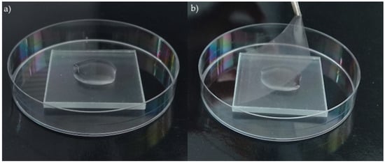
To determine the conditions for the spread of pathogenic fungi in a greenhouse or similar environment, the spore activity was measured by counting CFUs after collection from coated surfaces following an incubation period. Squares (50 mm × 50 mm) made of coated glass were sterilized with 70% ethanol and placed in a sterile Petri dish. Spore suspensions (1 mL) with a density of 103 mL−1, corresponding to the same number of CFU, were inoculated onto the surface of the coated glass (Figure 1a) and covered with sterile polypropylene foils (about 48 mm × 48 mm, 50 µm thick) (Figure 1b). The CFU of the inoculum of each microorganism was also verified on PDA medium.
Figure 1.
(a) Spore suspension on the surface of squares (50 mm × 50 mm) of coated glass; (b) covering spore suspensions with foil to protect the suspension from evaporation.
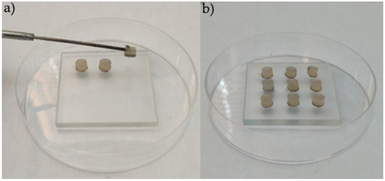
The experiment was conducted in a phytotron chamber (Biogenet®) under two lighting conditions: with light (L) (LED tube 1200 mm 15.5W 865 T8, light, about 1000 lux, spectral characteristics: 400–750 nm) and without light—dark (D) at 28 °C for 24 h. After incubation, the foils from the top of the tested surfaces were removed and the spore suspensions collected from the coated glass were sown on PDA medium. To determine the antimicrobial activity of the tested nanolayers, the CFU counts were compared with the number of colonies simultaneously grown after incubation on the control sample (diffuse glass without any coating). In order to evaluate the effect of the coatings on the hyphal growth, the contact of agar plugs with the coatings was assessed. For this purpose, 5 mm diameter agar disks were cut from several-day-old hyphae grown on PDA medium using a cork borer and placed on the surface of the coated glass (Figure 2a) in quantities of 9 disks. To ensure optimal humidity conditions (comparable to those in a greenhouse) during incubation, approximately 5 mL of sterile distilled water was added to the bottom of the Petri dish (Figure 2b). The experiment was conducted in a phytotron chamber (Biogenet®) under two lighting conditions at 28 °C for 3 days.
Figure 2.
(a) Deposition of microbial mycelium disks on the surface of squares (50 mm × 50 mm) made of coated glass; (b) full set of mycelium disks prepared for incubation.
For 3 consecutive days, after each 24 h contact between the coatings and a uniform layer of mycelium, three agar disks were gently transferred to fresh PDA medium and incubated for 7 days, during which the hyphal growth rate was determined. All tests were performed in triplicate.
To obtain Phytophthora infestans mycelium and to incubate it after contact with the coatings, the strain was cultivated on PDA medium supplemented with small pieces of sterilized tomato leaf as a substrate, to optimize growth conditions and enhance mycelial development [32].
The hyphal growth rate was determined each day during incubation, and the growth rate factor (R) was calculated according to the following formula and expressed as mm2 of mycelium per day:
where R—growth rate factor; D—diameter of the mycelium (mm); d—mycelia disks (11 mm); —3.14; T—incubation time (day).
The growth factor was calculated for each day of incubation, and then the average growth rate ratio over time (ΔT) was calculated and expressed as mm2/day:
where ΔT—growth rate ratio; R—growth rate factor from each day; n—number of incubation days and presented as a percentage relative to the control [33].
ΔT = ((R1 − R0) + (R2 − R1) + … + (Rn+1 − Rn))/n
2.3. Statistical Analysis
All statistical analyses were conducted in RStudio (version 2025.05.1+513; R Foundation for Statistical Computing, Vienna, Austria). Prior to inferential testing, data distributions were assessed for normality using the Shapiro–Wilk test. Differences between each alloy coating and the corresponding control were evaluated using one-way analysis of variance (ANOVA), followed by Dunnett’s post hoc test (α = 0.05), with the control (I. Control) group specified as the reference level. Descriptive statistics are reported as mean ± standard deviation (SD) based on three independent biological replicates (n = 3). Statistical significance is indicated in figures using asterisks: * p < 0.05; ** p < 0.01; *** p < 0.001.
The principal component analysis (PCA) was conducted on standardized data, with variables centered and scaled to unit variance, using the prcomp() function in R. The number of retained principal components was determined based on the proportion of explained variance (scree plot) and biological interpretability. Variable loadings and sample scores were visualized using biplots to identify clustering patterns and dominant sources of variation.
Heatmap analyses were used to visualize relative biological responses across fungal strains and alloy coatings. Data were normalized relative to the strain-specific control to emphasize trends in response, magnitude and direction. Hierarchical clustering of rows and columns was applied to identify similarity patterns among coatings and fungal responses.
3. Results
3.1. Antimicrobial Activity of Coatings Against Fungal and Chromista Spores
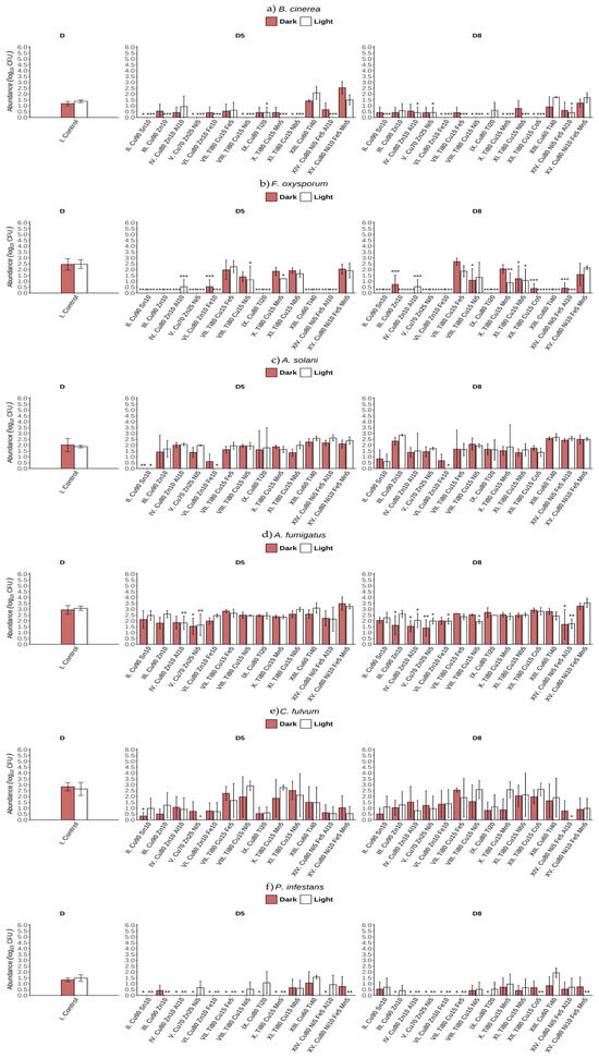
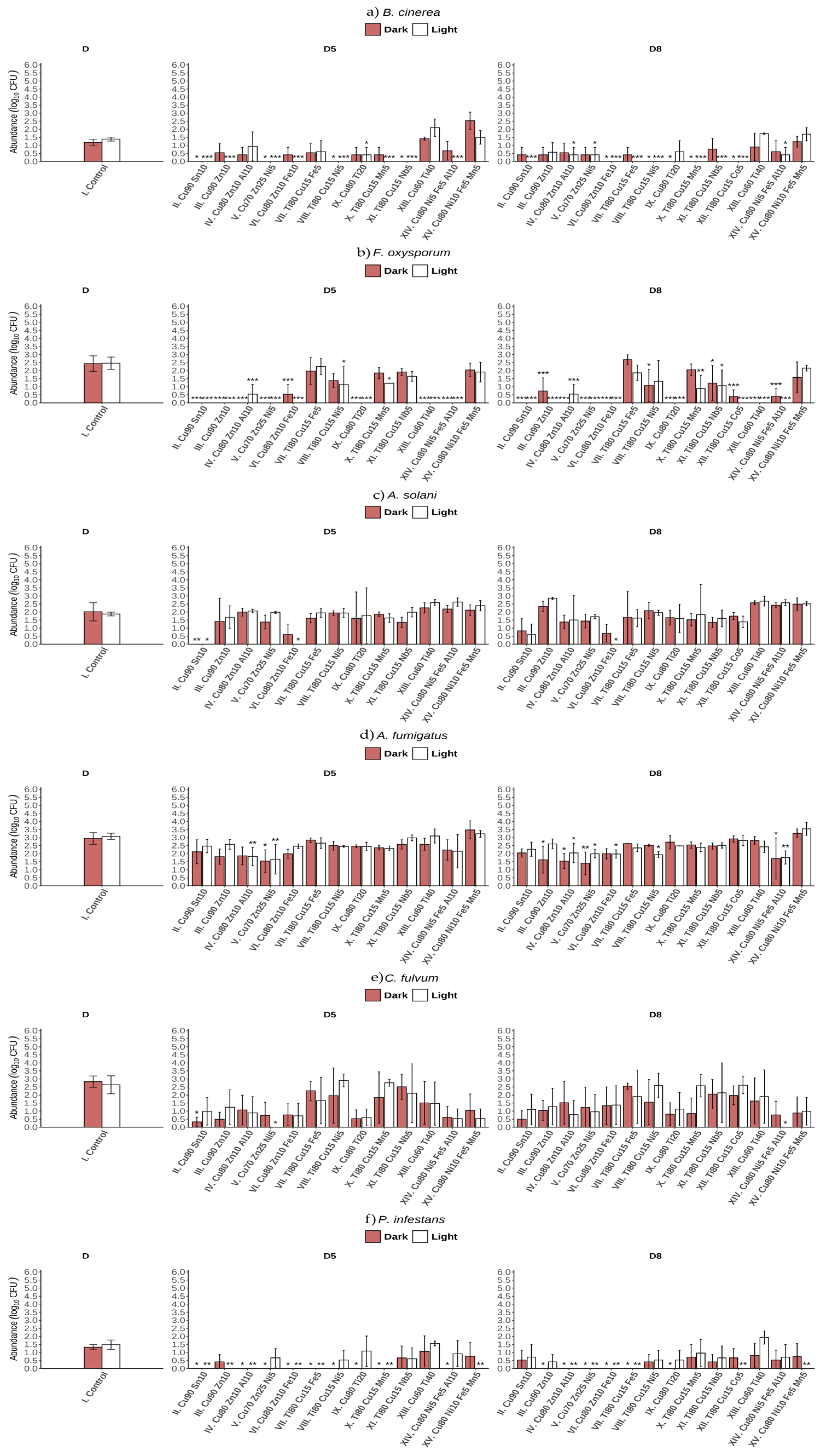
After 24 h of incubation, exposure of B. cinerea spores to most of the coatings (with the exception of XIII Cu60Ti40 and XV Cu80Ni10Fe5Mn5) resulted in either complete inhibition of germination or a statistically significant reduction in the number of CFU (Figure 3a), regardless of the transparency of the glass substrates onto which the coatings were applied (D5, D8). Moreover, a clear trend towards reduced spore viability was observed only for B. cinerea spores incubated on all coatings with a high Ti content, particularly under L conditions (Figure 4). All coatings with a high Cu content (II–VI and IX), as well as XIII and XIV, displayed pronounced biocidal and inhibitory activity on F. oxysporum spores (Figure 3b). Furthermore, incubation under L and D conditions did not significantly affect spore activity (Figure 4). Of all the fungal strains tested, A. solani and A. fumigatus spores showed the least activity when incubated on the investigated coatings (Figure 3c,d). An exception was observed for A. solani incubated on coating II Cu80Sn10, on which complete inhibition of spore germination occurred. In addition, the number of CFU in the suspension collected after 24 h of incubation on coating VI Cu80Zn10Fe10 decreased by almost 30% (Figure 3c). It should be noted that none of the tested coatings exhibited any statistically significant inhibition of A. fumigatus spore activity parameters following incubation on their surfaces (Figure 3d). Similarly, the decrease in CFU of C. fulvum in the suspension after incubation on the tested coatings was not statistically significant (Figure 3e).
Figure 3.
Number of microorganisms (Log10 CFU mL−1) after 24 h of spore incubation on coated glass (I-XV) in light and dark conditions for (a) B. cinerea, (b) F. oxysporum, (c) A. solani, (d) A. fumigatus, (e) C. fulvum, and (f) P. infestans; D5 ≥ 85% and D8 ≥ 88% direct transmittance; bars represent mean ± SD (n = 3). Statistically significant differences relative to the corresponding control (Dunnett’s test): * p < 0.05; ** p < 0.01; *** p < 0.001.
Figure 4.
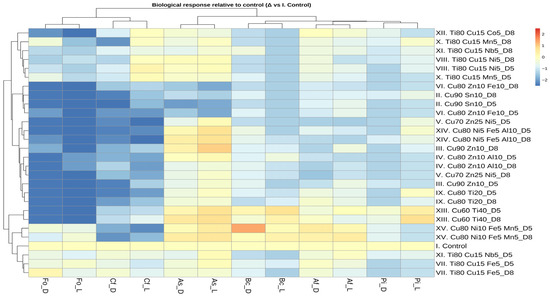
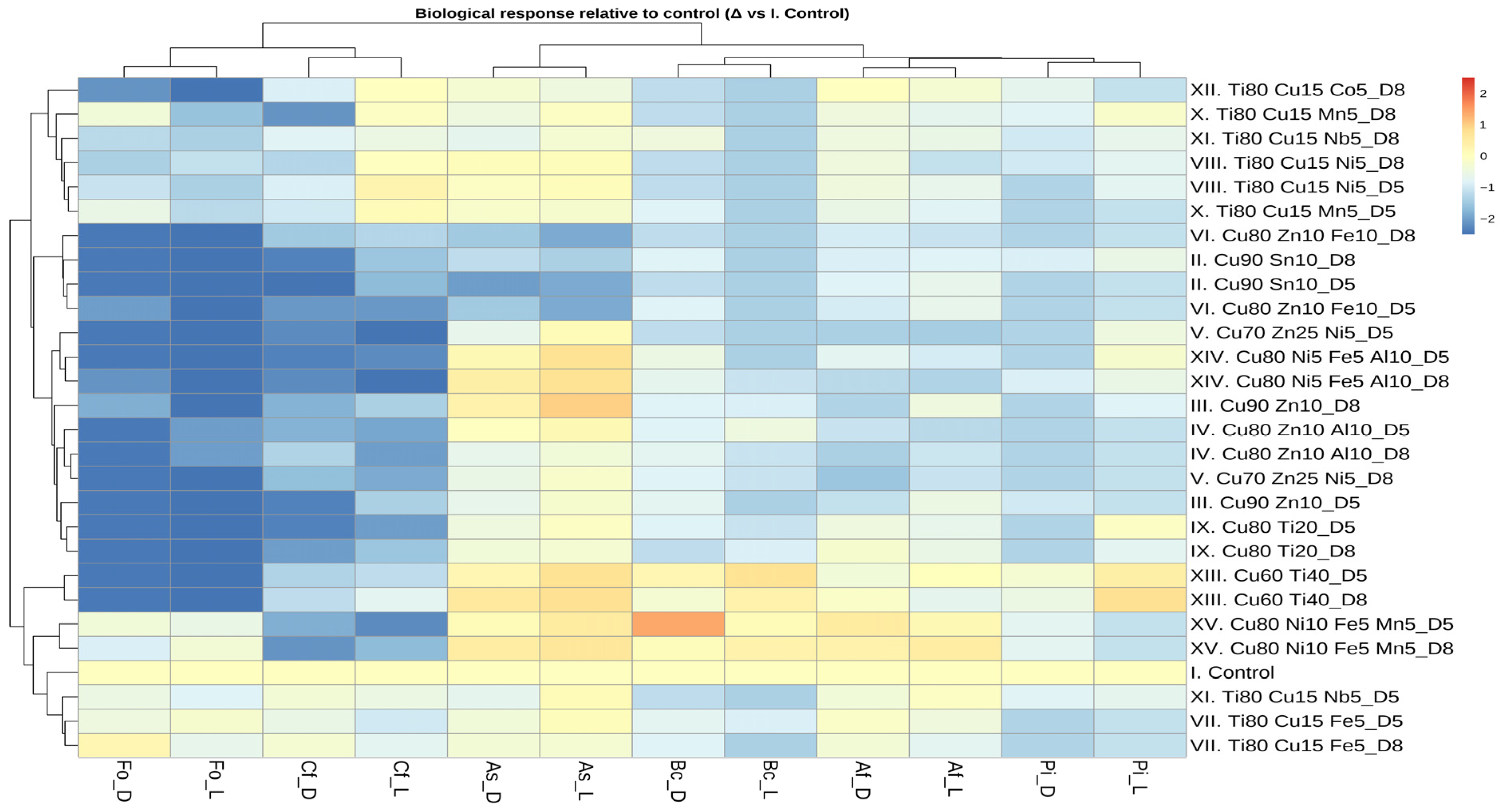
Heatmap with hierarchical clustering of mycelial radial growth of six microorganisms incubated on alloy-coated glass surfaces under light and dark conditions. Rows represent 14 coatings (D5 and D8), and columns correspond to microbial strains and light conditions. Color intensity indicates normalized CFU determined in microbial spore suspensions after 24 h, with lower values reflecting stronger inhibition relative to the control. Abbreviations: Bc—B. cinerea; Fo—F. oxysporum; As—A. solani; Af—A. fumigatus; Cf—C. fulvum; Pi—P. infestans. D5 ≥ 85% and D8 ≥ 88% direct transmittance.
It is noteworthy that a clear trend was observed, indicating that all Cu-predominant coatings inhibited the viability of C. fulvum and F. oxysporum (Figure A1) fungal spores. During the row clustering step of the heatmap analysis, the samples were grouped according to their compositional profiles. Cu-predominant coatings formed a distinct cluster, which was clearly separated from coatings characterized by different elemental compositions (Figure 4). After 24 h of incubation, exposure of P. infestans spores to most of the coatings (with the exception of XIII Cu60Ti40 and XV Cu80Ni10Fe5Mn5) resulted in the complete inhibition of germination or an approximately fourfold decrease in CFU (Figure 3a–f). These two coatings exhibited the weakest biocidal effects towards the other tested microorganisms (Figure 4). Additionally, coatings with a high Ti content did not exhibit a pronounced biocidal effect on the spores of Alternaria solani, Aspergillus fumigatus, Cladosporium fulvum (with the exception of Ti80Cu15Mn5 applied to D8 glass under D conditions), and Fusarium oxysporum (with the exception of Ti80Cu15Co5 applied to D8 glass) (Figure 4).
In the PCA, the first two eigenvalues together explained about 63.8% of the variation (Dim 1: 42% and Dim 2: 21.8%) (Figure A2). The variables Bc_L, As_L, Bc_D, As_D and Pi_L were positively correlated with each other and were associated with the coatings XIII and XV. Meanwhile, the variables Fo_L, Fo_D, Cf_L and Cf_D correlated with coatings VII, VIII, X and XI. Dim1 separated the control and coatings with a high Ti content (VII–X) from other coatings. The control was isolated from all coatings, indicating a strong effect of metal presence. Coatings with high contents of Cu, Zn and Ni clustered together and coatings with high Ti content were grouped together. Dim2 showed partial separation between D5 and D8, although these effects were weaker than those caused by metal composition. Overall, the PCA demonstrated that metal composition was the primary factor driving sample separation (Figure A2).
The incubation conditions of most microorganisms’ spore suspensions in L or D had no statistically significant effect on the spores’ activity on all tested coatings with diverse compositions. On the heatmap (Figure 4), L and D conditions for each strain were clustered together, indicating consistent activity patterns across both L and D incubation.
3.2. Antimicrobial Activity of Coatings Against Hyphal Growth of Fungi and Chromista
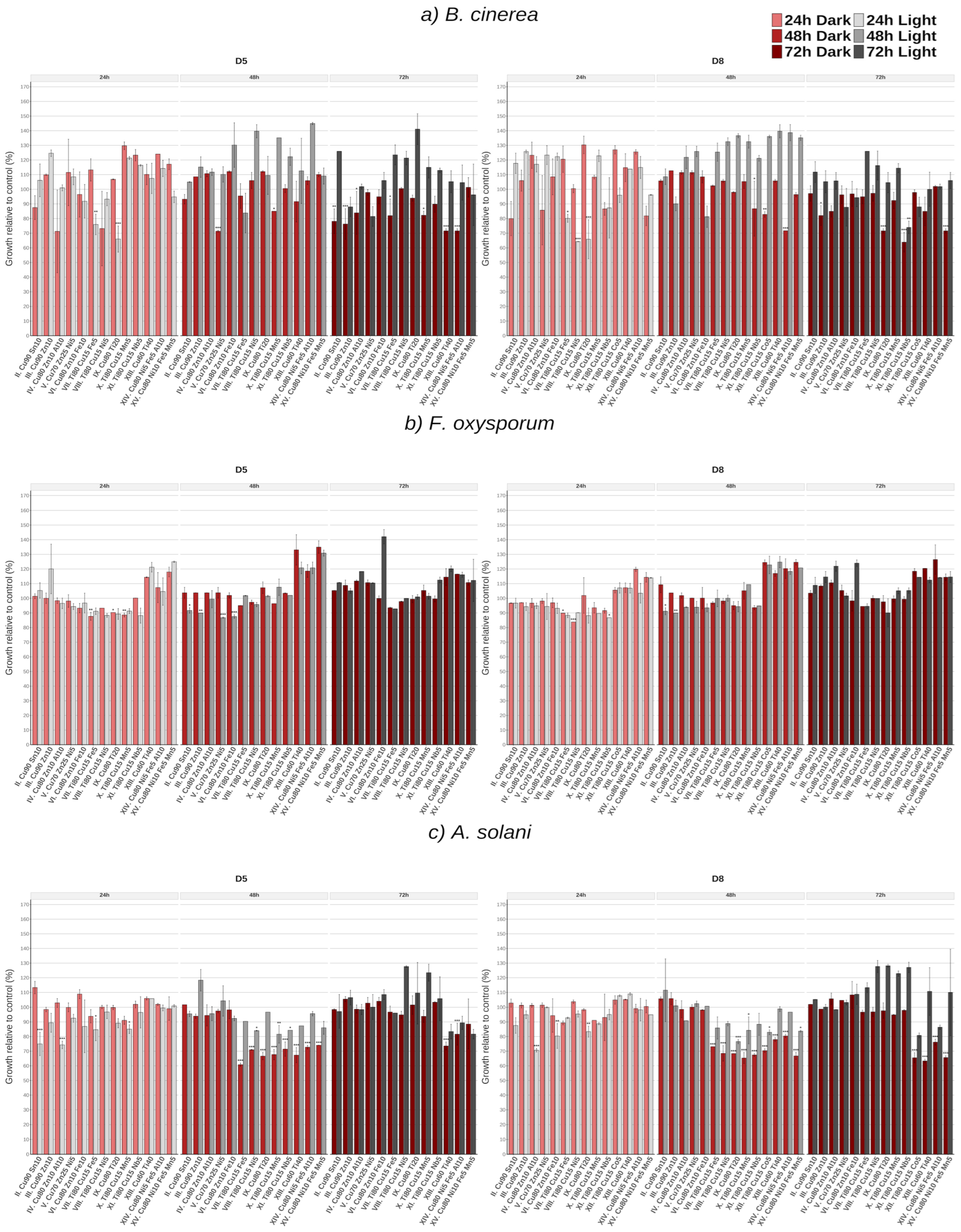
After 24 h of incubation, the mycelium of B. cinerea exhibited sensitivity to most of the tested glass coatings with D8, as evidenced by a pronounced inhibition of radial mycelial growth compared to the control. Notably, after 24 h of contact with coatings VIII Ti80Cu15Ni5 D5 and IX Cu80Ti20 D5 and D8, radial mycelial growth was inhibited by over 30% relative to the control mycelium incubated on uncoated glass. However, this sensitivity was not maintained after 48 h, as reflected by a reduced inhibition of radial mycelial growth (Figure 5a). After three days of incubation on the tested coatings, the strongest inhibition of mycelial growth was observed for coatings XIII Cu60Ti40 D5 and XIV Cu80Ni5Fe5Al10 D5, as well as XV Cu80Ni10Fe5Mn5 D8, with a statistically significant inhibition of radial mycelial growth of nearly 30%. In contrast, after 24 h of incubation, the F. oxysporum strain exhibited robust mycelial growth, showing a clear trend towards slight growth inhibition on glass coated with coatings III–XIII D5 and II–XII D8 (Figure 5b). However, no inhibitory effect was observed after 48 or 72 h of mycelium incubation on the tested coatings. Moreover, after 72 h of incubation, none of the tested coatings resulted in statistically significant inhibition of the radial mycelial growth of the F. oxysporum strain.
Figure 5.
Radial hyphal growth (mm) measured after 7 days of cultivation on PDA following 24, 48, and 72 h of incubation on coated glass (II–XV) under light and dark conditions for (a) B. cinerea, (b) F. oxysporum, (c) A. solani, D5 ≥ 85% and D8 ≥ 88% direct transmittance. Bars represent mean ± SD (n = 3). Statistically significant differences relative to the corresponding control (Dunnett’s test): * p < 0.05; ** p < 0.01; *** p < 0.001.
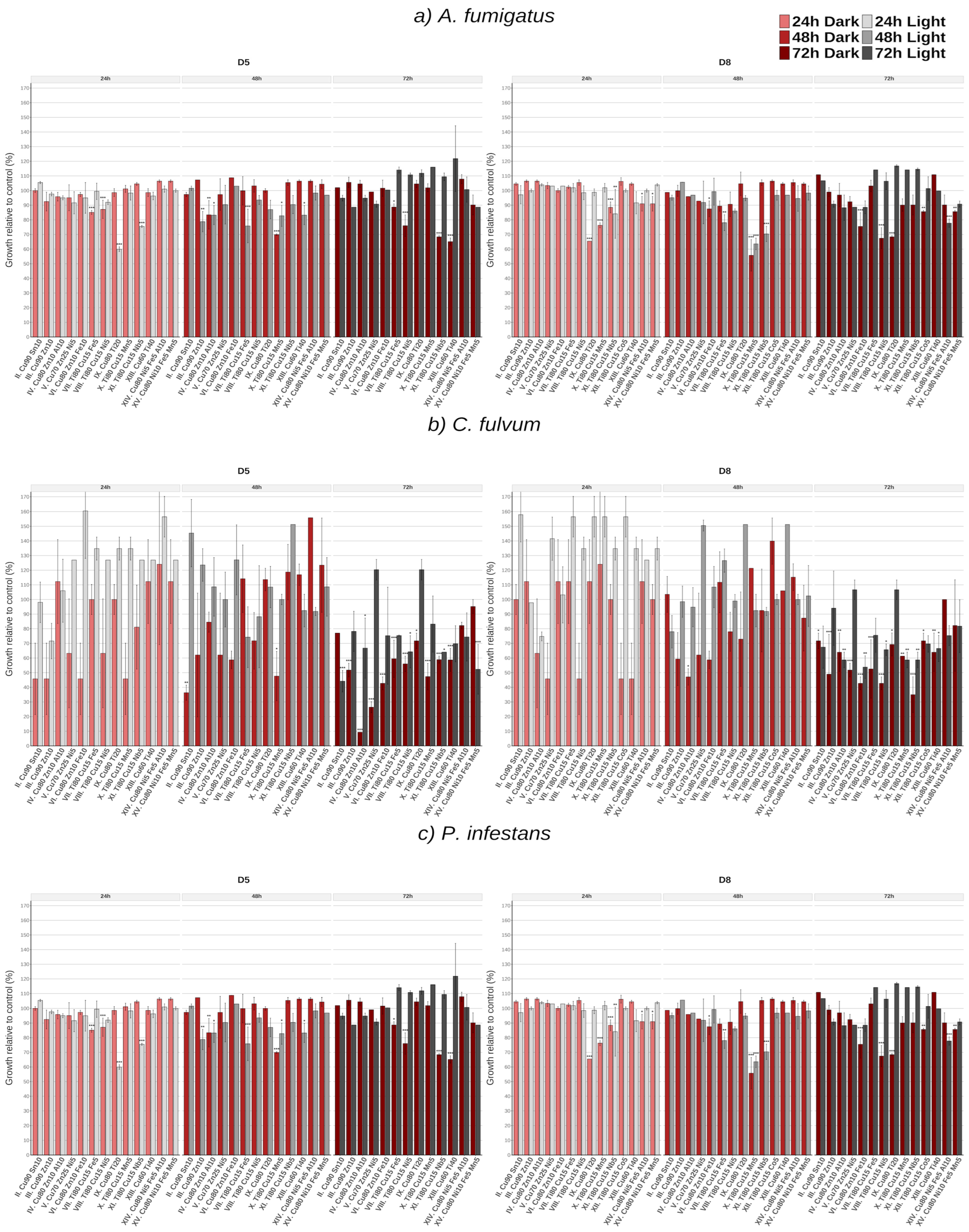
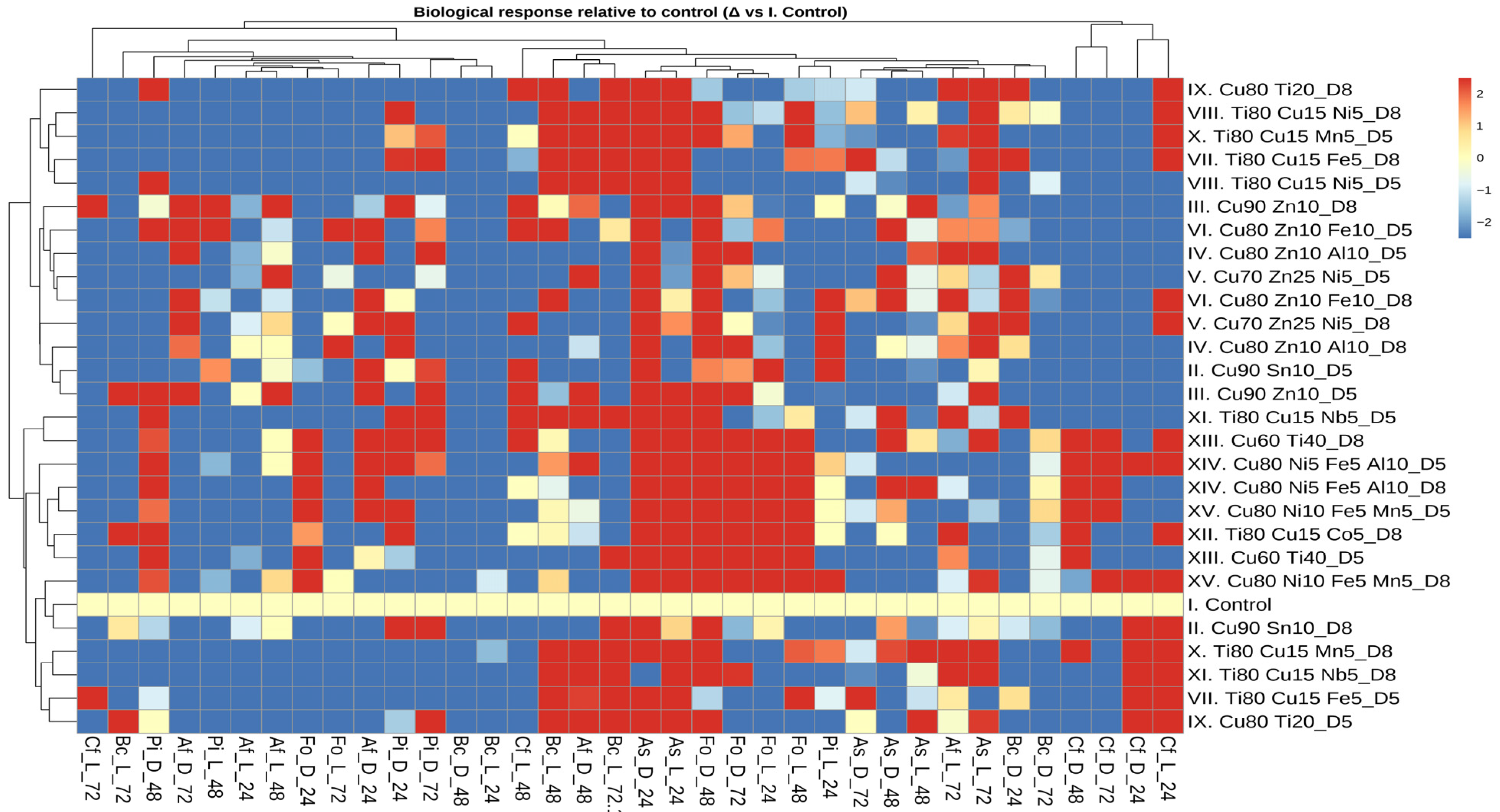
Following exposure to coatings with high Cu content (coatings II–IX), A. solani mycelium exhibited a slight decrease in growth intensity compared to the control after the first day of incubation (Figure 5c). After two days, the mycelium of A. solani (Figure A3) was markedly inhibited by coatings VII–XV, regardless of their direct transmittance values, and this effect was only observed under D conditions. Furthermore, exposure to coating VII Ti80Cu15Fe5 resulted in almost 40% inhibition of A. solani radial growth (Figure A3). After 72 h of incubation, statistically significant inhibition of A. solani mycelial growth was observed following exposure to coated glass XIII and XIV, including under D conditions, at approximately 30% and 20%, respectively, compared to the control (Figure 5c). After 48 h of incubation, A. fumigatus exhibited sensitivity to coatings VII–XI, whereas after 72 h, the strongest inhibitory effect on radial mycelial growth was observed for coatings XI and XII. After the first day of incubation, the mycelium of C. fulvum (Figure 6a) demonstrated a clearly reduced and/or inhibited growth under D conditions, a trend that continued over the following two days of incubation on a subset of the tested coatings. Moreover, this strain showed the highest sensitivity to certain coatings, with exposure to IV Cu80Zn10Al10 resulting in a more than 80% reduction in radial hyphal growth (Figure 6a). Both C. fulvum and B. cinerea exhibited similar sensitivity of their mycelium to the components of the tested coatings, and reduced sensitivity to certain coatings under L incubation conditions (Figure 7). A comparable, inhibitory effect of the coatings was also observed after 72 h of L incubation conditions for A. solani and for the chromista P. infestans (Figure 5c, Figure 6c and Figure 7). The growth of P. infestans was significantly slowed following 24 and 48 h incubations on coating X Ti80Cu15Mn5; however, no inhibition was observed after the subsequent day of incubation (Figure 6c).
Figure 6.
Radial hyphal growth (mm) measured after 7 days of cultivation on PDA following 24, 48, and 72 h of incubation on coated glass (II–XV) under light and dark conditions for (a) A. fumigatus, (b) C. fulvum, (c) P. infestans, D5 ≥ 85% and D8 ≥ 88% direct transmittance. Bars represent mean ± SD (n = 3). Statistically significant differences relative to the corresponding control (Dunnett’s test): * p < 0.05; ** p < 0.01; *** p < 0.001.
Figure 7.
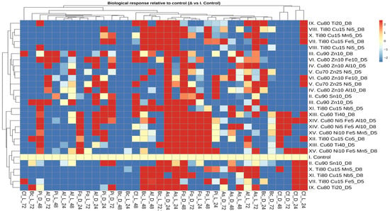
Heatmap with hierarchical clustering of hyphal radial growth of six microorganisms incubated on alloy-coated glass surfaces under light and dark conditions. Rows represent 14 coatings (D5, D8) and columns correspond to microbial strains and light conditions. Color intensity indicates normalized growth measured after 24, 48, and 72 h, with lower values reflecting stronger inhibition relative to the control. Abbreviations: Bc—B. cinerea, Fo—F. oxysporum, As—A. solani, Af—A. fumigatus, Cf—C. fulvum, Pi—P. infestans, D5 ≥ 85% and D8 ≥ 88% direct transmittance.
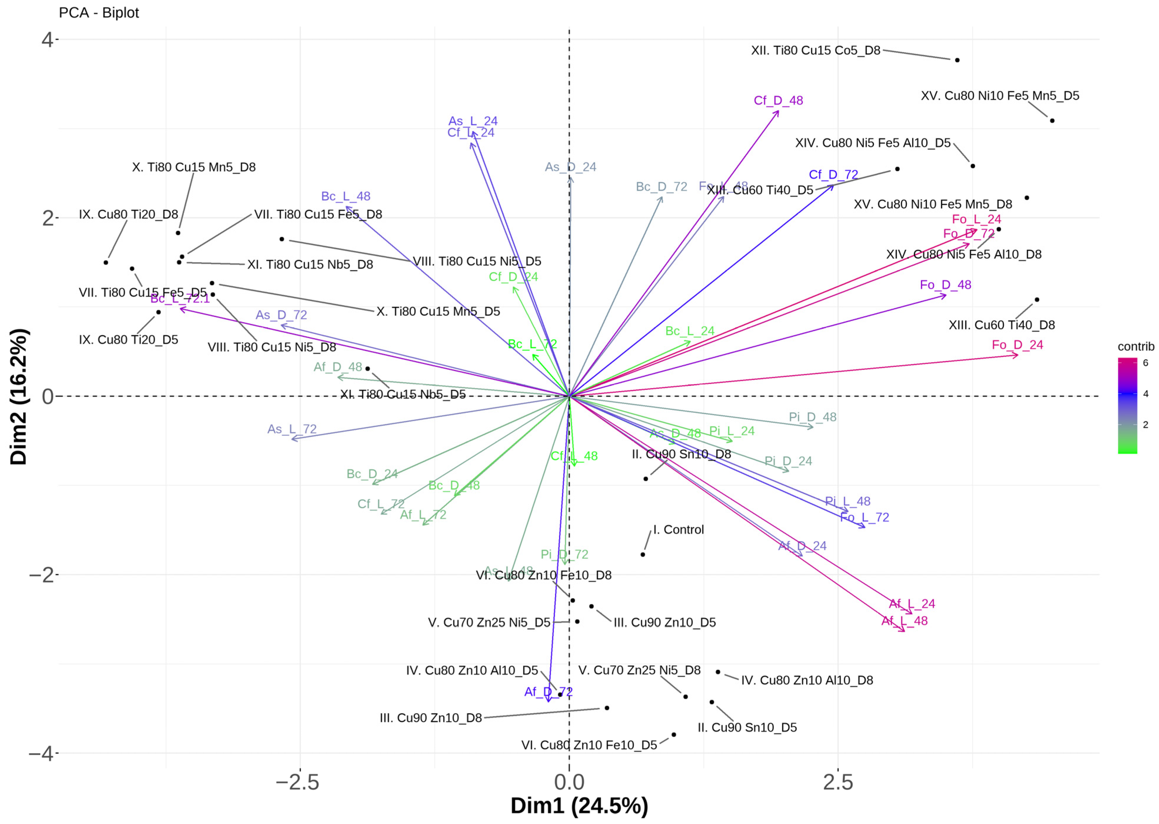
In the PCA, the first two eigenvalues together (Dim 1: 24.5% and Dim 2: 16.2%) explained about 40.7% of the variation (Figure A4). Dim1 primarily discriminated samples according to their metal composition and concentration. Coatings with higher concentrations of metal mixtures (e.g., Cu, Ni, Fe and Cu, Zn, Fe) clustered on the positive side of Dim1, whereas those with lower concentrations of metals were located on the negative side. Longer incubated samples (48–72 h) tended to separate along Dim2, while 24 h samples clustered closer to the center of the plot, suggesting weaker differentiation at initial incubation and time-dependent effects. Overall, the PCA demonstrated that combined effects of metal composition, concentration, and L/D conditions were the main reason for variability in the dataset, with higher metal concentrations producing the strong differentiation from the control condition (Figure A4).
4. Discussion
4.1. Effects of Glass Coatings on the Germination of Fungi and P. infestans
Magnetron sputtering (MS) is a PVD technique that enables the formation of uniform, dense, and highly adherent coatings, particularly on flat substrates. MS is costly, time-consuming, limited by line-of-sight deposition, and typically produces amorphous films [34]. Our studies evaluating the antimicrobial activity of such coatings are limited, particularly with regard to phytopathogenic fungi, including Botrytis cinerea, Fusarium oxysporum, Cladosporium fulvum, Alternaria solani, the chromista Phytophtora infestans, and the opportunistic pathogen Aspergillus fumigatus as a model organism. Studies of this type are scarce and are unique in their application of coatings against two types of fungal material (spores and hyphae) and chromista strains, as well as in their use of conditions representative of greenhouse environments, including controlled humidity and temperature, and incubation in both L and D conditions. The presented study evaluated the antimicrobial efficacy of the tested surface coatings against microbial spores under carefully controlled conditions. Spores were incubated in an aqueous solution within a specially designed “mini-chamber” that maintains high humidity levels (Figure 1). These conditions preserved spore viability and provided an optimal environment for germination (incubation for 24 h) on control glass (without any coatings). In the studies, the optimal temperature range for hyphal growth and spore germination of B. cinerea, A. solani, and F. oxysporum was around 20–30 °C, with efficient germination occurring after at least 6 h [35]. For A. fumigatus, germination was rapid, with most conidia completing germination within 10–12 h [36].
The presented study revealed that, after 24 h of incubation on uncoated glass on PDA medium, the CFU counts of F. oxysporum, A. fumigatus, and C. fulvum were approximately 3 log10 (Figure 3b,d,e), whereas for B. cinerea, A. solani, and P. infestans under control conditions, CFU counts were slightly lower, around 1.5–2 log10 (Figure 3a,c,f). This dual functionality ensures that any observed reduction in spore viability can be directly attributed to the antimicrobial properties of the coatings, rather than to unfavorable environmental conditions. In a study testing fungal spores, incubation conditions similar to those used in the present work had been applied to assess contact with the tested surfaces. The antifungal activity of both smooth and laser-microstructured pure Cu surfaces was assessed by exposing sterilized metal plates to a fungal spore suspension (106 spores mL−1) for up to 120 min [37]. According to the authors, these incubation conditions confirmed “wet killing contact on tested surfaces” and demonstrated further physiological effects of Cu exposure on fungal spores. Azamatov et al. [30] investigated Ta-Cu and Nb-Cu coatings deposited by MS on porous Ti alloy scaffolds and evaluated their antifungal properties against Candida albicans. It is worth emphasizing that this test provides optimal conditions for microbial growth, including access to nutrient-rich media similar to those present in the human body. Therefore, it is well-suited for assessing the biocidal properties of coatings intended for endoprosthetic applications [30,38].
Copper compounds have long been recognized for their broad-spectrum antifungal activity and have been widely used to control phytopathogenic fungi in agriculture. Their modes of action include disrupting cell membranes, interfering with enzymatic processes, and inducing oxidative stress in fungal cells, making them effective against a variety of fungal pathogens [34]. In one study, Aspergillus niger wild-type N402 spores were suspended (106 spores mL−1) and applied to sterilized smooth and laser-microstructured Cu plates for up to 120 min [37]. Spore survival, as determined by CFU, decreased by over 85% on Cu surfaces. These findings highlight the significant physiological impact of Cu exposure on fungal spores and provide insight into its potential for controlling spore viability. Notably, Cu oxychloride, Cu(II) sulfate, and dicopper chloride trihydroxide, which are widely used as plant protection products (fungicides), exhibit particularly high toxicity. Consequently, the ecotoxicological properties of these compounds render them unsuitable for contemporary integrated or organic crop protection, as their application to the large surface area of greenhouse glass could potentially serve as a site for growth and a source of invasive phytopathogens, having adverse effects on beneficial organisms [39]. In our study, the viability of the tested microorganisms’ spores decreased after incubation on coatings with a high Cu (Me%) content. This effect was particularly pronounced for B. cinerea, F. oxysporum, and P. infestans, whereas the highest reduction (including total inhibition) in CFU was likewise observed for the remaining strains following incubation on high-Cu coatings (Figure 3a–f and Figure 4). These results are consistent with those obtained in our earlier investigation concerning the antibacterial properties of these fourteen coatings [31]. In particular, the coatings II, III and IX derived from 90%Cu10%Sn, 90%Cu10%Zn, and 80%Cu20%Ti targets, respectively demonstrated some of the highest levels of antibacterial efficacy. Considering the selection of the most suitable coating for greenhouse applications, this study emphasizes that the high-Cu coatings examined are predominantly amorphous (Table 1), forming a dense surface that inhibits microbial adhesion. Moreover, according to literature data, such coatings exhibit long-lasting biocidal activity through the controlled leaching of metal ions [40].
Studies have confirmed that the presence of various metal ions in the growth medium can significantly affect the germination process of fungal spores and P. infestans. The experiment examined fungal isolates from two environments, ultramafic and agricultural soils, to evaluate spore germination in the presence of Ni, Mn, Mg, Fe, and Co ions [41]. The spore suspensions were spread on Czapek’s medium, supplemented with two different concentrations of the respective ions and incubated at 25 °C in the dark. Analysis revealed differences in spore sensitivity related to the origin of the isolates. Notably, within the group of isolates derived from agricultural soils, these differences were most evident for three metals, namely Ni, Mn, and Mg, and comparatively less pronounced for Co and Fe. Spore germination of strains belonging to the genera Aspergillus and Fusarium was particularly strongly inhibited by the presence of Ni ions in the medium. Moreover, supplementation of the medium with Fe ions (200 µg mL−1) completely inhibited the spore germination of both microorganisms [41]. In our study, the presence of Ni ions in the glass coatings (V Cu70Zn25Ni5; XIV Cu80Ni5Fe5Al10; and XV Cu80Ni10Fe5Mn5) did not significantly affect the germination of F. oxysporum and A. fumigatus spores (Figure 3b,f). In our opinion, the most critical factor in completely inhibiting the spore germination of the tested strains is the presence of Cu ions. This is indicated by the results (Figure 3a–f and Figure 4) for the two coatings prepared from targets IX Cu80Ti20 and XIII Cu60Ti40. For A. fumigatus, C. fulvum, and A. solani (Figure 3c–e), a clear trend was observed, showing that the IX coating exhibits stronger antimicrobial activity. Under D incubation conditions, the IX Cu80Ti20 coating completely suppressed spore activity for B. cinerea and P. infestans, whereas the XIII coating permitted colony formation, with CFU counts of 1 log10 or higher (Figure 3a,f).
Further studies should aim to determine the influence of individual ions and compositional parameters on the microbial response to the coatings by varying ion percentages and evaluating of their antimicrobial activity, efficacy and interactions with microbial cells. Unlike coatings obtained from the liquid phase, where a wider range of compositional variants can be produced, each target composition intended for magnetron-sputtered coatings must be carefully evaluated for theoretical metallurgical feasibility prior to production.
4.2. Effects of Glass Coatings on the Hyphal Growth of Fungi and P. infestans
Although mature fungal and chromista spores generally exhibit the highest tolerance to environmental stress, the processes of germination and vegetative growth often reduce this protection. The extent of adaptive resistance in germinated spores and established hyphae clearly depends on the species and the specific stressor, which may include the presence of certain ions at appropriately high concentrations [42]. All presented experiments were conducted under consistent temperature and humidity controls to guarantee the reproducibility and reliability of the results (Figure 3a–f, Figure 5a–c and Figure 6a–c). The antimicrobial efficacy of the tested surface coatings against spores and hyphal growth reflects greenhouse conditions, in which spores germinate in water droplets on glass surfaces, and hyphae can subsequently develop. Once dispersed by air movement to nutrient-rich substrates, the hyphae may infect plants. In the disk diffusion test, hypha on the disks is in contact with the coating for 24, 48 and 72 h to evaluate the effect of the glass coatings on the fully developed hypha (Figure 5a–c and Figure 6a–c).
It is worth emphasizing that the vast majority of studies investigating the effects of ions in the growth environment on hyphal development are conducted using medium inoculated with a defined fungal suspension onto which the test surface coated with the experimental layer is subsequently placed. Ibarra–Laclette et al. [43] demonstrated the strong antifungal activity of Cu-NPs against phytopathogenic fungi. Cu-NPs inhibited over 80% of Fusarium kuroshium mycelial growth at 0.5 and 0.75 mg mL−1, and exceeded 90% at 1 mg mL−1, corresponding to a reduction in hyphal growth relative to the control. The most pronounced effects were observed after 6 days of incubation. It should be noted that the fungal hyphae were in constant contact with the medium containing different concentrations of Cu, whereas in our study, contact was limited to a dry coating on glass for a maximum of 3 days, but inhibition of radial hyphal growth was still observed up to 7 days. In comparison with the experiments presented in this study, which assessed the effects of coatings on spore germination, the radial growth of hyphae for most of the tested strains was not statistically significant, even at such a high Cu content. However, this effect was clearly observed for F. oxysporum (nearly 30% inhibition of hyphal growth after incubation on coatings with high Cu content: VII, IX, and XIII-XV) and C. fulvum (nearly 90% inhibition of hyphal growth after incubation on coating IV Cu80Zn10Al10) (Figure 5a, Figure 6b and Figure 7). In these studies, investigating the effect on hyphal growth, a significant inhibition of fungal growth was observed in most cases after the incubation of the hyphal disk on the coating produced from multicomponent targets. For example, A. solani showed a reduction in hyphal growth of over 30% compared to the control after 3 days of incubation in D conditions on coatings XII Ti80Cu15Co5 and XV Cu80Ni10Fe5Mn5 (Figure 5c). Similarly, B. cinerea exhibited about 30% reduction in hyphal growth on coating XIV Cu80Ni5Fe5Al10 D5 (Figure 5a). These results are consistent with those reported by Rodríguez–Ramos et al. [44], who demonstrated that the concentrations capable of reducing the mycelial growth of B. cinerea by 50% (IC50) were 2.87 mM for Cu and 9.08 mM for Fe. A significant inhibitory effect was observed when applied in equimolar mixtures.
For most of the tested strains, radial hyphal growth displayed a clear pattern characterized by the absence of growth inhibition, and in some cases even enhanced growth, after incubation under L conditions compared to the control. Strains of B. cinerea and C. fulvum (Figure 5a and Figure 6b) exhibited comparable hyphal sensitivity to the tested coatings during the first two days, with reduced sensitivity under L conditions. A similar light-dependent effect after 72 h was observed for A. solani, A. fumigatus, and P. infestans (Figure 5c, Figure 6a,c and Figure 7). Fungal hyphae activate a range of metal-associated defense responses under metal-stress conditions, including metal binding and sequestration, chelator production, and the induction of antioxidant defense systems. Light acts as an additional regulatory factor capable of reprogramming fungal metabolism by downregulating specific metal transporters. Studies on A. fumigatus have shown that light exposure (blue and red wavelengths) changes fungal metabolism, affecting respiratory activity and downregulating heavy metal (particularly iron) transporter genes, while increasing hyphal pigmentation and enhancing resistance to oxidative and UV stress [45]. Given the complex interactions of multiple ions with fungal hyphae for individual microbial species, these observations suggest that coatings with a simple, two-component composition may be recommended for greenhouse applications.
Notably, the results of both studies investigating the effect of the coatings on spore germination and radial hyphal growth were independent of the transparency values of the coatings. Following the production of the coatings, it was demonstrated that samples with 85% transparency exhibited a thicker nanolayer [31]. Optimal light conditions are highly important for any indoor cultivation, as an increase in material transparency of approximately 1% typically leads to a corresponding increase in crop yield of 1% [46]. Therefore, in terms of practical application and greenhouse design, it is recommended that greenhouses use coatings with slightly higher transparency. Considering that greenhouse climatic conditions strongly favor the development and spread of fungi and chromista, environmentally friendly preventive strategies are particularly important. Accordingly, MS technology enables the production of functional coatings with potential antifungal properties, offering a sustainable and environmentally friendly solution for greenhouse applications by avoiding the extensive use of chemical fungicides.
5. Conclusions
The viability of the spores of the tested microorganisms was reduced after incubation on coatings with a high Cu content. These coatings showed the strongest inhibition of spore germination for most strains, particularly B. cinerea, F. oxysporum, and P. infestans. Notably, the effects on spore germination were independent of the direct transmittance value (≥85% or ≥88%) of the tested coated glass. Radial hyphal growth was generally less affected by the high Cu content of the glass coatings for most strains. However, clear inhibition was observed for F. oxysporum, C. fulvum, A. solani, and B. cinerea on selected multicomponent coatings, with reductions in hyphal growth exceeding 30% in some cases. For most strains, radial hyphal growth on the tested glass coatings showed no inhibition after light incubation, and growth was even enhanced in some cases compared to the control. After 72 h, light-dependent effects were observed for A. solani, A. fumigatus, and P. infestans, while B. cinerea and C. fulvum also showed reduced sensitivity on coated glass during the first two days. These results suggest that the presence of ions in the coatings induces a defensive response, which varies among species and should be investigated further in future studies.
Overall, coatings with a high Cu content are the most effective at inhibiting spore germination for most of the strains tested. However, hyphal growth on multicomponent coatings may allow for possible responses to different ions or interactions with various components. Therefore, two-component coatings with a high Cu content are recommended, confirming their suitability for practical greenhouse applications.
In the next phase of the research, the recommended type of glass coating will be used to construct experimental greenhouses to evaluate its performance when conventional plant protection products are applied at varying levels. Tomato (Solanum lycopersicum L.) will be used as the test species and will be exposed to the previously studied microorganisms.
Author Contributions
Conceptualization, P.Z., E.O., A.N. and J.J.-Ś.; methodology, E.O., A.N., M.M., A.H., R.S., P.Z. and K.R.; validation, A.N., E.O., A.H., M.M. and P.Z.; formal analysis, A.H., A.N. and E.O.; investigation, A.N., M.M., A.S., A.H., E.O., P.Z., J.J.-Ś. and R.S.; writing—original draft preparation, E.O., A.N., A.H. and P.Z.; writing—review and editing, E.O., A.N., A.H., P.Z. and J.J.-Ś.; visualization, A.N., E.O. and M.M.; supervision, J.J.-Ś. and K.R.; project administration, K.R.; funding acquisition, P.Z. and K.R. All authors have read and agreed to the published version of the manuscript.
Funding
This research project was financially supported by the National Center for Research and Development (NCBiR), Poland (Grant No. POIR.04.01.02-00-0111/17-00/2018).
Data Availability Statement
The raw data supporting the conclusions of this article will be made available by the authors on request.
Acknowledgments
Graphical abstract was created with https://BioRender.com/ysq2u99 (accessed on 27 January 2026).
Conflicts of Interest
Author Przemysław Ząbek was employed by the company D. A. Glass Sp. z.o.o. All authors declare that the research was conducted in the absence of any commercial or financial relationships that could be construed as a potential conflict of interest.
Abbreviations
The following abbreviations are used in this manuscript:
| CFU | Colony-forming units |
| PVD | Physical vapor deposition |
| MS | Magnetron sputtering |
| L | Light incubation |
| D | Dark incubation |
| D5 | Direct glass transmittance of no less than 85% |
| D8 | Direct glass transmittance of no less than 88% |
| PDA | Potato dextrose agar |
| PCA | Principal component analysis |
Appendix A
Figure A1.
Number of colony-forming units (CFUs) of Fusarium oxysporum after 24 h of incubation on (a) glass coatings V (Cu70Zn25Ni5) and VI (Cu80Zn10Fe10) and (b) uncoated glass (Control). A significant inhibition of spore germination was observed on coatings V and VI compared with the Control.
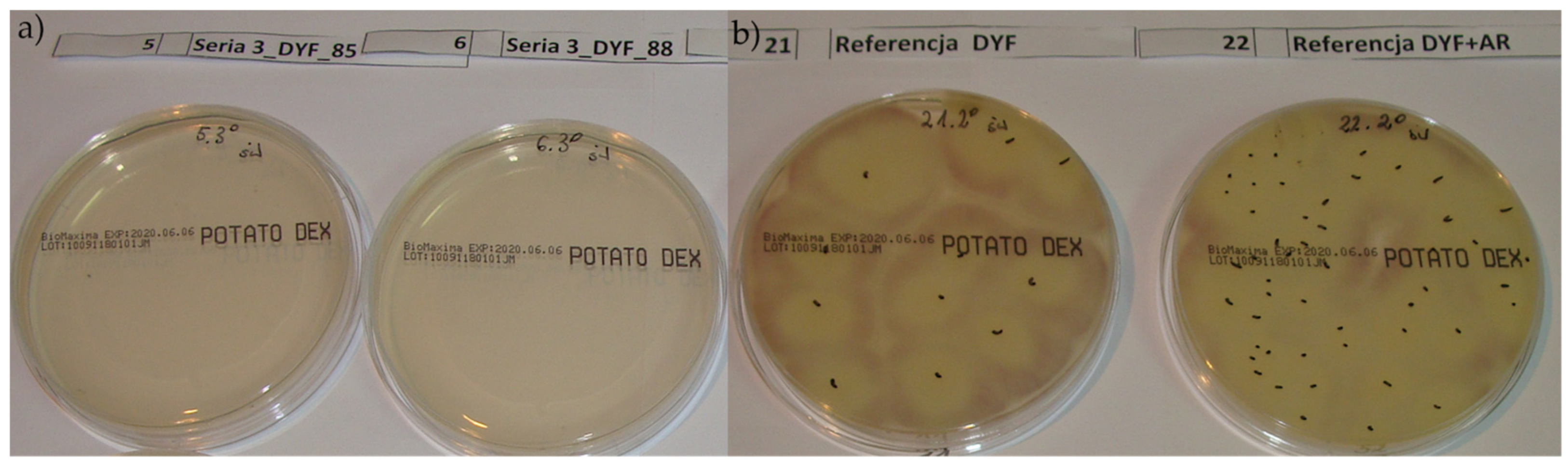
Figure A2.
Biplot of the first (Dim1) and second (Dim2) principal components of the principal component analysis (PCA) of spores of six microorganisms species (B. cinerea—Bc; F. oxysporum—Fo; A. solani—As; A. fumigatus—Af; C. fulvum—Cf; P. infestans—Pi) incubated on coated glass (II–XV) in light (L) and dark (D) conditions, D5 ≥ 85% and D8 ≥ 88% direct transmittance.
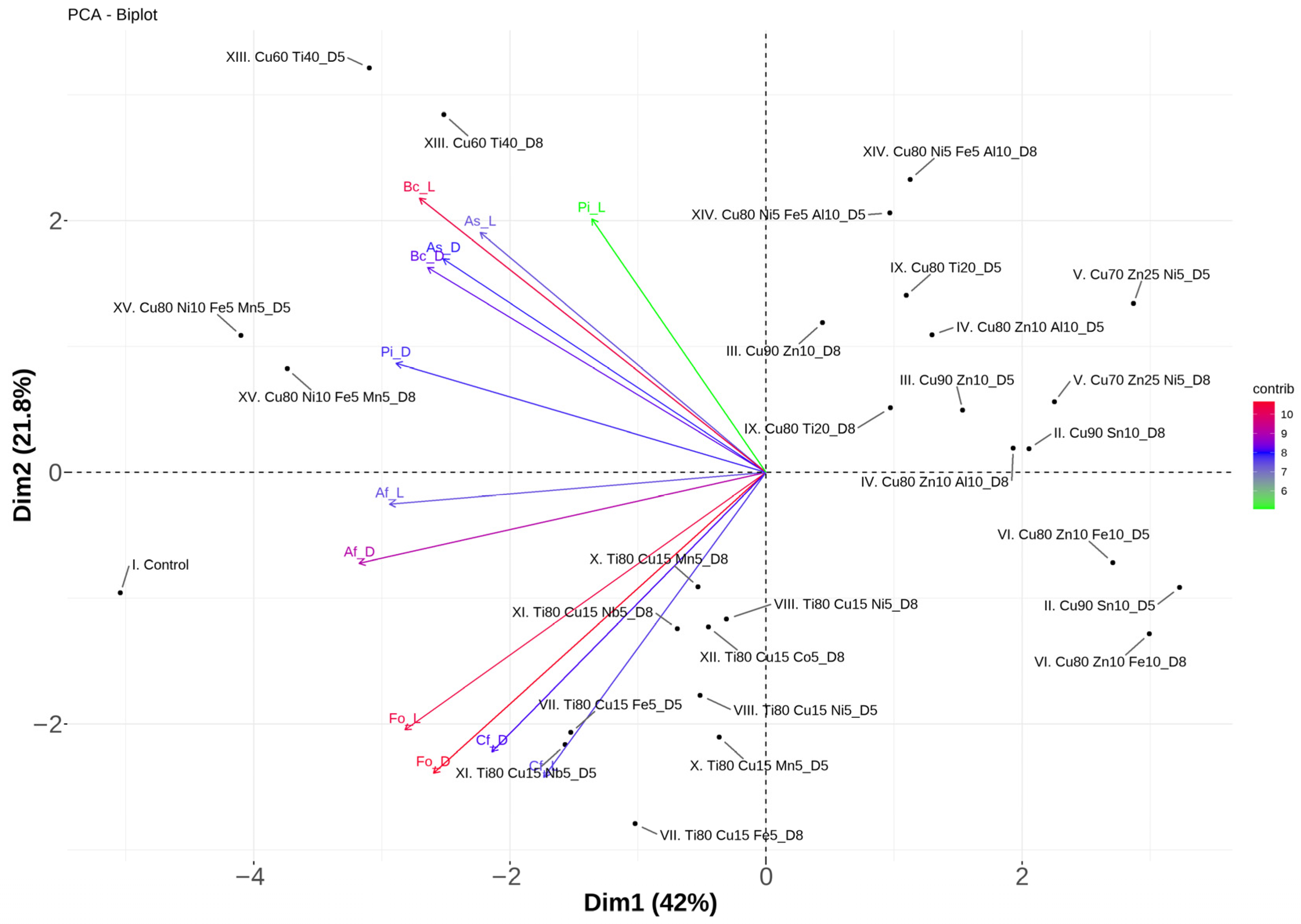
Figure A3.
Mycelial growth of Alternaria solani after 48 h incubation on (a) glass coating VII Ti80Cu15Fe80 and (b) uncoated glass (Control).
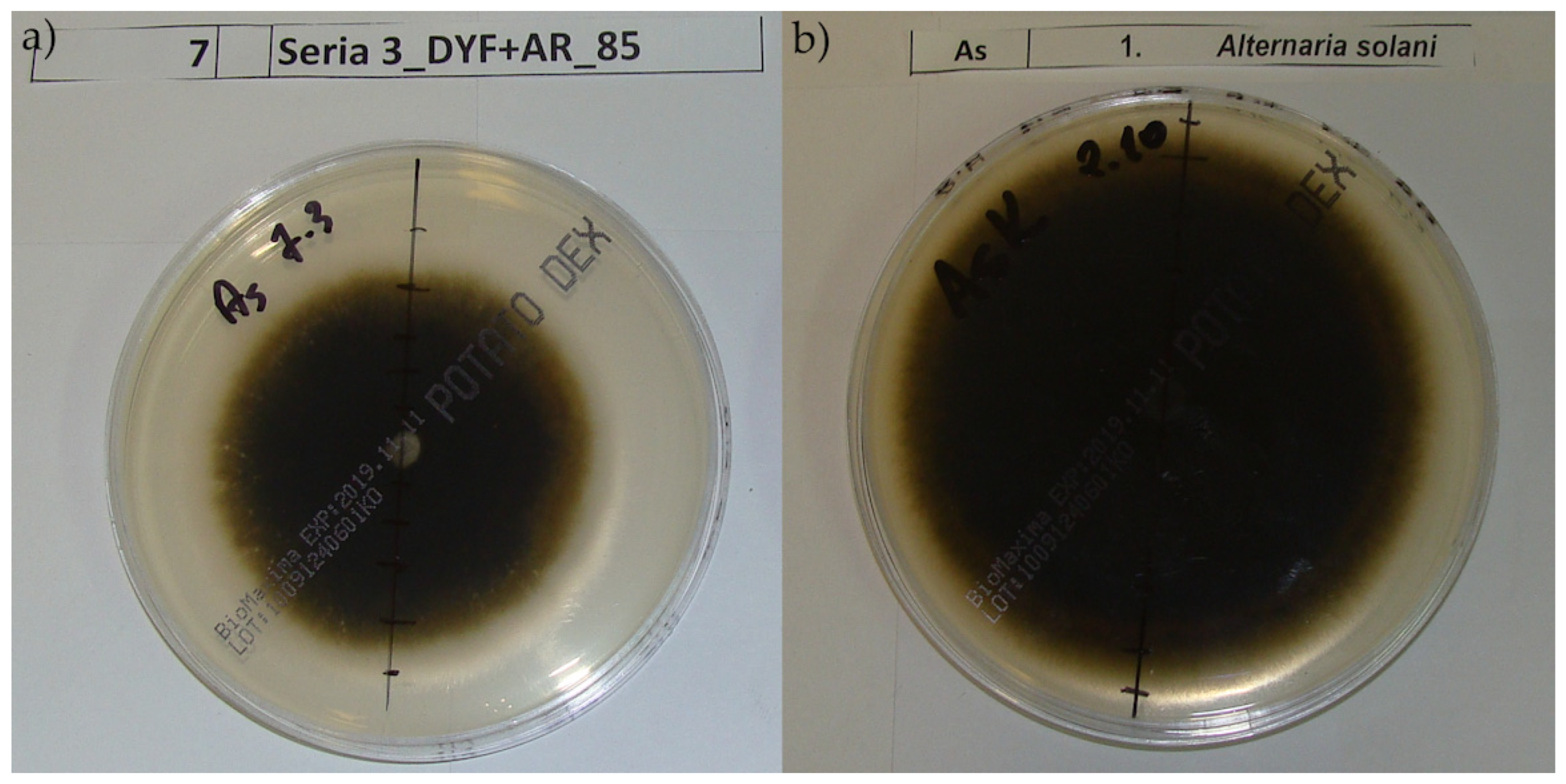
Figure A4.
Biplot of the first (Dim1) and second (Dim2) principal components of the principal component analysis (PCA) of the radial hyphal growth of six microorganisms species (B. cinerea—Bc; F. oxysporum—Fo; A. solani—As; A. fumigatus—Af; C. fulvum—Cf; P. infestans—Pi) incubated for 24, 48, and 72 h on coated glass (II–XV) in light (L) and dark (D) conditions, D5 ≥ 85% and D8 ≥ 88% direct transmittance.
References
- Imran, M.; Abo-Elyousr, K.A.M.; Mousa, M.A.A.; Saad, M.M. Use of Trichoderma Culture Filtrates as a Sustainable Approach to Mitigate Early Blight Disease of Tomato and Their Influence on Plant Biomarkers and Antioxidants Production. Front. Plant Sci. 2023, 14, 1192818. [Google Scholar] [CrossRef] [Scilit]
- Górnik, K.; Sas-Paszt, L.; Derkowska, E.; Mosa, W.F.A.; Trzciński, P.; Głuszek, S. Effects of Bacterial Inoculants and Irrigation Regimes on Yield, Mycorrhizal Colonisation, and Photosynthetic Efficiency in Strawberry Cultivars. Acta Sci. Pol. Hortorum Cultus 2025, 24, 17–35. [Google Scholar] [CrossRef] [Scilit]
- Gordon, T.R. Fusarium oxysporum and the Fusarium Wilt Syndrome. Annu. Rev. Phytopathol. 2017, 55, 23–39. [Google Scholar] [CrossRef] [Scilit]
- Yang, Y.; Wang, Y.; Gao, J.; Shi, Z.; Chen, W.; Huangfu, H.; Li, Z.; Liu, Y. Characterisation of Fusarium oxysporum f. sp. radicis-lycopersici in Infected Tomatoes in Inner Mongolia, China. J. Fungi 2024, 10, 622. [Google Scholar] [CrossRef] [Scilit]
- Adhikari, T.B.; Gao, A.; Ingram, T.; Louws, F.J. Pathogenomics Characterization of an Emerging Fungal Pathogen, Fusarium oxysporum f. sp. Lycopersici in Greenhouse Tomato Production Systems. Front. Microbiol. 2020, 11, 561236. [Google Scholar] [CrossRef] [Scilit]
- Palos-Fernández, R.; Aguilar-Pontes, M.V.; Puebla-Planas, G.; Berger, H.; Studt-Reinhold, L.; Strauss, J.; Di Pietro, A.; López-Berges, M.S. Copper Acquisition Is Essential for Plant Colonization and Virulence in a Root-Infecting Vascular Wilt Fungus. PLoS Pathog. 2024, 20, e1012671. [Google Scholar] [CrossRef] [Scilit]
- Jamiołkowska, A.; Michałek, W. Effect of Mycorrhiza Inoculation of Pepper Seedlings (Capsicum annuum L.) on the Growth Nd Protection against Fusarium oxysporum Infection. Acta Sci. Pol. Hortorum Cultus 2019, 18, 161–169. [Google Scholar] [CrossRef] [Scilit]
- Zhang, D.; Bao, Y.; Sun, Y.; Yang, H.; Zhao, T.; Li, H.; Du, C.; Jiang, J.; Li, J.; Xie, L.; et al. Comparative Transcriptome Analysis Reveals the Response Mechanism of Cf-16-Mediated Resistance to Cladosporium fulvum Infection in Tomato. BMC Plant Biol. 2020, 20, 33. [Google Scholar] [CrossRef] [Scilit]
- Rivas, S.; Thomas, C.M. Molecular Interactions between Tomato and the Leaf Mold Pathogen Cladosporium fulvum. Annu. Rev. Phytopathol. 2005, 43, 395–436. [Google Scholar] [CrossRef] [Scilit] [PubMed]
- Peng, R.; Sun, S.; Li, N.; Kong, L.; Chen, Z.; Wang, P.; Xu, L.; Wang, H.; Geng, X. Physiological and Transcriptome Profiling Revealed Defense Networks during Cladosporium fulvum and Tomato Interaction at the Early Stage. Front. Plant Sci. 2022, 13, 1085395. [Google Scholar] [CrossRef] [Scilit]
- Mosa, W.F.A.; Almutairi, K.F.; Sas-Paszt, L. Influence of Some Biostimulants Combined with Zinc and Boron Oxides on the Performance of Date Palm. BioResources 2025, 20, 9785–9803. [Google Scholar] [CrossRef] [Scilit]
- Anwaar, S.; Ijaz, D.e.S.; Anwar, T.; Qureshi, H.; Nazish, M.; Alrefaei, A.F.; Almutairi, M.H.; Alharbi, S.N. Boosting Solanum tuberosum Resistance to Alternaria solani through Green Synthesized Ferric Oxide (Fe2O3) Nanoparticles. Sci. Rep. 2024, 14, 2375. [Google Scholar] [CrossRef] [Scilit]
- Wang, S.; McLellan, H.; Bukharova, T.; He, Q.; Murphy, F.; Shi, J.; Sun, S.; Van Weymers, P.; Ren, Y.; Thilliez, G.; et al. Phytophthora infestans RXLR Effectors Act in Concert at Diverse Subcellular Locations to Enhance Host Colonization. J. Exp. Bot. 2019, 70, 343–356. [Google Scholar] [CrossRef] [Scilit]
- Fry, W. Phytophthora infestans: The Plant (and R Gene) Destroyer. Mol. Plant Pathol. 2008, 9, 385–402. [Google Scholar] [CrossRef] [Scilit]
- Aylor, D.E.; Fry, W.E.; Mayton, H.; Andrade-Piedra, J.L. Quantifying the Rate of Release and Escape of Phytophthora infestans Sporangia from a Potato Canopy. Phytopathology 2007, 91, 1189–1196. [Google Scholar] [CrossRef] [Scilit]
- Kwon-Chung, K.J.; Sugui, J.A. Aspergillus fumigatus—What Makes the Species a Ubiquitous Human Fungal Pathogen? PLoS Pathog. 2013, 9, e1003743. [Google Scholar] [CrossRef] [Scilit]
- Taha, M.P.M.; Pollard, S.J.T.; Sarkar, U.; Longhurst, P. Estimating Fugitive Bioaerosol Releases from Static Compost Windrows: Feasibility of a Portable Wind Tunnel Approach. Waste Manag. 2005, 25, 445–450. [Google Scholar] [CrossRef] [Scilit] [PubMed]
- Abdelaziz, A.M.; El-Wakil, D.A.; Attia, M.S.; Ali, O.M.; Abdelgawad, H.; Hashem, A.H. Inhibition of Aspergillus flavus Growth and Aflatoxin Production in Zea mays L. Using Endophytic Aspergillus fumigatus. J. Fungi 2022, 8, 482. [Google Scholar] [CrossRef] [Scilit] [PubMed]
- Pennerman, K.K.; Yin, G.; Glenn, A.E.; Bennett, J.W. Identifying Candidate Aspergillus Pathogenicity Factors by Annotation Frequency. BMC Microbiol. 2020, 20, 342. [Google Scholar] [CrossRef] [Scilit] [PubMed]
- Earle, K.; Valero, C.; Conn, D.P.; Vere, G.; Cook, P.C.; Bromley, M.J.; Bowyer, P.; Gago, S. Pathogenicity and Virulence of Aspergillus fumigatus. Virulence 2023, 14, 2172264. [Google Scholar] [CrossRef] [Scilit]
- Joshi, R. A Review of Fusarium oxysporum on Its Plant Interaction and Industrial Use. J. Med. Plants Stud. 2018, 6, 112–115. [Google Scholar] [CrossRef] [Scilit]
- Punja, Z.K. Epidemiology of Fusarium oxysporum Causing Root and Crown Rot of Cannabis (Cannabis sativa L., Marijuana) Plants in Commercial Greenhouse Production. Can. J. Plant Pathol. 2021, 43, 216–235. [Google Scholar] [CrossRef] [Scilit]
- El-sayed, A.S.; Refaaay, M.M.S. Development of a Greenhouse Steam-Powered, Self-Propelled Soil Sterilization Device. Turk. J. Agr. Eng. Res. 2024, 5, 284–302. [Google Scholar] [CrossRef] [Scilit]
- Tang, J.F.; Huang, P.Y.; Lin, J.H.; Liu, T.W.; Yang, F.C.; Chang, C.L. Microstructure and Antimicrobial Properties of Zr-Cu-Ti Thin-Film Metallic Glass Deposited Using High-Power Impulse Magnetron Sputtering. Materials 2022, 15, 2461. [Google Scholar] [CrossRef] [Scilit]
- Carrascal-Hernández, D.C.; Flórez-López, E.; Peralta-Ruiz, Y.; Chaves-López, C.; Grande-Tovar, C.D. Eco-Friendly Biocontrol Strategies of Alternaria Phytopathogen Fungus: A Focus on Gene-Editing Techniques. Agriculture 2022, 12, 1722. [Google Scholar] [CrossRef] [Scilit]
- Usmanova, A.; Brazhnikova, Y.; Omirbekova, A.; Kistaubayeva, A.; Savitskaya, I.; Ignatova, L. Biopolymers as Seed-Coating Agent to Enhance Microbially Induced Tolerance of Barley to Phytopathogens. Polymers 2024, 16, 376. [Google Scholar] [CrossRef] [Scilit]
- O’Gorman, J.; Humphreys, H. Application of Copper to Prevent and Control Infection. Where Are We Now? J. Hosp. Infect. 2012, 81, 217–223. [Google Scholar] [CrossRef] [Scilit]
- Chen, J.; Wu, L.; Song, K.; Zhu, Y.; Ding, W. Nonphytotoxic Copper Oxide Nanoparticles Are Powerful “Nanoweapons” That Trigger Resistance in Tobacco against the Soil-Borne Fungal Pathogen Phytophthora nicotianae. J. Integr. Agric. 2022, 21, 3245–3262. [Google Scholar] [CrossRef] [Scilit]
- Çimen, Ç.; Karakan, M.; Özgür, D.; Yıldız, E.P. Investigation of Wettability and Antimicrobial Activity of Ti-Cu and Ti-Ag Thin Film Metallic Glasses Coated on the Surface of 316L, CoCrW, Ti6Al4V Alloy Substrates Produced by Selective Laser Melting. Thin Solid Films 2025, 811, 140605. [Google Scholar] [CrossRef] [Scilit]
- Azamatov, B.; Borisov, A.; Maratuly, B.; Dogadkin, D.; Safarova, Y.; Yamanoglu, R.; Alontseva, D. Magnetron Sputtering of Antibacterial and Antifungal Tantalum-Copper and Niobium-Copper Coatings on Three Dimensional-Printed Porous Titanium Alloy Scaffolds: Part II. Johns. Matthey Technol. Rev. 2025, 69, 88–98. [Google Scholar] [CrossRef] [Scilit]
- Ząbek, P.; Jaroszuk-Ściseł, J.; Nowak, A.; Majewska, M.; Słomka, A.; Hanaka, A.; Ozimek, E.; Swadźba, R.; Liśkiewicz, M.; Radwański, K. New Technology for the Production of Transparent Glass Coatings from Multi-Alloy Targets with Antibacterial Activity. Materials 2026, 19, 175. [Google Scholar] [CrossRef] [Scilit]
- Shishkoff, N. Propagule Production by Phytophthora ramorum on Lilac (Syringa vulgaris) Leaf Tissue Left on the Surface of Potting Mix in Nursery Pots. Plant Dis. 2009, 93, 475–480. [Google Scholar] [CrossRef] [Scilit]
- Nowak, A.; Kutyła, M.; Kaczmarek, J.; Jaroszuk-Ściseł, J.; Jędryczka, M. Differences in the Production of Extracellular Polymeric Substances (EPS) and Other Metabolites of Plenodomus (Leptosphaeria) Infecting Winter Oilseed Rape (Brassica napus L.). Metabolites 2023, 13, 759. [Google Scholar] [CrossRef] [Scilit] [PubMed]
- Bharadishettar, N.; Bhat K, U.; Panemangalore, D.B. Coating Technologies for Copper Based Antimicrobial Active Surfaces: A Perspective Review. Metals 2021, 11, 711. [Google Scholar] [CrossRef] [Scilit]
- Thomidis, T.; Prodromou, I.; Paresidou, M.; Damos, P. Effects of Temperature and Leaf Wetness Duration on Pathogens Causing Preharvest Fruit Rots on Tomato. J. Plant Pathol. 2023, 105, 1431–1448. [Google Scholar] [CrossRef] [Scilit]
- Porter, L.D.; Johnson, D.A. Survival of Phytophthora infestans in Surface Water. Phytopathology 2007, 94, 380–387. [Google Scholar] [CrossRef] [Scilit]
- Timofeev, S.M.; Siems, K.; Müller, D.W.; Ahmed, A.S.; Schiele, A.; Brix, K.; Krämer, C.L.; Arndt, F.; Kautenburger, R.; Mücklich, F.; et al. Stress Response of Aspergillus niger Spores to Copper Surfaces and the Implications for Antifungal Surface Functionalization. Adv. Mater. Interfaces 2025, 12, 2400852. [Google Scholar] [CrossRef] [Scilit]
- Kudzin, M.H.; Giełdowska, M.; Król, P.; Sobańska, Z. Preparation of Cotton–Zinc Composites by Magnetron Sputtering Metallization and Evaluation of Their Antimicrobial Properties and Cytotoxicity. Materials 2022, 15, 2746. [Google Scholar] [CrossRef] [Scilit]
- Celar, F.A.; Kos, K. In Vitro Compatibility of Beauveria bassiana Strain ATCC 74040 with Copper Fungicides. J. Appl. Entomol. 2020, 144, 388–395. [Google Scholar] [CrossRef] [Scilit]
- Rashid, S.; Vita, G.M.; Persichetti, L.; Iucci, G.; Battocchio, C.; Daniel, R.; Visaggio, D.; Marsotto, M.; Visca, P.; Bemporad, E.; et al. Biocompatibility and Antibacterial Properties of TiCu(Ag) Thin Films Produced by Physical Vapor Deposition Magnetron Sputtering. Appl. Surf. Sci. 2022, 573, 151604. [Google Scholar] [CrossRef] [Scilit]
- Amir, H.; Pineau, R. Effects of Metals on the Germination and Growth of Fungal Isolates from New Caledonian Ultramafic Soils. Soil Biol. Biochem. 1998, 30, 2043–2054. [Google Scholar] [CrossRef] [Scilit]
- Cao, R.; Tan, L.; Li, K.; Wan, Q.; Wu, G.; Wang, J.; Tian, W.; Huang, T.; Wen, G. The Germination of Fungal Spores in Water and Enhanced Their Resistance to Chlor(Am)Ine: Characteristics and Mechanisms. Chem. Eng. J. 2023, 454, 140184. [Google Scholar] [CrossRef] [Scilit]
- Ibarra-Laclette, E.; Blaz, J.; Pérez-Torres, C.A.; Villafán, E.; Lamelas, A.; Rosas-Saito, G.; Ibarra-Juárez, L.A.; García-ávila, C.d.J.; Martínez-Enriquez, A.I.; Pariona, N. Antifungal Effect of Copper Nanoparticles against Fusarium kuroshium, an Obligate Symbiont of Euwallacea kuroshio Ambrosia Beetle. J. Fungi 2022, 8, 347. [Google Scholar] [CrossRef] [Scilit]
- Rodríguez-Ramos, F.; Briones-Labarca, V.; Plaza, V.; Castillo, L. Iron and copper on Botrytis cinerea: New inputs in the cellular characterization of their inhibitory effect. PeerJ 2023, 11, e15994. [Google Scholar] [CrossRef] [Scilit] [PubMed] [PubMed Central]
- Fuller, K.K.; Ringelberg, C.S.; Loros, J.J.; Dunlap, J.C. The Fungal Pathogen Aspergillus fumigatus Regulates Growth, Metabolism, and Stress Resistance in Response to Light. mBio 2013, 4, e00142-13. [Google Scholar] [CrossRef] [Scilit] [PubMed]
- Cockshull, K.E.; Graves, C.J.; Cave, C.R.J. The Influence of Shading on Yield of Glasshouse Tomatoes. J. Hortic. Sci. 1992, 67, 11–24. [Google Scholar] [CrossRef] [Scilit]

Disclaimer/Publisher’s Note: The statements, opinions and data contained in all publications are solely those of the individual author(s) and contributor(s) and not of MDPI and/or the editor(s). MDPI and/or the editor(s) disclaim responsibility for any injury to people or property resulting from any ideas, methods, instructions or products referred to in the content. |
© 2026 by the authors. Licensee MDPI, Basel, Switzerland. This article is an open access article distributed under the terms and conditions of the Creative Commons Attribution (CC BY) license.