


Abstract
Plant-derived biostimulants are gaining interest as sustainable tools to improve crop performance and quality. However, the potential of many wild plant species remains largely unexplored. This study characterized the biochemical composition of Silene vulgaris (syn. Silene inflata) leaves and roots and evaluated the biostimulant effects of their crude extracts, applied by irrigation at 1 and 10 mL/L, on greenhouse-grown lettuce (Lactuca sativa L. cv. Canasta). S. vulgaris leaves showed higher contents of proteins, chlorophylls, phenolic compounds, ascorbic acid and saponins than roots. The effects on lettuce varied according to the S. vulgaris organ used to prepare extracts, extract concentration and lettuce trait assessed. Leaf extract at 1 mL/L increased dry biomass, chlorophyll, protein, soluble sugar and phenolic contents, as well as antioxidant activity in lettuce, whereas at 10 mL/L it increased dry biomass and phenolic content. Fresh biomass remained unaffected. Root extract at 1 mL/L increased dry biomass, phenolic content and antioxidant activity, whereas at 10 mL/L it enhanced soluble sugar and phenolic contents. Overall, both leaf and root extracts influenced lettuce growth-related and biochemical traits in a concentration- and trait-dependent manner. These findings support further investigation of S. vulgaris as a potential source of plant-derived biostimulants for horticultural applications.
1. Introduction
Ensuring sustainable crop production while maintaining high yield and product quality is one of the major challenges facing modern agriculture. Climate change, increasing pressure on natural resources and the growing demand for food are driving the development of innovative agricultural strategies capable of improving crop performance while reducing the environmental impact of conventional inputs. In its report “The Future of Food and Agriculture”, the Food and Agriculture Organization (FAO) identified innovation as a key driver for the transition towards more sustainable and resilient agrifood systems [1]. Within this context, plant biostimulants are receiving increasing attention as promising tools for sustainable crop production, owing to their potential to improve nutrient use efficiency, enhance tolerance to abiotic stresses and promote crop quality [2,3].
The term plant biostimulant includes a heterogeneous group of substances and microorganisms that stimulate natural plant processes independently of their nutrient content [4]. These include products derived from humic substances, protein hydrolysates, seaweed and plant extracts, as well as beneficial microorganisms such as fungi, bacteria and microalgae [5,6]. Although their mechanisms of action are not yet fully understood, increasing evidence indicates that biostimulants enhance plant performance by modulating multiple physiological and metabolic pathways including nutrient uptake, hormonal signalling, primary and secondary metabolism, antioxidant defense systems and plant–microbe interactions in the rhizosphere [7,8,9,10,11]. These metabolic and physiological adjustments improve plant growth, nutritional quality and resilience to abiotic stresses [12,13], making biostimulants valuable tools for the transition towards more sustainable agricultural systems and contributing to several United Nations Sustainable Development Goals (SDGs), particularly those related to sustainable food production, responsible resource use and climate resilience [1].
Research in this field has expanded rapidly over the last decade, with growing interest in novel natural sources of bioactive compounds. In addition to established commercial products, unconventional plant biomasses, spontaneous plant species and agro-industrial by-products are being investigated as alternative resources within the framework of the circular bioeconomy [14,15,16,17]. Among higher plant-derived biostimulants, Moringa oleifera leaf extracts or Moringa-based formulations have been reported to improve growth, photosynthetic performance, crop quality and tolerance to abiotic stresses in several horticultural and field crops, including lettuce [18,19,20,21,22,23,24,25,26]. Similarly, Glycyrrhiza glabra root extract enhanced plant growth, nutrient accumulation and salinity tolerance in legumes [19,27,28]. Despite these promising findings, the biostimulant potential of many spontaneous plant species remains poorly investigated. Wild plants growing under variable environmental conditions may represent valuable sources of specialized metabolites involved in adaptation and stress tolerance [29]. Although these compounds have mainly attracted interest for their pharmaceutical and nutraceutical applications, their potential use in agricultural biostimulant formulations warrants further investigation.
Silene species (Caryophyllaceae), apart from their importance as model species in ecological and evolutionary research [30,31], are increasingly recognized as a rich source of specialized metabolites, including triterpenoid saponins, phenolic compounds, flavonoids and other antioxidant molecules [32,33,34]. Despite this phytochemical richness, their agricultural applications have received very limited attention. A notable example is the work of Buzón-Durán et al. [32], who demonstrated that Silene uniflora extracts reduced postharvest fungal decay in strawberry fruits, highlighting the largely unexplored agricultural potential of this genus. Among these species, Silene vulgaris (Moench) Garcke, historically referred to as Silene inflata Sm., is a spontaneous perennial species widely distributed across Europe and traditionally consumed as a wild edible plant in several Mediterranean countries. The documented phytochemical diversity of S. vulgaris, together with its ethnobotanical relevance and the limited investigation of its agricultural applications, provided the rationale for selecting this species. However, information on the comparative biochemical composition of its aerial and underground tissues remains limited. Moreover, to the best of our knowledge, the potential of S. vulgaris leaf- and root-derived extracts as plant biostimulants has not previously been investigated. Addressing these gaps may contribute to expanding the range of botanical resources considered for the development of plant-derived biostimulants.
The present study aimed to characterize the biochemical composition of leaf and root tissues of S. vulgaris and to evaluate the biostimulant potential of their extracts on lettuce (Lactuca sativa L. cv. Canasta). By comparing extracts derived from different plant organs and applied at two concentrations, this study provides an initial assessment of their effects on lettuce growth-related and biochemical traits. The findings contribute to the exploration of S. vulgaris as a potential source of botanical biostimulants and provide a basis for further investigations aimed at developing plant-derived formulations for horticultural applications.
2. Materials and Methods
2.1. Plant Material
Seeds of Silene vulgaris were collected from spontaneous plants growing in the Emilia-Romagna region (Italy). Mature capsules were harvested from at least 5 individuals located in Area Territoriale della Ricerca di Bologna (44°31′20.2″ N, 11°20′18.2″ E). Seeds were germinated and transplanted into 20 cm diameter pots and cultivated under greenhouse conditions. This approach allowed sufficient leaf and root biomass to be obtained for extract preparation while avoiding destructive harvesting of wild plants. At physiological maturity (defined as fully expanded leaves and complete root system development), leaves and roots were collected, immediately frozen in liquid nitrogen, and stored at −80 °C until extraction.
2.2. Protein Content and SDS-PAGE Analysis
Total soluble protein content was determined using the Bradford assay with the Biorad protein assay reagent (Bio-Rad, Hercules, CA, USA). Leaf and root tissues of S. vulgaris and leaf tissue of lettuce (150 mg fresh weight) were homogenized in 0.1 M Tris–HCl buffer (pH 7.8) containing 0.2 M NaCl, 1 mM EDTA, 2% (w/v) SDS, 0.2% (v/v) Triton X-100, 2% (v/v) β-mercaptoethanol, 1 mM PMSF, and 1× protease inhibitor cocktail. Homogenates were centrifuged at 10,000× g for 15 min at 4 °C, and the resulting supernatants were collected for analysis. Protein quantification was performed in triplicate using bovine serum albumin (BSA, Bio-Rad) as the standard (range 2–10 µg/mL, four calibration levels, R2 = 0.998). Results were expressed as mg/g FW (mean ± SD of three biological replicates).
The total protein profiles of leaf and root tissues of S. vulgaris were analyzed by 12% SDS-PAGE. Protein samples (5 µg per lane) were mixed with Laemmli sample buffer and heated at 95 °C for 10 min prior to electrophoresis. A prestained protein molecular weight marker (Bio-Rad) was included in each gel. Following electrophoresis, gels were stained with Coomassie Brilliant Blue R-250 and destained in methanol/acetic acid solution (30:10, v/v) until clear background was obtained. Gel images were acquired using a ChemiDoc™ Go Imaging System (Bio-Rad) and processed using Image Lab software (version 5.1 beta, Bio-Rad).
2.3. Pigment Determination
Chlorophyll and carotenoid contents were determined from methanolic extracts (1:1, w/v) according to the method described by Lichtenthaler [35]. Fresh leaf and root tissues of S. vulgaris and leaf tissue of lettuce (0.1 g) were homogenized in 100% (v/v) methanol (HPLC grade) and centrifuged at 13,000× g for 10 min at 4 °C. Samples were kept on ice in the dark during the experiment to protect them from the light. Absorbance of the extracts was measured at 665, 652 and 470 nm using a UV-Vis spectrophotometer MultiskanTM Sky microplate reader (Thermo Fisher Scientific, Waltham, MA, USA). Pigment determination was performed using five biological replicates for S. vulgaris tissues and seven biological replicates for lettuce leaves.
Pigment concentrations were calculated using the following equations:
Chl a = 16.72A665 − 9.16A652
Chl b = 34.09A652 − 15.28A665
Car = (1000A470 − 1.63Chl a − 104.96Chl b)/221
2.4. Total Polyphenol Content (TPC)
Total polyphenol content was determined using the Folin–Ciocalteu method, following the procedure described by Celano et al. [36] with minor modifications. Fresh plant material (100 mg) was extracted in 1 mL of 80% (v/v) methanol (HPLC grade) by sonication for 15 min at maximum power (20 kHz). The extracts were subsequently centrifuged at 20,000× g for 10 min at 4 °C, and the resulting supernatants were collected for analysis. The reaction mixture consisted of 20 µL sample, 5 µL of Folin–Ciocalteu reagent (Merck Life Science, Milano, Italy) and 30 µL of 20% (w/v) Na2CO3 in a final volume of 200 µL obtained with distilled water. Absorbance was measured at 765 nm using a UV-Vis microplate reader (MultiskanTM Sky, Thermo Fisher Scientific) in 96-well flat-bottom plates. All measurements were performed in four biological replicates. Total polyphenol content was quantified using gallic acid (Merck Life Science) as the calibration standard (range 250–0.312 µg/mL; R2 = 0.9929; y = 0.0053x + 0.0471).
2.5. Vitamin C Content
Reduced ascorbate content was determined according to the method described by Breniere et al. [37] with slight modifications. Briefly, 500 mg of fresh plant tissues were homogenized in 300 µL of ice-cold 6% (w/v) trichloroacetic acid (TCA, analytical grade) and incubated on ice for 15 min. Samples were then centrifuged at 25,000× g for 15 min at 4 °C, and 20 µL of the resulting supernatant was transferred to a clean tube containing 20 µL of 0.4 M phosphate buffer (pH 7.4) and 10 µL of distilled water. Subsequently, 80 µL of reagent solution was added, consisting of solution A [31% (v/v) H3PO4, 4.6% (w/v) TCA, and 0.6% (w/v) FeCl3] mixed with solution B [4% (w/v) 2,2′-dipyridyl in 70% (v/v) ethanol] at a ratio of 2.75:1 (v/v). Samples were incubated at 37 °C for 40 min, and absorbance was measured at 525 nm using a MultiskanTM SkyHigh Microplate reader (ThermoFisher Scientific) in a 96-well flat-bottom plate. All analyses were performed in triplicate. Quantification was carried out using sodium ascorbate (TCI Europe, Zwijndrecht, Belgium) as the calibration standard (6 concentration levels: 5–50 nmol; y = 0.0232x + 0.0237; R2 = 0.9937).
2.6. Saponin Content
Total saponin content was determined according to the method described by Le Bot et al. [38], with minor modifications. Fresh S. vulgaris plant material was lyophilized using a freeze-dryer alpha 1–2 LSC basic (Martin Christ Gefriertrocknungsanlagen GmbH, Osterode am Harz, Germany). Briefly, 0.5 g of freeze-dried samples were extracted in dichloromethane at a 1:100 ratio (w/v; 0.5 g in 50 mL solvent) and sonicated at 50 °C for 5 min. Samples were then centrifuged at 10,000× g for 10 min at 15 °C, and the supernatants were collected. The extraction with dichloromethane was repeated on the residual pellet, followed by two additional extractions using methanol. The combined residues were dried and subsequently resuspended in methanol to a final concentration of 10 mg/mL. The extracts were centrifuged again at 10,000× g for 10 min at 15 °C, and the resulting supernatants were used for saponin quantification. For the colorimetric assay, 100 µL of extract was mixed with 100 µL of p—anisaldehyde solution (50% v/v in methanol) and 2 mL of 50% (v/v) sulfuric acid in water. Reaction mixtures were incubated at 60 °C for 20 min and then rapidly cooled on ice for 10 min to stop the reaction. Absorbance was measured at 600 nm using a Multiskan™ SkyHigh Microplate Reader (Thermo Fisher Scientific) in 96-well flat-bottom plates. Quantification was performed using escin (ThermoFisher Scientific) as the reference standard (3 concentration levels, y = 0.1511x + 0.131, R2 = 0.9916). All measurements were carried out in triplicate.
2.7. Total Soluble Sugar Content
Total soluble sugar content was determined using the anthrone method with slight modifications [39]. Approximately 1 g of leaf tissue was homogenized in a mortar with 3 mL of distilled water and centrifuged at 10,000× g for 15 min at RT. The resulting supernatant was collected and analyzed. Anthrone reagent (10.3 mM) was prepared by dissolving anthrone in 95% H2SO4 and then was left to stand for 30–40 min before use. For the assay, 0.5 mL extract was carefully layered onto 2.5 mL of the anthrone reagent (Merck Life Science) and incubated on ice for 5 min, then mixed vigorously. The tubes were heated to 95 °C for 10 min and left to cool on ice. Absorbance was measured at 620 nm using a Multiskan™ SkyHigh Microplate Reader (Thermo Fisher Scientific). A calibration curve was carried out using glucose (Merck Life Science) as standard solution (range 0.125–0.5 mg/mL, y = 5.68x + 0.1519 R2 = 0.9987). All measurements were performed in triplicate.
2.8. Antioxidative Activity
Antioxidative activity of lettuce leaves treated with either leaf or root extracts of S. vulgaris was evaluated using the ABTS assay [40]. Briefly, the ABTS·+ radical cation was prepared by mixing 7 mM ABTS (Thermo Scientific, Hong Kong, China) with 2.45 mM potassium persulfate, kept in the dark at room temperature (RT) for 16 h. The working solution was prepared by diluting ABTS+ stock in PBS (1×) to an absorbance of 0.70 ± 0.02 at 734 nm. Samples were assayed in triplicate using 200 mg of fresh lettuce leaf material homogenized in 1 mL PBS (1×), vortexed and centrifuged at 10,000× g for 15′ at 10 °C. The supernatant was collected and kept on ice until the assay. Absorbance was measured at 734 nm using a Multiskan™ SkyHigh Microplate Reader (Thermo Fisher Scientific) in 96-well flat-bottom plates. Trolox (Merck Life Science) was used as the calibration standard (4 concentration levels, y = 617.73x + 15.241, R2 = 0.999). Antioxidant activity was expressed as Trolox Equivalent Antioxidant Capacity (µmol TEAC/g FW).
2.9. Preparation of Silene vulgaris Extracts, Lettuce Growth Conditions and Experimental Design
Crude extracts were prepared separately from leaves and roots of S. vulgaris. Fresh plant tissues collected from five individual plants were pooled to obtain representative composite samples for each plant organ. Fresh tissues were ground into a fine powder using liquid nitrogen, and 25 g of powdered tissue was suspended in 50 mL of phosphate-buffered saline PBS (1×) (137 mM NaCl, 2.7 mM KCl, 10 mM Na2HPO4, and 1.8 mM KH2PO4; pH 7.4), corresponding to a tissue-to-buffer ratio of 1:2 (w/v). Samples were sonicated for 30 min and subsequently centrifuged at 10,000× g for 10 min at 4 °C to remove insoluble particles. The resulting supernatants were collected as leaf extract (LES) and root extract (RES), without further concentration, and divided into aliquots, which were stored at −80 °C until use. A fresh aliquot was thawed for each treatment application to avoid repeated freeze–thaw cycles.
The biostimulant experiment was carried out in a greenhouse in the Area Territoriale della Ricerca di Bologna (44°31′20.2″ N, 11°20′18.2″ E) during autumn 2024. Daily air temperature ranged between 10 and 26 °C, while the night never dropped below 7 °C. Relative humidity ranged from 50 to 99%. The total duration of the experiment was 21 days. Lettuce (Lactuca sativa cv. Canasta) seedlings were purchased from a local plant nursery and transplanted into 1.4-L black plastic pots filled with a commercial peat-based substrate composed of Baltic blonde peat, dark peat, Irish peat, coconut granules, Ecofibra® wood fibre, pumice (3–8 mm), and clay (Vigorplant Italia srl, Fombio, Italy). Lettuce plants were cultivated in greenhouse conditions until complete head maturation.
After a 19-day pre-adaptation period, eleven lettuce plants were randomly assigned to each treatment group. The extracts were diluted in PBS 1× to final concentrations of 1 mL/L (LES1 and RES1) and 10 mL/L (LES10 and RES10). The concentrations were selected based on previous studies investigating plant-derived biostimulants in lettuce, which employed comparable application ranges [14,41]. Each lettuce plant received 10 mL of the corresponding treatment solution by irrigation, with three applications performed at 7-day intervals (days 0, 7, and 14). This corresponded to 10 µL of crude extract per application for LES1 and RES1 and 100 µL for LES10 and RES10. Over the three applications, each plant received a cumulative volume of 30 or 300 µL of crude extract, respectively, delivered in a total of 30 mL of treatment solution. Two control groups were included: one treated with water (10 mL per plant), and one treated with PBS 1× (10 mL per plant), with the latter serving as the vehicle control for the leaf and root extracts. Plants were randomly arranged and periodically repositioned within the experimental area to minimize potential positional effects. Additional irrigation with tap water was provided as needed (approximately 10–20 mL per plant).
The experimental treatment period lasted 21 days. Seven days after the final treatment (day 21), plants were harvested, and fresh and dry weights were measured. Fresh weight values were expressed as mean ± SD of eleven biological replicates. For dry weight determination, leaves were dried in an oven at 105 °C until constant weight was reached. For each treatment, dry weight was expressed as mean ± SD of four biological replicates.
2.10. Statistical Analysis
The number of biological replicates varied according to the analytical requirements of each parameter. In particular, a larger number of replicates was used for photosynthetic pigment determination to improve measurement precision, considering the susceptibility of chlorophylls to degradation during sample handling and analysis. Each biological replicate consisted of an independent lettuce plant, and no pooling was performed on lettuce samples used for statistical analysis. For each parameter, the same number of biological replicates was analysed across all treatments. The pooling of leaf and root tissues from five individual S. vulgaris plants was performed exclusively for the preparation of the crude extracts used in the biostimulant treatments and did not involve the lettuce samples analysed. Statistical comparisons were performed separately for each parameter using one-way analysis of variance (ANOVA) followed by Tukey’s multiple comparison test (p < 0.05). All statistical analyses were conducted with SigmaPlot 16.0 software (Systat Software, Inc., San Jose, CA, USA). Results were expressed as mean ± SD.
3. Results
3.1. Biochemical Characterization of S. vulgaris Tissues
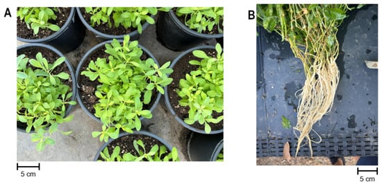
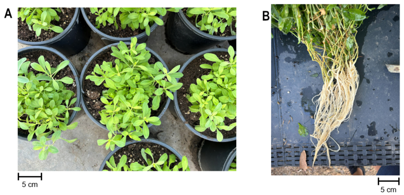
The biochemical characterization of S. vulgaris revealed clear tissue-specific differences between leaves and roots, confirming that the aerial and underground organs represent distinct metabolic compartments (Figure 1). Although such divergence is expected, this analysis was essential to verify the biochemical identity of the extracts used in the biostimulation treatments.
Figure 1.
Silene vulgaris leaves (A) and roots (B).
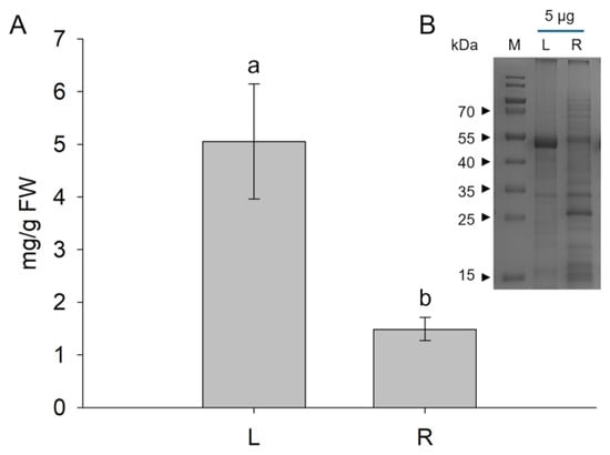
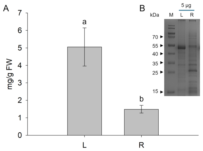
Protein quantification showed that leaves accumulated substantially higher protein levels (5 mg/g FW) compared to roots (1.5 mg/g FW) and SDS-PAGE profiles further highlighted their metabolic specialization (Figure 2). Leaf extracts were characterized by the predominance of a major protein band at approximately 45 kDa, likely corresponding to the Rubisco large subunit, consistent with their photosynthetic function. In contrast, root tissues displayed a different protein pattern dominated by an approximately 20 kDa band, indicating a markedly different protein composition and supporting the rationale for testing leaf and root extracts as separate biostimulant treatments.
Figure 2.
Total soluble protein content (A) and SDS-PAGE analysis (B) for leaf (L) and root (R) tissues of S. vulgaris. Values are presented as mean ± SD of biological replicates (n = 3). Different letters indicate statistically significant differences among treatments according to Tukey’s multiple comparison test (p < 0.05).
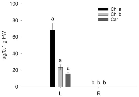
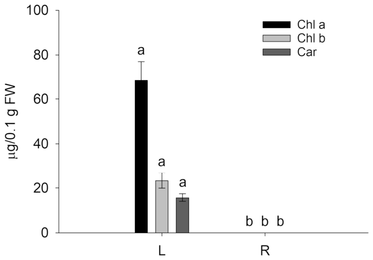
Pigment analysis further confirmed the functional differentiation between leaf and root tissues. Leaf tissues exhibited a typical photosynthetic pigment profile, with chlorophyll a (Chl a) representing the predominant photosynthetic pigment and a Chl a:b ratio of approximately 3:1, consistent with values reported for physiologically active green tissues in higher plants. Total chlorophyll content reached 108 µg/0.1 g FW (Figure 3) and was approximately 1.2-fold higher than carotenoid content, indicating an active photosynthetic apparatus and balanced pigment composition. As expected, no photosynthetic pigments were detectable in root tissues, confirming their non-photosynthetic nature.
Figure 3.
Pigment content as Chl a, Chl b and Car determination, in leaves (L) and roots (R) of S. vulgaris. Values are presented as mean ± SD of biological replicates (n = 5). Different letters indicate statistically significant differences among treatments according to Tukey’s multiple comparison test (p < 0.05).
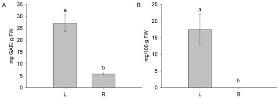
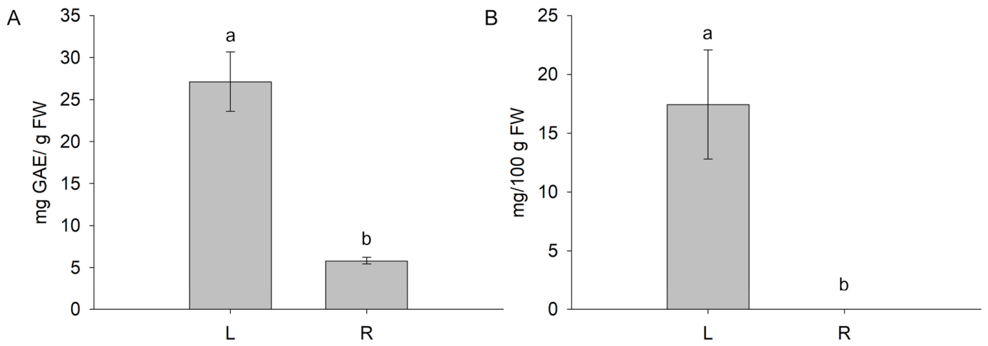
A pronounced accumulation of antioxidant-related metabolites was also observed in leaves. Total polyphenol content (TPC) was approximately five-fold higher in leaves than in roots (Figure 4A), confirming that aerial tissues represent the primary site of phenolic compound accumulation. Similarly, ascorbic acid was readily detected in leaf tissues, whereas negligible levels were observed in roots (Figure 4B).
Figure 4.
Total polyphenol (A) and ascorbic acid (B) contents in leaves (L) and roots (R) of S. vulgaris. Values are presented as mean ± SD of biological replicates (TPC, n = 4; ascorbic acid, n = 3). Different letters indicate statistically significant differences among treatments according to Tukey’s multiple comparison test (p < 0.05).
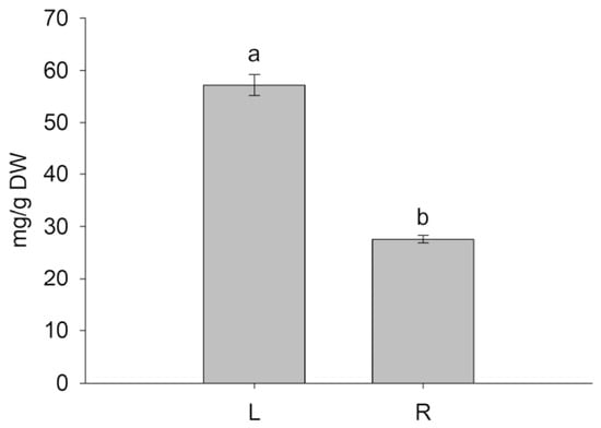
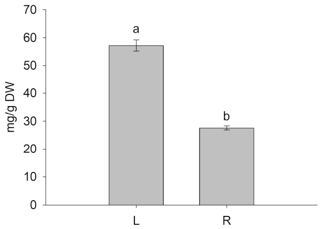
Total saponin analysis revealed the presence of these metabolites in both tissues; however, leaves accumulated approximately three-fold higher levels than roots (Figure 5).
Figure 5.
Total saponins in leaves (L) and roots (R) of S. vulgaris. Values are presented as mean ± SD of biological replicates (n = 3). Different letters indicate statistically significant differences among treatments according to Tukey’s multiple comparison test (p < 0.05).
3.2. Effects of S. vulgaris Extracts on Lettuce Growth in Physiological Conditions
To investigate the potential biostimulant activity of S. vulgaris extracts, lettuce plants (Lactuca sativa cv. Canasta) were treated with leaf (LES) and root (RES) extracts through irrigation.
No symptoms of phytotoxicity, such as chlorosis, necrosis or growth abnormalities, were observed in any experimental group (Supplementary Figure S1).
3.3. Plant Growth and Biomass Accumulation
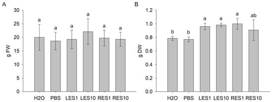
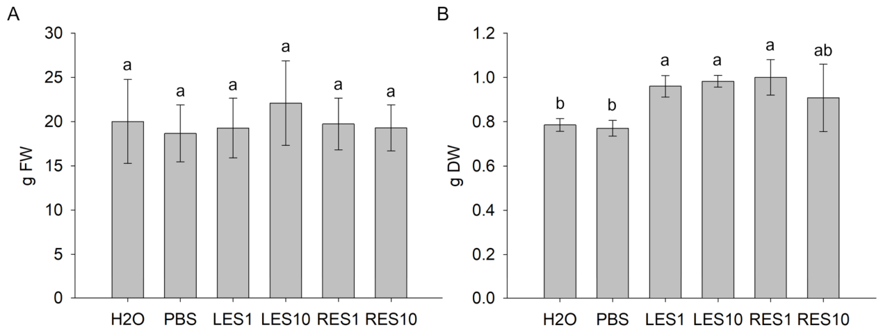
The effect of S. vulgaris extracts on lettuce growth was evaluated by measuring fresh and dry biomass at the end of the treatment period. No statistically significant differences were observed in fresh weight among treatments, with an average biomass of approximately 19.8 g per plant across all experimental groups (Figure 6A). Conversely, plants treated with the leaf (LES1 and LES10) and with the lower concentration of root extracts (RES1) exhibited a significant increase in dry biomass compared with both H2O- and PBS-treated controls (Figure 6B). The selective increase in dry weight observed in LES1-, LES10- and RES1-treated plants, despite the absence of significant differences in fresh biomass, indicates that the treatments affected dry matter accumulation. However, the relative contribution of different structural and metabolic constituents to this response remains to be determined.
Figure 6.
Fresh (A) and dry (B) weights of lettuce plants treated with leaf (LES) and root (RES) extracts of S. vulgaris at two concentrations (1 and 10 mL/L). Values are presented as mean ± SD of biological replicates (fresh weight, n = 11; dry weight, n = 4). Different letters indicate statistically significant differences among treatments according to Tukey’s multiple comparison test (p < 0.05).
3.4. Photosynthetic Pigments
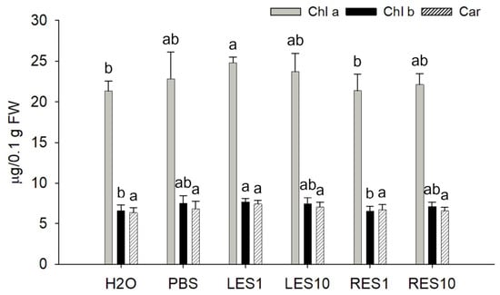
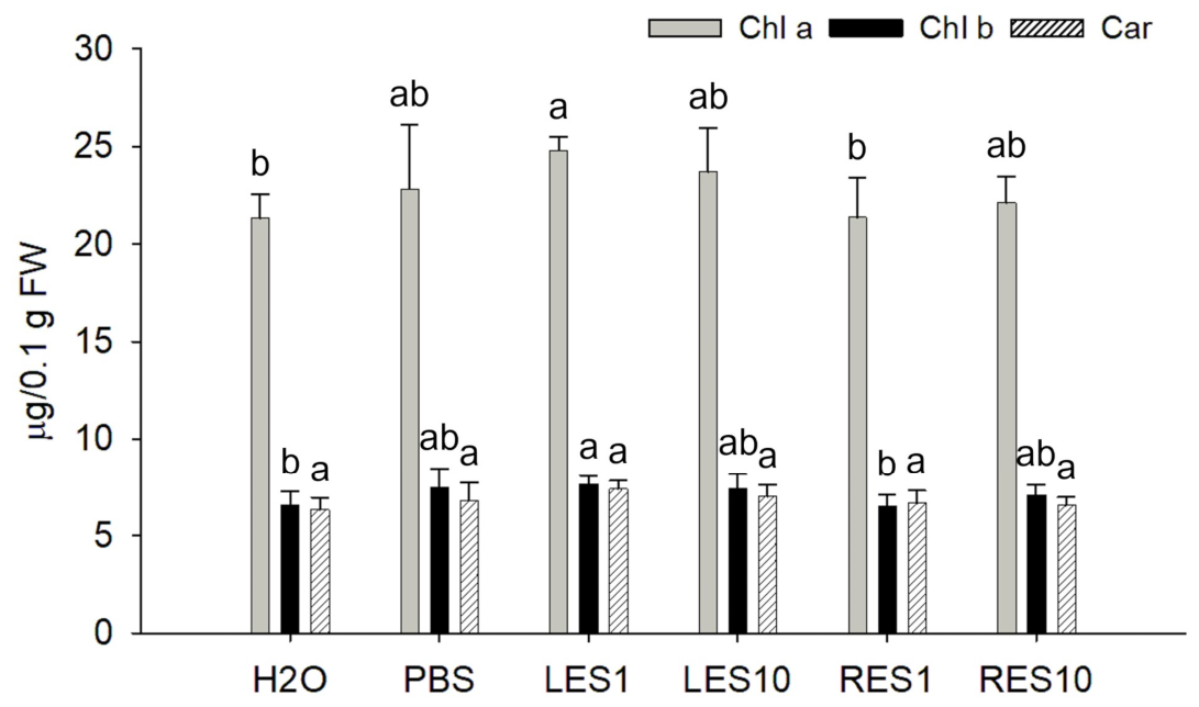
To evaluate the effects of S. vulgaris extracts on the physiological status of lettuce plants, chlorophyll and carotenoid contents were spectrophotometrically determined. Plants treated with the lower concentration of leaf extract (LES1) showed a statistically significant increase in chlorophyll a and b contents of up to 16% compared with H2O-treated controls (Figure 7). LES10- and RES10-treated plants displayed intermediate values that were not significantly different from either H2O- or PBS-treated plants (control groups). Root extracts at lower concentrations (RES1) did not significantly affect chlorophyll a and b contents compared with controls. Carotenoid levels remained unchanged across all the analyzed treatments and the chlorophyll-to-carotenoid (Chls:Car) ratio was stable, indicating that S. vulgaris extracts did not substantially alter the overall photosynthetic pigment balance of lettuce leaves. Together, these results show that LES1 moderately enhanced chlorophyll accumulation without affecting carotenoid content or pigment stoichiometry.
Figure 7.
Pigment content of lettuce plants treated with leaf (LES) and root (RES) extracts of S. vulgaris at two concentrations (1 and 10 mL/L). Values are expressed as mean ± SD (n = 7). Different letters indicate statistically significant differences among treatments according to Tukey’s multiple comparison test (p < 0.05).
3.5. Total Soluble Protein Content
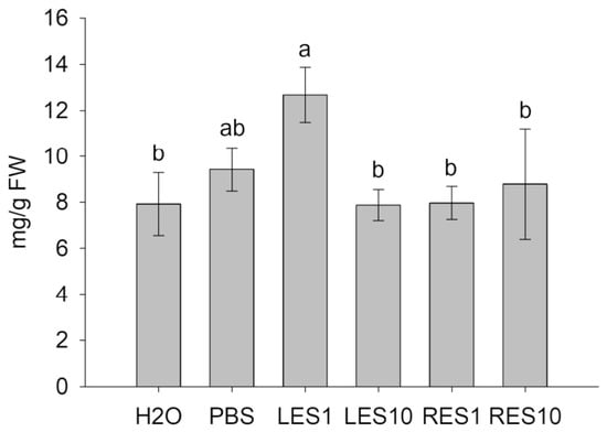
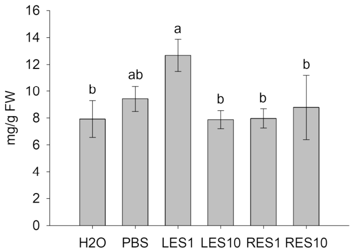
To investigate whether the increase in dry biomass was associated with changes in protein accumulation, total soluble protein content was quantified in lettuce tissues. Plants treated with the lower concentration of leaf extract (LES1) showed a significant increase in soluble protein content (up to 60%) compared with H2O-treated controls (Figure 8), with values comparable to those observed in PBS-treated plants. Conversely, LES10-treated plants displayed protein levels similar to H2O-treated controls, with no significant increase detected. Root extracts (RES1 and RES10) did not induce significant changes in total soluble protein content compared with control plants. Overall, the increase in soluble protein content observed in LES1-treated plants was consistent with the higher dry biomass detected under the same treatment, although these findings do not establish a direct contribution of soluble proteins to the increase in dry biomass.
Figure 8.
Total protein content of lettuce plants treated with leaf (LES) and root (RES) extracts of S. vulgaris at two concentrations (1 and 10 mL/L). Values are expressed as mean ± SD (n = 3). Different letters indicate significant differences among treatments according to Tukey’s multiple comparison test (p < 0.05).
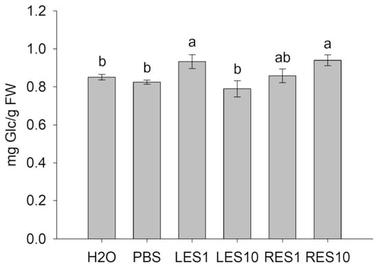
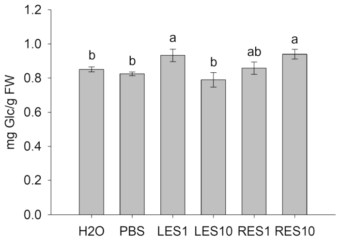
3.6. Total Soluble Sugar Content
To further investigate the biochemical changes associated with the increase in dry biomass, total soluble sugars were quantified in lettuce tissues. Plants treated with LES1 and RES10 exhibited significantly higher soluble sugar levels than both H2O- and PBS-treated controls (Figure 9). In LES1-treated plants, the increase in soluble sugars was consistent with the higher dry biomass observed under the same treatment. RES10 also increased soluble sugar content, although this response was not accompanied by a significant increase in dry biomass. In contrast, LES10 and RES1 treatments did not induce significant changes in total soluble sugar content.
Figure 9.
Total soluble sugar content in lettuce plants treated with leaf (LES) and root (RES) extracts of S. vulgaris at two concentrations (1 and 10 mL/L). Values are expressed as mean ± SD (n = 3). Different letters indicate significant differences among treatments according to Tukey’s multiple comparison test (p < 0.05).
3.7. Antioxidative Compounds and Antioxidant/Scavenging Activity
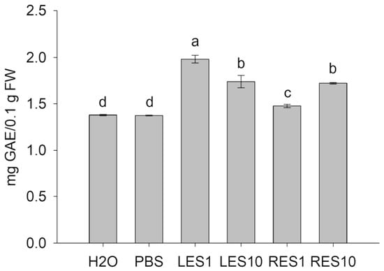
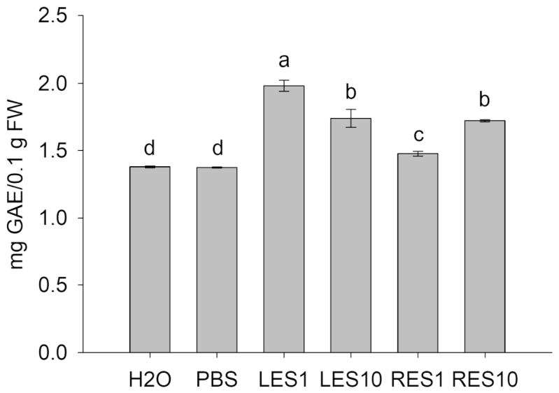
To evaluate whether S. vulgaris extracts affected the accumulation of antioxidant metabolites, total phenolic content (TPC) was evaluated using the Folin–Ciocalteu assay. All treatments with S. vulgaris extracts resulted in a statistically significant increase in TPC compared with controls, with LES1 showing the strongest response, since it displayed values up to 44% higher than control plants (Figure 10). This indicates that both leaf and root extracts increased total phenolic content, although the stimulatory effect was most pronounced at the lower leaf extract concentration.
Figure 10.
Total phenolic content (TPC) in lettuce plants treated with leaf (LES) and root (RES) extracts of S. vulgaris at two concentrations (1 and 10 mL/L). Values are expressed as mean ± SD (n = 4). Different letters indicate significant differences among treatments according to Tukey’s multiple comparison test (p < 0.05).
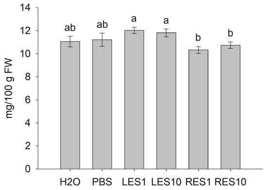
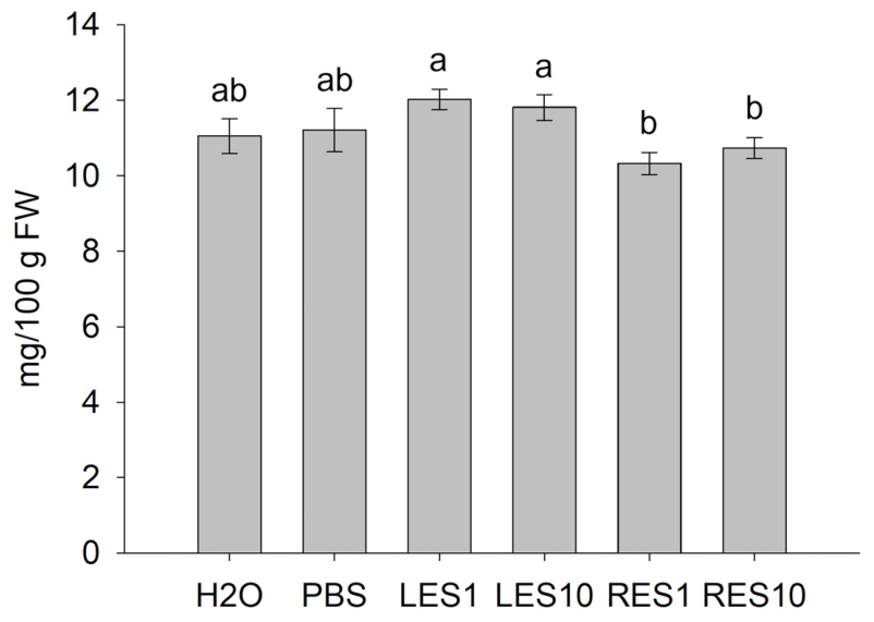
Ascorbic acid, a major antioxidant compound in lettuce, was also quantified. Treatments did not significantly affect vitamin C levels (Figure 11).
Figure 11.
Ascorbic acid content in lettuce plants treated with leaf (LES) and root (RES) extracts of S. vulgaris at two concentrations (1 and 10 mL/L). Values are expressed as mean ± SD (n = 3). Different letters indicate significant differences among treatments according to Tukey’s multiple comparison test (p < 0.05).
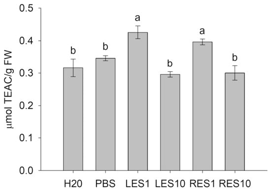
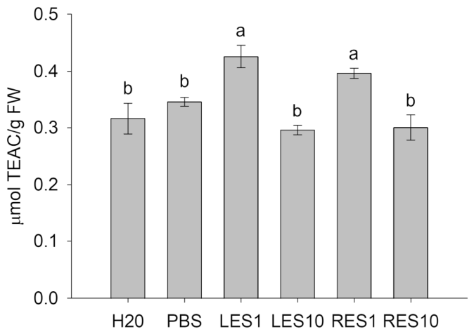
To evaluate whether the biochemical changes induced by the treatments were associated with enhanced antioxidant capacity, ABTS radical scavenging activity was assessed. Plants treated with LES1 and RES1 showed significantly higher antioxidant activity than all other treatments, including both H2O- and PBS-treated controls (Figure 12). In contrast, LES10- and RES10-treated plants did not show significant differences relative to controls. These findings indicate that, among the two concentrations tested, 1 mL/L was more effective in enhancing antioxidant capacity for both leaf and root extracts.
Figure 12.
Antioxidant activity (ABTS assay) of lettuce plants treated with leaf (LES) and root (RES) extracts of S. vulgaris at two concentrations (1 and 10 mL/L). Values are expressed as mean ± SD (n = 3). Different letters indicate significant differences among treatments according to Tukey’s multiple comparison test (p < 0.05).
4. Discussion
Wild plant species remain largely unexplored as potential sources of plant biostimulants, despite their richness in specialized metabolites with potential agricultural applications. In this context, the present study investigated the biochemical composition of Silene vulgaris leaf and root tissues and evaluated, for the first time to the best of our knowledge, the biostimulant potential of their crude extracts on lettuce (Lactuca sativa cv. Canasta).
The biochemical characterization of S. vulgaris revealed marked differences between leaf and root tissues, with leaves showing higher levels of proteins, phenolic compounds, ascorbic acid and saponins. These contrasting patterns reflect the distinct functional specialization of aerial and underground organs [42].
Several Silene species have a long history of traditional medicinal and nutritional use. Nevertheless, the phytochemical composition of many Silene species remains incompletely characterized, and quantitative information on the distribution of these compounds among different plant organs is still limited. This is particularly relevant for S. vulgaris, for which the available evidence supports the occurrence of marked tissue-dependent differences. Mohti et al. [43], for example, reported total phenolic content approximately nine-fold higher in leaf than in root extracts of S. vulgaris, consistent with the higher phenolic content observed in leaves in the present study. Variation in phenolic content has also been reported among other wild Silene species [44]. However, absolute values cannot be directly compared across studies because of differences in plant material, extraction procedures and the basis used to express phenolic content. In particular, Mohti et al. [43] and Zengin et al. [44] expressed total phenolic content per gram of extract, whereas in the present study it was expressed relative to fresh tissue weight.
Mohti et al. [43] also reported higher antioxidant activity in leaf than in root extracts of Silene vulgaris subsp. macrocarpa. The greater accumulation of phenolic compounds in aerial tissues may be related to their protective functions in plants. For example, the exposure to UV radiation has been shown to stimulate the accumulation of phenolic compounds in Silene littorea [45]. More generally, phenolic compounds contribute to antioxidant defense and photoprotection, and their accumulation can be induced by environmental stresses, such as UV radiation, excess light, drought and temperature extremes [46].
Triterpenoid saponins are another hallmark of Silene spp., although their composition and abundance vary considerably among species and plant organs [47,48,49,50].
All tested concentrations of S. vulgaris extracts were well tolerated by lettuce plants, since no visible phytotoxic symptoms were observed. The absence of visible phytotoxic effects is an important prerequisite for the development of novel plant biostimulants and is consistent with previous studies conducted with plant-derived protein hydrolysates, seaweed extracts and microbial-derived formulations [7,51].
No significant differences were observed in fresh biomass accumulation among treatments. However, lettuce plants treated with LES1, LES10 and RES1 showed a significant increase in dry biomass. Since fresh biomass comprises both water and dry matter, these parameters do not necessarily respond in parallel, and their relationship is influenced by tissue hydration [52]. Similar observations have been reported for several classes of plant biostimulants, whose effects may be more pronounced on dry matter accumulation and plant metabolism than on fresh biomass production [6,7,16,51,53]. Moreover, previous studies on lettuce have highlighted the variability of biomass responses to plant-derived protein hydrolysates, depending on the cultivar and specific treatment conditions [16].
Photosynthetic pigments are commonly used as indicators of plant physiological status. In the present study, the low concentration of leaf extract (LES1) significantly enhanced chlorophyll content compared with H2O-control, whereas carotenoid content remained unchanged. Similar responses have been reported in lettuce treated with other plant-derived biostimulants [15,53,54]. Although higher chlorophyll content may be associated with changes in the photosynthetic apparatus [17,55], it does not necessarily indicate enhanced photosynthetic activity, which was not directly assessed in the present study.
The increase in soluble sugar concentration detected in LES1- and RES10-treated plants suggests changes in carbohydrate metabolism, consistent with responses previously reported for protein hydrolysate-based biostimulants [7,56]. Similarly, transcriptomic analysis of drought-stressed potato plants treated with Trichoderma-based biostimulants revealed the modulation of genes involved in carbon fixation and sucrose and starch metabolism [11], suggesting that carbohydrate-related pathways may be responsive to different types of biostimulants. LES1 treatment also increased soluble protein content in lettuce as compared with H2O-control. The concurrent increases in protein and soluble sugar concentrations, expressed on a fresh weight basis, suggest changes in tissue biochemical composition that may be associated with the higher dry biomass. Indeed, proteins and carbohydrates are important constituents of plant dry matter [57]. However, these measurements do not establish which dry matter constituents contributed to the observed increase in biomass. Previous studies on protein hydrolysate-, enzyme- and Trichoderma-based biostimulants have reported changes in plant growth and metabolic processes, including nitrogen assimilation and carbon metabolism [11,51,56,58,59].
Treatments with S. vulgaris extracts affected antioxidant-related traits in lettuce plants, although the responses differed according to extract type and concentration. Total phenolic content increased following both leaf and root extract applications, whereas significantly higher ABTS antioxidant capacity was observed specifically in plants treated with LES1 and RES1. These findings suggest that the extracts may influence the accumulation of antioxidant-related compounds, although the biochemical pathways responsible for these changes were not investigated.
The observed increase in phenolic content is particularly relevant as phenolic compounds, together with vitamin C, are among the major determinants of lettuce nutritional quality and antioxidant capacity [60,61,62]. Enhancement of antioxidant-related metabolites is therefore considered a desirable outcome in modern biostimulant research, where the focus has progressively shifted from yield improvement to quality enhancement and metabolic priming [6,53].
The differences in biochemical composition observed between S. vulgaris leaves and roots may contribute to the distinct responses elicited by their respective extracts. Although the specific bioactive compounds involved remain unidentified, previous studies have shown that plant-derived biostimulants rich in secondary metabolites can modulate antioxidant metabolism and promote the accumulation of bioactive compounds in crops [6,14,53]. In this context, phenolic compounds are known to contribute to antioxidant metabolism and ROS scavenging, whereas saponins participate in constitutive and inducible defense mechanisms against biotic and abiotic stresses [46,63].
Apart from extract composition, concentration may also influence the observed responses. Interestingly, LES1 showed more pronounced effects on some of the physiological and biochemical parameters measured than LES10. Similar concentration-dependent effects have been reported for other plant biostimulants [6,7,64,65]. In particular, the highest concentrations of both leaf and root extracts, namely, LES10 and RES10, did not increase antioxidant capacity compared with the controls, unlike the corresponding treatments at 1 mL/L. However, additional concentrations would need to be tested to establish a dose–response relationship [6,66]. Interestingly, root extracts also affected several lettuce traits despite the lower levels of the metabolites quantified in the present work. RES1 increased dry biomass, phenolic content and antioxidant activity, whereas RES10 increased soluble sugar and phenolic contents. These responses may reflect the contribution of compounds not included in the present biochemical characterization or interactions among extract components.
Some limitations of the present study should be considered when interpreting these results. Although differences in the biochemical composition of S. vulgaris leaves and roots were identified, the specific bioactive compounds responsible for the effects observed in lettuce could not be identified in the present study. Moreover, the experiment was conducted on a single lettuce cultivar and included only two S. vulgaris extract concentrations. Finally, photosynthetic performance and other physiological processes potentially associated with the observed changes in biomass and biochemical traits were not directly measured. Therefore, the present results describe treatment-associated growth and biochemical responses but do not establish the physiological or molecular mechanisms underlying these effects.
5. Conclusions
Overall, the results indicate that S. vulgaris leaf and root extracts can influence lettuce growth and metabolism, with responses depending on the extract type, concentration and specific biochemical trait assessed. In particular, the observed effects on dry matter accumulation, photosynthetic pigments, soluble proteins, sugars, phenolic compounds and antioxidant capacity varied across treatments, indicating that these extracts may modulate plant physiological and metabolic processes. These findings highlight the potential of S. vulgaris as a source of plant-derived biostimulants, although the mechanisms underlying the observed effects remain unclear. Future research should aim to elucidate the physiological and molecular mechanisms involved, identify the bioactive constituents responsible and investigate their possible interactions. Such investigations could support the development and optimization of S. vulgaris-derived biostimulants for improving the performance and quality of horticultural crops.
Supplementary Materials
The following supporting information can be downloaded at https://www.mdpi.com/article/10.3390/agronomy16212183/s1, Supplementary Figure S1: Representative lettuce plants treated with leaf (LES) and root (RES) extracts of S. vulgaris compared with H2O-treated control. Extracts were applied at two concentrations (1 and 10 mL/L).
Author Contributions
Conceptualization, R.T.; methodology, R.T.; validation, R.T., P.I., E.G. and G.B.; investigation, R.T., G.B. and A.L.M.; resources, G.B.; writing—original draft preparation, R.T. and P.I.; writing—review and editing, R.T., P.I. and E.G.; visualization, R.T.; supervision, R.T.; project administration, R.T.; funding acquisition, R.T. All authors have read and agreed to the published version of the manuscript.
Funding
This research received no external funding.
Data Availability Statement
The original contributions presented in this study are included in the article/Supplementary Materials. Further inquiries can be directed to the corresponding author.
Acknowledgments
We thank Matteo Mari (CNR-IBE, Bologna, Italy) for assistance in plant growth and Massimiliano Magli (CNR-IBE, Bologna, Italy) for assistance in statistical analysis. The authors gratefully acknowledge Stefano Predieri for providing the plant material and for the valuable inspiration that contributed to the development of this study.
Conflicts of Interest
The authors declare no conflicts of interest.
Abbreviations
The following abbreviations are used in this manuscript:
| ABTS | 2, 2′-azino bis(3-ethylbenzothiazoline-6-sulfonic acid) |
| LES | Leaf extract of Silene vulgaris |
| RES | Root extract of Silene vulgaris |
| TPC | Total polyphenol content |
References
- FAO. The Future of Food and Agriculture—Drivers and Triggers for Transformation; The Future of Food and Agriculture; FAO: Rome, Italy, 2022. [Google Scholar]
- Ma, Y.; Freitas, H.; Dias, M.C. Strategies and Prospects for Biostimulants to Alleviate Abiotic Stress in Plants. Front. Plant Sci. 2022, 13, 1024243. [Google Scholar] [CrossRef] [Scilit] [PubMed]
- Di Sario, L.; Boeri, P.; Matus, J.T.; Pizzio, G.A. Plant Biostimulants to Enhance Abiotic Stress Resilience in Crops. Int. J. Mol. Sci. 2025, 26, 1129. [Google Scholar] [CrossRef] [Scilit] [PubMed]
- du Jardin, P. Plant Biostimulants: Definition, Concept, Main Categories and Regulation. Sci. Hortic. 2015, 196, 3–14. [Google Scholar] [CrossRef] [Scilit]
- Van Oosten, M.J.; Pepe, O.; De Pascale, S.; Silletti, S.; Maggio, A. The Role of Biostimulants and Bioeffectors as Alleviators of Abiotic Stress in Crop Plants. Chem. Biol. Technol. Agric. 2017, 4, 5. [Google Scholar] [CrossRef] [Scilit]
- Rouphael, Y.; Colla, G. Editorial: Biostimulants in Agriculture. Front. Plant Sci. 2020, 11, 40. [Google Scholar] [CrossRef] [Scilit] [PubMed]
- Colla, G.; Hoagland, L.; Ruzzi, M.; Cardarelli, M.; Bonini, P.; Canaguier, R.; Rouphael, Y. Biostimulant Action of Protein Hydrolysates: Unraveling Their Effects on Plant Physiology and Microbiome. Front. Plant Sci. 2017, 8, 2202. [Google Scholar] [CrossRef] [Scilit] [PubMed]
- Hasanuzzaman, M.; Parvin, K.; Bardhan, K.; Nahar, K.; Anee, T.I.; Masud, A.A.C.; Fotopoulos, V. Biostimulants for the Regulation of Reactive Oxygen Species Metabolism in Plants under Abiotic Stress. Cells 2021, 10, 2537. [Google Scholar] [CrossRef] [Scilit] [PubMed]
- Kaushal, P.; Ali, N.; Saini, S.; Pati, P.K.; Pati, A.M. Physiological and Molecular Insight of Microbial Biostimulants for Sustainable Agriculture. Front. Plant Sci. 2023, 14, 1041413. [Google Scholar] [CrossRef] [Scilit] [PubMed]
- Carillo, P. Can Biostimulants Enhance Plant Resilience to Heat and Water Stress in the Mediterranean Hotspot? Plant Stress 2025, 16, 100802. [Google Scholar] [CrossRef] [Scilit]
- Tamburino, R.; Sannino, L.; Palomba, F.; Russo, E.; Esposito, M.C.; Sacco, A.; Ruocco, M.; Scotti, N. Genotype-Specific Transcriptomic Response to Drought Stress in Potato Cultivars Modulated by Microbial Biostimulants. BMC Plant Biol. 2025, 25, 1777. [Google Scholar] [CrossRef] [Scilit] [PubMed]
- Nephali, L.; Piater, L.A.; Dubery, I.A.; Patterson, V.; Huyser, J.; Burgess, K.; Tugizimana, F. Biostimulants for Plant Growth and Mitigation of Abiotic Stresses: A Metabolomics Perspective. Metabolites 2020, 10, 505. [Google Scholar] [CrossRef] [Scilit] [PubMed]
- Gechev, T. Genomics Control of Biostimulant-Induced Stress Tolerance and Crop Yield Enhancement. Plant J. 2025, 123, e70382. [Google Scholar] [CrossRef] [Scilit] [PubMed]
- Bulgari, R.; Morgutti, S.; Cocetta, G.; Negrini, N.; Farris, S.; Calcante, A.; Spinardi, A.; Ferrari, E.; Mignani, I.; Oberti, R.; et al. Evaluation of Borage Extracts as Potential Biostimulant Using a Phenomic, Agronomic, Physiological, and Biochemical Approach. Front. Plant Sci. 2017, 8, 935. [Google Scholar] [CrossRef] [Scilit] [PubMed]
- Puglisi, I.; La Bella, E.; Rovetto, E.I.; Lo Piero, A.R.; Baglieri, A. Biostimulant Effect and Biochemical Response in Lettuce Seedlings Treated with a Scenedesmus quadricauda Extract. Plants 2020, 9, 123. [Google Scholar] [CrossRef] [Scilit] [PubMed]
- Cristofano, F.; El-Nakhel, C.; Pannico, A.; Giordano, M.; Colla, G.; Rouphael, Y. Foliar and Root Applications of Vegetal-Derived Protein Hydrolysates Differentially Enhance the Yield and Qualitative Attributes of Two Lettuce Cultivars Grown in Floating System. Agronomy 2021, 11, 1194. [Google Scholar] [CrossRef] [Scilit]
- Monterisi, S.; Garcia-Perez, P.; Buffagni, V.; Zuluaga, M.Y.A.; Ciriello, M.; Formisano, L.; El-Nakhel, C.; Cardarelli, M.; Colla, G.; Rouphael, Y.; et al. Unravelling the Biostimulant Activity of a Protein Hydrolysate in Lettuce Plants under Optimal and Low N Availability: A Multi-Omics Approach. Physiol. Plant 2024, 176, e14357. [Google Scholar] [CrossRef] [Scilit] [PubMed]
- Hassan, M.W.; Yasmeen, A.; Nawaz, H.; Khalofah, A. Moringa Bio-Stimulant Promoted Cotton Production via Coordinating Anti-Oxidants and Physiological Behaviors to Combat Reproductive Drought Stress. Sci. Rep. 2025, 16, 3425. [Google Scholar] [CrossRef] [Scilit] [PubMed]
- Zulfiqar, F.; Casadesús, A.; Brockman, H.; Munné-Bosch, S. An Overview of Plant-Based Natural Biostimulants for Sustainable Horticulture with a Particular Focus on Moringa Leaf Extracts. Plant Sci. 2020, 295, 110194. [Google Scholar] [CrossRef] [Scilit] [PubMed]
- Raza, Q.-U.-A.; Bashir, M.A.; Rehim, A.; Ejaz, R.; Raza, H.M.A.; Shahzad, U.; Ahmed, F.; Geng, Y. Biostimulants Induce Positive Changes in the Radish Morpho-Physiology and Yield. Front. Plant Sci. 2022, 13, 950393. [Google Scholar] [CrossRef] [Scilit] [PubMed]
- Admane, N.; Cavallo, G.; Hadjila, C.; Cavalluzzi, M.M.; Rotondo, N.P.; Salerno, A.; Cannillo, J.; Difonzo, G.; Caponio, F.; Ippolito, A.; et al. Biostimulant Formulations and Moringa Oleifera Extracts to Improve Yield, Quality, and Storability of Hydroponic Lettuce. Molecules 2023, 28, 373. [Google Scholar] [CrossRef] [Scilit] [PubMed]
- Khan, S.; Ibrar, D.; Hasnain, Z.; Nawaz, M.; Rais, A.; Ullah, S.; Gul, S.; Siddiqui, M.H.; Irshad, S. Moringa Leaf Extract Mitigates the Adverse Impacts of Drought and Improves the Yield and Grain Quality of Rice through Enhanced Physiological, Biochemical, and Antioxidant Activities. Plants 2023, 12, 2511. [Google Scholar] [CrossRef] [Scilit] [PubMed]
- Bhowmick, S.; Rai, G.; Mishra, S.K.; Bisht, N.; Chauhan, P.S. Bio-Stimulants from Medicinally and Nutritionally Significant Plant Extracts Mitigate Drought Adversities in Zea mays through Enhanced Physiological, Biochemical, and Antioxidant Activities. Plant Physiol. Biochem. 2024, 207, 108396. [Google Scholar] [CrossRef] [Scilit] [PubMed]
- Irshad, S.; Matloob, A.; Mehmood, K.; Nawaz, M.; Iqbal, S.; Wahid, M.A.; Ikram, R.M.; Ghafoor, M.A.; Siddiqui, M.H.; Alamri, S.; et al. Moringa Dried Leaf Extract as Bio-Foliar Fertilizer for Revitalizing Performance and Nutritional Status of Soybean. Sci. Rep. 2025, 15, 20431. [Google Scholar] [CrossRef] [Scilit] [PubMed]
- Saleh, H.A.; Tourky, S.M.N.; Ibraheem, F.; Abo-Hamed, S.A.; Shukry, W.M.; Elgamal, W.H.; Elghareeb, E.M. Enhancing Salinity Tolerance in Cultivated Rice through Introgression of African Rice Genes and Application of Moringa Leaf Extract. BMC Plant Biol. 2025, 25, 163. [Google Scholar] [CrossRef] [Scilit] [PubMed]
- Berouaken, A.; Calvano, C.D.D.; Schena, A.; Pascuzzi, S.; Paciolla, F.; Cocozza, C.; Losciale, P.; Ugenti, V.; di Leo, G.; Simini, A.; et al. A Moringa oleifera-Based Formulation for Growth and Mitigation of Drought Stress in Tomato Plants. Front. Plant Sci. 2026, 17, 1771042. [Google Scholar] [CrossRef] [Scilit] [PubMed]
- Desoky, E.-S.M.; ElSayed, A.I.; Merwad, A.-R.M.A.; Rady, M.M. Stimulating Antioxidant Defenses, Antioxidant Gene Expression, and Salt Tolerance in Pisum sativum Seedling by Pretreatment Using Licorice Root Extract (LRE) as an Organic Biostimulant. Plant Physiol. Biochem. 2019, 142, 292–302. [Google Scholar] [CrossRef] [Scilit] [PubMed]
- Rady, M.M.; Desoky, E.-S.M.; Elrys, A.S.; Boghdady, M.S. Can Licorice Root Extract Be Used as an Effective Natural Biostimulant for Salt-Stressed Common Bean Plants? S. Afr. J. Bot. 2019, 121, 294–305. [Google Scholar] [CrossRef] [Scilit]
- D’Amelia, V.; Docimo, T.; Crocoll, C.; Rigano, M.M. Specialized Metabolites and Valuable Molecules in Crop and Medicinal Plants: The Evolution of Their Use and Strategies for Their Production. Genes 2021, 12, 936. [Google Scholar] [CrossRef] [Scilit] [PubMed]
- Jafari, F.; Zarre, S.; Gholipour, A.; Eggens, F.; Rabeler, R.K.; Oxelman, B. A New Taxonomic Backbone for the Infrageneric Classification of the Species-Rich Genus Silene (Caryophyllaceae). TAXON 2020, 69, 337–368. [Google Scholar] [CrossRef] [Scilit]
- Karrenberg, S.; Bačovský, V.; Berardi, A.E.; Cauwer, I.D.; Giraud, T.; Hartmann, F.E.; Hobza, R.; Hudzieczek, V.; Marais, G.A.B.; Miladin, J.R.; et al. Silene, a Versatile Model System: From Sex and Genome Evolution to Ecology and Speciation. New Phytol. 2026, 250, 3613–3630. [Google Scholar] [CrossRef] [Scilit] [PubMed]
- Buzón-Durán, L.; Sánchez-Hernández, E.; Martín-Ramos, P.; Navas-Gracia, L.M.; García-González, M.C.; Oliveira, R.; Martín-Gil, J. Silene uniflora Extracts for Strawberry Postharvest Protection. Plants 2023, 12, 1846. [Google Scholar] [CrossRef] [Scilit] [PubMed]
- Bensaad, M.S.; Allaoua, Z.; Kahoul, M.A.; Pinto, D.C.G.A.; Bensouici, C.; Akkal, S. GC-Ms, UHPLC–MS/MSn Phytoconstituents Characterization and Physio-pharmacological Investigation of Silene colorata subsp. amphorina (Pomel) Batt. From Northeastern Algeria. Chem. Biodivers. 2026, 23, e71578. [Google Scholar] [CrossRef] [Scilit] [PubMed]
- Bilal, M.N.; Kutluay, V.M. Silene Genus: A Review of Its Traditional Uses, Phytochemistry and Pharmacology. Phytochem. Rev. 2026, 25, 2659–2743. [Google Scholar] [CrossRef] [Scilit]
- Lichtenthaler, H.K. [34] Chlorophylls and Carotenoids: Pigments of Photosynthetic Biomembranes. In Methods in Enzymology; Plant Cell Membranes; Academic Press: Cambridge, MA, USA, 1987; Volume 148, pp. 350–382. [Google Scholar] [CrossRef] [Scilit]
- Celano, R.; Piccinelli, A.L.; Pagano, I.; Roscigno, G.; Campone, L.; De Falco, E.; Russo, M.; Rastrelli, L. Oil Distillation Wastewaters from Aromatic Herbs as New Natural Source of Antioxidant Compounds. Food Res. Int. 2017, 99, 298–307. [Google Scholar] [CrossRef] [Scilit] [PubMed]
- Breniere, T.; Fanciullino, A.-L.; Dumont, D.; Le Bourvellec, C.; Riva, C.; Borel, P.; Landrier, J.-F.; Bertin, N. Effect of Long-Term Deficit Irrigation on Tomato and Goji Berry Quality: From Fruit Composition to In Vitro Bioaccessibility of Carotenoids. Front. Plant Sci. 2024, 15, 1339536. [Google Scholar] [CrossRef] [Scilit] [PubMed]
- Le Bot, M.; Thibault, J.; Pottier, Q.; Boisard, S.; Guilet, D. An Accurate, Cost-Effective and Simple Colorimetric Method for the Quantification of Total Triterpenoid and Steroidal Saponins from Plant Materials. Food Chem. 2022, 383, 132597. [Google Scholar] [CrossRef] [Scilit] [PubMed]
- Denova-Lozano, P.; Chamorro-Flores, A.; Cerón-Carpio, A.B.; Arroyo-Becerra, A. Determination of Total Soluble Sugars in Pteridophytes Using the Anthrone Method. Curr. Protoc. 2026, 6, e70373. [Google Scholar] [CrossRef] [Scilit] [PubMed]
- Chen, Z.; Jiang, X.; Liang, G. Non-Covalent Complexes of Chlorella pyrenoidosa Antioxidant Peptides with Chlorogenic Acid: Structure-Activity Relationships and Potential Synergistic Cytoprotection. Food Chem. 2026, 532, 151526. [Google Scholar] [CrossRef] [Scilit] [PubMed]
- Franzoni, G.; Ferrante, A. Plant Extract Improves Quality Traits of Green and Red Lettuce Cultivars. Heliyon 2024, 10, e39224. [Google Scholar] [CrossRef] [Scilit] [PubMed]
- Monson, R.K.; Trowbridge, A.M.; Lindroth, R.L.; Lerdau, M.T. Coordinated Resource Allocation to Plant Growth–Defense Tradeoffs. New Phytol. 2022, 233, 1051–1066. [Google Scholar] [CrossRef] [Scilit] [PubMed]
- Mohti, H.; Taviano, M.F.; Cacciola, F.; Dugo, P.; Mondello, L.; Zaid, A.; Cavò, E.; Miceli, N. Silene vulgaris subsp. Macrocarpa Leaves and Roots from Morocco: Assessment of the Efficiency of Different Extraction Techniques and Solvents on Their Antioxidant Capacity, Brine Shrimp Toxicity and Phenolic Characterization. Plant Biosyst.—Int. J. Deal. All Asp. Plant Biol. 2020, 154, 692–699. [Google Scholar] [CrossRef] [Scilit]
- Zengin, G.; Mahomoodally, M.F.; Aktumsek, A.; Ceylan, R.; Uysal, S.; Mocan, A.; Yilmaz, M.A.; Picot-Allain, C.M.N.; Ćirić, A.; Glamočlija, J.; et al. Functional Constituents of Six Wild Edible Silene Species: A Focus on Their Phytochemical Profiles and Bioactive Properties. Food Biosci. 2018, 23, 75–82. [Google Scholar] [CrossRef] [Scilit]
- Del Valle, J.C.; Buide, M.L.; Whittall, J.B.; Valladares, F.; Narbona, E. UV Radiation Increases Phenolic Compound Protection but Decreases Reproduction in Silene littorea. PLoS ONE 2020, 15, e0231611. [Google Scholar] [CrossRef] [Scilit] [PubMed]
- Saini, N.; Anmol, A.; Kumar, S.; Wani, A.W.; Bakshi, M.; Dhiman, Z. Exploring Phenolic Compounds as Natural Stress Alleviators in Plants- a Comprehensive Review. Physiol. Mol. Plant Pathol. 2024, 133, 102383. [Google Scholar] [CrossRef] [Scilit]
- Kikuchi, T.; Liu, D.; Otsuki, K.; Koike, K.; Li, W. Identification of Acidic Triterpenoid Saponins from Silene vulgaris Using a Methylation-Based Isolation and LC-MS Analysis Strategy. Chem. Pharm. Bull. 2025, 73, 179–188. [Google Scholar] [CrossRef] [Scilit] [PubMed]
- Bechkri, S.; Alabdul Magid, A.; Sayagh, C.; Berrehal, D.; Harakat, D.; Voutquenne-Nazabadioko, L.; Kabouche, Z.; Kabouche, A. Triterpene Saponins from Silene gallica Collected in North-Eastern Algeria. Phytochemistry 2020, 172, 112274. [Google Scholar] [CrossRef] [Scilit] [PubMed]
- Ün, R.N.; Masullo, M.; Karayildirim, T.; Nalbantsoy, A.; Alankus, O.; Piacente, S. Triterpene Glycosides from Silene odontopetala. Phytochemistry 2020, 176, 112404. [Google Scholar] [CrossRef] [Scilit] [PubMed]
- Mamadalieva, N.Z.; Koval, A.; Dusmuratov, M.M.; Hussain, H.; Katanaev, V.L. Chemical and Biological Characterization of Metabolites from Silene viridiflora Using Mass Spectrometric and Cell-Based Assays. Biomolecules 2024, 14, 1285. [Google Scholar] [CrossRef] [Scilit] [PubMed]
- Tamburino, R.; Docimo, T.; Sannino, L.; Gualtieri, L.; Palomba, F.; Valletta, A.; Ruocco, M.; Scotti, N. Enzyme-Based Biostimulants Influence Physiological and Biochemical Responses of Lactuca sativa L. Biomolecules 2023, 13, 1765. [Google Scholar] [CrossRef] [Scilit] [PubMed]
- Garnier, E.; Laurent, G.; Bellmann, A.; Debain, S.; Berthelier, P.; Ducout, B.; Roumet, C.; Navas, M.-L. Consistency of Species Ranking Based on Functional Leaf Traits. New Phytol. 2001, 152, 69–83. [Google Scholar] [CrossRef] [Scilit] [PubMed]
- Rouphael, Y.; Carillo, P.; Garcia-Perez, P.; Cardarelli, M.; Senizza, B.; Miras-Moreno, B.; Colla, G.; Lucini, L. Plant Biostimulants from Seaweeds or Vegetal Proteins Enhance the Salinity Tolerance in Greenhouse Lettuce by Modulating Plant Metabolism in a Distinctive Manner. Sci. Hortic. 2022, 305, 111368. [Google Scholar] [CrossRef] [Scilit]
- Rouphael, Y.; Giordano, M.; Cardarelli, M.; Cozzolino, E.; Mori, M.; Kyriacou, M.C.; Bonini, P.; Colla, G. Plant- and Seaweed-Based Extracts Increase Yield but Differentially Modulate Nutritional Quality of Greenhouse Spinach through Biostimulant Action. Agronomy 2018, 8, 126. [Google Scholar] [CrossRef] [Scilit]
- Ciriello, M.; Campana, E.; De Pascale, S.; Rouphael, Y. Implications of Vegetal Protein Hydrolysates for Improving Nitrogen Use Efficiency in Leafy Vegetables. Horticulturae 2024, 10, 132. [Google Scholar] [CrossRef] [Scilit]
- Malécange, M.; Sergheraert, R.; Teulat, B.; Mounier, E.; Lothier, J.; Sakr, S. Biostimulant Properties of Protein Hydrolysates: Recent Advances and Future Challenges. Int. J. Mol. Sci. 2023, 24, 9714. [Google Scholar] [CrossRef] [Scilit] [PubMed]
- Poorter, H.; Niinemets, Ü.; Poorter, L.; Wright, I.J.; Villar, R. Causes and Consequences of Variation in Leaf Mass per Area (LMA): A Meta-Analysis. New Phytol. 2009, 182, 565–588. [Google Scholar] [CrossRef] [Scilit] [PubMed]
- Sestili, F.; Rouphael, Y.; Cardarelli, M.; Pucci, A.; Bonini, P.; Canaguier, R.; Colla, G. Protein Hydrolysate Stimulates Growth in Tomato Coupled with N-Dependent Gene Expression Involved in N Assimilation. Front. Plant Sci. 2018, 9, 1233. [Google Scholar] [CrossRef] [Scilit] [PubMed]
- Lucini, L.; Miras-Moreno, B.; Rouphael, Y.; Cardarelli, M.; Colla, G. Combining Molecular Weight Fractionation and Metabolomics to Elucidate the Bioactivity of Vegetal Protein Hydrolysates in Tomato Plants. Front. Plant Sci. 2020, 11, 976. [Google Scholar] [CrossRef] [Scilit] [PubMed]
- Riga, P.; Benedicto, L.; Gil-Izquierdo, Á.; Collado-González, J.; Ferreres, F.; Medina, S. Diffuse Light Affects the Contents of Vitamin C, Phenolic Compounds and Free Amino Acids in Lettuce Plants. Food Chem. 2019, 272, 227–234. [Google Scholar] [CrossRef] [Scilit] [PubMed]
- De Gregorio, M.A.; Zengin, G.; Alp-Turgut, F.N.; Elbasan, F.; Ozfidan-Konakci, C.; Arikan, B.; Yildiztugay, E.; Zhang, L.; Lucini, L. Glutamate, Humic Acids and Their Combination Modulate the Phenolic Profile, Antioxidant Traits, and Enzyme-Inhibition Properties in Lettuce. Plants 2023, 12, 1822. [Google Scholar] [CrossRef] [Scilit] [PubMed]
- Shi, M.; Gu, J.; Wu, H.; Rauf, A.; Emran, T.B.; Khan, Z.; Mitra, S.; Aljohani, A.S.M.; Alhumaydhi, F.A.; Al-Awthan, Y.S.; et al. Phytochemicals, Nutrition, Metabolism, Bioavailability, and Health Benefits in Lettuce—A Comprehensive Review. Antioxidants 2022, 11, 1158. [Google Scholar] [CrossRef] [Scilit] [PubMed]
- Kundu, P.; Thorat, S.A.; Kiran, A.; Goel, K.; Zinta, G. Plant-Derived Triterpenoid Saponins: Multifaceted Roles and Bioengineering Prospects. Plant Cell Rep. 2025, 44, 172. [Google Scholar] [CrossRef] [Scilit] [PubMed]
- Cristofano, F.; El-Nakhel, C.; Rouphael, Y. Biostimulant Substances for Sustainable Agriculture: Origin, Operating Mechanisms and Effects on Cucurbits, Leafy Greens, and Nightshade Vegetables Species. Biomolecules 2021, 11, 1103. [Google Scholar] [CrossRef] [Scilit] [PubMed]
- Yuan, Y.; Dickinson, N. Revealing the Complex Interplay of Biostimulant Applications. Plants 2024, 13, 2188. [Google Scholar] [CrossRef] [Scilit] [PubMed]
- Mpai, S.; Mokganya, L.M.; Raphoko, L.; Masoko, P.; Ndhlala, A.R. Untargeted Metabolites and Chemometric Approach to Elucidate the Response of Growth and Yield Attributes on Different Concentrations of an Amino Acid Based Biostimulant in Two Lettuce Cultivars. Sci. Hortic. 2022, 306, 111478. [Google Scholar] [CrossRef] [Scilit]
Disclaimer/Publisher’s Note: The statements, opinions and data contained in all publications are solely those of the individual author(s) and contributor(s) and not of MDPI and/or the editor(s). MDPI and/or the editor(s) disclaim responsibility for any injury to people or property resulting from any ideas, methods, instructions or products referred to in the content. |
© 2026 by the authors. Licensee MDPI, Basel, Switzerland. This article is an open access article distributed under the terms and conditions of the Creative Commons Attribution (CC BY) license.