


Abstract
Nephrite is a significant jade resource, and systematic investigation of its deposits contributes to regional metallogenic synthesis and exploration targeting. The recently discovered white nephrite deposit in the Dikou area, Fujian Province, remains inadequately characterized. This study presents a comprehensive mineralogical investigation employing polarizing microscopy, scanning electron microscopy, electron probe microanalysis, X-ray powder diffraction and laser Raman spectroscopy to elucidate the mineralogical and petrochemical characteristics of Dikou nephrite and constrain its genesis. The results demonstrate that tremolite constitutes the predominant mineral phase, accompanied by abundant diopside and quartz, with minor dolomite, prehnite, and apatite. Based on subtle compositional variations, tremolite can be categorized into two generations: early metasomatic Tr-I and late-stage Tr-II. All tremolite samples exhibit Fe-depleted, Mg-enriched composition with Mg# > 0.99. The mineral assemblage and textural relationships record multiple episodes of hydrothermal metasomatism. Integrated with the regional geological constraints, the deposit formation is genetically linked to the Neoproterozoic–Early Paleozoic ocean–continent transition of the South China Plate and is classified as D-type nephrite. The Dikou nephrite exhibits the mineral assemblage typical of dolomite-related deposits, displaying a distinctive felt-like fibrous texture that yields a homogeneous structure and superior aesthetic quality. Its Fe-depleted composition imparts a notably lighter coloration relative to D-type nephrite from other deposits. This study advances understanding of Dikou nephrite genesis, highlights the diversity of metallogenic environments in Fujian Province, and provides a theoretical framework for exploration of analogous deposits.
1. Introduction
Nephrite is a fine-grained, compact mineral aggregate mainly composed of tremolite [Ca2Mg5(Si8O22)(OH)2] to actinolite [Ca2(Mg,Fe)5(Si8O22)(OH)2] within the calcic amphibole group [1]. According to the International Mineralogical Association classification, amphiboles are defined by their Mg/(Mg + Fe2+) ratio, with the end member tremolite requiring Mg# ≥ 0.90 [2]. Its crystal structure features double chains of silicon-oxygen tetrahedra [Si4O11] interconnected by M-site cations (Mg,Fe) in octahedral coordination and Ca in the M(4) site, with hydroxyl groups occupying the O(3) position [3,4]. This distinctive interlocking micro-texture confers superior toughness to nephrite, a key material property that underlies its historical and enduring value as a gem material [5].
Nephrite deposits are globally distributed, with significant occurrences in China (Xinjiang, Qinghai, Liaoning, Taiwan, etc.) [6,7,8,9,10,11], Russia [12,13], Korea [14], New Zealand [15], Canada [16], and several European countries. Based on geological setting and mineral assemblage, nephrite deposits are broadly classified into two genetic types: dolomite-related (D-type) and serpentinite-related (S-type) deposits [5]. D-type nephrite typically exhibits lighter coloration and superior quality, forming through retrograde metasomatic alteration of earlier prograde, diopside-rich skarn [17,18]. This type constitutes most of the high-quality white and greenish-white nephrite deposits in China, including notable occurrences in Hetian (Xinjiang) [6], Golmud (Qinghai) [9], and Xiuyan (Liaoning) [8].
Nephrite resources in Fujian Province were initially discovered in the Nanping area in 1997 [19]. Tang et al. reported that nephrite occurs in the Longbeixi Formation, associated with the retrograde alteration of regional metamorphic rocks, with ore bodies typically present as veins or massive lenses. Nevertheless, research data on Fujian nephrite have remained notably scarce over the following two decades. A systematic investigation into its mineralogical characteristics, crystal chemistry, and formation mechanisms is still lacking.
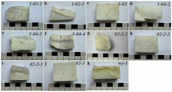
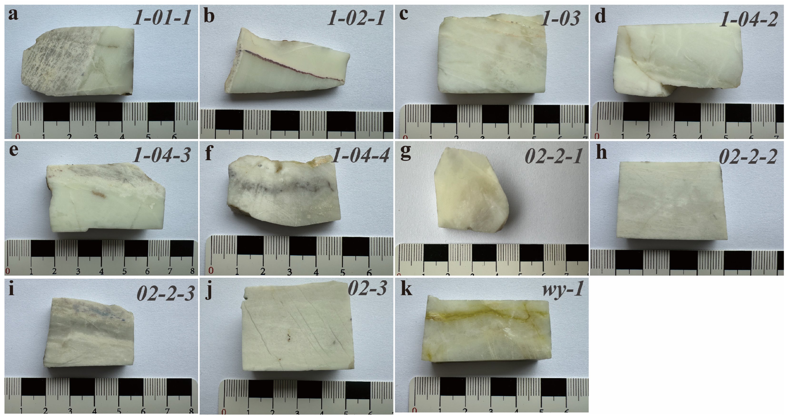
This study presents a systematic mineralogical investigation of white nephrites from the Dikou area, Fujian Province. The cut and polished specimens are shown in Figure 1. Using polarizing microscopy, scanning electron microscopy (SEM), electron probe microanalysis (EPMA), laser Raman spectroscopy, and X-ray Diffraction (XRD), we characterize the mineral assemblage, crystal chemistry, and textural features of the nephrite. Particular emphasis is placed on the compositional variations in white tremolite and their genetic implications. This work aims to enhance the understanding of regional nephrite formation and provide new insights into the crystallization mechanisms of tremolite in magnesian-skarn-type deposits.
Figure 1.
Cut and polished specimens of Dikou white nephrite. (a) Sample 1-01-1; (b) Sample 1-02-1; (c) Sample 1-03; (d) Sample 1-04-2; (e) Sample 1-04-3; (f) Sample 1-04-4; (g) Sample 02-2-1; (h) Sample 02-2-2; (i) Sample 02-2-3; (j) Sample 02-3; (k) Sample wy-1.
2. Geological Setting
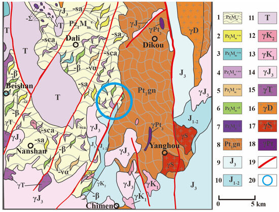
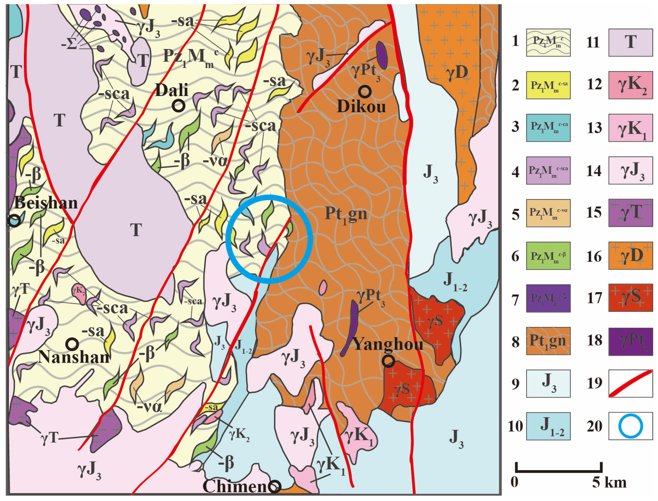
Fujian Province is located on the eastern margin of the Cathaysia Block in southeastern coastal China. It exhibits a complex geological setting, belonging to the South China Fold System [20], and possesses abundant mineral resources with favorable metallogenic conditions. The nephrite described in this study was sourced from the Zhengwei area, Dikou Town, southeast of Jian’ou City in northern Fujian Province (Figure 2). Tectonically, the mining area lies within the junction of the eastern and western Cathaysia blocks, which belong to the Yanping–Nanshan Early Paleozoic Mamianshan accretionary complex, which contains pelagic siliceous rocks [21].
Figure 2.
Geological map of the Dikou area, Fujian Province: 1. Mamianshan accretionary complex matrix: schist, leptynite. 2. Metasiliceous rock block: quartzite. 3. Metacarbonate rock block: marble, dolomite, diopsidite. 4. Metasiliceous limestone rock block: quartz diopsidite, diopside quartzite. 5. Meta-intermediate–acidic volcanic rock block: albite leptynite, K–feldspar leptynite. 6. Metabasalt rock block: plagioclase amphibolite, epidote-actinolite schist. 7. Ultramafic rock block: amphibolite, peridotite. 8. Gneiss block: Paleoproterozoic Dajinshan Formation, Qingshan Formation, Dikou Formation. 9. Late Jurassic Nanyuan Formation, Changlin Formation. 10. Early–Middle Jurassic Zhangping Formation, Lishan Formation, Fankeng Formation. 11. Triassic Wenbinshan Formation, Dakengcun Formation, Jiaokeng Formation, Anren Formation, Xikou Formation. 12. Late Cretaceous granitoids. 13. Early Cretaceous granitoids. 14. Late Jurassic granitoids. 15. Triassic granitoids. 16. Devonian granitoids. 17. Silurian granitoids. 18. Jinningian granitic gneiss. 19. Fault. 20. Mine.
The formation of this accretionary complex records the ocean–continent transition of the South China Block, which can be divided into five stages: (1) Early Neoproterozoic: development of basic–ultrabasic rift basins in the South China Ocean; (2) Middle to Late Neoproterozoic: intense intra-oceanic subduction leading to subduction–accretion complexes; (3) Late Neoproterozoic to Early Paleozoic: a period of relative tectonic quiescence; (4) Late Early Paleozoic: ocean–continent transition accompanied by intense magmatism and tectonic mixing; (5) Early Late Paleozoic: collisional orogeny with regional metamorphism and large-scale magmatic intrusion. During the Yanshanian period, large-scale ductile shearing overprinted the earlier structural framework [22].
The investigated nephrite originates from the accretionary complex belt, which is exposed as an NE-trending wedge in the Jian’ou area. This complex consists of quartzite, diopsidite, marble, and tremolite rock, occurring as mixed lithologies with widely distributed low-grade metamorphic clastic rocks and metamorphosed carbonate blocks. The protolith age ranges from the Neoproterozoic to the Early Paleozoic [21,22]. Quartzite and diopside quartzite layers, interpreted as metamorphosed marine siliceous rocks and limestones, constitute the principal parent lithologies for white nephrite mineralization, providing the essential calcium, magnesium, and silica chemical components for tremolite formation.
3. Materials and Methods
Nephrite samples were collected from the Dikou mining area. The material appears white with a faint yellowish-green tint (Figure 1) and exhibits a uniform, cryptocrystalline texture. Representative samples were selected for subsequent petrographic and geochemical analysis.
Polished thin sections (30 μm thickness) were prepared at China University of Geosciences, Beijing (CUGB). Petrographic observations were carried out using an OLYMPUS BX5 polarizing microscope (Olympus Corporation, Tokyo, Japan) at the Gemological Experimental Teaching Center, CUGB. Mineral compositions, textural characteristics, and paragenetic relationships were documented.
Scanning electron microscopy (SEM; ZEISS SUPRA 55, Carl Zeiss AG, Oberkochen, Germany) was employed to examine the surface characteristics and microstructure of the nephrite. Observations were performed on fresh fracture surfaces of representative samples under the following conditions: accelerating voltage of 20 kV, room temperature of 21 ± 0.5 °C, and humidity of 46% ± 1%.
Major element compositions of constituent minerals were determined by electron probe microanalysis (EPMA) using a JEOL JXA-8100 microprobe (JEOL Ltd., Tokyo, Japan) at the Electron Probe Laboratory, Chinese Academy of Geological Sciences, Beijing. Analytical conditions were: accelerating voltage 15 kV, beam current 1 × 10⁻⁸ Å, and beam diameter 5 μm. Natural and synthetic minerals were used for calibration. Data reduction was performed using the ZAF correction procedure. Detection limits for most elements were below 0.01 wt.%.
Raman spectroscopic analyses were performed using an HR-Evolution micro-Raman spectrometer (Horiba Scientific, Kyoto, Japan) at the Gemological Experimental Teaching Center, CUGB. A solid-state laser with an excitation wavelength of 532 nm was used, with an accumulation time of 1–2 s, laser power of 100%, and a beam spot diameter of approximately 1 μm. Spectra were collected in the range of 100–2000 cm−1 with a resolution of 1 cm−1. All measurements were conducted at room temperature (25 °C), and the spectrometer was calibrated using the 520.7 cm−1 Raman line of a silicon standard prior to analysis.
X-ray diffraction (XRD) analyses were conducted using a Y-2000 Automated X-ray Diffractometer (Chinese Academy of Geological Sciences, Beijing, China). The samples were finely ground in an agate mortar prior to measurement. Data were collected using Cu Kα radiation (λ = 0.154 nm) with a nickel filter, operating at 30 kV and 20 mA. Measurements were performed in continuous scanning mode over a 2θ range of 10–70° at a scan rate of 0.1°/min.
4. Results
4.1. Petrography
Microscopic observations indicate that the Dikou nephrite is predominantly composed of tremolite, accompanied by abundant diopside and quartz. Accessory minerals include calcite and prehnite, with trace amounts of magnetite, feldspar, and epidote.
4.1.1. Tremolite Microscopic Structure
Tremolite is the predominant mineral in the samples. Based on crystal morphology and texture, it can be classified into two types:
- (1)
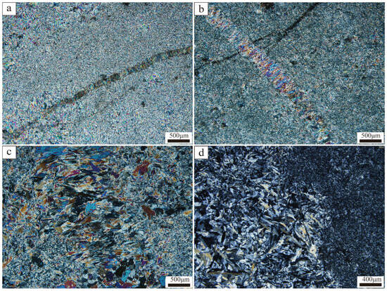
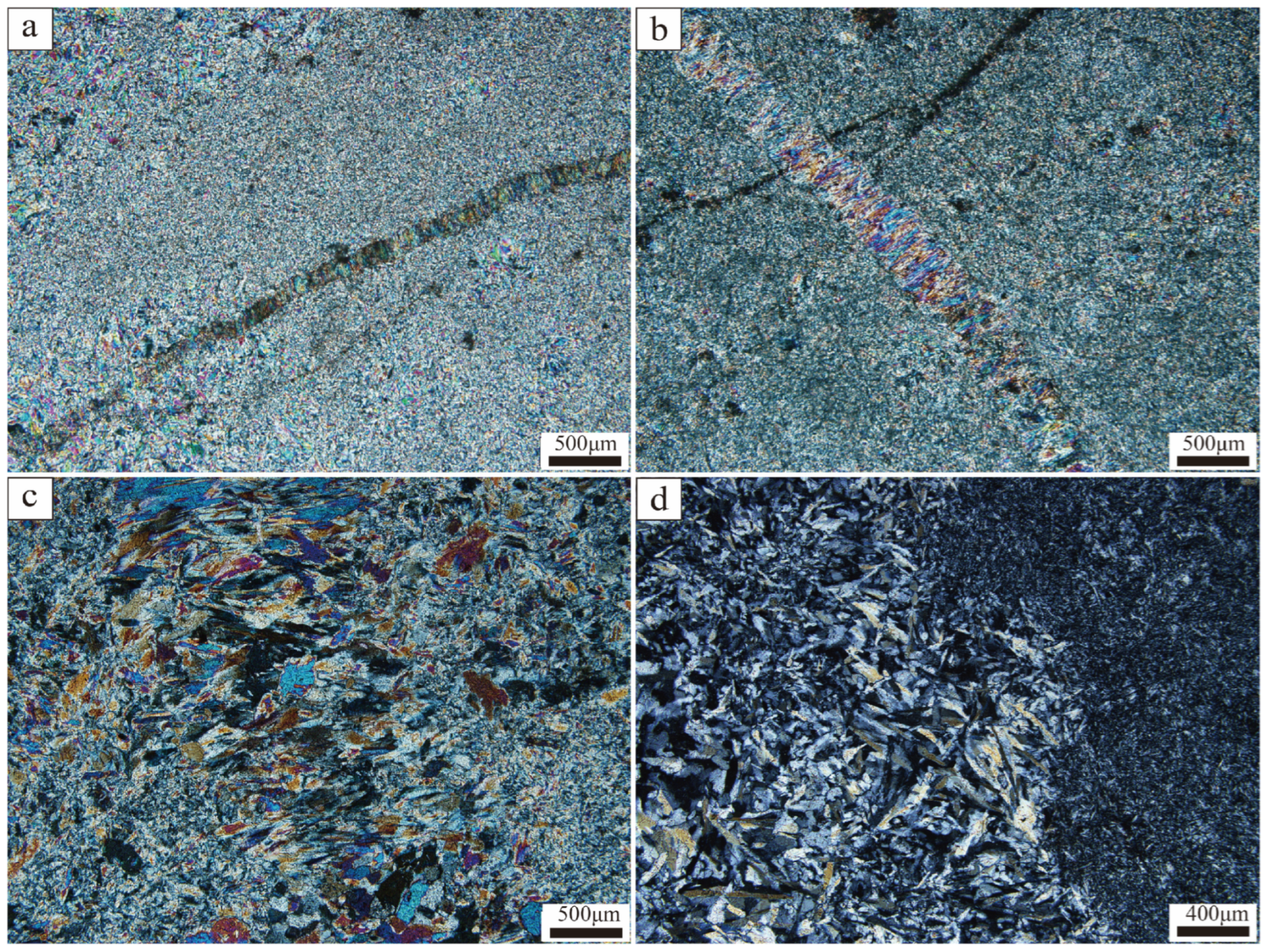
- Cryptocrystalline (Felt-like) texture: The most typical texture in amphibole jade, characterized by extremely fine-grained tremolite particles with sizes < 0.01 mm, rendering individual grain boundaries indistinguishable under polarized light. The particles are randomly oriented and densely interwoven, forming a felt-like appearance (Figure 3a,b). In contrast, acicular tremolite crystals infilling fractures are significantly larger, reaching up to 0.4 mm in length (Figure 3a,b). Regardless of texture, the tremolite is compositionally pure, displaying vivid green, blue, and purple interference colors under crossed polars.Figure 3. Photomicrographs of Dikou nephrite thin sections under crossed polars, illustrating the principal textural types: (a) Dense felt-like texture composed of fibrous tremolite. (b) Coexistence of felt-like matrix and needle-like tremolite. (c) Microcrystalline tremolite showing granular to columnar habits, with a preferred orientation of tremolite crystals resulting from subsequent tectonic deformation. (d) Textural contrast between columnar tremolite (left) and felt-like fibrous tremolite (right).
- (2)
- Microcrystalline texture: This type consists of granular, columnar, and acicular tremolite crystals ranging from 0.02 to 0.5 mm in size (Figure 3c,d). Interstices between these crystals are commonly filled with later fibrous tremolite. Figure 3d provides a direct comparison, showing columnar tremolite on the left and felt-like fibrous tremolite on the right, highlighting the pronounced grain-size contrast between the two textural types.
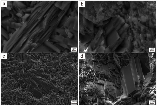
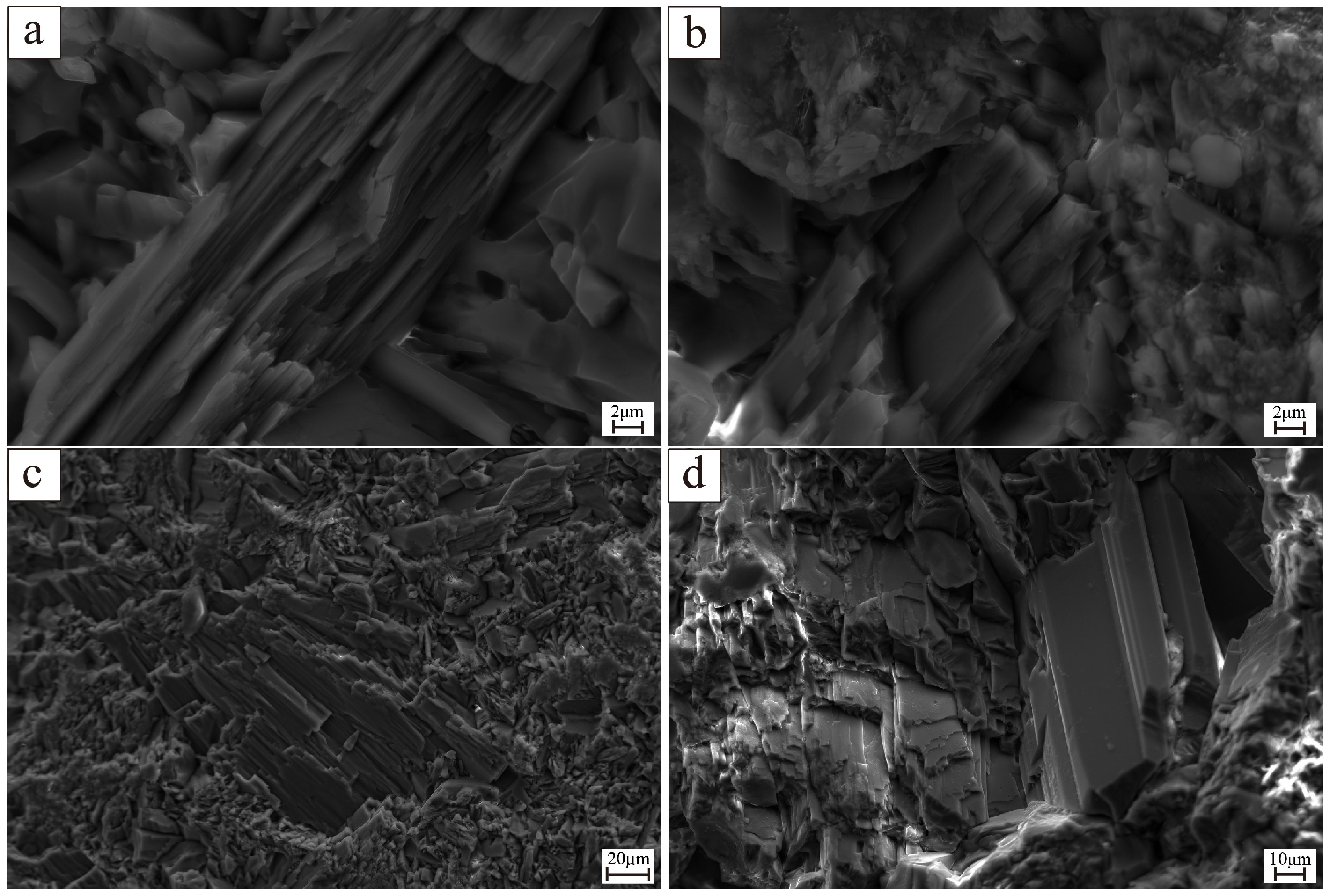
SEM observations reveal the coexistence of fibrous and platy textures in the samples (Figure 4). At higher magnifications (Figure 4a), randomly oriented fibrous tremolite crystals form a felt-like fibrous texture, with individual fibers approximately 2–5 μm in width and densely interwoven. A parallel fibrous texture is also present (Figure 4b), where tremolite fibers are sub-parallel and display occasional fracturing. This texture, analogous to the preferred orientation of tremolite crystals shown in Figure 3c, is interpreted to reflect tectonic deformation. At lower magnifications (Figure 4c,d), a platy texture composed of overlapping tabular crystals is evident, with individual plate plates about 10 μm thick. This platy texture commonly coexists with the felt-like fibrous texture, forming transitional textual types such as a platy–felt blastic texture.
Figure 4.
SEM images showing textual characteristics of tremolite in Dikou nephrite: (a) Dense felt-like texture of randomly oriented fibrous crystals. (b) Sub-parallel fibrous texture. (c,d) Platy texture formed by overlapping tabular crystals.
4.1.2. Other Minerals Microscopic Structure
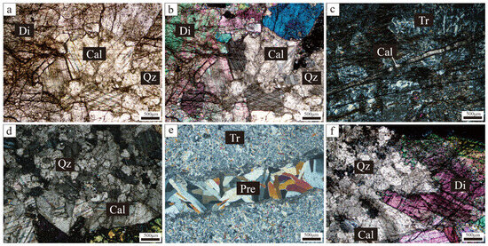
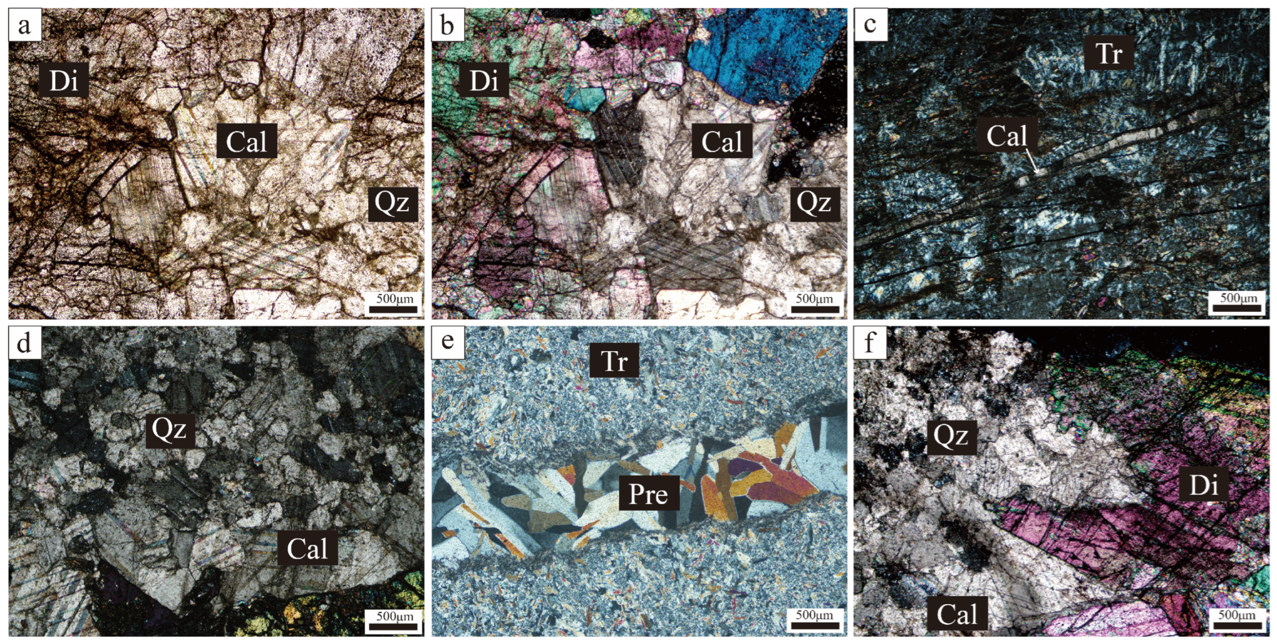
In addition to tremolite, polarized light microscopy identified several other minerals in the Dikou nephrite samples, including diopside, quartz, calcite, and prehnite (Figure 5).
Figure 5.
Photomicrographs showing associated minerals in Dikou nephrite: (a) Plane-polarized light; (b–f) Cross-polarized light. Mineral abbreviations: Tr—tremolite; Di—diopside; Cal—calcite; Qz—quartz; Pre—prehnite.
Diopside is the most common associated mineral in the nephrite, occurring as granular to short-columnar crystals with grain sizes ranging from approximately 0.1 to 0.5 mm (Figure 5a,b). At the contact between diopside-rich zones and tremolite-rich zones, diopside grains commonly display corroded margins and are partially replaced by fibrous tremolite, indicating a metasomatic relationship. This texture suggests that diopside represents a relict phase from the prograde skarn stage, which was subsequently altered during retrograde metamorphism.
Quartz occurs as anhedral granular crystals (Figure 5c), with grain sizes typically ranging from 0.05 to 0.25 mm. It exhibits undulose extinction under crossed polars, indicating post-crystallization deformation. A notable feature of these samples is their high quartz content, which is attributed to the regional geological setting—the protolith in the mining area is predominantly quartzite. Consequently, quartz is commonly observed in contact with diopside and calcite, and tremolite frequently fills interstices between quartz grains.
Calcite occurs as relatively coarse crystalline grains, reaching up to 0.5 mm in size, with well-developed cleavage, and frequent contact with quartz and diopside (Figure 5b). In a few samples, calcite is also observed filling open fractures within tremolite, interpreted as the late-stage fluid infiltration. The presence of calcite is consistent with the magnesium-rich carbonate protolith and the CO2-bearing nature of the hydrothermal system.
Prehnite occurs as veinlets cross-cutting tremolite aggregates, displaying sharp grain boundaries and a characteristic mosaic texture (Figure 5e). It represents the latest stage of hydrothermal activity, filling fractures after the main period of nephrite formation.
4.2. Mineral Chemistry
4.2.1. Tremolite
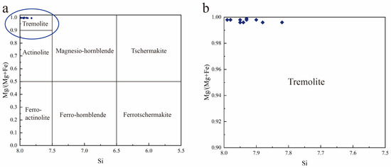
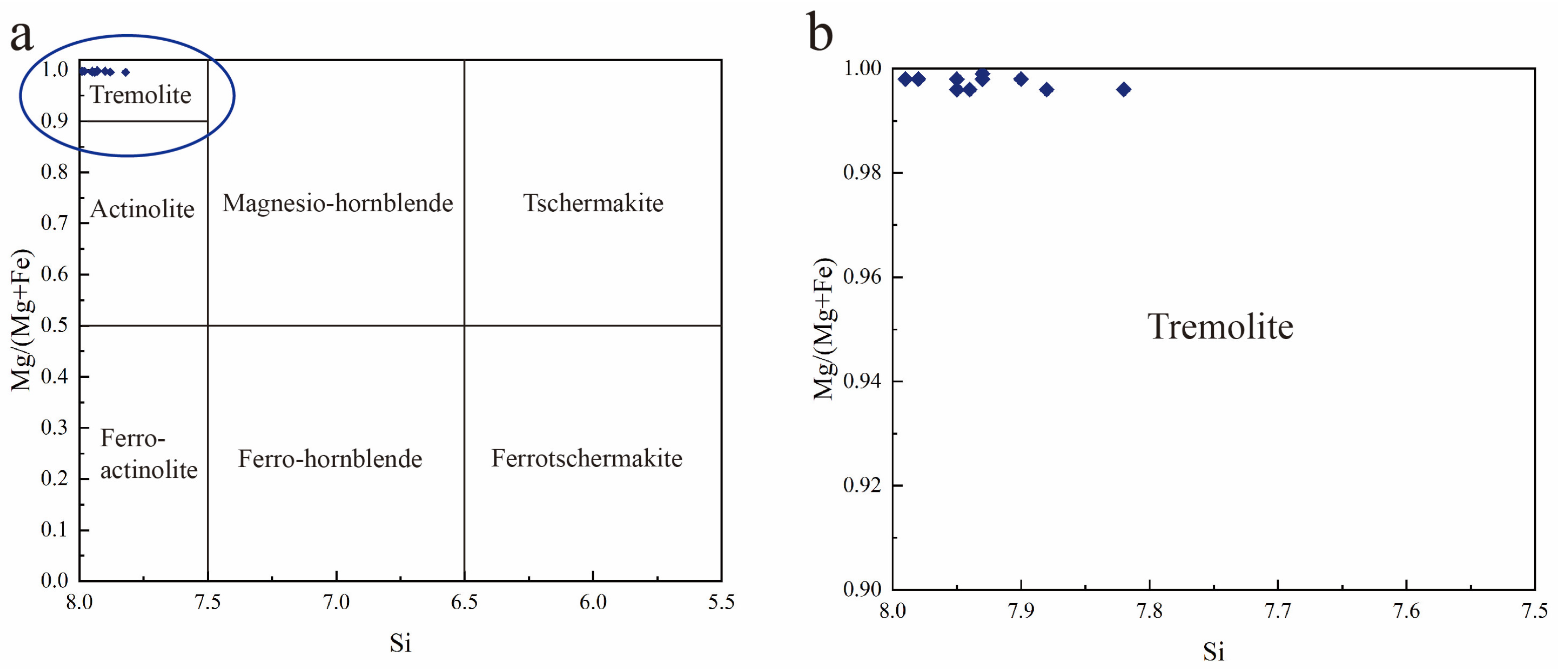
EPMA analyses of tremolite are presented in Table 1. The analyzed spots are representative, encompassing nephrite samples collected from various locations across the Dikou mining area, as well as all textural varieties identified in the sample suite. Following the IMA amphibole classification [2], all the analyzed points plot within the tremolite field (Figure 6a). The calculated Mg#[Mg/(Mg + Fe2+)] values range from 0.995 to 1.000 (average 0.997), indicating a highly Mg-enriched and Fe-depleted composition (Figure 6b).
Table 1.
Representative EPMA analysis of tremolite.
Figure 6.
Composition of tremolite plotted on the amphibole classification diagram: (a) Si cation coefficient vs. Mg# [R* = Mg/(Mg + Fe2+)]. All data points fall within the tremolite field. (b) Enlarged view of the blue-shaded area in (a), highlighting the extremely Mg-rich, Fe-depleted nature of the analyzed tremolite.
EPMA data indicate that the tremolite in the Dikou nephrite exhibits compositional homogeneity with limited variation, comprising predominantly SiO2 (57.51–58.89 wt.%), MgO (24.15–24.89 wt.%), and CaO (13.25–13.97 wt.%), with minor Al2O3 (0.11–1.52 wt.%), FeO (0.04–0.24 wt.%), Na2O (0.06–0.32 wt.%), and K2O (0.00–0.15 wt.%). All the analyzed tremolite grains exhibit high Mg# values (>0.99), reflecting an extremely Fe-depleted and Mg-enriched composition that precludes actinolite formation in the Dikou nephrite.
Based on compositional variations, two generations of tremolite can be distinguished:
Tr-I tremolite is characterized by relatively higher Al2O3 (0.69–1.52 wt.%, average 1.11 wt.%) and FeO (0.15–0.24 wt.%, average 0.20 wt.%) contents, and slightly lower SiO2 (57.51–57.74 wt.%) and MgO (24.15–24.50 wt.%). This type is interpreted as the product of incomplete metasomatic alteration of pre-existing diopside.
Tr-II tremolite has compositions closer to the ideal tremolite end-member, with lower Al2O3 (0.11–0.44 wt.%, average 0.20 wt.%) and FeO (0.04–0.14 wt.%, average 0.09 wt.%), alongside higher concentrations of SiO2 (57.99–58.89 wt.%) and MgO (24.52–24.89 wt.%). This compositional signature indicates that Tr-II tremolite formed through complete metasomatism and constitutes the predominant mineral phase of the nephrite.
In addition to its Fe-depleted character, Cr2O3 and NiO in the Dikou nephrite are consistently below detection limits. Detected Cr2O3 (0.00–0.03 wt.%) and NiO (0.00–0.09 wt.%) are lower than those reported for serpentinite-related (S-type) nephrites (0.07–0.43 wt.% and 0.08–0.36 wt.%, respectively), yet comparable to the ranges documented for dolomite-related (D-type) nephrites (0.00–0.07 wt.% and 0.00–0.08 wt.%, respectively) [23]. Fe, Ni, and Cr represent the principal chromophores responsible for green coloration in nephrite. The extremely low concentrations of these elements in the Dikou nephrite are insufficient to generate distinct green hues, accounting for the absence of green varieties at this deposit.
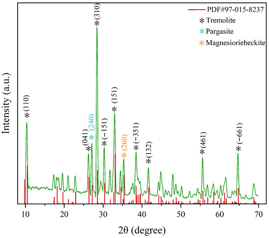
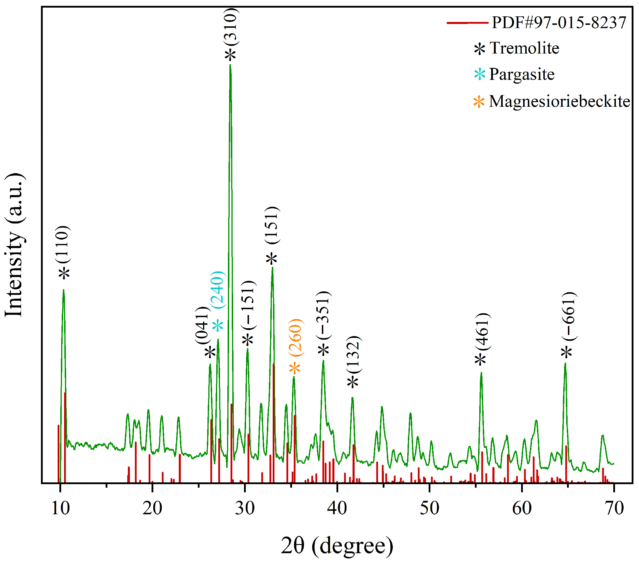
The XRD pattern demonstrates close correspondence between the major diffraction peaks of the sample and the standard tremolite reference (PDF#97-015-8237) (Figure 7). The sharp, intense diffraction peaks indicate well-crystallized tremolite with an intact crystal structure, confirming its dominance as the primary mineral phase. Additionally, weak diffraction peaks at 0.254 nm and 0.328 nm may be attributed to magnesioriebeckite and pargasite, respectively, suggesting trace abundances of other amphibole-group minerals.
Figure 7.
XRD patterns of Dikou nephrite samples. The green line represents the experimental data, while the red short straight lines represent the data from the database.
4.2.2. Other Minerals
In addition to tremolite, accessory and associated minerals identified in the samples and surrounding rocks include diopside, quartz, calcite, prehnite, and zoisite.
EPMA analyses of diopside are presented in Table 2. Analytical spots were selected from multiple textural settings, including diopside-rich domains, grain boundaries with tremolite, and contacts with quartz and calcite. The data demonstrate that diopside exhibits relatively homogeneous compositions, with SiO2 ranging from 54.77 to 55.61 wt.%, MgO from 17.62 to 18.19 wt.%, and CaO from 26.13 to 26.67 wt.%. FeO contents are notably low (0.01–0.25 wt.%), consistent with the Fe-depleted nature of the system.
Table 2.
Representative EPMA analyses of diopside.
Quartz analyses show nearly pure SiO2 (99.10–99.35 wt.%) with negligible impurities (Table 3). Calcite contains CaO and minor MgO, with no detectable SiO2, indicating a primary origin from marble protoliths rather than hydrothermal precipitation. Zoisite is characterized by high CaO and Al2O3 contents and very low FeO, consistent with Fe-poor zoisite formed during regional metamorphism.
Table 3.
EPMA analyses data of quartz, calcite and zoisite.
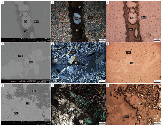
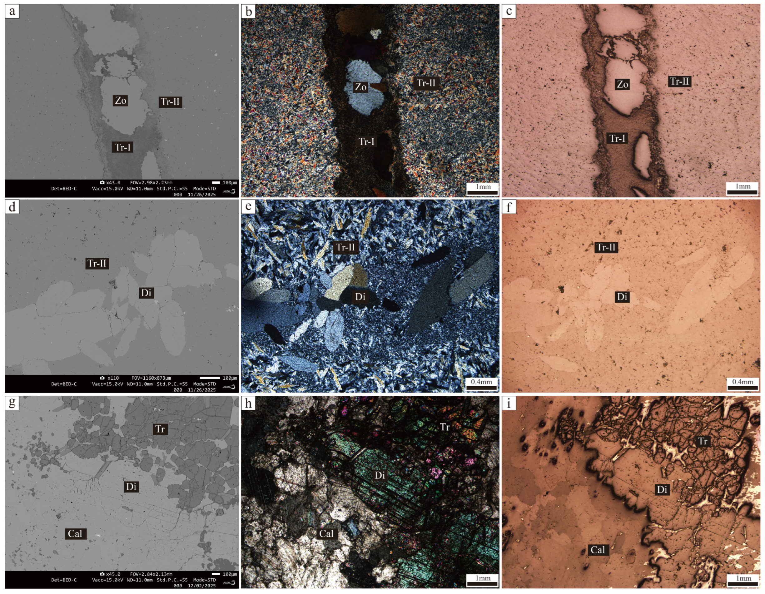
Comparative analysis of backscattered electron (BSE) images and optical micrographs further supports the metasomatic origin of tremolite (Figure 8). The BSE images reveal distinct compositional contrasts among different mineral phases, with diopside appearing brighter than tremolite due to its higher average atomic number. Two generations of tremolite exhibit distinct brightness variations: the slightly brighter Tr-I reflects its relatively higher Fe and Al contents, whereas the darker Tr-II corresponds to purer, Fe-depleted tremolite. Large diopside grains with irregular boundaries are partially replaced by tremolite, with relict cores preserved, indicating incomplete metasomatic reaction. Relict chlorite is also observed within tremolite veins, further suggesting multi-stage hydrothermal overprint. These textural features collectively document the progressive metasomatic replacement of pre-existing minerals by tremolite.
Figure 8.
Different micrograph of Dikou nephrite. (a,d,g) BSE images. (b,e,h) under crossed polars. (c,f,i) under reflected light.
4.3. Raman Spectroscopy
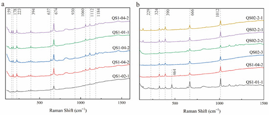
Raman spectroscopy was employed to further characterize the mineral phases in the Dikou nephrite samples. Representative Raman spectra of tremolite and diopside are shown in Figure 9.
Figure 9.
Representative Raman spectra of (a) tremolite and (b) diopside.
Tremolite, the predominant mineral in Dikou nephrite, was identified in all samples (Figure 9a). The most intense peak at 674 cm−1 corresponds to the symmetric stretching vibration of Si–O–Si bridges in the double-chain structure. The band at 1060 cm−1 is attributed to Si–O antisymmetric stretching vibrations. The peak at 394 cm−1 is assigned to Mg–OH antisymmetric translation, and the bands at 223 cm−1 and lower wavenumbers are related to [SiO4]4− lattice vibrations [24,25,26,27,28,29,30,31]. These characteristic peaks are consistent with those reported for tremolite from other nephrite deposits [32,33,34].
Diopside was identified in the samples based on its characteristic Raman spectrum (Figure 9b). The strong peak at 1012 cm−1 is assigned to Si–O symmetric stretching vibrations of [Si2O6]4− groups [28,35,36]. The intense band at 666 cm−1 corresponds to Si–O–Si symmetric stretching. The peaks at 390 and 324 cm−1, along with the weak absorption at 229 cm−1, are attributed to M–O stretching vibrations [35]. The peak at 464 cm−1 observed in some diopside spectra is interpreted as arising from minor quartz contamination [37,38].
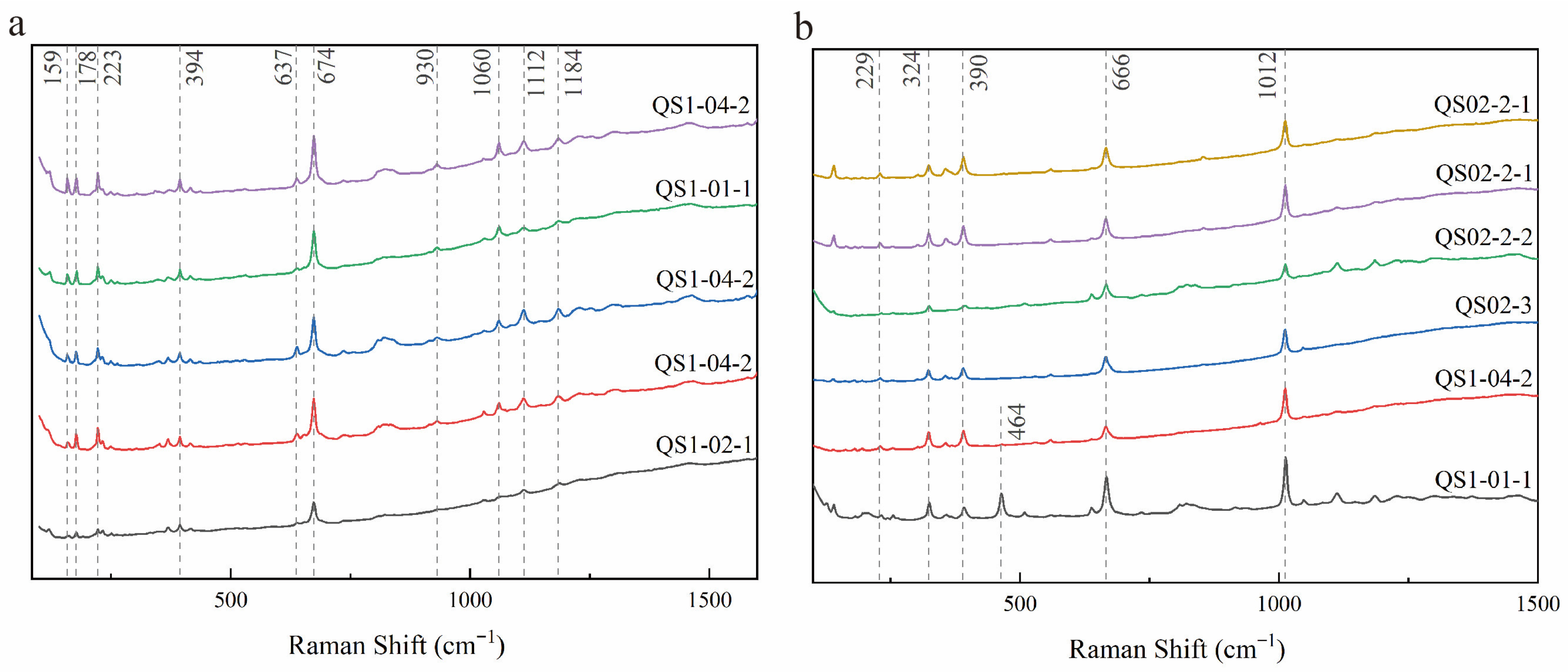
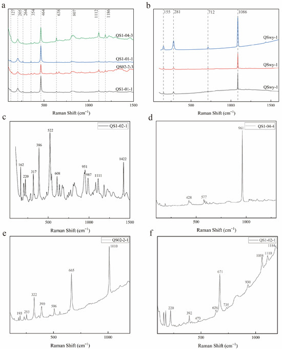
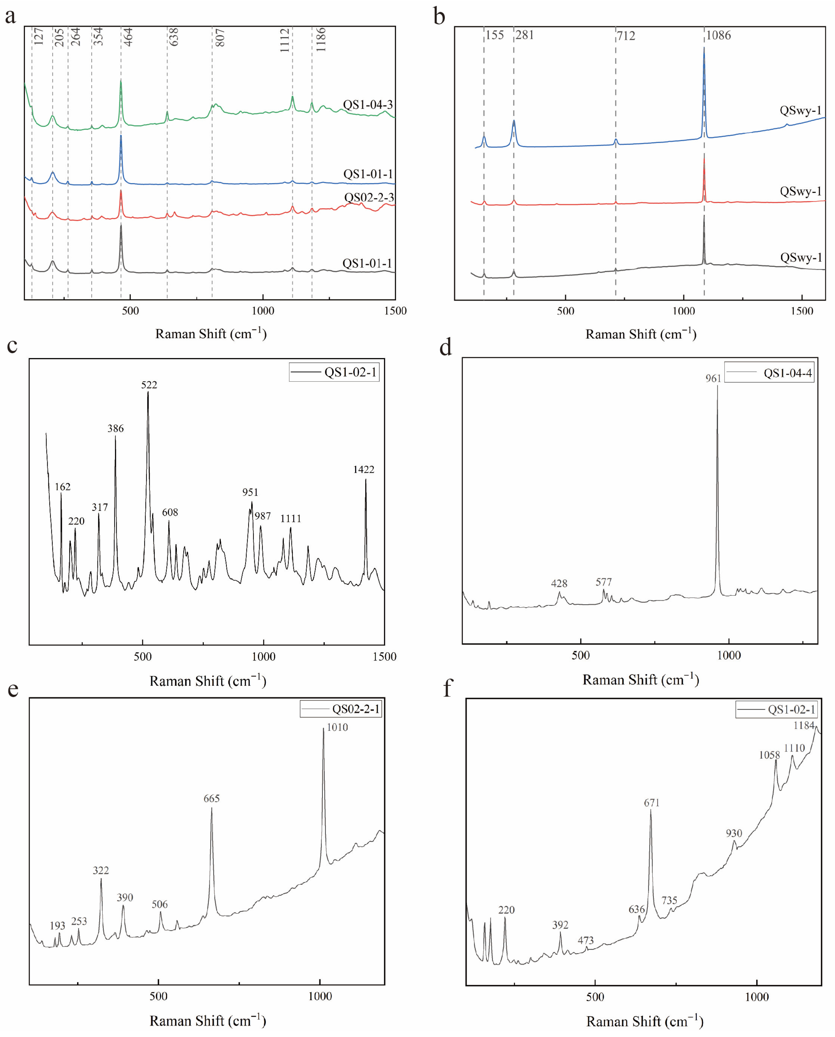
In addition to tremolite and diopside, several accessory minerals were identified by Raman spectroscopy (Figure 10). Quartz (Figure 10a) is characterized by its strong peak at 464 cm−1, assigned to Si–O–Si symmetric bending vibrations [37,38,39]. Calcite (Figure 10b) exhibits characteristic peaks at 1086 cm−1 (CO3 symmetric stretching) and 712 cm−1 (CO3 in-plane bending) [40,41]. Prehnite (Figure 10c) shows distinctive peaks at 987 cm−1 and 1080 cm−1, consistent with reference data [42,43]. Apatite (Figure 10d) is identified by its strong peak at 964 cm−1, attributed to PO4 symmetric stretching vibrations [44,45,46]. Gypsum (Figure 10e) displays characteristic peaks at 665 cm−1 (SO4 stretching) and 1010 cm−1 [47,48]. Talc (Figure 10f) exhibits peaks at 671 and 1058 cm−1, associated with Si–O–Si stretching vibrations [26,49]. The assignments of all characteristic Raman shifts are summarized in Table 4.
Figure 10.
Raman spectra of accessory mineral in Dikou nephrite samples: (a) Quartz. (b) Calcite. (c) Prehnite. (d) Apatite. (e) Gypsum. (f) Talc.
Table 4.
Assignments of characteristic Raman shifts for minerals identified in Dikou nephrite samples.
5. Discussion
5.1. Crystal Chemistry of Tremolite
The tremolite in Dikou nephrite is characterized by exceptionally low FeO contents (0.04–0.24 wt.%) and high Mg# values (>0.99), indicating crystallization from an Fe-depleted hydrothermal system. This compositional signature is diagnostic of high-quality white nephrite associated with dolomite-hosted deposits [17,23].
According to Hawthorne and Oberti, the general chemical formula for amphiboles is AB2C5T8O22W2 [4]. In tremolite, A = Na, K; B = Ca, Na; C = Mg, Fe2+, Al; T = Si, Al; and W = OH, F, Cl [1]. Based on the EPMA data obtained herein, the crystal-chemical formulas for tremolite in each sample were calculated and are presented in Table 5.
Table 5.
Calculated crystal-chemical formulas of tremolite.
The compositional disparity between Tr-I and Tr-II tremolite reflects variable degrees of metasomatic equilibration. Tr-I, enriched in Al and Fe, retains the chemical imprint of its diopside precursor, consistent with the great structural tolerance of clinopyroxene for these elements. In contrast, Tr-II approaches ideal tremolite stoichiometry, signifying complete equilibration with a hydrothermal fluid characterized by low Al and Fe activity.
5.2. Nephrite Formation
The Dikou nephrite deposit in Fujian Province represents a multi-stage, polyphase evolutionary product genetically linked to the Neoproterozoic–Early Paleozoic ocean–continent transition of the Cathaysia Block. It is classified as a skarn-type deposit in the broad sense, with the dominant metallogenic mechanism being contact metasomatism dominated by magnesian carbonates and accompanied by the involvement of mafic–ultramafic rocks [53].
From the Neoproterozoic through the Late Paleozoic, dolomitic marble and siliceous rocks derived from juvenile oceanic crust of the ancestral South China Ocean constituted the protoliths. During the Late Early Paleozoic, the ocean–continent transition triggered intensive magmatism, generating large-scale S-type granites that supplied K-, Na-, Al-, and Si-rich hydrothermal fluids [21,22]. These fluids migrated along lithological contact and reacted with dolomitic marble to produce diopside:
CaMg[CO3]2 + 2SiO2aq → CaMg[Si2O6] + 2CO2gas
dolomite diopside
dolomite diopside
Reaction 1 not only generated abundant diopside but also released substantial carbon dioxide. Thermal decomposition of carbonate minerals near the contact metamorphic zone provided an additional CO2 source. At this stage, lithostatic pressure prevailed, with CO2 accumulating in a relatively closed system. Therefore, diopside remained stable despite the presence of water, owing to the elevated CO2 concentrations.
Subsequent tectonic activity associated with collisional orogeny induced structural relaxation and fracture development, facilitating infiltration of external fluids, CO2 effervescence, system opening, and pressure reduction. This led to decreased CO2 activity, initiating retrograde metamorphism. Enhanced fluid-rock interaction promoted metasomatic replacement of diopside by Tr-I tremolite (Reaction 2). However, incomplete reaction at this stage produced tremolite relatively enriched in Al and Fe.
5CaMg[Si2O6] + H2O + 3CO2 → Ca2Mg5[Si8O22](OH)2 + 3CaCO3 + 2SiO2
diopside tremolite (Tr-I) calcite quartz
diopside tremolite (Tr-I) calcite quartz
Simultaneously, high-H2O-content hydrothermal fluids infiltrating along fractures reacted directly with surrounding carbonate rocks, generating coarse-grained Tr-I tremolite in direct contact with calcite (Reaction 3).
5CaMg[CO3]2 + 8SiO2aq + H2O → Ca2Mg5[Si8O22](OH)2 + 3CaCO3 + 7CO2gas
dolomite tremolite (Tr-I) calcite
dolomite tremolite (Tr-I) calcite
During the second stage, episodic fluid influx drove Reaction 2 to completion. Shifts in fluid chemistry coupled with enhanced fluid flux facilitated more exhaustive metasomatic replacement of diopside, yielding Tr-II tremolite with composition approaching ideal end-member stoichiometry (Reaction 4). This generation of tremolite exhibits a finer, more equigranular texture and is characterized by Fe-depleted and Mg-enriched compositions.
5CaMg[Si2O6] + H2O + 3CO2 → Ca2Mg5[Si8O22](OH)2 + 3CaCO3 + 2SiO2
diopside tremolite (Tr-II) calcite quartz
diopside tremolite (Tr-II) calcite quartz
Concurrently, as retrograde metamorphism advanced, system temperature and pressure progressively declined. Silica-rich hydrothermal fluids partially precipitated directly within open fractures, generating comb-textured and fibrous tremolite (Reaction 5) (Figure 3a,b).
5Ca2+ + 5Mg2+ + 8H4SiO4 → Ca2Mg5[Si8O22](OH)2 + 8H2O + 10H+
tremolite(Tr-II)
tremolite(Tr-II)
The foregoing reactions collectively constitute the complex nephrite-forming process of the Dikou deposit. BSE imaging further elucidates the textural relationships among constituent minerals (Figure 8).
Prehnite precipitation from late-stage fluids infilling fractures marks the terminal phase of retrograde evolution, reflecting the low-temperature conditions at the waning stages of metamorphism. This study systematically characterizes the metallogenic framework of nephrite in this district, furnishing critical petrological constraints on the complex tectonic evolution of the region. It complements the classic paradigm of magnesian skarn-type nephrite deposits in the broad sense and carries significant implications for the classification and genetic study of nephrite deposits worldwide.
6. Conclusions
Based on integrated petrographic, mineralogical, and geochemical data, the Dikou nephrite from Fujian Province comprises predominantly tremolite, with abundant diopside and quartz, and minor dolomite, prehnite, serpentine, apatite, gypsum, and talc.
Two generations of tremolite (Tr-I and Tr-II) are distinguished based on compositional variations. Tr-I (elevated Al, Fe) represents incomplete metasomatic replacement of diopside, whereas Tr-II (low Al, Fe) represents complete metasomatism and constitutes the principal nephrite body. All the analyzed tremolite samples exhibit exceptionally low FeO contents (<0.25 wt.%) and high Mg# values (>0.99), indicating crystallization from an Fe-depleted hydrothermal system.
The low concentrations of Fe, Cr, and Ni are insufficient to produce distinct green coloration, accounting for the absence of green varieties at this deposit. The absence of actinolite is attributed to these extremely low FeO contents, which preclude stabilization of the Fe-bearing actinolite end-member.
The Dikou nephrite represents a magnesian skarn-type deposit formed through multi-stage hydrothermal metasomatism associated with the Early Paleozoic orogeny of South China. The paragenetic sequence comprises prograde diopside formation followed by retrograde tremolite crystallization under declining temperature and Fe activity.
Author Contributions
Conceptualization, S.R. and Y.L.; methodology, S.R. and Y.L.; software, S.R.; validation, S.R. and Y.L.; formal analysis, S.R.; investigation, Y.L.; resources, Y.L.; data curation, S.R. and Y.L.; writing—original draft preparation, S.R.; writing—review and editing, Y.L.; visualization, S.R.; supervision, Y.L.; project administration, Y.L.; funding acquisition, Y.L. All authors have read and agreed to the published version of the manuscript.
Funding
This research was funded by the Innovation and Entrepreneurship Training Program for College Students of China University of Geosciences (Beijing) (S202511415144).
Data Availability Statement
The original contributions presented in this study are included in the article. Further inquiries can be directed to the corresponding author.
Acknowledgments
The authors gratefully acknowledge T. Nie, R. Chen and Y. Li et al. from the Fujian Provincial Geological Survey Bureau, for their support in sample collection and revision suggestions during this study. Sincere appreciation is also presented to the laboratory of the School of Gemmology, CUGB, for their technical assistance.
Conflicts of Interest
The authors declare no conflicts of interest.
References
- Hawthorne, F.C.; Oberti, R.; Harlow, G.E.; Maresch, W.V.; Martin, R.F.; Schumacher, J.C.; Welch, M.D. Nomenclature of the Amphibole Supergroup. Am. Mineral. 2012, 97, 2031–2048. [Google Scholar] [CrossRef] [Scilit]
- Leake, B.E.; Woolley, A.R.; Arps, C.E.S.; Birch, W.D.; Gilbert, M.C.; Grice, J.D.; Hawthorne, F.C.; Kato, A.; Kisch, H.J.; Krivovichev, V.G.; et al. Nomenclature of Amphiboles; Report of the Subcommittee on Amphiboles of the International Mineralogical Association, Commission on New Minerals and Mineral Names. Can. Mineral. 1997, 35, 219–246. [Google Scholar]
- Gottschalk, M.; Andrut, M.; Melzer, S. The Determination of the Cummingtonite Content of Synthetic Tremolite. Eur. J. Mineral. 1999, 11, 967–982. [Google Scholar] [CrossRef] [Scilit]
- Hawthorne, F.C.; Oberti, R. Amphiboles: Crystal Chemistry. Rev. Mineral. Geochem. 2007, 67, 1–54. [Google Scholar] [CrossRef] [Scilit]
- Harlow, G.E.; Sorensen, S.S. Jade (Nephrite and Jadeitite) and Serpentinite: Metasomatic Connections. Int. Geol. Rev. 2005, 47, 113–146. [Google Scholar] [CrossRef] [Scilit]
- Liu, Y.; Deng, J.; Shi, G.; Sun, X.; Yang, L. Geochemistry and Petrogenesis of Placer Nephrite from Hetian, Xinjiang, Northwest China. Ore Geol. Rev. 2011, 41, 122–132. [Google Scholar] [CrossRef] [Scilit]
- Yui, T.-F.; Yeh, H.-W.; Lee, C.W. Stable Isotope Studies of Nephrite Deposits from Fengtien, Taiwan. Geochim. Cosmochim. Acta 1988, 52, 593–602. [Google Scholar] [CrossRef] [Scilit]
- Zhang, C.; Yu, X.; Jiang, T. Mineral Association and Graphite Inclusions in Nephrite Jade from Liaoning, Northeast China: Implications for Metamorphic Conditions and Ore Genesis. Geosci. Front. 2019, 10, 425–437. [Google Scholar] [CrossRef] [Scilit]
- Yu, H.Y.; Wang, R.C.; Guo, J.C.; Li, J.G.; Yang, X.W. Study of the Minerogenetic Mechanism and Origin of Qinghai Nephrite from Golmud, Qinghai, Northwest China. Sci. China Earth Sci. 2016, 59, 1597–1609. [Google Scholar] [CrossRef] [Scilit]
- Bai, F.; Li, G.; Lei, J.; Sun, J. Mineralogy, Geochemistry, and Petrogenesis of Nephrite from Panshi, Jilin, Northeast China. Ore Geol. Rev. 2019, 115, 103171. [Google Scholar] [CrossRef] [Scilit]
- Zhang, Y.; Yu, H.; Lan, Y.; Ruan, Q. Mineralogy and Sr Isotope Characteristics of Dahua Stratified Tremolite Nephrite and Host Rocks, Guangxi Province, China. Minerals 2024, 14, 257. [Google Scholar] [CrossRef] [Scilit]
- Kislov, E.V.; Popov, M.P.; Nurmukhametov, F.M.; Posokhov, V.F.; Vanteev, V.V. Nyrdvomenshor Nephrite Deposit, Polar Urals, Russia. Minerals 2023, 13, 767. [Google Scholar] [CrossRef] [Scilit]
- Tsydenova, N.; Morozov, M.V.; Rampilova, M.V.; Vasil’ev, Y.A.; Matveeva, O.P.; Konovalov, P.B. Chemical and Spectroscopic Study of Nephrite Artifacts from Transbaikalia, Russia: Geological Sources and Possible Transportation Routes. Quat. Int. 2015, 355, 114–125. [Google Scholar] [CrossRef] [Scilit]
- Yui, T.-F.; Kwon, S.-T. Origin of a Dolomite-Related Jade Deposit at Chuncheon, Korea. Econ. Geol. 2002, 97, 593–601. [Google Scholar] [CrossRef]
- Grapes, R.H.; Yun, S.T. Geochemistry of a New Zealand Nephrite Weathering Rind. N. Z. J. Geol. Geophys. 2010, 53, 413–426. [Google Scholar] [CrossRef] [Scilit]
- Adams, C.J.; Beck, R.J.; Campbell, H.J. Characterisation and Origin of New Zealand Nephrite Jade Using Its Strontium Isotopic Signature. Lithos 2007, 97, 307–322. [Google Scholar] [CrossRef] [Scilit]
- Zhang, X.C.; Shi, G.H.; Zhang, X.M.; Gao, K. Formation of the Nephrite Deposit with Five Mineral Assemblage Zones in the Central Western Kunlun Mountains, China. J. Petrol. 2022, 63, egac117. [Google Scholar] [CrossRef] [Scilit]
- Liu, Y.; Zhang, R.-Q.; Abuduwayiti, M.; Wang, C.; Zhang, S.; Shen, C.; Zhang, Z.; He, M.; Zhang, Y.; Yang, X. SHRIMP U–Pb Zircon Ages, Mineral Compositions and Geochemistry of Placer Nephrite in the Yurungkash and Karakash River Deposits, West Kunlun, Xinjiang, Northwest China: Implication for a Magnesium Skarn. Ore Geol. Rev. 2016, 72, 699–727. [Google Scholar] [CrossRef] [Scilit]
- Tang, D.P.; Lin, G.X.; Jiang, A.G.; Yu, J.C.; Chen, W.B. The Nephrite First Found in Fujian, China. Geol. J. China Univ. 1997, 3, 396–399. [Google Scholar]
- Shu, L.; Faure, M.; Wang, B.; Zhou, X.; Song, B. Late Palaeozoic–Early Mesozoic Geological Features of South China: Response to the Indosinian Collision Events in Southeast Asia. C. R. Geosci. 2008, 340, 151–165. [Google Scholar] [CrossRef] [Scilit]
- Nie, T.C. Determination and Significance of Mamianshan Subduction Accretionary Complex in Jianou Area, Fujian Province. Geol. Fujian 2018, 37, 273–287. [Google Scholar]
- Nie, T.C. Geological Characteristics and Tectonic Evolution of Mamianshan Hyperplastic Miscellaneous Rock in Central—Northern Fujian Province. Geol. Fujian 2023, 43, 1–14. [Google Scholar]
- Liu, Y.; Deng, J.; Shi, G.; Yui, T.-F.; Zhang, G.; Abuduwayiti, M.; Yang, L.; Sun, X. Geochemistry and Petrology of Nephrite from Alamas, Xinjiang, NW China. J. Asian Earth Sci. 2011, 42, 440–451. [Google Scholar] [CrossRef] [Scilit]
- Bersani, D.; Andò, S.; Scrocco, L.; Gentile, P.; Salvioli-Mariani, E.; Fornasini, L.; Lottici, P.P. Composition of Amphiboles in the Tremolite–Ferro–Actinolite Series by Raman Spectroscopy. Minerals 2019, 9, 491. [Google Scholar] [CrossRef] [Scilit]
- Rinaudo, C.; Gastaldi, D.; Belluso, E. Characterization of Chrysotile, Antigorite and Lizardite by FT-Raman Spectroscopy. Can. Mineral. 2003, 41, 883–890. [Google Scholar] [CrossRef] [Scilit]
- Blaha, J.J.; Rosasco, G.J. Raman Microprobe Spectra of Individual Microcrystals and Fibers of Talc, Tremolite, and Related Silicate Minerals. Anal. Chem. 1978, 50, 892–896. [Google Scholar] [CrossRef] [Scilit]
- Wang, A.; Kuebler, K.E.; Jolliff, B.L.; Haskin, L.A. Raman Spectroscopy of Fe-Ti-Cr-Oxides, Case Study: Martian Meteorite EETA79001. Am. Mineral. 2004, 89, 665–680. [Google Scholar] [CrossRef] [Scilit]
- Wei, G.C.; You, J.L.; Ma, N.; Li, Y.K.; Wang, J.; Wang, M.; Huang, X.Y. Raman Spectroscopic Study of the Chain Silicate Minerals. Chin. J. Light Scatt. 2017, 29, 62–69. [Google Scholar] [CrossRef]
- Li, K.; Shen, X.P. Research on Identification Characteristics of Tremolite and Actinolite by Using Nondestructive Testing Techniques of Infrared Spectroscopy and Raman Spectroscopy. Bull. Mineral. Petrol. Geochem. 2019, 38, 405–408. [Google Scholar] [CrossRef]
- Zhang, C.; Yu, X.Y. Spectral Characteristic and Origin Identification of Tremolite Jade from Sangpiyu, Liaoning Province. J. Gems Gemmol. 2018, 20, 41–53. [Google Scholar] [CrossRef]
- Rösche, C.; Waeselmann, N.; Petrova, N.; Malcherek, T.; Schlüter, J.; Mihailova, B. Oxidation Processes and Thermal Stability of Actinolite. Phys. Chem. Miner. 2022, 49, 47. [Google Scholar] [CrossRef] [Scilit]
- Chen, Q.L.; Bao, D.Q.; Yin, Z.W. Study on XRD and Infrared Spectroscopy of Nephrites from Xinjiang and Xiuyan, Liaoning. Spectrosc. Spectr. Anal. 2013, 33, 3142–3146. [Google Scholar] [CrossRef]
- Liu, X.F.; Zhang, H.Q.; Liu, Y.; Zhang, Y.; Li, Z.J.; Zhang, J.H.; Zheng, F. Mineralogical Characteristics and Genesis of Green Nephrite from the World. Rock Miner. Anal. 2018, 37, 479–489. [Google Scholar] [CrossRef]
- Zhou, X. Study on the Indicative Significance of Diopside of Nephrite Minerals to Skarn Mineralization from Panshi Jilin, Southeast China. Master‘s Thesis, China University of Geosciences, Beijing, China, 2025. [Google Scholar] [CrossRef]
- Prencipe, M.; Mantovani, L.; Tribaudino, M.; Bersani, D.; Lottici, P.P. The Raman Spectrum of Diopside: A Comparison between Ab Initio Calculated and Experimentally Measured Frequencies. Eur. J. Mineral. 2012, 24, 457–464. [Google Scholar] [CrossRef] [Scilit]
- Lu, L.F.; Zhu, Q.Q.; Xie, G.Q.; Wu, C.X.; Wei, K.T.; Cai, H.A.; Shang, S.C. Raman spectroscopy characteristics and exploration implication of pyroxene from Fujiashan skarn tungsten deposit of Hubei Province. Miner. Depos. 2025, 44, 1127–1142. [Google Scholar] [CrossRef]
- Krishnan, R.S. Raman Spectrum of Quartz. Nature 1945, 155, 452. [Google Scholar] [CrossRef] [Scilit]
- Enami, M.; Nishiyama, T.; Mouri, T. Laser Raman Microspectrometry of Metamorphic Quartz: A Simple Method for Comparison of Metamorphic Pressures. Am. Mineral. 2007, 92, 1303–1315. [Google Scholar] [CrossRef] [Scilit]
- Krishnamurti, D. The Raman Spectrum of Quartz and Its Interpretation. Proc. Indian Acad. Sci. 1958, 47, 276–291. [Google Scholar] [CrossRef] [Scilit]
- Gunasekaran, S.; Anbalagan, G.; Pandi, S. Raman and Infrared Spectra of Carbonates of Calcite Structure—Gunasekaran—2006—Journal of Raman Spectroscopy—Wiley Online Library. J. Raman Spectrosc. 2006, 37, 892–899. [Google Scholar] [CrossRef] [Scilit]
- Sun, J.; Wu, Z.; Cheng, H.; Zhang, Z.; Frost, R.L. A Raman Spectroscopic Comparison of Calcite and Dolomite. Spectrochim. Acta Part A Mol. Biomol. Spectrosc. 2014, 117, 158–162. [Google Scholar] [CrossRef] [Scilit] [PubMed]
- Ross, N.L.; Detrie, T.A. High-Pressure Raman and Infrared Spectroscopic Study of Prehnite. Minerals 2020, 10, 312. [Google Scholar] [CrossRef] [Scilit]
- Zhang, J.Y.; Bai, Z.M.; Zhao, D. Raman and Infrared Spectrograms of Organic Borate Intercalated Hydrotalcite. Spectrosc. Spectr. Anal. 2013, 33, 704–708. [Google Scholar]
- Antonakos, A.; Liarokapis, E.; Leventouri, T. Micro-Raman and FTIR Studies of Synthetic and Natural Apatites. Biomaterials 2007, 28, 3043–3054. [Google Scholar] [CrossRef] [Scilit]
- Awonusi, A.; Morris, M.D.; Tecklenburg, M.M.J. Carbonate Assignment and Calibration in the Raman Spectrum of Apatite. Calcif. Tissue Int. 2007, 81, 46–52. [Google Scholar] [CrossRef] [Scilit]
- Khan, A.F.; Awais, M.; Khan, A.S.; Tabassum, S.; Chaudhry, A.A.; Rehman, I.U. Raman Spectroscopy of Natural Bone and Synthetic Apatites. Appl. Spectrosc. Rev. 2013, 48, 329–355. [Google Scholar] [CrossRef] [Scilit]
- Sarma, L.P.; Prasad, P.S.R.; Ravikumar, N. Raman Spectroscopic Study of Phase Transitions in Natural Gypsum. J. Raman Spectrosc. 1998, 29, 851–856. [Google Scholar] [CrossRef]
- Berenblut, B.J.; Dawson, P.; Wilkinson, G.R. The Raman Spectrum of Gypsum. Spectrochim. Acta Part A Mol. Spectrosc. 1971, 27, 1849–1863. [Google Scholar] [CrossRef] [Scilit]
- Longuinhos, R.; Cadore, A.R.; Bechtel, H.A.; De Matos, C.J.S.; Freitas, R.O.; Ribeiro-Soares, J.; Barcelos, I.D. Raman and Far-Infrared Synchrotron Nanospectroscopy of Layered Crystalline Talc: Vibrational Properties, Interlayer Coupling, and Symmetry Crossover. J. Phys. Chem. C 2023, 127, 5985–5995. [Google Scholar] [CrossRef] [Scilit]
- Ye, X.; Bai, F.; Li, M.; Sun, H. Gemology, Mineralogy, and Spectroscopy of an Attractive Tremolitized Diopside Anorthosite Gem Material from the Philippines: A New Type of Material with Similarities to Dushan Jade. Minerals 2021, 11, 152. [Google Scholar] [CrossRef] [Scilit]
- Karampelas, S.; Hennebois, U.; Delaunay, A. Raman Spectroscopy of Amphibole Inclusions in Emeralds. J. Raman Spectrosc. 2025, 56, 1228–1234. [Google Scholar] [CrossRef] [Scilit]
- Buzgar, N.; Apopei, A.I.; Diaconu, V.; Buzatu, A. The Composition and Source of the Raw Material of Two Stone Axes of Late Bronze Age from Neamt County (Romania)—A Raman Study. Analele Stiintifice Univ. AI Cuza Iasi Sect. Geol. 2013, 59, 5–22. [Google Scholar]
- Liu, F.; Yu, X.Y. Classification and Mineralogical Characteristics of Nephrite Deposits in China. Miner. Resour. Geol. 2009, 23, 375–380. [Google Scholar]

Disclaimer/Publisher’s Note: The statements, opinions and data contained in all publications are solely those of the individual author(s) and contributor(s) and not of MDPI and/or the editor(s). MDPI and/or the editor(s) disclaim responsibility for any injury to people or property resulting from any ideas, methods, instructions or products referred to in the content. |
© 2026 by the authors. Licensee MDPI, Basel, Switzerland. This article is an open access article distributed under the terms and conditions of the Creative Commons Attribution (CC BY) license.