Structure of Agmatinase from Klebsiella pneumoniae and the Active Site Comparison with Its Structural Homologues



Abstract
1. Introduction
2. Materials and Methods
2.1. Protein Expression and Purification
2.2. Crystallization and Data Collection
2.3. Structure Determination and Analysis
2.4. SEC-Multi Angle Light Scattering (MALS) Analysis
3. Results
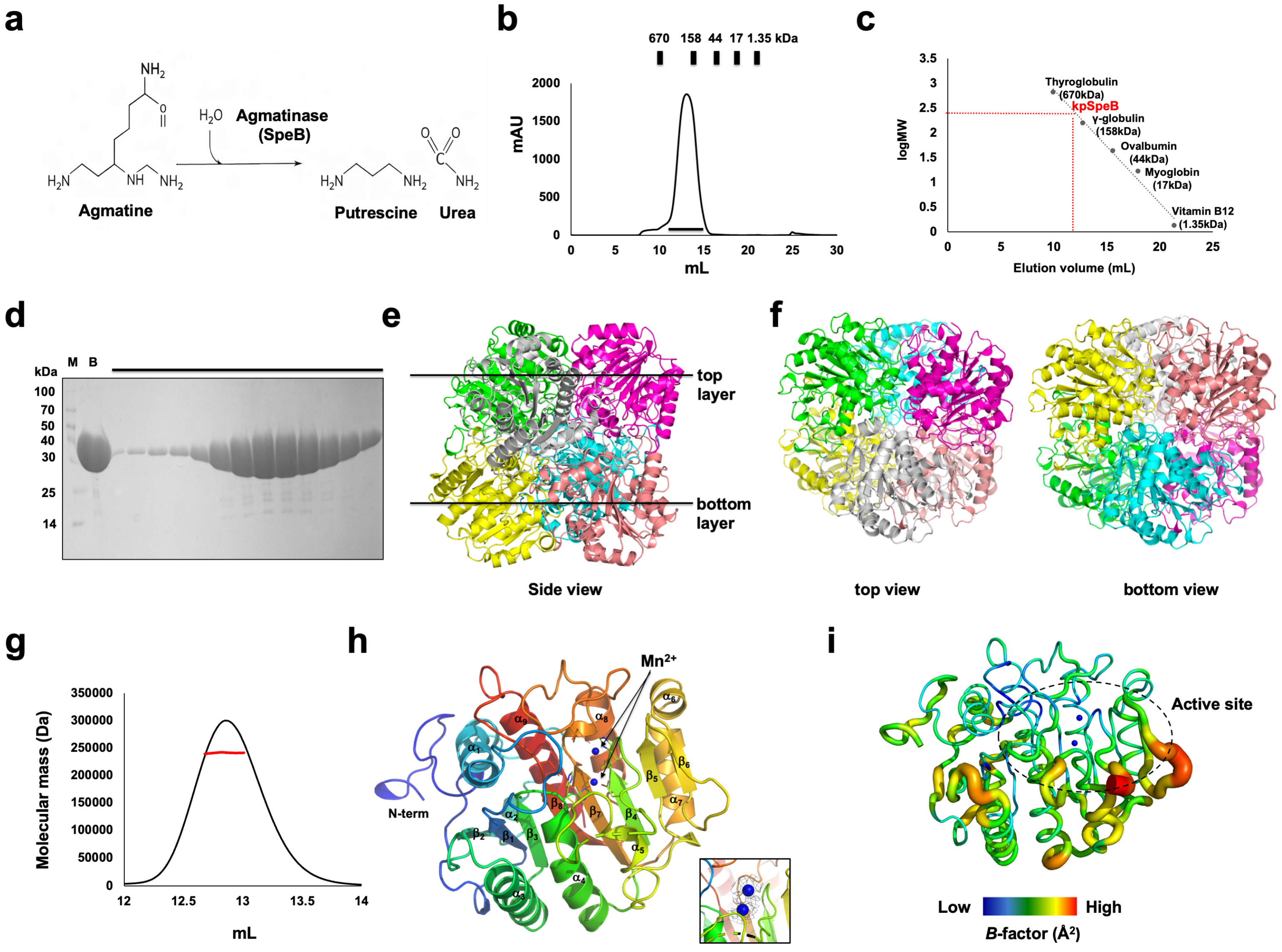
3.1. The Overall Structure and Oligomeric State of kpSpeB
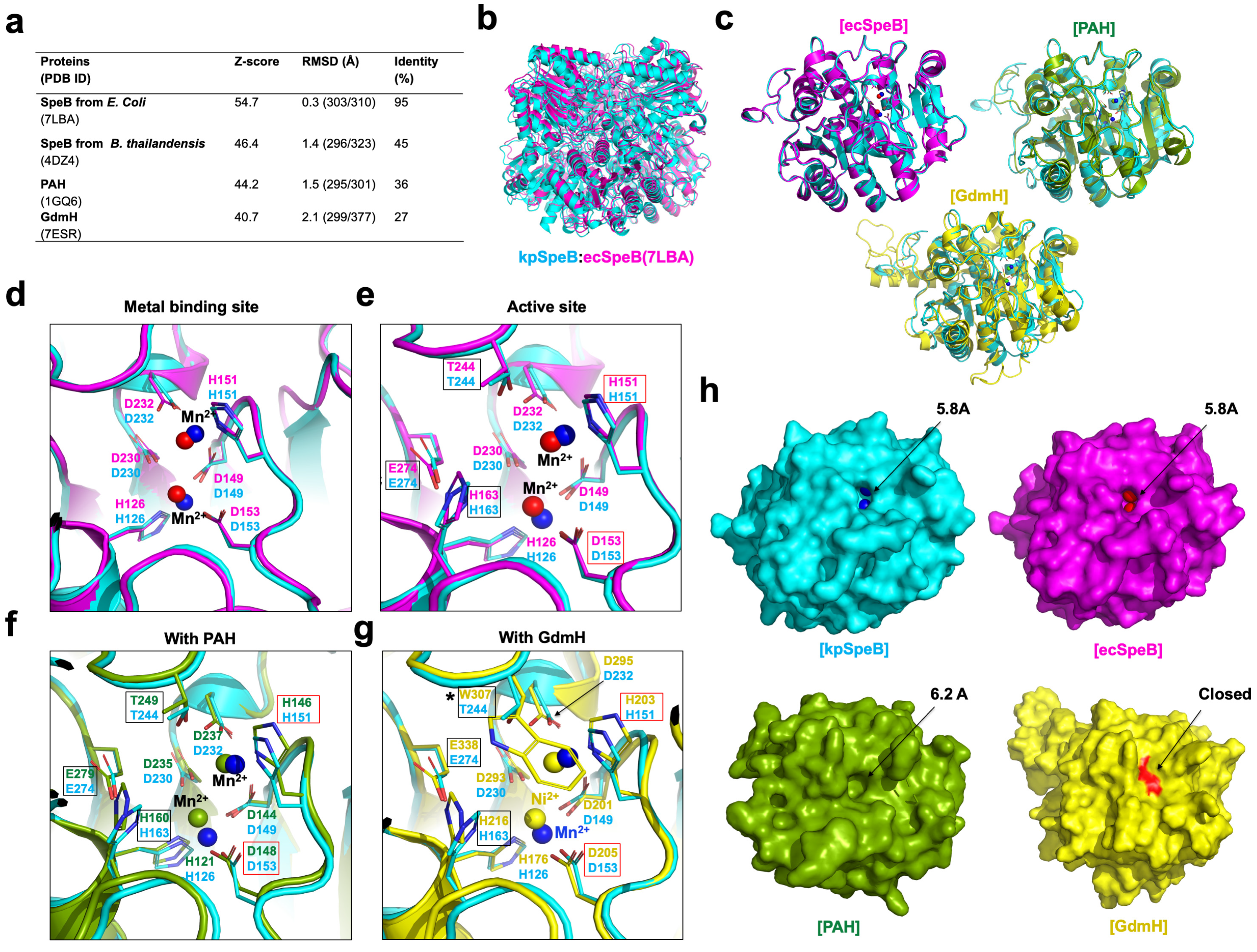
3.2. Structural Comparison of kpSpeB with Other Structural Isoforms
4. Discussion
Author Contributions
Funding
Institutional Review Board Statement
Informed Consent Statement
Data Availability Statement
Acknowledgments
Conflicts of Interest
References
- Miller-Fleming, L.; Olin-Sandoval, V.; Campbell, K.; Ralser, M. Remaining Mysteries of Molecular Biology: The Role of Polyamines in the Cell. J. Mol. Biol. 2015, 427, 3389–3406. [Google Scholar] [CrossRef] [Scilit] [PubMed]
- Pegg, A.E. Functions of Polyamines in Mammals. J. Biol. Chem. 2016, 291, 14904–14912. [Google Scholar] [CrossRef] [Scilit] [PubMed]
- Zhang, C.; Zhen, Y.; Weng, Y.; Lin, J.; Xu, X.; Ma, J.; Zhong, Y.; Wang, M. Research progress on the microbial metabolism and transport of polyamines and their roles in animal gut homeostasis. J. Anim. Sci. Biotechnol. 2025, 16, 57. [Google Scholar] [CrossRef] [Scilit] [PubMed]
- Kobayashi, T.; Sakamoto, A.; Kashiwagi, K.; Igarashi, K.; Takao, K.; Uemura, T.; Moriya, T.; Oshima, T.; Terui, Y. Putrescine Biosynthesis from Agmatine by Arginase (TtARG) in Thermus thermophilus. J. Biochem. 2023, 174, 81–88. [Google Scholar] [CrossRef] [Scilit] [PubMed]
- Benitez, J.; Garcia, D.; Romero, N.; Gonzalez, A.; Martinez-Oyanedel, J.; Figueroa, M.; Salas, M.; Lopez, V.; Garcia-Robles, M.; Dodd, P.R.; et al. Metabolic strategies for the degradation of the neuromodulator agmatine in mammals. Metabolism 2018, 81, 35–44. [Google Scholar] [CrossRef] [Scilit] [PubMed]
- Michael, A.J. Polyamines in Eukaryotes, Bacteria, and Archaea. J. Biol. Chem. 2016, 291, 14896–14903. [Google Scholar] [CrossRef] [Scilit] [PubMed]
- Dowling, D.P.; Di Costanzo, L.; Gennadios, H.A.; Christianson, D.W. Evolution of the arginase fold and functional diversity. Cell. Mol. Life Sci. 2008, 65, 2039–2055. [Google Scholar] [CrossRef] [Scilit] [PubMed]
- Hernandez, V.M.; Arteaga, A.; Dunn, M.F. Diversity, properties and functions of bacterial arginases. FEMS Microbiol. Rev. 2021, 45, fuab034. [Google Scholar] [CrossRef] [Scilit] [PubMed]
- Chitrakar, I.; Ahmed, S.F.; Torelli, A.T.; French, J.B. Structure of the E. coli agmatinase, SPEB. PLoS ONE 2021, 16, e0248991. [Google Scholar] [CrossRef] [Scilit] [PubMed]
- Baugh, L.; Gallagher, L.A.; Patrapuvich, R.; Clifton, M.C.; Gardberg, A.S.; Edwards, T.E.; Armour, B.; Begley, D.W.; Dieterich, S.H.; Dranow, D.M.; et al. Combining functional and structural genomics to sample the essential Burkholderia structome. PLoS ONE 2013, 8, e53851. [Google Scholar] [CrossRef] [Scilit] [PubMed]
- Tassoulas, L.J.; Wackett, L.P. Insights into the action of the pharmaceutical metformin: Targeted inhibition of the gut microbial enzyme agmatinase. iScience 2024, 27, 108900. [Google Scholar] [CrossRef] [Scilit] [PubMed]
- Lee, S.J.; Kim, D.J.; Kim, H.S.; Lee, B.I.; Yoon, H.J.; Yoon, J.Y.; Kim, K.H.; Jang, J.Y.; Im, H.N.; An, D.R.; et al. Crystal structures of Pseudomonas aeruginosa guanidinobutyrase and guanidinopropionase, members of the ureohydrolase superfamily. J. Struct. Biol. 2011, 175, 329–338. [Google Scholar] [CrossRef] [Scilit] [PubMed]
- Elkins, J.M.; Clifton, I.J.; Hernandez, H.; Doan, L.X.; Robinson, C.V.; Schofield, C.J.; Hewitson, K.S. Oligomeric structure of proclavaminic acid amidino hydrolase: Evolution of a hydrolytic enzyme in clavulanic acid biosynthesis. Biochem. J. 2002, 366, 423–434. [Google Scholar] [CrossRef] [Scilit] [PubMed]
- Funck, D.; Sinn, M.; Fleming, J.R.; Stanoppi, M.; Dietrich, J.; Lopez-Igual, R.; Mayans, O.; Hartig, J.S. Discovery of a Ni2+-dependent guanidine hydrolase in bacteria. Nature 2022, 603, 515–521. [Google Scholar] [CrossRef] [Scilit] [PubMed]
- Otwinowski, Z.; Minor, W. Processing of X-ray diffraction data collected in oscillation mode. Methods Enzymol. 1997, 276, 307–326. [Google Scholar] [PubMed]
- McCoy, A.J. Solving structures of protein complexes by molecular replacement with Phaser. Acta Crystallogr. D Biol. Crystallogr. 2007, 63, 32–41. [Google Scholar] [CrossRef] [Scilit] [PubMed]
- Adams, P.D.; Afonine, P.V.; Bunkoczi, G.; Chen, V.B.; Davis, I.W.; Echols, N.; Headd, J.J.; Hung, L.W.; Kapral, G.J.; Grosse-Kunstleve, R.W.; et al. PHENIX: A comprehensive Python-based system for macromolecular structure solution. Acta Crystallogr. D Biol. Crystallogr. 2010, 66, 213–221. [Google Scholar] [CrossRef] [Scilit] [PubMed]
- Emsley, P.; Cowtan, K. Coot: Model-building tools for molecular graphics. Acta Crystallogr. D Biol. Crystallogr. 2004, 60, 2126–2132. [Google Scholar] [CrossRef] [Scilit] [PubMed]
- Chen, V.B.; Arendall, W.B., 3rd; Headd, J.J.; Keedy, D.A.; Immormino, R.M.; Kapral, G.J.; Murray, L.W.; Richardson, J.S.; Richardson, D.C. MolProbity: All-atom structure validation for macromolecular crystallography. Acta Crystallogr. D Biol. Crystallogr. 2010, 66, 12–21. [Google Scholar] [CrossRef] [Scilit] [PubMed]



| Data Collection | |
|---|---|
| Space group | P 1 |
| Unit cell parameter a, b, c (Å) | |
| a, b, c (Å) | a = 77.63, b = 78.63, c = 79.07 |
| α, β, γ (°) | α = 105.349, β = 110.048, γ = 98.04 |
| Resolution range (Å) 1 | 29.28–2.77 (2.85–2.77) |
| Total reflections | 146,593 (14,896) |
| Unique reflections | 40,538 (4055) |
| Multiplicity | 3.6 (3.7) |
| Completeness (%) 1 | 97.11 (96.87) |
| Mean I/σ(I) 1 | 7.15 (1.88) |
| Rmerge (%) 1,2 | 15.93 (0.6566) |
| Wilson B-factor (Å2) | 44.16 |
| Refinement | |
| Resolution range (Å) | 29.28–2.77 |
| Reflections | 40,498 |
| Rwork (%) | 26.40 |
| Rfree (%) | 29.22 |
| No. of molecules in the asymmetric unit | 6 |
| No. of non-hydrogen atoms | 13,518 |
| Macromolecules | 13,439 |
| Ligands | 12 |
| Solvent | 67 |
| Average B-factor values (Å2) | 45.99 |
| Macromolecules | 46.01 |
| Ligands | 42.93 |
| Solvent | 42.67 |
| Ramachandran plot: | |
| favored/allowed/outliers (%) | 98.72/1.28/0.00 |
| Rotamer outliers (%) | 1.05 |
| Clash score | 15.69 |
| RMSD bonds (Å)/angles (°) | 0.005/1.02 |
Disclaimer/Publisher’s Note: The statements, opinions and data contained in all publications are solely those of the individual author(s) and contributor(s) and not of MDPI and/or the editor(s). MDPI and/or the editor(s) disclaim responsibility for any injury to people or property resulting from any ideas, methods, instructions or products referred to in the content. |
© 2026 by the authors. Licensee MDPI, Basel, Switzerland. This article is an open access article distributed under the terms and conditions of the Creative Commons Attribution (CC BY) license.
Share and Cite
Lee, S.Y.; Jin, H.B.; Park, H.H. Structure of Agmatinase from Klebsiella pneumoniae and the Active Site Comparison with Its Structural Homologues. Crystals 2026, 16, 285. https://doi.org/10.3390/cryst16050285
Lee SY, Jin HB, Park HH. Structure of Agmatinase from Klebsiella pneumoniae and the Active Site Comparison with Its Structural Homologues. Crystals. 2026; 16(5):285. https://doi.org/10.3390/cryst16050285
Chicago/Turabian StyleLee, So Yeon, Hyo Been Jin, and Hyun Ho Park. 2026. "Structure of Agmatinase from Klebsiella pneumoniae and the Active Site Comparison with Its Structural Homologues" Crystals 16, no. 5: 285. https://doi.org/10.3390/cryst16050285
APA StyleLee, S. Y., Jin, H. B., & Park, H. H. (2026). Structure of Agmatinase from Klebsiella pneumoniae and the Active Site Comparison with Its Structural Homologues. Crystals, 16(5), 285. https://doi.org/10.3390/cryst16050285