


Simple Summary
This study explores the role of 18F-fluorodeoxyglucose Positron Emission Tomography (18F-FDG-PET) as a preoperative predictor of Spread through Air Spaces (STAS) in early-stage non-small cell lung cancer (NSCLC), with the aim of improving surgical decision-making. STAS has emerged as an adverse prognostic factor, particularly after sublobar resections. This is particularly relevant for peripheral tumors ≤ 2 cm, where recent evidence supports sublobar resection as a feasible alternative to lobectomy. However, most available data derive from ground-glass opacity lesions, and evidence regarding PET-positive nodules remains limited. In this context, our study specifically investigates the predictive role of 18F-FDG-PET in stage IA NSCLC, focusing on tumors ≤ 2 cm. We identified an SUVmax ≥ 3 as significantly associated with the presence of STAS. These data suggest that, in patients with small peripheral tumors but elevated SUVmax, the presence of STAS should be considered to optimize the surgical strategy.
Abstract
Objective: Recent guidelines suggest sublobar resection as a viable option for peripheral lung cancer smaller than 2 cm. However, the presence of Spread through Air Spaces (STAS) is a well-known poor prognostic factor in early-stage lung cancer treated with sublobar resections. The aim of this study is to analyze the potential correlation between STAS and maximum standardized uptake volume (SUVmax) in this subset of patients to help define the optimal resection. Methods: A retrospective monocentric study was performed including patients diagnosed with stage IA lung cancer undergoing surgical resection. Patients were divided into two groups according to the presence/absence of STAS. As further investigation, we also separately analyzed the subgroup of patients with a peripheral nodule smaller than 2 cm. A p-value ≤ 0.05 was considered statistically significant. Results: The study cohort consists of 121 patients, 76 (62.8%) male, with a mean age of 74.2 ± 8.8 years. STAS was observed in 67 (55.4%) cases. The STAS-positive group showed a higher SUVmax value compared to the STAS-negative one (mean 5.5 ± 4.4 vs. 3.9 ± 3.0; p-value 0.007) with an AUC of 0.65 and an optimum SUVmax cut-off value of 3.0. Moreover, patients with SUVmax ≥ 3 showed a 77% increase in risk of having STAS (RR = 1.77; 95%CI = 1.21–2.59, p-value 0.014). This association was confirmed also in the subgroup of patients with nodules ≤ 2 cm (mean SUVmax 5.3 ± 4.6 vs. 3.6 ± 2.7; p value = 0.014). Conclusions: In our study, an SUVmax ≥ 3 in preoperative 18-FDG-PET is associated with the presence of STAS in stage IA lung cancer and peripheral cancer smaller than 2 cm. These findings, integrated with other clinical and radiological data, may guide surgeons to optimize the surgical strategy.
1. Introduction
Early diagnosis is the key factor for complete resection and potential cure in the case of lung cancer, a worldwide cause of tumor-related death [1]. In resectable early-stage non-small cell lung cancer (NSCLC), lobectomy still represents the gold standard [2,3]. However, in the case of small, peripheral and subsolid nodules, sublobar resections, particularly segmentectomy, have shown comparable outcomes and are now considered a viable and effective therapeutic alternative [2,3,4,5].
Spread through Air Spaces (STAS) has recently gained clinical attention due to its association with a worse prognosis and an increased risk of local recurrence, particularly in patients undergoing limited resections [6,7]. Intraoperative diagnosis of STAS could make the difference, but it is often unreliable; thus, it is crucial to develop tools for an effective preoperative assessment [8,9,10].
In this context, 18F-fluorodeoxyglucose Positron Emission Tomography (18F-FDG PET) has proven to be a promising noninvasive method to provide both morphologic and metabolic information. Recent studies showed the predictive value of variables such as SUVmax and consolidation-to-tumor ratio to identify STAS-positive tumors [11,12,13,14,15,16]. In addition, the integration of PET variables with traditional computed tomography (CT) imaging allowed the development of promising predictive models for risk stratification. However, studies specifically focusing on stage IA NSCLC remain limited.
Our study aims to investigate the association between 18F-FDG PET and STAS in patients diagnosed with stage IA lung cancer. As a further investigation, we analyzed the subgroup of patients with a peripheral cancer smaller than 2 cm. In those patients, guidelines indicate that both lobectomy and segmentectomy/wedge resection are considered viable options; having the possibility to know STAS preoperatively may change the surgical strategy.
2. Materials and Methods
We retrospectively reviewed a group of patients with lung cancer undergoing surgery at our institution between January 2017 and December 2023, according to the following inclusion criteria: preoperative 18-FDG PET scan within 3 months before surgery; pathological stage IA NSCLC according to the 9th TNM classification of the International Association for the Study of Lung Cancer; and assessment of STAS in the surgical specimen.
Patients with synchronous lung lesions, lung cancer recurrence or large cell neuroendocrine carcinoma and other rare lung tumors were not included.
STAS was defined in histopathological specimens according to the International Association for the Study of Lung Cancer as the presence of tumor cells within air spaces in the lung parenchyma beyond the edge of the main tumor. Histological evaluation was performed on formalin-fixed, paraffin-embedded, hematoxylin and eosin-stained sections. STAS assessment was performed by two experienced thoracic pathologists blinded to imaging data.
We anonymously collected the following clinical and radiological data: demographics (age, sex), medical history (smoking habits, previous diseases, comorbidities), imaging data (maximum diameter, aspect of the nodule, SUVmax), pathological data (tumor histotype and subtypes, tumor size, grading, presence or absence of STAS), and surgical procedure (lobectomy, segmentectomy or wedge resection; surgical approach).
The statistical analysis was performed using IBM SPSS Statistics v.27 (IBM Corp., Armonk, NY, USA). Categorical data are presented as numbers and percentages; continuous data as mean ± standard deviation (SD). The Shapiro–Wilk test was used to ensure the normal distribution of continuous data. The chi-square test was used to compare categorical data while Student’s t-test and the Mann–Whitney U test were applied to compare normally distributed and non-normally distributed continuous data, respectively. The Youden Index was utilized to determine the optimal cut-off value in the Receiver Operating Characteristic (ROC) curve. Risk ratios (RRs) were computed by comparing event probabilities across groups using contingency tables. Odds ratios (ORs) with 95% confidence intervals (CIs) were obtained through univariable logistic regression. Multivariable analysis was performed using logistic regression models to identify independent predictors of STAS. Due to collinearity between SUVmax as a continuous variable and the dichotomized variable SUVmax ≥ 3, two separate multivariable models were developed. The following covariates were included based on clinical relevance and univariable analysis results: tumor size, grading, radiological appearance, and SUVmax (continuous and dichotomized). Results were reported as odds ratios (ORs) with 95% CIs. A p-value ≤ 0.05 was considered statistically significant.
This study was approved by the Ethical Committee of Policlinico Umberto I (Protocol Number 1020/2024). Considering the retrospective nature of the study, the requirement of a specific informed consent was waived.
3. Results
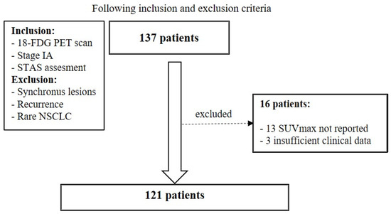
A hundred thirty-seven patients were initially included in the study. Among them, 13 patients were excluded because SUVmax was not explicitly indicated, and 3 because clinical data were incomplete (Figure 1).
Figure 1.
Flow diagram of study enrollment.
Therefore, the final cohort resulted in 121 patients, 76 (62.8%) male and 45 (37.2%) female, with a mean age of 74.2 ± 8.8 years. Among them, 62 (51.2%) were ex-smokers, 40 (33.0%) were current smokers, and 19 (15.7%) were never smokers. Hypertension (59.5%), dyslipidemia (52.0%), COPD (43.8%) and diabetes (29.8%) were the most frequently observed comorbidities, resulting in an ASA I score in 7.4%, an ASA II score in 43.8% and an ASA III score in 48.8% of the patients. All of them underwent surgical resection: 52 (42.9%) received lobectomy, four (3.3%) segmentectomy, and 65 (53.7%) wedge resection. The tumor was in the right upper lobe in 37 (30.5%) patients, in the middle lobe in four (3.3%), in the right lower lobe in 26 (21.4%), in the left upper lobe in 34 (28%), and in the left lower lobe in 20 (16.5%). All patients underwent a lobe-specific mediastinal lymph node dissection as the institutional standard approach. CT scan analysis showed a solid lesion in 59 (48.8%) cases, a mixed nodule in 44 (36.4%), and pure ground glass opacity in 18 (14.9%) cases. Histology showed adenocarcinoma in 100 (82.6%) patients, squamous cell carcinoma in 20 (16.5%) and mucoepidermoid carcinoma in one (0.8%). Adenocarcinomas showed an acinar predominant pattern in 58 (58.0%), a solid predominant pattern in 17 (17.0%), a lepidic predominant pattern in 10 (10.0%), a papillary predominant pattern in eight (8.0%) and others in seven (7.0%). Three patients with a lepidic predominant pattern received a diagnosis of minimally invasive adenocarcinoma (MIA). Sixty-three patients showed a moderately differentiated cancer (G2, 52.1%), 50 were poorly differentiated (G3, 41.3%), and four had a well-differentiated tumor (G1, 3.3%). The mean tumor size was 16.8 ± 5.8 mm (range 4–29 mm). No perioperative mortality was observed. The mean postoperative hospital stay was 5.8 ± 3.9 days. Postoperative complications were observed in 21 (17.3%) patients, most commonly cardiac arrhythmia (6, 5.0%) and prolonged air leaks (3, 2.5%).
All the patients underwent preoperative 18-FDG PET. Overall, the mean SUVmax was 4.8 ± 3.9 (range 1–27.6). STAS was found in the surgical specimen in 67 (55.4%) cases, while 54 (44.6%) patients were STAS negative. In our cohort, STAS was not statistically correlated with sex, age, smoking history and histological subtype as shown in Table 1. However, STAS was significantly associated with tumor grading (p-value = 0.005), acinar and solid adenocarcinoma pattern (p-value 0.031) and the presence of solid lesions at CT scan (p-value 0.041) (Table 1).
Table 1.
Correlation analysis between STAS-positive and STAS-negative groups.
3.1. Correlation Between Stas and Suvmax Values
Patients with STAS showed significantly higher SUVmax values compared to those without STAS (5.5 ± 4.4 vs. 3.9 ± 3.0; p = 0.007).
The ROC curve analysis showed an AUC (area under the curve) of 0.65 with an optimum SUVmax cut-off value measured through the Youden Index of 3.0.
Therefore, patients were dichotomized into two groups according to whether SUVmax was higher or lower than 3. In the SUVmax ≥ 3 group, 47 patients (68.1%) were STAS positive, while 22 (31.9%) were STAS negative. Conversely, among patients with an SUVmax < 3, 20 (38.5%) were STAS positive and 32 (61.5%) were negative. This difference resulted in a statistically significant result, with a p-value of 0.001 after chi-squared analysis. Moreover, the SUVmax ≥ 3 group showed an RR of 1.77 (95% CI, 1.21–2.59), indicating a 77% increase in risk of having STAS compared to SUVmax < 3. Univariate logistic regression confirmed this association, yielding an OR of 3.42 (95% CI, 1.61–7.27).
After multivariable analysis including tumor size, grading, and radiological appearance, both SUVmax (as a continuous variable) and SUVmax ≥ 3 were independently associated with STAS. In particular, SUVmax showed an OR of 1.16 (95% CI 1.03–1.31; p = 0.015), while SUVmax ≥ 3 was associated with a more than two-fold increased risk of STAS (OR 2.94, 95% CI 1.32–6.54; p = 0.008). Tumor grading also remained independently associated with STAS, whereas tumor size and radiological appearance were not significant predictors (Table 2).
Table 2.
Multivariable logistic regression analysis for predictors of STAS.
3.2. Analysis of the Subgroup of Patients with T ≤ 2 cm
As a further investigation, we performed the analysis in the subgroup of patients with a peripheral nodule smaller than 2 cm in diameter. This cohort consisted of 94 patients (76 (80.6%) male; mean age 73.8 ± 8.62 years) with a mean tumor diameter of 14.4 ± 3.9 mm. Thirty-seven (39.4%) underwent lobectomy and 57 (60.6%) received segmentectomy/wedge resection. Histological examination showed adenocarcinoma in 78 patients (83.0%) and squamous cell carcinoma in 16 patients (17.0%). The most frequent prevalent pattern in adenocarcinoma was acinar in 44 patients (56.4%), solid in 13 (16.7%) and lepidic in 8 (10.3%). STAS was present in 51 (54.3%) patients. A statistically significant association between the presence of STAS and SUVmax was confirmed also in this subgroup of patients: the STAS-positive and -negative groups showed a mean SUVmax of 5.3 ± 4.6 and 3.6 ± 2.7 respectively (p value = 0.014). The SUVmax ≥ 3 group showed a significantly higher STAS (65.4% vs. 40.5%; p value = 0.016) with a 67% increase in risk of having STAS than the SUVmax < 3 group (RR 1.61, 95% CI 1.06–2.44). Multivariable logistic regression analysis confirmed that both SUVmax (OR 1.14, 95% CI 1.01–1.30; p = 0.041) and SUVmax ≥ 3 (OR 2.71, 95% CI 1.13–6.47; p = 0.025) were independent predictors of STAS also in this subgroup of patients.
4. Discussion
In this study, we assessed the potential correlation between 18F-FDG PET parameters and STAS in stage IA NSCLC, particularly those smaller than 2 cm.
Previous studies showed a possible relationship between STAS and metabolic 18F-FDG PET parameters in lung cancer [10,11,12,13,14], but just a few reports focused on stage I NSCLC. Nishimori et al. [15] proved that SUVmax is the most reliable metabolic parameter to predict STAS in clinical stage I lung adenocarcinoma with a sensitivity of 89.5%, a specificity of 52.8% and an AUC of 0.74 using an SUVmax cut-off of 2.48.
In a recent study, Wang Y and colleagues [17] showed the predictive value of SUVmax and consolidation-tumor ratio in determining the presence of STAS in stage IA NSCLC, creating a nomogram with 78.1% accuracy in their validation test.
Suh JW et al. [16] proposed a stepwise flowchart using the SUV tumor/liver ratio at 18F-FDG PET to predict STAS negativity in NSCLC with T ≤ 2 cm achieving good results. However, their cohort also included pathological T2a, N1 and N2 patients, thus mixing stages IB, IIB and IIIA in the analysis.
While previous studies have demonstrated an association between PET-derived features and STAS, to our knowledge this is the first study specifically assessing this correlation in stage IA lung cancer smaller than 2 cm. This specific cohort of patients is clinically relevant, as sublobar resection is increasingly considered a valid alternative to lobectomy.
Our analysis showed how a preoperative SUVmax ≥ 3 is strongly correlated with the STAS in the pathological specimen. This information could be crucial in the case of small peripheral NSCLC to better personalize the surgical treatment considering that many studies showed that STAS is associated with worse prognosis and increased local recurrence after sublobar resection [5,6]. Importantly, the association between SUVmax and STAS persisted after adjustment for tumor size, grading, and radiological appearance, suggesting that metabolic activity reflects tumor aggressiveness beyond conventional clinicopathological features.
However, the discriminatory performance of SUVmax alone was limited, reaching an AUC of 0.65. Moreover, even if consistent with the literature, the SUVmax ≥ 3 cut-off is data-driven and needs validation. These findings indicate that SUVmax alone provides limited discriminatory value and may be better interpreted within a multimodal framework [18,19,20]. Advanced approaches such as radiomics and artificial intelligence-based tools may improve STAS preoperative prediction by integrating metabolic, radiological, and clinical data [13,14,21]. However, these tools are not yet widely implemented in clinical practice due to limited external validation, lack of standardization, and concerns regarding generalizability [9,22].
This study presents some limitations. First, it is a retrospective single-center study, potentially leading to a selection bias in patient selection, surgical strategy (lobectomy vs. segmentectomy vs. wedge) and STAS assessment. Interestingly, our cohort shows a relatively high prevalence of STAS in stage IA NSCLC. This finding is likely related to the PET-based selection of patients and the high predominance of solid nodules. In addition, PET/CT examinations were performed across different centers according to routine clinical practice, resulting in heterogeneity in acquisition protocols and representing a potential source of bias. Second, this study focused only on the correlation between SUVmax and STAS, not considering other 18-FDG PET parameters. We chose SUVmax because it is the most common metabolic feature measured at 18-FDG PET/CT and other parameters are not routinely reported. In addition, we did not perform a survival analysis because of incomplete follow-up data and thus discussion on surgical strategy must be interpreted cautiously and in the context of existing literature. Finally, the cohort number is relatively small and larger multi-centric studies are required to confirm these results.
5. Conclusions
In conclusion, an SUVmax ≥ 3 on preoperative 18-FDG PET was associated with the presence of STAS in stage IA NSCLC and in peripheral lung cancers smaller than 2 cm. These findings might improve preoperative risk stratification, particularly in early-stage lung cancer patients who may be candidates for sublobar resection. Integration of SUVmax data within the clinical and radiological assessment could offer an additional guide to plan the most appropriate surgical approach.
Author Contributions
Conceptualization, M.B., M.A., F.V. and D.D.; methodology, B.Z., R.V.S. and A.P.; validation, P.G., C.P., M.A. and T.D.G.; formal analysis, M.B.; investigation, R.V.S., S.D.M. and S.A.; data curation, B.Z., R.V.S., A.P., S.D.M. and S.A.; writing—original draft preparation, M.B. and B.Z.; writing—review and editing, P.G., F.V. and D.D. All authors have read and agreed to the published version of the manuscript.
Funding
This research received no external funding.
Institutional Review Board Statement
The study was conducted in accordance with the Declaration of Helsinki and approved by the Ethics Committee of Policlinico Umberto I of Rome (Protocol Number 1020/2024, 13 November 2024).
Informed Consent Statement
Patient consent was waived due to the retrospective nature of the study.
Data Availability Statement
The data underlying this article will be shared on reasonable request to the corresponding author.
Conflicts of Interest
The authors declare no conflicts of interest.
Abbreviations
The following abbreviations are used in this manuscript:
| NSCLC | non-small cell lung cancer |
| STAS | Spread through Air Spaces |
| 18-FDG PET | 18F-fluorodeoxyglucose Positron Emission Tomography |
| CT | computed tomography |
| SD | standard deviation |
| ROC | Receiver Operating Characteristic |
| RR | Risk ratio |
| OR | Odds ratio |
| CI | confidence intervals |
| MIA | minimally invasive adenocarcinoma |
| AUC | area under the curve |
References
- Moyer, V.A.; U.S. Preventive Services Task Force. Screening for lung cancer: U.S. preventive services task force recommendation statement. Ann. Intern. Med. 2014, 160, 330–338. [Google Scholar] [CrossRef] [Scilit]
- Postmus, P.E.; Kerr, K.M.; Oudkerk, M.; Senan, S.; Waller, D.A.; Vansteenkiste, J.; Escriu, C.; Peters, S.; ESMO Guidelines Committee. Early and locally advanced non-small-cell lung cancer (NSCLC): ESMO Clinical Practice Guidelines for diagnosis, treatment and follow-up. Ann. Oncol. 2017, 28, iv1–iv21. [Google Scholar] [CrossRef] [Scilit] [PubMed]
- Smythe, W.R.; American College of Chest Physicians. Treatment of stage I non-small cell lung carcinoma. Chest 2003, 123, 181S–187S. [Google Scholar] [CrossRef] [Scilit] [PubMed]
- Brunelli, A.; Decaluwe, H.; Gonzalez, M.; Gossot, D.; Petersen, R.H.; Panel, E.; Augustin, F.; Assouad, J.; Baste, J.M.; Steering Group; et al. European Society of Thoracic Surgeons expert consensus recommendations on technical standards of segmentectomy for primary lung cancer. Eur. J. Cardiothorac. Surg. 2023, 63, ezad224. [Google Scholar] [CrossRef] [Scilit]
- Bertolaccini, L.; Tralongo, A.C.; Del Re, M.; Facchinetti, F.; Ferrara, R.; Franchina, T.; Graziano, P.; Malapelle, U.; Menis, J.; Passaro, A.; et al. Segmentectomy vs. Lobectomy in stage IA non-small cell lung cancer: A systematic review and meta-analysis of perioperative and survival outcomes. Lung Cancer 2024, 197, 107990. [Google Scholar] [CrossRef] [Scilit]
- Mantovani, S.; Pernazza, A.; Bassi, M.; Amore, D.; Vannucci, J.; Poggi, C.; Diso, D.; D’aMati, G.; Della Rocca, C.; Rendina, E.A.; et al. Prognostic impact of spread through air spaces in lung adenocarcinoma. Interact. Cardiovasc. Thorac. Surg. 2022, 34, 1011–1015. [Google Scholar] [CrossRef] [Scilit] [PubMed]
- Travis, W.D.; Eisele, M.; Nishimura, K.K.; Aly, R.G.; Bertoglio, P.; Chou, T.-Y.; Detterbeck, F.C.; Donnington, J.; Fang, W.; Joubert, P.; et al. The International Association for the Study of Lung Cancer (IASLC) Staging Project for Lung Cancer: Recommendation to Introduce Spread Through Air Spaces as a Histologic Descriptor in the Ninth Edition of the TNM Classification of Lung Cancer. Analysis of 4061 Pathologic Stage I NSCLC. J. Thorac. Oncol. 2024, 19, 1028–1051. [Google Scholar] [CrossRef] [Scilit]
- Ding, Y.; Chen, Y.; Wen, H.; Li, J.; Chen, J.; Xu, M.; Geng, H.; You, L.; Pan, X.; Sun, D. Pretreatment prediction of tumour spread through air spaces in clinical stage I non-small-cell lung cancer. Eur. J. Cardiothorac. Surg. 2022, 62, ezac248. [Google Scholar] [CrossRef] [Scilit]
- Bassi, M.; Russomando, A.; Vannucci, J.; Ciardiello, A.; Dolciami, M.; Ricci, P.; Pernazza, A.; D’aMati, G.; Terracciano, C.M.; Faccini, R.; et al. Role of radiomics in predicting lung cancer spread through air spaces in a heterogeneous dataset. Transl. Lung Cancer Res. 2022, 11, 560–571. [Google Scholar] [CrossRef] [Scilit]
- Jiang, Q.; Wang, X.; Jiang, X.; Sun, H.; Chen, Q.; Cheng, Y.; Wang, Y.; Xing, T.; Zhang, X.; Xiao, Y. A narrative review of preoperative CT for predicting spread through air spaces of lung cancer. Transl. Lung Cancer Res. 2025, 14, 1471–1481. [Google Scholar] [CrossRef] [Scilit]
- Li, H.; Li, L.; Liu, Y.; Deng, Y.; Zhu, Y.; Huang, L.; Long, T.; Zeng, L.; Shu, Y.; Peng, D. Predictive value of CT and 18F-FDG PET/CT features on spread through air space in lung adenocarcinoma. BMC Cancer 2024, 24, 434. [Google Scholar] [CrossRef] [Scilit]
- Falay, O.; Selçukbiricik, F.; Tanju, S.; Erus, S.; Kapdağli, M.; Cesur, E.; Yavuz, Ö.; Bulutay, P.; Firat, P.; Mandel, N.M.; et al. The prediction of spread through air spaces with preoperative 18F-FDG PET/CT in cases with primary lung adenocarcinoma, its effect on the decision for an adjuvant treatment and its prognostic role. Nucl. Med. Commun. 2021, 42, 922–927. [Google Scholar] [CrossRef] [Scilit]
- Zheng, C.; Cai, Y.; Miao, J.; Zheng, B.; Gao, Y.; Shen, C.; Bao, S.; Tan, Z.; Sun, C. A PET/CT-based 3D deep learning model for predicting spread through air spaces in stage I lung adenocarcinoma. Clin. Transl. Oncol. 2025, 27, 3364–3374. [Google Scholar] [CrossRef] [Scilit] [PubMed]
- Gao, Z.; An, P.; Li, R.; Wu, F.; Sun, Y.; Wu, J.; Yang, G.; Wang, Z. Development and validation of a clinic-radiological model to predict tumor spread through air spaces in stage I lung adenocarcinoma. Cancer Imaging 2024, 24, 25. [Google Scholar] [CrossRef] [Scilit]
- Nishimori, M.; Iwasa, H.; Miyatake, K.; Nitta, N.; Nakaji, K.; Matsumoto, T.; Yamanishi, T.; Yoshimatsu, R.; Iguchi, M.; Tamura, M.; et al. 18F FDG-PET/CT analysis of spread through air spaces (STAS) in clinical stage I lung adenocarcinoma. Ann. Nucl. Med. 2022, 36, 897–903. [Google Scholar] [CrossRef] [Scilit] [PubMed]
- Suh, J.W.; Jeong, Y.H.; Cho, A.; Kim, D.J.; Chung, K.Y.; Shim, H.S.; Lee, C.Y. Stepwise flowchart for decision making on sublobar resection through the estimation of spread through air space in early stage lung cancer1. Lung Cancer 2020, 142, 28–33. [Google Scholar] [CrossRef] [Scilit]
- Wang, Y.; Lyu, D.; Cheng, C.; Zhou, T.; Tu, W.; Xiao, Y.; Zuo, C.; Fan, L.; Liu, S. Preoperative nomogram for predicting spread through air spaces in clinical-stage IA non-small cell lung cancer using 18F-fluorodeoxyglucose positron emission tomography/computed tomography. J. Cancer Res. Clin. Oncol. 2024, 150, 185. [Google Scholar] [CrossRef] [Scilit] [PubMed]
- Liu, X.; Zou, Q.; Sun, Y.; Liu, H.; Cailiang, G. Role of multiple dual-phase 18F-FDG PET/CT metabolic parameters in differentiating adenocarcinomas from squamous cell carcinomas of the lung. Heliyon 2023, 9, e20180. [Google Scholar] [CrossRef] [Scilit]
- Wang, X.Y.; Zhao, Y.F.; Yang, L.; Liu, Y.; Yang, Y.K.; Wu, N. Correlation analysis between metabolic tumor burden measured by positron emission tomography/computed tomography and the 2015 World Health Organization classification of lung adenocarcinoma, with a risk prediction model of tumor spread through air spaces. Transl. Cancer Res. 2020, 9, 6412–6422. [Google Scholar] [CrossRef] [Scilit]
- Toyokawa, G.; Yamada, Y.; Tagawa, T.; Kozuma, Y.; Matsubara, T.; Haratake, N.; Takamori, S.; Akamine, T.; Oda, Y.; Maehara, Y. Significance of Spread Through Air Spaces in Resected Pathological Stage I Lung Adenocarcinoma. Ann. Thorac. Surg. 2018, 105, 1655–1663. [Google Scholar] [CrossRef] [Scilit]
- Chen, X.; Qi, H.; Zou, Y.; Chen, Y.; Li, H.; Hu, D.; Jiang, L.; Wang, M.; Chen, L.; Chen, H.; et al. Predicting the spread through air spaces in lung adenocarcinoma from preoperative 18 F-FDG PET/CT radiomics. Nucl. Med. Commun. 2025, 46, 570–578. [Google Scholar] [CrossRef] [Scilit]
- Chen, J.; Zhang, X.; Xu, C.; Liu, K. Diagnostic performance of radiomics analysis for pulmonary cancer airway spread: A systematic review and meta-analysis. Diagn. Interv. Radiol. 2025, 31, 215–225. [Google Scholar] [CrossRef] [Scilit]

Disclaimer/Publisher’s Note: The statements, opinions and data contained in all publications are solely those of the individual author(s) and contributor(s) and not of MDPI and/or the editor(s). MDPI and/or the editor(s) disclaim responsibility for any injury to people or property resulting from any ideas, methods, instructions or products referred to in the content. |
© 2026 by the authors. Licensee MDPI, Basel, Switzerland. This article is an open access article distributed under the terms and conditions of the Creative Commons Attribution (CC BY) license.