


Simple Summary
Adrenal tumors are increasingly found by chance on scans performed for unrelated reasons. Most are harmless and need no treatment, but a minority produce excess hormones or are cancerous, and telling these apart requires a structured imaging and hormonal evaluation. This narrative review summarizes the contemporary work-up and management of incidentally discovered adrenal tumors, with particular attention to adrenocortical carcinoma and pheochromocytoma. We compare the recommendations of the major European and North American guidelines, show where they disagree and how strong the supporting evidence is, and appraise the data behind commonly used imaging and laboratory thresholds. We also review current surgical approaches, perioperative care, and emerging systemic therapies. Our aim is to give clinicians a practical, evidence-graded framework for deciding which adrenal tumors can safely be left alone and which need further testing, surgery, or specialist referral.
Abstract
Background/Objectives: Widespread use of high-resolution cross-sectional imaging has made the incidentally discovered adrenal mass a common clinical problem. Although most adrenal incidentalomas are non-functioning benign adenomas, clinicians must distinguish them from actionable pathology such as functional adenomas, adrenocortical carcinoma, and pheochromocytoma. This review outlines the contemporary diagnostic algorithm for adrenal incidentalomas and the main indications for surgical and medical intervention. Methods: We performed a narrative review of current society guidelines and of the primary literature underlying their recommendations on anatomic, biochemical, and surgical evaluation, comparing recommendations and evidence grades where guidelines differ. Results: Management is driven primarily by imaging phenotype, lesion size, and hormonal function. Unenhanced computed tomography attenuation ≤ 10 Hounsfield units is highly specific for a benign lipid-rich adenoma. Hormonal evaluation should exclude hypercortisolism in all patients and pheochromocytoma in all but asymptomatic patients with homogeneous, lipid-rich lesions, with testing for primary aldosteronism and androgen excess when clinically indicated. Minimally invasive adrenalectomy is standard for hormonally active benign adenomas and most pheochromocytomas, whereas open adrenalectomy remains preferred for suspected adrenocortical carcinoma that is large or locally invasive, to avoid tumor capsule rupture. Novel therapies, including peptide receptor radionuclide therapy, show efficacy in advanced disease. Conclusions: Society guidelines agree on most of the diagnostic pathway but diverge on surveillance of large benign lesions, interpretation of cortisol testing, and the surgical approach to suspected carcinoma. A standardized, multidisciplinary approach that recognizes these differences and the strength of the underlying evidence optimizes outcomes while preventing unnecessary procedures.
1. Introduction
Widespread use of high-resolution cross-sectional imaging has given rise to a modern clinical phenomenon termed the incidentaloma epidemic. Adrenal tumors are identified on approximately 5% of abdominal computed tomography (CT) scans performed for unrelated indications [1]. Although these lesions are common, the vast majority (>75%) are non-functioning benign adenomas that require no intervention [1].
Clinicians must therefore distinguish clinically insignificant lesions from actionable pathology, such as functional adenomas, adrenocortical carcinoma (ACC), pheochromocytoma, and metastatic disease [1].
Work-up of adrenal lesions requires assessment of both hormonal function and oncologic risk. Several societies have published broadly similar recommendations on diagnosis, treatment, and follow-up [2,3,4]. However, there are a few key variations in management, which are highlighted throughout this manuscript.
Herein, we review the contemporary diagnostic algorithm for adrenal incidentalomas, detailing the clinical, radiographic, and biochemical evaluations required to differentiate non-functioning adenomas from functional or malignant lesions (Figure 1). Guidelines from the European Society of Endocrinology (ESE) and the European Network for the Study of Adrenal Tumors (ENSAT), the Canadian Urological Association (CUA) and American Urological Association (AUA), the Endocrine Society, and the National Comprehensive Cancer Network (NCCN) are integrated into each section. Unlike existing reviews, we tabulate where these guidelines diverge, together with the strength of each recommendation and its evidence grade, and appraise the primary data behind commonly used diagnostic thresholds. We then cover the indications for surgical intervention and perioperative management. Finally, we address the management of specific adrenal pathologies—ACC, pheochromocytoma and paraganglioma (PPGL), and metastatic disease—highlighting current surgical controversies, updates in functional imaging, and emerging therapies.
Figure 1.
Diagnostic algorithm for evaluation of the adrenal incidentaloma. Lesions are classified on unenhanced CT as benign, indeterminate, or suspicious for malignancy, and hormonal evaluation proceeds in parallel. Yellow panels mark the points at which the ESE-ENSAT, CUA/AUA, and NCCN guidelines differ and gray panels the points on which they agree; the corresponding strength of recommendation and evidence grades are given in Table 1. Abbreviations: ACC, adrenocortical carcinoma; ACTH, adrenocorticotropic hormone; APW, absolute percentage washout; AUA, American Urological Association; AVS, adrenal vein sampling; CT, computed tomography; CUA, Canadian Urological Association; DHEA-S, dehydroepiandrosterone sulfate; DST, dexamethasone suppression test; ENSAT, European Network for the Study of Adrenal Tumors; ESE, European Society of Endocrinology; FDG-PET, fluorodeoxyglucose positron emission tomography; HU, Hounsfield units; MACS, mild autonomous cortisol secretion; MRI, magnetic resonance imaging; NCCN, National Comprehensive Cancer Network; PA, primary aldosteronism; PPGL, pheochromocytoma and paraganglioma; RPW, relative percentage washout; SI, signal intensity.
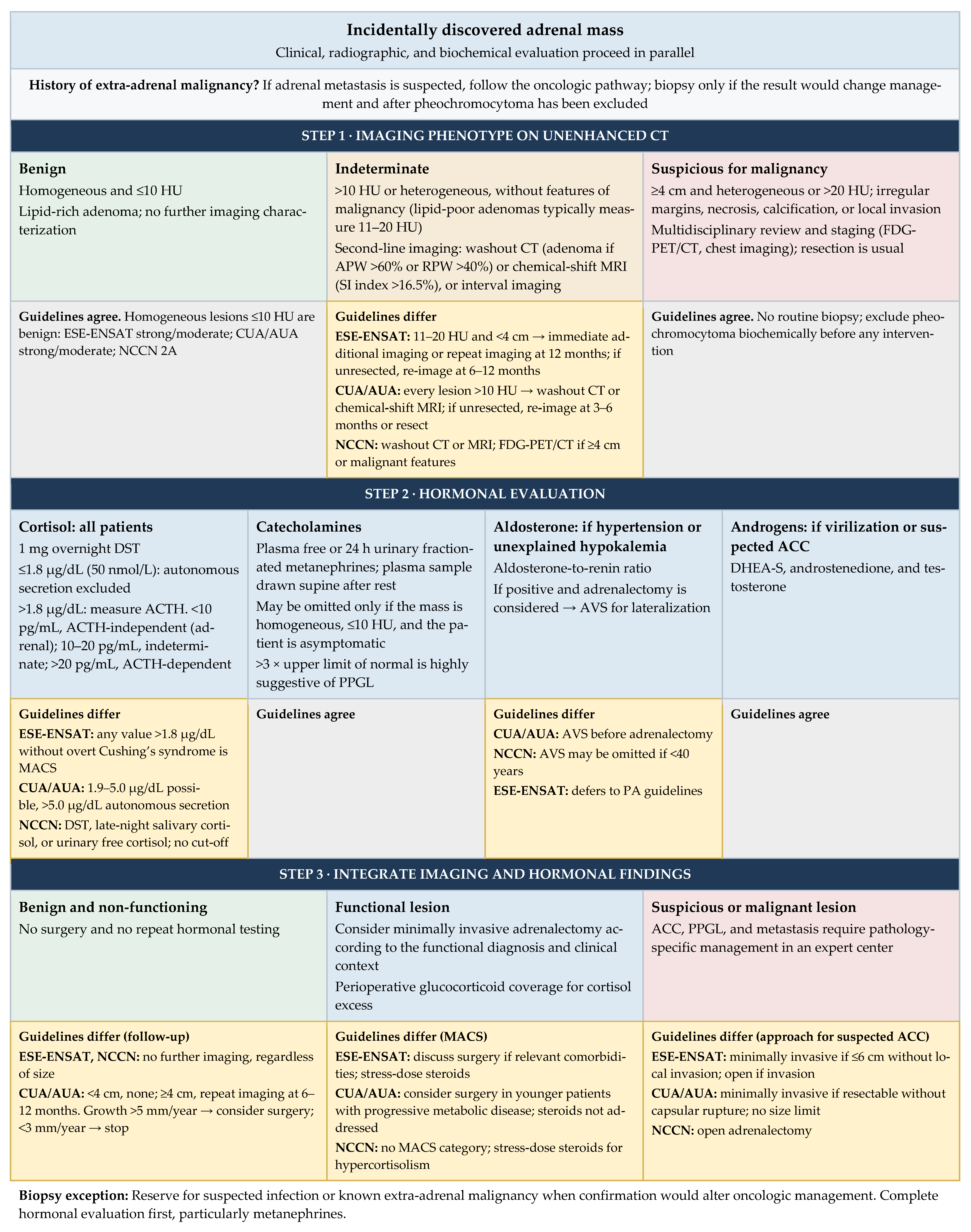
Table 1.
Principal differences among the ESE-ENSAT, CUA/AUA, and NCCN recommendations for the adrenal incidentaloma, with the strength of each recommendation and the quality of the supporting evidence.
2. Societal Guidelines
Three prominent guidelines address the management of adrenal incidentalomas. The ESE-ENSAT guideline was developed by the ESE with representatives from ENSAT and has been endorsed by the Endocrine Society [2]. The CUA developed a separate guideline with a panel of urologists, endocrinologists, and radiologists, which has been endorsed by the AUA [3]. Finally, the NCCN addresses adrenal masses through the lens of cancer detection [4]. The ESE-ENSAT and CUA/AUA guidelines used the Grading of Recommendations Assessment, Development and Evaluation (GRADE) process, stating each recommendation as ‘strong’ or ‘weak’ and rating the quality of the supporting evidence. NCCN recommendations reflect the evidence-informed consensus of an expert panel of medical and surgical oncologists and are assigned a category of evidence and consensus (category 2A unless otherwise noted); the GRADE scale is not used. The three guidelines agree on most of the diagnostic pathway. The principal points of divergence, with the corresponding strength of recommendation and evidence grade, are summarized in Table 1 and flagged in Figure 1.
This narrative review is anchored on these guidelines and on the Endocrine Society guidelines for primary aldosteronism (PA) and PPGL [5,6,7], supplemented by targeted, non-systematic searches for the primary studies that underpin key diagnostic thresholds and for recent therapeutic trials. No formal search strategy or risk-of-bias assessment was applied, and selection bias is therefore possible. Where guidelines conflict, we did not adjudicate by consensus; instead, we present each position with its grade, describe the underlying evidence, and indicate where that evidence favors one approach. Throughout, we distinguish graded guideline recommendations from expert opinion and from preliminary, hypothesis-generating data.
3. Initial Work-Up
3.1. Anatomic/Imaging Evaluation
Initial radiographic characterization of an adrenal mass is the cornerstone of the diagnostic algorithm and the primary filter for distinguishing benign from malignant lesions. All three guidelines recommend unenhanced CT as the first-line modality [2,3,4].
3.1.1. Size
Although size alone is not an indication for resection, historical data suggest that adrenal lesions ≥ 4 cm more often prove to be ACC on final pathology. Accordingly, the CUA/AUA guideline recommends no follow-up for non-functioning, radiologically benign lesions < 4 cm (strong recommendation, moderate-quality evidence) and repeat imaging at 6–12 months for the same lesions ≥ 4 cm (weak, low) [3] (Figure 1). In contrast, the ESE-ENSAT (strong, moderate) and NCCN (category 2A) guidelines recommend no further imaging for homogeneous lesions with attenuation ≤ 10 Hounsfield units (HU), regardless of size [2,4] (Table 1). Repeat imaging at 6–12 months is therefore not appropriate for every lesion ≥ 4 cm: it applies only to benign-appearing lesions under the CUA/AUA guideline, whereas large lesions that are heterogeneous or measure > 20 HU are managed as suspicious for malignancy. For lesions measuring 11–20 HU and <4 cm, the ESE-ENSAT guideline suggests either immediate additional imaging or interval imaging at 12 months (weak, very low) [2]. Interval growth prompts consideration of resection under both guidelines—an increase of >20% in maximum diameter with an absolute increase of ≥5 mm for ESE-ENSAT, and >5 mm per year for CUA/AUA—although both recommendations are weak [2,3].
3.1.2. Attenuation and Washout Characteristics
Benign adenomas are characterized by high intracellular lipid content, which results in low attenuation on unenhanced CT. A homogeneous mass measuring ≤ 10 HU is considered diagnostic of a benign lipid-rich adenoma by all three guidelines [2,3,4] (Table 2).
Table 2.
Imaging modalities and diagnostic performance for adrenal lesion characterization, with the source and design of the supporting studies and the corresponding guideline evidence level.
Lipid-poor adenomas are also benign but, lacking abundant intracellular lipid, display higher attenuation on CT (typically 10–20 HU). An adrenal mass measuring > 10 HU therefore requires further diagnostic evaluation.
In this setting, contrast-enhanced CT with washout analysis is used to evaluate vascular dynamics. Benign adenomas typically show rapid enhancement and rapid loss of contrast; an absolute percentage washout (APW) > 60% or a relative percentage washout (RPW) > 40% suggests a benign adenoma [3,14]. Malignant lesions (ACC or metastasis) and pheochromocytomas typically retain contrast and fall below these thresholds, although overlap is substantial (Table 2).
Chemical-shift magnetic resonance imaging (MRI) is an accepted alternative to washout CT for lesion characterization [3,4]. In-phase and opposed-phase sequences detect the intracellular lipid typical of adenomas, and a signal intensity (SI) index > 16.5% is commonly used as the threshold to confirm a benign adenoma [19] (Table 2). MRI is particularly useful for patients with an allergy to iodinated contrast and for those in whom ionizing radiation should be avoided, such as pediatric or pregnant patients.
3.1.3. Analysis of Evidence
Size (4 cm)
The 4 cm cut-off was first proposed on the basis of a retrospective national Italian survey that pooled 1004 adrenal incidentalomas over 15 years, in which a diameter ≥ 4 cm had 93% sensitivity for malignancy [12]. A 2024 meta-analysis of 40 studies supported this threshold (sensitivity 85%, specificity 75%), which offered the best balance among the size cut-offs compared; however, 19 of the 40 studies had at least one domain at high risk of bias [13]. Retrospective surgical series likewise support the threshold [23]; in one, malignancy was found in 0% of non-functioning incidentalomas < 4 cm versus 13.6% of those > 6 cm [24]. Nevertheless, small, dense ACCs, lymphomas, and metastases escape the size criterion: most non-adrenocortical malignancies measure < 4 cm [25], contemporary ACC series include tumors as small as 20 mm, all measuring > 20 HU [26], and one single-center series described 32 ACCs measuring ≤ 4 cm (median 27 mm; median attenuation 31 HU) [27]. For this reason, guidelines apply size in combination with attenuation rather than alone (Table 1).
Attenuation (10 and 20 HU)
Risk stratification by attenuation differs among societies (Table 1). The evidence for the 10 HU cut-off consists largely of small, retrospective studies at high or unclear risk of bias. In the systematic review that informed the ESE-ENSAT guideline, only 19 of 37 studies were suitable for meta-analysis, and only two (102 lesions) evaluated the 10 HU threshold in true incidentalomas, in which attenuation > 10 HU had 100% sensitivity for malignancy [2,10]. In the prospective, multicenter EURINE-ACT study (2017 patients, 98 with ACC), raising the threshold from >10 HU to >20 HU improved specificity for malignancy from 64% to 80% while preserving sensitivity (99% versus 100%) [9], a finding since supported by retrospective surgical data [11]. All three guidelines accordingly regard homogeneous lesions ≤ 10 HU as benign. Above 10 HU they diverge: only ESE-ENSAT stratifies by both attenuation (11–20 versus > 20 HU) and size, whereas CUA/AUA and NCCN treat every lesion > 10 HU as indeterminate and proceed to second-line imaging [2,3,4].
CT Washout
The absolute (>60%) and relative (>40%) washout thresholds were developed in mixed or oncologic cohorts rather than in true incidentalomas, with sensitivity and specificity of 98% and 92% in the derivation series [10,14]. Subsequent validation has been less favorable (Table 2). In a retrospective single-center cohort of 252 lesions, the absolute and relative thresholds misclassified 35.9% and 35.2%, respectively, of masses measuring > 10 HU [15]. Pheochromocytomas met washout criteria for adenoma in 29–50% of cases [16,17,18]. Consequently, the ESE-ENSAT guideline does not designate washout CT as a preferred second-line test, whereas CUA/AUA (weak, moderate) and NCCN (category 2A) offer it for indeterminate lesions [2,3,4] (Table 1). These estimates should be compared cautiously, because reference standards (histology versus imaging follow-up) and case mix differ among studies.
Chemical-Shift MRI
The 16.5% SI index cut-off was derived in a single-center study and is not explicitly endorsed by any society guideline [19]. Optimal thresholds are also field-strength dependent (>21.6% at 1.5 T versus > 7.4% at 3 T in an intra-individual comparison), which limits generalizability across imaging protocols [20]. Nevertheless, in a meta-analysis of 18 studies (1280 lesions), chemical-shift MRI had a pooled sensitivity of 94% and specificity of 95% for adenoma, with an area under the curve of 0.98 for the SI index [21]. Given the lack of a uniform, prospectively validated threshold, a fixed SI index cut-off should be regarded as supportive rather than definitive, although it may guide further work-up of suspicious lesions.
3.2. Functional/Hormonal Evaluation
Although most adrenal incidentalomas (approximately 75%) are benign and non-functioning, up to one quarter are hormone-secreting or malignant. Hormonal evaluation is therefore recommended for all patients, because functional lesions are frequently an indication for surgical excision [2,3].
3.2.1. Cortisol
Evaluation for hypercortisolism is essential in all patients with adrenal incidentalomas, even in the absence of the classic stigmata of Cushing’s syndrome, to identify mild autonomous cortisol secretion (MACS) [2,3]. MACS is associated with a high prevalence of cardiometabolic comorbidities, including hypertension (approximately 64%) and type 2 diabetes mellitus (30–40%), and with an increased risk of vertebral fractures, which warrants assessment of bone health [28].
The 1 mg overnight dexamethasone suppression test (DST) is the standard and most widely used screening test. The patient takes 1 mg of oral dexamethasone between 11:00 PM and midnight, and serum cortisol is measured the following morning between 8:00 and 10:00 AM. The test evaluates suppression of the hypothalamic–pituitary–adrenal (HPA) axis by exogenous glucocorticoid: a normal response is suppression of endogenous cortisol, whereas failure to suppress indicates autonomous secretion. A post-dexamethasone serum cortisol > 1.8 µg/dL (50 nmol/L) indicates non-suppression. Historically, the degree of non-suppression was used to grade severity; under the 2023 ESE-ENSAT guideline, however, any patient above this threshold without overt Cushing’s syndrome is classified as having MACS, with no further stratification (strong, low) [2] (Figure 1). The CUA/AUA guideline retains an intermediate tier, regarding 1.9–5.0 µg/dL as possible and >5.0 µg/dL (138 nmol/L) as evidence of autonomous secretion, and the NCCN accepts the DST, late-night salivary cortisol, or 24 h urinary free cortisol without specifying a cut-off [3,4] (Table 1).
In patients with a positive DST, plasma adrenocorticotropic hormone (ACTH) should be measured to confirm ACTH independence, thereby localizing the pathology to the adrenal gland and excluding pituitary or ectopic sources [2]. Normal plasma ACTH typically ranges from 10 to 60 pg/mL, and autonomous adrenal cortisol production suppresses it. An ACTH < 10 pg/mL therefore supports an ACTH-independent (adrenal) source, values of 10–20 pg/mL are indeterminate, and a non-suppressed ACTH > 20 pg/mL suggests ACTH-dependent hypercortisolism [2,29]. These thresholds are assay dependent and have not been validated across laboratory platforms, so a single ACTH measurement can be misleading. In one case series, five patients had falsely elevated ACTH (>20 pg/mL) on a single immunoassay platform because of assay interference, leading to unnecessary testing and surgery [30]. When the ACTH result conflicts with the clinical picture or imaging, the measurement should be repeated on a different laboratory platform.
The most common site of ectopic ACTH secretion is the lung, which accounts for nearly half of cases. Lung neuroendocrine tumors, including bronchial carcinoids, are the most frequent source (approximately 25%), followed by small-cell lung cancer (20%). Less common sources include thymic and pancreatic neuroendocrine tumors, medullary thyroid carcinoma, and PPGL [31].
3.2.2. Catecholamines
All three guidelines agree that biochemical testing for pheochromocytoma may be omitted in patients with a homogeneous adrenal mass measuring ≤ 10 HU on unenhanced CT who have no symptoms of catecholamine excess (e.g., paroxysmal hypertension, headache, palpitations, and diaphoresis) [2,3,4]. This recommendation (weak, low in the CUA/AUA guideline) rests on a multicenter retrospective study in which 374 of 376 pheochromocytomas measured > 10 HU and the remaining two measured exactly 10 HU [16]. All other lesions—those that are indeterminate, lipid-poor, or associated with symptoms—require biochemical screening.
The recommended initial tests are plasma free metanephrines or 24 h urinary fractionated metanephrines [3,7]. A plasma or urinary metanephrine or normetanephrine level more than three times the upper limit of normal is highly suggestive of PPGL; this threshold reflects NCCN consensus (category 2A) and Endocrine Society guidance rather than a prospectively validated diagnostic cut-off, and lesser elevations warrant repeat or confirmatory testing [4,7]. For plasma testing, strict adherence to collection protocols minimizes false-positive results: blood should be drawn with the patient supine after at least 20–30 min of rest, because upright posture and physical stress stimulate catecholamine release [7,28]. Tricyclic antidepressants and serotonin–norepinephrine reuptake inhibitors can cause false-positive results (specifically elevated normetanephrine) and should be held before testing when possible [7,28].
3.2.3. Aldosterone
Screening for PA is indicated in patients with an adrenal incidentaloma and concomitant hypertension or unexplained hypokalemia [2,3]. The cornerstone of screening is the aldosterone-to-renin ratio (ARR), calculated from the plasma aldosterone concentration and either plasma renin activity (PRA) or direct renin concentration (DRC) [5,6]. Suppressed renin with inappropriately high aldosterone yields an elevated ratio, the hallmark of the disease. According to the 2025 Endocrine Society guideline, screening is positive when PRA is ≤1 ng/mL/h or DRC is ≤8.2 mU/L, together with an elevated aldosterone (≥10 ng/dL by immunoassay or ≥7.5 ng/dL by liquid chromatography–tandem mass spectrometry [LC-MS/MS]) and an elevated ARR (>20 ng/dL per ng/mL/h with PRA, or >70 pmol/mU with DRC) [5]. Because these thresholds vary with the assay method, the units reported, and concurrent medications, results should be interpreted against laboratory-specific reference values and in clinical context.
Interpretation of the ARR is frequently confounded by antihypertensive medications. Mineralocorticoid receptor antagonists (spironolactone, eplerenone) and epithelial sodium channel blockers (amiloride) raise renin and can produce false-negative results, whereas beta-blockers suppress renin and can produce false-positive results; when it is safe to do so, these agents should be withdrawn before testing [6].
Patients with a positive screen who are candidates for adrenalectomy should undergo adrenal vein sampling (AVS) to lateralize aldosterone secretion, because CT alone does not reliably distinguish an aldosterone-producing adenoma, which is managed surgically, from bilateral adrenal hyperplasia, which is managed medically [6]. The CUA/AUA guideline recommends AVS before adrenalectomy (strong, moderate), the NCCN considers its omission reasonable in patients younger than 40 years, and the ESE-ENSAT incidentaloma guideline defers to dedicated PA guidelines [2,3,4] (Table 1). Recognized exceptions include young patients (<35 years) with marked hypokalemia, a plasma aldosterone concentration > 30 ng/dL, and a unilateral adenoma; patients with bilateral macronodular hyperplasia; patients with concomitant adrenal Cushing’s syndrome and a unilateral mass; and patients with mild PA who proceed directly to medical management [5,6]. AVS is generally performed by an interventional radiologist via percutaneous femoral venous access. Samples are obtained from both adrenal veins and from the inferior vena cava (IVC) or external iliac vein to measure aldosterone and cortisol. A selectivity index (adrenal vein cortisol divided by IVC cortisol) above the institutional threshold (typically 1.4–3) confirms that the adrenal veins were sampled successfully. A lateralization index (the aldosterone-to-cortisol ratio of the dominant adrenal vein divided by that of the contralateral vein) ≥ 4 indicates unilateral secretion, and a contralateral suppression index (the aldosterone-to-cortisol ratio of the non-dominant adrenal vein divided by that of the IVC) ≤ 1 supports lateralization [6].
3.2.4. Androgens
Dehydroepiandrosterone sulfate (DHEA-S) is the preferred biochemical marker of adrenal androgen hypersecretion; its long half-life and high serum concentration provide a stable measure that avoids the circadian fluctuation of other androgens [32]. Evaluation of a suspected androgen-producing mass should also include serum androstenedione and free testosterone [32]. All three guidelines reserve androgen testing for patients with virilization or imaging features suspicious for ACC [2,3,4].
Careful history-taking helps to differentiate the underlying etiology. Benign conditions such as polycystic ovary syndrome typically cause mild-to-moderate symptoms (hirsutism, acne) over an extended period, whereas rapid-onset virilization in a female patient should raise suspicion for a malignant process such as ACC [32]. A free testosterone level > 8.85 pg/mL yielded a sensitivity of 82% and a specificity of 97% for distinguishing androgen-secreting adrenocortical tumors from non-tumoral causes of androgen excess [33].
3.3. Biopsy
Percutaneous biopsy has no role in the routine initial work-up of an adrenal incidentaloma, a point on which all three guidelines agree [2,3,4]. The procedure carries risks of pneumothorax, hemorrhage, and infection, and in unsuspected pheochromocytoma it can precipitate a life-threatening hypertensive crisis [1]. Perhaps most critically, if the lesion is an ACC, violating the tumor capsule risks seeding along the needle track, potentially upstaging the malignancy and compromising curative resection [1,34].
Biopsy is reserved for lesions concerning for infection and for patients with a known extra-adrenal malignancy (e.g., lung cancer, breast cancer, or melanoma) in whom the adrenal mass is a suspected metastasis, and only when confirmation would alter oncologic management (e.g., a change in systemic regimen or clinical trial eligibility) [2,3,4]. Hormonal evaluation, particularly plasma metanephrines, should be completed before sampling to avoid a hypertensive crisis [3] (Figure 1).
4. Surgical Management of Diagnostically Confirmed Benign Adrenal Cortical Adenomas
Minimally invasive surgery (MIS) is the standard approach for most adrenal pathology, including hormonally active benign adenomas [2,3]. Whether MACS itself warrants adrenalectomy is graded differently: ESE-ENSAT recommends discussing surgery when MACS coexists with relevant comorbidities (strong, very low), whereas CUA/AUA suggests considering it in younger patients with progressive cortisol-related metabolic disease (weak, low) (Table 1).
For patients with overt Cushing’s syndrome or MACS, perioperative glucocorticoid coverage is recommended: ESE-ENSAT advises stress-dose coverage for every patient with a post-DST cortisol > 1.8 µg/dL (strong, ungraded good-practice statement) and the NCCN for hypercortisolism (category 2A), whereas the CUA/AUA guideline does not address the issue [2,4] (Table 1). Removal of the hyperfunctioning tumor unmasks suppression of the remaining adrenal axis, placing the patient at risk of acute adrenal insufficiency. The standard protocol is intraoperative stress-dose hydrocortisone (typically 50–100 mg intravenously), followed by a slow postoperative taper of oral glucocorticoid (e.g., hydrocortisone or prednisone) guided by clinical recovery and cortisol levels until the HPA axis recovers [28].
Recent data challenge the universal application of this practice. In a prospective single-center pilot study of 12 patients with pituitary or adrenal Cushing’s syndrome undergoing curative surgery as first-line treatment, Trummer et al. withheld glucocorticoids until the first postoperative day without clinical hypocortisolism, suggesting that routine perioperative replacement may be unnecessary in selected patients when close postoperative monitoring is available [35].
The need for steroids is even more debated in MACS. Contrary to earlier literature suggesting high rates of dependency, a 2025 study by Ibrahimli et al. found that only a minority (23%) of patients with MACS required postoperative steroid replacement [36]. Independent predictors of replacement were a preoperative plasma ACTH < 7.0 pg/mL (p = 0.02) and a post-DST cortisol > 4.2 µg/dL (p = 0.008) [36]. In patients requiring replacement, steroids were weaned within a median of 78 days, and with this selective algorithm there was no evidence of adrenal insufficiency at a median follow-up of 15 months.
These data do not constitute level I evidence; most studies are small, single-center, and hypothesis-generating. Contemporary guidelines continue to recommend glucocorticoid coverage, and a selective algorithm has not been prospectively validated.
5. Adrenocortical Carcinoma
5.1. Epidemiology and Genetics
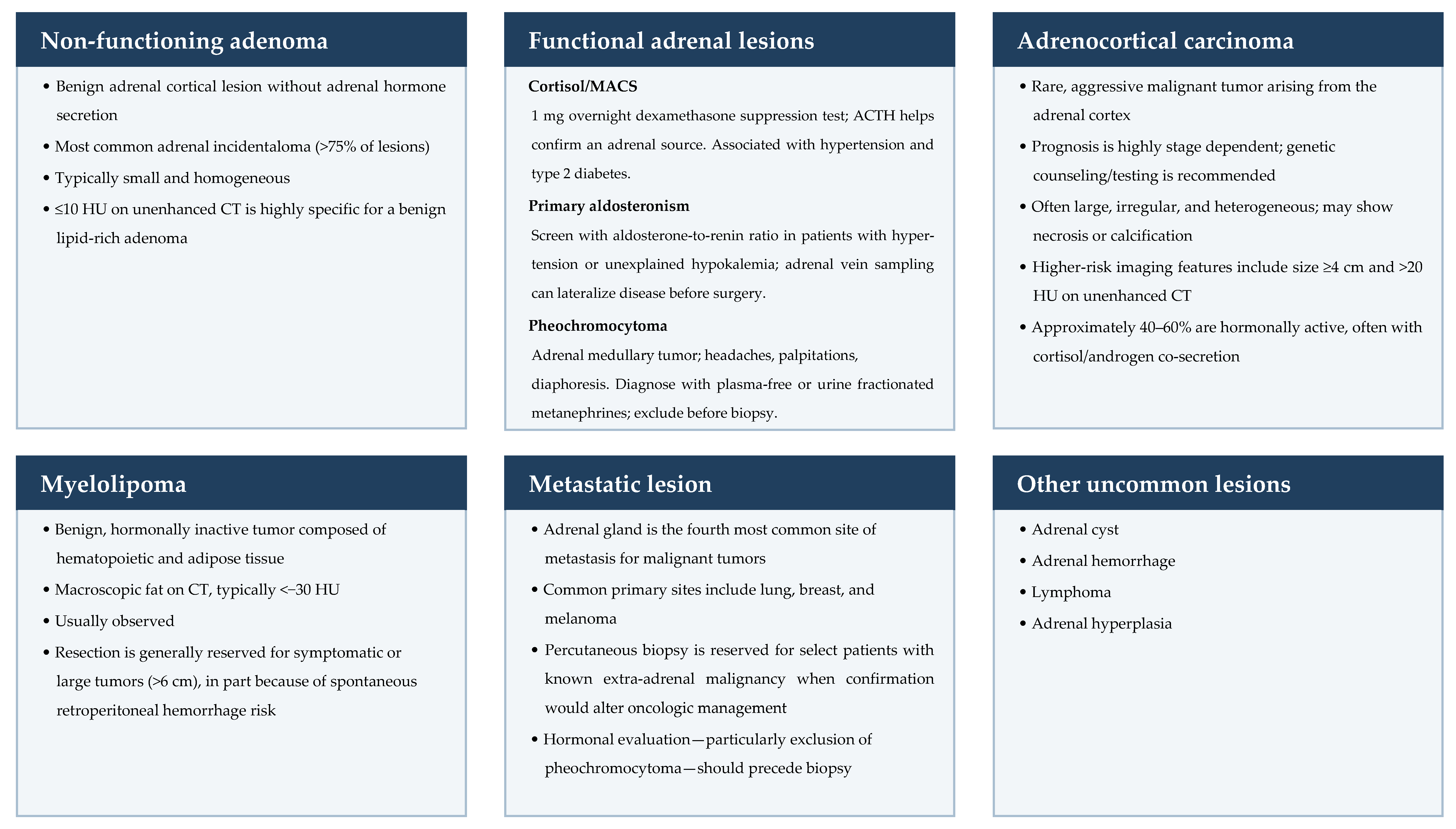
ACC is a rare and aggressive malignancy arising from the adrenal cortex (Figure 2), with an estimated annual incidence of 0.7–2.0 cases per million population [34]. The disease has a bimodal age distribution, with a first peak in early childhood (typically <5 years of age) and a second, broader peak in the fourth to fifth decades of life [37].
The overall prognosis for ACC remains poor but is highly stage dependent. Under the ENSAT staging system, 5-year survival ranges from 65 to 82% for stage I to 10–20% for stage IV [37]. Although survival has improved slightly over the past two decades, outcomes remain guarded, particularly for patients with a high Ki-67 index, cortisol hypersecretion, or advanced age [37].
A substantial proportion of ACC cases, particularly in children, are associated with hereditary cancer syndromes such as Li–Fraumeni syndrome (germline TP53 mutation), which accounts for 50–80% of pediatric cases [4]. Lynch syndrome is also relevant, because mismatch repair deficiency (MMR-D) with high microsatellite instability (MSI-H) increases the risk of adrenocortical malignancy. Genetic counseling and testing for inherited syndromes are therefore recommended for all patients diagnosed with ACC (NCCN category 2A) [4] (Figure 2).
5.2. Clinical Presentation and Diagnosis
5.2.1. Radiologic Presentation
Although many patients present with symptoms of hormonal excess (40–60%) or tumor growth (abdominal pain, fullness, or early satiety), approximately 20–30% of ACCs are diagnosed incidentally as large (approximately 10–13 cm), asymptomatic masses on imaging [32]. Tumor size informs the diagnosis: masses ≥ 4 cm are traditionally considered high risk for malignancy, and only a minority of ACCs (3%) measure ≤ 4 cm. Size also has prognostic value, as patients with smaller tumors (<5 cm) may have better overall survival (OS) [32]. In addition, ACCs tend to be irregular and heterogeneous and commonly show central necrosis or calcification [38,39]. In EURINE-ACT, the combination of attenuation > 20 HU, tumor size ≥ 4 cm, and a high-risk urine steroid metabolomics profile had a positive predictive value of 76.4% and a negative predictive value of 99.7% for ACC [9].
5.2.2. Functional Status
Approximately 40–60% of ACCs are hormonally active at diagnosis [37]. The most common functional presentation is a mixed picture, typically co-secretion of cortisol and androgens (Figure 2), which can manifest as rapidly progressive Cushing’s syndrome with virilization (hirsutism, male-pattern baldness, clitoromegaly, deepening of the voice, and severe menstrual abnormalities) [37].
Figure 2.
Differential diagnosis of the adrenal incidentaloma. Abbreviations: ACTH, adrenocorticotropic hormone; CT, computed tomography; HU, Hounsfield units; MACS, mild autonomous cortisol secretion.
5.2.3. Pathologic Evaluation
The definitive diagnosis of malignancy in adrenocortical tumors rests on histopathologic scoring of the resected specimen. The Weiss score is the most widely used system, with a score ≥ 3 (of nine histologic criteria) indicating malignancy [34,40]. The criteria are high nuclear grade, >5 mitoses per 50 high-power fields, atypical mitoses, clear cells comprising ≤ 25% of the tumor, diffuse architecture, necrosis, and venous, sinusoidal, and capsular invasion [40]. The score has recognized limitations. Because a single criterion can move a tumor from a score of 2 to the diagnostic threshold of 3, interobserver variability is consequential: in a French national program, agreement among 12 pathologists was only moderate to good (κ = 0.70) and improved after dedicated training [41]. Adequate sampling of large, heterogeneous specimens also affects the result. Furthermore, oncocytic tumors meet three Weiss criteria by definition, leading to overdiagnosis; myxoid tumors have a morphology that complicates identification of individual criteria; and the score overestimates malignant potential in pediatric tumors. Alternative systems are recommended for these variants in the 2022 World Health Organization classification [40,42]. Finally, because ACC is rare, applicability among non-expert pathologists is limited: in the German ACC registry, reference pathology revised the diagnosis in 13% of cases [43]. Suspected ACC should therefore be reviewed by an expert adrenal pathologist, as suggested by the ESE-ENSAT guideline (weak recommendation, low-quality evidence) [34].
The Ki-67 proliferation index, assessed by immunohistochemistry, adds prognostic information. Ki-67 is a non-histone nuclear protein expressed during the active phases of the cell cycle that stains strongly in rapidly dividing ACC cells. A Ki-67 index > 10–15% indicates a high-grade tumor with a significantly elevated risk of recurrence and poor long-term survival [34,37].
5.3. Surgical Management
Complete (R0) resection without capsular violation is the only curative treatment for ACC, and the choice of approach is governed by the ability to achieve it. For suspected ACC that is large (>6 cm) or shows radiographic evidence of local invasion, nodal disease, or venous tumor thrombus, open adrenalectomy remains the preferred approach [3,4,34]. Open resection reduces the risk of tumor rupture and spillage, which is associated with local recurrence rates approaching 100% [44].
Historically, MIS was strongly discouraged for ACC because of higher rates of positive resection margins and peritoneal dissemination than with open adrenalectomy [44]. Recent evidence suggests that this absolute contraindication is softening for smaller, localized tumors in which complete resection is feasible without tumor rupture [45]. The preoperative features that identify such patients are consistent across sources: the European Society of Endocrine Surgeons and ENSAT, in a consensus statement acknowledged to rest on low-quality evidence, and the ESE-ENSAT guidelines (weak, very low) accept MIS for tumors ≤ 6 cm without imaging evidence of local or nodal invasion when surgery is performed in an expert center [2,34,46]. The guidelines differ at the margins: CUA/AUA sets no size limit and permits MIS whenever resection without capsular rupture is achievable (weak, low), whereas the NCCN favors open adrenalectomy for any suspected ACC [3,4] (Table 1). Among minimally invasive techniques, robotic approaches may shorten hospitalization and reduce blood loss compared with laparoscopy [44]; either is acceptable provided the surgeon uses the technique with which they are most experienced and adheres strictly to oncologic principles [45].
Regardless of technique, oncologic principles apply: preservation of the tumor capsule, en bloc resection of involved structures, intact specimen extraction, and locoregional lymphadenectomy, which was associated with a lower risk of recurrence and disease-related death in a retrospective registry analysis of 283 patients [46,47]. Because outcomes depend on surgical experience, referral to a center with expertise in adrenal surgery is strongly recommended [34,46].
5.4. Systemic Therapy
5.4.1. Standard of Care
For advanced, recurrent, or metastatic ACC, the first-line regimen is etoposide, doxorubicin, and cisplatin plus mitotane (EDP-M); mitotane is an adrenolytic agent that targets the adrenal cortex [4,48]. Despite its toxicity, this combination is the established standard of care on the basis of the landmark FIRM-ACT trial, which randomized 304 patients with advanced ACC to EDP-M or streptozocin plus mitotane, with OS as the primary endpoint [48]. OS did not differ significantly between the EDP-M and streptozocin–mitotane groups (median 14.8 versus 12.0 months; hazard ratio 0.79, 95% confidence interval 0.61–1.02; p = 0.07). EDP-M did, however, improve the objective response rate (23.2% versus 9.2%; p < 0.001) and median progression-free survival (PFS; 5.0 versus 2.1 months; hazard ratio 0.55; p < 0.001) [48]. Rates of serious adverse events did not differ significantly between groups (58.1% with EDP-M versus 41.6% with streptozocin–mitotane) but were clinically important and included bone marrow toxicity, thromboembolic events, and liver dysfunction [48]. Despite the improvement in PFS, the prognosis of advanced ACC remains dismal, and given the toxicity of these regimens, better treatment options are needed.
5.4.2. Immunotherapy
The treatment landscape of ACC has expanded to include immunotherapy. Pembrolizumab carries a tumor-agnostic approval for MSI-H or MMR-D solid tumors, and the NCCN lists it, with or without mitotane, as an option for advanced ACC irrespective of microsatellite status (category 2A) [4]. In a single-center, single-arm phase II study from Memorial Sloan Kettering, 39 patients with advanced ACC, 72% of whom had received prior therapy, were treated with pembrolizumab 200 mg every 3 weeks [49]. The primary endpoint was objective response rate by Response Evaluation Criteria in Solid Tumors (RECIST) version 1.1. Nine patients (23%) had a partial response and seven (18%) had stable disease. Neither MSI-H/MMR-D status nor programmed death-ligand 1 (PD-L1) expression predicted response: two of six patients with MSI-H/MMR-D tumors responded, and seven of the nine responders had microsatellite-stable tumors [49]. Median PFS was 2.1 months and median OS was 24.9 months at a median follow-up of 17.8 months, and grade 3–4 treatment-related adverse events occurred in 13% of patients [49]. Although these outcomes are encouraging, interpretation is limited by the small, single-arm design, and the efficacy of checkpoint inhibitors in ACC is frequently blunted by tumor biology. A major limitation is the systemic immunosuppression caused by cortisol excess, which can dampen the T-cell response required for immunotherapy to work effectively [37].
5.4.3. Neoadjuvant Therapy
The role of neoadjuvant therapy remains debated because prospective data are lacking. Existing retrospective studies are limited by selection bias, making it difficult to determine whether systemic therapy truly renders unresectable tumors amenable to surgery [45]. Neoadjuvant systemic therapy allows treatment to begin at the earliest possible time and may reduce tumor size, maximizing the probability of an R0 resection while minimizing the magnitude of the operation [45]. It also acts as a biological selection mechanism, identifying patients who respond to induction therapy and are therefore most suitable for surgery. It has also been suggested that immunosuppression after extensive upfront surgery could increase the chance of metastasis. Currently, the recommended approach is upfront EDP-M, with repeat imaging after 2–6 cycles to evaluate resectability [45].
5.4.4. Adjuvant Mitotane
Given the high propensity of ACC to recur even after radical resection, adjuvant therapy should be considered [45]. The ESE-ENSAT guideline suggests adjuvant mitotane for patients with high-risk disease (ENSAT stage III, microscopically positive [R1] margins, or Ki-67 > 10%) [34], whereas the NCCN lists it as a category 3 option, reflecting major disagreement within the panel [4]. Current expert practice has evolved to recommend adjuvant mitotane for all patients except those at low risk of recurrence [45]. This exception is supported by the randomized ADIUVO trial, in which 5-year recurrence-free survival among 91 patients with low-to-intermediate-risk ACC was similar with mitotane (79%) and with surveillance alone (75%) [50].
6. Pheochromocytoma and Paraganglioma
6.1. Genetics
Pheochromocytomas and paragangliomas are rare neuroendocrine tumors arising from chromaffin cells (Figure 2). The distinction between the two is primarily anatomic: tumors originating from the adrenal medulla are pheochromocytomas, whereas those arising from extra-adrenal autonomic paraganglia are paragangliomas [7]. Although these tumors are typically detected at a localized stage, 15–20% of patients present with de novo metastatic disease or develop metastatic recurrence after primary curative treatment [51]. Hereditary germline mutations—most commonly those underlying von Hippel–Lindau (VHL) disease and multiple endocrine neoplasia type 2 (MEN2) and those in the succinate dehydrogenase subunit genes (SDHx)—are identified in 30–40% of patients [7,51].
6.2. Diagnosis
6.2.1. Biochemical Testing
Diagnosis is supported by elevated plasma free metanephrines, ideally measured by LC-MS/MS [7]. Clinicians must remain vigilant for false-positive results, which occur in approximately 20–25% of cases and are frequently caused by interfering medications or physiologic stressors such as obstructive sleep apnea [51].
6.2.2. Anatomic and Functional Imaging
Once biochemical evidence suggests a catecholamine-secreting tumor, CT or MRI is the standard modality for initial anatomic localization [7]. For functional characterization, 123I-metaiodobenzylguanidine (MIBG) scintigraphy is less sensitive for metastatic and multifocal PPGL than positron emission tomography (PET) with 18F-fluorodeoxyglucose (FDG) or 68Ga-DOTATATE [4]. In a meta-analysis, pooled lesion detection rates were 93% for 68Ga-labeled somatostatin analog PET, 74% for FDG PET, and 38% for MIBG [22] (Table 2). The NCCN lists somatostatin receptor PET alongside FDG PET and MIBG for suspected extra-adrenal, multifocal, or metastatic PPGL (category 2A) [4].
6.3. Perioperative Management
The primary goals of preoperative management are blood pressure control and expansion of intravascular volume. Alpha-adrenergic blockade is recommended for all patients with functional tumors and should be started 7–14 days before surgery (NCCN category 2A), using a non-selective agent such as phenoxybenzamine or a selective alpha-1 antagonist such as doxazosin [4,7]. In the randomized PRESCRIPT trial (134 patients), the proportion of intraoperative time outside the blood pressure target did not differ between phenoxybenzamine and doxazosin [52]. After alpha-blockade is initiated, patients should be assessed objectively for surgical readiness. Although no randomized data have established optimal targets, expert consensus suggests a seated blood pressure of 120–130/80 mmHg, a standing systolic blood pressure ≥ 90 mmHg, and a heart rate of 60–70 beats per minute seated and 70–80 beats per minute standing [53]. All patients should have a preoperative electrocardiogram, with an echocardiogram for those with suspected cardiomyopathy, prior major cardiac events, or prolonged catecholamine excess. Patients with uncontrolled blood pressure, symptomatic hypotension or hypertension, syncope, or signs of volume depletion should be admitted preoperatively.
Alpha-blockade must be paired with intravascular volume repletion. Patients with pheochromocytoma have chronic vasoconstriction and a contracted total blood volume; once alpha receptors are blocked and vessels relax, this relative hypovolemia can precipitate severe orthostatic hypotension or intraoperative circulatory collapse. Guidelines therefore recommend a high-salt diet and liberal hydration starting 7–14 days preoperatively and continued until orthostatic stability is achieved [4,7].
Although alpha-blockade with doxazosin remains the mainstay of preoperative preparation, recent data suggest that adding metyrosine (alpha-methyl-para-tyrosine) may improve hemodynamic stability in selected high-risk patients [54]. In a retrospective single-center study of 51 patients (14 of whom received the combination), doxazosin plus metyrosine reduced the requirement for intraoperative vasodilators (e.g., diltiazem) and blunted hypertensive surges compared with doxazosin alone, at the cost of a lower intraoperative systolic blood pressure nadir but without severe adverse events [54]. Given this low level of evidence (retrospective and non-randomized), metyrosine should be regarded as an option rather than a standard, particularly for patients with inadequate blood pressure control on alpha-blockers or those with large, biochemically active tumors in whom significant catecholamine release is anticipated.
Beta-blockers may be added to control tachycardia but only after adequate alpha-blockade has been established; the cardinal rule of pheochromocytoma management is never to start beta-blockade first [7]. Doing so inhibits beta-2-mediated vasodilation in skeletal muscle and leaves alpha-adrenergic vasoconstriction unopposed, which can precipitate a paradoxical and potentially lethal hypertensive crisis.
6.4. Operative Management
Surgical resection remains the definitive treatment for localized PPGL. Laparoscopic adrenalectomy is the standard approach for most adrenal pheochromocytomas, as it reduces postoperative pain, shortens hospitalization, and speeds recovery compared with open surgery [7,51]. Open adrenalectomy is generally reserved for large tumors (>6 cm in the Endocrine Society guideline, although some contemporary series extend laparoscopy to tumors of 8–10 cm) or cases with a high suspicion of local invasion into adjacent organs or major vessels [7,51].
Partial (cortical-sparing) adrenalectomy should be considered for patients with hereditary pheochromocytoma syndromes (e.g., VHL disease, MEN2) or bilateral tumors [4,7]. The goal is to preserve sufficient adrenocortical tissue to maintain endogenous cortisol production, thereby avoiding permanent steroid dependence and the life-threatening risk of adrenal crisis. The oncologic and functional trade-off was quantified in a retrospective international registry study of 625 patients with bilateral pheochromocytoma treated over seven decades (1950–2018) [55]. Of 324 planned cortical-sparing procedures, 248 (76.5%) were successful. Adrenal insufficiency occurred in all patients after total adrenalectomy versus 23.5% after attempted cortical-sparing surgery; among steroid-dependent patients, 18% experienced adrenal crisis and 13% developed iatrogenic Cushing’s syndrome. Recurrence in the adrenal remnant occurred in 33 patients (13%) after a median of 8 years and was treated with repeat surgery in every case, with no difference in survival between approaches [55]. In practical terms, approximately one in eight patients required a repeat operation in exchange for three in four avoiding lifelong steroid replacement. Partial adrenalectomy is best suited to patients with syndromes characterized by frequent bilateral disease and low metastatic risk (MEN2 and VHL disease, but not SDHB-related disease), those with small tumors, and those who have already undergone contralateral adrenalectomy [7,55].
6.5. Advanced Disease
6.5.1. Peptide Receptor Radionuclide Therapy
For patients with metastatic or unresectable PPGL, peptide receptor radionuclide therapy (PRRT) with 177Lu-DOTATATE has emerged as a therapeutic option [51], although the NCCN describes the supporting data as limited [4]. This targeted therapy relies on the high expression of somatostatin receptors on tumor cells. After binding to these receptors, the radiolabeled somatostatin analog is internalized and delivers a highly localized, cytotoxic dose of beta radiation [56], inducing double-strand DNA breaks and cell death while minimizing exposure of surrounding healthy tissue. Consequently, 68Ga-DOTATATE PET serves not only as a diagnostic tool but also as a predictive biomarker: tumors with high avidity are the most likely to respond to PRRT. The reported median PFS with 177Lu-DOTATATE in patients with progressive, somatostatin receptor-positive disease is 21.6 months [51].
6.5.2. Targeted Molecular Therapy
Sunitinib, a multi-targeted tyrosine kinase inhibitor, has demonstrated efficacy in patients with progressive metastatic PPGL: in the randomized, placebo-controlled phase II FIRSTMAPPP trial (78 patients), median PFS was 8.9 months with sunitinib versus 3.6 months with placebo [57].
Belzutifan, an oral hypoxia-inducible factor 2α inhibitor, was approved in 2025 for patients with advanced PPGL after producing a 26% objective response rate among 72 patients in the single-arm phase II LITESPARK-015 trial [58].
7. Adrenal Metastasis and Other Rare Tumors
7.1. Metastasis
The adrenal gland is a frequent site of metastatic disease because of its rich sinusoidal blood supply. It is the fourth most common site of metastasis, most frequently from primary malignancies of the lung and breast and from melanoma [59] (Figure 2). Metastasectomy is a viable option for selected patients with oligometastatic disease whose primary tumor is controlled [59].
7.2. Adrenal Myelolipoma
Adrenal myelolipomas are benign, hormonally inactive tumors composed of hematopoietic and adipose tissue [59] (Figure 2). They are readily identified on CT by the presence of macroscopic fat, typically with an attenuation below −30 HU [59]. Most myelolipomas are managed conservatively with observation. Resection is generally reserved for large tumors (typically >6 cm) or symptomatic patients (e.g., those with abdominal pain), primarily to eliminate the risk of spontaneous retroperitoneal hemorrhage [59].
8. Conclusions
Widespread use of cross-sectional imaging has led to the increasing discovery of adrenal incidentalomas. Although most of these lesions are benign, non-functioning adenomas, clinicians must conduct a meticulous diagnostic evaluation to exclude malignancy and hormonally active masses. Navigating this complex landscape is highly nuanced; however, endocrine surgeons, urologists, radiologists, and endocrinologists play a pivotal role in the multidisciplinary management of these adrenal masses. This review serves as a practical guide for clinicians, summarizing contemporary best practices from the initial anatomic and biochemical work-up to definitive surgical indications, including the selection of minimally invasive versus open surgical techniques. The major guidelines agree on most of this pathway but differ on surveillance of large benign lesions, the definition of autonomous cortisol secretion, and the surgical approach to suspected carcinoma, and many widely used thresholds rest on low-quality, retrospective evidence (Table 1 and Table 2). Furthermore, as the field rapidly evolves, understanding the role of advanced treatments, such as neoadjuvant systemic therapy for ACC and PRRT for selected patients with metastatic PPGL, remains essential for providing comprehensive, state-of-the-art patient care.
Author Contributions
Conceptualization, methodology, software, validation, formal analysis, investigation, resources, and data curation were performed primarily by G.R. and P.O. Writing—original draft preparation was performed by G.R. writing—review and editing, visualization was performed by G.R., P.O. and B.T.R. Supervision performed by B.T.R. All authors have read and agreed to the published version of the manuscript.
Funding
This research received no external funding.
Data Availability Statement
No new data were created or analyzed in this study. Data sharing is not applicable to this article.
Conflicts of Interest
The authors declare no conflicts of interest.
Abbreviations
The following abbreviations are used in this manuscript:
| ACC | Adrenocortical carcinoma |
| ACTH | Adrenocorticotropic hormone |
| APW | Absolute percentage washout |
| ARR | Aldosterone-to-renin ratio |
| AUA | American Urological Association |
| AVS | Adrenal vein sampling |
| CT | Computed tomography |
| CUA | Canadian Urological Association |
| DHEA-S | Dehydroepiandrosterone sulfate |
| DRC | Direct renin concentration |
| DST | Dexamethasone suppression test |
| EDP-M | Etoposide, doxorubicin, and cisplatin plus mitotane |
| ENSAT | European Network for the Study of Adrenal Tumors |
| ESE | European Society of Endocrinology |
| FDG | Fluorodeoxyglucose |
| GRADE | Grading of Recommendations Assessment, Development and Evaluation |
| HPA | Hypothalamic–pituitary–adrenal |
| HU | Hounsfield unit |
| IVC | Inferior vena cava |
| LC-MS/MS | Liquid chromatography–tandem mass spectrometry |
| MACS | Mild autonomous cortisol secretion |
| MDT | Multidisciplinary team |
| MEN2 | Multiple endocrine neoplasia type 2 |
| MIBG | Metaiodobenzylguanidine |
| MIS | Minimally invasive surgery |
| MMR-D | Mismatch repair deficiency |
| MRI | Magnetic resonance imaging |
| MSI-H | High microsatellite instability |
| NCCN | National Comprehensive Cancer Network |
| OS | Overall survival |
| PA | Primary aldosteronism |
| PD-L1 | Programmed death-ligand 1 |
| PET | Positron emission tomography |
| PFS | Progression-free survival |
| PPGL | Pheochromocytoma and paraganglioma |
| PRA | Plasma renin activity |
| PRRT | Peptide receptor radionuclide therapy |
| RECIST | Response Evaluation Criteria in Solid Tumors |
| RPW | Relative percentage washout |
| SI | Signal intensity |
| VHL | von Hippel–Lindau |
References
- Schmidt, J.; Strother, M. Evaluation of the Adrenal Mass in Urologic Practice. Urol. Clin. N. Am. 2025, 52, 181–192. [Google Scholar] [CrossRef] [Scilit] [PubMed]
- Fassnacht, M.; Tsagarakis, S.; Terzolo, M.; Tabarin, A.; Sahdev, A.; Newell-Price, J.; Pelsma, I.; Marina, L.; Lorenz, K.; Bancos, I.; et al. European Society of Endocrinology clinical practice guidelines on the management of adrenal incidentalomas, in collaboration with the European Network for the Study of Adrenal Tumors. Eur. J. Endocrinol. 2023, 189, G1–G42. [Google Scholar] [CrossRef] [Scilit] [PubMed]
- Rowe, N.E.; Kumar, R.; Schieda, N.; Siddiqi, F.; McGregor, T.; McAlpine, K.; Violette, P.; Bathini, V.; Eng, M.; Izard, J. Diagnosis, Management, and Follow-Up of the Incidentally Discovered Adrenal Mass: CUA Guideline Endorsed by the AUA. J. Urol. 2023, 210, 590–599. [Google Scholar] [CrossRef] [Scilit] [PubMed]
- National Comprehensive Cancer Network. NCCN Clinical Practice Guidelines in Oncology: Neuroendocrine and Adrenal Tumors. Version 3. 2025. Available online: https://www.nccn.org/professionals/physician_gls/pdf/neuroendocrine.pdf (accessed on 7 February 2026).
- Adler, G.K.; Stowasser, M.; Correa, R.R.; Khan, N.; Kline, G.; McGowan, M.J.; Mulatero, P.; Murad, M.H.; Touyz, R.M.; Vaidya, A.; et al. Primary Aldosteronism: An Endocrine Society Clinical Practice Guideline. J. Clin. Endocrinol. Metab. 2025, 110, 2453–2495, Erratum in J. Clin. Endocrinol. Metab. 2025, 110, e3933–e3934. https://doi.org/10.1210/clinem/dgaf472. [Google Scholar] [CrossRef] [Scilit] [PubMed]
- Funder, J.W.; Carey, R.M.; Mantero, F.; Murad, M.H.; Reincke, M.; Shibata, H.; Stowasser, M.; Young, W.F., Jr. The Management of Primary Aldosteronism: Case Detection, Diagnosis, and Treatment: An Endocrine Society Clinical Practice Guideline. J. Clin. Endocrinol. Metab. 2016, 101, 1889–1916, Erratum in J. Clin. Endocrinol. Metab. 2025, 110, e3549. https://doi.org/10.1210/clinem/dgaf445. [Google Scholar] [CrossRef] [Scilit] [PubMed]
- Lenders, J.W.M.; Duh, Q.Y.; Eisenhofer, G.; Gimenez-Roqueplo, A.P.; Grebe, S.K.G.; Murad, M.H.; Naruse, M.; Pacak, K.; Young, W.F., Jr. Pheochromocytoma and paraganglioma: An Endocrine Society clinical practice guideline. J. Clin. Endocrinol. Metab. 2014, 99, 1915–1942, Erratum in J. Clin. Endocrinol. Metab. 2023, 108, e200. https://doi.org/10.1210/clinem/dgad064. [Google Scholar] [CrossRef] [Scilit] [PubMed]
- Boland, G.W.; Lee, M.J.; Gazelle, G.S.; Halpern, E.F.; McNicholas, M.M.; Mueller, P.R. Characterization of adrenal masses using unenhanced CT: An analysis of the CT literature. AJR Am. J. Roentgenol. 1998, 171, 201–204. [Google Scholar] [CrossRef] [Scilit] [PubMed]
- Bancos, I.; Taylor, A.E.; Chortis, V.; Sitch, A.J.; Jenkinson, C.; Davidge-Pitts, C.J.; Lang, K.; Tsagarakis, S.; Macech, M.; Riester, A.; et al. Urine steroid metabolomics for the differential diagnosis of adrenal incidentalomas in the EURINE-ACT study: A prospective test validation study. Lancet Diabetes Endocrinol. 2020, 8, 773–781. [Google Scholar] [CrossRef] [Scilit] [PubMed]
- Dinnes, J.; Bancos, I.; Ferrante di Ruffano, L.; Chortis, V.; Davenport, C.; Bayliss, S.; Sahdev, A.; Guest, P.; Fassnacht, M.; Deeks, J.J.; et al. Management of endocrine disease: Imaging for the diagnosis of malignancy in incidentally discovered adrenal masses: A systematic review and meta-analysis. Eur. J. Endocrinol. 2016, 175, R51–R64. [Google Scholar] [CrossRef] [Scilit] [PubMed]
- Özbaş, B.; Etleç, M.M.; Dursun, H.; Gök, M.; Öz, A.B.; Akcan, A.C.; Hacıoğlu, A.; Karahan, Ö.İ.; Özdamar Karaca, Z.C.; Ünlühizarcı, K. Hounsfield unit thresholds in differentiating adenoma from nonadenoma adrenal masses: A retrospective adrenalectomy-based study. Turk. J. Med. Sci. 2026, 56, 1176–1184. [Google Scholar] [CrossRef] [Scilit] [PubMed]
- Mantero, F.; Terzolo, M.; Arnaldi, G.; Osella, G.; Masini, A.M.; Alì, A.; Giovagnetti, M.; Opocher, G.; Angeli, A. A survey on adrenal incidentaloma in Italy. J. Clin. Endocrinol. Metab. 2000, 85, 637–644. [Google Scholar] [CrossRef] [Scilit]
- Wilson, M.P.; Randhawa, S.; Bao, B.; Croutze, R.; Murad, M.H.; McInnes, M.D.F.; Low, G. Impact of size thresholds on the diagnosis of incidental adrenal lesions: A systematic review and meta-analysis. J. Am. Coll. Radiol. 2024, 21, 107–117. [Google Scholar] [CrossRef] [Scilit] [PubMed]
- Caoili, E.M.; Korobkin, M.; Francis, I.R.; Cohan, R.H.; Platt, J.F.; Dunnick, N.R.; Raghupathi, K.I. Adrenal masses: Characterization with combined unenhanced and delayed enhanced CT. Radiology 2002, 222, 629–633. [Google Scholar] [CrossRef] [Scilit] [PubMed]
- Schloetelburg, W.; Ebert, I.; Petritsch, B.; Weng, A.M.; Dischinger, U.; Kircher, S.; Buck, A.K.; Bley, T.A.; Deutschbein, T.; Fassnacht, M. Adrenal wash-out CT: Moderate diagnostic value in distinguishing benign from malignant adrenal masses. Eur. J. Endocrinol. 2021, 186, 183–193. [Google Scholar] [CrossRef] [Scilit] [PubMed]
- Canu, L.; Van Hemert, J.A.W.; Kerstens, M.N.; Hartman, R.P.; Khanna, A.; Kraljevic, I.; Kastelan, D.; Badiu, C.; Ambroziak, U.; Tabarin, A.; et al. CT characteristics of pheochromocytoma: Relevance for the evaluation of adrenal incidentaloma. J. Clin. Endocrinol. Metab. 2019, 104, 312–318. [Google Scholar] [CrossRef] [Scilit] [PubMed]
- Patel, J.; Davenport, M.S.; Cohan, R.H.; Caoili, E.M. Can established CT attenuation and washout criteria for adrenal adenoma accurately exclude pheochromocytoma? AJR Am. J. Roentgenol. 2013, 201, 122–127. [Google Scholar] [CrossRef] [Scilit] [PubMed]
- Altinmakas, E.; Perrier, N.D.; Grubbs, E.G.; Lee, J.E.; Prieto, V.G.; Ng, C.S. Diagnostic performance of adrenal CT in the differentiation of adenoma and pheochromocytoma. Acta Radiol. 2020, 61, 1080–1086. [Google Scholar] [CrossRef] [Scilit] [PubMed]
- Fujiyoshi, F.; Nakajo, M.; Fukukura, Y.; Tsuchimochi, S. Characterization of adrenal tumors by chemical shift fast low-angle shot MR imaging: Comparison of four methods of quantitative evaluation. AJR Am. J. Roentgenol. 2003, 180, 1649–1657. [Google Scholar] [CrossRef] [Scilit] [PubMed]
- Ream, J.M.; Gaing, B.; Mussi, T.C.; Rosenkrantz, A.B. Characterization of adrenal lesions at chemical-shift MRI: A direct intraindividual comparison of in- and opposed-phase imaging at 1.5 T and 3 T. AJR Am. J. Roentgenol. 2015, 204, 536–541. [Google Scholar] [CrossRef] [Scilit] [PubMed]
- Platzek, I.; Sieron, D.; Plodeck, V.; Borkowetz, A.; Laniado, M.; Hoffmann, R.T. Chemical shift imaging for evaluation of adrenal masses: A systematic review and meta-analysis. Eur. Radiol. 2019, 29, 806–817. [Google Scholar] [CrossRef] [Scilit] [PubMed]
- Han, S.; Suh, C.H.; Woo, S.; Kim, Y.J.; Lee, J.J. Performance of 68Ga-DOTA-conjugated somatostatin receptor-targeting peptide PET in detection of pheochromocytoma and paraganglioma: A systematic review and metaanalysis. J. Nucl. Med. 2019, 60, 369–376. [Google Scholar] [CrossRef] [Scilit] [PubMed]
- Ye, Y.L.; Yuan, X.X.; Chen, M.K.; Dai, Y.P.; Qin, Z.K.; Zheng, F.F. Management of adrenal incidentaloma: The role of adrenalectomy may be underestimated. BMC Surg. 2016, 16, 41. [Google Scholar] [CrossRef] [Scilit] [PubMed]
- Vural, V.; Kılınç, E.M.; Sarıdemir, D.; Gök, İ.B.; Hüseynov, A.; Akbarov, A.; Yaprak, M. Association between tumor size and malignancy risk in hormonally inactive adrenal incidentalomas. Cureus 2020, 12, e6574. [Google Scholar] [CrossRef] [Scilit] [PubMed]
- Jackson, B.S. Adrenal incidentaloma controversial size recommendations. Urol. Res. Pract. 2023, 49, 96–99. [Google Scholar] [CrossRef] [Scilit] [PubMed]
- Kostiainen, I.; Hakaste, L.; Kejo, P.; Parviainen, H.; Laine, T.; Löyttyniemi, E.; Pennanen, M.; Arola, J.; Haglund, C.; Heiskanen, I.; et al. Adrenocortical carcinoma: Presentation and outcome of a contemporary patient series. Endocrine 2019, 65, 166–174. [Google Scholar] [CrossRef] [Scilit] [PubMed]
- Villavicencio Torres, C.A.; Bancos, I. Clinical presentation, management, and outcomes of patients with small (≤4 cm) adrenocortical carcinoma: Single-center retrospective cohort study. Endocr. Abstr. 2024, 107, 003. [Google Scholar] [CrossRef] [Scilit]
- Johnson, J.H. Nuances of Adrenal Metabolic Workup. Urol. Clin. N. Am. 2025, 52, 243–252. [Google Scholar] [CrossRef] [Scilit] [PubMed]
- Reincke, M.; Fleseriu, M. Cushing Syndrome: A Review. JAMA 2023, 330, 170–181. [Google Scholar] [CrossRef] [Scilit] [PubMed]
- Greene, L.W.; Geer, E.B.; Page-Wilson, G.; Findling, J.W.; Raff, H. Assay-specific spurious ACTH results lead to misdiagnosis, unnecessary testing, and surgical misadventure-a case series. J. Endocr. Soc. 2019, 3, 763–772. [Google Scholar] [CrossRef] [Scilit] [PubMed]
- Gadelha, M.; Gatto, F.; Wildemberg, L.E.; Fleseriu, M. Cushing’s syndrome. Lancet 2023, 402, 2237–2252. [Google Scholar] [CrossRef] [Scilit] [PubMed]
- Olson, P.; Ristau, B.T. Review of Adrenal Androgen Synthesis, Hypersecretion, and Blockade. Urol. Clin. N. Am. 2025, 52, 217–227. [Google Scholar] [CrossRef] [Scilit] [PubMed]
- d’Alva, C.B.; Abiven-Lepage, G.; Viallon, V.; Groussin, L.; Dugue, M.A.; Bertagna, X.; Bertherat, J. Sex steroids in androgen-secreting adrenocortical tumors: Clinical and hormonal features in comparison with non-tumoral causes of androgen excess. Eur. J. Endocrinol. 2008, 159, 641–647. [Google Scholar] [CrossRef] [Scilit] [PubMed]
- Fassnacht, M.; Dekkers, O.M.; Else, T.; Baudin, E.; Berruti, A.; de Krijger, R.R.; Haak, H.R.; Mihai, R.; Assie, G.; Terzolo, M. European Society of Endocrinology Clinical Practice Guidelines on the management of adrenocortical carcinoma in adults, in collaboration with the European Network for the Study of Adrenal Tumors. Eur. J. Endocrinol. 2018, 179, G1–G46. [Google Scholar] [CrossRef] [Scilit] [PubMed]
- Trummer, C.; Pandis, M.; Theiler-Schwetz, V.; Schmitt, L.; Obermayer-Pietsch, B.; Gellner, V.; Simon, A.; Pilz, S. No requirement of perioperative glucocorticoid replacement in patients with endogenous Cushing’s syndrome—A pilot study. Endocrine 2024, 85, 926–936. [Google Scholar] [CrossRef] [Scilit] [PubMed]
- Ibrahimli, A.; Memisoglu, E.; Perez-Soto, R.; Rao, P.; Correa, R.; Li, D.; Veeramachaneni, R.; Bendaram, S.R.; Berber, E. Steroid replacement after adrenalectomy for mild autonomous cortisol secretion: Clinical predictors and a practical algorithm. Surgery 2025, 191, 109967. [Google Scholar] [CrossRef] [Scilit] [PubMed]
- Sarrafan-Chaharsoughi, Z.; Yazdian Anari, P.; Malayeri, A.A.; Naraev, B.; Del Rivero, J. Update on Adrenocortical Carcinoma. Urol. Clin. N. Am. 2025, 52, 275–286. [Google Scholar] [CrossRef] [Scilit] [PubMed]
- Thampi, A.; Shah, E.; Elshimy, G.; Correa, R. Adrenocortical carcinoma: A literature review. Transl. Cancer Res. 2020, 9, 1253. [Google Scholar] [CrossRef] [Scilit] [PubMed]
- Bharwani, N.; Rockall, A.G.; Sahdev, A.; Gueorguiev, M.; Drake, W.; Grossman, A.B.; Reznek, R.H. Adrenocortical carcinoma: The range of appearances on CT and MRI. AJR Am. J. Roentgenol. 2011, 196, W706–W714. [Google Scholar] [CrossRef] [Scilit] [PubMed]
- Mete, O.; Erickson, L.A.; Juhlin, C.C.; de Krijger, R.R.; Sasano, H.; Volante, M.; Papotti, M.G. Overview of the 2022 WHO classification of adrenal cortical tumors. Endocr. Pathol. 2022, 33, 155–196. [Google Scholar] [CrossRef] [Scilit] [PubMed]
- Tissier, F.; Aubert, S.; Leteurtre, E.; Al Ghuzlan, A.; Patey, M.; Decaussin, M.; Doucet, L.; Gobet, F.; Hoang, C.; Mazerolles, C.; et al. Adrenocortical tumors: Improving the practice of the Weiss system through virtual microscopy: A National Program of the French Network INCa-COMETE. Am. J. Surg. Pathol. 2012, 36, 1194–1201. [Google Scholar] [CrossRef] [Scilit] [PubMed]
- Swart, C.C.; de Krijger, R.R. Histopathological and molecular characteristics of pediatric adrenocortical tumors: Where do we stand? Endocr. Pathol. 2026, 37, 9. [Google Scholar] [CrossRef] [Scilit] [PubMed]
- Johanssen, S.; Hahner, S.; Saeger, W.; Quinkler, M.; Beuschlein, F.; Dralle, H.; Haaf, M.; Kroiss, M.; Jurowich, C.; Langer, P.; et al. Deficits in the management of patients with adrenocortical carcinoma in Germany. Dtsch. Arztebl. Int. 2010, 107, 885–891. [Google Scholar] [CrossRef] [Scilit] [PubMed]
- Sholklapper, T.; Omil-Lima, D.; Kutikov, A. Adrenal Surgery: Open, Laparoscopic, and Robotic Approaches. Urol. Clin. N. Am. 2025, 52, 261–273. [Google Scholar] [CrossRef] [Scilit] [PubMed]
- Fassnacht, M.; Puglisi, S.; Kimpel, O.; Terzolo, M. Adrenocortical carcinoma: A practical guide for clinicians. Lancet Diabetes Endocrinol. 2025, 13, 438–452. [Google Scholar] [CrossRef] [Scilit] [PubMed]
- Gaujoux, S.; Mihai, R. European Society of Endocrine Surgeons (ESES) and European Network for the Study of Adrenal Tumours (ENSAT) recommendations for the surgical management of adrenocortical carcinoma. Br. J. Surg. 2017, 104, 358–376. [Google Scholar] [CrossRef] [Scilit] [PubMed]
- Reibetanz, J.; Jurowich, C.; Erdogan, I.; Nies, C.; Rayes, N.; Dralle, H.; Behrend, M.; Allolio, B.; Fassnacht, M.; German ACC Study Group. Impact of lymphadenectomy on the oncologic outcome of patients with adrenocortical carcinoma. Ann. Surg. 2012, 255, 363–369. [Google Scholar] [CrossRef] [Scilit] [PubMed]
- Fassnacht, M.; Terzolo, M.; Allolio, B.; Baudin, E.; Haak, H.; Berruti, A.; Welin, S.; Schade-Brittinger, C.; Lacroix, A.; Jarzab, B.; et al. Combination chemotherapy in advanced adrenocortical carcinoma. N. Engl. J. Med. 2012, 366, 2189–2197. [Google Scholar] [CrossRef] [Scilit] [PubMed]
- Raj, N.; Zheng, Y.; Kelly, V.; Katz, S.S.; Chou, J.; Do, R.K.G.; Capanu, M.; Zamarin, D.; Saltz, L.B.; Ariyan, C.E.; et al. PD-1 blockade in advanced adrenocortical carcinoma. J. Clin. Oncol. 2020, 38, 71–80. [Google Scholar] [CrossRef] [Scilit] [PubMed]
- Terzolo, M.; Fassnacht, M.; Perotti, P.; Libé, R.; Kastelan, D.; Lacroix, A.; Arlt, W.; Haak, H.R.; Loli, P.; Decoudier, B.; et al. Adjuvant mitotane versus surveillance in low-grade, localised adrenocortical carcinoma (ADIUVO): An international, multicentre, open-label, randomised, phase 3 trial and observational study. Lancet Diabetes Endocrinol. 2023, 11, 720–730, Erratum in Lancet Diabetes Endocrinol. 2023, 11, e14. https://doi.org/10.1016/S2213-8587(23)00319-4. [Google Scholar] [CrossRef] [Scilit] [PubMed]
- Ben-David, R.; Eraky, A.; Mehrazin, R.; Waingankar, N. Pheochromocytoma and Paragangliomas: Current Management Strategies. Urol. Clin. N. Am. 2025, 52, 229–242. [Google Scholar] [CrossRef] [Scilit] [PubMed]
- Buitenwerf, E.; Osinga, T.E.; Timmers, H.J.L.M.; Lenders, J.W.M.; Feelders, R.A.; Eekhoff, E.M.W.; Haak, H.R.; Corssmit, E.P.M.; Bisschop, P.H.L.T.; Valk, G.D.; et al. Efficacy of α-blockers on hemodynamic control during pheochromocytoma resection: A randomized controlled trial. J. Clin. Endocrinol. Metab. 2020, 105, 2381–2391. [Google Scholar] [CrossRef] [Scilit] [PubMed]
- Patel, D.; Phay, J.E.; Yen, T.W.F.; Dickson, P.V.; Wang, T.S.; Garcia, R.; Yang, A.D.; Kim, L.T.; Solórzano, C.C. Update on pheochromocytoma and paraganglioma from the SSO Endocrine and Head and Neck Disease Site Working Group, part 2 of 2: Perioperative management and outcomes of pheochromocytoma and paraganglioma. Ann. Surg. Oncol. 2020, 27, 1338–1347. [Google Scholar] [CrossRef] [Scilit] [PubMed]
- Ohmachi, Y.; Yamamoto, M.; Inaba, Y.; Makino, S.; Urai, S.; Matsumoto, R.; Bando, H.; Kanie, K.; Tsujimoto, Y.; Motomura, Y.; et al. The combination of doxazosin and metyrosine as a preoperative treatment for pheochromocytomas and paragangliomas. Endocrine 2024, 84, 694–703. [Google Scholar] [CrossRef] [Scilit] [PubMed]
- Neumann, H.P.H.; Tsoy, U.; Bancos, I.; Amodru, V.; Walz, M.K.; Tirosh, A.; Kaur, R.J.; McKenzie, T.; Qi, X.; Bandgar, T.; et al. Comparison of pheochromocytoma-specific morbidity and mortality among adults with bilateral pheochromocytomas undergoing total adrenalectomy vs cortical-sparing adrenalectomy. JAMA Netw. Open. 2019, 2, e198898. [Google Scholar] [CrossRef] [Scilit] [PubMed]
- Tobimatsu, A.; Mukai, K.; Obata, Y.; Isohashi, K.; Miyashita, K.; Fukuhara, A.; Kato, H.; Shimomura, I. Potentially fatal crisis after 177Lu-DOTATATE therapy for paraganglioma: A case report with review of literature. Endocr. J. 2025, 72, 1143–1150. [Google Scholar] [CrossRef] [Scilit] [PubMed]
- Baudin, E.; Goichot, B.; Berruti, A.; Hadoux, J.; Moalla, S.; Laboureau, S.; Nölting, S.; de la Fouchardière, C.; Kienitz, T.; Deutschbein, T.; et al. Sunitinib for metastatic progressive phaeochromocytomas and paragangliomas: Results from FIRSTMAPPP, an academic, multicentre, international, randomised, placebo-controlled, double-blind, phase 2 trial. Lancet 2024, 403, 1061–1070. [Google Scholar] [CrossRef] [Scilit] [PubMed]
- Jimenez, C.; Andreassen, M.; Durand, A.; Moog, S.; Hendifar, A.; Welin, S.; Spada, F.; Sharma, R.; Wolin, E.; Ruether, J.; et al. Belzutifan for advanced pheochromocytoma or paraganglioma. N. Engl. J. Med. 2025, 393, 2012–2022. [Google Scholar] [CrossRef] [Scilit] [PubMed]
- Leonard, S.; Smaldone, M.C. Rare Adrenal Tumors and Adrenal Metastasis. Urol. Clin. N. Am. 2025, 52, 287–296. [Google Scholar] [CrossRef] [Scilit] [PubMed]

Disclaimer/Publisher’s Note: The statements, opinions and data contained in all publications are solely those of the individual author(s) and contributor(s) and not of MDPI and/or the editor(s). MDPI and/or the editor(s) disclaim responsibility for any injury to people or property resulting from any ideas, methods, instructions or products referred to in the content. |
© 2026 by the authors. Licensee MDPI, Basel, Switzerland. This article is an open access article distributed under the terms and conditions of the Creative Commons Attribution (CC BY) license.