


Simple Summary
Kidney tumors include many distinct entities that may appear similar under the microscope but differ in their molecular features, clinical behavior, hereditary implications, and response to treatment. This review explains how molecular biomarkers can be integrated with routine microscopic examination and immunohistochemistry to improve tumor classification, identify newly recognized and hereditary renal tumor types, and support prognosis and treatment selection. It also highlights the practical limitations of current testing and discusses emerging approaches, including methylation profiling, transcriptomic classifiers, liquid biopsy, and artificial intelligence.
Abstract
Background: Kidney tumors comprise a biologically diverse and diagnostically challenging group of neoplasms. Many entities share overlapping morphologic features despite having distinct molecular profiles. Although histomorphology remains the cornerstone of diagnosis, molecular biomarker testing has become increasingly important for accurate classification, recognition of emerging entities, identification of hereditary cancer syndromes, and therapeutic decision-making. Focus of the Review: This narrative review provides a practical, pathology-oriented framework for molecular biomarker testing in kidney tumors. It integrates immunohistochemistry, fluorescence in situ hybridization, polymerase chain reaction-based methods, and next-generation sequencing with morphologic assessment. Particular attention is given to diagnostically relevant alterations involving VHL, TFE3, TFEB, MET, FH, ELOC, and ALK, genes encoding SDH subunits, and components of the mTOR signaling pathway, together with commonly used immunohistochemical surrogates. The prognostic and predictive roles of selected biomarkers, including their potential value in hereditary risk assessment and treatment selection, are also discussed. Practical limitations, such as limited biopsy material, intratumoral heterogeneity, and technical variability, are considered. Conclusions: Molecular biomarker testing is reshaping kidney tumor pathology by supporting an integrated diagnostic approach that complements, rather than replaces, careful morphologic evaluation. Its appropriate use can improve diagnostic accuracy, refine clinical stratification, and facilitate the recognition of emerging renal tumor entities with hereditary or therapeutic relevance. Future developments are expected to include methylation profiling, transcriptomic classifiers, liquid biopsy platforms, and artificial intelligence-assisted interpretation.
1. Introduction
Kidney tumors represent a biologically heterogeneous and increasingly complex family of neoplasms, with marked differences in morphology, genomic alterations, clinical behavior, and therapeutic responsiveness [1,2,3,4]. Contemporary renal tumor diagnostics encompass newly recognized molecularly defined entities, indolent neoplasms with distinctive genetic signatures, aggressive high-grade carcinomas, hereditary syndrome-associated lesions, and emerging provisional subtypes [1,3]. This evolution has transformed renal tumor diagnostics from a predominantly histologic exercise into an integrated discipline that combines morphology, immunophenotype, and molecular pathology for precise classification and clinical management. Although thorough morphologic assessment remains the cornerstone of renal tumor diagnosis, with architectural pattern and cytologic features providing the initial and often most informative clues, morphology alone is insufficient in a substantial subset of cases due to overlapping patterns across biologically distinct entities. In this context, molecular testing and immunohistochemistry (IHC) have become essential for resolving challenging differentials and confirming molecularly defined tumor types [5,6].
Reflecting this shift, the evolving WHO classification of renal tumors, including the forthcoming sixth-edition framework, recognizes an expanding spectrum of renal neoplasms in which molecular alterations are central to classification, including TFE3-rearranged renal cell carcinoma (RCC), TFEB-altered RCC, ELOC-mutated RCC, fumarate hydratase (FH)-deficient RCC, succinate dehydrogenase (SDH)-deficient RCC, ALK-rearranged RCC, and SMARCB1-deficient renal medullary carcinoma [1]. In parallel, immunohistochemical surrogates such as CA9, KRT7, AMACR, FH, SDHB, cathepsin K, and GPNMB have become highly valuable adjuncts in routine practice, while fluorescence in situ hybridization (FISH), polymerase chain reaction (PCR), and DNA- and RNA-based next-generation sequencing (NGS) enable direct detection of rearrangements, mutations, and copy number alterations [3,5,6,7,8,9].
Beyond diagnosis, biomarker testing increasingly influences patient management [10]. Molecular findings may refine prognostic stratification, predict response or resistance to antiangiogenic therapy, identify candidates for immune checkpoint blockade, and support selection of targeted therapies, while germline alterations may signal inherited predisposition with implications for affected individuals and their families [10,11]. Implementation must nonetheless be balanced against real-world challenges, including resource-limited laboratory settings, restricted biopsy tissue, intratumoral heterogeneity, pre-analytic variability, and context-dependent interpretation.
Recent reviews, including the work by Akgul et al., have provided focused guidance on the diagnosis of molecularly defined RCCs [5]. The present review extends this foundation by integrating molecularly defined and conventional renal tumor categories within a unified morphology-driven testing framework. In addition to diagnostic classification, it links immunohistochemical and molecular findings to therapeutic selection, hereditary risk assessment, tissue stewardship, assay-specific limitations, and emerging technologies. The proposed three-tier strategy and consolidated diagnostic summary are intended to translate this broader scope into a practical workflow for routine surgical pathology. For this narrative review, the literature was selected through targeted searches of PubMed and major pathology and oncology publications, with priority given to the WHO classification, consensus documents, clinically relevant studies, and recent original reports describing emerging renal tumor entities. The literature was critically synthesized according to diagnostic relevance, strength of clinical evidence, hereditary implications, and applicability to routine practice. Accordingly, we provide a practical and contemporary framework for molecular biomarker testing in kidney tumors, emphasizing diagnostic applications, therapeutic relevance, hereditary risk assessment, common pitfalls, and future directions in precision renal oncology.
2. Diagnostic and Predictive Biomarkers in Renal Tumors
The diagnostic evaluation of renal tumors increasingly relies on an integrated framework that combines morphology with a combination of biomarker assays [1,6]. Although many renal neoplasms retain characteristic histologic patterns, substantial overlap exists. In this context, biomarker testing is most helpful when applied in a morphology-driven and clinically targeted manner. The optimal diagnostic strategy therefore begins with recognition of the dominant morphologic phenotype, followed by selection of confirmatory markers tailored to the most relevant differential diagnoses. Although no single biomarker currently dictates management across all renal tumor subtypes, accumulating evidence supports a clinically meaningful framework in which pathway alterations, tumor microenvironment features, and lineage-specific molecular events can refine precision treatment approaches in renal neoplasia [10,12]. The principal morphologic, immunohistochemical, and molecular features of the renal tumor entities discussed below are consolidated in Table 1.
Table 1.
Practical morphology-driven diagnostic framework for renal tumor entities, including key immunohistochemical profiles, defining or supportive molecular alterations, and principal clinical implications.
2.1. Renal Tumors with Clear Cytoplasm
Clear cell RCC (ccRCC) is the most common malignant renal epithelial neoplasm and is typically characterized by nests, alveolar structures, or acinar growth composed of cells with clear cytoplasm and a delicate vascular network [13]. Molecularly, ccRCC is driven by chromosome 3p loss and inactivation of the VHL pathway, leading to stabilization of hypoxia-inducible factors and downstream expression of hypoxia-responsive genes [14]. Immunohistochemically, diffuse membranous CA9 with a characteristic box-like staining pattern remains one of the most useful diagnostic markers, whereas KRT7 is absent or only focal in most conventional cases [6]. A practical initial panel for tumors with clear cytoplasm includes CA9, KRT7, and CD10. GATA3 may be added as a subtype-refining marker when clear cell papillary renal cell tumor is included in the differential diagnosis. Diffuse membranous CA9 with CD10 positivity and absent or only focal KRT7 strongly supports ccRCC, whereas diffuse KRT7, cup-like CA9 positivity, and GATA3 expression favor clear cell papillary renal cell tumor [15]. When morphology is unusual (papillary, cystic, or eosinophilic), CA9 and/or KRT7 pattern is nonclassic, or the tumor has leiomyomatous stroma, the differential expands to TFE3-rearranged RCC, TFEB-altered RCC, and ELOC-mutated RCC, prompting the use of second-line markers such as cathepsin K, GPNMB, and TFE3 IHC, with FISH or NGS reserved for confirmation [7,16].
Loss of functional VHL prevents oxygen-dependent degradation of HIF transcription factors and results in constitutive activation of HIF-regulated programs, including VEGFA-mediated angiogenesis, metabolic reprogramming, cell survival, and tumor progression. This pathway has several distinct biomarker implications that should not be conflated. VHL/HIF pathway activation is a defining biological feature of ccRCC, whereas HIF and VEGF expression have been investigated primarily as prognostic or treatment-associated biomarkers. HIF2α is also a therapeutically validated target, as demonstrated by the clinical activity of HIF2α inhibition; however, immunohistochemical expression of HIF or VEGF is not currently an established stand-alone predictive test for treatment selection. Importantly, nuclear and cytoplasmic HIF expression may convey different clinicopathological information because transcriptional activity depends principally on nuclear localization.
Emerging data also suggest that alterations such as PBRM1, BAP1, SETD2, and KDM5C are associated with certain morphologic patterns and may offer useful diagnostic, biological or therapeutic insights in challenging cases, although they are not required for routine diagnosis [17,18,19,20]. In particular, BAP1-mutated ccRCC tends to display voluminous granular to densely eosinophilic cytoplasm with prominent eosinophilic hyaline globules, and high-grade nuclei, frequently accompanied by an adjacent conventional ccRCC component with an abrupt transition [19]. Immunohistochemically, these tumors typically show loss of nuclear BAP1 expression [19]. BAP1 alterations have reproducible prognostic associations and are linked to aggressive behavior, including renal vein invasion, advanced pathological stage, and distant metastasis [19]. By contrast, their predictive significance remains unvalidated. Preclinical and early translational studies have suggested potential sensitivity to PARP inhibition and radiation and possible differences in response to mTOR inhibition [21,22]. These observations should be regarded as hypothesis-generating and do not currently support the use of BAP1 status as a stand-alone clinical treatment-selection biomarker.
2.2. Papillary Renal Neoplasms
Papillary renal neoplasms encompass a heterogeneous group. Traditional subdivision into type 1 and type 2 tumors has been replaced by distinct RCC subtypes, predominantly reflecting the biologic diversity of this category [1,23,24]. Molecularly, classic papillary RCC (formerly “type 1”) is characterized by gains of chromosomes 7 and 17, loss of chromosome Y, and activation of the MET signaling pathway [23,24]. MET alterations may take the form of activating point mutations, MET gene amplification, or copy number gains through chromosome 7 polysomy that effectively duplicate the wild-type MET allele [24,25,26]. Germline activating MET mutations underlie hereditary papillary RCC, in which affected individuals develop multifocal and bilateral papillary tumors, whereas somatic MET alterations drive a substantial subset of sporadic cases [27]. MET pathway activation provides the biologic rationale for MET-directed therapy, and selective MET inhibitors have shown activity in selected MET-altered papillary RCC cohorts [28]. However, therapeutic benefit varies according to the specific alteration and agent, and MET status should not be regarded as a universally validated predictive biomarker across all papillary RCCs. Within the MET-driven spectrum, biphasic squamoid alveolar RCC has emerged as a morphologically distinct subtype frequently harboring MET alterations [29]. It is characterized by a biphasic architecture composed of nests of larger squamoid cells surrounded by smaller alveolar cells, frequently demonstrating emperipolesis [29]. The proportion of the larger cell component is variable, and they typically show cyclin D1 expression [29].
Papillary adenoma (PA) is regarded as the precursor lesion of classic papillary RCC, and the two share a common molecular backbone: gains of chromosomes 7 and 17 and loss of chromosome Y are present in both, occur early in tumorigenesis, and likely represent the initiating cytogenetic events upon which subsequent oncogenic alterations are layered [30]. Integrated copy-number, miRNA, and whole-exome sequencing analyses have shown that PA and the major papillary RCCs share recurrently gained chromosomal regions, recurrent frameshift mutations in KMT2C, and pathogenic DUSP5 variants [30]. Aberrant nuclear or cytoplasmic β-catenin accumulation is identifiable in the majority of PAs, consistent with WNT pathway activation as an early driver [30].
A recently recognized entity within the papillary tumor spectrum is papillary renal neoplasm with reverse polarity, characterized by eosinophilic cells with apically oriented low-grade nuclei and recurrent KRAS mutations [31,32]. Although KRAS mutations are typically actionable in other tumor types, the indolent course of these renal tumors generally obviates the need for targeted therapy.
The molecular landscape of former “type 2” papillary RCC is substantially more heterogeneous and no longer represents a unified entity. Many such tumors are now reclassified into molecularly defined tumors that carry distinct biology and management [1].
For practical work-up, an initial morphology-directed panel includes KRT7 and AMACR. GATA3 may be added as a subtype-refining marker when papillary renal neoplasm with reverse polarity is considered. Papillary RCC typically shows KRT7 and AMACR positivity. Papillary renal neoplasm with reverse polarity often expresses GATA3 [33]. When the morphology or initial IHC panel does not support classic papillary RCC, the differential should expand to FH-deficient RCC, TFE3-rearranged RCC, TFEB-altered RCC, or other molecularly defined entities.
2.3. Chromophobe RCC, Renal Oncocytoma, Low-Grade Oncocytic Tumor, and Other Low-Grade Oncocytic Neoplasms
The differential diagnosis between chromophobe RCC, renal oncocytoma, and the newly recognized low-grade oncocytic tumor (LOT) is one of the most common biomarker-driven challenges in renal tumor pathology. A practical initial panel includes KRT7 and KIT. Subtype-refining markers, including GATA3, L1CAM, SDHB, FH, cathepsin K, and GPNMB, may then be selected according to the morphology and the resulting differential diagnosis. Diffuse KRT7 with KIT positivity supports chromophobe RCC, which is also characterized by multiple chromosomal losses typically involving chromosomes 1, 2, 6, 10, 13, 17, and 21, whereas strong KIT with absent or only focal KRT7 supports oncocytoma [6,34].
Although chromophobe RCC has historically been considered to have a low mutational burden, recent studies have refined the molecular landscape. MTOR, TSC1, or TSC2 mutations occur in approximately 10–17% of chromophobe RCCs, are enriched in the eosinophilic variant, and are independently associated with worse disease-specific survival and increased metastatic risk, raising the possibility of mTOR inhibitor benefit in this subset [35]. A small but clinically important subset of chromophobe RCC displays high-grade, nonsarcomatoid morphology, defined by nondegenerative nuclear atypia, increased cellularity, atypical mitoses, and necrosis. These tumors present at an advanced stage and harbor RB1 mutations with corresponding loss of RB expression (or strong, diffuse P16 expression as a surrogate for functional RB loss) almost exclusively in the high-grade component, alongside chromosomal gains involving chromosomes 7, 16, 18, and 20 consistent with imbalanced chromosomal duplication [36,37]. Awareness of this subset is important because routine grading is not applied to chromophobe RCC, yet RB1-driven high-grade transformation is associated with markedly aggressive behavior [36,37].
KRT7-positive/KIT-negative tumors should raise consideration of LOT, now recognized as a distinct entity in the WHO classification [1]. LOT presents as a solitary, organ-confined tumor in older adults and behaves indolently [38]. Histologically, it shows compact nested to solid growth that transitions sharply into hypocellular edematous stromal areas containing loosely arranged tumor cells in cords, reticular structures, and individual cell patterns [38]. The cells display oncocytic cytoplasm with uniform, low-grade round-to-oval nuclei [38]. Importantly, LOT lacks the multiple chromosomal losses characteristic of chromophobe RCC, and aggregated NGS data from published series demonstrate strong involvement of the mTOR pathway, with MTOR, TSC1, and TSC2 representing the most frequently mutated genes [38,39,40]. Additional non-canonical mTOR-activating alterations occur in PIK3CA, STK11, PTEN, and NF2, with PIK3CA mutations sometimes appearing as the sole driver event [40].
For low-grade oncocytic/eosinophilic renal neoplasms that do not fulfill the diagnostic criteria for oncocytoma, chromophobe RCC, LOT, EVT, ESC RCC, sporadic FLCN-mutated neoplasms, or syndrome-associated oncocytic tumors, the designation renal oncocytic neoplasm, NOS/NCF may be appropriate. This category should be restricted to morphologically low-risk tumors and should not be used in the presence of coagulative tumor necrosis, lymphovascular invasion, frequent or atypical mitoses, prominent nuclear atypia, sarcomatoid or rhabdoid features, or infiltrative/desmoplastic growth.
2.4. TFE3-Rearranged RCC and TFEB-Altered RCC
TFE3-rearranged RCC and TFEB-altered RCC are recognized as separate entities in the evolving WHO classification. TFEB-altered RCC encompasses TFEB-rearranged and TFEB-amplified tumors, which differ in their molecular background and clinical behavior. TFE3-rearranged RCC is a heterogeneous entity in which fusion partners may correlate with distinct morphologic patterns, immunophenotypic features, and clinical behavior [41,42]. Certain fusion partners appear to show relatively characteristic features. MED15::TFE3 RCC is often associated with a predominantly multicystic architecture and has generally been linked to a more favorable outcome; ASPSCR1::TFE3 RCC more frequently contains psammoma bodies and poor prognosis, whereas SFPQ::TFE3 RCC has been reported to show melanotic pigment more often than others [41,42,43,44]. MED15::TFE3 RCC may also demonstrate more frequent expression of melanocytic markers such as Melan-A [42]. Fusion partners may also have therapeutic relevance. In particular, ASPSCR1::TFE3 RCC has been associated with a more angiogenic and immune-enriched tumor microenvironment compared with other TFE3 fusion partners, and emerging evidence suggests that combined immune checkpoint inhibitor and VEGFR tyrosine kinase inhibitor therapy may provide particular benefit in this subgroup [41].
In view of their marked morphologic heterogeneity, ancillary testing is necessary to establish the diagnosis. Nuclear TFE3 expression may be helpful as a screening tool but can be affected by fixation and laboratory protocols [7,45]. Both TFE3- and TFEB-rearranged RCCs have reduced pankeratin and EMA expression. Cathepsin K and GPNMB are valuable supportive markers [7,45]. Break-apart FISH remains a widely used confirmatory method, while RNA-based sequencing increasingly enables precise fusion partner identification and TFE3-rearranged tumors with cryptic inversions [7].
Recognition of TFE3-rearranged RCC and TFEB-altered RCC is clinically important because these tumors may occur in younger patients and may behave aggressively in adults; however, biomarker-directed therapeutic strategies remain investigational [46].
2.5. Fumarate Hydratase-Deficient Renal Cell Carcinoma
FH-deficient RCC is an uncommon entity that is defined by biallelic inactivation of the FH gene [47]. A majority of FH-deficient RCCs arise in the setting of hereditary leiomyomatosis and renal cell carcinoma (HLRCC) syndrome, whereas a significant minority arise sporadically through somatic-only FH inactivation [47,48]. Loss of FH activity drives intratumoral accumulation of fumarate, which acts as an oncometabolite, succinating cysteine residues on cellular proteins to generate 2-succino-cysteine (2SC), inhibiting α-ketoglutarate-dependent dioxygenases, and producing pseudohypoxic and hypermethylator phenotypes that promote aggressive tumor behavior [49].
Morphologically, FH-deficient RCC is heterogeneous, with papillary, tubulocystic, solid, cribriform, sieve-like, and collecting duct-like architectures often coexisting within the same tumor [47]. The most frequently emphasized cytologic clue is the presence of prominent eosinophilic macronucleoli with perinucleolar clearing reminiscent of viral inclusions [47]. The diagnosis rests on the combination of compatible morphology with characteristic IHC: loss of FH expression and/or aberrant 2SC positivity. FH loss is highly specific but incompletely sensitive because some functionally deficient tumors retain detectable FH expression, whereas 2SC is highly sensitive but may be less specific [48,50]. An oncocytic subtype of FH-deficient RCC is increasingly recognized, typically showing lower-grade cytologic features and morphologic resemblance to SDH-deficient RCC. These tumors appear to follow a less aggressive course than conventional FH-deficient RCC.
The recognition of FH-deficient RCC has direct therapeutic implications. Conventional VEGF tyrosine kinase inhibitor monotherapy and single-agent mTOR inhibitors produce limited responses, and single-agent immune checkpoint blockade has shown essentially no objective responses in retrospective metastatic series [51]. In contrast, combined antiangiogenic and EGFR-directed therapy has emerged as the historically favored regimen, with more recent prospective data on combined VEGFR tyrosine kinase inhibitor plus anti-PD-1 combinations reporting substantially higher objective response rates [52,53].
2.6. Succinate Dehydrogenase-Deficient Renal Cell Carcinoma
SDH-deficient RCC is among the rarest renal entities, accounting for roughly 0.05–0.2% of all RCCs [54]. It is defined by biallelic inactivation of one of the genes encoding the four subunits of the mitochondrial complex II enzyme (SDHA, SDHB, SDHC, SDHD), with SDHB mutations representing the majority of cases [54,55]. Loss of SDH activity causes accumulation of succinate, a second oncometabolite that inhibits α-ketoglutarate-dependent dioxygenases and drives pseudohypoxic signaling and aberrant DNA and histone methylation [56,57]. Most cases arise on a germline background, and patients are generally diagnosed at younger ages than those with sporadic RCC [54].
Histologically, SDH-deficient RCC has a distinctive appearance characterized by solid or nested growth of cuboidal cells with bubbly, flocculent, vacuolated, or pale eosinophilic cytoplasm [54]. Most tumors display low-grade nuclei and lack significant pleomorphism, although a small subset shows high-grade nuclei, sarcomatoid differentiation, or coagulative necrosis associated with worse outcome [54,58,59]. The diagnostic immunohistochemical hallmark is complete loss of SDHB expression in tumor cells regardless of which specific SDH subunit harbors the underlying mutation [54,59]. Also, GATA3 positivity with no KIT and KRT7 expression helps to identify these tumors [60]. Identification of SDH-deficient RCC essentially mandates referral for genetic counseling and germline testing.
Most SDH-deficient RCCs are low-grade and clinically indolent, and partial nephrectomy with negative margins is generally curative; the presence of adverse pathologic features, high-grade nuclei, coagulative necrosis, or sarcomatoid differentiation identifies a small subset with substantially higher metastatic potential that warrants closer follow-up [54]. Systemic therapy evidence in metastatic SDH-deficient RCC is essentially limited to anecdotal case reports, and no class of agents has been prospectively validated in this entity; selection is typically extrapolated from non-clear cell RCC management, with VEGF receptor tyrosine kinase inhibitors most commonly employed [61].
2.7. Other Molecularly Defined Renal Cell Carcinomas
Beyond the TFE3-rearranged or TFEB-altered renal tumors, FH-deficient RCC, and SDH-deficient RCC, the 5th edition WHO classification formally recognizes three additional renal cell carcinomas defined primarily by their molecular alterations rather than by morphology alone: ELOC-mutated RCC, ALK-rearranged RCC, and SMARCB1-deficient renal medullary carcinoma [1]. These tumors are individually rare but collectively important because each carries distinctive diagnostic features and, in selected cases, direct therapeutic implications that diverge from those of conventional RCCs.
ELOC-mutated (formerly TCEB1-mutated) RCC is defined by hotspot somatic missense mutations in ELOC accompanied by monosomy 8 (encompassing the ELOC locus on 8q21) [16,62]. The resulting elongin C dysfunction disrupts the VHL-elongin complex and produces constitutive HIF stabilization [62,63]. Morphologically, ELOC-mutated RCC shows clear cells arranged in nests or tubules surrounded by prominent smooth muscle stroma [16,64]. These tumors are diffusely positive for CA9 and KRT7 [64]. Diagnosis is supported by characteristic morphology and immunoprofile, with confirmation by targeted ELOC sequencing [64,65]. Behavior is generally indolent, and most cases are managed surgically; given the HIF-driven biology, antiangiogenic VEGF-targeted therapy has a mechanistic rationale; however, there are no prospective clinical data demonstrating entity-specific benefit [16].
ALK-rearranged RCC is a rare molecularly defined RCC characterized by structural alterations resulting in ALK gene fusions. Reported fusion partners include EML4, TPM3, VCL, TPM1, SLIT1, STRN, HOOK1, KIF5B, CLIP1, KIAA1217, PLEKHA7, NUMA1, and ARNT [66,67]. Morphology is highly heterogeneous and may include papillary, tubular, tubulocystic, cribriform, solid, trabecular, or corded architectures, frequently with eosinophilic cytoplasm and extracellular mucin. ALK immunohistochemistry typically shows strong and diffuse cytoplasmic and/or membranous expression and is useful as a screening tool. However, definitive diagnosis requires demonstration of an ALK rearrangement or fusion by break-apart FISH, next-generation sequencing, or RT-PCR. TFE3 immunoreactivity may occur despite the absence of a TFE3 rearrangement and represents a potential diagnostic pitfall. Although many localized tumors follow an indolent course, metastatic progression is well documented. ALK inhibitors have produced clinically meaningful responses in selected metastatic cases, but the available predictive evidence remains limited and heterogeneous [68,69,70].
SMARCB1-deficient renal medullary carcinoma (RMC) is a highly aggressive carcinoma arising almost exclusively in young patients with sickle cell trait or, less commonly, sickle cell disease or hemoglobin SC disease [71]. It is characterized by biallelic inactivation of SMARCB1 (INI1), a core subunit of the SWI/SNF chromatin remodeling complex, most often through deletion or rearrangement rather than point mutation [71]. Tumors arise in the renal medulla and display infiltrative growth with reticular, cribriform, or solid architecture, high-grade rhabdoid cytomorphology, and a brisk neutrophilic and lymphocytic infiltrate [71]. The diagnostic hallmark is complete loss of INI1 nuclear expression by immunohistochemistry [71,72]. OCT3/4 expression has also been described and may be a useful adjunct [73]. The differential diagnosis includes urothelial carcinoma with rhabdoid features, collecting duct carcinoma, and SMARCB1-deficient sarcomatoid neoplasms, all of which require careful integration of morphology, clinical context, and immunohistochemistry. Therapeutically, they are resistant to conventional cytotoxic regimens used for clear cell RCC and to single-agent VEGF-targeted therapy [74]. Platinum-based combination chemotherapy, particularly cisplatin, paclitaxel, and gemcitabine, remains the backbone of systemic management [74,75]. The addition of the proteasome inhibitor bortezomib to platinum-based chemotherapy has been associated with complete responses in two reported pediatric cases of renal medullary carcinoma; however, the evidence remains limited to individual case reports, and the efficacy of this approach requires further investigation [76].
2.8. Other Renal Tumors with Distinct Molecular Alterations
A growing number of renal neoplasms display reproducible morphologic, immunophenotypic, and molecular features, although their current classification status and level of supporting evidence vary. Several tumors that were initially described as emerging or provisional categories have now acquired defined diagnostic criteria and recognition as distinct entities in the evolving WHO classification, whereas other molecularly characterized renal neoplasms remain incompletely defined. These categories should therefore not be considered equivalent with respect to classification status or strength of supporting evidence. Awareness of these tumors is increasingly important as targeted sequencing becomes more widely available.
Biphasic hyalinizing psammomatous RCC is a recently characterized renal neoplasm composed of a biphasic population of larger and smaller epithelial cells, with the smaller cells commonly clustering around basement membrane material to produce pseudorosette-like or glomeruloid structures. Hyalinized or sclerotic stroma and psammomatous calcifications are frequent [77]. The tumors usually express PAX8, vimentin, and AMACR, whereas KRT7 expression is variable. Loss of merlin, the protein product of NF2, is a useful immunohistochemical surrogate for NF2 inactivation and has shown approximately 92% sensitivity and 94% specificity for tumors within the classic BHP RCC spectrum [78]. Molecularly, these tumors demonstrate biallelic NF2 inactivation or, less commonly, NF2 promoter methylation. Biphasic hyalinizing psammomatous RCC is recognized as a distinct renal tumor entity characterized by its defining biphasic morphology together with NF2 inactivation and/or loss of merlin expression. Importantly, NF2 inactivation alone is not sufficient for this diagnosis, as NF2 alterations may also occur in other renal tumor types. Renal tumors showing NF2 inactivation but lacking the characteristic morphologic features of BHP RCC should therefore not be classified as BHP RCC solely on molecular grounds. Available clinical data remain limited, although metastatic and clinically aggressive behavior has been documented in a subset of cases [79,80].
Follicular (thyroid-like) RCC is a molecularly defined renal epithelial neoplasm characterized by EWSR1::PATZ1 fusion [81,82]. Histologically, it is composed of closely packed, variably sized tubules resembling thyroid follicles and containing eosinophilic colloid-like material. Tumor cells consistently express PAX8, KRT7, and cyclin D1, whereas TTF1, thyroglobulin, and WT1 are negative, supporting renal origin and helping exclude metastatic thyroid carcinoma. Demonstration of EWSR1::PATZ1 fusion or EWSR1 rearrangement is essential for diagnosis in the appropriate morphologic and clinical context. Break-apart FISH may fail to detect some EWSR1::PATZ1 fusions because the two genes are located in close proximity on chromosome 22 and the fusion may arise through paracentric inversion; NGS-based fusion detection is therefore preferable when suspicion remains high despite negative FISH [82,83]. Although available clinical experience remains limited, regional and distant metastases have been documented, particularly in tumors showing sarcomatoid transformation [83].
The mTOR pathway-altered renal tumor spectrum has expanded substantially and encompasses several morphologically distinct neoplasms with recurrent alterations involving TSC1, TSC2, MTOR, PIK3CA, or PTEN (Figure 1) [39].
Figure 1.
PI3K/AKT/mTOR signaling pathway and representative renal neoplasms characterized by mTOR pathway alterations. Schematic overview of the canonical PI3K/AKT/mTOR signaling cascade and its relevance in renal tumorigenesis. Activation of upstream RTKs, integrins, and cytokine receptors stimulates PI3K, leading to conversion of PIP2 to PIP3, recruitment of PDK1, and activation of AKT. PTEN acts as a key negative regulator by antagonizing PIP3 accumulation. Activated AKT suppresses the TSC1/TSC2 complex, promoting accumulation of active RHEB-GTP and subsequent activation of mTORC1, while mTORC2 contributes to AKT activation through feedback phosphorylation. Downstream mTORC1 signaling promotes protein synthesis and cellular growth through S6K1 and 4E-BP1 while suppressing autophagy via ULK1 inhibition. Representative renal neoplasms with recurrent mTOR pathway alterations are shown, including eosinophilic solid and cystic RCC, eosinophilic vacuolated tumor, low-grade oncocytic tumor, and fibromyomatous RCC with hemangioblastoma-like areas, together with selected characteristic immunohistochemical features. The relative frequencies of TSC1, TSC2, and MTOR alterations vary among tumor types and published cohorts and should not be interpreted as fixed entity-specific percentages. Abbreviations: AKT: protein kinase B, Cathepsin K: cathepsin K, ESC RCC: eosinophilic solid and cystic renal cell carcinoma, EVT: eosinophilic vacuolated tumor, GATA3: GATA binding protein 3, KIT: KIT proto-oncogene receptor tyrosine kinase (CD117), KRT20: cytokeratin 20, LOT: low-grade oncocytic tumor, mTOR: mechanistic target of rapamycin, mTORC1: mechanistic target of rapamycin complex 1, mTORC2: mechanistic target of rapamycin complex 2, PDK1: phosphoinositide-dependent kinase 1, PI3K: phosphoinositide 3-kinase, PIP2: phosphatidylinositol-4,5-bisphosphate, PIP3: phosphatidylinositol-3,4,5-trisphosphate, PTEN: phosphatase and tensin homolog, RCC: renal cell carcinoma, RHEB: Ras homolog enriched in brain, RTK: receptor tyrosine kinase, S6K1: ribosomal protein S6 kinase beta-1, TSC1/TSC2: tuberous sclerosis complex 1/2, ULK1: unc-51-like kinase 1.
Eosinophilic solid and cystic RCC (ESC-RCC) is defined by an admixture of solid eosinophilic sheets and macrocysts lined by hobnail cells, with abundant granular cytoplasm and basophilic cytoplasmic stippling [84,85]. Tumor cells frequently express CK20 in a focal patchy pattern [84,85]. ESC-RCC is the most strongly TSC1/2-driven entity in the spectrum, with biallelic TSC2 alterations identifiable in over 80% of cases [39]. The female predominance, sporadic occurrence in adults, and indolent course are now well established [84,85].
Eosinophilic vacuolated tumor (EVT) is characterized by solid, nested, acinar, or trabecular growth of large eosinophilic cells with prominent nucleoli and conspicuous intracytoplasmic vacuoles [86,87]. EVT shows a reproducible immunophenotype, with expression of CD117, cathepsin K, CD10, cytokeratin 8/18, and frequently GPNMB, whereas KRT7 is negative or limited to focal or patchy staining. Vimentin, HMB45, and Melan-A are typically negative [8,86,87]. Molecular alterations involve TSC1, TSC2, or activating MTOR mutations, with reported frequencies varying among published cohorts because of small sample sizes and differences in case selection and molecular platforms [39,86,87]. These alterations converge on mTORC1 pathway activation. Although most EVTs behave indolently, rare metastatic examples have now been documented [88].
Hemangioblastoma-like morphology has been documented within TSC/mTOR-altered fibromyomatous RCC and is characterized by an arborizing capillary network and stromal-cell–like tumor cells with pale, vacuolated, or eosinophilic cytoplasm [89]. These hemangioblastoma-like areas typically express inhibin-α, S100, and GPNMB but lack KRT7, whereas the conventional fibromyomatous RCC component is KRT7-positive and -negative for inhibin-α and S100. Recognition of this biphasic immunophenotype supports interpretation of hemangioblastoma-like areas as part of the fibromyomatous RCC spectrum rather than as a separate renal tumor entity [89].
Folliculin-mutated renal tumors are characterized by biallelic inactivation of FLCN, either in association with Birt–Hogg–Dubé syndrome or, less commonly, through purely somatic alterations [90,91,92,93]. Conventional tumors commonly contain an admixture of eosinophilic and clear cells, sometimes forming a mosaic or checkerboard pattern, whereas non-conventional tumors may show papillary, tubulopapillary, microcystic, tubulocystic, or other heterogeneous architectures. Immunohistochemically, these tumors usually show diffuse PAX8 and GPNMB expression, with mosaic or variable staining for KRT7, GATA3, CD117, and L1CAM [90,91,92,93]. Because morphologic and immunophenotypic overlap with TSC/MTOR-altered, TFE3-rearranged, and TFEB-altered renal tumors can be substantial, demonstration of biallelic FLCN inactivation may be required for definitive classification. Importantly, an apparently sporadic FLCN-mutated renal neoplasm may represent either a tumor with purely somatic biallelic alterations or the first manifestation of previously unrecognized Birt–Hogg–Dubé syndrome. Detection of pathogenic FLCN alterations, particularly in patients with multifocal or bilateral tumors or compatible personal or family history, should therefore prompt genetic counseling and consideration of confirmatory germline testing [90,91,92,93].
3. Therapeutic Implications and an Integrated Testing Strategy
Beyond classification, several molecular alterations in renal tumors have therapeutic relevance, although the strength of evidence differs substantially among biomarkers and tumor entities (Table 2). In ccRCC, VHL pathway inactivation provides the biological basis for established VEGF-targeted therapies and HIF2α inhibition [94,95,96]. However, VHL status and HIF/VEGF immunohistochemical expression are not routinely used as stand-alone predictive tests. Transcriptomic profiling suggests that angiogenic and immune-inflamed phenotypes may be associated with differential therapeutic outcomes, but no single biomarker reliably predicts response to immune checkpoint blockade in RCC [10]. In papillary RCC, selected MET alterations may identify tumors with sensitivity to MET-directed therapy, although the evidence varies by alteration and agent [97]. For rare tumors harboring ALK rearrangements or TSC1/TSC2/MTOR alterations, therapeutic evidence is based mainly on small series, case reports, retrospective observations, or biologic rationale rather than prospective validation [39,69,70,98]. Accordingly, the identification of a potentially actionable alteration should not automatically be interpreted as evidence of a clinically validated predictive biomarker. Intratumoral heterogeneity and clonal evolution remain additional limitations, and future predictive models will likely require integration of genomic, transcriptomic, and microenvironmental data [10,99].
Table 2.
Diagnostic, prognostic, and predictive roles of selected molecular biomarkers in renal tumors and the corresponding level of evidence.
In this context, a coherent diagnostic strategy must integrate morphology, immunohistochemistry, and molecular testing in a stepwise, hypothesis-driven manner rather than relying on indiscriminate panel ordering. A practical diagnostic framework can be conceptualized in three tiers. Tier 1 includes morphology-guided, lineage- and phenotype-defining immunostains (e.g., PAX8, CA9, KRT7, AMACR, CD10, KIT). Tier 2 incorporates subtype-refining markers (e.g., FH, 2SC, SDHB, TFE3, cathepsin K, GATA3, GPNMB, L1CAM, INI1), selected according to the narrowed differential diagnosis. Tier 3 comprises confirmatory molecular assays, employed when classification remains uncertain or when molecular findings may influence therapeutic decisions or hereditary risk assessment.
Escalation to molecular testing is most appropriate in morphologically ambiguous tumors following initial immunohistochemical work-up, in cases suspicious for molecularly defined entities, when morphology and immunophenotype are discordant, in tumors occurring in younger patients, in bilateral or multifocal neoplasms, or when hereditary syndromes are a consideration. Tissue stewardship is critical, particularly in core biopsies, where excessive reflex testing may deplete material before clinically meaningful analyses are performed.
Selection of the appropriate molecular platform requires balancing resolution, genomic breadth, and clinical necessity (Figure 2). FISH is well suited for targeted detection of predefined rearrangements, including TFE3 and TFEB, but is inherently limited by probe design and may miss cryptic rearrangements [7]. Conventional cytogenetics, in contrast, provides genome-wide chromosomal information but has substantially lower resolution and requires viable dividing cells; it should therefore be considered separately from probe-based FISH assays. DNA-based short-read NGS is optimal for detecting single-nucleotide variants and small insertions/deletions, with limited capability for identifying large structural variants or balanced translocations [100]. In contrast, RNA-based NGS is particularly effective for detecting gene fusions, including novel chimeric transcripts involving known drivers; however, its performance is highly dependent on RNA quality and expression levels, which may be suboptimal in archival specimens [101,102]. Importantly, somatic NGS findings may also raise suspicion for germline predisposition, particularly when high variant allele frequencies or syndrome-associated alterations are observed, although confirmatory germline testing remains essential [103,104]. Figure 2 also summarizes emerging platforms (including methylation profiling, spatial transcriptomics, single-cell sequencing, optical genome mapping, and long-read sequencing) that remain primarily investigational in renal tumor pathology and are discussed further in Section 5.
Figure 2.
Overview of molecular testing platforms used in kidney tumors. This figure complements the morphology-driven diagnostic strategy described in the text by summarizing the molecular platforms available after histomorphologic and immunohistochemical assessment. FFPE tissue is suitable for FISH, DNA- and RNA-based next-generation sequencing, methylation profiling, and selected spatial transcriptomic approaches, whereas fresh-frozen tissue enables advanced applications, including single-cell sequencing, optical genome mapping, and long-read sequencing. The strengths and major limitations of each platform are summarized. Abbreviations: CNV, copy-number variant; DNA, deoxyribonucleic acid; FFPE, formalin-fixed, paraffin-embedded; FISH, fluorescence in situ hybridization; NGS, next-generation sequencing; RNA, ribonucleic acid; SNV, single-nucleotide variant.
Hereditary Risk Assessment and Germline Testing
Molecular biomarker testing in renal tumors also plays an important role in identifying patients who may harbor a hereditary cancer predisposition. Features that should prompt consideration of genetic counseling and germline testing include early age at diagnosis, bilateral or multifocal renal tumors, a suggestive personal or family history, multiple oncocytic neoplasms, and the diagnosis of a tumor type strongly associated with an inherited syndrome [11,105]. In this setting, pathology may provide the first indication of an otherwise unrecognized hereditary disorder.
Several tumor findings warrant particular attention. FH-deficient RCC and SDH-deficient RCC should generally trigger genetic counseling and germline evaluation because of their strong associations with germline alterations in FH and the SDHx genes, respectively [47,54]. Likewise, multiple clear cell RCCs may raise suspicion for von Hippel–Lindau syndrome, whereas bilateral or multifocal papillary RCCs can suggest hereditary papillary RCC associated with germline MET alterations [106,107]. Multiple oncocytic or hybrid oncocytic/chromophobe tumors should prompt consideration of Birt–Hogg–Dubé syndrome, particularly when accompanied by renal oncocytosis or compatible extra-renal manifestations [108,109,110]. Renal tumors occurring in the setting of tuberous sclerosis complex may similarly show alterations involving TSC1, TSC2, or mTOR [39,111].
Immunohistochemical biomarkers can serve as practical screening tools in this context. Loss of FH expression and/or diffuse 2SC positivity supports FH deficiency, whereas loss of SDHB expression identifies SDH-deficient neoplasia regardless of the affected SDH subunit [48,54,112]. However, immunohistochemistry does not distinguish germline from purely somatic inactivation. Similarly, detection of a pathogenic variant in a syndrome-associated gene by tumor-only sequencing should be regarded as a potential indication for germline evaluation rather than as proof of hereditary disease. Variant allele frequency, tumor purity, copy-number status, and the specific gene involved should be considered when interpreting such findings [103,104].
Accordingly, suspected hereditary findings should be clearly communicated in the pathology report, with a recommendation for genetic counseling and confirmatory testing using a non-neoplastic specimen. Conversely, a negative tumor-based assay does not completely exclude a germline predisposition when the clinical or pathologic suspicion remains high [113]. Integrating morphology, immunohistochemistry, somatic molecular findings, and clinical context therefore allows biomarker testing to function not only as a diagnostic tool but also as a gateway to appropriate hereditary risk assessment.
4. Pitfalls and Limitations
Although molecular biomarker testing has substantially advanced renal tumor diagnostics, its real-world value depends on specimen quality, pre-analytic conditions, biologic context, and correlation with morphology. Biomarkers should not be interpreted as stand-alone truth tests, and overreliance on isolated marker results may lead to diagnostic overcalling, under-recognition of unusual entities, or inappropriate escalation to costly molecular studies.
The most common immunohistochemical pitfalls follow this same principle and each marker behaves predictably within a defined morphologic context. KRT7 supports the diagnosis of papillary RCC, classic chromophobe RCC, and LOT but its distribution and intensity must be interpreted alongside morphology [6]. For example, the eosinophilic chromophobe RCC may show focal or even absent KRT7 staining. AMACR is frequently positive in papillary RCC but variable expression has been documented in several molecularly defined entities and BAP1-mutated ccRCCs [19]. GATA3 expression can be seen in both LOT and SDH-deficient RCC [60]. Technical performance is similarly vulnerable to pre-analytic variability, with fixation quality, ischemic time, antibody clone selection, and platform differences all influencing results. Nuclear markers such as TFE3 are particularly prone to both false-positive staining and false-negative staining [114]. FISH-based confirmation has its own blind spots. Break-apart TFE3 FISH probes can fail to detect rearrangements that arise from small paracentric inversions on the X chromosome, in which the two halves of the split probe remain too close to be resolved as separate signals [7,115]. RNA-based NGS is more reliable in this setting and should be considered when morphology and immunophenotype strongly suggest a TFE3-rearranged tumor despite negative FISH. NGS assays more broadly may fail because of low tumor fraction, nucleic acid degradation, or insufficient yield, and somatic NGS in particular carries a non-trivial risk of both false-positive and false-negative calls owing to limited panel coverage and the inability to detect non-targeted or cryptic fusions [100,102]. A technically negative molecular result should therefore be interpreted cautiously whenever clinicopathologic suspicion remains high.
Two further considerations apply broadly. First, RCC is biologically heterogeneous at both morphologic and genomic levels, so a single core biopsy may not capture clinically relevant grade, sarcomatoid transformation, immune microenvironment, or subclonal alterations, a limitation especially relevant when biomarkers are used for therapeutic prediction or when negative results are used to exclude a targeted alteration [99]. Second, detection of a pathogenic alteration on tumor-only sequencing does not automatically establish hereditary disease, and a negative somatic result does not exclude a germline syndrome [103,104]. Definitive distinction requires matched germline testing with appropriate counseling [113].
Finally, the diagnostic landscape itself is moving. Renal tumor taxonomy continues to evolve, with entities increasingly subdivided by molecular features. Practical considerations also shape what is achievable in routine practice: access to advanced molecular testing remains uneven across institutions and health systems, and cost, turnaround time, and reimbursement constraints all influence test utilization. Throughout, morphology remains the foundation on which biomarker and molecular data must be interpreted in a morphology-driven differential.
5. Future Directions
The next phase of renal tumor pathology will be defined by deeper biologic profiling, more dynamic biomarkers, and tighter integration between pathology and precision oncology. Future classification systems are expected to incorporate multilayered data that better capture tumor lineage, evolution, therapeutic vulnerability, and microenvironmental complexity, with biomarkers functioning as decision-support tools across diagnosis, prognosis, treatment selection, and disease monitoring rather than as static labels. The relationship between these emerging technologies, specimen requirements, and currently established molecular assays is summarized in Figure 2.
Gene expression profiling is already revealing biologically meaningful subgroups within morphologically similar renal tumors, and future classifiers will likely integrate genomic alterations with transcriptomic, proteomic, and metabolomic signatures to distinguish aggressive from indolent disease more accurately than morphology alone [116]. DNA methylation profiling, which has transformed central nervous system tumor diagnostics, may offer similar value in renal neoplasia, particularly for unclassified tumors, oncocytic neoplasms with overlapping morphology, and metabolically driven entities in which oncometabolite accumulation directly influences chromatin regulation [56,117].
Newer sequencing technologies are also reshaping what can be resolved. As summarized in Figure 2, optical genome mapping provides a genome-wide view of structural architecture and excels in detecting large-scale, cryptic translocations and inversions that define certain RCC subtypes, although it does not yield nucleotide-level data [118]. Long-read sequencing resolves complex breakpoints and can simultaneously capture DNA methylation profiles [119]. In parallel, liquid biopsy approaches may eventually assist in minimal residual disease detection, treatment response monitoring, and tracking clonal evolution [120].
Experimental studies have implicated Hedgehog/SHH–SMO–GLI signaling in ccRCC biology. Expression-based analyses have suggested an association between SHH/GLI1 signaling and the VHL–HIF–VEGFA axis, although the direction and prognostic significance of GLI1 expression remain inconsistent across studies [121]. More recent experimental evidence indicates that primary cilia-dependent Hedgehog signaling may promote tumor growth and metastatic progression, particularly in VHL-wild-type ccRCC, and that pathway inhibition can induce autophagic cell death in preclinical models [122]. Nevertheless, Hedgehog signaling remains an investigational research area and is not currently established as a routine diagnostic, prognostic, or predictive biomarker in RCC.
Artificial intelligence and spatial biology are expected to complement rather than replace molecular pathology. Computational analysis of whole-slide images may help predict molecular alterations, quantify intratumoral heterogeneity, and triage cases for reflex biomarker testing, while spatial transcriptomics, multiplex imaging, and single-cell analysis can clarify why morphologically similar tumors behave differently and how stromal and immune compartments shape treatment response [123]. Taken together, these developments suggest that future renal tumor reports will increasingly include therapeutic vulnerabilities and hereditary risk flags.
6. Conclusions
Molecular biomarkers complement, rather than replace, expert morphologic assessment in kidney tumor pathology. Their principal clinical value lies in resolving challenging differential diagnoses, confirming molecularly defined entities, identifying hereditary risk, and, in selected settings, informing prognosis and treatment. Because assay performance is influenced by specimen quality, intratumoral heterogeneity, and technical limitations, testing should remain morphology-driven and supported by careful tissue stewardship. As transcriptomic, liquid biopsy, spatial, and artificial intelligence-assisted approaches mature, integrated interpretation of morphologic, immunophenotypic, and molecular findings will become increasingly important for accurate classification and personalized patient management.
Author Contributions
Conceptualization, B.Y.B., E.O. and L.C.; methodology, B.Y.B., E.O. and L.C.; investigation, B.Y.B., E.O. and L.C.; data curation, B.Y.B., E.O. and L.C.; resources, B.Y.B., E.O., N.A.Y., K.T., M.C., A.C., G.M., M.A. and L.C.; writing—original draft preparation, B.Y.B., E.O., M.A. and L.C.; writing—review and editing, B.Y.B., E.O., N.A.Y., K.T., M.C., A.C., G.M., M.A. and L.C.; supervision, M.A. and L.C.; project administration, B.Y.B. and L.C. All authors have read and agreed to the published version of the manuscript.
Funding
This research received no external funding.
Data Availability Statement
No new data were created or analyzed in this study. Data sharing is not applicable to this article.
Conflicts of Interest
The authors declare no conflicts of interest.
References
- Smith, S.C.; Fine, S.W.; Finn, S.P. Urinary and Male Genital Tumours; International Agency for Research on Cancer: Lyon, France, 2022; Available online: https://tumourclassification.iarc.who.int/chapters/36 (accessed on 10 October 2026).
- Trpkov, K.; Hes, O.; Williamson, S.R.; Adeniran, A.J.; Agaimy, A.; Alaghehbandan, R.; Amin, M.B.; Argani, P.; Chen, Y.B.; Cheng, L.; et al. New developments in existing WHO entities and evolving molecular concepts: The Genitourinary Pathology Society (GUPS) update on renal neoplasia. Mod. Pathol. 2021, 34, 1392–1424. [Google Scholar] [CrossRef] [Scilit]
- Trpkov, K.; Williamson, S.R.; Gill, A.J.; Adeniran, A.J.; Agaimy, A.; Alaghehbandan, R.; Amin, M.B.; Argani, P.; Chen, Y.B.; Cheng, L.; et al. Novel, emerging and provisional renal entities: The Genitourinary Pathology Society (GUPS) update on renal neoplasia. Mod. Pathol. 2021, 34, 1167–1184, Erratum in Mod. Pathol. 2021, 34, 1037. https://doi.org/10.1038/s41379-021-00775-0. [Google Scholar] [CrossRef] [Scilit]
- Ricketts, C.J.; De Cubas, A.A.; Fan, H.; Smith, C.C.; Lang, M.; Reznik, E.; Bowlby, R.; Gibb, E.A.; Akbani, R.; Beroukhim, R.; et al. The Cancer Genome Atlas Comprehensive Molecular Characterization of Renal Cell Carcinoma. Cell Rep. 2018, 23, 313–326.e5, Erratum in Cell Rep. 2018, 23, 3698. https://doi.org/10.1016/j.celrep.2018.06.032. Erratum in Cell Rep. 2024, 43, 113063. https://doi.org/10.1016/j.celrep.2023.113063. [Google Scholar] [CrossRef] [Scilit]
- Akgul, M.; George, R.S.; Siegmund, S.E.; Oktay, M.; Sangoi, A.R.; Williamson, S.R.; Cheng, L. Molecularly defined renal cell carcinomas: Practical approaches for surgical pathologists. Histopathology 2026, 88, 137–148. [Google Scholar] [CrossRef] [Scilit]
- Akgul, M.; Williamson, S.R. Immunohistochemistry for the diagnosis of renal epithelial neoplasms. Semin. Diagn. Pathol. 2022, 39, 1–16. [Google Scholar] [CrossRef] [Scilit]
- Whaley, R.D.; Sill, D.R.; Tekin, B.; McCarthy, M.R.; Cheville, J.C.; Ebare, K.; Stanton, M.L.; Reynolds, J.P.; Raghunathan, A.; Herrera Hernandez, L.P.; et al. Evaluation of 3606 renal cell tumors for TFE3 rearrangements and TFEB alterations via fluorescence in situ hybridization, next generation sequencing, and GPNMB immunohistochemistry. Hum. Pathol. 2025, 159, 105797. [Google Scholar] [CrossRef] [Scilit]
- Salles, D.C.; Asrani, K.; Woo, J.; Vidotto, T.; Liu, H.B.; Vidal, I.; Matoso, A.; Netto, G.J.; Argani, P.; Lotan, T.L. GPNMB expression identifies TSC1/2/mTOR-associated and MiT family translocation-driven renal neoplasms. J. Pathol. 2022, 257, 158–171. [Google Scholar] [CrossRef] [Scilit]
- Bakouny, Z.; Sadagopan, A.; Ravi, P.; Metaferia, N.Y.; Li, J.; AbuHammad, S.; Tang, S.; Denize, T.; Garner, E.R.; Gao, X.; et al. Integrative clinical and molecular characterization of translocation renal cell carcinoma. Cell Rep. 2022, 38, 110190. [Google Scholar] [CrossRef] [Scilit]
- Motzer, R.J.; Banchereau, R.; Hamidi, H.; Powles, T.; McDermott, D.; Atkins, M.B.; Escudier, B.; Liu, L.F.; Leng, N.; Abbas, A.R.; et al. Molecular Subsets in Renal Cancer Determine Outcome to Checkpoint and Angiogenesis Blockade. Cancer Cell 2020, 38, 803–817.e4. [Google Scholar] [CrossRef] [Scilit]
- Bratslavsky, G.; Mendhiratta, N.; Daneshvar, M.; Brugarolas, J.; Ball, M.W.; Metwalli, A.; Nathanson, K.L.; Pierorazio, P.M.; Boris, R.S.; Singer, E.A.; et al. Genetic risk assessment for hereditary renal cell carcinoma: Clinical consensus statement. Cancer 2021, 127, 3957–3966. [Google Scholar] [CrossRef] [Scilit]
- Linehan, W.M.; Ricketts, C.J. The Cancer Genome Atlas of renal cell carcinoma: Findings and clinical implications. Nat. Rev. Urol. 2019, 16, 539–552. [Google Scholar] [CrossRef] [Scilit]
- Grignon, D.J.; Che, M. Clear cell renal cell carcinoma. Clin. Lab. Med. 2005, 25, 305–316. [Google Scholar] [CrossRef] [Scilit]
- Clark, P.E. The role of VHL in clear-cell renal cell carcinoma and its relation to targeted therapy. Kidney Int. 2009, 76, 939–945. [Google Scholar] [CrossRef] [Scilit]
- Williamson, S.R.; Eble, J.N.; Cheng, L.; Grignon, D.J. Clear cell papillary renal cell carcinoma: Differential diagnosis and extended immunohistochemical profile. Mod. Pathol. 2013, 26, 697–708. [Google Scholar] [CrossRef] [Scilit]
- Wang, J.J.; Huang, R.R.; Cone, B.D.; Kang, S.L.; Setoodeh, R.; Sisk, A.E.; Sajed, D.P.; Shuch, B.M.; Sowalsky, A.G.; Ye, H. ELOC-Mutated Renal Cell Carcinoma is a Rare Indolent Tumor with Distinctive Genomic Characteristics. Mod. Pathol. 2025, 38, 100777. [Google Scholar] [CrossRef] [Scilit]
- Muller, M.; Zodel, K.; Abhari, B.A.; Cuomo, F.; Nizamuddin, S.; Metzger, P.; Boerries, M.; Timmers, H.T.M.; Frew, I.J. KDM5C and KDM5D mutations have different consequences in clear cell renal cell carcinoma cells. Commun. Biol. 2025, 8, 244. [Google Scholar] [CrossRef] [Scilit]
- Aili, A.; Wen, J.; Xue, L.; Wang, J. Mutational Analysis of PBRM1 and Significance of PBRM1 Mutation in Anti-PD-1 Immunotherapy of Clear Cell Renal Cell Carcinoma. Front. Oncol. 2021, 11, 712765. [Google Scholar] [CrossRef] [Scilit]
- Gallan, A.J.; Parilla, M.; Segal, J.; Ritterhouse, L.; Antic, T. BAP1-Mutated Clear Cell Renal Cell Carcinoma. Am. J. Clin. Pathol. 2021, 155, 718–728. [Google Scholar] [CrossRef] [Scilit]
- Liu, Y.; Li, Y.; Xu, H.; Zhou, L.; Yang, X.; Wang, C. Exploration of Morphological Features of Clear Cell Renal Cell Carcinoma with PBRM1, SETD2, BAP1, or KDM5C Mutations. Int. J. Surg. Pathol. 2023, 31, 1485–1494. [Google Scholar] [CrossRef] [Scilit]
- Pena-Llopis, S.; Vega-Rubin-de-Celis, S.; Liao, A.; Leng, N.; Pavia-Jimenez, A.; Wang, S.; Yamasaki, T.; Zhrebker, L.; Sivanand, S.; Spence, P.; et al. BAP1 loss defines a new class of renal cell carcinoma. Nat. Genet. 2012, 44, 751–759, Erratum in Nat Genet. 2012, 44, 1072. [Google Scholar] [CrossRef] [Scilit]
- Hsieh, J.J.; Purdue, M.P.; Signoretti, S.; Swanton, C.; Albiges, L.; Schmidinger, M.; Heng, D.Y.; Larkin, J.; Ficarra, V. Renal cell carcinoma. Nat. Rev. Dis. Primers 2017, 3, 17009. [Google Scholar] [CrossRef] [Scilit]
- Przybycin, C.G. Papillary Renal Cell Carcinoma: Evolving Classification by Combined Morphologic and Molecular Means. Adv. Anat. Pathol. 2024, 31, 147–156. [Google Scholar] [CrossRef] [Scilit]
- Murugan, P.; Jia, L.; Dinatale, R.G.; Assel, M.; Benfante, N.; Al-Ahmadie, H.A.; Fine, S.W.; Gopalan, A.; Sarungbam, J.; Sirintrapun, S.J.; et al. Papillary renal cell carcinoma: A single institutional study of 199 cases addressing classification, clinicopathologic and molecular features, and treatment outcome. Mod. Pathol. 2022, 35, 825–835. [Google Scholar] [CrossRef] [Scilit]
- Jiang, F.; Richter, J.; Schraml, P.; Bubendorf, L.; Gasser, T.; Sauter, G.; Mihatsch, M.J.; Moch, H. Chromosomal imbalances in papillary renal cell carcinoma: Genetic differences between histological subtypes. Am. J. Pathol. 1998, 153, 1467–1473. [Google Scholar] [CrossRef] [Scilit]
- Pecci, F.; Nakazawa, S.; Ricciuti, B.; Harada, G.; Lee, J.K.; Alessi, J.V.; Barrichello, A.; Vaz, V.R.; Lamberti, G.; Di Federico, A.; et al. Activating Point Mutations in the MET Kinase Domain Represent a Unique Molecular Subset of Lung Cancer and Other Malignancies Targetable with MET Inhibitors. Cancer Discov. 2024, 14, 1440–1456. [Google Scholar] [CrossRef] [Scilit]
- Jacoba, I.M.; Lu, Z. Hereditary papillary renal cell carcinoma. Semin. Diagn. Pathol. 2024, 41, 28–31. [Google Scholar] [CrossRef] [Scilit]
- Rhoades Smith, K.E.; Bilen, M.A. A Review of Papillary Renal Cell Carcinoma and MET Inhibitors. Kidney Cancer 2019, 3, 151–161. [Google Scholar] [CrossRef] [Scilit]
- Denize, T.; Just, P.A.; Sibony, M.; Blons, H.; Timsit, M.O.; Drossart, T.; Jakubowicz, D.; Broudin, C.; Morini, A.; Molina, T.; et al. MET alterations in biphasic squamoid alveolar papillary renal cell carcinomas and clinicopathological features. Mod. Pathol. 2021, 34, 647–659. [Google Scholar] [CrossRef] [Scilit]
- Saleeb, R.M.; Farag, M.; Ding, Q.; Downes, M.; Bjarnason, G.; Brimo, F.; Plant, P.; Rotondo, F.; Lichner, Z.; Finelli, A.; et al. Integrated Molecular Analysis of Papillary Renal Cell Carcinoma and Precursor Lesions Unfolds Evolutionary Process from Kidney Progenitor-Like Cells. Am. J. Pathol. 2019, 189, 2046–2060. [Google Scholar] [CrossRef] [Scilit]
- Al-Obaidy, K.I.; Saleeb, R.M.; Trpkov, K.; Williamson, S.R.; Sangoi, A.R.; Nassiri, M.; Hes, O.; Montironi, R.; Cimadamore, A.; Acosta, A.M.; et al. Recurrent KRAS mutations are early events in the development of papillary renal neoplasm with reverse polarity. Mod. Pathol. 2022, 35, 1279–1286. [Google Scholar] [CrossRef] [Scilit]
- Kim, B.; Lee, S.; Moon, K.C. Papillary renal neoplasm with reverse polarity: A clinicopathologic study of 43 cases with a focus on the expression of KRAS signaling pathway downstream effectors. Hum. Pathol. 2023, 142, 1–6. [Google Scholar] [CrossRef] [Scilit]
- Lee, Y.I.; Park, J.M.; Yoon, S.Y.; Song, C.; Cho, Y.M. Papillary renal neoplasm with reverse polarity shows benign behavior: Results from a 77-case clinicopathological and molecular study. Ann. Diagn. Pathol. 2026, 80, 152498. [Google Scholar] [CrossRef] [Scilit]
- Brunelli, M.; Eble, J.N.; Zhang, S.; Martignoni, G.; Delahunt, B.; Cheng, L. Eosinophilic and classic chromophobe renal cell carcinomas have similar frequent losses of multiple chromosomes from among chromosomes 1, 2, 6, 10, and 17, and this pattern of genetic abnormality is not present in renal oncocytoma. Mod. Pathol. 2005, 18, 161–169. [Google Scholar] [CrossRef] [Scilit]
- Roldan-Romero, J.M.; Santos, M.; Lanillos, J.; Caleiras, E.; Anguera, G.; Maroto, P.; Garcia-Donas, J.; de Velasco, G.; Martinez-Montes, A.M.; Calsina, B.; et al. Molecular characterization of chromophobe renal cell carcinoma reveals mTOR pathway alterations in patients with poor outcome. Mod. Pathol. 2020, 33, 2580–2590. [Google Scholar] [CrossRef] [Scilit]
- Baraban, E.G.; Elias, R.; Lin, M.T.; Ged, Y.; Zhu, J.; Pallavajjala, A.; Singla, N.; Lotan, T.L.; Argani, P.; Eshleman, J.R.; et al. High-Grade, Nonsarcomatoid Chromophobe Renal Cell Carcinoma: A Series of 22 Cases with Novel Molecular Features on a Subset. Mod. Pathol. 2024, 37, 100472. [Google Scholar] [CrossRef] [Scilit]
- Collins, K.; Acosta, A.M.; Siegmund, S.E.; Cheng, L.; Hirsch, M.S.; Idrees, M.T. Genetic Profiling Uncovers Genome-Wide Loss of Heterozygosity and Provides Insight into Mechanisms of Sarcomatoid Transformation in Chromophobe Renal Cell Carcinoma. Mod. Pathol. 2024, 37, 100396. [Google Scholar] [CrossRef] [Scilit]
- Trpkov, K.; Williamson, S.R.; Gao, Y.; Martinek, P.; Cheng, L.; Sangoi, A.R.; Yilmaz, A.; Wang, C.; San Miguel Fraile, P.; Perez Montiel, D.M.; et al. Low-grade oncocytic tumour of kidney (CD117-negative, cytokeratin 7-positive): A distinct entity? Histopathology 2019, 75, 174–184. [Google Scholar] [CrossRef] [Scilit]
- Bhardwaj, S.; Amaral, A.; Dairo, O.; Akbari, A.; Pivovarcikova, K.; Williamson, S.R.; Antic, T.; Argani, P.; Asrani, K.; Lotan, T.L. Association of Genomic Driver Mutation, Histologic Subtype, and mTORC1 Biomarker Expression in mTOR Pathway-Altered Renal Tumors. Hum. Pathol. 2026, 175, 106154. [Google Scholar] [CrossRef] [Scilit]
- Ricci, C.; Ambrosi, F.; Franceschini, T.; Giunchi, F.; Grillini, A.; Franchini, E.; Grillini, M.; Schiavina, R.; Massari, F.; Mollica, V.; et al. Evaluation of an institutional series of low-grade oncocytic tumor (LOT) of the kidney and review of the mutational landscape of LOT. Virchows Arch. 2023, 483, 687–698. [Google Scholar] [CrossRef] [Scilit]
- Zhao, J.; Tang, Y.; Hu, X.; Yin, X.; Chen, Y.; Chen, J.; Liu, H.; Liu, H.; Liang, J.; Zhang, X.; et al. Patients with ASPSCR1-TFE3 fusion achieve better response to ICI based combination therapy among TFE3-rearranged renal cell carcinoma. Mol. Cancer 2024, 23, 132. [Google Scholar] [CrossRef] [Scilit]
- Tretiakova, M.S. Chameleon TFE3-translocation RCC and How Gene Partners Can Change Morphology: Accurate Diagnosis Using Contemporary Modalities. Adv. Anat. Pathol. 2022, 29, 131–140. [Google Scholar] [CrossRef] [Scilit]
- Argani, P.; Matoso, A.; Baraban, E.G.; Epstein, J.I.; Antonescu, C.R. MED15::TFE3 Renal Cell Carcinomas: Report of Two New Cases and Review of the Literature Confirming Nearly Universal Multilocular Cystic Morphology. Int. J. Surg. Pathol. 2023, 31, 409–414. [Google Scholar] [CrossRef] [Scilit]
- Wang, Y.; Wu, X.; Cheng, X.; Fang, R.; Zhang, P.; Wang, H.; Yu, S.; Zhang, H.; Zhao, M. Single-Cell Sequencing Plus Spatial Transcriptomics: Dissecting Tumor Heterogeneity, Immunosuppression, and Potential Targets for ASPSCR1::TFE3 Renal Cell Carcinoma. Am. J. Pathol. 2026, 196, 1514–1529. [Google Scholar] [CrossRef] [Scilit]
- Akgul, M.; Williamson, S.R.; Ertoy, D.; Argani, P.; Gupta, S.; Calio, A.; Reuter, V.; Tickoo, S.; Al-Ahmadie, H.A.; Netto, G.J.; et al. Diagnostic approach in TFE3-rearranged renal cell carcinoma: A multi-institutional international survey. J. Clin. Pathol. 2021, 74, 291–299. [Google Scholar] [CrossRef] [Scilit]
- Argani, P. MiT family translocation carcinomas of the kidney and related entities. Histopathology 2026, 88, 193–213. [Google Scholar] [CrossRef] [Scilit]
- Lau, H.D.; Chan, E.; Fan, A.C.; Kunder, C.A.; Williamson, S.R.; Zhou, M.; Idrees, M.T.; Maclean, F.M.; Gill, A.J.; Kao, C.S. A Clinicopathologic and Molecular Analysis of Fumarate Hydratase-deficient Renal Cell Carcinoma in 32 Patients. Am. J. Surg. Pathol. 2020, 44, 98–110. [Google Scholar] [CrossRef] [Scilit]
- Trpkov, K.; Hes, O.; Agaimy, A.; Bonert, M.; Martinek, P.; Magi-Galluzzi, C.; Kristiansen, G.; Luders, C.; Nesi, G.; Comperat, E.; et al. Fumarate Hydratase-deficient Renal Cell Carcinoma Is Strongly Correlated with Fumarate Hydratase Mutation and Hereditary Leiomyomatosis and Renal Cell Carcinoma Syndrome. Am. J. Surg. Pathol. 2016, 40, 865–875. [Google Scholar] [CrossRef] [Scilit]
- Schmidt, C.; Sciacovelli, M.; Frezza, C. Fumarate hydratase in cancer: A multifaceted tumour suppressor. Semin. Cell Dev. Biol. 2020, 98, 15–25. [Google Scholar] [CrossRef] [Scilit]
- Buelow, B.; Cohen, J.; Nagymanyoki, Z.; Frizzell, N.; Joseph, N.M.; McCalmont, T.; Garg, K. Immunohistochemistry for 2-Succinocysteine (2SC) and Fumarate Hydratase (FH) in Cutaneous Leiomyomas May Aid in Identification of Patients with HLRCC (Hereditary Leiomyomatosis and Renal Cell Carcinoma Syndrome). Am. J. Surg. Pathol. 2016, 40, 982–988. [Google Scholar] [CrossRef] [Scilit]
- Carril-Ajuria, L.; Colomba, E.; Cerbone, L.; Romero-Ferreiro, C.; Crouzet, L.; Laguerre, B.; Thibault, C.; Vicier, C.; de Velasco, G.; Flechon, A.; et al. Response to systemic therapy in fumarate hydratase-deficient renal cell carcinoma. Eur. J. Cancer 2021, 151, 106–114. [Google Scholar] [CrossRef] [Scilit]
- Srinivasan, R.; Gurram, S.; Singer, E.A.; Sidana, A.; Al Harthy, M.; Ball, M.W.; Friend, J.C.; Mac, L.; Purcell, E.; Vocke, C.D.; et al. Bevacizumab and Erlotinib in Hereditary and Sporadic Papillary Kidney Cancer. N. Engl. J. Med. 2025, 392, 2346–2356. [Google Scholar] [CrossRef] [Scilit]
- Xu, Y.; Kong, W.; Cao, M.; Wang, J.; Wang, Z.; Zheng, L.; Wu, X.; Cheng, R.; He, W.; Yang, B.; et al. Genomic Profiling and Response to Immune Checkpoint Inhibition plus Tyrosine Kinase Inhibition in FH-Deficient Renal Cell Carcinoma. Eur. Urol. 2023, 83, 163–172. [Google Scholar] [CrossRef] [Scilit]
- Gill, A.J.; Hes, O.; Papathomas, T.; Sedivcova, M.; Tan, P.H.; Agaimy, A.; Andresen, P.A.; Kedziora, A.; Clarkson, A.; Toon, C.W.; et al. Succinate dehydrogenase (SDH)-deficient renal carcinoma: A morphologically distinct entity: A clinicopathologic series of 36 tumors from 27 patients. Am. J. Surg. Pathol. 2014, 38, 1588–1602. [Google Scholar] [CrossRef] [Scilit]
- Williamson, S.R.; Eble, J.N.; Amin, M.B.; Gupta, N.S.; Smith, S.C.; Sholl, L.M.; Montironi, R.; Hirsch, M.S.; Hornick, J.L. Succinate dehydrogenase-deficient renal cell carcinoma: Detailed characterization of 11 tumors defining a unique subtype of renal cell carcinoma. Mod. Pathol. 2015, 28, 80–94. [Google Scholar] [CrossRef] [Scilit]
- Xiao, M.; Yang, H.; Xu, W.; Ma, S.; Lin, H.; Zhu, H.; Liu, L.; Liu, Y.; Yang, C.; Xu, Y.; et al. Inhibition of α-KG-dependent histone and DNA demethylases by fumarate and succinate that are accumulated in mutations of FH and SDH tumor suppressors. Genes Dev. 2012, 26, 1326–1338, Erratum in Genes Dev. 2015, 29, 887. [Google Scholar] [CrossRef] [Scilit]
- Selak, M.A.; Armour, S.M.; MacKenzie, E.D.; Boulahbel, H.; Watson, D.G.; Mansfield, K.D.; Pan, Y.; Simon, M.C.; Thompson, C.B.; Gottlieb, E. Succinate links TCA cycle dysfunction to oncogenesis by inhibiting HIF-α prolyl hydroxylase. Cancer Cell 2005, 7, 77–85. [Google Scholar] [CrossRef] [Scilit]
- Ajmal, N.; Lallas, C.D.; McCue, P.; Li, L. Succinate Dehydrogenase Deficient Renal Cell Carcinoma with Sarcomatoid and Rhabdoid Features—A Diagnostic Dilemma. Int. J. Surg. Pathol. 2024, 32, 1508–1513. [Google Scholar] [CrossRef] [Scilit]
- Fuchs, T.L.; Maclean, F.; Turchini, J.; Vargas, A.C.; Bhattarai, S.; Agaimy, A.; Hartmann, A.; Kao, C.S.; Ellis, C.; Bonert, M.; et al. Expanding the clinicopathological spectrum of succinate dehydrogenase-deficient renal cell carcinoma with a focus on variant morphologies: A study of 62 new tumors in 59 patients. Mod. Pathol. 2022, 35, 836–849. [Google Scholar] [CrossRef] [Scilit]
- Sangoi, A.R.; Williamson, S.R.; Oktay, M.; Gill, A.J.; Trpkov, K.; Siadat, F.; MacLean, F.; Galea, L.A.; Baydar, D.E.; Cakir, C.; et al. Succinate dehydrogenase deficient renal cell carcinoma frequently expresses GATA3 and L1CAM. Virchows Arch. 2025, 486, 1247–1256, Erratum in Virchows Arch. 2025, 487, 723–724. https://doi.org/10.1007/s00428-025-04194-3. [Google Scholar] [CrossRef] [Scilit]
- McEvoy, C.R.; Koe, L.; Choong, D.Y.; Leong, H.S.; Xu, H.; Karikios, D.; Plew, J.D.; Prall, O.W.; Fellowes, A.P.; Fox, S.B. SDH-deficient renal cell carcinoma associated with biallelic mutation in succinate dehydrogenase A: Comprehensive genetic profiling and its relation to therapy response. npj Precis. Oncol. 2018, 2, 9. [Google Scholar] [CrossRef] [Scilit]
- Hakimi, A.A.; Tickoo, S.K.; Jacobsen, A.; Sarungbam, J.; Sfakianos, J.P.; Sato, Y.; Morikawa, T.; Kume, H.; Fukayama, M.; Homma, Y.; et al. TCEB1-mutated renal cell carcinoma: A distinct genomic and morphological subtype. Mod. Pathol. 2015, 28, 845–853. [Google Scholar] [CrossRef] [Scilit]
- Sato, Y.; Yoshizato, T.; Shiraishi, Y.; Maekawa, S.; Okuno, Y.; Kamura, T.; Shimamura, T.; Sato-Otsubo, A.; Nagae, G.; Suzuki, H.; et al. Integrated molecular analysis of clear-cell renal cell carcinoma. Nat. Genet. 2013, 45, 860–867. [Google Scholar] [CrossRef] [Scilit]
- Hou, J.; Xiong, Y.; Yang, Y.; Pan, X.; Qian, J.; Guo, X.; Zhong, J.; Zheng, L.; Yin, X.; Yu, T.; et al. ELOC-mutated Renal Cell Carcinoma: Clinicopathologic, Immunohistochemical, and Molecular Genetic Analysis of 35 Cases. Mod. Pathol. 2026, 39, 100977. [Google Scholar] [CrossRef] [Scilit]
- Batavia, A.A.; Rutishauser, D.; Sobottka, B.; Schraml, P.; Beerenwinkel, N.; Moch, H. Biallelic ELOC-Inactivated Renal Cell Carcinoma: Molecular Features Supporting Classification as a Distinct Entity. Mod. Pathol. 2023, 36, 100194. [Google Scholar] [CrossRef] [Scilit]
- Kuroda, N.; Trpkov, K.; Gao, Y.; Tretiakova, M.; Liu, Y.J.; Ulamec, M.; Takeuchi, K.; Agaimy, A.; Przybycin, C.; Magi-Galluzzi, C.; et al. ALK rearranged renal cell carcinoma (ALK-RCC): A multi-institutional study of twelve cases with identification of novel partner genes CLIP1, KIF5B and KIAA1217. Mod. Pathol. 2020, 33, 2564–2579. [Google Scholar] [CrossRef] [Scilit]
- Lobo, A.; Akgul, M.; Sangoi, A.R.; Al-Obaidy, K.I.; Acosta, A.M.; Kandukuri, S.R.; Kapoor, R.; Mishra, S.K.; Jha, S.; Kaushal, S.; et al. ALK-Rearranged Renal Cell Carcinoma: Morphologic Spectrum and Genomic Landscape from a Multi-Institutional Cohort of 16 Cases. Am. J. Surg. Pathol. 2026, 50, 513–523. [Google Scholar] [CrossRef] [Scilit]
- Debelenko, L.V.; Raimondi, S.C.; Daw, N.; Shivakumar, B.R.; Huang, D.; Nelson, M.; Bridge, J.A. Renal cell carcinoma with novel VCL–ALK fusion: New representative of ALK-associated tumor spectrum. Mod. Pathol. 2011, 24, 430–442. [Google Scholar] [CrossRef] [Scilit]
- Kathuria-Prakash, N.; Lopez, L.P.; Raman, S.; Ye, H.; Anaokar, J.; Sisk, A.; Pantuck, A.J.; Drakaki, A. ALK Inhibition with Alectinib for Refractory Metastatic Renal Cell Carcinoma with ALK Rearrangement: A Rare Case Report and Literature Review. JCO Precis. Oncol. 2024, 8, e2400154. [Google Scholar] [CrossRef] [Scilit]
- Pal, S.K.; Bergerot, P.; Dizman, N.; Bergerot, C.; Adashek, J.; Madison, R.; Chung, J.H.; Ali, S.M.; Jones, J.O.; Salgia, R. Responses to Alectinib in ALK-rearranged Papillary Renal Cell Carcinoma. Eur. Urol. 2018, 74, 124–128. [Google Scholar] [CrossRef] [Scilit]
- Jia, L.; Carlo, M.I.; Khan, H.; Nanjangud, G.J.; Rana, S.; Cimera, R.; Zhang, Y.; Hakimi, A.A.; Verma, A.K.; Al-Ahmadie, H.A.; et al. Distinctive mechanisms underlie the loss of SMARCB1 protein expression in renal medullary carcinoma: Morphologic and molecular analysis of 20 cases. Mod. Pathol. 2019, 32, 1329–1343. [Google Scholar] [CrossRef] [Scilit]
- Cheng, J.X.; Tretiakova, M.; Gong, C.; Mandal, S.; Krausz, T.; Taxy, J.B. Renal medullary carcinoma: Rhabdoid features and the absence of INI1 expression as markers of aggressive behavior. Mod. Pathol. 2008, 21, 647–652. [Google Scholar] [CrossRef] [Scilit]
- Rao, P.; Tannir, N.M.; Tamboli, P. Expression of OCT3/4 in renal medullary carcinoma represents a potential diagnostic pitfall. Am. J. Surg. Pathol. 2012, 36, 583–588. [Google Scholar] [CrossRef] [Scilit]
- Shah, A.Y.; Karam, J.A.; Malouf, G.G.; Rao, P.; Lim, Z.D.; Jonasch, E.; Xiao, L.; Gao, J.; Vaishampayan, U.N.; Heng, D.Y.; et al. Management and outcomes of patients with renal medullary carcinoma: A multicentre collaborative study. BJU Int. 2017, 120, 782–792. [Google Scholar] [CrossRef] [Scilit]
- Msaouel, P.; Hong, A.L.; Mullen, E.A.; Atkins, M.B.; Walker, C.L.; Lee, C.H.; Carden, M.A.; Genovese, G.; Linehan, W.M.; Rao, P.; et al. Updated Recommendations on the Diagnosis, Management, and Clinical Trial Eligibility Criteria for Patients with Renal Medullary Carcinoma. Clin. Genitourin. Cancer 2019, 17, 1–6. [Google Scholar] [CrossRef] [Scilit]
- Carden, M.A.; Smith, S.; Meany, H.; Yin, H.; Alazraki, A.; Rapkin, L.B. Platinum plus bortezomib for the treatment of pediatric renal medullary carcinoma: Two cases. Pediatr. Blood Cancer 2017, 64, e26402. [Google Scholar] [CrossRef] [Scilit]
- Argani, P.; Reuter, V.E.; Eble, J.N.; Vlatkovic, L.; Yaskiv, O.; Swanson, D.; Dickson, B.C.; Antonescu, C.R.; Matoso, A.; Gagan, J.; et al. Biphasic Hyalinizing Psammomatous Renal Cell Carcinoma (BHP RCC): A Distinctive Neoplasm Associated with Somatic NF2 Mutations. Am. J. Surg. Pathol. 2020, 44, 901–916. [Google Scholar] [CrossRef] [Scilit]
- Collins, K.; Hwang, M.; Antic, T.; Paintal, A.; Argani, P.; Matoso, A.; Gopinath, A.; Baskovich, B.; Mehra, R.; Williamson, S.R.; et al. Merlin immunohistochemistry is useful in diagnosis of tumours within the spectrum of biphasic hyalinizing psammomatous renal cell carcinoma. Histopathology 2022, 81, 577–586. [Google Scholar] [CrossRef] [Scilit]
- Chen, Y.B.; Xu, J.; Skanderup, A.J.; Dong, Y.; Brannon, A.R.; Wang, L.; Won, H.H.; Wang, P.I.; Nanjangud, G.J.; Jungbluth, A.A.; et al. Molecular analysis of aggressive renal cell carcinoma with unclassified histology reveals distinct subsets. Nat. Commun. 2016, 7, 13131. [Google Scholar] [CrossRef] [Scilit]
- Yekedüz, E.; Bian, W.; Siegmund, S.E.; Machaalani, M.; El Masri, J.; Saleh, M.; Saad, E.; Ascione, L.; El Hajj Chehade, R.; Steiner, C.; et al. Integrative clinical and molecular evaluation of renal cell carcinoma with merlin protein deficiency and biallelic loss of NF2. Clin. Cancer Res. 2026, 32, 1169–1179. [Google Scholar] [CrossRef] [Scilit]
- Amin, M.B.; Gupta, R.; Ondrej, H.; McKenney, J.K.; Michal, M.; Young, A.N.; Paner, G.P.; Junker, K.; Epstein, J.I. Primary thyroid-like follicular carcinoma of the kidney: Report of 6 cases of a histologically distinctive adult renal epithelial neoplasm. Am. J. Surg. Pathol. 2009, 33, 393–400. [Google Scholar] [CrossRef] [Scilit]
- Al-Obaidy, K.I.; Bridge, J.A.; Cheng, L.; Sumegi, J.; Reuter, V.E.; Benayed, R.; Hameed, M.; Williamson, S.R.; Hes, O.; Alruwaii, F.I.; et al. EWSR1-PATZ1 fusion renal cell carcinoma: A recurrent gene fusion characterizing thyroid-like follicular renal cell carcinoma. Mod. Pathol. 2021, 34, 1921–1934. [Google Scholar] [CrossRef] [Scilit]
- Perret, R.; Lefort, F.; Bernhard, J.C.; Baud, J.; Le Loarer, F.; Yacoub, M. Thyroid-like follicular renal cell carcinoma with sarcomatoid differentiation and an aggressive clinical course: A case report confirming the presence of the EWSR1::PATZ1 fusion gene. Histopathology 2022, 80, 745–748. [Google Scholar] [CrossRef] [Scilit]
- Trpkov, K.; Hes, O.; Bonert, M.; Lopez, J.I.; Bonsib, S.M.; Nesi, G.; Comperat, E.; Sibony, M.; Berney, D.M.; Martinek, P.; et al. Eosinophilic, Solid, and Cystic Renal Cell Carcinoma: Clinicopathologic Study of 16 Unique, Sporadic Neoplasms Occurring in Women. Am. J. Surg. Pathol. 2016, 40, 60–71. [Google Scholar] [CrossRef] [Scilit]
- Trpkov, K.; Abou-Ouf, H.; Hes, O.; Lopez, J.I.; Nesi, G.; Comperat, E.; Sibony, M.; Osunkoya, A.O.; Zhou, M.; Gokden, N.; et al. Eosinophilic Solid and Cystic Renal Cell Carcinoma (ESC RCC): Further Morphologic and Molecular Characterization of ESC RCC as a Distinct Entity. Am. J. Surg. Pathol. 2017, 41, 1299–1308. [Google Scholar] [CrossRef] [Scilit]
- Hes, O.; Trpkov, K. Do we need an updated classification of oncocytic renal tumors? Emergence of low-grade oncocytic tumor (LOT) and eosinophilic vacuolated tumor (EVT) as novel renal entities. Mod. Pathol. 2022, 35, 1140–1150. [Google Scholar] [CrossRef] [Scilit]
- Kapur, P.; Gao, M.; Zhong, H.; Rakheja, D.; Cai, Q.; Pedrosa, I.; Margulis, V.; Xu, L.; Kinch, L.; Brugarolas, J. Eosinophilic Vacuolated Tumor of the Kidney: A Review of Evolving Concepts in This Novel Subtype with Additional Insights from a Case with MTOR Mutation and Concomitant Chromosome 1 Loss. Adv. Anat. Pathol. 2021, 28, 251–257. [Google Scholar] [CrossRef] [Scilit]
- Coiner, B.L.; Akgul, M.; Ayanambakkam, A.; Fung, K.M.; Doytcheva, K.; Hirsch, M.S. Eosinophilic vacuolated tumour has metastatic potential. Histopathology 2026, 88, 741–744. [Google Scholar] [CrossRef] [Scilit]
- Trpkov, K.; Salut, N.; Ribera-Cortada, I.; Xipell, E.T.; Puigsureda, I.T.; Yilmaz, A.; Husain, A.R.; Nohr, E.; Box, A.; Siadat, F.; et al. Renal hemangioblastoma and renal cell carcinoma with fibromyomatous stroma and hemangioblastoma-like areas belong to the spectrum of one entity. Virchows Arch. 2026, 488, 767–776. [Google Scholar] [CrossRef] [Scilit]
- Ding, C.C.; Chan, E.; Lotan, T.L.; Hang, J.F.; Greenland, N.Y.; Stohr, B.A.; Simko, J.P.; Sirohi, D. Renal tumors harboring FLCN mutations: Case series from clinical practice. Hum. Pathol. 2026, 173, 106122. [Google Scholar] [CrossRef] [Scilit]
- Wan, L.; Xie, B.; Li, Q.; Ma, Q.; Huang, Y.; Hu, Z.; Feng, J.; Tong, K. Conventional FLCN-mutated tumor: A retrospective study of 5 cases with emphasis on diagnostic challenges. Hum. Pathol. 2026, 167, 105986. [Google Scholar] [CrossRef] [Scilit]
- Zhao, M.; Song, J.; Xu, J.; Ru, G.; Ye, L.; Zhang, H.; Wang, S. FLCN-mutated eosinophilic renal tumors: Clinicopathologic and molecular analysis of five cases highlighting morphologic heterogeneity. Virchows Arch. 2026, 488, 263–275. [Google Scholar] [CrossRef] [Scilit]
- Argani, P.; Baraban, E.; Yaskiv, O.; Li, H.; Bhardwaj, S.; Dombrowski, K.; Lotan, T.L.; Zou, Y.S.; Patel, S.H.; Katz, B.; et al. Clinically Sporadic Folliculin-mutated Renal Epithelial Neoplasms Represent a Mixture of True Somatic Folliculin-mutated and Occult Birt-Hogg-Dube Syndrome-associated Cases: Morphologic and Molecular Overlap with TSC/MTOR-mutated Eosinophilic Renal Neoplasms and MiT Family Translocation Renal Cell Carcinoma. Am. J. Surg. Pathol. 2025, 49, 859–872. [Google Scholar] [CrossRef] [Scilit]
- Choueiri, T.K.; Motzer, R.J. Systemic Therapy for Metastatic Renal-Cell Carcinoma. N. Engl. J. Med. 2017, 376, 354–366. [Google Scholar] [CrossRef] [Scilit]
- Choueiri, T.K.; Powles, T.; Peltola, K.; de Velasco, G.; Burotto, M.; Suarez, C.; Ghatalia, P.; Iacovelli, R.; Lam, E.T.; Verzoni, E.; et al. Belzutifan versus Everolimus for Advanced Renal-Cell Carcinoma. N. Engl. J. Med. 2024, 391, 710–721. [Google Scholar] [CrossRef] [Scilit]
- Jonasch, E.; Donskov, F.; Iliopoulos, O.; Rathmell, W.K.; Narayan, V.K.; Maughan, B.L.; Oudard, S.; Else, T.; Maranchie, J.K.; Welsh, S.J.; et al. Belzutifan for Renal Cell Carcinoma in von Hippel–Lindau Disease. N. Engl. J. Med. 2021, 385, 2036–2046. [Google Scholar] [CrossRef] [Scilit]
- Choueiri, T.K.; Heng, D.Y.C.; Lee, J.L.; Cancel, M.; Verheijen, R.B.; Mellemgaard, A.; Ottesen, L.H.; Frigault, M.M.; L’Hernault, A.; Szijgyarto, Z.; et al. Efficacy of Savolitinib vs Sunitinib in Patients with MET-Driven Papillary Renal Cell Carcinoma: The SAVOIR Phase 3 Randomized Clinical Trial. JAMA Oncol. 2020, 6, 1247–1255, Erratum in JAMA Oncol. 2020, 6, 1473. https://doi.org/10.1001/jamaoncol.2020.2873. [Google Scholar] [CrossRef] [Scilit]
- Kwiatkowski, D.J.; Choueiri, T.K.; Fay, A.P.; Rini, B.I.; Thorner, A.R.; de Velasco, G.; Tyburczy, M.E.; Hamieh, L.; Albiges, L.; Agarwal, N.; et al. Mutations in TSC1, TSC2, and MTOR Are Associated with Response to Rapalogs in Patients with Metastatic Renal Cell Carcinoma. Clin. Cancer Res. 2016, 22, 2445–2452. [Google Scholar] [CrossRef] [Scilit]
- Gerlinger, M.; Rowan, A.J.; Horswell, S.; Math, M.; Larkin, J.; Endesfelder, D.; Gronroos, E.; Martinez, P.; Matthews, N.; Stewart, A.; et al. Intratumor heterogeneity and branched evolution revealed by multiregion sequencing. N. Engl. J. Med. 2012, 366, 883–892, Erratum in N. Engl. J. Med. 2012, 367, 976. [Google Scholar] [CrossRef] [Scilit]
- Li, M.M.; Datto, M.; Duncavage, E.J.; Kulkarni, S.; Lindeman, N.I.; Roy, S.; Tsimberidou, A.M.; Vnencak-Jones, C.L.; Wolff, D.J.; Younes, A.; et al. Standards and Guidelines for the Interpretation and Reporting of Sequence Variants in Cancer: A Joint Consensus Recommendation of the Association for Molecular Pathology, American Society of Clinical Oncology, and College of American Pathologists. J. Mol. Diagn. 2017, 19, 4–23. [Google Scholar] [CrossRef] [Scilit]
- Davies, K.D.; Lomboy, A.; Lawrence, C.A.; Yourshaw, M.; Bocsi, G.T.; Camidge, D.R.; Aisner, D.L. DNA-Based versus RNA-Based Detection of MET Exon 14 Skipping Events in Lung Cancer. J. Thorac. Oncol. 2019, 14, 737–741. [Google Scholar] [CrossRef] [Scilit]
- Heyer, E.E.; Deveson, I.W.; Wooi, D.; Selinger, C.I.; Lyons, R.J.; Hayes, V.M.; O’Toole, S.A.; Ballinger, M.L.; Gill, D.; Thomas, D.M.; et al. Diagnosis of fusion genes using targeted RNA sequencing. Nat. Commun. 2019, 10, 1388, Erratum in Nat. Commun. 2020, 11, 1810. https://doi.org/10.1038/s41467-020-15697-9. [Google Scholar] [CrossRef] [Scilit]
- Mandelker, D.; Zhang, L.; Kemel, Y.; Stadler, Z.K.; Joseph, V.; Zehir, A.; Pradhan, N.; Arnold, A.; Walsh, M.F.; Li, Y.; et al. Mutation Detection in Patients with Advanced Cancer by Universal Sequencing of Cancer-Related Genes in Tumor and Normal DNA vs Guideline-Based Germline Testing. JAMA 2017, 318, 825–835, Erratum in JAMA 2018, 320, 2381. https://doi.org/10.1001/jama.2018.17511. [Google Scholar] [CrossRef] [Scilit]
- Carlo, M.I.; Mukherjee, S.; Mandelker, D.; Vijai, J.; Kemel, Y.; Zhang, L.; Knezevic, A.; Patil, S.; Ceyhan-Birsoy, O.; Huang, K.C.; et al. Prevalence of Germline Mutations in Cancer Susceptibility Genes in Patients with Advanced Renal Cell Carcinoma. JAMA Oncol. 2018, 4, 1228–1235. [Google Scholar] [CrossRef] [Scilit]
- Shuch, B.; Vourganti, S.; Ricketts, C.J.; Middleton, L.; Peterson, J.; Merino, M.J.; Metwalli, A.R.; Srinivasan, R.; Linehan, W.M. Defining early-onset kidney cancer: Implications for germline and somatic mutation testing and clinical management. J. Clin. Oncol. 2014, 32, 431–437. [Google Scholar] [CrossRef] [Scilit]
- Latif, F.; Tory, K.; Gnarra, J.; Yao, M.; Duh, F.M.; Orcutt, M.L.; Stackhouse, T.; Kuzmin, I.; Modi, W.; Geil, L.; et al. Identification of the von Hippel-Lindau disease tumor suppressor gene. Science 1993, 260, 1317–1320. [Google Scholar] [CrossRef] [Scilit]
- Schmidt, L.S.; Nickerson, M.L.; Angeloni, D.; Glenn, G.M.; Walther, M.M.; Albert, P.S.; Warren, M.B.; Choyke, P.L.; Torres-Cabala, C.A.; Merino, M.J.; et al. Early onset hereditary papillary renal carcinoma: Germline missense mutations in the tyrosine kinase domain of the met proto-oncogene. J. Urol. 2004, 172, 1256–1261. [Google Scholar] [CrossRef] [Scilit]
- Nickerson, M.L.; Warren, M.B.; Toro, J.R.; Matrosova, V.; Glenn, G.; Turner, M.L.; Duray, P.; Merino, M.; Choyke, P.; Pavlovich, C.P.; et al. Mutations in a novel gene lead to kidney tumors, lung wall defects, and benign tumors of the hair follicle in patients with the Birt-Hogg-Dube syndrome. Cancer Cell 2002, 2, 157–164. [Google Scholar] [CrossRef] [Scilit]
- Toro, J.R.; Wei, M.H.; Glenn, G.M.; Weinreich, M.; Toure, O.; Vocke, C.; Turner, M.; Choyke, P.; Merino, M.J.; Pinto, P.A.; et al. BHD mutations, clinical and molecular genetic investigations of Birt–Hogg–Dube syndrome: A new series of 50 families and a review of published reports. J. Med. Genet. 2008, 45, 321–331. [Google Scholar] [CrossRef] [Scilit]
- Pavlovich, C.P.; Walther, M.M.; Eyler, R.A.; Hewitt, S.M.; Zbar, B.; Linehan, W.M.; Merino, M.J. Renal tumors in the Birt-Hogg-Dube syndrome. Am. J. Surg. Pathol. 2002, 26, 1542–1552. [Google Scholar] [CrossRef] [Scilit]
- Guo, J.; Tretiakova, M.S.; Troxell, M.L.; Osunkoya, A.O.; Fadare, O.; Sangoi, A.R.; Shen, S.S.; Lopez-Beltran, A.; Mehra, R.; Heider, A.; et al. Tuberous sclerosis-associated renal cell carcinoma: A clinicopathologic study of 57 separate carcinomas in 18 patients. Am. J. Surg. Pathol. 2014, 38, 1457–1467. [Google Scholar] [CrossRef] [Scilit]
- Chen, Y.B.; Brannon, A.R.; Toubaji, A.; Dudas, M.E.; Won, H.H.; Al-Ahmadie, H.A.; Fine, S.W.; Gopalan, A.; Frizzell, N.; Voss, M.H.; et al. Hereditary leiomyomatosis and renal cell carcinoma syndrome-associated renal cancer: Recognition of the syndrome by pathologic features and the utility of detecting aberrant succination by immunohistochemistry. Am. J. Surg. Pathol. 2014, 38, 627–637. [Google Scholar] [CrossRef] [Scilit]
- Stadler, Z.K.; Maio, A.; Chakravarty, D.; Kemel, Y.; Sheehan, M.; Salo-Mullen, E.; Tkachuk, K.; Fong, C.J.; Nguyen, B.; Erakky, A.; et al. Therapeutic Implications of Germline Testing in Patients with Advanced Cancers. J. Clin. Oncol. 2021, 39, 2698–2709. [Google Scholar] [CrossRef] [Scilit]
- Sharain, R.F.; Gown, A.M.; Greipp, P.T.; Folpe, A.L. Immunohistochemistry for TFE3 lacks specificity and sensitivity in the diagnosis of TFE3-rearranged neoplasms: A comparative, 2-laboratory study. Hum. Pathol. 2019, 87, 65–74. [Google Scholar] [CrossRef] [Scilit]
- Argani, P.; Zhang, L.; Reuter, V.E.; Tickoo, S.K.; Antonescu, C.R. RBM10-TFE3 Renal Cell Carcinoma: A Potential Diagnostic Pitfall Due to Cryptic Intrachromosomal Xp11.2 Inversion Resulting in False-negative TFE3 FISH. Am. J. Surg. Pathol. 2017, 41, 655–662. [Google Scholar] [CrossRef] [Scilit]
- Clark, D.J.; Dhanasekaran, S.M.; Petralia, F.; Pan, J.; Song, X.; Hu, Y.; da Veiga Leprevost, F.; Reva, B.; Lih, T.M.; Chang, H.Y.; et al. Integrated Proteogenomic Characterization of Clear Cell Renal Cell Carcinoma. Cell 2019, 179, 964–983.e31, Erratum in Cell 2020, 180, 207. https://doi.org/10.1016/j.cell.2019.12.026. [Google Scholar] [CrossRef] [Scilit]
- Capper, D.; Jones, D.T.W.; Sill, M.; Hovestadt, V.; Schrimpf, D.; Sturm, D.; Koelsche, C.; Sahm, F.; Chavez, L.; Reuss, D.E.; et al. DNA methylation-based classification of central nervous system tumours. Nature 2018, 555, 469–474. [Google Scholar] [CrossRef] [Scilit]
- Yang, H.; Garcia-Manero, G.; Sasaki, K.; Montalban-Bravo, G.; Tang, Z.; Wei, Y.; Kadia, T.; Chien, K.; Rush, D.; Nguyen, H.; et al. High-resolution structural variant profiling of myelodysplastic syndromes by optical genome mapping uncovers cryptic aberrations of prognostic and therapeutic significance. Leukemia 2022, 36, 2306–2316. [Google Scholar] [CrossRef] [Scilit]
- Mantere, T.; Kersten, S.; Hoischen, A. Long-Read Sequencing Emerging in Medical Genetics. Front. Genet. 2019, 10, 426. [Google Scholar] [CrossRef] [Scilit]
- Wan, J.C.M.; Massie, C.; Garcia-Corbacho, J.; Mouliere, F.; Brenton, J.D.; Caldas, C.; Pacey, S.; Baird, R.; Rosenfeld, N. Liquid biopsies come of age: Towards implementation of circulating tumour DNA. Nat. Rev. Cancer 2017, 17, 223–238. [Google Scholar] [CrossRef] [Scilit]
- Kotulak-Chrzaszcz, A.; Kiezun, J.; Czajkowski, M.; Matuszewski, M.; Klacz, J.; Krazinski, B.E.; Godlewski, J.; Kmiec, Z.; Wierzbicki, P.M. The Immunoreactivity of GLI1 and VEGFA Is a Potential Prognostic Factor in Kidney Renal Clear Cell Carcinoma. BMC Cancer 2023, 23, 1110. [Google Scholar] [CrossRef] [Scilit]
- Tian, S.; Du, S.; Wang, C.; Zhang, Y.; Wang, H.; Fan, Y.; Gao, Y.; Gu, L.; Huang, Q.; Wang, B.; et al. Inhibition of Primary Cilia-Hedgehog Signaling Axis Triggers Autophagic Cell Death and Suppresses Malignant Progression of VHL Wild-Type ccRCC. Cell Death Dis. 2024, 15, 739. [Google Scholar] [CrossRef] [Scilit]
- Coudray, N.; Ocampo, P.S.; Sakellaropoulos, T.; Narula, N.; Snuderl, M.; Fenyo, D.; Moreira, A.L.; Razavian, N.; Tsirigos, A. Classification and mutation prediction from non-small cell lung cancer histopathology images using deep learning. Nat. Med. 2018, 24, 1559–1567. [Google Scholar] [CrossRef] [Scilit]
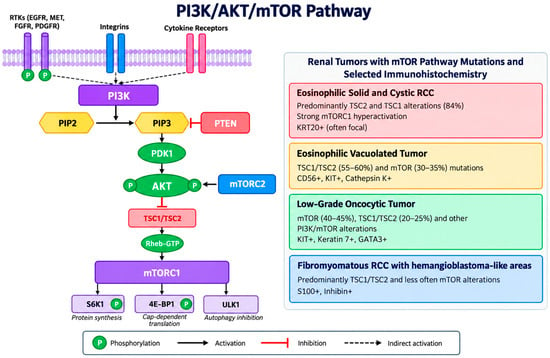
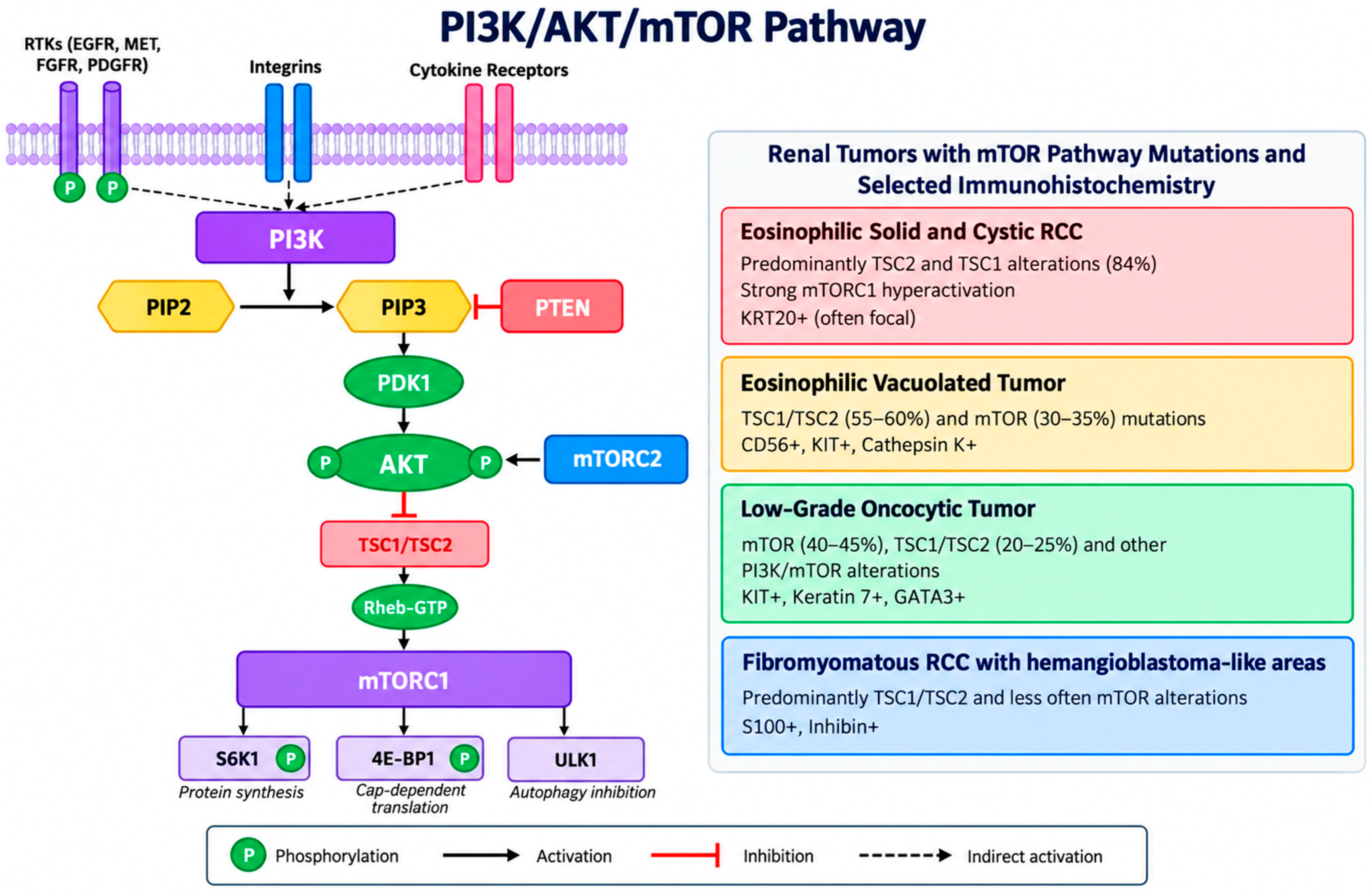
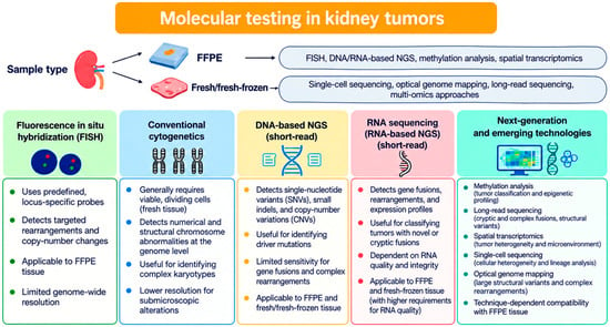
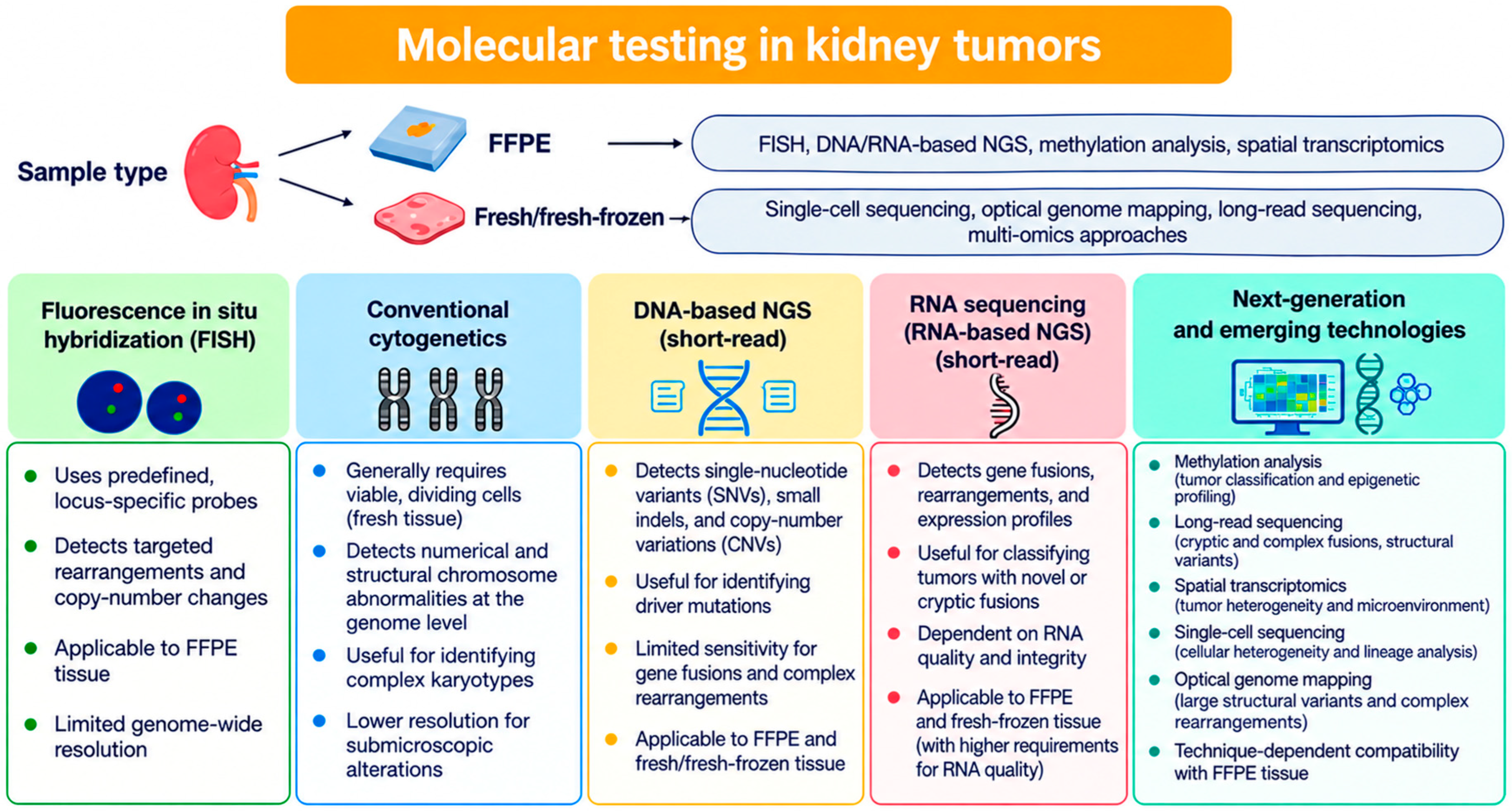

Disclaimer/Publisher’s Note: The statements, opinions and data contained in all publications are solely those of the individual author(s) and contributor(s) and not of MDPI and/or the editor(s). MDPI and/or the editor(s) disclaim responsibility for any injury to people or property resulting from any ideas, methods, instructions or products referred to in the content. |
© 2026 by the authors. Licensee MDPI, Basel, Switzerland. This article is an open access article distributed under the terms and conditions of the Creative Commons Attribution (CC BY) license.