


Simple Summary
Bone metastases represent a serious and common challenge in advanced cancer patients, making early detection crucial. The growing implementation of artificial intelligence (AI) in various medical fields is revolutionizing healthcare by enhancing diagnostic accuracy. This systematic review includes 59 articles and sought to assess the potential of machine learning models in the fields of nuclear medicine, clinical research, radiology, and molecular biology. The review provides a comprehensive analysis of AI’s effectiveness in detecting bone metastases, which had not yet been thoroughly conducted. The findings highlight the benefits of integrating AI technologies in the inspected fields, encouraging further advancements and adoption of AI to ultimately improve patient care and treatment planning. This study provides invaluable perceptions into the promising application of computational intelligence in the detection of bone metastases, demonstrating its superiority over standard tests. However, further research is necessary to substantiate these findings with the aim of developing better-trained data-driven models soon.
Abstract
Background: Metastasis commonly occur in the bone tissue. Artificial intelligence (AI) has become increasingly prevalent in the medical sector as support in decision-making, diagnosis, and treatment processes. The objective of this systematic review was to assess the reliability of AI systems in clinical, radiological, and pathological aspects of bone metastases. Methods: We included studies that evaluated the use of AI applications in patients affected by bone metastases. Two reviewers performed a digital search on 31 December 2023 on PubMed, Scopus, and Cochrane library and extracted authors, AI method, interest area, main modalities used, and main objectives from the included studies. Results: We included 59 studies that analyzed the contribution of computational intelligence in diagnosing or forecasting outcomes in patients with bone metastasis. Six studies were specific for spine metastasis. The study involved nuclear medicine (44.1%), clinical research (28.8%), radiology (20.4%), or molecular biology (6.8%). When a primary tumor was reported, prostate cancer was the most common, followed by lung, breast, and kidney. Conclusions: Appropriately trained AI models may be very useful in merging information to achieve an overall improved diagnostic accuracy and treatment for metastasis in the bone. Nevertheless, there are still concerns with the use of AI systems in medical settings. Ethical considerations and legal issues must be addressed to facilitate the safe and regulated adoption of AI technologies. The limitations of the study comprise a stronger emphasis on early detection rather than tumor management and prognosis as well as a high heterogeneity for type of tumor, AI technology and radiological techniques, pathology, or laboratory samples involved.
1. Introduction
Metastasis commonly occur in the bone tissue, making it one of the principal sites of invasion, notably for breast, prostate, and lung cancers [1]. The onset of bone metastases results in limitation of daily activities, pain, and reduced 5-year survival in cancer patients. Reports indicate that a poor prognosis is observed in 60–70% of patients with spinal metastases [2]. The typical survival time for individuals impacted by bone metastases from melanoma, breast, or prostate malignancy is 6 months, 19–25 months, and 53 months, respectively [3]. Furthermore, bone metastases are linked to a decline in the overall well-being, especially when hypercalcemia, uncontrolled pain, or skeletal-related events (SREs) occur [4]. These incidents characterize up to 70% of cases, particularly in patients in whom preventive interventions like the utilization of orthotic devices or bone-targeting agents (BTAs) are not implemented [5,6,7]. SREs include pathological fractures, spinal compression, radiation therapy, and metastasis surgery [8]. The rise in life expectancy among patients with bone metastases has led to a corresponding increase in SRE occurrences [9]. In the case of pathological fracture, surgical treatment aims to stabilize the fractured fragment [10], while in the presence of pathological vertebral collapse with spinal cord compression, urgent decompression surgery with possible stabilization of adjacent segments is required [11,12].
The diagnosis of bone metastases uses traditional radiology firstly, followed by second-level imaging techniques such as computed tomography (CT) and magnetic resonance imaging (MRI), and finally bone scintigraphy (BS) or positron emission tomography and computed tomography (PET-CT) [13].
Over the past few years, machine intelligence has proliferated in the medical domain as an aid in decision making, diagnosis, and treatment [14,15]. Already, in several fields, artificial intelligence (AI) applications such as convolutional neural networks (CNN), deep learning (DL), and machine learning (ML) have proven useful in determining predictive factors inherent in the pathology in question [16,17]. However, several limitations reduce the full integration of AI into clinical practice. In particular, AI systems for the treatment and diagnosis of tumors need to be implemented, and there are also concerns related to the availability and security of using big data [18,19]. Li et al. already analyzed the contribution of neural networks in the diagnosis of skeletal muscle oncological disorders [20].
However, a thorough examination of the efficacy and function of AI in bone metastasis remains unaccomplished. Hence, this study aims to evaluate the effectiveness of AI systems in the early detection, management, treatment, and evaluation of the prognosis of bone metastasis as a complementary tool useful in daily clinical practice. Therefore, this systematic review assesses the accuracy of the implementation of AI in nuclear medicine, clinical research, radiology, and molecular biology techniques for bone metastasis.
2. Materials and Methods
2.1. Inclusion Criteria
In this systematic review, studies that assessed the use of AI applications such as CNN, DL, or ML in patients with bone metastases were included. We excluded studies not written in English and animal and cadaveric studies.
2.2. Information Sources and Search Strategy
Two reviewers (G.F.P. and G.M.) performed an electronic search on PubMed, Scopus, and Cochrane Library using the search strings (“deep learning” [MeSH Terms] OR (“deep” [All Fields] AND “learning” [All Fields]) OR “deep learning” [All Fields] OR (“machine learning” [MeSH Terms] OR (“machine” [All Fields] AND “learning” [All Fields]) OR “machine learning” [All Fields]) OR (“artificial intelligence” [MeSH Terms] OR (“artificial” [All Fields] AND “intelligence” [All Fields]) OR “artificial intelligence” [All Fields])) AND ((“bone and bones” [MeSH Terms] OR (“bone” [All Fields] AND “bones” [All Fields]) OR “bone and bones” [All Fields] OR “bone” [All Fields]) AND (“metastasi” [All Fields] OR “neoplasm metastasis” [MeSH Terms] OR (“neoplasm” [All Fields] AND “metastasis” [All Fields]) OR “neoplasm metastasis” [All Fields] OR “metastasis” [All Fields])). The research was conducted on 31 December 2023. Following the PRISMA 2020 statement [21], duplicates were removed from the identified records, and the two independent reviewers proceeded to screen the manuscripts respecting the inclusion and exclusion criteria. Finally, the fully eligible articles were read by the reviewers to select the studies to include in the review.
2.3. Data Collection, Analysis, and Outcomes
Two reviewers (P.B. and G.M.) extracted these data from the included studies: authors, year, artificial intelligence method, interest area, main modalities used, and main objectives. The analysis included the assessment of data concerning the fields of nuclear medicine, clinical research, radiology, and molecular biology. This systematic review was not previously registered in databases.
3. Results
3.1. Search Results
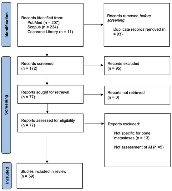
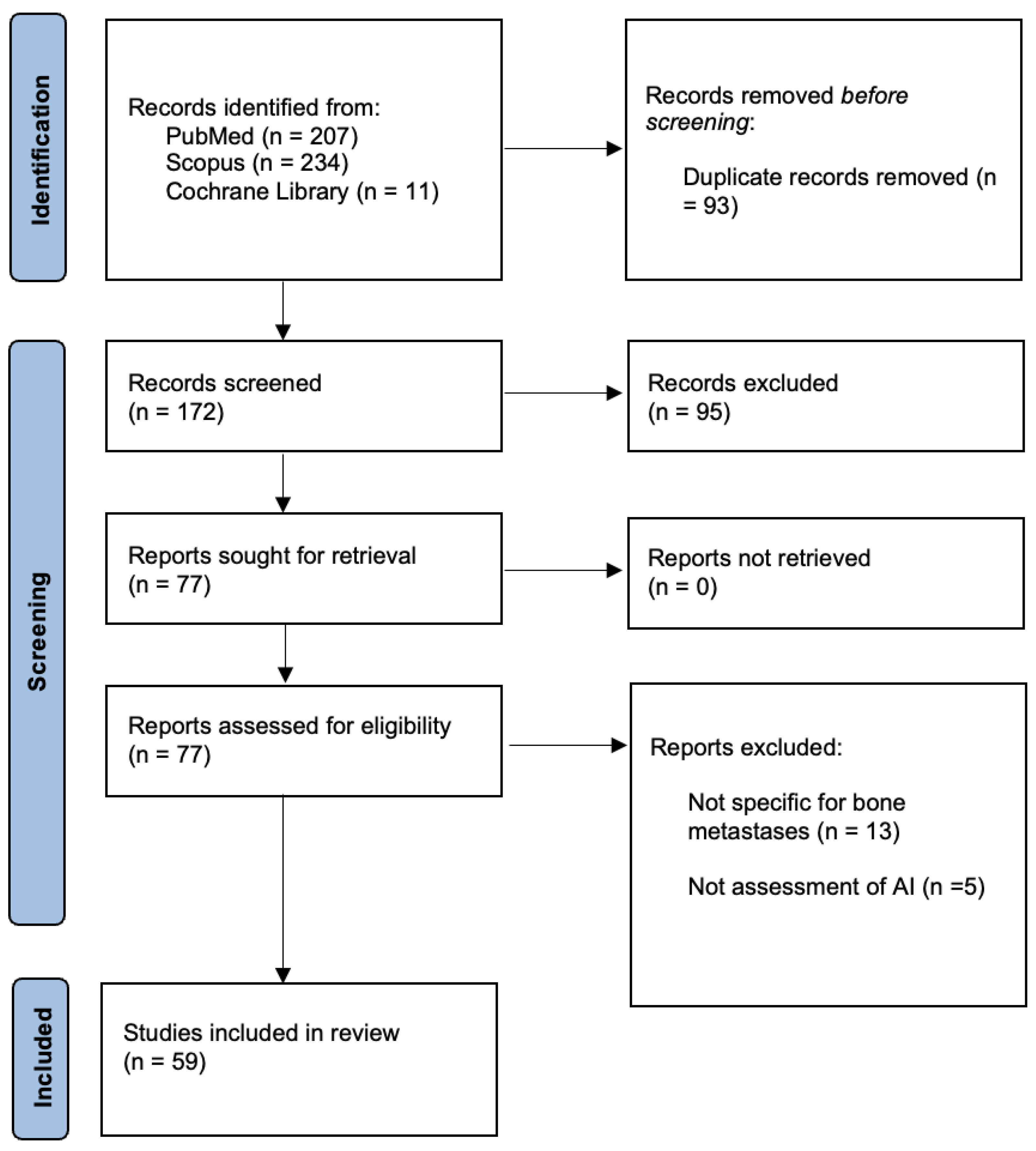
The literary investigation identified 474 articles. After duplicate removal, 380 articles were screened based on title and abstract. The entire text of 77 papers was read, and 18 were excluded for the following reasons: not specific for bone metastases (n = 13) and not an assessment of AI (n = 5). The articles included in this review were 59 (Figure 1).
Figure 1.
PRISMA 2020 flow diagram.
All the articles analyzed the role of AI in the diagnosis of skeletal metastasis or prognosis among individuals experiencing bone metastasis, of which six (10.2%) were specific for spine metastasis. The study involved nuclear medicine (N = 26 [44.1%]), clinical research (N = 17 [28.8%]), radiology (N = 12 [20.4%]), or molecular biology (N = 4 [6.8%]). When a primary tumor was reported, prostate neoplasms were the prevailing cancer (N = 13 [22%]), preceding lung (N = 7 [11.9%]), breast (N = 3 [5.1%]), kidney (N = 3 [5.1%]), thyroid (N = 1 [1.7%]), and colorectal cancer (N = 1 [1.7%]).
3.2. Nuclear Medicine
Imaging modalities in nuclear medicine represent a fundamental tool in the diagnosis and management of metastatic bone disease [22]. The use of AI in nuclear medicine for bone metastasis is evolving quickly, showing new opportunities for diagnosis, treatment, and patient care. AI can improve the analysis of nuclear medicine imaging techniques by offering detailed quantitative evaluations of tracer uptake and delivering accurate localization of bone metastases. In our research, nuclear medicine was the most common category (N = 26).
3.2.1. Bone Scintigraphy
The main modality used was bone scintigraphy (N = 21 [80.77%]). Research demonstrated that machine learning algorithms utilizing BS images can accurately differentiate the metastatic bone tissue from normal tissue [13]. Zhao et al. [23] demonstrated a significant diagnostic performance of ML techniques based on Tc-MDP BS in the diagnosis of skeletal metastasis, showing an area under the curve (AUC) of receiver operating characteristic (ROC) of 0.988 for breast cancer, 0.955 for prostate cancer, 0.957 for lung cancer, and 0.971 for other cancers. Groot et al. [24] developed an NLP algorithm designed to classify single or multiple metastases in bone scans of individuals chosen for surgical treatment of bone metastases, which had a sensitivity of 0.94 and specificity of 0.82.
Koizumi et al. [25] evaluated the performance of the BONEVAVI version 2 for bone metastasis diagnosis among individuals having or lacking skeletal metastasis. The study reported high levels of sensitivity and specificity of patient artificial neural network (ANN) data, reaching values higher than 80%.
Koizumi et al. [26] also investigated the diagnostic performance of the computer-assisted diagnostic system for BS BONENAVI in the presence or absence of bone involvement in prostate cancer, assessing an accuracy of 82% for metastasis detection. Papandrianos et al. [27] used a CNN algorithm for BS in determining the occurrence or non-occurrence of prostate cancer metastasis. They demonstrated that the method is adequately accurate in distinguishing between bone metastasis and degenerative or normal tissues, achieving an overall classification accuracy of 91.42% ± 1.64%. Finally, the best-performing CNN method outperformed commonly used CNN methods in nuclear medicine for diagnosing metastatic prostate cancer in bones [28].
3.2.2. Single-Photon Emission-Computed Tomography
In our study, single-photon emission-computed tomography (SPECT) ranked as the second most frequently utilized modality (N = 5 [19.23%]). It was shown that deep learning models can accurately identify hotspots of metastasis in bone SPECT images, achieving values of 0.9920, 0.7721, and 0.6788 for accuracy, precision, and recall, respectively [29]. Moreover, Lin et al. presented similar results in other manuscripts [30,31]. Acar et al. [32] used ML algorithms based on 68Ga-prostate-specific membrane antigen (PSMA) PET-CT images to distinguish sclerotic lesions from metastasis or completely responded lesions in individuals with acknowledged bone metastasis who had undergone prior treatment.
3.3. Clinical Research
AI models for clinical prediction have been established for many purposes using clinical data obtained from medical records. AI can examine large clinical datasets to obtain an early diagnosis, create individualized treatment plans, and deliver customized prognostic insights for each patient. De Groot et al. [33] analyzed ML algorithms in predicting the 90-day and 1-year survival in subjects with skeletal metastasis who had surgery. Zhou et al. analyzed the diagnostic performance of ML algorithms in patients with lung adenocarcinoma bone metastasis [34]. In both the training and test groups, the AUC values of all ML classifiers exceeded 0.8, except for random forest (RF) and logistic regression (LR). However, the combined algorithm did not enhance the AUC value for any individual machine learning algorithm. The accuracy of all ML classifiers, except for the RF algorithm, surpassed 70%. ML algorithms were used also in predicting outcomes for cancer-specific survival (CSS) and overall survival (OS). For example, Chen et al. [35] found that patients with small-cell lung cancer (SCLC) with skeletal metastasis experienced a reduced median survival time (MTS) for OS compared to patients with SCLC without bone metastasis (6 vs. 10 months).
Le et al. [36] investigated the function of ML algorithms in forecasting the 3-year OS of patients diagnosed with bone metastasis from clear cell renal cell carcinoma, proving that the extreme gradient boosting (XGB) algorithm model achieved the best accuracy and specificity (0.792 and 0.806, respectively) compared to other models. Finally, Paulino Pereira et al. [37] analyzed the performance of ML algorithms in estimating survival in patients surgically treated for spinal metastases.
3.4. Radiology
In our review, radiology-related articles involved CT and MRI. CT (N = 8 [66.7%]) was the most common imaging modality used.
3.4.1. Computed Tomography
AI applications in CT for bone metastasis provide substantial potential benefits such as great diagnostic accuracy, efficient segmentation of the lesions (Image S1) and treatment planning, and patient follow-up by identifying early signs of disease progression or treatment response. In the study by Noguchi et al. [38] to evaluate efficiency in skeletal metastases detection on CT, in the validation dataset, the deep learning-based algorithm attained a sensitivity of 89.8% with 0.775 false positives per case.
3.4.2. Magnetic Resonance Imaging
The second most common modality used was MRI imaging (N = 4 [33.3%]), of which one was pelvic MRI, and three were whole-body MRI. AI algorithms can greatly improve the sensitivity and specificity of MRI scans for detecting bone metastases, identifying subtle changes in bone marrow and other structures that human radiologists might overlook. Additionally, AI can automate MRI image analysis, which can reduce the time needed for diagnosis and result in faster and more efficient clinical workflows. Tajima et al. [39] assessed the image acquisition time in 17 patients with prostate malignancy who had undergone diffusion-weighted whole-body imaging with background body signal suppression by 1.5T MRI with 2 excitations (NEX2) and 8 (NEX8).
AI can be also used in diagnosis of spine metastasis. In particular, Wang et al. [40] analyzed the CNN algorithm based on MRI spine scans to diagnose and predict spinal metastasis. They found that this model could accurately forecast spinal metastases, with an accuracy up to 96.45%. Furthermore, Jakubicek et al. [41] examined how ML techniques utilizing vertebrae in 3D CT scans perform in diagnosing potentially incomplete spines in patients with bone metastases and vertebral compressions, showing a mean error of intervertebral discs localization of 4.4 mm.
3.5. Molecular Biology
AI can process extensive datasets from genomics, proteomics, and metabolomics to reveal the molecular mechanisms associated with bone metastasis. This could enhance the understanding of the disease and help identify potential therapeutic targets. Additionally, AI can spot molecular biomarkers indicative of bone metastasis, facilitating early detection and personalized treatment strategies. Shao et al. [42] used label-free surface-enhanced Raman spectroscopy for screening prostate carcinoma bone metastasis, finding high values for mean training accuracy (99.51%), testing accuracy (81.70%), testing sensitivity (80.63%), and testing specificity (82.82%). Albaradei et al. [43] used bone metastasis-related genes from gene expression datasets in Gene Expression Omnibus (GEO) to predict bone metastases development. The deep neural network (DNN) model achieved the peak prediction accuracy (AUC of 92.11%) by utilizing the top 34 genes identified based on their betweenness centrality scores. Hsu et al. [17] analyzed patients who underwent local MRI-guided focused ultrasound ablation to determine an appropriate treatment plan for skeletal metastasis. The most accurate predictive few-shot learning model was obtained by integrating clinical features with the amount of cytokines IL-6, IL-13, IP-10, and eotaxin (accuracy of 85.2% and sensitivity of 88.6%). Park et al. [44] examined breast cancer genes frequently expressed during bone metastasis and in osteoblasts using data from the GEO database to create a potential causal network. They discovered 33 genes strongly linked to the onset of breast cancer skeletal metastasis. Additional model assessments revealed that 16 genes were sufficient to make the model statistically significant for the maximum likelihood of causal Bayesian networks and accurately predict breast cancer bone metastasis.
All the included studies with their characteristics are reported in Table 1.
Table 1.
AI methods and results of included studies.
4. Discussion
AI has gained great importance in the medical field over the last 10 years, representing a breakthrough in the scientific literature [78]. AI use has been primarily confined to evaluating bone metastasis using bone scintigraphy and analyzing histopathology samples in the diagnosis of both solid and hematological neoplasms [79]. Electronic health records or molecular datasets are still relatively unknown fields for AI models. The application and continued usage of these models may revolutionize these domains, making them a more powerful tool than those available at this moment [80]. The field of musculoskeletal oncology may benefit from a mix of discoveries both from clinical and non-clinical specialties [81]. Nuclear medicine has long played a significant role in musculoskeletal oncology, with numerous new imaging techniques investigated over time [82], such as PET/CT and multiparameter MRI. Despite this, bone scintigraphy with 9mTc-MDP continue to have a noteworthy position thanks to the advantages of being less expensive and still having decent sensitivity [83]. As shown by Zhao et al. [23], AI models with deep neural networks using image features from bone scintigraphy with 9mTc-MDP demonstrated notable time efficiency, accuracy, specificity, and sensitivity in diagnosing bone metastasis, making it potentially valuable in distant or economically disadvantaged areas where interpreting bone scintigraphy images remains challenging.
Single-photon emission-computed tomography is also a widely used technique of nuclear medicine, which can evaluate the metabolism of radioactively labeled compounds, bringing useful information about a specified area of concern that may present itself with malignancy features, also referred to as hotspots [84]. Deep learning-based segmentation models using SPECT images showed promising results in detecting and segmenting hotspots [85], with propitious future directions including the necessity of optimization and improvement of the models to develop a more straightforward, efficient, and productive computer-assisted diagnosis system and to evolve into multi-category and multi-condition models for identifying hotspots of different diseases in SPECT thorax images [29].
Computed tomography is a primary imaging technique employed in diagnosing a wide range of illnesses. In particular, even though it is considered inferior to MRI and PET for detecting bone metastases, especially those affecting the spine, its widespread adoption allows for routinely performed exams, making it an ideal candidate for computer-aided detection (CAD) systems [86] under AI, including ML and DL [87,88]. For all the above-mentioned reasons, Koike et al. [75] created a fully automated two-step CAD method with AI assistance for identifying lytic spinal bone metastases and showed promising results, as the system was both accurate and rapid compared to the conventional diagnosis of spinal metastasis, which usually comes with the possibility of overlooking the lesions if the examinations are not specifically designed to assess them. On the other hand, MRI offers an overall accuracy of 70–100% in identifying bony metastases [89]. It is broadly used in the diagnosis and staging of bone lesions. The combination of MRI and PET/CT parameters has been shown to have the possibility of predicting early metastatic disease if analyzed by AI models such as model-averaged neural network (avNNet) [90]. However, all the above-mentioned radiological and nuclear medicine techniques are associated with high costs and radioactive injury, which are variables not to be ignored. For this reason, low-cost, rapid, and accurate laboratory tests that specifically evaluate alterations in bone metabolism in the presence of bone metastasis have sparked considerable interest. For example, increased levels of procollagen N-terminal propeptides (PIPNs), osteocalcin, and bone-specific-alkaline phosphatase (BAP) could indicate heightened osteoblastic activity, which may represent an underlying metastatic condition of the patient [91]. Yet, the assessment of bone metabolic parameters is not currently advised for routine clinical practice due to conflicting findings. For this reason, Raman spectroscopy (SERS) has seen increased usage over the years as a method for blood analysis thanks to the relatively fast results and the possibility of predicting disease recurrence based on serum parameters, for example, in the case of the prostate [92,93]. Shao et al. [42] implemented Raman spectra parameters on prostate screening tests for convolutional neural networks (CNNs) to identify early disease, with promising results. Raman spectroscopy was also used in the evaluation of breast carcinoma genes frequently expressed in individuals with bone metastasis to construct a plausible causal network. In the study by Park et al. [44], 33 genes that were significantly associated and likely involved in the progression of breast cancer bone metastasis were identified. Among these, 16 genes were sufficient for a model to accurately predict breast cancer bone metastasis. In conclusion, clinical-prediction AI models have been developed over the years with many applications, one of which is the evaluation of disease-free survival (DFS) and overall survival (OS) in cancer patients [2]. Regarding bone metastasis, survival after the diagnosis is quite variable and has an essential role in care decision making. For example, the prophylactic stabilization of impending pathologic fractures in the case of bone metastasis is heavily influenced by the predicted survival of the patient, with a relative contraindication in patients with 3 months or less of estimated survival [94]. For this reason, many predictive models for the survival of patients with various diseases and at different stages and for candidates for various medical procedures proposed over time span from traditional statistics to boosting algorithms and, more recently, machine learning models [95,96]. The latter has the advantage of being able to delineate predictive models based on larger datasets. In addition to this, AI models are subjected to retraining because their predictive performance tends to decline over time as treatment regimens tend to advance. As the complexity of these networks increases, AI-based models require significant computational power and memory. Moreover, such complex networks may not always lead to faster performance, especially when implemented on devices with limited resources. In medical cases, time is crucial for early diagnosis of bone metastasis to improve the clinical outcomes of the patients with bone metastasis using the best treatment choices [97]. Nowadays, the greatest challenge is identifying how to improve the AI-based model systems used in medical applications in terms of feasibility, reproducibility, and fastness in processing data. AI applications are increasing dramatically, with promising but intricate prospects. Regarding the early detection of metastasis, enhancing detection accuracy may be crucial [98]. This will be driven by expanding datasets, advancing machine learning algorithms, and improving imaging technologies [99]. Concurrently, advanced imaging algorithms may aid in distinguishing metastasis from other pathologies, enhancing the resolution and clarity of medical images, and shortening the diagnostic phase, thereby expediting the initiation of patient treatment [100]. Furthermore, integrating established AI models may help reduce human error during image evaluation. The clinical management and prognosis of tumor metastasis may also be shaped by the deployment of well-trained AI models. For instance, the integration of genetic, demographic, and clinical data could lead to personalized treatment plans becoming standard practice, where treatments are tailored to individual patients rather than generalized. This integration offers a holistic perspective on a patient’s health, which aids in making informed decisions and coordinating care more effectively [101]. Moreover, by integrating patient data, it becomes possible to predict the progression of bone metastasis, assisting in the formulation of long-term treatment strategies and the management of disease progression [102]. Nevertheless, there are still some concerns with the use of AI systems in medical settings. For instance, it is essential to ensure that the diverse datasets on which AI systems are trained can precisely represent different patient populations, that they are effectively integrated into routine clinical workflows, and that they can be improved in overall interpretability and transparency. Furthermore, ethical considerations and regulatory and legal issues must be addressed to facilitate the safe and regulated adoption of AI technologies in bone metastasis detection and overall management [103]. There are some limitations to this study. The majority of studies included focused on early detection of bone metastasis, and only a few focused on tumor management and the treatment and evaluation of prognosis. Hence, future studies that evaluate the clinical management of metastasis rather than the radiological features must be considered. Furthermore, the included studies presented a high grade of heterogeneity regarding the type of tumor evaluated and both the AI technology and the radiological techniques, pathology, or laboratory samples implemented.
5. Conclusions
The diagnosis and management of tumors in the musculoskeletal system are extremely complex, requiring a multidisciplinary approach. In this context, appropriately trained AI models hold significant promise for enhancing the precision of diagnosing and treating bone metastasis. By effectively merging and analyzing data from radiologic imaging, pathology, and laboratory samples, AI can provide a comprehensive and nuanced understanding of tumor characteristics. This can lead to more accurate and timely diagnoses, personalized treatment plans, and improved patient outcomes. Furthermore, the continuous advancement of AI technologies and their integration into clinical workflows could revolutionize the standard of care in musculoskeletal oncology, paving the way for more effective and efficient management of bone metastasis. Future research should focus on the development of robust and reliable AI algorithms, validation of these algorithms in diverse clinical settings, and addressing potential ethical and implementation challenges to fully realize the potential of AI in the field.
Supplementary Materials
The following supporting information can be downloaded at: https://www.mdpi.com/article/10.3390/cancers16152700/s1, Image S1: AI-based CT segmentation model for identification of bone metastatic lesion; File S1: List of acronyms.
Author Contributions
Conceptualization, G.F.P., E.F. and F.P.; methodology, G.F.P., P.B., L.S. and G.M.; validation, E.F., B.V. and G.T.; formal analysis, G.F.P., P.B. and G.M.; investigation, G.F.P. and L.S.; data curation, G.F.P., P.B. and D.S.; writing—original draft preparation, P.B., L.S. and G.M.; writing—review and editing, G.F.P., E.F., D.S. and F.P.; visualization, B.V. and R.P.; supervision, G.T. and V.D.; project administration, R.P. and V.D.; funding acquisition, R.P. and V.D. All authors have read and agreed to the published version of the manuscript.
Funding
This research received no external funding.
Conflicts of Interest
The authors declare no conflicts of interest.
References
- Siegel, R.L.; Miller, K.D.; Fuchs, H.E.; Jemal, A. Cancer Statistics, 2022. CA. Cancer J. Clin. 2022, 72, 7–33. [Google Scholar] [CrossRef] [Scilit]
- He, X.; Jiao, Y.; Yang, X.; Hu, Y. A Novel Prediction Tool for Overall Survival of Patients Living with Spinal Metastatic Disease. World Neurosurg. 2020, 144, e824–e836. [Google Scholar] [CrossRef] [Scilit]
- Faiella, E.; Santucci, D.; Calabrese, A.; Russo, F.; Vadalà, G.; Zobel, B.B.; Soda, P.; Iannello, G.; De Felice, C.; Denaro, V. Artificial Intelligence in Bone Metastases: An MRI and CT Imaging Review. Int. J. Environ. Res. Public. Health 2022, 19, 1880. [Google Scholar] [CrossRef] [Scilit]
- D’Oronzo, S.; Coleman, R.; Brown, J.; Silvestris, F. Metastatic Bone Disease: Pathogenesis and Therapeutic Options. J. Bone Oncol. 2019, 15, 100205. [Google Scholar] [CrossRef] [Scilit]
- Clemons, M.; Gelmon, K.A.; Pritchard, K.I.; Paterson, A.H.G. Bone-Targeted Agents and Skeletal-Related Events in Breast Cancer Patients with Bone Metastases: The State of the Art. Curr. Oncol. 2012, 19, 259–268. [Google Scholar] [CrossRef] [Scilit]
- Coleman, R. The Role of Zoledronic Acid in Cancer: Clinical Studies in the Treatment and Prevention of Bone Metastases. Semin. Oncol. 2001, 28, 11–16. [Google Scholar] [CrossRef] [Scilit]
- Stopeck, A.T.; Lipton, A.; Body, J.-J.; Steger, G.G.; Tonkin, K.; De Boer, R.H.; Lichinitser, M.; Fujiwara, Y.; Yardley, D.A.; Viniegra, M.; et al. Denosumab Compared with Zoledronic Acid for the Treatment of Bone Metastases in Patients With Advanced Breast Cancer: A Randomized, Double-Blind Study. J. Clin. Oncol. 2010, 28, 5132–5139. [Google Scholar] [CrossRef] [Scilit]
- Santini, D.; Pantano, F.; Iuliani, M.; Ribelli, G.; Manca, P.; Vincenzi, B.; Tonini, G. Bone-Modifying Agents and Anticancer Agents with Bone Effects. In Management of Bone Metastases; Denaro, V., Di Martino, A., Piccioli, A., Eds.; Springer International Publishing: Cham, Switzerland, 2019; pp. 13–25. ISBN 978-3-319-73484-2. [Google Scholar]
- Silvestris, N.; Pantano, F.; Ibrahim, T.; Gamucci, T.; De Vita, F.; Di Palma, T.; Pedrazzoli, P.; Barni, S.; Bernardo, A.; Febbraro, A.; et al. Natural History of Malignant Bone Disease in Gastric Cancer: Final Results of a Multicenter Bone Metastasis Survey. PLoS ONE 2013, 8, e74402. [Google Scholar] [CrossRef] [Scilit]
- Di Martino, A.; Martinelli, N.; Loppini, M.; Piccioli, A.; Denaro, V. Is Endoprosthesis Safer than Internal Fixation for Metastatic Disease of the Proximal Femur? A Systematic Review. Injury 2017, 48 (Suppl. S3), S48–S54. [Google Scholar] [CrossRef] [Scilit]
- Di Martino, A.; Vincenzi, B.; Denaro, L.; Barnaba, S.A.; Papalia, R.; Santini, D.; Tonini, G.; Denaro, V. “Internal Bracing” Surgery in the Management of Solid Tumor Metastases of the Thoracic and Lumbar Spine. Oncol. Rep. 2009, 21, 431–435. [Google Scholar] [CrossRef] [Scilit]
- Denaro, V.; Di Martino, A. Spinal Metastases: Diagnosis and Management. In Management of Bone Metastases; Denaro, V., Di Martino, A., Piccioli, A., Eds.; Springer International Publishing: Cham, Switzerland, 2019; pp. 137–147. ISBN 978-3-319-73484-2. [Google Scholar]
- Elfarra, F.-G.; Calin, M.A.; Parasca, S.V. Computer-Aided Detection of Bone Metastasis in Bone Scintigraphy Images Using Parallelepiped Classification Method. Ann. Nucl. Med. 2019, 33, 866–874. [Google Scholar] [CrossRef] [Scilit]
- Xu, C.; Liu, W.; Yin, C.; Li, W.; Liu, J.; Sheng, W.; Tang, H.; Li, W.; Zhang, Q. Establishment and Validation of a Machine Learning Prediction Model Based on Big Data for Predicting the Risk of Bone Metastasis in Renal Cell Carcinoma Patients. Comput. Math. Methods Med. 2022, 2022, 5676570. [Google Scholar] [CrossRef] [Scilit]
- Ali, O.; Abdelbaki, W.; Shrestha, A.; Elbasi, E.; Alryalat, M.A.A.; Dwivedi, Y.K. A Systematic Literature Review of Artificial Intelligence in the Healthcare Sector: Benefits, Challenges, Methodologies, and Functionalities. J. Innov. Knowl. 2023, 8, 100333. [Google Scholar] [CrossRef] [Scilit]
- Thio, Q.C.B.S.; Karhade, A.V.; Ogink, P.T.; Bramer, J.A.M.; Ferrone, M.L.; Calderón, S.L.; Raskin, K.A.; Schwab, J.H. Development and Internal Validation of Machine Learning Algorithms for Preoperative Survival Prediction of Extremity Metastatic Disease. Clin. Orthop. 2020, 478, 322–333. [Google Scholar] [CrossRef] [Scilit]
- Hsu, F.-C.; Lee, H.-L.; Chen, Y.-J.; Shen, Y.-A.; Tsai, Y.-C.; Wu, M.-H.; Kuo, C.-C.; Lu, L.-S.; Yeh, S.-D.; Huang, W.-S.; et al. A Few-Shot Learning Approach Assists in the Prognosis Prediction of Magnetic Resonance-Guided Focused Ultrasound for the Local Control of Bone Metastatic Lesions. Cancers 2022, 14, 445. [Google Scholar] [CrossRef] [Scilit]
- Siddique, S.; Chow, J.C.L. Artificial Intelligence in Radiotherapy. Rep. Pract. Oncol. Radiother. 2020, 25, 656–666. [Google Scholar] [CrossRef] [Scilit]
- Chow, J.C.L. Artificial Intelligence in Radiotherapy and Patient Care. In Artificial Intelligence in Medicine; Lidströmer, N., Ashrafian, H., Eds.; Springer International Publishing: Cham, Switzerland, 2021; pp. 1–13. ISBN 978-3-030-58080-3. [Google Scholar]
- Li, M.D.; Ahmed, S.R.; Choy, E.; Lozano-Calderon, S.A.; Kalpathy-Cramer, J.; Chang, C.Y. Artificial Intelligence Applied to Musculoskeletal Oncology: A Systematic Review. Skeletal Radiol. 2022, 51, 245–256. [Google Scholar] [CrossRef] [Scilit]
- Page, M.J.; McKenzie, J.E.; Bossuyt, P.M.; Boutron, I.; Hoffmann, T.C.; Mulrow, C.D.; Shamseer, L.; Tetzlaff, J.M.; Akl, E.A.; Brennan, S.E.; et al. The PRISMA 2020 Statement: An Updated Guideline for Reporting Systematic Reviews. BMJ 2021, 372, n71. [Google Scholar] [CrossRef] [Scilit]
- Isaac, A.; Dalili, D.; Dalili, D.; Weber, M.-A. State-of-the-Art Imaging for Diagnosis of Metastatic Bone Disease. Radiologe 2020, 60, 1–16. [Google Scholar] [CrossRef] [Scilit]
- Zhao, Z.; Pi, Y.; Jiang, L.; Xiang, Y.; Wei, J.; Yang, P.; Zhang, W.; Zhong, X.; Zhou, K.; Li, Y.; et al. Deep Neural Network Based Artificial Intelligence Assisted Diagnosis of Bone Scintigraphy for Cancer Bone Metastasis. Sci. Rep. 2020, 10, 17046. [Google Scholar] [CrossRef] [Scilit]
- Groot, O.Q.; Bongers, M.E.R.; Karhade, A.V.; Kapoor, N.D.; Fenn, B.P.; Kim, J.; Verlaan, J.J.; Schwab, J.H. Natural Language Processing for Automated Quantification of Bone Metastases Reported in Free-Text Bone Scintigraphy Reports. Acta Oncol. 2020, 59, 1455–1460. [Google Scholar] [CrossRef] [Scilit]
- Koizumi, M.; Wagatsuma, K.; Miyaji, N.; Murata, T.; Miwa, K.; Takiguchi, T.; Makino, T.; Koyama, M. Evaluation of a Computer-Assisted Diagnosis System, BONENAVI Version 2, for Bone Scintigraphy in Cancer Patients in a Routine Clinical Setting. Ann. Nucl. Med. 2015, 29, 138–148. [Google Scholar] [CrossRef] [Scilit]
- Koizumi, M.; Motegi, K.; Koyama, M.; Terauchi, T.; Yuasa, T.; Yonese, J. Diagnostic Performance of a Computer-Assisted Diagnosis System for Bone Scintigraphy of Newly Developed Skeletal Metastasis in Prostate Cancer Patients: Search for Low-Sensitivity Subgroups. Ann. Nucl. Med. 2017, 31, 521–528. [Google Scholar] [CrossRef] [Scilit]
- Papandrianos, N.; Papageorgiou, E.; Anagnostis, A.; Papageorgiou, K. Efficient Bone Metastasis Diagnosis in Bone Scintigraphy Using a Fast Convolutional Neural Network Architecture. Diagnostics 2020, 10, 532. [Google Scholar] [CrossRef] [Scilit] [PubMed]
- Papandrianos, N.; Papageorgiou, E.; Anagnostis, A.; Papageorgiou, K. Bone Metastasis Classification Using Whole Body Images from Prostate Cancer Patients Based on Convolutional Neural Networks Application. PLoS ONE 2020, 15, e0237213. [Google Scholar] [CrossRef] [Scilit]
- Lin, Q.; Luo, M.; Gao, R.; Li, T.; Man, Z.; Cao, Y.; Wang, H. Deep Learning Based Automatic Segmentation of Metastasis Hotspots in Thorax Bone SPECT Images. PLoS ONE 2020, 15, e0243253. [Google Scholar] [CrossRef] [Scilit] [PubMed]
- Lin, Q.; Gao, R.; Luo, M.; Wang, H.; Cao, Y.; Man, Z.; Wang, R. Semi-Supervised Segmentation of Metastasis Lesions in Bone Scan Images. Front. Mol. Biosci. 2022, 9, 956720. [Google Scholar] [CrossRef] [Scilit]
- Lin, Q.; Chen, X.; Liu, L.; Cao, Y.; Man, Z.; Zeng, X.; Huang, X. Detecting Multiple Lesions of Lung Cancer-Caused Metastasis with Bone Scans Using a Self-Defined Object Detection Model Based on SSD Framework. Phys. Med. Biol. 2022, 67, 225009. [Google Scholar] [CrossRef] [Scilit]
- Acar, E.; Leblebici, A.; Ellidokuz, B.E.; Başbınar, Y.; Kaya, G.Ç. Machine Learning for Differentiating Metastatic and Completely Responded Sclerotic Bone Lesion in Prostate Cancer: A Retrospective Radiomics Study. Br. J. Radiol. 2019, 92, 20190286. [Google Scholar] [CrossRef] [Scilit]
- De Groot, T.M.; Ramsey, D.; Groot, O.Q.; Fourman, M.; Karhade, A.V.; Twining, P.K.; Berner, E.A.; Fenn, B.P.; Collins, A.K.; Raskin, K.; et al. Does the SORG Machine-Learning Algorithm for Extremity Metastases Generalize to a Contemporary Cohort of Patients? Temporal Validation From 2016 to 2020. Clin. Orthop. 2023, 481, 2419–2430. [Google Scholar] [CrossRef] [Scilit] [PubMed]
- Zhou, C.-M.; Wang, Y.; Xue, Q.; Zhu, Y. Differentiation of Bone Metastasis in Elderly Patients with Lung Adenocarcinoma Using Multiple Machine Learning Algorithms. Cancer Control 2023, 30, 10732748231167958. [Google Scholar] [CrossRef] [Scilit] [PubMed]
- Chen, Q.; Liang, H.; Zhou, L.; Lu, H.; Chen, F.; Ge, Y.; Hu, Z.; Wang, B.; Hu, A.; Hong, W.; et al. Deep Learning of Bone Metastasis in Small Cell Lung Cancer: A Large Sample-Based Study. Front. Oncol. 2023, 13, 1097897. [Google Scholar] [CrossRef] [Scilit] [PubMed]
- Le, Y.; Xu, W.; Guo, W. The Construction and Validation of a New Predictive Model for Overall Survival of Clear Cell Renal Cell Carcinoma Patients with Bone Metastasis Based on Machine Learning Algorithm. Technol. Cancer Res. Treat. 2023, 22, 153303382311651. [Google Scholar] [CrossRef] [Scilit] [PubMed]
- Paulino Pereira, N.R.; Janssen, S.J.; Van Dijk, E.; Harris, M.B.; Hornicek, F.J.; Ferrone, M.L.; Schwab, J.H. Development of a Prognostic Survival Algorithm for Patients with Metastatic Spine Disease. J. Bone Jt. Surg. 2016, 98, 1767–1776. [Google Scholar] [CrossRef] [Scilit] [PubMed]
- Noguchi, S.; Nishio, M.; Sakamoto, R.; Yakami, M.; Fujimoto, K.; Emoto, Y.; Kubo, T.; Iizuka, Y.; Nakagomi, K.; Miyasa, K.; et al. Deep Learning–Based Algorithm Improved Radiologists’ Performance in Bone Metastases Detection on CT. Eur. Radiol. 2022, 32, 7976–7987. [Google Scholar] [CrossRef] [Scilit] [PubMed]
- Tajima, T.; Akai, H.; Sugawara, H.; Furuta, T.; Yasaka, K.; Kunimatsu, A.; Yoshioka, N.; Akahane, M.; Abe, O.; Ohtomo, K.; et al. Feasibility of Accelerated Whole-Body Diffusion-Weighted Imaging Using a Deep Learning-Based Noise-Reduction Technique in Patients with Prostate Cancer. Magn. Reson. Imaging 2022, 92, 169–179. [Google Scholar] [CrossRef] [Scilit] [PubMed]
- Wang, D.; Sun, Y.; Tang, X.; Liu, C.; Liu, R. Deep Learning-Based Magnetic Resonance Imaging of the Spine in the Diagnosis and Physiological Evaluation of Spinal Metastases. J. Bone Oncol. 2023, 40, 100483. [Google Scholar] [CrossRef] [Scilit]
- Jakubicek, R.; Chmelik, J.; Jan, J.; Ourednicek, P.; Lambert, L.; Gavelli, G. Learning–Based Vertebra Localization and Labeling in 3D CT Data of Possibly Incomplete and Pathological Spines. Comput. Methods Programs Biomed. 2020, 183, 105081. [Google Scholar] [CrossRef] [Scilit] [PubMed]
- Shao, X.; Zhang, H.; Wang, Y.; Qian, H.; Zhu, Y.; Dong, B.; Xu, F.; Chen, N.; Liu, S.; Pan, J.; et al. Deep Convolutional Neural Networks Combine Raman Spectral Signature of Serum for Prostate Cancer Bone Metastases Screening. Nanomed. Nanotechnol. Biol. Med. 2020, 29, 102245. [Google Scholar] [CrossRef] [Scilit]
- Albaradei, S.; Uludag, M.; Thafar, M.A.; Gojobori, T.; Essack, M.; Gao, X. Predicting Bone Metastasis Using Gene Expression-Based Machine Learning Models. Front. Genet. 2021, 12, 771092. [Google Scholar] [CrossRef] [Scilit]
- Park, S.B.; Chung, C.K.; Gonzalez, E.; Yoo, C. Causal Inference Network of Genes Related with Bone Metastasis of Breast Cancer and Osteoblasts Using Causal Bayesian Networks. J. Bone Metab. 2018, 25, 251. [Google Scholar] [CrossRef] [Scilit] [PubMed]
- Koizumi, M.; Motegi, K.; Koyama, M.; Ishiyama, M.; Togawa, T.; Makino, T.; Arisaka, Y.; Terauchi, T. Diagnostic Performance of a Computer-Assisted Diagnostic System: Sensitivity of BONENAVI for Bone Scintigraphy in Patients with Disseminated Skeletal Metastasis Is Not so High. Ann. Nucl. Med. 2020, 34, 200–211. [Google Scholar] [CrossRef] [Scilit] [PubMed]
- Lin, Q.; Li, T.; Cao, C.; Cao, Y.; Man, Z.; Wang, H. Deep Learning Based Automated Diagnosis of Bone Metastases with SPECT Thoracic Bone Images. Sci. Rep. 2021, 11, 4223. [Google Scholar] [CrossRef] [Scilit] [PubMed]
- Papandrianos, N.; Papageorgiou, E.I.; Anagnostis, A. Development of Convolutional Neural Networks to Identify Bone Metastasis for Prostate Cancer Patients in Bone Scintigraphy. Ann. Nucl. Med. 2020, 34, 824–832. [Google Scholar] [CrossRef] [Scilit] [PubMed]
- Aoki, Y.; Nakayama, M.; Nomura, K.; Tomita, Y.; Nakajima, K.; Yamashina, M.; Okizaki, A. The Utility of a Deep Learning-Based Algorithm for Bone Scintigraphy in Patient with Prostate Cancer. Ann. Nucl. Med. 2020, 34, 926–931. [Google Scholar] [CrossRef] [Scilit] [PubMed]
- Cheng, D.-C.; Hsieh, T.-C.; Yen, K.-Y.; Kao, C.-H. Lesion-Based Bone Metastasis Detection in Chest Bone Scintigraphy Images of Prostate Cancer Patients Using Pre-Train, Negative Mining, and Deep Learning. Diagnostics 2021, 11, 518. [Google Scholar] [CrossRef] [Scilit] [PubMed]
- Chiu, J.-S.; Wang, Y.-F.; Su, Y.-C.; Wei, L.-H.; Liao, J.-G.; Li, Y.-C. Artificial Neural Network to Predict Skeletal Metastasis in Patients with Prostate Cancer. J. Med. Syst. 2009, 33, 91–100. [Google Scholar] [CrossRef] [Scilit] [PubMed]
- Liao, C.-W.; Hsieh, T.-C.; Lai, Y.-C.; Hsu, Y.-J.; Hsu, Z.-K.; Chan, P.-K.; Kao, C.-H. Artificial Intelligence of Object Detection in Skeletal Scintigraphy for Automatic Detection and Annotation of Bone Metastases. Diagnostics 2023, 13, 685. [Google Scholar] [CrossRef] [Scilit]
- Ntakolia, C.; Diamantis, D.E.; Papandrianos, N.; Moustakidis, S.; Papageorgiou, E.I. A Lightweight Convolutional Neural Network Architecture Applied for Bone Metastasis Classification in Nuclear Medicine: A Case Study on Prostate Cancer Patients. Healthcare 2020, 8, 493. [Google Scholar] [CrossRef] [Scilit]
- Inaki, A.; Nakajima, K.; Wakabayashi, H.; Mochizuki, T.; Kinuya, S. Fully Automated Analysis for Bone Scintigraphy with Artificial Neural Network: Usefulness of Bone Scan Index (BSI) in Breast Cancer. Ann. Nucl. Med. 2019, 33, 755–765. [Google Scholar] [CrossRef] [Scilit]
- Kikushima, S.; Hanawa, N.; Kotake, F. Diagnostic Performance of Bone Scintigraphy Analyzed by Three Artificial Neural Network Systems. Ann. Nucl. Med. 2015, 29, 125–131. [Google Scholar] [CrossRef] [Scilit] [PubMed]
- Liu, Y.; Yang, P.; Pi, Y.; Jiang, L.; Zhong, X.; Cheng, J.; Xiang, Y.; Wei, J.; Li, L.; Yi, Z.; et al. Automatic Identification of Suspicious Bone Metastatic Lesions in Bone Scintigraphy Using Convolutional Neural Network. BMC Med. Imaging 2021, 21, 131. [Google Scholar] [CrossRef] [Scilit] [PubMed]
- Liu, S.; Feng, M.; Qiao, T.; Cai, H.; Xu, K.; Yu, X.; Jiang, W.; Lv, Z.; Wang, Y.; Li, D. Deep Learning for the Automatic Diagnosis and Analysis of Bone Metastasis on Bone Scintigrams. Cancer Manag. Res. 2022, 14, 51–65. [Google Scholar] [CrossRef] [Scilit]
- Pi, Y.; Zhao, Z.; Xiang, Y.; Li, Y.; Cai, H.; Yi, Z. Automated Diagnosis of Bone Metastasis Based on Multi-View Bone Scans Using Attention-Augmented Deep Neural Networks. Med. Image Anal. 2020, 65, 101784. [Google Scholar] [CrossRef] [Scilit] [PubMed]
- Higashiyama, S.; Yoshida, A.; Kawabe, J. Study of the Usefulness of Bone Scan Index Calculated From 99m-Technetium-Hydroxymethylene Diphosphonate (99mTc-HMDP) Bone Scintigraphy for Bone Metastases from Prostate Cancer Using Deep Learning Algorithms. Curr. Med. Imaging Former. Curr. Med. Imaging Rev. 2021, 17, 89–96. [Google Scholar] [CrossRef] [Scilit] [PubMed]
- Yu, C.-C.; Ting, C.-Y.; Yang, M.-H.; Chan, H.-P. Comparison of Irregular Flux Viewer System with BONENAVI Version for Identification of Tc-99m MDP Whole Body Bone Scan Metastasis Images. J. X-ray Sci. Technol. 2021, 29, 617–633. [Google Scholar] [CrossRef] [Scilit] [PubMed]
- Anderson, A.B.; Grazal, C.; Wedin, R.; Kuo, C.; Chen, Y.; Christensen, B.R.; Cullen, J.; Forsberg, J.A. Machine Learning Algorithms to Estimate 10-Year Survival in Patients with Bone Metastases Due to Prostate Cancer: Toward a Disease-Specific Survival Estimation Tool. BMC Cancer 2022, 22, 476. [Google Scholar] [CrossRef] [Scilit] [PubMed]
- Liu, W.; Li, Z.; Luo, Z.; Liao, W.; Liu, Z.; Liu, J. Machine Learning for the Prediction of Bone Metastasis in Patients with Newly Diagnosed Thyroid Cancer. Cancer Med. 2021, 10, 2802–2811. [Google Scholar] [CrossRef] [Scilit] [PubMed]
- Liu, W.-C.; Li, M.-X.; Qian, W.-X.; Luo, Z.-W.; Liao, W.-J.; Liu, Z.-L.; Liu, J.-M. Application of Machine Learning Techniques to Predict Bone Metastasis in Patients with Prostate Cancer. Cancer Manag. Res. 2021, 13, 8723–8736. [Google Scholar] [CrossRef] [Scilit]
- Xiong, F.; Cao, X.; Shi, X.; Long, Z.; Liu, Y.; Lei, M. A Machine Learning–Based Model to Predict Early Death among Bone Metastatic Breast Cancer Patients: A Large Cohort of 16,189 Patients. Front. Cell Dev. Biol. 2022, 10, 1059597. [Google Scholar] [CrossRef] [Scilit]
- Li, M.-P.; Liu, W.-C.; Sun, B.-L.; Zhong, N.-S.; Liu, Z.-L.; Huang, S.-H.; Zhang, Z.-H.; Liu, J.-M. Prediction of Bone Metastasis in Non-Small Cell Lung Cancer Based on Machine Learning. Front. Oncol. 2023, 12, 1054300. [Google Scholar] [CrossRef] [Scilit] [PubMed]
- Cui, Y.; Wang, Q.; Shi, X.; Ye, Q.; Lei, M.; Wang, B. Development of a Web-Based Calculator to Predict Three-Month Mortality among Patients with Bone Metastases from Cancer of Unknown Primary: An Internally and Externally Validated Study Using Machine-Learning Techniques. Front. Oncol. 2022, 12, 1095059. [Google Scholar] [CrossRef] [Scilit] [PubMed]
- Li, T.; Huang, H.; Zhang, S.; Zhang, Y.; Jing, H.; Sun, T.; Zhang, X.; Lu, L.; Zhang, M. Predictive Models Based on Machine Learning for Bone Metastasis in Patients with Diagnosed Colorectal Cancer. Front. Public Health 2022, 10, 984750. [Google Scholar] [CrossRef] [Scilit] [PubMed]
- Ji, L.; Zhang, W.; Huang, J.; Tian, J.; Zhong, X.; Luo, J.; Zhu, S.; He, Z.; Tong, Y.; Meng, X.; et al. Bone Metastasis Risk and Prognosis Assessment Models for Kidney Cancer Based on Machine Learning. Front. Public Health 2022, 10, 1015952. [Google Scholar] [CrossRef] [Scilit] [PubMed]
- Cui, Y.; Shi, X.; Wang, S.; Qin, Y.; Wang, B.; Che, X.; Lei, M. Machine Learning Approaches for Prediction of Early Death among Lung Cancer Patients with Bone Metastases Using Routine Clinical Characteristics: An Analysis of 19,887 Patients. Front. Public Health 2022, 10, 1019168. [Google Scholar] [CrossRef] [Scilit] [PubMed]
- Jacobson, D.; Cadieux, B.; Higano, C.S.; Henry, D.H.; Bachmann, B.A.; Rehn, M.; Stopeck, A.T.; Saad, H. Risk Factors Associated with Skeletal-Related Events Following Discontinuation of Denosumab Treatment among Patients with Bone Metastases from Solid Tumors: A Real-World Machine Learning Approach. J. Bone Oncol. 2022, 34, 100423. [Google Scholar] [CrossRef] [Scilit]
- Fan, X.; Zhang, X.; Zhang, Z.; Jiang, Y. Deep Learning on MRI Images for Diagnosis of Lung Cancer Spinal Bone Metastasis. Contrast Media Mol. Imaging 2021, 2021, 5294379. [Google Scholar] [CrossRef] [Scilit] [PubMed]
- Han, S.; Oh, J.S.; Lee, J.J. Diagnostic Performance of Deep Learning Models for Detecting Bone Metastasis on Whole-Body Bone Scan in Prostate Cancer. Eur. J. Nucl. Med. Mol. Imaging 2022, 49, 585–595. [Google Scholar] [CrossRef] [Scilit] [PubMed]
- Huo, T.; Xie, Y.; Fang, Y.; Wang, Z.; Liu, P.; Duan, Y.; Zhang, J.; Wang, H.; Xue, M.; Liu, S.; et al. Deep Learning-Based Algorithm Improves Radiologists’ Performance in Lung Cancer Bone Metastases Detection on Computed Tomography. Front. Oncol. 2023, 13, 1125637. [Google Scholar] [CrossRef] [Scilit]
- Hong, J.H.; Jung, J.-Y.; Jo, A.; Nam, Y.; Pak, S.; Lee, S.-Y.; Park, H.; Lee, S.E.; Kim, S. Development and Validation of a Radiomics Model for Differentiating Bone Islands and Osteoblastic Bone Metastases at Abdominal CT. Radiology 2021, 299, 626–632. [Google Scholar] [CrossRef] [Scilit]
- Hoshiai, S.; Hanaoka, S.; Masumoto, T.; Nomura, Y.; Mori, K.; Okamoto, Y.; Saida, T.; Ishiguro, T.; Sakai, M.; Nakajima, T. Effectiveness of Temporal Subtraction Computed Tomography Images Using Deep Learning in Detecting Vertebral Bone Metastases. Eur. J. Radiol. 2022, 154, 110445. [Google Scholar] [CrossRef] [Scilit] [PubMed]
- Koike, Y.; Yui, M.; Nakamura, S.; Yoshida, A.; Takegawa, H.; Anetai, Y.; Hirota, K.; Tanigawa, N. Artificial Intelligence-Aided Lytic Spinal Bone Metastasis Classification on CT Scans. Int. J. Comput. Assist. Radiol. Surg. 2023, 18, 1867–1874. [Google Scholar] [CrossRef] [Scilit] [PubMed]
- Jin, J.; Zhou, H.; Sun, S.; Tian, Z.; Ren, H.; Feng, J.; Jiang, X. Machine Learning Based Gray-Level Co-Occurrence Matrix Early Warning System Enables Accurate Detection of Colorectal Cancer Pelvic Bone Metastases on MRI. Front. Oncol. 2023, 13, 1121594. [Google Scholar] [CrossRef] [Scilit] [PubMed]
- Masoudi, S.; Mehralivand, S.; Harmon, S.A.; Lay, N.; Lindenberg, L.; Mena, E.; Pinto, P.A.; Citrin, D.E.; Gulley, J.L.; Wood, B.J.; et al. Deep Learning Based Staging of Bone Lesions from Computed Tomography Scans. IEEE Access 2021, 9, 87531–87542. [Google Scholar] [CrossRef] [Scilit] [PubMed]
- Paudyal, R.; Shah, A.D.; Akin, O.; Do, R.K.G.; Konar, A.S.; Hatzoglou, V.; Mahmood, U.; Lee, N.; Wong, R.J.; Banerjee, S.; et al. Artificial Intelligence in CT and MR Imaging for Oncological Applications. Cancers 2023, 15, 2573. [Google Scholar] [CrossRef] [Scilit] [PubMed]
- Ong, W.; Zhu, L.; Tan, Y.L.; Teo, E.C.; Tan, J.H.; Kumar, N.; Vellayappan, B.A.; Ooi, B.C.; Quek, S.T.; Makmur, A.; et al. Application of Machine Learning for Differentiating Bone Malignancy on Imaging: A Systematic Review. Cancers 2023, 15, 1837. [Google Scholar] [CrossRef] [Scilit]
- Stadlbauer, A.; Meyer-Bäse, A. Artificial Intelligence in Oncology: A Topical Collection in 2022. Cancers 2023, 15, 1065. [Google Scholar] [CrossRef] [Scilit] [PubMed]
- Bonnelye, E.; Juárez, P. Targeting Bone Metastasis in Cancers. Cancers 2021, 13, 4490. [Google Scholar] [CrossRef] [Scilit] [PubMed]
- Salem, A.E.; Shah, H.R.; Covington, M.F.; Koppula, B.R.; Fine, G.C.; Wiggins, R.H.; Hoffman, J.M.; Morton, K.A. PET-CT in Clinical Adult Oncology: I. Hematologic Malignancies. Cancers 2022, 14, 5941. [Google Scholar] [CrossRef] [Scilit]
- Davila, D.; Antoniou, A.; Chaudhry, M.A. Evaluation of Osseous Metastasis in Bone Scintigraphy. Semin. Nucl. Med. 2015, 45, 3–15. [Google Scholar] [CrossRef] [Scilit]
- Van Der Meulen, N.P.; Strobel, K.; Lima, T.V.M. New Radionuclides and Technological Advances in SPECT and PET Scanners. Cancers 2021, 13, 6183. [Google Scholar] [CrossRef] [Scilit]
- Usama, S.M.; Marker, S.C.; Hernandez Vargas, S.; AghaAmiri, S.; Ghosh, S.C.; Ikoma, N.; Tran Cao, H.S.; Schnermann, M.J.; Azhdarinia, A. Targeted Dual-Modal PET/SPECT-NIR Imaging: From Building Blocks and Construction Strategies to Applications. Cancers 2022, 14, 1619. [Google Scholar] [CrossRef] [Scilit]
- Takenaka, D.; Ohno, Y.; Matsumoto, K.; Aoyama, N.; Onishi, Y.; Koyama, H.; Nogami, M.; Yoshikawa, T.; Matsumoto, S.; Sugimura, K. Detection of Bone Metastases in Non-small Cell Lung Cancer Patients: Comparison of Whole-body Diffusion-weighted Imaging (DWI), Whole-body MR Imaging without and with DWI, Whole-body FDG-PET/CT, and Bone Scintigraphy. J. Magn. Reson. Imaging 2009, 30, 298–308. [Google Scholar] [CrossRef] [Scilit] [PubMed]
- Santucci, D.; Faiella, E.; Cordelli, E.; Calabrese, A.; Landi, R.; De Felice, C.; Beomonte Zobel, B.; Grasso, R.F.; Iannello, G.; Soda, P. The Impact of Tumor Edema on T2-Weighted 3T-MRI Invasive Breast Cancer Histological Characterization: A Pilot Radiomics Study. Cancers 2021, 13, 4635. [Google Scholar] [CrossRef] [Scilit]
- Faiella, E.; Vaccarino, F.; Ragone, R.; D’Amone, G.; Cirimele, V.; Piccolo, C.L.; Vertulli, D.; Grasso, R.F.; Zobel, B.B.; Santucci, D. Can Machine Learning Models Detect and Predict Lymph Node Involvement in Prostate Cancer? A Comprehensive Systematic Review. J. Clin. Med. 2023, 12, 7032. [Google Scholar] [CrossRef] [Scilit]
- Ellmann, S.; Beck, M.; Kuwert, T.; Uder, M.; Bäuerle, T. Multimodal Imaging of Bone Metastases: From Preclinical to Clinical Applications. J. Orthop. Transl. 2015, 3, 166–177. [Google Scholar] [CrossRef] [Scilit]
- Ellmann, S.; Seyler, L.; Evers, J.; Heinen, H.; Bozec, A.; Prante, O.; Kuwert, T.; Uder, M.; Bäuerle, T. Prediction of Early Metastatic Disease in Experimental Breast Cancer Bone Metastasis by Combining PET/CT and MRI Parameters to a Model-Averaged Neural Network. Bone 2019, 120, 254–261. [Google Scholar] [CrossRef] [Scilit]
- Jung, K.; Lein, M. Bone Turnover Markers in Serum and Urine as Diagnostic, Prognostic and Monitoring Biomarkers of Bone Metastasis. Biochim. Biophys. Acta BBA-Rev. Cancer 2014, 1846, 425–438. [Google Scholar] [CrossRef] [Scilit] [PubMed]
- Chen, N.; Rong, M.; Shao, X.; Zhang, H.; Liu, S.; Dong, B.; Xue, W.; Wang, T.; Li, T.; Pan, J. Surface-Enhanced Raman Spectroscopy of Serum Accurately Detects Prostate Cancer in Patients with Prostate-Specific Antigen Levels of 4–10 Ng/mL. Int. J. Nanomed. 2017, 12, 5399–5407. [Google Scholar] [CrossRef] [Scilit] [PubMed]
- Pan, J.; Shao, X.; Zhu, Y.; Dong, B.; Wang, Y.; Kang, X.; Chen, N.; Chen, Z.; Liu, S.; Xue, W. Surface-Enhanced Raman Spectroscopy before Radical Prostatectomy Predicts Biochemical Recurrence Better than CAPRA-S. Int. J. Nanomed. 2019, 14, 431–440. [Google Scholar] [CrossRef] [Scilit]
- Nathan, S.S.; Healey, J.H.; Mellano, D.; Hoang, B.; Lewis, I.; Morris, C.D.; Athanasian, E.A.; Boland, P.J. Survival in Patients Operated on for Pathologic Fracture: Implications for End-of-Life Orthopedic Care. J. Clin. Oncol. 2005, 23, 6072–6082. [Google Scholar] [CrossRef] [Scilit] [PubMed]
- Willeumier, J.J.; Van Der Linden, Y.M.; Van Der Wal, C.W.P.G.; Jutte, P.C.; Van Der Velden, J.M.; Smolle, M.A.; Van Der Zwaal, P.; Koper, P.; Bakri, L.; De Pree, I.; et al. An Easy-to-Use Prognostic Model for Survival Estimation for Patients with Symptomatic Long Bone Metastases. J. Bone Jt. Surg. 2018, 100, 196–204. [Google Scholar] [CrossRef] [Scilit] [PubMed]
- Janssen, S.J.; Van Der Heijden, A.S.; Van Dijke, M.; Ready, J.E.; Raskin, K.A.; Ferrone, M.L.; Hornicek, F.J.; Schwab, J.H. 2015 Marshall Urist Young Investigator Award: Prognostication in Patients with Long Bone Metastases: Does a Boosting Algorithm Improve Survival Estimates? Clin. Orthop. Relat. Res. 2015, 473, 3112–3121. [Google Scholar] [CrossRef] [Scilit] [PubMed]
- Umirzakova, S.; Mardieva, S.; Muksimova, S.; Ahmad, S.; Whangbo, T. Enhancing the Super-Resolution of Medical Images: Introducing the Deep Residual Feature Distillation Channel Attention Network for Optimized Performance and Efficiency. Bioengineering 2023, 10, 1332. [Google Scholar] [CrossRef] [Scilit] [PubMed]
- Aggarwal, R.; Sounderajah, V.; Martin, G.; Ting, D.S.W.; Karthikesalingam, A.; King, D.; Ashrafian, H.; Darzi, A. Diagnostic Accuracy of Deep Learning in Medical Imaging: A Systematic Review and Meta-Analysis. NPJ Digit. Med. 2021, 4, 65. [Google Scholar] [CrossRef] [Scilit]
- Hosny, A.; Parmar, C.; Quackenbush, J.; Schwartz, L.H.; Aerts, H.J.W.L. Artificial Intelligence in Radiology. Nat. Rev. Cancer 2018, 18, 500–510. [Google Scholar] [CrossRef] [Scilit] [PubMed]
- Jiang, Y.; Yang, M.; Wang, S.; Li, X.; Sun, Y. Emerging Role of Deep Learning-based Artificial Intelligence in Tumor Pathology. Cancer Commun. 2020, 40, 154–166. [Google Scholar] [CrossRef] [Scilit] [PubMed]
- Saxena, S.; Jena, B.; Gupta, N.; Das, S.; Sarmah, D.; Bhattacharya, P.; Nath, T.; Paul, S.; Fouda, M.M.; Kalra, M.; et al. Role of Artificial Intelligence in Radiogenomics for Cancers in the Era of Precision Medicine. Cancers 2022, 14, 2860. [Google Scholar] [CrossRef] [Scilit] [PubMed]
- Huang, S.; Yang, J.; Fong, S.; Zhao, Q. Artificial Intelligence in Cancer Diagnosis and Prognosis: Opportunities and Challenges. Cancer Lett. 2020, 471, 61–71. [Google Scholar] [CrossRef] [Scilit]
- Naik, N.; Hameed, B.M.Z.; Shetty, D.K.; Swain, D.; Shah, M.; Paul, R.; Aggarwal, K.; Ibrahim, S.; Patil, V.; Smriti, K.; et al. Legal and Ethical Consideration in Artificial Intelligence in Healthcare: Who Takes Responsibility? Front. Surg. 2022, 9, 862322. [Google Scholar] [CrossRef] [Scilit]

Disclaimer/Publisher’s Note: The statements, opinions and data contained in all publications are solely those of the individual author(s) and contributor(s) and not of MDPI and/or the editor(s). MDPI and/or the editor(s) disclaim responsibility for any injury to people or property resulting from any ideas, methods, instructions or products referred to in the content. |
© 2024 by the authors. Licensee MDPI, Basel, Switzerland. This article is an open access article distributed under the terms and conditions of the Creative Commons Attribution (CC BY) license (https://creativecommons.org/licenses/by/4.0/).