


Simple Summary
The increasing advancements in genetic testing have led to a rise in the number of patients undergoing cancer genetic testing who receive a VUS (variant of uncertain significance) result. This outcome leaves both patients and their healthcare providers perplexed, as they are unsure about the actual cancer risk and the necessary preventive measures. To address this issue, our retrospective study aimed to assess the occurrence of VUSs in patients tested for two prevalent cancer genetic syndromes. Additionally, we sought to explore the demographic and clinical characteristics of the population who received a VUS result. Our findings revealed that nearly one third of patients tested for common cancer genetic syndromes obtained a VUS test result. Furthermore, we discovered that age, personal history of breast cancer, and family history of breast or ovarian cancer were associated with VUS results. Further research is imperative to identify individuals at risk of receiving a VUS report and, more importantly, to develop tests that can accurately determine the associated cancer risk.
Abstract
Hereditary Breast and Ovarian Cancer (HBOC) and Lynch Syndrome (LS) are the most common inherited cancer syndromes identified with genetic testing. Testing, though, commonly reveals variants of uncertain significance (VUSs). This is a retrospective observational study designed to determine the prevalence of pathogenic mutations and VUSs in patients tested for HBOC and/or LS and to explore the characteristics of the VUS population. Patients 18–80 years old that met NCCN criteria for HBOC and/or LS genetic screening were tested between 2006 and 2020 at Mount Auburn Hospital in Cambridge, Massachusetts. A total of 663 patients were included in the study, with a mean age of 50 years old and 90% being females. Pathogenic mutations were identified in 12.5% and VUSs in 28.3%. VUS prevalence was associated with race (p-value = 0.019), being particularly higher in Asian populations. Patients with a personal history of breast cancer or family history of breast or ovarian cancer were more likely to have a VUS (personal breast: OR: 1.55; CI: 1.08–2.25; family breast: OR: 1.68; CI: 1.08–2.60, family ovarian OR: 2.29; CI: 1.04–5.45). In conclusion, VUSs appear to be detected in almost one third patients tested for cancer genetic syndromes, and thus future work is warranted to determine their significance in cancer development.
1. Introduction
Germline genetic testing is slowly becoming a regular part of clinical practice. With the surge of next-generation sequencing technologies, many novel genetic variants have been identified, most of which are variants of uncertain significance (VUSs) [1,2]. The latter are DNA changes in a genetic sequence with an unknown effect on the gene function and risk for cancer development [3]. Thus, genetic tests for inherited mutations are reported as positive (pathogenic mutations likely to cause malignancy), negative (no mutation), or VUS (variation in a genetic sequence for which the association with disease risk is unclear) [4]. The two most common hereditary cancer syndromes are Hereditary Breast and Ovarian Cancer Syndrome (HBOC) and Lynch syndrome [5]. HBOC is responsible for 10–15% of all breast cancers and 24% of epithelial ovarian cancers [6,7]. Tumor suppressor genes BRCA1 and BRCA2 are most commonly involved in HBOC [6]. Lynch syndrome is an autosomal dominant disorder characterized by germline mutations, leading to impaired function in one of the multiple mismatch repair genes. As a result of impaired DNA processing correction, patients with Lynch syndrome are at 80% risk for colorectal cancer, 60% for endometrial cancer, and at increased risk for several other malignancies [8,9]. The mismatch repair genes commonly involved are MLH1, MSH2, MSH6, and PMS2 [8,9].
As our understanding of hereditary cancer syndromes advances, a noteworthy convergence has emerged among these syndromes. This intersection is particularly evident in the increased risks associated with certain cancers. Both pancreatic and ovarian cancers, for instance, exhibit heightened risks for hereditary cancer syndromes [10,11,12,13]. Lynch syndrome (LS) alone accounts for 13–15% of ovarian cancer cases [14]. The literature also underscores the association between specific mutations and an elevated risk of certain cancers. BRCA1 and BRCA2 mutations, for instance, are linked to increased risks of endometrial and colorectal cancers [15], while Lynch syndrome is associated with a higher breast cancer risk [16]. Notably, Lynch syndrome patients with specific mutations, such as PMS2 and those exhibiting MMR deficiency, face an elevated risk of developing breast cancer [17,18]. The accumulating evidence designates breast cancer as an extra colonic manifestation of Lynch syndrome [17,18]. Furthermore, the co-occurrence of pathogenic variants in both Hereditary Breast and Ovarian Cancer Syndrome (HBOC) and Lynch syndrome is on the rise, underscoring the complex interplay between these genetic predispositions [19].
Genetic testing of high-risk individuals for HBOC and Lynch syndrome allows patients and their children to undergo close surveillance with more frequent screening, including breast imaging or colonoscopies, endometrial sampling, and pelvic ultrasounds with CA-125 levels, leading to early detection and improved morbidity and mortality [20,21]. Depending on the results, some may be placed on risk-reducing medications or decide to undergo risk-reducing surgery, like mastectomy and salpingo-oophorectomy [21,22]. Unfortunately, as NGS (Neo-Genomic Sequencing) use is becoming more widely available, an increasing proportion of patients tested for HBOC or Lynch receive a diagnosis of VUS, leaving them and their physicians unsure of how to proceed with screening intensity [1,23]. The reports of VUS rates vary, ranging from 10–40% in patients undergoing genetic testing for hereditary cancer syndromes [1,24,25]. Specific patient demographics or risk factors associated with a VUS result continue to remain unclear. The aim of our study is to determine the prevalence of pathogenic mutations and VUS in patients tested for HBOC and/or Lynch syndrome and to further explore the demographic and clinical characteristics of the population receiving a VUS result.
2. Materials and Methods
2.1. Study Design
This is retrospective observational study designed to determine the incidence of genetic mutations and VUS in patients who underwent genetic risk assessment for HBOC and/or Lynch syndrome at Mount Auburn Hospital (MAH), an academic community hospital in Boston, Massachusetts, between 2006 and 31 July 2020. Patient demographics, clinical characteristics, and follow-up were gathered through electronic medical records. For data storage, a master code spreadsheet was generated linking a study number to direct identifiers (patient names, dates of birth, dates of therapy, dates of access to medical records, and medical record numbers) Then, a de-identified data spreadsheet was created with each patient’s study number, race, ethnicity, medical history, treatment history, family history, disease characteristics, and outcomes. Given this was a medical record review study without interventions, no informed consent was required. With regards to confidentiality, all spreadsheets were maintained as password-protection-accessible only to members of the study protocol, which was approved by the Institutional Review Board of our network.
2.2. Study Population
Included in the study were patients between 18 and 80 years who met the criteria for HBOC or Lynch syndrome genetic testing between 1 January 2006 and 31 July 2020. For HBOC, we used the NCCN recognized criteria for genetic testing as summarized in Table 1(a) [26]. For Lynch syndrome, genetic testing was pursued in individuals that met Amsterdam II or Bethesda criteria, as described in Table 1(b) [27,28].
Table 1.
(a) Indications for HBOC genetic testing. (b) Indications for Lynch syndrome genetic testing.
2.3. Genetic Testing
Genetic mutation information was extracted from patients’ genetic testing laboratory reports. All our patients underwent testing via the commercial Invitae or Myriad genetic panels. These panels encompass a comprehensive 20+ gene hereditary cancer panel, inclusive of both HBOC and Lynch genes, along with several others. The 20+ common genes for which patients are typically, at minimum, tested for are as follows: BRCA1, BRCA2, MLH1, MSH2, MSH6, PMS2, EPCAM, APC, MUTYH • CDK4, CDKN2A (p16INK4a, p14ARF), TP53, PTEN, STK11, CDH1, BMPR1A, SMAD4, PALB2, ATM, CHEK2, NBN, BARD1, BRIP1, RAD51C, RAD51D, POLD1, POLE, and GREM1. Patient records were collected for the period spanning from 2006 to 2020. Notably, genetic panels underwent modifications over time, resulting in variations among tests. These changes were influenced by the addition of new genes or, in some instances, patients opting for more limited panels based on their preferences or insurance coverage. Our analysis considered all Variants of Uncertain Significance (VUSs) detected through genetic testing, extending beyond the confines of NCCN-designated HBOC and Lynch syndrome genes. For result interpretations, reports without identified mutations were categorized as “negative”. Conversely, if a mutation was identified, it was classified as “positive” (indicating a pathogenic mutation), “VUS”, or grouped with the negative results if deemed benign.
To classify mutations, we utilized ClinVar 19, designating results as “positive” if they had at least one pathogenic report in ClinVar. Variants were considered VUS if they lacked pathogenic reports in ClinVar but had at least one report of unclear significance. Variants were designated as negative if they lacked pathogenic or unclear significance reports in ClinVar. The determination of results as positive, negative, or VUS was based on ClinVar reports published as of 9 July 2023. Mutations not previously reported in ClinVar were considered VUS and were included in our analysis.
2.4. Outcomes
Our primary goal was to determine the prevalence of pathogenic mutations and VUS in patients that underwent genetic risk assessment for HBOC and/or Lynch syndrome. We also assessed the demographic and clinical characteristics of the population that underwent genetic testing and tried to explore potential risk factors that could be associated with the detection of VUSs.
2.5. Statistical Analysis
The descriptive data for demographic and clinic characteristics are presented as median ± standard deviation (SD) or percentages (%). The Chi-square test was used to evaluate VUS proportion within different subgroups. A p-value ≤ 0.05 was considered significant. The Chi-square test was performed with GraphPad Prism, version 9.5.1. Effect size was measured with Odds Ratio (OR) for retrospective data, and the Confidence Interval (CI) was set at 95%.
3. Results
There were 663 individuals included in our study. All patients met the criteria for genetic testing for HBOC or Lynch syndrome (or both) and were tested during the period 2006–2020 at MAH. The mean age was 50 years (SD:15), with 90% being females and with a high proportion of patients of Ashkenazi Jewish descent (14.3%). Regarding personal history of cancer, 162 (24.4%) had a previous breast cancer and 25 (3.7%) had a precancerous breast lesion. Colon cancer rates were much lower, at 3.01%. The majority of patients had a family history of cancer, with 351 (52.9%) in first- and 396 (59.7%) in second-degree relatives. In our cohort, 20 (3.01%) patients reported active smoking and 47 (7.08%) reported previous smoking history. Almost no patients reported heavy alcohol drinking (i.e., >7 drinks/week for females and >14 drinks/week for males). Baseline characteristics are available in Table 2.
Table 2.
Patient characteristics (n = 663).
The most common indication for genetic testing was HBOC (558, 84.2%). A total of 90 pathogenic mutations were identified in 83 (12.5%) patients, with the most common being BRCA mutations (24, 28.9%), followed by MUTYH (11,13.2%) and CHEK2 mutations (10, 12.0%). All pathogenic mutations were reported classified as pathogenic in ClinVar, and among them, 12 out of 90 (13.3%) had conflicting reports indicating uncertain significance.
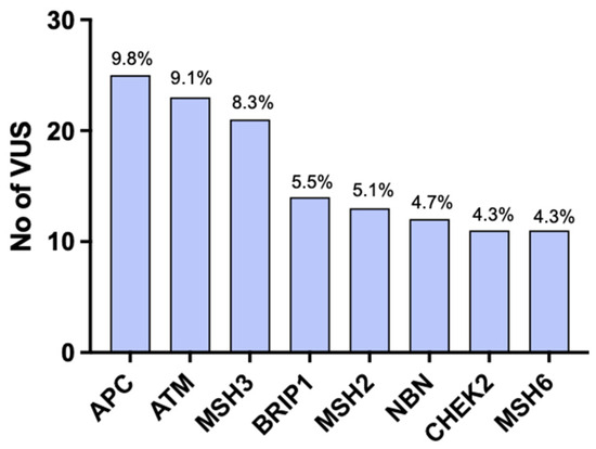
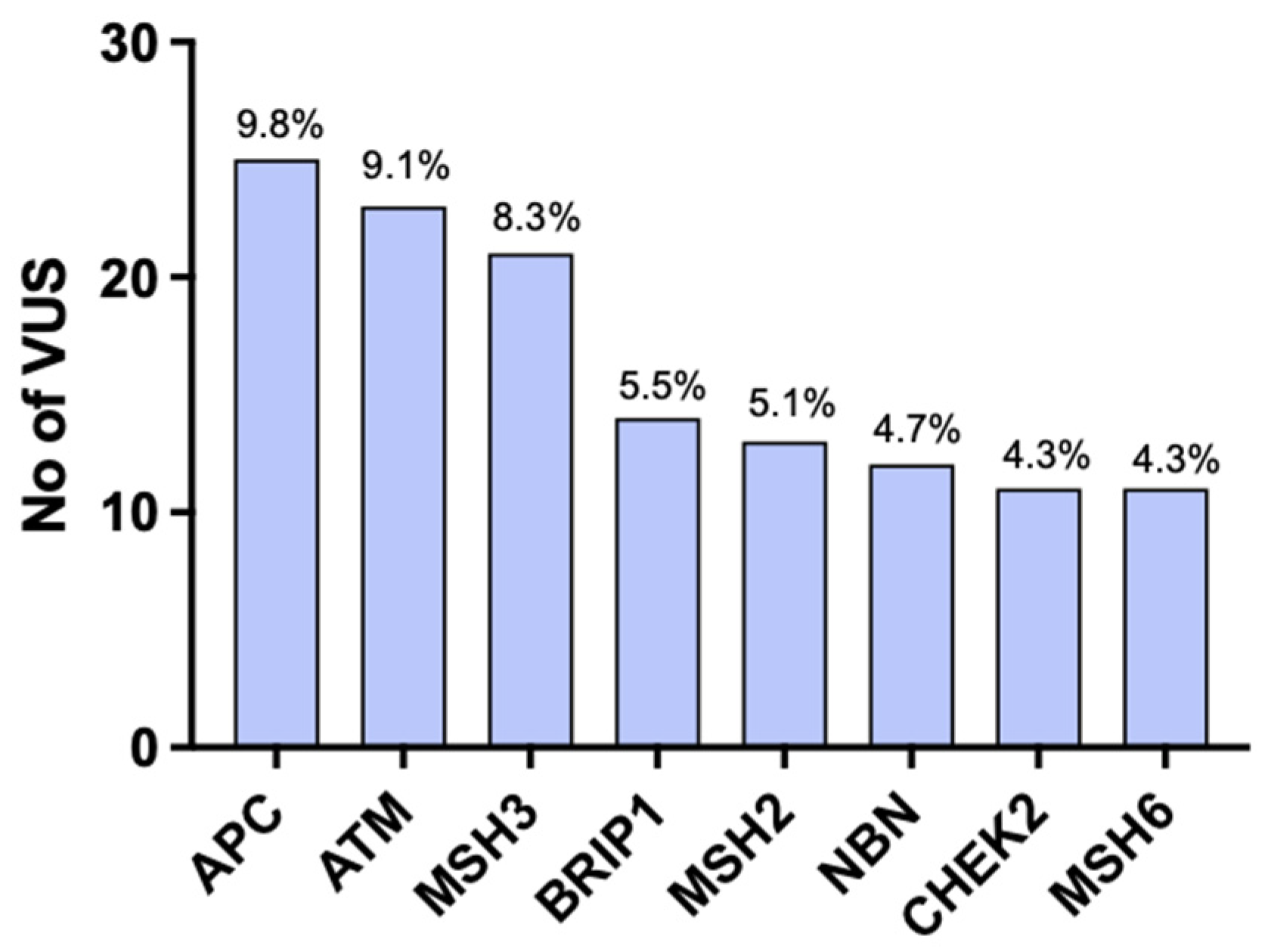
Among the whole group, 188 (28.3%) patients had a VUS, with 253 total VUSs identified (Table 3). Eleven of those mutations had not been previously reported in ClinVar (Table 4). Fifty-two patients (26.6%) had two or more VUSs within the same or different genes. Most VUSs were in APC, followed by ATM and MSH3 (Figure 1). Other common genes in which VUSs were detected were CHEK2, NBN, BRIP1, MSH2, and MSH6 (Figure 1). ATM and MSH6 were the most common genes in the Asian population. In the African-American population, the most common genes were NBN and APC. All variants found were previously described in ClinVar. VUSs and pathogenic mutations were present simultaneously in nine patients (0.90%).
Table 3.
VUSs in patients tested for HBOC and/or Lynch syndrome.
Table 4.
VUSs not previously reported.
Figure 1.
Most common genes in which VUSs were detected.
During the period from June 2021 to June 2023, we observed significant changes in the distribution of pathogenic, VUS, and negative findings within our cohort, driven by the updates to ClinVar. Notably, our preliminary analysis of 663 patients in June 2021 revealed 76 deleterious mutations, whereas the June 2023 analysis identified 89 such mutations. Additionally, the number of VUSs also saw an upward trend, rising from 200 in June 2021 to 253 by June 2023. We also identified VUSs that had conflicting data on ClinVar (Table 5).
Table 5.
VUSs with conflicting data on ClinVar.
Rates of VUS were not different with relation to age (≤40 vs. >40) or ethnicity (Hispanic vs Non-Hispanic) (Table 6). Interestingly, VUS incidence rates were significantly associated with race (Table 6). Post hoc analysis showed that VUSs were much more commonly detected in our Asian population compared to White patients (OR: 2.44; CI: 1.18–5.15). There was no difference between White and African-American, or African-American vs Asian. Furthermore, patients with a personal history of breast cancer were almost two times more likely to have a VUS compared to patients without a previous history (OR: 1.55; CI: 1.08–2.25) (Table 6). Patients with a family history of breast or ovarian cancer in a first-degree relative were also more likely to have a VUS (OR: 1.68; CI: 1.08–2.60 for breast and OR: 2.29; CI: 1.04–5.45 for ovarian) (Table 6). Family history of prostate or colon cancer in a close relative did not affect VUS rates (Table 6). Finally, the most common recommendation following a VUS report was annual breast magnetic resonance imaging (MRI) (70/188, 37.2%) as per review of clinical notes on electronic medical records. The next most common recommendations were following up with the primary care doctor (PCP) (20/188, 10.6%) or oncologist (30/188, 15.9%) in high-risk clinics.
Table 6.
VUS incidence rate in relation to age as well as personal and family history of cancer.
4. Discussion
To our knowledge, this is the first study evaluating the prevalence of VUS in patients meeting genetic test criteria for HBOC or Lynch syndrome in a USA-based population between 2006 and 2020. Our results show that 12.5% of the 663 patients that met the criteria for HBOC and/or Lynch genetic testing were found to have a pathogenic mutation, while almost one third of patients (28.3%) were found to have a VUS. VUS detection appeared to be associated with race, personal history of breast cancer, and family history of breast or ovarian cancer.
In our patient population meeting genetic test criteria for HBOC or Lynch syndrome, 12.5% of patients were found to have a pathogenic mutation, with the most common being in BRCA1/2 followed by MUTYH and CHEK2. BRCA1/2 are the most common genes involved in HBOC, and mutations in either of the BRCA genes increase a woman’s risk of breast cancer to 45–65% by 70 years old [29]. CHEK2 DNA repair gene pathogenic mutations also account for a significant amount of breast and colon cancer [30]. In contrast, MUTYH is primarily associated with MUTYH-associated polyposis, exhibiting some phenotypic similarities to Lynch syndrome [31]. In a Europe-based study, MUTYH accounted for only 3.6% of Lynch syndrome–like cases. In our study, however, we identified MUTYH pathogenic mutations in 13.2% of cases. This disparity may be attributed to incidental factors, population-specific genetic variability, or other contributing factors.
The main finding of our manuscript is the high prevalence of VUSs in patients tested for HBOC and/or Lynch. Almost one-third (28.3%) of the patients received a report for a VUS. There have only been a few studies evaluating VUS prevalence across the world, generating inconsistent results. For example, VUS prevalence was 46.1% in a Brazilian population tested for HBOC, while it was 9.2% in a Jordanian-Arab population [32,33]. This variability can be attributed to the availability of genetic testing, the number of genes tested in each panel, and possibly demographics and specific population characteristics. It is also important to consider that the relevance of genetic variants to disease is undergoing constant evaluation and change as NGS continues to advance. This is evident from the significant difference in prevalence numbers observed between our June 2021 and June 2023 analyses. The shifting prevalence of numbers highlights the continuous discovery of new variants and the evolving landscape of genetic research. Staying updated on the latest advancements is essential to ensure accurate and up-to-date genetic analysis and interpretation.
In addition, VUS prevalence showed no significant difference between the Hispanic and non-Hispanic populations in our study. However, when examining different racial groups, we observed significantly higher VUS rates in all non-White groups. A subgroup analysis further revealed that a significant difference in VUS rates was specifically observed between the Asian and White populations. However, the small sample size in the non-White groups and uneven distribution due to the observational study design may pose limitations to our findings. The literature data regarding the prevalence of VUSs based on race or ethnicity are currently scarce. Some studies find no correlation, while others, consistent with our results, find a higher prevalence of VUSs in non-White populations [34,35]. In a recent large study, Ndugga-Kabuye et al. evaluated VUS prevalence in 50,000 patients and confirmed the higher prevalence of VUS in non-European populations (i.e., Hispanics, African Americans, Asians, and Pacific Islanders). The absence of ancestral variety in genomic investigations is most likely to blame for the racial/ethnic discrepancies. For example, European ancestral populations account for the majority of observations (55.8%) in ClinVar, used to determine if a mutation is pathogenic or of undetermined significance.
Furthermore, we show that individuals with a personal history of breast cancer or family history of breast/ovarian cancer are more likely to have a VUS compared to their counterparts without history. This association may be an unintended consequence of VUS predominance in our population that will be later reclassified as pathogenic. If not incidental, it raises the question of whether VUS is the outcome of the increased mutational tendency seen in patients with pathogenic mutations and/or a history of cancer. To the best of our knowledge, no previous studies have compared the prevalence of VUS in patients with and without a personal or family history of cancer. Ndugga-Kabuye et al. performed separate analyses for VUS prevalence for patients with a positive and negative history of cancer. Although they did not statistically compare the prevalence, based on descriptive data, a VUS in HBOC or LS genes was found in 6.8% of patients with a personal history of cancer and in 6.4% of patients without, which is more suggestive of a similar distribution [35]. Another study that looked at VUS prevalence in breast cancer found no link between VUS rates and triple-negative malignancy [33]. Larger, prospective, and randomized studies are thus needed to investigate the possible correlations of VUS with ancestry and cancer history.
The unclear diagnosis resulting from a VUS result can have a negative impact on a patient’s health. The lack of structured medical guidance leads to inconsistent management based on the approach of the PCP or oncologists [3]. Some patients may not be offered predictive testing, while others may enter a cycle of intense screening and/or risk-reducing interventions of unclear benefit [3]. Furthermore, studies show that an inconclusive diagnosis may affect patients’ psychology, increasing stress and anxiety [3,36]. Similarly, the accuracy of pathogenic reports may also need to be questioned. In our study, all pathogenic mutations were listed as pathogenic on ClinVar, but 12/90 (13.3%) also had conflicting reports for uncertain significance or benign classification. This can lead to overestimation of the pathogenicity of the assessed variants and underestimation of VUSs. Thus, there is an unmet need to understand and better classify VUSs.
Towards this end, the ongoing development of functional assays holds promise in accurately determining the cancer risk associated with VUSs in a timely way [37,38]. These assays are designed to assess the impact of a particular variant on the structure and functionality of the corresponding protein, providing an estimated VUS pathogenicity [37]. Large investigator consortia, like ENIGMA or INSIGHT, have started to form, with the aim of collecting a large amount of data on VUSs and subsequently using machine learning prediction models or functional assays in determining the significance of variants [39,40,41].
To further address the complexity of sequence interpretation, the American Society of Medical Genetics and Genomics (ACMG) has released a set of guidelines for the classification of new sequences [4]. According to the ACMG, sequences should be categorized as pathogenic, likely pathogenic, of uncertain significance, likely benign, or benign. The criteria for classifying a sequence as pathogenic or likely pathogenic take various factors into consideration, such as the type of mutation (e.g., frameshift, nonsense), whether the mutation is causing an amino acid change of known pathogenicity, and the patient’s demographics. On the other hand, the criteria for benign or likely benign involve considerations like patient demographics, the frequency of the sequence, and existing functional studies. It is important to highlight that both sets of criteria take into great consideration the sequence distribution in healthy vs. affected individuals. Sequences identified in patients with a personal history provide robust evidence of pathogenicity, while those observed in healthy adults are more likely to be benign. Of note, these guidelines are designed for the interpretation of sequences related to inherited (Mendelian diseases) and not somatic genetic variations.
For sequence interpretation, the ACMG recommends that healthcare providers work in collaboration with clinical laboratories, as the patient’s clinical information plays a crucial role in enabling the laboratory to accurately classify genetic variants [4]. Given the growing complexity of analysis and interpretation, the ACMG strongly advocates for these tests to be conducted in Clinical Laboratory Improvement Amendments (CLIA)-approved laboratories, and the results should be interpreted by a board-certified clinical molecular geneticist or pathologist [4]. While acknowledging the current imperfections in variant classification, the ACMG recommends that mutations deemed pathogenic or likely pathogenic have substantial evidence to be used in clinical decision making, always within the appropriate clinical context [4]. On the other hand, variants of uncertain significance (VUSs) are discouraged from influencing clinical decisions [4]. Some experts recommend that laboratories do not release VUS reports and alert the clinician only if the VUS is reclassified as pathogenic [42]. However, this approach might threaten patients’ autonomy. As per the ACMG, as efforts to reclassify VUS are ongoing, vigilant monitoring of patients with VUS results is advisable [4]. This approach ensures a cautious and informed approach to utilizing genetic information in clinical practice.
5. Conclusions
In conclusion, VUSs appear to be detected in almost one out of three patients tested for cancer genetic syndromes, like HBOC and Lynch, thus having a huge impact on patients’ psychology and health management. Further studies are required to identify patients at risk for VUSs and, most importantly, to develop tests to determine the associated cancer risk.
Author Contributions
Conceptualization, C.T.J., A.D. and P.L.; methodology, C.T.J., P.C., M.L. and P.L.; software, P.C. and C.T.J.; formal analysis, P.C. and C.T.J.; investigation, P.C., C.T.J., O.A.O., H.S., K.S., L.A., A.R., A.Q.D. and A.D.; resources, C.T.J., M.L., A.D. and P.L.; data curation, P.C., C.T.J. and A.D.; writing—original draft preparation, P.C. and C.T.J.; writing—review and editing, O.A.O., M.L., A.D. and P.L.; visualization, P.C.; supervision, P.L.; project administration, C.T.J. All authors have read and agreed to the published version of the manuscript.
Funding
This research received no external funding.
Institutional Review Board Statement
The study was conducted in accordance with the Declaration of Helsinki, and approved by the Institutional Review Board of Mount Auburn Hospital/Beth Israel Deaconess Medical Center Protocol # 2020P1261.
Informed Consent Statement
Patient consent was waived due to the retrospective nature of the study.
Data Availability Statement
Data are available upon request.
Conflicts of Interest
The authors declare no conflict of interest.
References
- Federici, G.; Soddu, S. Variants of uncertain significance in the era of high-throughput genome sequencing: A lesson from breast and ovary cancers. J. Exp. Clin. Cancer Res. 2020, 39, 46. [Google Scholar] [CrossRef] [Scilit] [PubMed]
- Yohe, S.; Thyagarajan, B. Review of Clinical Next-Generation Sequencing. Arch. Pathol. Lab. Med. 2017, 141, 1544–1557. [Google Scholar] [CrossRef] [Scilit] [PubMed]
- Burke, W.; Parens, E.; Chung, W.K.; Berger, S.M.; Appelbaum, P.S. The Challenge of Genetic Variants of Uncertain Clinical Significance: A Narrative Review. Ann. Intern. Med. 2022, 175, 994–1000. [Google Scholar] [CrossRef] [Scilit] [PubMed]
- Richards, S.; Aziz, N.; Bale, S.; Bick, D.; Das, S.; Gastier-Foster, J.; Grody, W.W.; Hegde, M.; Lyon, E.; Spector, E.; et al. Standards and guidelines for the interpretation of sequence variants: A joint consensus recommendation of the American College of Medical Genetics and Genomics and the Association for Molecular Pathology. Genet. Med. 2015, 17, 405–424. [Google Scholar] [CrossRef] [Scilit]
- Rahner, N.; Steinke, V. Hereditary cancer syndromes. Dtsch. Arztebl. Int. 2008, 105, 706–714. [Google Scholar] [CrossRef] [Scilit]
- Samadder, N.J.; Giridhar, K.V.; Baffy, N.; Riegert-Johnson, D.; Couch, F.J. Hereditary Cancer Syndromes—A Primer on Diagnosis and Management: Part 1: Breast-Ovarian Cancer Syndromes. Mayo Clin. Proc. 2019, 94, 1084–1098. [Google Scholar] [CrossRef] [Scilit]
- Samuel, D.; Diaz-Barbe, A.; Pinto, A.; Schlumbrecht, M.; George, S. Hereditary Ovarian Carcinoma: Cancer Pathogenesis Looking beyond BRCA1 and BRCA2. Cells 2022, 11, 539. [Google Scholar] [CrossRef] [Scilit]
- Giardiello, F.M.; Allen, J.I.; Axilbund, J.E.; Boland, C.R.; Burke, C.A.; Burt, R.W.; Church, J.M.; Dominitz, J.A.; Johnson, D.A.; Kaltenbach, T.; et al. Guidelines on genetic evaluation and management of Lynch syndrome: A consensus statement by the US Multi-Society Task Force on Colorectal Cancer. Dis. Colon. Rectum. 2014, 57, 1025–1048. [Google Scholar] [CrossRef] [Scilit]
- Sehgal, R.; Sheahan, K.; O’Connell, P.R.; Hanly, A.M.; Martin, S.T.; Winter, D.C. Lynch syndrome: An updated review. Genes 2014, 5, 497–507. [Google Scholar] [CrossRef] [Scilit]
- Saam, J.; Arnell, C.; Theisen, A.; Moyes, K.; Marino, I.; Roundy, K.M.; Wenstrup, R.J. Patients Tested at a Laboratory for Hereditary Cancer Syndromes Show an Overlap for Multiple Syndromes in Their Personal and Familial Cancer Histories. Oncology 2015, 89, 288–293. [Google Scholar] [CrossRef] [Scilit]
- Lynch, H.T.; Casey, M.J.; Snyder, C.L.; Bewtra, C.; Lynch, J.F.; Butts, M.; Godwin, A.K. Hereditary ovarian carcinoma: Heterogeneity, molecular genetics, pathology, and management. Mol. Oncol. 2009, 3, 97–137. [Google Scholar] [CrossRef] [Scilit] [PubMed]
- Iqbal, J.; Ragone, A.; Lubinski, J.; Lynch, H.T.; Moller, P.; Ghadirian, P.; Foulkes, W.D.; Armel, S.; Eisen, A.; Neuhausen, S.L.; et al. The incidence of pancreatic cancer in BRCA1 and BRCA2 mutation carriers. Br. J. Cancer 2012, 107, 2005–2009. [Google Scholar] [CrossRef] [Scilit] [PubMed]
- Malander, S.; Rambech, E.; Kristoffersson, U.; Halvarsson, B.; Ridderheim, M.; Borg, A.; Nilbert, M. The contribution of the hereditary nonpolyposis colorectal cancer syndrome to the development of ovarian cancer. Gynecol. Oncol. 2006, 101, 238–243. [Google Scholar] [CrossRef] [Scilit] [PubMed]
- Nakamura, K.; Banno, K.; Yanokura, M.; Iida, M.; Adachi, M.; Masuda, K.; Ueki, A.; Kobayashi, Y.; Nomura, H.; Hirasawa, A.; et al. Features of ovarian cancer in Lynch syndrome (Review). Mol. Clin. Oncol. 2014, 2, 909–916. [Google Scholar] [CrossRef] [Scilit]
- Watson, P.; Vasen, H.F.A.; Mecklin, J.P.; Bernstein, I.; Aarnio, M.; Jarvinen, H.J.; Myrhøj, T.; Sunde, L.; Wijnen, J.T.; Lynch, H.T. The risk of extra-colonic, extra-endometrial cancer in the Lynch syndrome. Int. J. Cancer 2008, 123, 444–449. [Google Scholar] [CrossRef] [Scilit]
- Segev, Y.; Iqbal, J.; Lubinski, J.; Gronwald, J.; Lynch, H.T.; Moller, P.; Ghadirian, P.; Rosen, B.; Tung, N.; Kim-Sing, C.; et al. The incidence of endometrial cancer in women with BRCA1 and BRCA2 mutations: An international prospective cohort study. Gynecol. Oncol. 2013, 130, 127–131. [Google Scholar] [CrossRef] [Scilit]
- Sheehan, M.; Heald, B.; Yanda, C.; Kelly, E.D.; Grobmyer, S.; Eng, C.; Kalady, M.; Pederson, H. Investigating the Link between Lynch Syndrome and Breast Cancer. Eur. J. Breast Health 2020, 16, 106–109. [Google Scholar] [CrossRef] [Scilit]
- Buerki, N.; Gautier, L.; Kovac, M.; Marra, G.; Buser, M.; Mueller, H.; Heinimann, K. Evidence for breast cancer as an integral part of Lynch syndrome. Genes Chromosomes Cancer 2012, 51, 83–91. [Google Scholar] [CrossRef] [Scilit]
- Infante, M.; Arranz-Ledo, M.; Lastra, E.; Abella, L.E.; Ferreira, R.; Orozco, M.; Hernández, L.; Martínez, N.; Durán, M. Increased Co-Occurrence of Pathogenic Variants in Hereditary Breast and Ovarian Cancer and Lynch Syndromes: A Consequence of Multigene Panel Genetic Testing? Int. J. Mol. Sci. 2022, 23, 11499. [Google Scholar] [CrossRef] [Scilit]
- Clarke, J.E.; Magoon, S.; Forghani, I.; Alessandrino, F.; D’Amato, G.; Jonczak, E.; Subhawong, T.K. Radiologic screening and surveillance in hereditary cancers. Eur. J. Radiol. Open 2022, 9, 100422. [Google Scholar] [CrossRef] [Scilit]
- Hall, M.J.; Obeid, E.I.; Schwartz, S.C.; Mantia-Smaldone, G.; Forman, A.D.; Daly, M.B. Genetic testing for hereditary cancer predisposition: BRCA1/2, Lynch syndrome, and beyond. Gynecol. Oncol. 2016, 140, 565–574. [Google Scholar] [CrossRef] [Scilit] [PubMed]
- Mittendorf, K.F.; Knerr, S.; Kauffman, T.L.; Lindberg, N.M.; Anderson, K.P.; Feigelson, H.S.; Gilmore, M.J.; Hunter, J.E.; Joseph, G.; Kraft, S.A.; et al. Systemic Barriers to Risk-Reducing Interventions for Hereditary Cancer Syndromes: Implications for Health Care Inequities. JCO Precis. Oncol. 2021, 5, 1709–1718. [Google Scholar] [CrossRef] [Scilit] [PubMed]
- Balmana, J.; Digiovanni, L.; Gaddam, P.; Walsh, M.F.; Joseph, V.; Stadler, Z.K.; Nathanson, K.L.; Garber, J.E.; Couch, F.J.; Offit, K.; et al. Conflicting Interpretation of Genetic Variants and Cancer Risk by Commercial Laboratories as Assessed by the Prospective Registry of Multiplex Testing. J. Clin. Oncol. 2016, 34, 4071. [Google Scholar] [CrossRef] [Scilit]
- Ready, K.; Gutierrez-Barrera, A.M.; Amos, C.; Meric-Bernstam, F.; Lu, K.; Hortobagyi, G.; Arun, B. Cancer risk management decisions of women with BRCA1 or BRCA2 variants of uncertain significance. Breast J. 2011, 17, 210–212. [Google Scholar] [CrossRef] [Scilit] [PubMed]
- Kurian, A.W.; Ward, K.C.; Abrahamse, P.; Bondarenko, I.; Hamilton, A.S.; Deapen, D.; Morrow, M.; Berek, J.S.; Hofer, T.P.; Katz, S.J. Time Trends in Receipt of Germline Genetic Testing and Results for Women Diagnosed with Breast Cancer or Ovarian Cancer, 2012–2019. J. Clin. Oncol. 2021, 39, 1631–1640. [Google Scholar] [CrossRef] [Scilit]
- National Comprehensive Cancer Network. Genetic/Familial High-Risk Assessment: Breast, Ovarian, and Pancreatic, NCCN Clinical Practice Guidelines in Oncology (NCCN Guidelines®) Version 2.2024—27 September 2023; National Comprehensive Cancer Network: Plymouth Meeting, PA, USA, 2023.
- Vasen, H.F.; Watson, P.; Mecklin, J.P.; Lynch, H.T. New clinical criteria for hereditary nonpolyposis colorectal cancer (HNPCC, Lynch syndrome) proposed by the International Collaborative group on HNPCC. Gastroenterology 1999, 116, 1453–1456. [Google Scholar] [CrossRef] [Scilit] [PubMed]
- Umar, A.; Boland, C.R.; Terdiman, J.P.; Syngal, S.; de la Chapelle, A.; Ruschoff, J.; Fishel, R.; Lindor, N.M.; Burgart, L.J.; Hamelin, R.; et al. Revised Bethesda Guidelines for hereditary nonpolyposis colorectal cancer (Lynch syndrome) and microsatellite instability. J. Natl. Cancer Inst. 2004, 96, 261–268. [Google Scholar] [CrossRef] [Scilit]
- Kuchenbaecker, K.B.; Hopper, J.L.; Barnes, D.R.; Phillips, K.A.; Mooij, T.M.; Roos-Blom, M.J.; Jervis, S.; Van Leeuwen, F.E.; Milne, R.L.; Andrieu, N.; et al. Risks of Breast, Ovarian, and Contralateral Breast Cancer for BRCA1 and BRCA2 Mutation Carriers. JAMA 2017, 317, 2402–2416. [Google Scholar] [CrossRef] [Scilit]
- Stolarova, L.; Kleiblova, P.; Janatova, M.; Soukupova, J.; Zemankova, P.; Macurek, L.; Kleibl, Z. CHEK2 Germline Variants in Cancer Predisposition: Stalemate Rather than Checkmate. Cells 2020, 9, 2675. [Google Scholar] [CrossRef] [Scilit]
- Weiss, J.M.; Gupta, S.; Burke, C.A.; Axell, L.; Chen, L.M.; Chung, D.C.; Clayback, K.M.; Dallas, S.; Felder, S.; Gbolahan, O.; et al. NCCN Guidelines(R) Insights: Genetic/Familial High-Risk Assessment: Colorectal, Version 1.2021. J. Natl. Compr. Cancer Netw. 2021, 19, 1122–1132. [Google Scholar]
- Guindalini, R.S.C.; Viana, D.V.; Kitajima, J.; Rocha, V.M.; Lopez, R.V.M.; Zheng, Y.; Freitas, É.; Monteiro, F.P.M.; Valim, A.; Schlesinger, D.; et al. Detection of germline variants in Brazilian breast cancer patients using multigene panel testing. Sci. Rep. 2022, 12, 4190. [Google Scholar] [CrossRef] [Scilit] [PubMed]
- Abdel-Razeq, H.; Tamimi, F.; Abujamous, L.; Abdel-Razeq, R.; Abunasser, M.; Edaily, S.; Abdulelah, H.; Khashabeh, R.A.; Bater, R. Rates of Variants of Uncertain Significance Among Patients with Breast Cancer Undergoing Genetic Testing: Regional Perspectives. Front. Oncol. 2022, 12, 673094. [Google Scholar] [CrossRef] [Scilit] [PubMed]
- Caswell-Jin, J.L.; Gupta, T.; Hall, E.; Petrovchich, I.M.; Mills, M.A.; Kingham, K.E.; Koff, R.; Chun, N.M.; Levonian, P.; Lebensohn, A.P.; et al. Racial/ethnic differences in multiple-gene sequencing results for hereditary cancer risk. Genet. Med. 2018, 20, 234–239. [Google Scholar] [CrossRef] [Scilit] [PubMed]
- Ndugga-Kabuye, M.K.; Issaka, R.B. Inequities in multi-gene hereditary cancer testing: Lower diagnostic yield and higher VUS rate in individuals who identify as Hispanic, African or Asian and Pacific Islander as compared to European. Fam. Cancer 2019, 18, 465–469. [Google Scholar] [CrossRef] [Scilit] [PubMed]
- Makhnoon, S.; Shirts, B.H.; Bowen, D.J. Patients’ perspectives of variants of uncertain significance and strategies for uncertainty management. J. Genet. Couns. 2019, 28, 313–325. [Google Scholar] [CrossRef] [Scilit]
- Monteiro, A.N.; Bouwman, P.; Kousholt, A.N.; Eccles, D.M.; Millot, G.A.; Masson, J.Y.; Schmidt, M.K.; Sharan, S.K.; Scully, R.; Wiesmüller, L.; et al. Variants of uncertain clinical significance in hereditary breast and ovarian cancer genes: Best practices in functional analysis for clinical annotation. J. Med. Genet. 2020, 57, 509–518. [Google Scholar] [CrossRef] [Scilit]
- Woods, N.T.; Baskin, R.; Golubeva, V.; Jhuraney, A.; De-Gregoriis, G.; Vaclova, T.; Goldgar, D.E.; Couch, F.J.; Carvalho, M.A.; Iversen, E.S.; et al. Functional assays provide a robust tool for the clinical annotation of genetic variants of uncertain significance. NPJ Genom. Med. 2016, 1, 16001. [Google Scholar] [CrossRef] [Scilit]
- Tricarico, R.; Kasela, M.; Mareni, C.; Thompson, B.A.; Drouet, A.; Staderini, L.; Gorelli, G.; Crucianelli, F.; Ingrosso, V.; Kantelinen, J.; et al. Assessment of the InSiGHT Interpretation Criteria for the Clinical Classification of 24 MLH1 and MSH2 Gene Variants. Hum. Mutat. 2017, 38, 64–77. [Google Scholar] [CrossRef] [Scilit]
- Khandakji, M.; Habish, H.H.A.; Abdulla, N.B.S.; Kusasi, S.A.A.; Abdou, N.M.G.; Al-Mulla, H.; Al Sulaiman, R.J.A.; Bu Jassoum, S.M.; Mifsud, B. BRCA1-specific machine learning model predicts variant pathogenicity with high accuracy. Physiol. Genom. 2023, 55, 315–323. [Google Scholar] [CrossRef] [Scilit]
- Stolarova, L.; Kleiblova, P.; Zemankova, P.; Stastna, B.; Janatova, M.; Soukupova, J.; Achatz, M.I.; Ambrosone, C.; Apostolou, P.; Arun, B.K.; et al. ENIGMA CHEK2gether Project: A Comprehensive Study Identifies Functionally Impaired CHEK2 Germline Missense Variants Associated with Increased Breast Cancer Risk. Clin. Cancer Res. 2023, 29, 3037–3050. [Google Scholar] [CrossRef] [Scilit]
- Hoffman-Andrews, L. The known unknown: The challenges of genetic variants of uncertain significance in clinical practice. J. Law Biosci. 2017, 4, 648–657. [Google Scholar] [CrossRef] [Scilit] [PubMed]

Disclaimer/Publisher’s Note: The statements, opinions and data contained in all publications are solely those of the individual author(s) and contributor(s) and not of MDPI and/or the editor(s). MDPI and/or the editor(s) disclaim responsibility for any injury to people or property resulting from any ideas, methods, instructions or products referred to in the content. |
© 2023 by the authors. Licensee MDPI, Basel, Switzerland. This article is an open access article distributed under the terms and conditions of the Creative Commons Attribution (CC BY) license (https://creativecommons.org/licenses/by/4.0/).