


Abstract
Magnetite (Fe3O4) micro- and nanoparticles are critical for advanced core–shell applications, but their susceptibility to oxidation into maghemite (γ-Fe2O3) degrades magnetic performance. This study evaluates the influence of solvothermal duration and autoclave filling factor on the size, morphology, and magnetic response of magnetite particles and introduces an X-ray-diffraction-based method for quantifying oxidation. Across the evaluated conditions, the 18 h series consistently produced particles approaching the intended 250 nm size while retaining saturation magnetization values of 80–85 A·m2kg−1. The 18 h series with 40% autoclave filling factor (18h40) yielded a particle diameter of ~243 nm, comparable to the 24 h samples, indicating no size benefit from extending the synthesis to 24 h. To track phase, a hybrid Rietveld refinement protocol was implemented to deconvolute peak overlap between the isomorphic phases. A 60-day aging study of the 18h40 condition demonstrated progressive oxidation, with the maghemite fraction increasing from 1.64 wt.% to 22.76 wt.%. VSM corroborated this degradation, showing a 9.1% reduction in saturation magnetization. A 60-day control study showed that sealed storage limited oxidation, with the particles containing only 0.9 wt.% maghemite. These findings support further investigation of the 18 h synthesis conditions for future core–shell studies. The localized Rietveld methodology provides a tool for monitoring magnetite-to-maghemite oxidation.
1. Introduction
Nanomaterials represent a significant focus in scientific research due to their excellent, size-dependent physical and chemical properties that differ markedly from those of their bulk counterparts. Their applications have spanned a wide range of fields, including medicine, energy, automobiles, smart infrastructure, coatings, and more [1,2,3,4].
Importantly, such size-dependent properties can be governed not only by particle dimensions but also by structural design, for example through the incorporation of functional coatings that enable hybrid architectures with tailored interfacial properties [5]. Magnetic core–shell hybrid particles are a prime example of this, as they exhibit tunable magnetic behavior and versatile surface functionalities, rendering them ideal for applications such as hyperthermia, lithium-ion batteries, microwave absorption, and catalysis [6,7,8,9]. These structures typically consist of a magnetic core enclosed in a functional shell, allowing the tuning of their properties through independent or combined modification of the core and the shell. This approach integrates features such as superparamagnetism and magnetic susceptibility, with enhanced stability, dispersibility, and dielectric behavior, while also protecting the core from oxidation [10]. The design of the magnetic core is a key determinant of particle behavior and real-world applicability. Parameters such as average diameter, dispersity, crystallinity, and defect chemistry directly influence crucial responses including saturation magnetization, coercive field, Brownian/Néel relaxation, and hysteretic losses [11]. Notably, the ideal size for a magnetic core depends entirely on its intended application. For biomedical applications such as magnetic hyperthermia, nanoscale iron-oxide particles are widely employed, with dimensions and magnetic behavior varying based on whether the system consists of individual single-core particles or larger multicore assemblies, while aggregation state and surface characteristics also need to be considered [11,12]. By contrast, advanced electromagnetic applications, such as microwave absorption and EMI shielding, often employ larger submicron magnetic cores, typically in the 200–500 nm range, although nanoscale ones may also be effective depending on the system design [13]. At this larger size, the particles transition into multi-domain states. This natural transition boosts interfacial polarization, resonance, and magnetic hysteresis losses, all of which work together to drastically improve the material’s ability to absorb electromagnetic waves [14]. The ability to control the size and magnetic properties of Fe3O4 cores is also particularly relevant for the growth of core–shell architectures. The magnetic core and the surrounding shell can be designed to provide complementary functionalities. In electromagnetic applications, Fe3O4-based core–shell structures have attracted considerable interest for microwave absorption [14] because the combination of magnetic loss from the iron-oxide core with dielectric and interfacial polarization effects introduced by the shell can improve impedance matching and broaden the effective absorption response. Core–shell magnetic particles are also widely investigated in biomedical applications such as magnetic hyperthermia, where the shell may improve colloidal stability, biocompatibility, surface functionalization, and protection of the magnetic core against oxidation [5,15]. However, the particle size requirements differ substantially between these applications; whereas submicron magnetic particles may be advantageous for electromagnetic-wave attenuation, magnetic hyperthermia generally requires careful control of particle size, magnetic domain state, aggregation, and surface chemistry to achieve efficient and biologically suitable heating. Therefore, reproducible control of the magnetic core prior to shell formation represents an important prerequisite for application-specific core–shell design.
Among the various techniques used for the synthesis of magnetic cores, hydrothermal and solvothermal methods offer significant control over particle characteristics. These approaches can be tuned to produce quasi-spherical, octahedral, or cubic morphologies while retaining a single-phase spinel structure for magnetite cores [16]. Therefore, they have been widely adopted due to their ability to produce particles with higher crystallinity, narrower size distributions, and improved control over morphology compared to conventional co-precipitation methods, resulting in more predictable magnetic behavior and improved batch-to-batch reproducibility [17]. In this context, hydrothermal and solvothermal synthetic methods have become a practical bridge between laboratory discovery and scalable manufacturing.
In such methods, manipulating synthesis parameters such as precursor concentration and reactor filling rates is critical for tailoring the core’s morphology, density, and crystallinity, as shown by Wang et al. [18]. The general guidelines Wang and his team suggest include varying temperatures from 20 to 200 °C, omitting time variation and choosing a fixed time for a variety of materials, while time dependency is currently missing, especially in the literature on Fe3O4 magnetic particle synthesis. Lv et al. [19] have synthesized Fe3O4 coated with carbon particles with an average size of 300 nm for use in Anode Materials via the solvothermal method. Still, their group has not elucidated the time effect on its synthesis while maintaining the safe, well-known 24 h solvothermal synthesis. Kim J. et al. [17] managed to synthesize Fe3O4 at pilot production scale, using a fixed synthesis time while varying the iron precursor concentrations, producing magnetic cores within a similar size range to those targeted in the present study, while confirming their suitability for applications such as EMI shielding, bio-separation and theragnostic probes. Collectively, these findings highlight the importance of precise particle size control during solvothermal synthesis, as particle dimensions directly influence both functional performance and application specificity.
However, beyond morphological optimization, the long-term stability of magnetite cores remains a critical limitation for their practical implementation, primarily due to their susceptibility to progressive surface oxidation. While the functional degradation caused by magnetite-to-maghemite oxidation has been extensively documented by Schwaminger et al. [20], accurately quantifying this phase transition remains analytically challenging due to severe crystallographic overlap between the two isomorphic phases. Standard X-ray diffraction cannot reliably resolve magnetite and maghemite independently, forcing previous studies to rely either on complex and time-intensive Mössbauer spectroscopy for accurate phase quantification or on empirical calibration approaches based on deconvolution of the characteristic (511) reflection [21]. Although such methods have demonstrated partial success, they remain dependent on external calibration standards and experimentally derived fitting procedures. To overcome these limitations, the present study introduces a localized hybrid Rietveld tracking methodology capable of mathematically isolating the overlapping phases directly from X-ray diffraction data through the use of crystallographic phase models and a 3rd-order background polynomial, thereby reducing reliance on additional spectroscopic techniques or empirical calibration curves.
To bridge this gap between high-quality synthesis and long-term practical performance, this study focuses on the optimization of solvothermal production and the stability of highly crystalline magnetite cores. Specifically, we examine whether key synthesis parameters, such as reactor filling rate and processing time, can be adjusted to achieve a desired particle size distribution and magnetic characteristics consistent with those reported as optimal for core–shell electromagnetic absorption applications (i.e., ~250 nm and Ms > 80 Am2/kg for magnetite). The resulting particles are further evaluated in terms of their composition and distinct magnetic behavior, as well as their stability under ambient conditions.
2. Materials and Methods
The materials required for solvothermal synthesis included iron(III) chloride hexahydrate, ACS reagent, 97%, which was purchased from Sigma-Aldrich (St. Louis, MO, USA), and anhydrous sodium acetate, ACS reagent; ethylene glycol, Reag. Ph. Eur., ≥99.5%; and ethanol, absolute ≥99.8%, which were purchased from Honeywell (Charlotte, NC, USA).
2.1. Solvothermal Synthesis
Fe3O4 microspheres were synthesized via a solvothermal method. Concisely, 9.03 g of FeCl3·6H2O was dissolved in 210 mL of ethylene glycol, while 15 g of CH3COONa was dissolved in 90 mL of ethylene glycol. Subsequently, the two solutions were mixed and stirred vigorously for 30 min. The resulting homogeneous yellow solution was then transferred to Teflon-lined stainless-steel autoclaves, sealed, and heated to 200 °C for varying times, as specified in our protocol, yielding three solvothermal times of 12 h, 18 h, and 24 h. The autoclave filling rate was not fixed and varied, resulting in three distinct rates: 40%, 50%, and 60%. After the reaction, the autoclave was allowed to cool to room temperature naturally. The black magnetite particles were collected by magnetic separation using a neodymium magnet, and then the supernatant was decanted. The precipitates were washed three times with deionized water and then with absolute ethanol to remove organic residues, and finally dried at 40 °C.
2.2. Characterization Techniques
The particle sizes and surface morphology were studied using a JSM-6610LV scanning electron microscope (SEM) from JEOL Ltd. (Akishima, Tokyo, Japan) equipped with an Oxford Instruments X-Max 80 energy-dispersive spectroscopy (EDS) detector for elemental validation. The crystalline phase and structural analysis were conducted using a D8 ADVANCE X-ray diffractometer (XRD; Bruker, Billerica, MA, USA) with a CuKα radiation source set to 40 kV and 40 mA. X-ray diffraction measurements were performed over a 2θ range of 15–90° using a continuous position-sensitive detector (PSD) scan. Data were acquired with a counting time of 2.5 s per step, yielding a total of 7359 measurement steps across the investigated angular range. The PSD angular opening was set to 2.908°. These acquisition conditions were selected to provide adequate counting statistics and angular sampling for phase identification and subsequent analysis of the magnetite and maghemite diffraction contributions. All samples were measured using the same acquisition settings to ensure consistency and comparability between the different synthesis conditions and aging time points.
The magnetic properties of the particles were assessed using a room-temperature vibrating sample magnetometer (VSM-1.2H/CF/HT from Oxford Instruments) within a magnetic field range of ±1 T. For the quantitative analysis of the aged samples, a hybrid Rietveld refinement technique was applied using Bruker’s TOPAS v.5 software.
Prior to X-ray diffraction (XRD) analysis, the as-produced magnetite particles were dispersed in ethanol and vortex-mixed for three minutes to ensure a uniform, agglomerate-free suspension. Droplets of this suspension were then carefully applied onto a silicon zero-background sample holder. To accumulate a sufficient quantity of material for accurate analysis, the substrate was dried at 40 °C for several minutes, and this drop-casting and drying cycle was repeated multiple times.
While measures were taken to suppress thermal oxidation by limiting the drying temperature to 40 °C, it is important to acknowledge the potential for subtle phase changes during the preparation protocol. Magnetite (Fe3O4) is known to be susceptible to surface oxidation, particularly in the presence of water and atmospheric oxygen, even at relatively mild temperatures. The repeated washing with DI water and ethanol, coupled with subsequent drying at 40 °C in air after synthesis and after each XRD sample preparation, could facilitate the slow surface oxidation of a small fraction of the magnetite particles to maghemite (γ-Fe2O3).
This pre-analysis oxidation potentially contributes to the initial maghemite content observed in the 0-day samples. While a certain degree of maghemite formation might be inherent to the synthesis process itself, the sample preparation steps could augment this baseline. Therefore, the ‘0-day’ XRD data should be interpreted as representing the phase composition of the material immediately prior to the controlled oxidation studies, encompassing any minor oxidation that occurred during synthesis and subsequent preparation.
2.3. Hybrid Rietveld Refinement for Oxidation Analysis
Phase evolution was quantified using localized Rietveld refinement of their overlapping (511) reflections, as conventional whole-pattern XRD refinement cannot reliably distinguish between magnetite and maghemite. By fitting isolated reflection clusters directly to specific crystallographic models, we utilized TOPAS v.5 (Bruker, Billerica, MA, USA) to calculate the phase weight percentages of overlapping reflections, without the need for empirical Voigt peak integration and external empirical secondary calibration.
The refinement was performed within a carefully selected 55–59° 2θ window. This expanded range improves baseline modeling by capturing sufficient data points on both sides of the (511) peaks, which is essential for accurate background subtraction. Furthermore, it ensures the complete intensity profile—including the full peak tails of both phases—is recorded, preventing the underestimation of peak areas affected by nanocrystalline broadening. Applying this consistent fitting window across all samples provides the necessary resolution to accurately track oxidative growth, starting from the minor surface oxidation present in the 0-day baseline sample.
Instrumental background and amorphous scattering were modeled utilizing a 3rd-order polynomial to prevent artificial intensity suppression of the broadened peaks. Structural refinements were based on Crystallography Open Database (COD) references: COD 9006194 for the primary magnetite phase and COD 9006316 for the oxidized maghemite phase. Both phases were refined within the cubic Fdm space group. During mild oxidation of magnetite to maghemite, Fe2+ is converted to Fe3+, while charge neutrality is maintained through the formation of cation vacancies that are randomly distributed over the octahedral sublattice [22]. Consequently, the crystal retains its original cubic Fdm symmetry rather than shifting into a lower-symmetry ordered state [23]. Treating maghemite as a disordered defect spinel in this higher symmetry group was considered appropriate for these samples, as the observed peak broadening associated with their small crystallite dimensions was expected to limit the detection of any weak superlattice reflections. Furthermore, the peak profiles were modeled using a Double-Voigt approach to account for size- and strain-related contributions to peak broadening.
2.4. Statistics
Particle size data were analyzed using a two-way analysis of variance (ANOVA), with synthesis duration (12, 18, and 24 h) and autoclave filling capacity (40%, 50%, and 60%) treated as fixed factors, together with their interaction. Statistical significance was set at p < 0.05. Because the large sample size of the dataset increases the sensitivity of conventional p-value-based post hoc testing to small differences, pairwise comparisons between specific synthesis conditions were evaluated using Cohen’s d. This approach was utilized to quantify the magnitude of differences between groups and assess their practical significance, specifically, the degree of physical overlap of the resulting particle size distributions. Absolute d values below 0.20 were interpreted as negligible and used to assign conditions to practical similarity groups. Conditions sharing at least one letter were considered to exhibit comparable mean particle sizes according to this effect-size criterion.
3. Results and Discussion
3.1. Size and Morphology
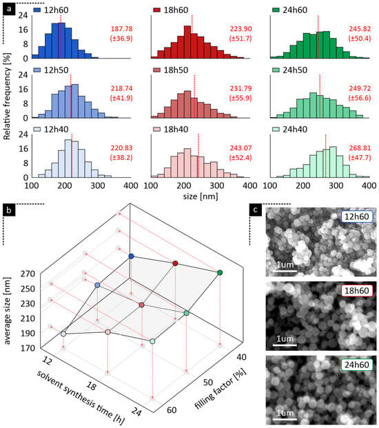
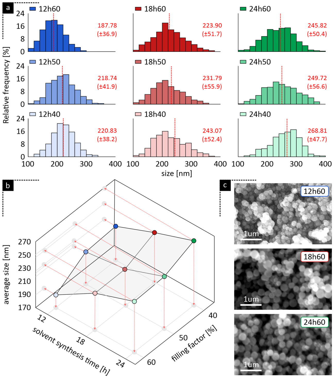
Particle size distributions obtained under various synthesis conditions are presented in Figure 1a. All combinations (i.e., time and filling factor) exhibited approximately normal particle size distributions with relatively narrow dispersion ranges, indicating a high degree of morphological uniformity.
Figure 1.
(a) Particle size distributions for all synthesis conditions, (b) combined influence of solvothermal duration and autoclave filling factor on the resulting average particle diameter and (c) indicative SEM micrographs, demonstrating the influence of synthesis parameters on particle morphology and diameter.
Table 1 summarizes the mean particle sizes, standard deviation, and effect-size-based similarity grouping for each synthesis condition.
Table 1.
Mean particle size (nm), standard deviation (SD), and effect-size-based similarity grouping for each synthesis condition. Conditions sharing at least one letter exhibited a negligible pairwise standardized difference (∣d∣ < 0.20). The similarity groups represent practical similarity based on effect size rather than statistical equivalence.
It is well established that rapid aqueous co-precipitation routes, such as the 5 to 8 min core synthesis reported by Furlan et al. [24], can achieve nanoparticle formation at low temperatures, but the resulting ultrafine cores (~10–40 nm) remain highly susceptible to oxidation without extensive protective encapsulation. Achieving highly crystalline, phase-pure Fe3O4 instead requires closed-system, high-temperature conditions: Chen et al. [25] reported that a 200 °C hydrothermal treatment requires at least 10 h to fully reduce α-Fe2O3 to Fe3O4, with the resulting phase remaining stable for treatments exceeding up to 70 h. The synthesis durations examined here (12, 18, and 24 h) fall well within this stable, phase-pure window, supporting progressive coarsening as a plausible contributor to the observed size increase.
The combined influence of synthesis duration and autoclave filling factor on average particle size is summarized in Figure 1b. The observed increase in particle size with increasing synthesis duration may be attributed to the progressive extension of the Ostwald ripening regime under elevated temperature and pressure conditions [26]. During prolonged solvothermal treatment, smaller Fe3O4 nuclei progressively dissolve and reprecipitate onto larger particles, thereby favoring thermodynamically more stable structures and resulting in increased average particle diameters.
In contrast, the autoclave filling factor exhibited an inverse relationship with particle size. For each synthesis duration, decreasing the filling factor from 60% to 40% resulted in progressively larger particles, with the effect being most pronounced at 24 h synthesis, where the average particle size increased from 245.82 ± 50.4 nm at 60% filling to 268.81 ± 47.7 nm at 40% filling. This behavior deviates from the conventional solvothermal expectation in which elevated autogenous pressure generally promotes crystal growth [27]. This behavior may be associated with differences in vapor–liquid equilibrium within the autoclave. At lower filling rates, the increased headspace volume could favor partial redistribution of water derived from the FeCl3·6H2O precursor into the vapor phase, thereby reducing the effective water concentration available for hydrolysis. Because water acts as a critical reactant for metal ion hydrolysis in such syntheses (Fiévet et al., 1989), this reduced water concentration inherently suppresses the hydrolysis kinetics [28]. Following classical nucleation theory (LaMer & Dinegar, 1950) [29], a suppressed hydrolysis rate limits the initial supersaturation spike, resulting in the formation of fewer nuclei that subsequently undergo sustained growth into larger microspheres. In addition, since solvothermal crystal growth is strongly influenced by pressure, solvent concentration, and precursor supersaturation [30], such conditions may suppress nucleation kinetics and reduce the density of initially formed nuclei, ultimately favoring the growth of larger particles during subsequent ripening stages.
Representative SEM micrographs of selected sample series are presented in Figure 1c to illustrate the morphology and relative particle dimensions obtained under the different synthesis conditions. Independent of synthesis parameters, all samples consisted predominantly of quasi-spherical magnetite particles with relatively homogeneous morphology and limited agglomeration. Consistent with the preceding quantitative analysis, increasing synthesis duration resulted in visibly larger particle diameters.
A two-way ANOVA revealed a statistically significant interaction between synthesis time and filling capacity on particle size, F(4, 7771) = 25.866, p < 0.001, = 0.013. Because the extremely large sample size renders even trivial amounts of variance highly statistically significant—as indicated by the low partial relying solely on the omnibus p-value overstates the practical importance of the interaction. Therefore, analysis of the pairwise effect sizes (Cohen’s d) was used to characterize the magnitude of differences among individual synthesis conditions and generate the practical similarity groups presented in Table 1. The most distinct synthesis condition was observed at 12 h with a 60% filling capacity, which yielded the smallest mean particle size (M = 187.78). This parameter set produced a very large, standardized difference (d = 1.75) when compared with the largest particles synthesized at 24 h and 40% capacity (M = 268.82), indicating almost entirely non-overlapping size distributions. Conversely, specific parameter shifts resulted in negligible practical differences; for instance, increasing the capacity from 40% to 50% at 12 h synthesis (d = 0.05) or from 50% to 60% at 24 h (d = 0.07) produced comparable mean particle sizes, differing by approximately 2 and 4 nm, respectively, as reflected by their shared similarity groups in Table 1.
Ultimately, these metrics indicate that the highest filling capacity with the shortest synthesis duration was the most effective of the tested conditions for producing smaller magnetic microparticles. Relative to the targeted particle size of approximately 250 nm, the 18 h and 24 h series produced mean particle sizes within approximately 11% and 8% of the target, respectively, across all evaluated filling capacities. By contrast, the mean particle sizes obtained after 12 h remained approximately 12–25% below the target.
For industrial-scale applications, however, selection of an appropriate synthesis protocol must consider not only particle size but also reactor utilization, synthesis-cycle duration, and energy consumption per unit of product, as these parameters directly influence production throughput and operational expenditure (OPEX). The used configuration is consistent with established pilot-scale solvothermal protocols, such as the 200 g/batch mass production reactor reported by Kim et al. [17]. Although the aforementioned research achieved synthesis in 10 h using a slightly modified chemical route (e.g., employing sodium citrate as a stabilizer), a significant portion of the broader literature often adopts a fixed synthesis duration as a default standard without explicit justification. Those studies have shown that scaling up solvothermal processes preserves consistent product characteristics [17]. Consequently, the duration of synthesis functions as a critical and legitimate optimization metric.
Based on this rationale, 18 h represented the shortest evaluated synthesis duration that consistently produced particles approaching the intended size domain across all filling capacities. Within the 18 h series, the 18h40 condition produced particles with a mean diameter of approximately 243 nm, corresponding to a deviation of less than 3% from the targeted size of approximately 250 nm. Moreover, the revised similarity groups showed that the 18h40 condition produced particle sizes comparable to those obtained with both 24h50 and 24h60 (d ≈ 0.12 and d ≈ 0.05, respectively), indicating that extending the synthesis duration to 24 h did not yield a practically distinct particle size. By contrast, increasing the filling capacity within the 18 h series progressively shifted the mean particle size further below the intended target, while the increase from 50% to 60% produced only a negligible difference (d = 0.15). Together, these findings identified the 18h40 condition as the most suitable candidate for subsequent analysis, as it combined the shortest synthesis duration capable of closely approaching the targeted particle size with particle dimensions comparable to those obtained using longer 24 h protocols. Nevertheless, particle size considerations alone could not establish the overall superiority of a single set of synthesis parameters, necessitating complementary evaluation of phase composition, magnetic performance, and energy consumption per unit of product, as examined in the following sections.
3.2. Elemental and Structural Characterization
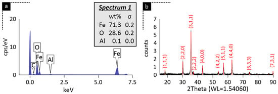
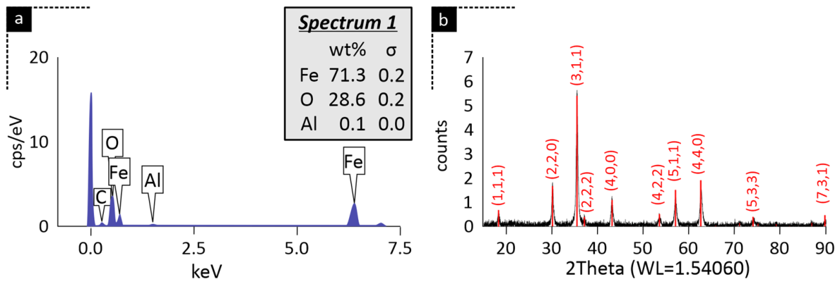
Following the morphological analysis, the elemental composition and crystallographic structure of the synthesized microparticles were evaluated using energy-dispersive X-ray spectroscopy (EDS) and X-ray diffraction (XRD), respectively. Representative EDS and XRD results for the 18h40 sample series are presented in Figure 2a and Figure 2b, respectively.
Figure 2.
Elemental and crystallographic characterization of the synthesized magnetite microparticles shown indicatively for the 18h40 sample. (a) EDS spectrum showing the dominant Fe and O signals together with the corresponding quantitative elemental composition, closely matching the stoichiometric ratio of magnetite (Fe3O4), and (b) XRD pattern corresponding to the cubic inverse spinel structure of Fe3O4.
The EDS spectrum is dominated by strong characteristic peaks corresponding to iron and oxygen, confirming these as the primary constituents of the sample, as also visible in the quantitative analysis (inset of Figure 2a). These values are in excellent agreement with the theoretical stoichiometric composition of magnetite (Fe3O4), which consists of 72.4 wt% Fe and 27.6 wt% O. A negligible trace of aluminum (0.1 wt%) and carbon was detected, likely originating from the substrate or sample holder and the carbon tape, respectively, but no other significant impurities were observed, indicating a high degree of chemical purity.
Representative XRD data for the same sample series are presented in Figure 2b. The diffraction peaks observed at approximately 18.3°, 30.1°, 35.5°, 43.1°, 53.5°, 57.0°, and 62.6° 2θ were indexed to the (111), (220), (311), (400), (422), (511), and (440) crystal planes of the cubic inverse spinel structure of magnetite (space group: Fd3m), showing excellent agreement with PDF Card 88-0866 (International Centre for Diffraction Data—ICDD). No secondary phases, such as hematite (α-Fe2O3), were detected within the sensitivity limits of the instrument. However, due to the crystallographic similarity between magnetite and maghemite (γ-Fe2O3), conventional XRD alone cannot reliably distinguish between the two phases [21].
Consequently, potential surface oxidation effects are further evaluated in the subsequent oxidation and magnetic characterization sections.
3.3. Magnetic Phase Verification
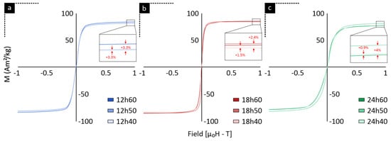
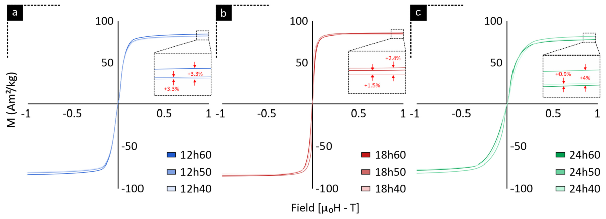
Room-temperature field-dependent magnetization (M–H) curves for the synthesized samples are presented in Figure 3. All sample series exhibited characteristic ferrimagnetic behavior with narrow hysteresis loops, near-zero coercivity (Hc), and negligible remanent magnetization (Mr), indicating high magnetic susceptibility and limited residual magnetization after removal of the external magnetic field.
Figure 3.
Room-temperature field-dependent magnetization (M–H) curves of samples synthesized at: (a) 12 h, (b) 18 h, and (c) 24 h under different autoclave filling conditions. The saturation magnetization values confirm the formation of a magnetic Fe3O4 phase across all samples.
The saturation magnetization (Ms) values obtained for all synthesis conditions ranged approximately between 80 and 85 Am2/kg, consistent with the strong magnetic response expected for magnetite-rich Fe3O4 particles.
Notably, the 12 h synthesis series (Figure 3a) exhibited the highest saturation magnetization for the 60% filling condition, which exceeded both the 50% and 40% filling conditions by approximately 3.3%, while the latter two differed by only about 0.1%. In the 18 h synthesis series (Figure 3b), the trend shifted, with the 50% filling condition exhibiting the highest saturation magnetization, exceeding the 40% and 60% filling conditions by approximately 2.4% and 1.5%, respectively. A different response was observed after 24 h of synthesis (Figure 3c), where the 50% filling condition again exhibited the highest saturation magnetization, exceeding the 60% filling condition by approximately 4%, whereas the 40% filling condition was only marginally higher than the 60% filling condition (≈0.9%).
3.4. Sample Stability and Oxidation
Standard Rietveld refinements cannot reliably quantify magnetite and maghemite. To address this limitation, we adapted, to some extent, the step-scan peak deconvolution method by Kim W et al. [21]. However, rather than relying on simple pseudo-Voigt peak integration and the external empirical calibration curves used by Kim and his group, we used a different approach: applying crystallographic models directly to the isolated reflection cluster using TOPAS. While the original method used a 56–58.5° 2θ range for the (511) peaks, our slightly broader window of 55–59° 2θ improved background subtraction and baseline fitting. Additionally, this broader scope ensures the complete intensity profile, including the full peak tails of both phases, is recorded, reducing the chance of underestimating peak areas when peak broadening occurs. The hybrid Rietveld refinement above used a 3rd-order background polynomial to prevent overfitting of the broad maghemite peak tails. A 5th-order background polynomial was also tested among others and gave a satisfactory fit similar to the 3rd-order, with the same compositions but a slightly higher standard deviation; therefore, it was not preferred.
The localized TOPAS refinements produced consistently strong agreement between the calculated and experimental diffraction profiles, with Goodness-of-Fit (GOF) values as low as 1.02 and weighted profile R-factors (Rwp) remaining below 4.0% across all datasets. These refinement metrics supported reliable quantification of the relative magnetite and maghemite phase contributions during the aging study.
The 18h40 samples initially contained approximately 1.64 wt.% maghemite on Day 0, indicating the presence of a limited surface-oxidized fraction before controlled aging. The maghemite content increased steadily to approximately 11 wt.% after 30 days and to 22.76 wt.% after 60 days, demonstrating progressive oxidation of the magnetite phase during repeated ambient exposure and sample preparation.
By contrast, the sealed control sample contained only 0.9 wt.% maghemite after 60 days of undisturbed storage. The marked difference between the repeatedly handled samples and the sealed control suggests that ambient exposure, repeated washing, and thermal drying contributed substantially to the observed oxidation, whereas sealed storage effectively preserved the phase composition of magnetic particles.
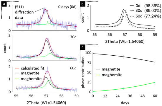
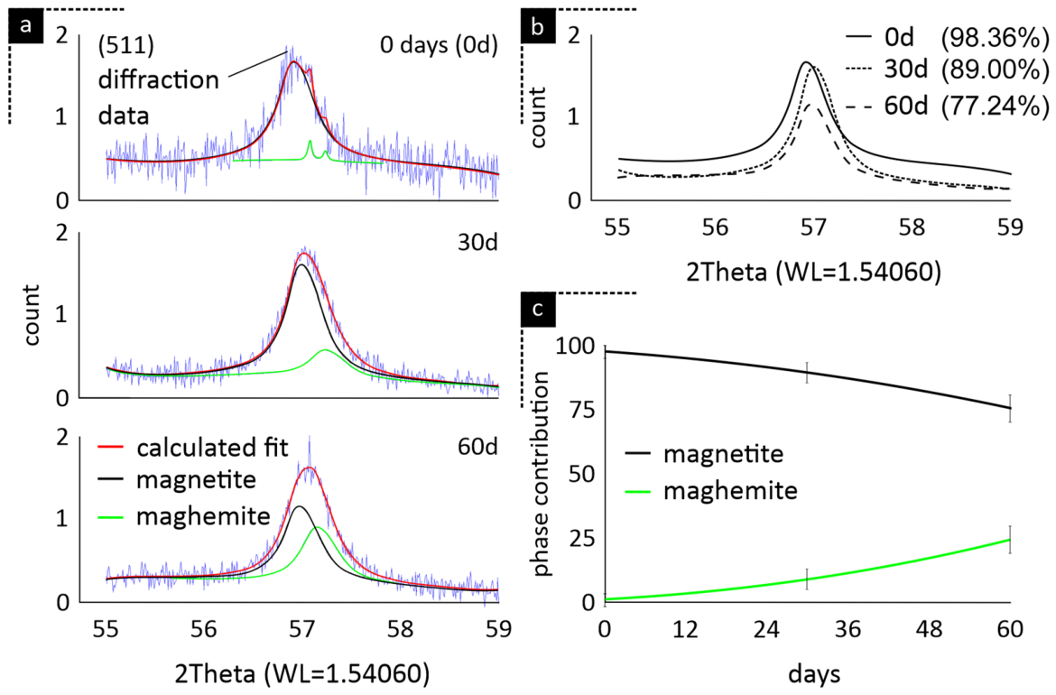
Figure 4a illustrates the progressive increase in the maghemite contribution within the refined (511) region from Day 0 to Day 60. The calculated profiles closely followed the experimental diffraction data at each time point, while the increasing intensity and broadening of the maghemite contribution were accompanied by a corresponding reduction in the magnetite contribution. This progressive phase evolution is further summarized in Figure 4b and Figure 4c, which show the decline in magnetite content and the concurrent increase in maghemite during the 60-day aging period.
Figure 4.
Hybrid TOPAS Rietveld refinement analysis of oxidation kinetics, indicatively shown for the 18h40 sample series: (a) Localized refinement of the (511) diffraction region (55–59° 2θ) at 0, 30, and 60 days, showing the experimental diffraction data, calculated fit, and the deconvoluted magnetite and maghemite phase contributions, (b) progressive reduction of the magnetite contribution within the (511) reflection during ambient aging, and (c) evolution of the relative magnetite and maghemite phase contributions as a function of storage time. Error bars represent the estimated standard deviations obtained from the TOPAS refinement.
The progressive evolution of the fitted XRD profiles provided support for the quantitative phase analysis. At Day 0, the maghemite contribution was limited, consistent with minor surface oxidation during sample preparation and handling. After 30 and 60 days of ambient exposure, the maghemite contribution progressively increased and broadened, accompanied by a corresponding reduction in the magnetite contribution. These changes are consistent with the progressive conversion of the magnetite core into an oxidized maghemite-rich surface fraction. The evolution of the fitted profiles in Figure 4a therefore complements the quantitative changes in magnetite and maghemite content presented in Figure 4b,c.
The estimated standard deviations associated with the refined maghemite fractions were approximately ±3 percentage points at day 0, ±7 percentage points at day 30, and ±13 percentage points at day 60, reflecting the uncertainty of the individual refinement-derived phase estimates rather than variability among independent replicate measurements. Accordingly, the absolute phase fraction obtained at each individual time point, particularly the low maghemite contribution at day 0, should be interpreted with appropriate caution. Importantly, however, the primary outcome of the aging analysis is not the absolute phase fraction at any single time point, but the temporal evolution of the relative phase contributions. Across the 60-day observation period, the refinements consistently indicate a progressive increase in the maghemite contribution accompanied by a corresponding decrease in magnetite, demonstrating a clear directional evolution toward oxidation. This time-dependent pattern is further supported by the independent VSM measurements, which showed a reduction in saturation magnetization after 60 days of aging, consistent with the same progressive magnetite-to-maghemite transformation.
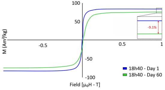
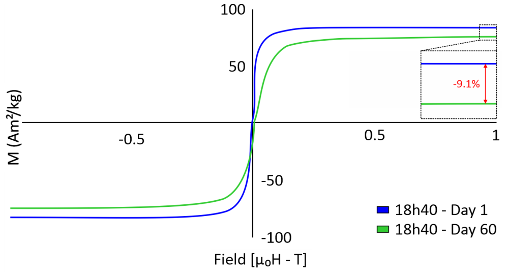
To evaluate whether this structural oxidation was accompanied by a corresponding change in magnetic performance, the 18h40 particles were additionally characterized by VSM one day after synthesis and again after the degradation period of 60 days (Figure 5). This longer-term comparison extends the XRD-based oxidation analysis and directly evaluates the functional consequences of prolonged ambient exposure on the magnetic response of the particles.
Figure 5.
Room-temperature field-dependent magnetization curves of the 18h40 sample measured one day after synthesis and after 60 days of degradation, demonstrating a 9.1% reduction in saturation magnetization associated with progressive oxidation.
VSM analysis revealed a 9.1% reduction in saturation magnetization (Ms) for the 18h40 particles between Day 1 and Day 60 of ambient-air exposure (Figure 5). Magnetite-rich Fe3O4 particles typically exhibit room-temperature saturation magnetization values approaching approximately 86 Am2/kg [31] for the bulk form and lower for nanoparticles (e.g., 50–80 Am2/kg etc.) [32], whereas oxidized, maghemite-rich γ-Fe2O3 systems are commonly associated with lower values near 74 Am2/kg for the bulk form and much lower values for nanoparticles [32], owing to the oxidation of Fe2+ to Fe3+ and the accompanying formation of cation vacancies within the spinel structure. The 9.1% macroscopic magnetic loss observed via VSM was consistent with the direction of the phase evolution detected by XRD refinement, which calculated a corresponding increase in the maghemite fraction from 1.64 to 22.76 wt.% over the same period. While previous studies have utilized magnetization curves to indirectly model magnetite/maghemite core–shell compositions [32], pairing VSM with high-resolution Rietveld deconvolution provides complementary structural and magnetic evidence consistent with progressive oxidation. Taken together, these structural and magnetic findings quantitatively demonstrate how progressive ambient oxidation compromises functional performance, further supporting sealed storage as an effective means of limiting oxidation and preserving the pristine magnetite core.
The present approach builds upon the step-scan deconvolution methodology originally introduced by Kim et al. [21], who validated the use of the overlapping (511) reflection for magnetite–maghemite quantification using standard mixtures of known composition. In that study, mixtures containing 0–100 wt.% maghemite were analyzed in triplicate, yielding a highly linear calibration relationship (R2 = 0.9941) between the maghemite intensity fraction and the known phase composition. The present work extends this concept by replacing empirical pseudo-Voigt peak integration and external calibration with direct crystallographic-model-based refinement of the overlapping phases in TOPAS. The good agreement between calculated and experimental profiles, together with the consistent decrease in saturation magnetization observed by VSM during aging, supports the ability of the method to track relative phase evolution. Nevertheless, because the present crystallographic-model-based implementation was not independently calibrated against newly prepared reference mixtures or Mössbauer spectroscopy, the reported phase fractions should be interpreted as refinement-based estimates, and future validation would be valuable for establishing absolute accuracy and method-specific detection limits.
3.5. Implications for Core–Shell Applications
The particle characteristics obtained in the present study may be relevant to electromagnetic-wave absorption both as independently employed magnetite particles and as magnetic cores in Fe3O4-based core–shell structures. Submicron magnetite particles can contribute to microwave attenuation through magnetic-loss mechanisms, while incorporation of an appropriate dielectric or conductive shell may additionally promote interfacial polarization and dielectric losses and improve impedance matching between the absorber and free space [13,14]. In this context, the 18 h synthesis conditions are of interest because they produced particles close to the predefined ~250 nm size range while maintaining relatively high saturation magnetization values of approximately 80–85 A·m2 kg−1. Such a combination of controlled submicron dimensions and strong magnetic response may provide a suitable magnetic-core platform for subsequent shell engineering, where the overall electromagnetic response can be further tuned through the composition, thickness, and interfacial characteristics of the shell [8,13].
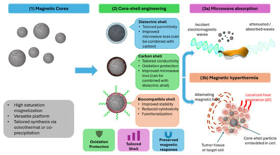
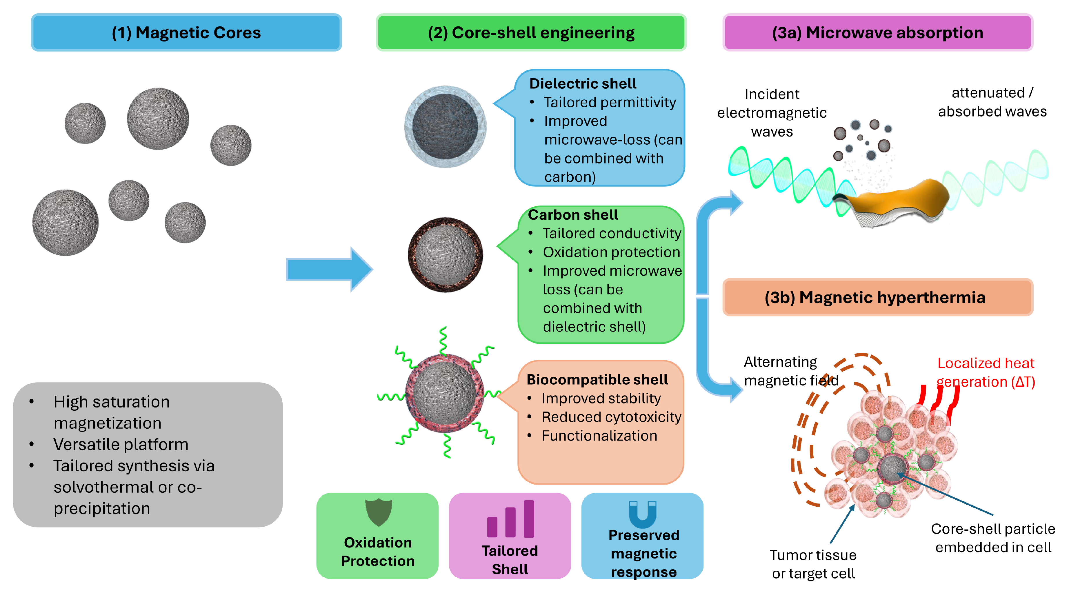
For biomedical applications such as magnetic hyperthermia, the quasi-spherical submicron particles observed by SEM may also be of interest, particularly if they comprise multicore assemblies of smaller magnetite crystallites. Such assemblies can exhibit cooperative magnetic behavior [16] and enhanced heat generation under alternating magnetic fields [12]. SEM alone does not establish whether the synthesized particles are single crystals or multicore assemblies, and their heating performance cannot be inferred from morphology and saturation magnetization alone. Nevertheless, the combination of controlled particle dimensions and strong magnetic response supports consideration of these particles, either independently or following suitable surface functionalization, for application-specific evaluation [11,12] as shown in Figure 6.
Figure 6.
Schematic representation of potential application pathways for the synthesized Fe3O4 particles, including independent use and further functionalization through core–shell engineering for electromagnetic-wave absorption and magnetic hyperthermia.
Overall, the present findings identify the 18 h magnetite cores as a versatile platform with potential relevance to electromagnetic-wave absorption and, subject to application-specific requirements, magnetic hyperthermia, either independently or as components of core–shell architectures.
4. Conclusions
This study demonstrates that solvothermal duration and autoclave filling factor can be used to modulate the size of quasi-spherical, magnetite-rich particles while maintaining a strong magnetic response across the evaluated synthesis conditions. The 18 h series showed particular promise, producing particles approaching the targeted 250 nm size domain without the additional processing time required by the 24 h protocols. Within this series, the 18h40 condition yielded a mean particle diameter of approximately 243 nm and was comparable to the 24h50 and 24h60 conditions, supporting its selection as a representative formulation for further investigation. Localized TOPAS refinement additionally enabled quantitative monitoring of progressive magnetite-to-maghemite oxidation, with the maghemite fraction increasing from 1.36 to 22.76 wt.% over 60 days of repeated ambient exposure and sample preparation, accompanied by a 9.1% reduction in saturation magnetization. Conversely, the limited maghemite content observed after 60 days of sealed storage highlights the importance of controlled handling and storage in preserving the magnetite-rich phase. These results should, however, not be interpreted as establishing an application-ready or universally preferable core system, as further work is required to evaluate coating compatibility, interfacial stability, and post-coating magnetic performance.
Nevertheless, the combination of controlled submicron dimensions, strong magnetic response, and measurable oxidation behavior provides a useful basis for further development of tailored core–shell systems for electromagnetic and biomedical applications.
Author Contributions
Conceptualization, D.T. and A.T.; methodology, D.T., A.M. and A.T.; software, D.T. and A.M.; validation, D.T., M.A. and A.T.; investigation, D.T. and A.M.; resources, M.A. and A.T.; writing—original draft preparation, D.T. and A.M.; writing—review and editing, M.A. and A.T.; visualization, D.T. and A.T.; supervision, M.A. and A.T.; project administration, A.T. All authors have read and agreed to the published version of the manuscript.
Funding
This research received no external funding.
Institutional Review Board Statement
Not applicable.
Informed Consent Statement
Not applicable.
Data Availability Statement
Data is contained within the article.
Conflicts of Interest
The authors declare no conflicts of interest.
References
- Lehner, R.; Wang, X.; Marsch, S.; Hunziker, P. Intelligent Nanomaterials for Medicine: Carrier Platforms and Targeting Strategies in the Context of Clinical Application. Nanomedicine 2013, 9, 742–757. [Google Scholar] [CrossRef] [Scilit] [PubMed]
- Yu, Y.; Tu, S.; Zhu, Z.; Ma, T.; Hwang, S.; Huang, H. Metal Single-Atom Materials: Unlocking the Future of Sustainable Energy and Environment in Automobiles. Adv. Mater. 2025, 37, e2507831. [Google Scholar] [CrossRef] [Scilit] [PubMed]
- Vignesh, J.; Ramesh, B.; Xavier, J.R. Recent Advances in Nano-Engineered Cement Composites for Sustainable and Smart Infrastructure. Nanotechnol. Environ. Eng. 2025, 10, 81. [Google Scholar] [CrossRef] [Scilit]
- Tsamos, D.; Artemiou, P.; Fasnakis, D.; Loizos, D.; Gerardis, S.; Maropoulos, S. Optimizing the Annealing Process of Sol–Gel Hybrid Coating Technology for Corrosion Barrier Protection of Al. J. Coat. Technol. Res. 2026, 23, 569–578. [Google Scholar] [CrossRef] [Scilit]
- Lu, A.; Salabas, E.L.; Schüth, F. Magnetic Nanoparticles: Synthesis, Protection, Functionalization, and Application. Angew. Chem. Int. Ed. 2007, 46, 1222–1244. [Google Scholar] [CrossRef] [Scilit] [PubMed]
- Pinakidou, F.; Simeonidis, K.; Myrovali, E.; Brzhezinskaya, M.; Paloura, E.C.; Angelakeris, M.; Katsikini, M. Addressing the Effect of Magnetic Particle Hyperthermia Application on the Composition and Spatial Distribution of Iron Oxide Nanoparticles Using X-Ray Spectroscopic Techniques. J. Phys. Chem. C 2022, 126, 10101–10109. [Google Scholar] [CrossRef] [Scilit]
- Kang, E.; Jung, Y.S.; Cavanagh, A.S.; Kim, G.-H.; George, S.M.; Dillon, A.C.; Kim, J.K.; Lee, J. Fe3O4 Nanoparticles Confined in Mesocellular Carbon Foam for High Performance Anode Materials for Lithium-Ion Batteries. Adv. Funct. Mater. 2011, 21, 2430–2438. [Google Scholar] [CrossRef] [Scilit]
- Wang, T.; Liu, Z.; Lu, M.; Wen, B.; Ouyang, Q.; Chen, Y.; Zhu, C.; Gao, P.; Li, C.; Cao, M.; et al. Graphene–Fe3O4 Nanohybrids: Synthesis and Excellent Electromagnetic Absorption Properties. J. Appl. Phys. 2013, 113, 024314. [Google Scholar] [CrossRef] [Scilit]
- Wu, Z.-S.; Yang, S.; Sun, Y.; Parvez, K.; Feng, X.; Müllen, K. 3D Nitrogen-Doped Graphene Aerogel-Supported Fe3O4 Nanoparticles as Efficient Electrocatalysts for the Oxygen Reduction Reaction. J. Am. Chem. Soc. 2012, 134, 9082–9085. [Google Scholar] [CrossRef] [Scilit] [PubMed]
- Ghosh Chaudhuri, R.; Paria, S. Core/Shell Nanoparticles: Classes, Properties, Synthesis Mechanisms, Characterization, and Applications. Chem. Rev. 2012, 112, 2373–2433. [Google Scholar] [CrossRef] [Scilit] [PubMed]
- Myrovali, E.; Maniotis, N.; Samaras, T.; Angelakeris, M. Spatial Focusing of Magnetic Particle Hyperthermia. Nanoscale Adv. 2020, 2, 408–416. [Google Scholar] [CrossRef] [Scilit] [PubMed]
- Martinez-Boubeta, C.; Simeonidis, K.; Makridis, A.; Angelakeris, M.; Iglesias, O.; Guardia, P.; Cabot, A.; Yedra, L.; Estradé, S.; Peiró, F.; et al. Learning from Nature to Improve the Heat Generation of Iron-Oxide Nanoparticles for Magnetic Hyperthermia Applications. Sci. Rep. 2013, 3, 1652. [Google Scholar] [CrossRef] [Scilit] [PubMed]
- Du, Y.; Liu, W.; Qiang, R.; Wang, Y.; Han, X.; Ma, J.; Xu, P. Shell Thickness-Dependent Microwave Absorption of Core–Shell Fe3O4@C Composites. ACS Appl. Mater. Interfaces 2014, 6, 12997–13006. [Google Scholar] [CrossRef] [Scilit] [PubMed]
- Ni, S.; Lin, S.; Pan, Q.; Yang, F.; Huang, K.; He, D. Hydrothermal Synthesis and Microwave Absorption Properties of Fe3O4 Nanocrystals. J. Phys. D Appl. Phys. 2009, 42, 055004. [Google Scholar] [CrossRef] [Scilit]
- Rezaei, B.; Yari, P.; Sanders, S.M.; Wang, H.; Chugh, V.K.; Liang, S.; Mostufa, S.; Xu, K.; Wang, J.; Gómez-Pastora, J.; et al. Magnetic Nanoparticles: A Review on Synthesis, Characterization, Functionalization, and Biomedical Applications. Small 2024, 20, e2304848. [Google Scholar] [CrossRef] [Scilit] [PubMed]
- Belew, A.A.; Assege, M.A. Solvothermal Synthesis of Metal Oxide Nanoparticles: A Review of Applications, Challenges, and Future Perspectives. Results Chem. 2025, 16, 102438. [Google Scholar] [CrossRef] [Scilit]
- Kim, J.; Tran, V.T.; Oh, S.; Kim, C.-S.; Hong, J.C.; Kim, S.; Joo, Y.-S.; Mun, S.; Kim, M.-H.; Jung, J.-W.; et al. Scalable Solvothermal Synthesis of Superparamagnetic Fe3O4 Nanoclusters for Bioseparation and Theragnostic Probes. ACS Appl. Mater. Interfaces 2018, 10, 41935–41946, Correction in ACS Appl. Mater. Interfaces 2019, 11, 18885. https://doi.org/10.1021/acsami.9b07059. [Google Scholar] [CrossRef] [Scilit] [PubMed]
- Wang, X.; Zhuang, J.; Peng, Q.; Li, Y. A General Strategy for Nanocrystal Synthesis. Nature 2005, 437, 121–124. [Google Scholar] [CrossRef] [Scilit] [PubMed]
- Lv, H.; Xue, Y.; Feng, X.; Wang, H.; Zheng, M.; Zhang, B. One-Step Synthesisof Highly Uniform Fe3O4@C Nanospheres as Anode Materials for LIBs. Int. J. Electrochem. Sci. 2020, 15, 2157–2165. [Google Scholar] [CrossRef] [Scilit]
- Schwaminger, S.P.; Bauer, D.; Fraga-García, P.; Wagner, F.E.; Berensmeier, S. Oxidation of Magnetite Nanoparticles: Impact on Surface and Crystal Properties. CrystEngComm 2017, 19, 246–255. [Google Scholar] [CrossRef] [Scilit]
- Kim, W.; Suh, C.-Y.; Cho, S.-W.; Roh, K.-M.; Kwon, H.; Song, K.; Shon, I.-J. A New Method for the Identification and Quantification of Magnetite–Maghemite Mixture Using Conventional X-Ray Diffraction Technique. Talanta 2012, 94, 348–352. [Google Scholar] [CrossRef] [Scilit] [PubMed]
- Andersen, H.L.; Frandsen, B.A.; Gunnlaugsson, H.P.; Jørgensen, M.R.V.; Billinge, S.J.L.; Jensen, K.M.Ø.; Christensen, M. Local and Long-Range Atomic/Magnetic Structure of Non-Stoichiometric Spinel Iron Oxide Nanocrystallites. IUCrJ 2021, 8, 33–45. [Google Scholar] [CrossRef] [Scilit] [PubMed]
- Kinebuchi, I.; Kyono, A. Study on Magnetite Oxidation Using Synchrotron X–Ray Diffraction and X–Ray Absorption Spectroscopy: Vacancy Ordering Transition in Maghemite (γ–Fe2O3). J. Mineral. Petrol. Sci. 2021, 116, 210304. [Google Scholar] [CrossRef] [Scilit]
- Furlan, P.Y.; Furlan, A.Y.; Kisslinger, K.; Melcer, M.E.; Shinn, D.W.; Warren, J.B. Water as the Solvent in the Stober Process for Forming Ultrafine Silica Shells on Magnetite Nanoparticles. ACS Sustain. Chem. Eng. 2019, 7, 15578–15584. [Google Scholar] [CrossRef] [Scilit]
- Chen, M.; Shao, L.-L.; Li, J.-J.; Pei, W.-J.; Chen, M.-K.; Xie, X.-H. One-Step Hydrothermal Synthesis of Hydrophilic Fe3O4/Carbon Composites and Their Application in Removing Toxic Chemicals. RSC Adv. 2016, 6, 35228–35238. [Google Scholar] [CrossRef] [Scilit]
- Huang, W.; Wang, Y.; Wei, S.; Wang, B.; Liang, Y.; Huang, Y.; Xu, B. Effect of Reaction Time on Microwave Absorption Properties of Fe3O4 Hollow Spheres Synthesized via Ostwald Ripening. Materials 2019, 12, 2921. [Google Scholar] [CrossRef] [Scilit] [PubMed]
- Huo, Y.; Xiu, S.; Meng, L.-Y.; Quan, B. Solvothermal Synthesis and Applications of Micro/Nano Carbons: A Review. Chem. Eng. J. 2023, 451, 138572. [Google Scholar] [CrossRef] [Scilit]
- Fievet, F.; Lagier, J.P.; Figlarz, M. Preparing Monodisperse Metal Powders in Micrometer and Submicrometer Sizes by the Polyol Process. MRS Bull. 1989, 14, 29–34. [Google Scholar] [CrossRef] [Scilit]
- LaMer, V.K.; Dinegar, R.H. Theory, Production and Mechanism of Formation of Monodispersed Hydrosols. J. Am. Chem. Soc. 1950, 72, 4847–4854. [Google Scholar] [CrossRef] [Scilit]
- Whitehead, C.B.; Özkar, S.; Finke, R.G. LaMer’s 1950 Model of Particle Formation: A Review and Critical Analysis of Its Classical Nucleation and Fluctuation Theory Basis, of Competing Models and Mechanisms for Phase-Changes and Particle Formation, and Then of Its Application to Silver Halide, Semiconductor, Metal, and Metal-Oxide Nanoparticles. Mater. Adv. 2021, 2, 186–235. [Google Scholar] [CrossRef] [Scilit]
- Unni, M.; Uhl, A.M.; Savliwala, S.; Savitzky, B.H.; Dhavalikar, R.; Garraud, N.; Arnold, D.P.; Kourkoutis, L.F.; Andrew, J.S.; Rinaldi, C. Thermal Decomposition Synthesis of Iron Oxide Nanoparticles with Diminished Magnetic Dead Layer by Controlled Addition of Oxygen. ACS Nano 2017, 11, 2284–2303. [Google Scholar] [CrossRef] [Scilit] [PubMed]
- Sharifi Dehsari, H.; Ksenofontov, V.; Möller, A.; Jakob, G.; Asadi, K. Determining Magnetite/Maghemite Composition and Core–Shell Nanostructure from Magnetization Curve for Iron Oxide Nanoparticles. J. Phys. Chem. C 2018, 122, 28292–28301. [Google Scholar] [CrossRef] [Scilit]

Disclaimer/Publisher’s Note: The statements, opinions and data contained in all publications are solely those of the individual author(s) and contributor(s) and not of MDPI and/or the editor(s). MDPI and/or the editor(s) disclaim responsibility for any injury to people or property resulting from any ideas, methods, instructions or products referred to in the content. |
© 2026 by the authors. Licensee MDPI, Basel, Switzerland. This article is an open access article distributed under the terms and conditions of the Creative Commons Attribution (CC BY) license.