


Abstract
Aflatoxin B1 (AFB1) is a highly toxic mycotoxin which can be carried over into animal products and cause deterioration of livestock productivity when fed to livestock and wildlife. This review proposes a biomarker-guided framework for improving the early assessment of AFB1 exposure and toxicological responses in animal models. Oral exposure leads to the absorption of AFB1, which is bioactivated in the liver to the reactive AFB1-exo-8,9-epoxide that causes DNA and protein adduct formation, inflammation, mitochondrial apoptosis, and other effects. Cytochrome P450 activation and glutathione-dependent detoxification are in balance in determining susceptibility species, and this balance is different for poultry, pigs, ruminants, and rodents. In addition to traditional liver enzymes and histopathology, we highlight mechanistically informative biomarkers such as metabolites of aflatoxin, DNA and albumin adduct, lipid peroxidation products, antioxidant indices, cytokines, apoptotic markers, as well as signals involved in the Nrf2/NFκB pathway. AFB1 also damages the integrity of the intestinal barrier, the maintenance of the intestinal gut microbiota, reproductive function, growth performance, and development, thus creating a gut–liver-systemic toxic cascade. Finally, an assessment of stage-targeted interventions such as aluminosilicate binders, adsorbents derived from yeast, probiotics and nano-enabled interventions is conducted as viable tools for the reduction in exposure and injury. This review offers targeted mitigation strategies for early diagnosis of aflatoxicosis in animal production systems based on a biomarker approach.
Key Contribution:
This review establishes a biomarker-guided framework linking AFB1-DNA/albumin adducts, oxidative stress, inflammatory signaling, and Nrf2/NF-κB pathways to species-specific susceptibility, revealing the gut–liver-systemic cascade as a central pathological axis. By systematically comparing biotransformation capacities across poultry, pigs, ruminants, and rodents, this approach provides a translational foundation for species-selective biomarker application and stage-targeted mitigation of aflatoxicosis.
1. Introduction
Aflatoxin contamination, primarily caused by Aspergillus spp, is a global food and feed-safety problem that poses public health and agricultural threats, particularly in tropical, subtropical and now also temperate regions [1,2]. Survey and meta-analytical data from decades of research show that levels of aflatoxins in processed products and in major staple crops, such as maize, groundnuts, sorghum and millet, have routinely exceeded regulatory limits for chronic exposure to humans and food-producing animals via dietary and contaminated feed chains, respectively [3].The threat is not static: the frequency and magnitude of climatic stressors such as high temperature, altered precipitation patterns, and droughts, which can make plants more susceptible to Aspergillus colonization and enable the production of mycotoxins, may increase [4], while the proportion of high-level contamination events occurs in areas which were previously classified as low-risk areas is likely to increase as a result of climate change [5]. Similar dynamics have been observed in the international assessments and sectoral analysis linking the future climate scenarios with the heightened aflatoxin risk to key food crops (particularly maize and groundnut) and the need to adapt to climate change in the value chain throughout the entire value chain [6,7]. Feed contamination affects animal production directly by inducing immunosuppression and making the animals more susceptible to infectious diseases by causing reproductive failure and by causing high morbidity/mortality in severe cases [5,8]. These biological effects can be monetized using the losses in production, increased veterinary costs, and the value of market rejection [9]. Animal-product (meat, milk, offal, eggs) surveillance confirms that carry-over and residues are present in real-world feeding conditions, and present both animal-health and downstream food-safety risks [10,11]. Aflatoxins have also been associated with impaired cell-mediated immune suppression and stunted growth in highly exposed populations after chronic exposure [12]. This also makes the problem One Health, as the contamination of the feed with AFB1 affects animal health, and can be passed on through food chains, creating a risk for human food safety and public health [13]. Aflatoxin is a target for regulatory harmonization, investment in rapid detection and biomarker development, cross-sectoral agriculture–veterinary–public health mitigation and is endemic in the environment, which is expected to become more of a problem under the increasing stresses of climate change. AFB1 has been widely reported to be the most toxic and mutagenic aflatoxin among the naturally occurring aflatoxins and the relative sequence of toxicity is AFB1 > AFG1 > AFB2 > AFG2 [14,15,16]. The hydroxylated form of AFB1 (AFM1) that can be excreted into milk is less toxic but is also a carcinogen and a public health concern as it can be exposed to humans through dairy products [17,18,19]. The International Agency on Research on Cancer (IARC) classifies AFB1 in the Group 1 category of human carcinogens, meaning there is high confidence that chronic exposure to AFB1 increases the risk of hepatocellular carcinoma [20]. Apart from cancer, chronic exposure to aflatoxins has been reported to adversely affect child development and immune function of exposed populations, further highlighting its importance as a food-safety and One Health issue [21]. There are interspecies differences in the susceptibility of livestock species to the toxicity of AFB1, which result from differences in hepatic enzymes that bioactivate AFB1, phase II enzymes that detoxify AFB1, rumen microbial metabolism, and antioxidant defenses [22]. Ducks are extremely sensitive to AFB1 as they readily convert it to the highly reactive epoxide form and have a relatively weak detoxification system, resulting in liver damage, immune system suppression, growth inhibition, and an increased incidence of deaths at higher exposures [23,24]. Swine are also susceptible, exposure has been associated with decreased feed efficiency and growth, impaired immune response, hepatobiliary metabolic changes and susceptibility to secondary infections [25,26]. Ruminants are, however, partially protected because some of the toxins are bio-transformed by ruminal microorganisms and some are bound by digesta but chronic exposure can reduce productivity and damage the liver. Importantly, AFB1 can be bio-transformed to AFM1 and released into milk, creating a serious potential food-safety risk [11,27]. Rodent models, particularly rats and mice, are the most commonly used experimental models for investigating AFB1 toxicity, because they have high and reproducible sensitivity to bioactivation of AFB1 and hepatocarcinogenesis, which is helpful for mechanistic research and chemoprevention studies [28,29,30]. AFB1 is the most important toxicological problem of aflatoxins, because it has the highest carcinogenicity, is able to undergo metabolic activation to form DNA-reactive metabolites, and there are interspecies differences in the metabolic activation and detoxification of AFB1 [31]. These mechanistic and species-specific variations are the key to proper risk assessment, biomarker development, and effective mitigation strategies in livestock production systems [32]. The significance of early warning of aflatoxin B1 (AFB1) exposure lies in the fact that the conventional approaches mainly verify the damage when the toxic harm has already been done, but biomarker science is in its infancy to improve early detection and mitigation assessment [32,33]. Veterinary practice assessment is typically done using feed analysis, clinical signs, serum liver enzymes (ALT and AST), and histopathology, although feed testing can be inaccurate because of aflatoxin distribution inequality in feed batches, and sampling error is a major cause of error [34]. ALT and AST are usually elevated once hepatic damage has already been done, and histopathology only confirms hepatocellular damage once tissues have been harvested, making these methods helpful in confirmation but not in early screening or screening at the herd level [35]. Recent advances in multi-omics have therefore shifted biomarker discovery toward earlier and more mechanistic signals: metabolomics has revealed perturbations in amino acid metabolism, lipid oxidation, and bile-acid-related pathways; transcriptomic and proteomic studies have identified xenobiotic-response, detoxification, mitochondrial, and liver-development changes, and miRNA profiling is increasingly being explored as a complementary early indicator of AFB1 toxicity [36,37]. Another promising frontier is the microbiome: AFB1 can alter gut microbial composition and intestinal barrier integrity, and fecal microbiota profiling has already been proposed as a non-invasive approach for screening exposure and resilience in exposed animals [38,39]. A paradigm shift in conventional diagnostics to biomarker-based diagnosing is a shift towards precision livestock health management. By diagnosing at an early stage, feed can be detoxified, nutritional supplements provided, and the microbiome regulated before irreversible tissue damage occurs. The present review helps to create a mechanism-based, species-specific model of aflatoxin B1 (AFB1) toxicity by integrating biomarkers, molecular pathways, and systemic effects between the species. It extends beyond classical definitions of contamination or hepatotoxicity and combines exposure biomarkers, oxidative stress, inflammation, and apoptotic signaling into a unified toxicological cascade. It is important to note that new interventions, including adsorbents, probiotics, nano-enabled systems, and multimodal strategies, are superimposed to discrete phases of this cascade, which provides a translational and stage-selective solution to reduce aflatoxicosis in animal production systems.
Literature Search Strategy and Study Selection
This article was conducted as a narrative review with a structured literature search to comprehensively summarize current evidence on aflatoxin B1 (AFB1)-induced toxicity, biomarker discovery, systemic toxicological effects, and mitigation strategies in animal models. Although a formal systematic review protocol was not applied, the literature search and study selection were performed using predefined criteria to improve transparency and reproducibility, consistent with recommendations for high-quality narrative reviews [40].
A comprehensive search was conducted in PubMed, Web of Science Core Collection, Scopus, and Google Scholar for studies published from database inception to March 2026. Additional relevant publications were identified through manual screening of reference lists from key review articles and highly cited original studies. The search strategy combined medical subject headings (MeSHs) and free-text terms using Boolean operators, including “Aflatoxin B1” OR “AFB1”, “animal models”, “livestock”, “poultry”, “ruminants”, “swine”, “rodents”, “biomarkers”, “DNA adducts”, “albumin adducts”, “oxidative stress”, “inflammation”, “hepatotoxicity”, “gut–liver axis”, “mitigation”, “adsorbents”, “probiotics”, and “mycotoxin detoxification” [41].
Peer-reviewed original research articles, controlled animal experiments, mechanistic studies, comparative toxicology investigations, and authoritative review articles published in English were considered eligible. Studies were included if they investigated AFB1 exposure in livestock or laboratory animal models and reported toxicokinetic characteristics, molecular mechanisms, biomarkers of exposure or toxicity, pathological outcomes, or mitigation strategies. Studies focusing exclusively on other mycotoxins, lacking sufficient methodological information, conference abstracts, editorials, duplicate publications, or non-peer-reviewed reports were excluded.
Titles and abstracts were initially screened for relevance, followed by full-text assessment of potentially eligible articles. Data extracted from the selected studies included animal species, experimental model, AFB1 dose and exposure duration, biological matrix, analytical methods, biomarkers evaluated, mechanistic pathways, toxicological outcomes, and mitigation approaches. The studies summarized in tables were selected based on their scientific quality, methodological rigor, relevance to the review objectives, and their contribution to comparative interpretation across animal species. Owing to the substantial heterogeneity of experimental designs, animal species, exposure conditions, and outcome measures, findings were synthesized qualitatively without formal meta-analysis or risk-of-bias assessment.
2. Chemical Structure and Physicochemical Properties of Aflatoxin B1 (AFB1)
Aspergillus flavus and Aspergillus parasiticus primarily produce aflatoxin B1 (AFB1), a very toxic secondary metabolite, which is a major contaminant of crops and feed in warm and damp climates [42]. Its molecular formula is C17H12O6 and its molecular weight is approximately 312.27 g/mol, reflecting a relatively small but highly reactive molecule [43,44]. AFB1 is a difuranocoumarin, structurally a coumarin (benzopyrone) nucleus containing a bifuran moiety, a planar and highly conjugated aromatic system [45]. The fused-ring structure is known to stabilize the compound physicochemically and to promote the interaction with biological macromolecules, including DNA and proteins [46]. The terminal furan ring (and the C8C9 double bond in particular) is the major toxicological determinant and the major site of bioactivation by the cytochrome P450 [47]. These structural features cause AFB1 to be regarded as the strongest mutagenic and carcinogenic aflatoxin and a Group 1 human carcinogen [46]. Its high fluorescence is also linked to the intact lactone-containing structure, commonly utilized in the analysis of the structure, and typically seen at excitation wavelengths of 365 nm [48]. From a physicochemical standpoint, AFB1 is sparingly soluble in water but readily soluble in polar organic solvents such as methanol, chloroform, and acetone [49]. Its logP is about 1.60, which is moderately lipophilic, and thus it will diffuse passively across the membrane and be absorbed by the gastrointestinal tract easily [50]. AFB1 is also bound by plasma proteins, e.g., albumin, and this influences its systemic distribution and retention in the circulation [51]. The molecule is quite stable over a broad pH range; but in highly alkaline environments, lactone-ring hydrolysis/opening is induced to produce less toxic derivatives [52]. Photochemical degradation of AFB1 is possible under UV light, but the rate of degradation is extremely dependent on irradiation intensity, wavelength, matrix structure, and exposure time, so UV degradation is generally a controlled-laboratory or surface-treatment technology, rather than a large-scale agricultural detoxification technology [53]. Non-ruminant systems have limited enzymatic detoxification of AFB1, but the rumen partially bio-transforms the toxin, though not completely and inconsistently [54]. AFB1 belongs to the B-series of aflatoxins, which is blue in the UV light, and G-series of aflatoxins, which is green in the UV light, because of the differences in chemical structure and oxygenation pattern [55]. Among the naturally occurring aflatoxins, AFB1 is the most toxic since its structure is ideally positioned to be metabolically activated, bind DNA and persist, all of which help to make it highly mutagenic and carcinogenic [56]. Major change endpoints can alter toxic potency: AFM1 hydroxylation can alter its activity and allow milk carry-over, and saturation of the double bond in AFB2 is more likely to decrease carcinogenic potential, which is a clear structure–activity relationship in aflatoxin toxicology [31]. In addition to milk, AFM1 and aflatoxin residues in eggs represent a potential food safety concern and should be considered when evaluating carry-over in animal-derived foods [57].
3. AFB1 Toxicokinetic and Bioactivation in Animals
The major route of exposure of AFB1 in animals is by consuming contaminated feed, and oral exposure is the most common route of exposure in poultry, pigs, and dairy cattle [26,58,59]. AFB1 is easily absorbed across the intestinal mucosa of monogastric animals largely due to its small size and it being lipophilic, and thus is readily absorbed across the intestinal mucosa of monogastric species such as pigeon, rat, sheep, and goat [23,60]. Following intestinal absorption, AFB1 enters the portal circulation and is delivered to the liver, where first-pass biotransformation becomes the critical determinant of systemic toxicity in animals [15]. Part of the absorbed toxin may also be degraded in the gut before reaching the liver since rumen microorganisms are capable of partially degrading AFB1, although this detoxification is incomplete and differs by species and rumen conditions [61]. In pigs, serum AFB1-lysine adducts are already known as an internal-dose biomarker, and adsorption-based interventions have been shown to lower adduct formation, which confirms their usefulness in monitoring exposure in livestock [62]. Both serum AFB1-albumin adducts and blood AFB1-DNA adducts are being used to assess chronic exposure, further supporting albumin-related adducts as biologically significant exposure-recording events in ruminants [63].
After absorption, AFB1 is subjected to extensive hepatic biotransformation, with the liver being the primary site of phase I activation and phase II detoxification of AFB1 in livestock species [64]. Recent mechanistic studies in cattle indicate that cytochrome P450 systems, in particular CYP1A1 and CYP3A74, are central in AFM1 metabolism and in the regulation of AFM1 formation, AFBO synthesis and cytotoxicity [15]. The products of phase I metabolism include a variety of hydroxylated and related products, such as aflatoxicol, AFM1, AFQ1, and hydrated products including AFB2a, O-demethylated products such as AFP1, reduced products such as aflatoxicol, and the highly reactive epoxide intermediates, AFB1-endo-8,9-epoxide and AFB1-exo-8,9-epoxide [59,65]. The exo-8,9-epoxide is the most important bioactivated metabolite of AFB1 that is responsible for genotoxicity and carcinogenicity. This is the metabolite that is produced via the epoxidation catalyzed by CYP450 and is the major electrophilic intermediate between hepatic metabolism and downstream DNA damage in animals, including bovine hepatocyte models and comparative hepatocyte studies across species [15,66]. The exo-epoxide can easily attack the nucleophilic sites in DNA, proteins, phospholipids, and other cellular macromolecules, thus causing cellular dysfunction, membrane damage, and toxic harm [67]. The alkylation of DNA takes place mostly at the N7 position of guanine, and it results in the formation of the pro-mutagenic AFB1-N7-Gua adduct. This lesion is also a canonical marker of AFB1 exposure, and it has been shown by mechanistic studies of the aflatoxin–DNA reaction chemistry [68]. Since the N7-guanine adduct is unstable, it can be subjected to further processing to form more persistent ring-opened products, particularly AFB1-formamidopyridine (AFB1-FAPY) adducts. The lesions are more enduring in DNA and noteworthy because animal studies show that the lesions are highly mutagenic in vivo [69]. The one major mutational impact of this adduct chemistry is the G-to-T transversion at the third nucleotide of codon 249 of the p53 tumor suppressor gene, which is the paradigm molecular signature of AFB1-related hepatocarcinogenesis. Despite this hotspot having been defined in both studies of human hepatocytes and tumors, it is the best-established downstream readout of the same bioactivation-to-adduct pathway that is also observed in animal mechanistic systems [70]. Detoxification is competing with AFB1 bioactivation, as species selectivity is extremely dependent upon the extent of disparity on the expression and catalytic functionality of the biotransformation enzymes. Recent comparative reviews point out the fact that AFB1 toxicity is not species fixed, but rather it does reflect the efficiency with which an animal partitions the toxin between activation and detoxification routes [22]. The major route of protection is conjugation of AFB1-exo-8,9-epoxide (AFBO) with glutathione (GSH), which is catalyzed by glutathione S-transferases (GSTs), followed by additional processing through the mercapturic acid pathway to more water-soluble excretion products [71]. If this detoxification pathway is compromised, species are at risk because the AFBO may persist long enough to damage cellular macromolecules, causing genotoxicity. This is more so in sensitive animal models where the GST functionalities are compromised or less effective against the reactive epoxide [72,73]. Similar to GSH conjugation, AFBO may also be subjected to epoxide hydrolase-mediated conversion to AFB1-dihydrodiol, which can, in turn, break down to AFB1-dialdehyde [74]. This dialdehyde is biologically significant in that it is a binding molecule of proteins, especially lysine residues in albumins, and thus causes acute toxicity and protein adduct formation. Protein adducts are currently considered as useful internal-dose markers in animal investigations [63]. The balance between activation and detoxification is therefore strongly associated with species resistance. Animals with higher GST capacity or higher rate of clearance of detoxified products are more resistant. This pattern explains why rodents, poultry, pigs, and ruminants differ so markedly in AFB1 susceptibility and biomarker profiles. In pigs, porcine CYP2A19 has recently been shown to play a key role in hepatic AFB1 bioactivation [75]; in poultry, turkey GST studies show deficient AFBO detoxification [73]; in ducks, unusually high hepatic AFB1-dihydrodiol production helps explain their extreme sensitivity [74]; and in mice, rapid detoxification is associated with strain resistance. In general, the phase of absorption and hepatic activation should be considered as the biological bottleneck that defines downstream damage. A combination of passive intestinal uptake, the formation of AFBO under the influence of CYP450, and competing GST-dependent detoxification explain why a combination of early biomarkers of exposure and metabolic stress is especially informative in animal model studies.
Species-specific susceptibility to AFB1 is largely determined by differences in its biotransformation rather than by the toxicant itself. Although the principal metabolic pathways are conserved among vertebrates, substantial interspecies variation exists in the expression and catalytic activity of cytochrome P450 enzymes responsible for AFB1 bioactivation, as well as glutathione S-transferase-mediated detoxification pathways. Consequently, the balance between metabolic activation to the highly reactive AFB1-exo-8,9-epoxide and detoxification differs markedly among animal species, leading to pronounced differences in hepatotoxicity, carcinogenicity, and biomarker responses. Therefore, findings obtained in one species should not be directly extrapolated to another without considering these physiological and metabolic differences [22].
The graphical representation of absorption, hepatic activation, and formation of AFB1-epoxide is shown in Figure 1.
Figure 1.
Mechanistic overview of aflatoxin B1 (AFB1) absorption, hepatic bioactivation, and downstream toxicity in animal species. Following oral ingestion of contaminated feed, AFB1 is absorbed in the small intestine and transported via the portal circulation to the liver, where cytochrome P450 enzymes (e.g., CYP1A1, CYP3A74, CYP2A19) convert it to the reactive AFB1-exo-8,9-epoxide. This metabolite forms DNA (AFB1-N7-Gua, AFB1-FAPY) and protein (albumin, lysine) adducts and contributes to mutational events such as p53 codon 249 transversion. Detoxification occurs via glutathione S-transferase (GST)-mediated conjugation and epoxide hydrolase pathways. The balance between bioactivation and detoxification determines species-specific susceptibility (poultry, pigs, cattle, ducks, mice) and is reflected by circulating biomarkers including AFB1-albumin, AFB1-lysine, and DNA adducts, as well as metabolites such as AFM1 and AFQ1.
4. Biomarkers of AFB1 Exposure and Early Injury
It should be noted that not all biomarkers discussed in this review are uniquely specific to AFB1 exposure. Several biochemical, oxidative stress, inflammatory, and physiological biomarkers (e.g., ALT, AST, MDA, SOD, CAT, GPx, and pro-inflammatory cytokines) are general indicators of tissue injury and may also be altered by other toxicological, nutritional, or environmental stressors. However, the studies included in this review were conducted under controlled experimental conditions, in which AFB1 exposure was the primary experimental variable, thereby minimizing potential confounding factors. Consequently, these biomarkers are discussed as AFB1-associated response biomarkers that provide insight into the biological effects and mechanistic pathways of AFB1 toxicity in animal models, rather than as biomarkers uniquely diagnostic of AFB1 exposure. Exposure-specific biomarkers, such as AFB1–albumin adducts, AFB1–N7-guanine DNA adducts, and AFM1, remain the most specific indicators of AFB1 exposure.
4.1. Exposure Biomarkers: AFB1 Metabolites, DNA Adducts, Albumin Adducts
In animal aflatoxin research, biomarkers are usually more informative than feed contamination data alone because AFB1 biotransformation and susceptibility differ markedly among species [22,26]. The most useful exposure biomarkers are excreted metabolites for recent intake, DNA adducts for biologically effective dose, and albumin/lysine adducts for longer-term integrated exposure [76]. In broiler chickens, AFB1 was identified as an optimal biomarker in plasma and excreta in a detoxifier-efficacy study, and a separate chicken biomarker method found AFB1-N7-Gua only in excreta [77]. AFM1 was detected in both ileal content and excreta and was about five times higher in excreta, indicating that excretion is a major elimination route in this model [78]. For pigs, urinary AFB1 and AFM1 are established short-term exposure biomarkers. A controlled feeding study reported that about 30% of the oral dose was recovered in urine as AFB1 plus AFM1, with an AFB1-to-AFM1 ratio of about 23%, and later pig biomarker reviews reported urinary recoveries of 20–48% depending on dose and sampling conditions [79,80].
AFB1-lysine adducts are formed from albumin-bound aflatoxin adducts after enzymatic digestion, so they represent a longer-integrated exposure marker than free urinary metabolites [32]. In pigs, AFB1-lysine has been validated in serum and plasma by LC-MS/MS as a biomarker of AFB1 exposure [81]. In dairy cattle, aflatoxin-albumin adducts in blood have been proposed as a marker of chronic aflatoxin exposure and were compared with AFM1 in milk [82]. A cattle study in 2025 further expanded the biomarker approach by evaluating serum AFB1-albumin adducts together with blood AFB1-DNA adducts for assessment of individual chronic exposure [63]. Because ruminal degradation of AFB1 varies with species, feed type, and ruminal conditions, internal-dose biomarkers are still needed in cattle rather than relying on feed contamination alone [54,61].
DNA adducts are the most mechanistically informative exposure biomarkers because AFB1 is bioactivated to the reactive exo-8,9-epoxide, which preferentially forms N7-guanine adducts in DNA [83,84]. In rats, urinary AFB1-N7-Gua has been validated as a biomarker of internal aflatoxin dose and liver DNA adduct formation [83,85]. The value of measuring DNA adducts is that it links aflatoxin exposure to the earliest mutagenic event rather than to a later clinical outcome [86]. Among aflatoxin DNA lesions, AFB1-N7-Gua is the major adduct in rodent and human systems, and it is chemically unstable, rapidly released from DNA, and excreted in urine [87]. The ring-opened AFB1-FAPY adduct persists much longer than AFB1-N7-Gua, so it better reflects longer-lasting genomic injury [88]. Albumin adducts complement DNA adducts because they remain in circulation longer and provide a wider exposure window than rapidly excreted urinary metabolites [32,76]. Serum AFB1-albumin adducts have been described as the most reliable biomarker for chronic aflatoxin exposure in comparative work, and in cattle and pigs they provide a biologically meaningful bridge between absorbed dose, hepatic bioactivation, and systemic exposure burden [89]. Taken together, a combined biomarker strategy provides the strongest framework for linking contaminated feed, species-specific metabolism, and early injury in animal models. Animal matrices used for monitoring AFB1 exposure biomarkers and their practical interpretation are shown in Table 1.
Table 1.
Animal matrices used for monitoring AFB1 exposure biomarkers and their practical interpretation.
4.2. Early Injury Biomarkers: ALT, AST, ALP, MDA, GSH, SOD, CAT
In animal AFB1 studies, early injury biomarkers are useful because AFB1 hepatotoxicity is driven by mitochondrial dysfunction, oxidative stress, and loss of antioxidant defenses, so biochemical changes can appear before overt structural damage becomes obvious [92]. Across poultry, duck, rat, and other experimental models, the most reproducible toxic response is a simultaneous increase in serum liver enzymes, depletion of antioxidant defenses, and accumulation of lipid peroxidation products [93]. A pattern of these biomarkers suggests that AFB1 injury is not mass-scale structural damage but rather the impairment of hepatocyte membrane integrity, mitochondrial redox homeostasis, and intracellular detoxification systems [94]. The most popular first-line markers of hepatocellular injury are ALT and AST [95]. ALT is predominantly a cytosolic enzyme; AST is found in the cytosol and mitochondria, such that an increase in AST may often be indicative of deeper injury and mitochondrial involvement [96]. Alternation of the ALT and AST by exposure of ducklings to AFB1 was accompanied by liver injury and derangement of the bile metabolism, which demonstrate that transaminase elevation is initial hepatocellular dysfunction in an avian model [97,98]. Rodents remain valuable experimental models for investigating AFB1 toxicity; however, important metabolic differences exist between rodents, livestock species, and humans. Consequently, rodent findings should be interpreted within the context of species-specific AFB1 biotransformation pathways. Rats acutely exposed to AFB1 also had their ALT and AST impacted, which argues in favor of the enzymes as being sensitive biomarkers of early hepatic toxicity across animal species [99]. AST is among the chief serum enzymes that are elevated in aflatoxicosis, and it is utilized to indicate the presence of hepatic injury in ruminants [100]. Studies conducted in rabbits observe an increase in ALT following exposure to AFB1, and the increase is usually accompanied by oxidative stress and degraded protein status [101,102]. In sheep, exposure to AFB1 at low doses is likely to induce minimal or no change in ALT in concert with AST, but will do so at low doses in goats [103,104]. Mechanistically, bioactivation of AFB1 induces the formation of reactive intermediates, which damage mitochondria and destabilize hepatocyte membranes, leading to leakage of ALT and AST into serum [92]. Once membrane permeability increases, these enzymes escape into serum, which makes them practical readouts of both cell injury and the intensity of intracellular stress [105]. This is particularly critical when using animal models because the progression of enzyme changes in most cases precedes the development of severe necrosis; it is possible to detect subclinical toxicity in poultry and livestock feeding trials [106]. Another layer to the injury profile of that it is compared to ALT and AST since it is more strongly correlated with hepatobiliary and bile-canalicular dysfunction than with mere leakage of the hepatocytes [107]. Mechanistically, ALP comes in handy as it tends to increase when AFB1 damage is not a result of membrane leakage, but rather due to biliary stress, impaired canalicular transport, and altered hepatocyte polarization [108]. So, in animal models, a concomitant elevation of the levels of ALT, AST, and ALP is an indicator of a broader hepatic syndrome, which is a cytosolic injury, perturbation of mitochondria, and altered bile management rather than a single, well-defined lesion [109].
MDA is the most commonly reported biochemical readout of lipid peroxidation, and one of the simplest signatures of AFB1-induced oxidative damage, as AFB1 activates the production of free radicals that attack lipids in membranes [110]. MDA can be considered a reflection of phospholipid damage by ROS formed after AFB1 bioactivation. Increasing MDA levels indicates that cellular and organelle membranes are already exposed to oxidative stress [111]. Since MDA is a downstream product of lipid oxidation, it reflects cumulative oxidative burden, but not a transient stress signal, and a high level of MDA tends to indicate cumulative oxidative burden, but not a momentary stress signal [112]. AFB1 exposure of animal models demonstrates that MDA is an early injury biomarker. Studies have shown that the exposure of broilers to AFB1 causes an increase in the amount of MDA, which is an indication of lipid peroxidation and oxidative damage to the membranes [113]. AFB1 has been demonstrated to cause oxidative damage markers like MDA in swine, showing the toxin promotes oxidative damage in swine membrane lipids [114]. Research studies of dairy cows have shown that exposure to AFB1 leads to an increase in MDA and a decrease in SOD-related antioxidant balance, indicating systemic oxidative stress in ruminants [115]. Hepatic and cardiac rat studies show that hepatic and cardiac MDA increase following acute exposure to AFB1 [99]. Studies with rabbits have reported a significant increase in ROS, MDA, and 4-HNE after exposure to AFB1, with a pronounced lipid peroxidation in this species [116]. GSH is at the center of aflatoxin defense, having a role in maintaining intracellular redox balance and in providing the thiol donor necessary to facilitate GST-mediated conjugation of the reactive AFB1-8,9-epoxide [117]. Mechanistically, the GSH reduction results in a reduced ability of the liver to neutralize the reactive epoxide, leading to an increase in propensity of the DNA and protein adducts to form and an increase in risk of downstream genotoxic injury occurring [118]. A loss of GSH is a frequent theme in the larger animal literature, which is a loss of detoxification capacity in the cell [119]. The tests on cattle have shown that GSH depletion occurs with aflatoxicosis, which is not surprising considering that ruminants still experience the loss of GSH, despite the partial detoxification of the rumen [100]. SOD and CAT are some of the most commonly used enzymatic antioxidant markers as they will indicate whether or not the animal still has enough first-line capacity to counter reactive oxygen species [120]. SOD transforms the superoxide to hydrogen peroxide, whereas catalase (CAT) catalyzes the decomposition of hydrogen peroxide (H2O2) into water and molecular oxygen, thereby protecting cells from oxidative damage [121]. Mechanistically, a fall in SOD implies that the removal of superoxide cannot be as efficient, whereas a fall in CAT allows the persistence of hydrogen peroxide and its ability to feed other reactions leading to the production of radicals. It is this disruption of the enzymatic defense that allows the self-amplifying oxidative cascade that increases membrane damage, mitochondrial dysfunction, and apoptosis following AFB1 exposure [122].
These markers, together with the others, make up a coherent panel of early injury in the animal models. The ALT, AST, and ALP are best interpreted as the hepatocellular/hepatobiliary injury-related markers, but the MDA, GSH, SOD, and CAT describe oxidative stress and antioxidant failure. An informative interpretation of the combination of the biomarkers should be their pattern, rather than a single number [67]. Early injury biomarkers of AFB1 toxicity in animal studies are given in Table 2.
Table 2.
Early injury biomarkers of AFB1 toxicity in animal studies.
4.3. Mechanism-Linked Biomarkers: Inflammatory Cytokines, Apoptosis Markers, Nrf2/NF-κB-Related Markers
Mechanism-linked biomarkers are among the most informative early-response indicators in AFB1-exposed animals because they appear downstream of exposure and reflect the actual biological pathways that drive tissue injury [126]. AFB1 typically induces a pro-inflammatory response with an increase in IL-1b, IL-6, and TNF-α, and in several poultry datasets also with increasing IL-18 and IFN-γ; thus, cytokine activation is a common early response to exposure [127]. In the case of research in bovine liver cells, it has been found that AFB1 activates an inflammatory transcriptional program dominated by TNF signaling via NF-κB and interferon-gamma response, which is species-specific in the case of bovine and ruminant hepatocytes [64]. Inflammatory liver injury in sheep studies has been reported to be linked with increases in TNF-alpha and IL-6 in response to AFB1-linked increases in the plasma concentration of AFB1 [104]. Exposure of AFB1 to rabbit intestine/kidney/liver models increases TNF-α, IL-1β, IL-6, and IFN-γ, which proves the existence of evident inflammatory injury and immune response activation [128]. The corresponding changes have been noted in the thymus and bursa of Fabricius of broiler chickens, where AFB1 regulates the expression of Bax, Bcl-2, and caspase-3, and it is indicated that lymphoid organs are also highly sensitive to apoptotic injury [129]. Reduction in Bcl-2 and increased Bax and caspase-related signaling have also been suggested to mediate liver injury induced by AFB1 in ducks, again supporting a mitochondria-centered apoptotic pathway [130]. In intestine models, AFB1 increases Bax and caspase-3 and decreases Bcl-2, which demonstrates AFB1 does not cause apoptosis in the liver but, rather, is also a contributor to barrier injury [131]. A significant part of the AFB1 toxicology is the apoptosis markers, since the toxin leads to the destruction of mitochondria and caspase-dependent cell death in the liver, intestine, kidney, thymus, and several other target tissues. The most frequently used apoptosis markers in the animal AFB1 studies include Bax, Bcl-2, Bax/Bcl-2 ratio, cytochrome c, caspase-9, caspase-3, and p53, by virtue of the fact that they are all useful in describing the mitochondrial death pathway [132]. Exposure of mice to AFB1 has been shown to increase caspase-3 activity and the extent of apoptosis in genetically susceptible strains, which suggests that genetic background can determine the magnitude of the apoptotic response [133].
Nrf2 and NF-κB can be framed as two tightly linked but functionally opposite regulatory systems, with Nrf2 governing antioxidant defense and NF-κB driving inflammatory transcription [134]. In chicken liver models, AFB1 has been shown to suppress Nrf2 signaling while increasing NF-κB p65 activation, whereas protective phytochemicals such as astaxanthin, phillygenin, curcumin, and α-lipoic acid restore Nrf2 activity and dampen NF-κB signaling [135]. Duck studies show the same overall pattern: resveratrol and curcumin alleviate AFB1-induced liver or ileum injury by restoring Nrf2-related defense and suppressing NF-κB-linked inflammatory signaling, while reducing oxidative stress and tissue damage [136]. The causal role of Nrf2 has been demonstrated in rodents: Nrf2-knockout rats are much more vulnerable to AFB1 toxicity, show greater hepatotoxicity, and accumulate more AFB1-N7-guanine DNA adducts than wild-type rats after exposure [30]. Mechanism-linked biomarkers of AFB1 toxicity in animal studies are given in Table 3. Although numerous biomarkers have demonstrated promising diagnostic potential in experimental animal models, relatively few have undergone comprehensive validation for routine veterinary or field applications. Biomarker performance may vary according to animal species, biological matrix, exposure duration, analytical platform, and environmental conditions. Consequently, many currently reported biomarkers should be regarded as exploratory rather than fully validated diagnostic tools. Future studies should establish standardized analytical protocols, diagnostic thresholds, sensitivity, specificity, and inter-laboratory reproducibility to facilitate translation into practical livestock health monitoring.
Table 3.
Mechanism-linked biomarkers of AFB1 toxicity in animal studies.
5. Systemic Effects
5.1. Hepatic Toxicity: The Central Organ of Injury
The liver is the main target organ for aflatoxin B1 in animals because absorbed toxin reaches it through the portal circulation, and it is also the principal site where cytochrome P450 enzymes carry out AFB1 bioactivation [137]. The entry of AFB1 into hepatocytes is metabolized to the reactive AFB1-8,9-epoxide (AFB1-exo-8,9 epoxide) which can bind DNA and other cellular macromolecules and thereby triggering the downstream cascade of oxidative stress, inflammation, and cell death that characterize aflatoxicosis [92,138]. At the tissue level, aflatoxin injury generally switches between reversible hepatocellular stress and overt structural damage, which initially involves hepatocyte swelling, fatty degeneration, and disruption of hepatic cord organization, followed by necrosis, inflammatory infiltration, and hyperplasia of the bile ducts [139,140]. Even a single acute exposure can cause permanent liver damage that may be identified days after exposure cessation in rats. The injury does not need to be resolved promptly after termination of exposure in rats and with high levels of lipid peroxidation and apoptosis [99]. A fibrotic remodeling is now recognized to be a part of the chronic hepatic response to AFB1 in animals, and recent work with ducklings has described hepatic steatosis and fibrosis involving PPARa- and TGFb-related pathways [141]. One of the earliest biochemical consequences of AFB1 exposure in animal liver is suppression of protein synthesis, so impaired synthetic function appears very early in aflatoxicosis [142]. Rat liver studies showed that AFB1 reduces hepatic expression of albumin and other acute-phase-related proteins, indicating that the toxin alters normal protein production rather than causing only later structural injury [143]. Bile metabolism is another major downstream target of AFB1 in animals, particularly in duck and rodent models [97]. The duck studies reported increased ALT, AST, and ALP together with altered expression of bile-acid-related genes such as CYP7A1, CYP8B1, and BSEP, which supports a cholestatic injury pattern combined with oxidative stress [93]. In mice, AFB1 induced hepatic bile duct proliferation, oxidative stress, inflammation, and liver injury, while also causing gut microbiota dysbiosis, reduced fecal bile salt hydrolase activity, increased hepatic bile acid synthesis, and suppression of intestinal FXR/FGF-15 signaling [144]. Chronic AFB1-related liver injury can progress from inflammation and oxidative stress into preneoplastic and neoplastic change, and low-dose chronic exposure in Sprague–Dawley rats has been reported to produce toxicant-associated fatty liver disease with fatty infiltration, multiple foci, preneoplastic lesions, and adenomas [145]. At the molecular level, AFB1 is bioactivated to the exo-8,9-epoxide, which forms unstable N7-guanine adducts and more persistent FAPY lesions; these long-lived DNA lesions promote mutation fixation and genomic instability [146]. The best-known mutational signature in AFB1-associated hepatocarcinogenesis is the p53 codon 249 G-to-T hotspot, and mechanistic studies show that AFB1-related lipid peroxidation can inhibit DNA repair and increase mutational sensitivity, linking oxidative injury to hotspot fixation [147]. Mouse studies further show that deficient repair of AFB1-induced Fapy lesions increases mutation frequency and HCC susceptibility, which supports the idea that impaired repair is a key step in AFB1-driven carcinogenesis [148].
5.2. Gastrointestinal and Barrier Dysfunction
The first anatomical and functional barrier met by dietary AFB1 is the intestine, and current animal studies do support the concept that the intestine is a direct target organ of dietary AFB1, and not just a passive route of absorption [149,150]. In animal models, AFB1 may damage the small intestine, both through direct toxicity to the epithelial cells and by local bioactivation, due to the ability of intestinal tissues to contribute to the metabolic activation of AFB1 and subsequent cellular injury [151]. Intestinal mucosal injury is a common lesion in animal models, which is typically observed as a loss of the villus-height-to-crypt-depth ratio, disorganization of the epithelial layer, and shedding or apoptosis of the absorptive cells [152]. Exposure to AFB1 in broiler research has been repeatedly associated with the deterioration of intestinal surface morphology and the reduced ratio of the villus-height-to-crypt-depth, which indicates the loss of absorptive surface area and digestive efficiency [153]. The main tight-junction proteins, which have been perturbed in animal studies, include those of ZO-1, occludin, and claudins, which are essential in animal studies in the context of paracellular sealing and epithelial polarity [154]. AFB1 suppressed FXR in mice and ZO-1, occludin, and claudin-1, linking failure of the barrier with disturbed bile-acid signaling [155]. Pigs fed a chronic low level of AFB1 inhibited the expression of ZO-1 and increased the serum levels of the intestinal barrier, which is the typical damage and leakage of the intestinal barrier [25]. Functionally, AFB1-related structural damage can translate into higher intestinal permeability, but the size of that effect varies with species, dose, exposure duration, and the assay used to measure leakiness [156].
AFB1 exposure alters the composition and function of the intestinal microbiota, contributing to intestinal dysbiosis and impaired gut health [38]. In rats with AFB1 disruption the profile of gut microbial populations was disrupted, and was linked with overgrowth of Alloprevotella which, as the authors established, is associated with gut injury and carcinogenic potential [157]. The exposure to AFB1 in pigs has been associated with an increase in abundance of Escherichia coli in the colon, a decrease in intestinal antioxidant capacity and an increase in the expression of pro-inflammatory cytokines [65,114].
The intestinal barrier is compromised, and the epithelium is more easily penetrated by bacterial products like LPS [158]. Barrier-compromising experiments in animals have established a mechanism whereby the loss of epithelial integrity promotes bacterial translocation, and liver work has specifically explored this in intestinal permeability and liver bacterial translocation in AFB1-related settings [159]. Minimal noninjury doses of LPS clearly enhanced AFB1 hepatotoxicity in rats, showing that exposure to endotoxins can boost aflatoxin liver toxicity [160]. This biological interaction is important because LPS induces hepatic innate immune response signaling, including, but not limited to, Kupffer cell and TLR4-linked inflammatory signaling, which increases liver damage [161]. In practical terms, AFB1-induced barrier damage can convert a local intestinal insult into a systemic inflammatory syndrome by increasing the host’s exposure to microbial products. This is why intestinal injury, microbiota dysbiosis, and hepatic inflammation often appear together in animal aflatoxicosis rather than as isolated events. Mechanistic overview of AFB1-induced systemic toxicity through gut–liver axis in animals is shown in Figure 2.
Figure 2.
Mechanistic overview of AFB1-induced systemic toxicity in animals. Dietary AFB1 is absorbed in the intestine and delivered to the liver, where CYP450-mediated bioactivation to AFB1-exo-8,9-epoxide drives oxidative stress, DNA adduct formation, impaired protein synthesis, cholestasis, and progressive hepatocellular injury leading to fibrosis and carcinogenic change. In parallel, AFB1 disrupts intestinal integrity by damaging villi and tight junction proteins, inducing microbiota dysbiosis and increased permeability. Translocation of endotoxin (LPS) via the portal circulation activates hepatic inflammatory pathways (e.g., Kupffer cell/TLR4 signaling), amplifying liver injury and establishing a gut–liver axis of systemic aflatoxicosis.
5.3. Reproductive and Developmental Toxicity
In animal models, AFB1 reproductive toxicity is attributed to the result of oxidative stress, mitochondrial dysfunction, endocrine disruption, and apoptosis in gonadal and embryonic tissues [162]. While the specific phenotype depends on the species, the most common effects of prenatal exposure involve reduction in fertility, decreased gamete quality, loss of ovarian follicles, changes in sex-steroid levels, and fetal growth or structural defects [163]. In males, AFB1 compromises fertility by disrupting spermatogenesis, epididymal maturation, and sperm function [164]. In Swiss albino mice, AFB1 caused a marked drop in fertility, lowered epididymal sperm concentration and motility, increased sperm abnormalities, and increased abnormal cytoplasmic droplet retention, which indicates injury to both sperm production and post-testicular maturation [165]. In male rats, oral AFB1 disturbed the hypothalamic–pituitary–gonadal axis by lowering testosterone and LH and raising FSH and prolactin, and the authors linked these changes to injury in Leydig cells, Sertoli cells, germ cells, and possibly pituitary regulation [166]. Duck reproductive cell studies show that AFB1 damages granulosa cell viability and induces NRF2-linked ferroptotic or oxidative injury, which are highly relevant to ovarian function because granulosa cells support follicle growth, steroidogenesis, and oocyte maturation [167]. In pigs, porcine testicular and Sertoli cell studies also show that AFB1 disrupts testicular development through Ras/PI3K/Akt-related signaling, linking germ cell injury to developmental interference in the male gonad [168]. Bull spermatozoa exposed to AFB1 show impaired proteome profiles, reduced sperm viability, and compromised fertilization competence, indicating that AFB1 can directly reduce male reproductive capacity in cattle [169].
The oxidative stress, endocrine disruption, apoptotic damage, sub-optimal development of follicles, and decreased developmental competence of gametes and embryos are well-documented effects of AFB1 in animal models and are the most prominent observed effects in females [170]. In White Leghorn laying hens, chronic low-dose oral AFB1 had a significant effect on both the small, medium, and large size follicles that were normal and capable of developing into a mature ovule and on the level of follicular atresia and follicular death, indicating a progressive reduction in ovarian reserves and ovulatory potential even when levels of exposure are subclinical for long periods [171]. In breeder hens, reduced egg production, fertility and hatchability were observed from the ingestion of AFB1, revealing a direct effect on reproductive output in poultry, and a decrease in egg quality and AFB1 residues in eggs and tissues were observed, suggesting the possibility of an effect on egg quality and residue transfer [172,173]. Studies on bovine embryo cultures indicated that AFB1 and AFM1 have an impact on embryo development and morphogenetics and that early embryonic progression is sensitive to AFB1 exposure in cattle [174]. Lab studies of ovine oocytes demonstrate a negative effect of AFB1 on nuclear and cytoplasmic maturation and early competence, and the production of ROS and apoptosis indicates that the embryo is at risk from the very beginning at the oocyte stage [175]. Aflatoxin B1 has the potential to affect the production of sex steroids in animals, causing detectable reproductive endocrine disruption. Oral AFB1 decreased LH and increased FSH and prolactin, and estradiol was also decreased at the highest dose, causing a disruption in the pituitary–gonadal signaling in male rats [162,176]. In goats, prolonged oral AFB1 changed the blood pattern of oestradiol-17β and progesterone during both the luteal phase and synchronized oestrus, showing that ruminants also develop measurable endocrine disruption [177,178]. Comparative reproductive and developmental toxicity of AFB1 in animals are shown in Table 4.
Table 4.
Comparative reproductive and developmental toxicity of AFB1 across animal species.
5.4. Growth Performance and Metabolic Effects
Although growth performance, feed intake, feed efficiency, and nutrient digestibility are frequently evaluated in AFB1 toxicity studies, these parameters should not be considered specific biomarkers of AFB1 exposure. Rather, they represent functional physiological outcomes that reflect the cumulative impact of AFB1 on nutrient utilization, intestinal integrity, hepatic metabolism, and overall animal health. Because these production-related traits are influenced by multiple factors, including genetics, age, nutritional status, management practices, and environmental conditions, their interpretation should be made in conjunction with molecular, biochemical, and histopathological biomarkers obtained under controlled experimental conditions. The studies discussed in this review were selected because AFB1 was the primary experimental variable, thereby minimizing potential confounding factors and allowing these functional outcomes to be interpreted within the context of AFB1-induced toxicity [32,183].
In animal AFB1 toxicology, reduced feed intake and lower weight gain are among the earliest production-level outcomes, and this pattern is already visible in avian and piglet models [184]. That performance loss is consistent with a multiorgan toxic burden in which intestinal injury, hepatic dysfunction, oxidative stress, and inflammation occur together rather than as isolated events [97]. In broilers, dietary AFB1 significantly decreased body-weight gain and feed intake and increased feed conversion ratio, making growth performance one of the most sensitive readouts of avian aflatoxicosis [185,186]. In ducks, AFB1 also reduced body weight and worsened feed conversion efficiency, showing that avian growth suppression reflects a broader hepatointestinal toxic phenotype [187]. In pigs, chronic low-level AFB1 suppresses growth performance and average daily gain and also reduces apparent total tract digestibility [188]. The additional effects of AFB1 on the integrity of the intestinal barrier and on apoptosis contribute to the fact that growth loss can continue even after the supply of feed is available [131]. For juvenile hybrid groupers, dietary AFB1 resulted in dose-dependent growth inhibition and increased feed conversion ratio and liver injury markers, suggesting that decreased growth in biomass is a conserved vertebrate response to aflatoxin stress [189]. The effects of AFB1 on lambs were shown to affect meat quality and to correlate with oxidative stress, inflammation, and gut microbiota disturbance in sheep [190]. In goats, similarly, AFB1 caused losses in total weight gain, average daily gain, and also in nutrient digestibility [191].
AFB1 affects the handling of nutrients in all three pathways (protein, lipid, carbohydrate), and the growth deficit it causes is not just a feeding problem, but also a metabolic reprogramming problem [192]. In dairy goats, low-dose AFB1 affects serum metabolites associated with multiple pathways of central energy and substrate metabolism, a result indicating that these pathways are not only changed, but also involve shifts in more than one nutrient class [193]. In dairy cows, the metabolomic profiling identified metabolites in the rumen fluid, plasma, and milk that were related to AFB1, such as pathways involving amino acids, representing changes in the protein turnover and nitrogen economy at the whole-animal level [194]. Sheep studies show that AFB1 can lower nitrogen retention and reduce the efficiency with which dietary protein is converted into productive tissue, confirming that ruminants also suffer measurable production losses [123]. Taking together, these species differences still point to one common endpoint: AFB1 creates a low-efficiency metabolic state in which feed is consumed, but a smaller fraction is converted into productive output. Comparative growth performance and metabolic effects of AFB1 across animal species are summarized in Table 5.
Table 5.
Functional effects of AFB1 on growth performance, nutrient utilization, and metabolic outcomes in experimental animal models. (The parameters summarized in this table represent functional toxicological outcomes rather than AFB1-specific biomarkers. Because production performance and nutrient digestibility may be influenced by multiple biological and environmental factors, the reported findings should be interpreted alongside biochemical, molecular, and histopathological evidence obtained under controlled experimental conditions).
6. Emerging Therapeutic Strategies
Intervention strategies can target multiple stages of AFB1-induced toxicity, including prevention of gastrointestinal absorption, inhibition of cytochrome P450-mediated bioactivation, enhancement of phase II detoxification, attenuation of oxidative stress and inflammatory signaling, preservation of intestinal barrier integrity, and mitigation of organ-specific injury [22] (Figure 3). This is a stage-based approach that aligns with the animal literature, in which AFB1 toxicity is shown to occur through feed contamination, gastrointestinal absorption, liver activation, oxidative stress, inflammation, barrier injury, and downstream organ dysfunction, rather than as a single isolated lesion [153]. The pre-ingestion stage aims to reduce the available biologically active AFB1 in the feed before the animal consumes it, primarily through feed decontamination, binders, and other additives [196]. Studies indicate that there are interventions on the feed level that can enhance their growth performance and minimize AFB1-related losses, such as clay, yeast, and detoxifiers, all of which serve as barriers to their exposure, the first of which is feed-level mitigation [186]. Given that AFB1 damages intestinal morphology, changes the microbiota, and compromises barrier integrity during the intestinal phase, this process is now seen as an active therapeutic target [60]. In mice, a combination of Lactobacillus rhamnosus GG (LGG), Lactobacillus acidophilus (LAC), and montmorillonite (MMT) reduced AFB1-induced liver and intestinal injury and improved microbiota disorder, showing that gut-directed interventions can blunt systemic toxicity [105]. A logical intervention strategy is to block the internal activation (hepatic bioactivation) at the level of the cytochrome P450 system that converts AFB1 to the reactive 8,9-epoxide [197]. The metabolism of AFB1 in bovine hepatocyte-like cells showed that CYP1A1 had different metabolic abilities than CYP3A74, with the former linked to the formation of AFM1 and the latter strongly involved in the production of 8,9-exo-epoxide, which leads to cytotoxicity [15]. It is important that the species-specific biotransformation pattern is considered because the same intervention can have different effects in poultry, pigs, ruminants, and rodents [22]. Commercial poultry species, particularly turkeys and ducks, are generally among the most susceptible domestic animal species to AFB1, whereas susceptibility varies considerably among avian species and even among different turkey genotypes owing to differences in hepatic biotransformation and detoxification capacity. The outputs at the system injury stage are oxidative stress, inflammation, apoptosis, and barrier failure, and AFB1-induced dysbiosis may exacerbate liver inflammation at this stage [198].
Figure 3.
Schematic illustration of a multi-layered intervention framework showing the progression of AFB1 from contaminated feed to systemic toxicity, alongside targeted mitigation strategies at each stage. Pre-ingestion control reduces exposure at the feed level, while intestinal-phase interventions (mineral and organic adsorbents, probiotics) limit toxin bioavailability and preserve gut integrity. Advanced approaches, including nano-adsorbents and nanoencapsulation, enhance binding efficiency and delivery of protective compounds. Host-directed mechanisms attenuate hepatic bioactivation (AFBO formation), oxidative stress, and inflammation. Integrated multimodal strategies provide synergistic protection across multiple stages, ultimately reducing system injury and improving animal health outcomes.
Feed-level prevention or decontamination measures aim to reduce the amount of biologically available AFB1 before it reaches the animal at the pre-ingestion stage [199]. Among the clays, HSCAS and related aluminosilicates are most extensively studied and continue to be considered practical and field-ready feed-borne aflatoxin binders in production animals [200]. More recent broiler studies also demonstrated that supplementation using HSCAS will enhance growth and health in AFB1-challenged birds and support the use of HSCAS as a viable and practical mitigation tool [201]. Regarding clay chemistry, the AFB1 binding is associated with the interaction with surface functional groups and changes in crystallinity, pore structure, ion substitution, and lattice strain, which are properties that make these materials suitable as sorbents, but not necessarily as hepatic enzyme modulators [202]. In broilers, 0.5% HSCAS reduced the growth depression and liver damage caused by the presence of aflatoxin, and also increased the indices of serum proteins like albumin and total protein [203]. In a separate broiler residue study, 0.5% HSCAS reduced AFB1 and its metabolites in the liver and kidney, but did not totally remove the lesions from liver (and kidney) tissues [204]. Protection was also shown in older turkey studies, with an observed dose-responsive protective effect for all poultry models at approximately 0.5% HSCAS [205]. In dairy cows, certain binders proved to be much more effective in blocking the transfer of AFM1 into milk than others, suggesting that the efficacy of a binder is species- and product-specific and is not a universal trait [206].
Organic adsorbents are considered an important pre-absorption tool as they reduce the exposure of AFB1 in the gut and also promote intestinal health, and thus limit the activation of the systemic toxic cascade [153]. The most exemplified are products from the cell wall of yeast, such as β-glucans, mannans, mannan oligosaccharides, etc., which have been reviewed recently as practical feed additives; they are biodegradable and can interact with adsorption with host support, not acting as an inert binder only [207,208]. At a mechanistic level, binding to yeast cell walls is a two-fold process: binding is mediated by adsorption sites on the surface of polysaccharide-rich yeast cells, but broiler studies have shown that the binding of AFB1 may be mediated by hydrogen bonding, van der Waals forces, and ionic interactions, and that the structure of β-glucan plays an important role in sequestration [209]. In broiler chickens challenged with AFB1, supplementation with a fraction of yeast cell wall (YCWF) enhanced weight gain and feed conversion ratio, increased villus height, villus area, crypt depth, number of goblet cells, and decreased mortality and intestinal damage, demonstrating non-in vitro activity of YCWF in toxin binding [210]. In another broiler experiment, mannan oligosaccharides significantly increased ADFI, ADG, and FCR when challenged with aflatoxin, and decreased the populations of Escherichia coli, Salmonella, and Klebsiella in the ileum and restored some intestinal morphology, indicating the presence of both adsorption and microbiota effects [211]. Organic adsorbents have also been found to be useful in ruminants, as in dairy ewes fed with aflatoxin-contaminated feed; a modified yeast cell wall extract increased the excretion of aflatoxins in the feces after the animals had been fed for an extended period of time, while it decreased the urinary excretion of AFM1, suggesting lower absorption and higher elimination in the gut [212]. The evidence for polysaccharide-based binders like β-mannan hydrolysates is more promising but less consistent, with a recent pig study showing that the net binding of β-mannan hydrolysate was significantly less than for the clay-based additives, which performed best [213].
Probiotics are increasingly considered as functional biotherapeutics in the control of aflatoxins, as they can bind or degrade AFB1, decrease the absorption in the gut, increase the elimination of toxins in feces, and help to stabilize the gut barrier, gut microbiota, and redox balance [214]. Lactobacillus, Pediococcus, and Bacillus are the genera commonly screened for their AFB1 detoxification capabilities in the current probiotic literature and have recently been proposed as biological detoxifiers and not as typical feed additives [215]. The binding step is mostly an adsorption phenomenon at the cell wall level, since the ability of the probiotic to bind AFB1 efficiently is influenced by the cell wall structure, which includes the presence of teichoic acids and other strain-specific components of the cell wall [216]. In the Lactobacillus casei Shirota study, the binding was dose-dependent, with 98% binding being produced by live cells, while the same rat study demonstrated a decrease in circulating AFB1 and a corresponding alleviation of growth suppression caused by AFB1 [151]. In case of Lactobacillus strains, the evidence is strongest for its effect in broilers, which showed significant benefits on growth performance, liver function, meat quality, antibody and IFN-γ response against challenge with Salmonella in the presence of AFB1 [154]. In a more recent study in geese with SNK-6, L. salivarius SNK-6 had beneficial effects on growth and intestinal function, reduced intestinal permeability when exposed to AFB1, and helped to reduce liver inflammation [217]. Under practical exposure conditions, a Bacillus-based direct-fed microbial was shown to interrupt the toxic cascade as it was able to drastically reduce the severe performance, biochemical, immunologic and hepatic damage induced by 2 ppm AFB1 in broiler chickens [218]. Pediococcus is promising, but the available in vivo data for AFB1 is less extensive than for Lactobacillus: a chicken-gut isolate study found that Pediococcus pentosaceus was able to bind 36.24% of the AFB1. In a recent detoxification screening, Pediococcus strains were found to have measurable AFB1-removal activity [219]. When considering AFB1 control, probiotics may be best summarized as a dual mechanism, with Lactobacillus as the most advanced group, Bacillus as the fastest growing group, and Pediococcus as an emerging probiotic still in need of further in vivo validation.
Nano-adsorbents can be envisioned as a new kind of traditional binders, as recent reviews focused on nano-materials for aflatoxin studies show that they are emerging mitigation tools with potential for the future [220]. The primary scientific benefit of their use is that when particle size is reduced and composite architectures are constructed, surface area can be increased and more active adsorption sites are formed alongside interaction with the target matrix, making pre-absorption toxin interception more efficient [221]. The recent reviews on mycotoxin decontamination have included nanoparticle adsorbents as newer control measures, but also recognized that adsorption, reactive-species treatment, and biotransformation were different approaches, and that the scale-up and feasibility of practical use in field application of the conventional binders were limiting factors for immediate adoption of this technology [222]. The low cost and high adsorption capacity for AFB1 make sepiolite-based nanostructures very attractive for use in removing AFB1 in engineered systems, as has already been demonstrated for sepiolite nanofiber composites [223]. An example of a toxin binder that is useful for poultry is the bentonite–curcumin toxin binder that decreased AFB1-induced liver damage, enhanced intestinal morphology, increased the expression of CLDN-1 and JAM-2, and gave the greatest protection when used in combination at 90% bentonite and 10% curcumin [224].
Nanoencapsulation is conceptually different from adsorption, which does not aim at sequestrating AFB1 itself but focuses on delivering protection compounds that counteract AFB1 injury [225]. Nano-curcumin has been evaluated in broilers combined with Saccharomyces cell wall, as part of an aflatoxicosis-mitigation strategy, with positive effect on the aflatoxin residues and production-related outcomes. In a later study in 2025, free curcumin was tested to be more effective than nano-curcumin on some endpoints [226,227]. The hepatotoxicity effects of AFB1 have also been demonstrated in the broiler model, with curcumin shown to reduce expression and activity of CYP450 and increase the expression of DNA methyltransferase enzyme and overall DNA methylation, reaching the internal target nanoencapsulation aims to deliver more effectively [228]. In recent studies on broilers, it was demonstrated that curcumin was able to ameliorate the liver and intestinal damage induced by AFB1 by activating the Nrf2 pathway, and also by decreasing intestinal permeability, suggesting a dual mechanism of action of nano-encapsulated phytochemicals that involves barrier protection as well as antioxidant activity [229]. The application of nanoencapsulation is practical, as it will protect active compounds that have poor water solubility, low gastrointestinal stability, and variable gastrointestinal bioavailability. It will increase the residence time of active compounds and achieve controlled release within the gastrointestinal tracts and liver. Emerging therapeutic strategies for the mitigation of AFB1 toxicity in animal systems are given in Table 6. The strength of evidence supporting mitigation strategies differs considerably. Clay-based adsorbents and yeast-derived products have been extensively evaluated under controlled feeding conditions and are already applied in commercial livestock production. In contrast, many probiotics, phytochemicals, enzyme-based detoxification systems, and nano-enabled interventions remain supported primarily by laboratory or experimental animal studies. Their long-term efficacy, safety, nutrient interactions, economic feasibility, and regulatory acceptance require further evaluation before widespread field implementation. Accordingly, mitigation strategies should be interpreted according to their current level of evidence rather than assuming equivalent practical readiness. The efficacy of mitigation strategies should be interpreted in the context of study quality, experimental conditions, and practical applicability. Although numerous adsorbents, probiotics, phytogenic compounds, and biological detoxification approaches have demonstrated promising efficacy under controlled experimental conditions, their effectiveness may vary under commercial production systems. Furthermore, clay-based adsorbents differ substantially in AFB1-binding capacity according to their mineral composition, physicochemical properties, and gastrointestinal conditions. Some inorganic adsorbents may also interact with essential nutrients, vitamins, or trace minerals, highlighting the importance of evaluating both efficacy and safety before routine application. Regulatory approval and permitted inclusion levels also vary among countries and should be considered when selecting mitigation strategies for livestock production.
Table 6.
Emerging therapeutic strategies for the mitigation of AFB1 toxicity in animal systems.
7. Future Perspective and Conclusions
Moving forward, animal studies of the toxicity of AFB1 should shift from descriptive to mechanism-specific, stage-specific prevention and intervention. One of the key challenges is the development of standardized panels of species-specific biomarkers, which include exposure markers, early injury indices, markers of oxidative stress, markers of inflammation, markers of apoptosis, and pathway-specific markers including Nrf2 and NF-κB. Integrated panels would aid in early diagnosis and enable more comparisons between poultry, pigs, ruminants, rodents, and other models, and would facilitate more reliable assessment of mitigation strategies. The establishment of longitudinal studies that document the progression from subclinical exposure to system injury is also critical: most economic losses in livestock probably occur prior to the clinical disease’s noticeable symptoms. Future comparative studies integrating toxicokinetics, metabolic enzyme profiling, and exposure biomarkers across multiple animal species are needed to improve the translation of experimental findings to livestock health and risk assessment.
The second priority is to increase the number of multimodal therapeutic interventions targeting various stages of the AFB1 cascade. The diversity of adsorbents, probiotics, enzymatic detoxifiers, and systems based on nanotechnology allows each system to target only one aspect of the toxic process, while combined systems could offer broader protection by decreasing bioavailability, preserving the integrity of the gut, limiting hepatic bioactivation, and supporting antioxidant and anti-inflammatory defense. Under realistic feeding conditions, such as mixed mycotoxins, variable mycotoxin loads, and developmental stages, future studies should compare the effectiveness of single and combined interventions.
Last but not least, more attention needs to be given to the process of translating validation. A large number of promising approaches have yielded good results in experimental studies, but have not yet been tested in field conditions, particularly in species with different digestive physiology and metabolic susceptibility. Further research is also needed to determine the safety, sustainability, and feasibility of the use of advanced binders, probiotics, and nano-formulations over the long term. This should be seen as a systems-level challenge, with the issue of AFB1 mitigation being one of several additive challenges that must be coordinated between feed safety, gut health, hepatic protection, and precision animal nutrition.
The absorption, hepatic bioactivation, oxidative stress, inflammation, barrier dysfunction, apoptosis, and systemic metabolic injury pathways are all linked together, making AFB1 one of the most important feed-borne toxins in animal production, responsible for disrupting health. The toxic effect is influenced by different exposures, biotransformation, detoxification, and sensitivity of tissues; therefore, the biomarkers and clinical effects are seen to be varied between poultry, pigs, ruminants, rodents, rabbits, and fish. This review points out that AFB1 toxicity is multisystemic and not just liver-specific because it causes growth retardation, metabolic dysfunction, immune deficiency, reproductive failure, and developmental incompetence.
This review provides an integrated perspective of exposure biomarkers, early injury markers, mechanism-linked pathways, systemic effects, and emerging therapies for aflatoxicosis in animals. The evidence suggests the best future interventions should be stage-specific, species-specific, combinational, and capable of lowering internal exposure, keeping the intestinal and hepatic function intact, and sustaining productive performance. This approach facilitates transition from ‘reactive treatment’ to ‘proactive prevention’ of AFB1 toxicity in animal production systems.
Author Contributions
R.M.U.D.: Software, data curation, writing—original draft. X.Z.: Writing—review and editing. S.E.: Writing—review and editing. A.A.S.: Writing—review and editing. M.Y.A.: Writing—review and editing. H.M.H.: Writing—review and editing. S.u.R.: Writing—review and editing. X.G.: Writing—review and editing. N.C. & M.W.: Writing—review and editing, supervision, project administration. All authors have read and agreed to the published version of the manuscript.
Funding
This work was supported by the Bintuan Key Research and Development Program (2024AB033, 2023AB078) and the Priority Academic Program Development of Jiangsu Higher Education Institutions (PAPD).
Institutional Review Board Statement
Not applicable.
Informed Consent Statement
Not applicable.
Data Availability Statement
No new data were created or analyzed in this study. Data sharing is not applicable to this article.
Conflicts of Interest
The authors declare no conflicts of interest.
References
- Shabeer, S.; Asad, S.; Jamal, A.; Ali, A. Aflatoxin Contamination, Its Impact and Management Strategies: An Updated Review. Toxins 2022, 14, 307. [Google Scholar] [CrossRef] [Scilit] [PubMed]
- Jallow, A.; Xie, H.; Tang, X.; Qi, Z.; Li, P. Worldwide Aflatoxin Contamination of Agricultural Products and Foods: From Occurrence to Control. Compr. Rev. Food Sci. Food Saf. 2021, 20, 2332–2381. [Google Scholar] [CrossRef] [Scilit] [PubMed]
- Libamila, H.L.; Wakhisi, C.A.; Wanjala, A.K. Aflatoxin Contamination in Food Crops in Kenya: A Systematic Review. Int. J. Res. Med. Sci. 2025, 13, 4884–4889. [Google Scholar] [CrossRef] [Scilit]
- Haerani, H.; Apan, A.; Basnet, B. The Climate-Induced Alteration of Future Geographic Distribution of Aflatoxin in Peanut Crops and Its Adaptation Options. Mitig. Adapt. Strateg. Glob. Change 2020, 25, 1149–1175. [Google Scholar] [CrossRef] [Scilit]
- Tajdar-Oranj, B.; Garshasbi, S.; Akbari, N.; Shavali-Gilani, P.; Akbari, A.; Sadighara, P. Climate Change and Aflatoxin B 1 in Agriculture Products: A Systematic Review. Food Sci. Nutr. 2026, 14, e71608. [Google Scholar] [CrossRef] [Scilit] [PubMed]
- Kos, J.; Anić, M.; Radić, B.; Zadravec, M.; Hajnal, E.J.; Pleadin, J. Climate Change—A Global Threat Resulting in Increasing Mycotoxin Occurrence. Foods 2023, 12, 2704. [Google Scholar] [CrossRef] [Scilit] [PubMed]
- Nji, Q.N.; Babalola, O.O.; Mwanza, M. Aflatoxins in Maize: Can Their Occurrence Be Effectively Managed in Africa in the Face of Climate Change and Food Insecurity? Toxins 2022, 14, 574. [Google Scholar] [CrossRef] [Scilit] [PubMed]
- Mavrommatis, A.; Giamouri, E.; Tavrizelou, S.; Zacharioudaki, M.; Danezis, G.; Simitzis, P.E.; Zoidis, E.; Tsiplakou, E.; Pappas, A.C.; Georgiou, C.A.; et al. Impact of Mycotoxins on Animals’ Oxidative Status. Antioxidants 2021, 10, 214. [Google Scholar] [CrossRef] [Scilit] [PubMed]
- Kolawole, O.; Siri-anusornsak, W.; Petchkongkaw, A.; Meneely, J.; Elliott, C. The Efficacy of Additives for the Mitigation of Aflatoxins in Animal Feed: A Systematic Review and Network Meta-Analysis. Toxins 2022, 14, 707. [Google Scholar] [CrossRef] [Scilit] [PubMed]
- Yohannis, E.; Urugo, M.M.; Teka, T.A.; Getachew, P.; Tola, Y.B.; Forsido, S.F.; Kebede, Y.S.; Teferra, T.F. Aflatoxin Contamination in Agri-Food Systems: A Comprehensive Review of Toxicity, Food Security, Economic Impacts, and Sustainable Mitigation Across the Value Chain. Food Sci. Nutr. 2025, 13, e71104. [Google Scholar] [CrossRef] [Scilit] [PubMed]
- Zentai, A.; Jóźwiak, Á.; Süth, M.; Farkas, Z. Carry-Over of Aflatoxin B1 from Feed to Cow Milk—A Review. Toxins 2023, 15, 195. [Google Scholar] [CrossRef] [Scilit] [PubMed]
- Gong, Y.Y.; Watson, S.; Routledge, M.N. Aflatoxin Exposure and Associated Human Health Effects, a Review of Epidemiological Studies. Food Saf. 2016, 4, 14–27. [Google Scholar] [CrossRef] [Scilit] [PubMed]
- Frazzoli, C.; Gherardi, P.; Saxena, N.; Belluzzi, G.; Mantovani, A. The Hotspot for (Global) One Health in Primary Food Production: Aflatoxin M1 in Dairy Products. Front. Public Health 2017, 4, 294. [Google Scholar] [CrossRef] [Scilit] [PubMed]
- Schrenk, D.; Bignami, M.; Bodin, L.; Chipman, J.K.; Grasl-kraupp, B.; Hogstrand, C.; Hoogenboom, L.R.; Leblanc, J.; Nebbia, C.S.; Nielsen, E.; et al. Risk Assessment of Aflatoxins in Food. EFSA J. 2020, 18, e06040. [Google Scholar] [CrossRef] [Scilit] [PubMed]
- Iori, S.; Lahtela-Kakkonen, M.; D’oNofrio, C.; Maietti, F.; Mucignat, G.; Bardhi, A.; Barbarossa, A.; Zaghini, A.; Pauletto, M.; Dacasto, M.; et al. New Insights into Aflatoxin B1 Mechanistic Toxicology in Cattle Liver: An Integrated Approach Using Molecular Docking and Biological Evaluation in CYP1A1 and CYP3A74 Knockout BFH12 Cell Lines. Arch. Toxicol. 2024, 98, 3097–3108. [Google Scholar] [CrossRef] [Scilit] [PubMed]
- Dai, C.; Tian, E.; Hao, Z.; Tang, S.; Wang, Z.; Sharma, G.; Jiang, H.; Shen, J. Aflatoxin B1 Toxicity and Protective Effects of Curcumin: Molecular Mechanisms and Clinical Implications. Antioxidants 2022, 11, 2031. [Google Scholar] [CrossRef] [Scilit] [PubMed]
- Giovati, L.; Magliani, W.; Ciociola, T.; Santinoli, C.; Conti, S.; Polonelli, L. AFM1 in Milk: Physical, Biological, and Prophylactic Methods to Mitigate Contamination. Toxins 2015, 7, 4330–4349. [Google Scholar] [CrossRef] [Scilit] [PubMed]
- Kortei, N.K.; Annan, T.; Kyei-Baffour, V.; Essuman, E.K.; Boakye, A.A.; Tettey, C.O.; Boadi, N.O. Exposure Assessment and Cancer Risk Characterization of Aflatoxin M 1 (AFM 1) through Ingestion of Raw Cow Milk in Southern Ghana. Toxicol. Rep. 2022, 9, 1189–1197. [Google Scholar] [CrossRef] [Scilit] [PubMed]
- Ghaffarian-Bahraman, A.; Mohammadi, S.; Dini, A. Occurrence and Risk Characterization of Aflatoxin M 1 in Milk Samples from Southeastern Iran Using the Margin of Exposure Approach. Food Sci. Nutr. 2023, 11, 7100–7108. [Google Scholar] [CrossRef] [Scilit] [PubMed]
- Marchese, S.; Polo, A.; Ariano, A.; Velotto, S.; Costantini, S.; Severino, L. Aflatoxin B1 and M1: Biological Properties and Their Involvement in Cancer Development. Toxins 2018, 10, 214. [Google Scholar] [CrossRef] [Scilit] [PubMed]
- Nejad, B.G.; Mostafaei, Z.; Rezaabad, A.B.; Mehravar, F.; Zarei, M.; Dehghani, A.; Estabragh, M.A.R.; Karami-Mohajeri, S.; Alizadeh, H. A Systematic Review with Meta—Analysis of the Relation of Aflatoxin B1 to Growth Impairment in Infants/Children. BMC Pediatr. 2023, 23, 614. [Google Scholar] [CrossRef] [Scilit] [PubMed]
- Eaton, D.L.; Williams, D.E.; Coulombe, R.A. Species Differences in the Biotransformation of Aflatoxin B1: Primary Determinants of Relative Carcinogenic Potency in Different Animal Species. Toxins 2025, 17, 30. [Google Scholar] [CrossRef] [Scilit] [PubMed]
- Monson, M.S.; Coulombe, R.A.; Reed, K.M. Aflatoxicosis: Lessons from Toxicity and Responses to Aflatoxin B1 in Poultry. Agriculture 2015, 5, 742–777. [Google Scholar] [CrossRef] [Scilit]
- Rawal, S.; Kim, J.E.; Coulombe, R. Aflatoxin B 1 in Poultry: Toxicology, Metabolism and Prevention. Res. Vet. Sci. 2010, 89, 325–331. [Google Scholar] [CrossRef] [Scilit] [PubMed]
- Pu, J.; Yuan, Q.; Yan, H.; Tian, G.; Chen, D. Effects of Chronic Exposure to Low Levels of Dietary Aflatoxin B 1 on Growth Performance, Apparent Total Tract Digestibility. Animals 2021, 11, 336. [Google Scholar] [CrossRef] [Scilit] [PubMed]
- Popescu, R.G.; Rădulescu, A.L.; Georgescu, S.E.; Dinischiotu, A. Aflatoxins in Feed: Types, Metabolism, Health Consequences in Swine and Mitigation Strategies. Toxins 2022, 14, 853. [Google Scholar] [CrossRef] [Scilit] [PubMed]
- Loh, Z.H.; Ouwerkerk, D.; Klieve, A.V.; Hungerford, N.L.; Fletcher, M.T. Toxin Degradation by Rumen Microorganisms: A Review. Toxins 2020, 12, 664. [Google Scholar] [CrossRef] [Scilit] [PubMed]
- Hamid, A.S.; Tesfamariam, I.G.; Zhang, Y.; Zhang, Z.G. Aflatoxin B1-Induced Hepatocellular Carcinoma in Developing Countries: Geographical Distribution, Mechanism of Action and Prevention (Review). Oncol. Lett. 2013, 5, 1087–1092. [Google Scholar] [CrossRef] [Scilit] [PubMed]
- Ilic, Z.; Crawford, D.; Egner, P.A.; Sell, S. Glutathione-S-Transferase A3 Knockout Mice Are Sensitive to Acute Cytotoxic and Genotoxic Effects of Aflatoxin B1. Toxicol. Appl. Pharmacol. 2010, 242, 241–246. [Google Scholar] [CrossRef] [Scilit] [PubMed]
- Taguchi, K.; Takaku, M.; Egner, P.A.; Morita, M.; Kaneko, T.; Mashimo, T.; Kensler, T.W.; Yamamoto, M. Generation of a New Model Rat: Nrf2 Knockout Rats Are Sensitive to Aflatoxin B 1 Toxicity. Toxicol. Sci. 2016, 152, 40–52. [Google Scholar] [CrossRef] [Scilit] [PubMed]
- Falade, T.D.O.; Neya, A.; Bonkoungou, S.; Dagno, K.; Basso, A.; Senghor, A.L.; Atehnkeng, J.; Ortega-Beltran, A.; Bandyopadhyay, R. Aflatoxin Contamination of Maize, Groundnut, and Sorghum Grown in Burkina Faso, Mali, and Niger and Aflatoxin Exposure Assessment. Toxins 2022, 14, 700. [Google Scholar] [CrossRef] [Scilit] [PubMed]
- Turner, P.C.; Snyder, J.A. Development and Limitations of Exposure Biomarkers to Dietary Contaminants Mycotoxins. Toxins 2021, 13, 314. [Google Scholar] [CrossRef] [Scilit] [PubMed]
- Inflammation, I.; Mitophagy, I. Aflatoxin-B1-Exposure-Induced Hepatic Injury Could Be Alleviated by Polydatin through Reducing Oxidative Stress, Inhibiting Inflammation and Improving Mitophagy. Toxics 2023, 11, 309. [Google Scholar] [CrossRef] [Scilit] [PubMed]
- Donnelly, R.; Elliott, C.; Zhang, G.; Baker, B.; Meneely, J. Understanding Current Methods for Sampling of Aflatoxins in Corn and to Generate a Best Practice Framework. Toxins 2022, 14, 819. [Google Scholar] [CrossRef] [Scilit] [PubMed]
- Trombetti, S.; Cimbalo, A.; Grosso, M.; Vila-donat, P.; Mañes, J.; Manyes, L. Proteomic Analysis of the Murine Liver Response to Oral Exposure to Aflatoxin B1 and Ochratoxin A: The Protective Role to Bioactive Compounds. Toxins 2025, 17, 29. [Google Scholar] [CrossRef] [Scilit] [PubMed]
- Gao, Y.-N.; Wu, C.-Q.; Wang, J.-Q.; Zheng, N. Metabolomic Analysis Reveals the Mechanisms of Hepatotoxicity Induced by Aflatoxin M1 and Ochratoxin A. Toxins 2022, 14, 141. [Google Scholar] [CrossRef] [Scilit] [PubMed]
- Aflatoxin, B.; Aflatoxin, M.; Cells, N.C.M. Multi-Omics Reveal Additive Cytotoxicity Effects of Aflatoxin B1 and Aflatoxin M1 toward Intestinal NCM460 Cells. Toxins 2022, 14, 368. [Google Scholar] [CrossRef] [Scilit] [PubMed]
- Xia, D.; Mo, Q.; Yang, L.; Wang, W. Crosstalk between Mycotoxins and Intestinal Microbiota and the Alleviation Approach via Microorganisms. Toxins 2022, 14, 859. [Google Scholar] [CrossRef] [Scilit] [PubMed]
- Lin, L.; Fu, P.; Zhang, C.; Xu, T.; Cao, Q.; Shaukat, A.; Yue, K.; Liu, F.; Dong, H.; Huang, S.; et al. Evaluation of Gut Microbiota Composition to Screening for Potential Biomarker in AFB1—Exposed Sheep. 3 Biotech 2023, 13, 409. [Google Scholar] [CrossRef] [Scilit] [PubMed]
- Baethge, C.; Goldbeck-wood, S.; Mertens, S. SANRA—A Scale for the Quality Assessment of Narrative Review Articles. Res. Integr. Peer Rev. 2019, 4, 5. [Google Scholar] [CrossRef] [Scilit] [PubMed]
- Heath, A.; Levay, P.; Tuvey, D. Literature Searching Methods or Guidance and Their Application to Public Health Topics: A Narrative Review. Health Inf. Libr. J. 2022, 39, 6–21. [Google Scholar] [CrossRef] [Scilit] [PubMed]
- Xing, F.; Wang, L.; Liu, X.; Selvaraj, J.N.; Wang, Y.; Zhao, Y.; Liu, Y. Aflatoxin B1 inhibition in Aspergillus flavus by Aspergillus niger through down-regulating expression of major biosynthetic genes and AFB1 degradation by atoxigenic A. flavus. Int. J. Food Microbiol. 2017, 256, 1–10. [Google Scholar] [CrossRef] [Scilit] [PubMed]
- Söylemez, T.; Berger, R.G.; Krings, U.; Yamaç, M. Aflatoxin B 1 (AFB 1) Biodegradation by a Lignolytic Phenoloxidase of Trametes Hirsuta. Sci. Rep. 2025, 15, 6330. [Google Scholar] [CrossRef] [Scilit] [PubMed]
- Muthusamy, S.P.; Jagadeeswaran, A.; Natarajan, A. Pharmacokinetics, Dynamics, Toxicology and Molecular Docking of Bioactive Alkaloid Vasicine from Adhatoda vasica: A Promising Toxin Binder against Aflatoxin B1 and Ochratoxin A. Poult. Sci. 2024, 103, 103272. [Google Scholar] [CrossRef] [Scilit] [PubMed]
- Zhang, A.; Yang, J. A Review of Research Progress on the Microbial or Enzymatic Degradation and Mechanism of Aflatoxin B1. J. Microbiol. Biotechnol. 2025, 35, e2504044. [Google Scholar] [CrossRef] [Scilit] [PubMed]
- Mart, J.; Hern, M.; Abraham, M.; Guillermo, T.; Mera, E. Computational Studies of Aflatoxin B 1 (AFB 1): A Review. Toxins 2023, 15, 135. [Google Scholar] [CrossRef] [Scilit] [PubMed]
- Kutasi, K.; Recek, N.; Zaplotnik, R.; Mozetič, M.; Krajnc, M.; Gselman, P.; Primc, G. Approaches to Inactivating Aflatoxins—A Review and Challenges. Int. J. Mol. Sci. 2021, 22, 13322. [Google Scholar] [CrossRef] [Scilit] [PubMed]
- Nguyen, T.D.; Nguyen, T.N.H.; Ly, T.K.; Nguyen, Q.H.; Le, T.T.; Chu, V.H.; Nguyen, T.D.; Le, D.V. A High-Performance Method for Quantitation of Aflatoxins B1, B2, G1, G2: Full Validation for Raisin, Peanut Matrices, and Survey of Related Products at Ho Chi Minh City. Food Sci. Nutr. 2023, 11, 6509–6521. [Google Scholar] [CrossRef] [Scilit] [PubMed]
- Choochuay, S.; Phakam, J.; Jala, P.; Maneeboon, T.; Tansakul, N. Determination of Aflatoxin B 1 in Feedstuffs without Clean-Up Step by High-Performance Liquid Chromatography. Int. J. Anal. Chem. 2018, 2018, 4650764. [Google Scholar] [CrossRef] [Scilit] [PubMed]
- De Boevre, M.; Ning, J.; Gasthuys, E.; Van Bocxlaer, J.; De Saeger, S.; Vermeulen, A. Building a Human Physiologically Based Pharmacokinetic Model for Aflatoxin B1 to Simulate Interactions with Drugs. Pharmaceutics 2023, 15, 894. [Google Scholar] [CrossRef] [Scilit] [PubMed]
- Bteich, M. Heliyon An Overview of Albumin and Alpha-1-Acid Glycoprotein Main Characteristics: Highlighting the Roles of Amino Acids in Binding Kinetics and Molecular Interactions. Heliyon 2019, 5, e02879. [Google Scholar] [CrossRef] [Scilit] [PubMed]
- Lalah, J.O.; Omwoma, S.; Orony, D.A.O. Aflatoxin B1: Chemistry, Environmental and Diet Sources and Potential Exposure in Human in Kenya. In Xi-Dai Long Aflatoxin B1 Occurrence, Detection and Toxicological Effects; IntechOpen: London, UK, 2019. [Google Scholar]
- Stanley, J.; Patras, A.; Pendyala, B.; Vergne, M.J.; Bansode, R.R. Performance of a UV-A LED system for degradation of aflatoxins B1 and M1 in pure water: kinetics and cytotoxicity study. Sci. Rep. 2020, 10, 13473. [Google Scholar] [CrossRef] [Scilit] [PubMed]
- Upadhaya, S.D.; Sung, H.G.; Lee, C.H.; Lee, S.Y.; Kim, S.W.; Cho, K.J.; Ha, J.K. Comparative Study on the Aflatoxin B1 Degradation Ability of Rumen Fluid from Holstein Steers and Korean Native Goats. J. Vet. Sci. 2009, 10, 29–34. [Google Scholar] [CrossRef] [Scilit] [PubMed]
- Soltanabadi, O.; Atri, M.S.; Bagheri, M. Fluorescence Spectroscopy and Molecular Modeling Studies on the Interaction of Aflatoxin B1 and G1 with Bovine α-Lactalbumin. Vet. Res. Forum 2024, 15, 551–558. [Google Scholar] [CrossRef] [Scilit] [PubMed]
- Iram, W.; Anjum, T.; Iqbal, M.; Ghaffar, A.; Abbas, M. Structural Elucidation and Toxicity Assessment of Degraded Products of Aflatoxin B1 and B2 by Aqueous Extracts of Trachyspermum Ammi. Front. Microbiol. 2016, 7, 346. [Google Scholar] [CrossRef] [Scilit] [PubMed]
- Wolzak, A.; Pearson, A.; Coleman, T.; Pestka, J.; Gray, J. Aflatoxin Deposition and Clearance in the Eggs of Laying Hens. Food Chem. Toxicol. 1985, 23, 1057–1061. [Google Scholar] [CrossRef] [Scilit] [PubMed]
- Fouad, A.M.; Ruan, D.; El-senousey, H.K.; Chen, W.; Jiang, S.; Zheng, C. Harmful Effects and Control Strategies of Aflatoxin B 1 Produced by Aspergillus Flavus and Aspergillus Parasiticus Strains on Poultry: Review. Toxins 2019, 11, 176. [Google Scholar] [CrossRef] [Scilit] [PubMed]
- Min, L.; Fink-gremmels, J.; Li, D.; Tong, X.; Tang, J.; Nan, X.; Yu, Z.; Chen, W.; Wang, G. An Overview of Aflatoxin B1 Biotransformation and Aflatoxin M1 Secretion in Lactating Dairy Cows. Anim. Nutr. 2021, 7, 42–48. [Google Scholar] [CrossRef] [Scilit] [PubMed]
- Sarker, T.; Wan, X.L.; Yang, H.M.; Wang, Z.Y. AflatoxinB 1 (AFB 1) and Its Toxic Effect on the Broilers Intestine: A review. Vet. Med. Sci. 2023, 9, 1646–1655. [Google Scholar] [CrossRef] [Scilit] [PubMed]
- Peles, F.; Sipos, P.; Győri, Z.; Pfliegler, W.P.; Giacometti, F.; Serraino, A.; Pagliuca, G.; Gazzotti, T.; Pócsi, I. Adverse Effects, Transformation and Channeling of Aflatoxins into Food Raw Materials in Livestock. Front. Microbiol. 2019, 10, 2861. [Google Scholar] [CrossRef] [Scilit] [PubMed]
- Di Gregorio, M.C.; Jager, A.V.; Souto, P.C.M.C.; Costa, A.A.; Rottinghaus, G.E.; Passarelli, D.; Budiño, F.E.L.; Corassin, C.H.; Oliveira, C.A.F. Determination of Serum Aflatoxin B1-Lysine to Evaluate the Efficacy of an Aflatoxin-Adsorbing Feed Additive in Pigs Fed an Aflatoxin B1-Contaminated Diet. Mycotoxin Res. 2016, 33, 93–102. [Google Scholar] [CrossRef] [Scilit] [PubMed]
- Ponnusamy, P.; Rajendran, U.S.; Nair, M.G.; Sambath, U.; Kumar, R.; Thanislass, J.; Lakkawar, A.W.; Padmanaban, V.; Subbarayan, P. Biomarker-Based Evaluation of Aflatoxin B1 Exposure in Cattle. Vet. World 2025, 18, 1297–1305. [Google Scholar] [CrossRef] [Scilit] [PubMed]
- Pauletto, M.; Tolosi, R.; Giantin, M.; Guerra, G.; Barbarossa, A.; Zaghini, A.; Dacasto, M. Insights into Aflatoxin B1 Toxicity in Cattle: An In Vitro Whole-Transcriptomic Approach. Toxins 2020, 12, 429. [Google Scholar] [CrossRef] [Scilit] [PubMed]
- Chałaśkiewicz, K.; Kępka-Borkowska, K.; Starzyński, R.R.; Ogłuszka, M.; Borkowski, M.; Poławska, E.; Lepczyński, A.; Lichwiarska, E.; Sultana, S.; Kalra, G.; et al. Impact of Aflatoxins on the Digestive, Immune, and Nervous Systems: The Role of Microbiota and Probiotics in Toxicity Protection. Int. J. Mol. Sci. 2025, 26, 8258. [Google Scholar] [CrossRef] [Scilit] [PubMed]
- Uengerich, F.P.E.G. Reaction of Aflatoxin B1 Exo-8,9-Epoxide with DNA: Kinetic Analysis of Covalent Binding and DNA-Induced Hydrolysis. Proc. Natl. Acad. Sci. USA 1997, 94, 6121–6125. [Google Scholar] [CrossRef] [Scilit] [PubMed]
- Li, C.; Liu, X.; Wu, J.; Ji, X.; Xu, Q. Research Progress in Toxicological Effects and Mechanism of Aflatoxin B-1 Toxin. PeerJ 2022, 10, e13850. [Google Scholar] [CrossRef] [Scilit] [PubMed]
- Chawanthayatham, S.; Valentine, C.C.; Fedeles, B.I.; Fox, E.J.; Loeb, L.A.; Levine, S.S.; Slocum, S.L.; Wogan, G.N.; Croy, R.G.; Essigmann, J.M. Mutational Spectra of Aflatoxin B1 in Vivo Establish Biomarkers of Exposure for Human Hepatocellular Carcinoma. Proc. Natl. Acad. Sci. USA 2017, 114, E3101–E3109. [Google Scholar] [CrossRef] [Scilit] [PubMed]
- Croy, R.; Wogan, G. Temporal Patterns of Covalent DNA Adducts in Rat Liver after Single and Multiple Doses of Aflatoxin B1. Cancer Res. 1981, 41, 197–203. [Google Scholar] [PubMed]
- Gramantieri, L.; Gnudi, F.; Vasuri, F.; Mandrioli, D.; Fornari, F.; Tovoli, F.; Suzzi, F.; Vornoli, A.; D’errico, A.; Piscaglia, F.; et al. Aflatoxin B1 DNA-Adducts in Hepatocellular Carcinoma from a Low Exposure Area. Nutrients 2022, 14, 1652. [Google Scholar] [CrossRef] [Scilit] [PubMed]
- Nayak, S.; Tanuja, P.; Sashidhar, R.B. Synthesis and Characterization of Mercapturic Acid (N-Acetyl-L-Cysteine)-Aflatoxin B1 Adduct and Its Quantitation in Rat Urine by an Enzyme Immunoassay. J. AOAC Int. 2009, 92, 487–495. [Google Scholar] [CrossRef] [Scilit]
- Kim, J.E.; Bunderson, B.R.; Croasdell, A.; Reed, K.M.; Coulombe, R.A., Jr. Alpha-Class Glutathione S-Transferases in Wild Turkeys (Meleagris gallopavo): Characterization and Role in Resistance to the Carcinogenic Mycotoxin Aflatoxin B 1. PLoS ONE 2013, 8, e60662. [Google Scholar] [CrossRef] [Scilit] [PubMed]
- Kim, J.E.; Bunderson, B.R.; Croasdell, A.; Coulombe, R.A. Functional Characterization of Alpha-Class Glutathione S-Transferases from the Turkey (Meleagris gallopavo). Toxicol. Sci. 2011, 124, 45–53. [Google Scholar] [CrossRef] [Scilit] [PubMed]
- Diaz, G.J.; Murcia, H.W. An Unusually High Production of Hepatic Aflatoxin B1-Dihydrodiol, the Possible Explanation for the High Susceptibility of Ducks to Aflatoxin B1. Sci. Rep. 2019, 9, 18–21. [Google Scholar] [CrossRef] [Scilit] [PubMed]
- Wang, Z.; Huang, Q.; Zhang, F.; Wu, J.; Wang, L.; Sun, Y.; Deng, Y.; Jiang, J. Key Role of Porcine Cytochrome P450 2A19 in the Bioactivation of Aflatoxin B1 in the Liver. J. Agric. Food Chem. 2024, 72, 2334–2346. [Google Scholar] [CrossRef] [Scilit] [PubMed]
- Srinivasan, B.; Ghosh, S.; Webb, P.; Griswold, S.P.; Xue, K.S.; Wang, J.-S.; Mehta, S. Assessing an Aflatoxin Exposure Biomarker: Exploring the Interchangeability and Correlation between Venous and Capillary Blood Samples. Environ. Res. 2022, 215, 114396. [Google Scholar] [CrossRef] [Scilit] [PubMed]
- Lauwers, M.; Croubels, S.; Letor, B.; Gougoulias, C.; Devreese, M. Biomarkers for Exposure as a Tool for Efficacy Testing of a Mycotoxin Detoxifier in Broiler Chickens and Pigs. Toxins 2019, 11, 187. [Google Scholar] [CrossRef] [Scilit] [PubMed]
- Jurišić, N.; Schwartz-Zimmermann, H.E.; Kunz-Vekiru, E.; Moll, W.D.; Schweiger, W.; Fowler, J.; Berthiller, F. Determination of Aflatoxin Biomarkers in Excreta and Ileal Content of Chickens. Poult. Sci. 2002, 98, 5551–5561. [Google Scholar] [CrossRef] [Scilit] [PubMed]
- Thieu, N.Q.; Pettersson, H. Zearalenone, Deoxynivalenol and Aflatoxin B 1 and Their Metabolites in Pig Urine as Biomarkers for Mycotoxin Exposure. Mycotoxin Res. 2009, 25, 59–66. [Google Scholar] [CrossRef] [Scilit] [PubMed]
- Tkaczyk, A.; Jedziniak, P. Mycotoxin Biomarkers in Pigs—Current State of Knowledge and Analytics. Toxins 2021, 13, 586. [Google Scholar] [CrossRef] [Scilit] [PubMed]
- Di Gregorio, M.C.; Jager, A.V.; Costa, A.A.; Bordin, K.; Rottinhghaus, G.E.; Petta, T.; Souto, P.C.M.C.; Budiño, F.E.L.; Oliveira, C.A.F. Determination of Aflatoxin B1-Lysine in Pig Serum and Plasma by Liquid Chromatography—Tandem Mass Spectrometry. J. Anal. Toxicol. 2017, 41, 236–241. [Google Scholar] [CrossRef] [Scilit] [PubMed]
- Saltzmann, J.; Xu, Y.; Gong, Y.Y.; Lindahl, J.; Kersten, S.; Dänicke, S.; Routledge, M.N. Preliminary Study on the Relationship between Aflatoxin-Bovine Serum Albumin Adducts in Blood and Aflatoxin M1 Levels in Milk of Dairy Cows. Mycotoxin Res. 2020, 36, 207–211. [Google Scholar] [CrossRef] [Scilit] [PubMed]
- Zarba, A.; Hmieleski, R.; Hemenway, D.R.; Jakab, G.J.; Groopman, J.D. Aflatoxin Bj—DNA Adduct Formation in Rat Liver Following Exposure by Aerosol Inhalation. Carcinogenesis 1992, 13, 1031–1033. [Google Scholar] [CrossRef] [Scilit] [PubMed]
- Luo, Y.; Wang, L.-J.; Wang, C.-L. Metalloproteinase-3 in Hepatocellular Carcinoma. World J. Hepatol. 2025, 17, 109391. [Google Scholar] [CrossRef] [Scilit] [PubMed]
- Kensler, T.W.; Eaton, D.L. 65 Years on—Aflatoxin Biomarkers Blossoming: Whither Next? Toxins 2024, 16, 496. [Google Scholar] [CrossRef] [Scilit] [PubMed]
- Kensler, T.W.; Roebuck, B.D.; Wogan, G.N.; Groopman, J.D. Aflatoxin: A 50-Year Odyssey of Mechanistic and Translational Toxicology. Toxins 2011, 120, 28–48. [Google Scholar] [CrossRef] [Scilit] [PubMed]
- Coskun, E.; Jaruga, P.; Vartanian, V.; Erdem, O.; Egner, P.A.; Groopman, J.D.; Lloyd, R.S.; Dizdaroglu, M. Aflatoxin-Guanine DNA Adducts and Oxidatively Induced DNA Damage in Aflatoxin-Treated Mice in Vivo as Measured by Liquid Chromatography-Tandem Mass Spectrometry with Isotope Dilution. Chem. Res. Toxicol. 2019, 32, 80–89. [Google Scholar] [CrossRef] [Scilit] [PubMed]
- Minko, I.G.; Kellum, A.H., Jr.; Stone, M.P.; Lloyd, R.S. The Aflatoxin B1-induced Imidazole Ring-opened Guanine Adduct: High Mutagenic Potential That Is Minimally Affected by Sequence Context. Environ. Mol. Mutagen. 2024, 65, 9–13. [Google Scholar] [CrossRef] [Scilit] [PubMed]
- Benkerroum, N. Retrospective and Prospective Look at Aflatoxin Research and Development from a Practical Standpoint. Int. J. Environ. Res. Public Health 2019, 16, 3633. [Google Scholar] [CrossRef] [Scilit] [PubMed]
- Lizárraga-Paulín, E.G.; Moreno-Martínez, E.; Miranda-Castro, S.P. Aflatoxins and Their Impact on Human and Animal Health: An Emerging Problem. Aflatoxins-Biochem. Mol. Biol. 2011, 13, 255–262. [Google Scholar]
- Gonz, E. Monitoring Mycotoxin Exposure in Food-Producing Animals (Cattle, Pig, Poultry, and Sheep). Toxins 2024, 16, 218. [Google Scholar] [CrossRef] [Scilit] [PubMed]
- Moloi, T.P.; Ziqubu, K.; Mazibuko-mbeje, S.E.; Mabaso, N.H.; Ndlovu, Z. Aflatoxin B1-Induced Hepatotoxicity through Mitochondrial Dysfunction, Oxidative Stress, and Inflammation as Central Pathological Mechanisms: A Review of Experimental Evidence. Toxicology 2024, 509, 153983. [Google Scholar] [CrossRef] [Scilit] [PubMed]
- Yu, A.; Wang, H.; Cheng, Q.; Rajput, S.A.; Qi, D. The Effects of Aflatoxin B 1 on Liver Cholestasis and Its Nutritional Regulation in Ducks. Toxins 2024, 16, 239. [Google Scholar] [CrossRef] [Scilit] [PubMed]
- Yan, J.; Chen, L.; Zhang, L.; Zhang, Z.; Zhao, Y.; Wang, Y.; Ou, J. New Insights into the Persistent Effects of Acute Exposure to AFB 1 on Rat Liver. Front. Microbiol. 2022, 13, 911757. [Google Scholar] [CrossRef] [Scilit] [PubMed]
- Thakur, S.; Kumar, V.; Das, R.; Sharma, V.; Mehta, D.K. Biomarkers of Hepatic Toxicity: An Overview. Curr. Ther. Res. 2024, 100, 100737. [Google Scholar] [CrossRef] [Scilit] [PubMed]
- Investigation, O. Validity and Clinical Utility of the Aspartate Aminotransferase–Alanine Aminotransferase Ratio in Assessing Disease Severity and Prognosis in Patients with Hepatitis C Virus–Related Chronic Liver Disease. Arch. Intern. Med. 2015, 163, 218–224. [Google Scholar]
- Chu, Y.; Yu, A.; Wang, H.; Rajput, S.A.; Yu, Q.; Qi, D. Biological Mechanisms of Aflatoxin B1-Induced Bile Metabolism Abnormalities in Ducklings. Animals 2024, 14, 2996. [Google Scholar] [CrossRef] [Scilit] [PubMed]
- Wang, Y.; Wu, J.; Wang, L.; Yang, P.; Liu, Z.; Rajput, S.A.; Hassan, M.; Qi, D. Epigallocatechin Gallate and Glutathione Attenuate Aflatoxin B1-Induced Acute Liver Injury in Ducklings via Mitochondria-Mediated Apoptosis and the Nrf2 Signalling Pathway. Toxins 2022, 14, 876. [Google Scholar] [CrossRef] [Scilit] [PubMed]
- Altyar, A.E.; Kensara, O.A.; Sayed, A.A.; Aleya, L.; Almutairi, M.H.; Zaazouee, M.S.; Elshanbary, A.A.; El-Demerdash, F.M.; Abdel-Daim, M.M. Acute Aflatoxin B1-Induced Hepatic and Cardiac Oxidative Damage in Rats: Ameliorative Effects of Morin. Heliyon 2023, 9, e21837. [Google Scholar] [CrossRef] [Scilit] [PubMed]
- Elgioushy, M.M.; Elgaml, S.A.; El-adl, M.M.; Hegazy, A.M.; Hashish, E.A. Aflatoxicosis in Cattle: Clinical Findings and Biochemical Alterations. Environ. Sci. Pollut. Res. 2020, 27, 35526–35534. [Google Scholar] [CrossRef] [Scilit] [PubMed]
- Farag, M.R.; Gharib, H.S.; El-Naggar, K.; Hendam, B.M.; Ahmad, E.A.; Alagawany, M.; El-Ghazali, H.M. Origanum majorana Essential Oil Ameliorated the Behavioral, Biochemical, Physiological and Performance Perturbations Induced by Aflatoxin B1 in Growing Rabbits. Ann. Anim. Sci. 2023, 23, 1201–1210. [Google Scholar] [CrossRef] [Scilit]
- Al-Afifi, S.H.H.; Saudi, K.R. Biochemical Effects of Aflatoxin on Rabbit Serum and Meat Chemical Constituents. Anim. Health Res. J. 2019, 7, 16–30. [Google Scholar]
- Bodas, R.; Giráldez, F.J.; Olmedo, S.; Herrera, M.; Lorán, S.; Ariño, A.; López, S.; Benito, A.; Juan, T. The Effects of Aflatoxin B1 Intake in Assaf Dairy Ewes on Aflatoxin M1 Excretion, Milk Yield, Haematology and Biochemical Profile. Animals 2023, 13, 436. [Google Scholar] [CrossRef] [Scilit] [PubMed]
- Sui, Y.; Lu, Y.; Zuo, S.; Wang, H.; Bian, X.; Chen, G.; Huang, S. Based on Oxidative Stress, Inflammatory Injury, Apoptosis, and Gut Microbiota Analysis. Toxins 2022, 14, 840. [Google Scholar] [CrossRef] [Scilit] [PubMed]
- Cheng, J.; Gao, Y.; Lv, H.; Li, J.; Sun, X.; An, T.; Liu, H.; Wang, J.; Zhang, H.; Wang, H.; et al. LGG/LAC-MMT Combination Mitigates AFB1-Induced Liver and Intestinal Injury in Mice Based on Intestinal Microbiota Modulation. Front. Vet. Sci. 2025, 12, 1654294. [Google Scholar] [CrossRef] [Scilit] [PubMed]
- Ye, X.; Yang, Y.; Yao, Q.; Huang, M.; Balasubramanian, B.; Jha, R.; Liu, W. The Ameliorative Role of Phlorotannin on Aflatoxin B1—Induced Liver Oxidative Stress and Mitochondrial Injury Is Related to the Activation of Nrf2 and Nrf1 Signaling Pathways in Broilers. J. Anim. Sci. Biotechnol. 2025, 16, 75. [Google Scholar] [CrossRef] [Scilit] [PubMed]
- Minisola, S.; Cipriani, C.; Colangelo, L.; Labbadia, G.; Pepe, J.; Magnusson, P. Diagnostic Approach to Abnormal Alkaline. Mayo Clin. Proc. 2025, 100, 712–728. [Google Scholar] [CrossRef] [Scilit] [PubMed]
- Lala, V.; Zubair, M.; Minter, D. Liver Function Tests; StatPearls: St. Petersburg, FL, USA, 2023. [Google Scholar]
- Qian, G.; Wang, F.; Tang, L.; Massey, M.E.; Mitchell, N.J.; Su, J.; Williams, J.H.; Phillips, T.D.; Wang, J.-S. Integrative Toxicopathological Evaluation of Aflatoxin B 1 Exposure in F344 Rats. Toxicol. Pathol. 2013, 41, 1093–1105. [Google Scholar] [CrossRef] [Scilit] [PubMed]
- Jobe, M.C.; Mthiyane, D.M.N.; Dludla, P.V.; Mazibuko-mbeje, S.E.; Onwudiwe, D.C.; Mwanza, M. Pathological Role of Oxidative Stress in Aflatoxin-Induced Toxicity in Different Experimental Models and Protective Effect of Phytochemicals: A Review. Molecules 2023, 28, 5369. [Google Scholar] [CrossRef] [Scilit] [PubMed]
- Ma, J.; Liu, Y.; Guo, Y.; Ma, Q.; Ji, C.; Zhao, L. Transcriptional profiling of aflatoxin B1-induced oxidative stress and inflammatory response in macrophages. Toxins 2021, 13, 401. [Google Scholar] [CrossRef] [Scilit] [PubMed]
- Singh, M.; Kapoor, A.; Bhatnagar, A. Oxidative and Reductive Metabolism of Lipid-Peroxidation Derived Carbonyls. Chem. Biol. Interact. 2015, 234, 261–273. [Google Scholar] [CrossRef] [Scilit] [PubMed]
- Sang, R.; Ge, B.; Li, H.; Zhou, H.; Yan, K.; Wang, W.; Cui, Q.; Zhang, X. Taraxasterol Alleviates Aflatoxin B1-Induced Liver Damage in Broiler Chickens via Regulation of Oxidative Stress, Apoptosis and Autophagy. Ecotoxicol. Environ. Saf. 2023, 251, 114546. [Google Scholar] [CrossRef] [Scilit] [PubMed]
- Taranu, I.; Marin, D.E.; Palade, M.; Pistol, G.C.; Chedea, V.S.; Gras, M.A.; Rotar, C. Assessment of the Efficacy of a Grape Seed Waste in Counteracting the Changes Induced by Aflatoxin B1 Contaminated Diet on Performance, Plasma, Liver and Intestinal Tissues of Pigs after Weaning. Toxicon 2019, 162, 24–31. [Google Scholar] [CrossRef] [Scilit] [PubMed]
- Wang, Q.; Zhang, Y.; Zheng, N.; Guo, L.; Song, X.; Zhao, S.; Wang, J. Biological System Responses of Dairy Cows to Aflatoxin B1 Exposure Revealed with Metabolomic Changes in Multiple Biofluids. Toxins 2019, 11, 77. [Google Scholar] [CrossRef] [Scilit] [PubMed]
- Hassan, A.; Mansour, M. Aflatoxicosis in Rabbits with Particular Reference to Its Control. Int. J. Curr. Res. 2016, 8, 25547–25560. [Google Scholar]
- Stewart, R.K.; Serabjit-Singh, C.J.; Massey, T.E. GlutathioneS-Transferase-Catalyzed Conjugation of Bioactivated Aflatoxin B1in Rabbit Lung and Liver. Toxicol. Appl. Pharmacol. 1996, 140, 499–507. [Google Scholar] [CrossRef] [Scilit] [PubMed]
- Rotimi, O.A.; Rotimi, S.O.; Goodrich, J.M.; Adelani, I.B.; Agbonihale, E.; Talabi, G. Time-Course Effects of Acute Aflatoxin B1 Exposure on Hepatic Mitochondrial Lipids and Oxidative Stress in Rats. Front. Pharmacol. 2019, 10, 467. [Google Scholar] [CrossRef] [Scilit] [PubMed]
- Kövesi, B.; Cserháti, M.; Erdélyi, M.; Zándoki, E.; Mézes, M.; Balogh, K. Lack of Dose-and Time-Dependent Effects of Aflatoxin B1 on Gene Expression and Enzymes Associated with Lipid Peroxidation and the Glutathione Redox System in Chicken. Toxins 2020, 12, 84. [Google Scholar] [CrossRef] [Scilit] [PubMed]
- Surai, P.F.; Earle-payne, K. Antioxidant Defences and Redox Homeostasis in Animals. Antioxidants 2022, 11, 1012. [Google Scholar] [CrossRef] [Scilit] [PubMed]
- Zheng, M.; Liu, Y.; Zhang, G.; Yang, Z.; Xu, W.; Chen, Q. The Applications and Mechanisms of Superoxide Dismutase in Medicine, Food, and Cosmetics. Antioxidants 2023, 12, 1675. [Google Scholar] [CrossRef] [Scilit] [PubMed]
- Yuan, S.; Wu, B.; Yu, Z.; Fang, J.; Liang, N.; Zhou, M.; Huang, C.; Peng, X. The Mitochondrial and Endoplasmic Reticulum Pathways Involved in the Apoptosis of Bursa of Fabricius Cells in Broilers Exposed to Dietary Aflatoxin B 1. Oncotarget 2016, 7, 65295. [Google Scholar] [CrossRef] [Scilit] [PubMed]
- Zhang, M.; Jiao, P.; Wang, X.; Sun, Y.; Liang, G.; Xie, X.; Zhang, Y. Evaluation of Growth Performance, Nitrogen Balance and Blood Metabolites of Mutton Sheep Fed an Ammonia-Treated Aflatoxin B1-Contaminated Diet. Toxins 2022, 14, 361. [Google Scholar] [CrossRef] [Scilit] [PubMed]
- Velazhahan, R.; Al-Sadi, A.M.; Waly, M.I.; Pandian, S.B.S.; Al-Sabahi, J.; Al-Farsi, K. Aflatoxin B1 Detoxification and Antioxidant Effect of Selected Omani Medicinal Plants against Aflatoxin B1-Induced Oxidative Stress Pathogenesis in the Mouse Liver. Appl. Sci. 2024, 14, 5378. [Google Scholar] [CrossRef] [Scilit]
- El-Bahr, S.M. Effect of Curcumin on Hepatic Antioxidant Enzymes Activities and Gene Expressions in Rats Intoxicated with Aflatoxin B1. Phyther. Res. 2015, 29, 134–140. [Google Scholar] [CrossRef] [Scilit] [PubMed]
- Guo, J.; Yan, W.-R.; Tang, J.-K.; Jin, X.; Xue, H.-H.; Wang, T.; Zhang, L.-W.; Sun, Q.-Y.; Liang, Z.-X. Dietary Phillygenin Supplementation Ameliorates Aflatoxin B1-Induced Oxidative Stress, Inflammation, and Apoptosis in Chicken Liver. Ecotoxicol. Environ. Saf. 2022, 236, 113481. [Google Scholar] [CrossRef] [Scilit] [PubMed]
- Wan, F.; Tang, L.; Rao, G.; Zhong, G.; Jiang, X.; Wu, S.; Huang, R.; Tang, Z.; Ruan, Z.; Chen, Z.; et al. Curcumin Activates the Nrf2 Pathway to Alleviate AFB1-Induced Immunosuppression in the Spleen of Ducklings. Toxicon 2022, 209, 18–27. [Google Scholar] [CrossRef] [Scilit] [PubMed]
- Saghir, S.A.; Al Hroob, A.M.; Al-Tarawni, A.H.; Abdulghani, M.A.; Tabana, Y.; Aldhalmi, A.K.; Mothana, R.A.; Al-Yousef, H.M. Effect of Lactiplantibacillus plantarum on the Growth, Hemato-Biochemical, Inflammation, Apoptosis, Oxidative Stress Markers, Involved Gens and Histopathological Alterations in Growing Rabbits Challenged with Aflatoxin B1. Poult. Sci. 2024, 103, 104002. [Google Scholar] [CrossRef] [Scilit] [PubMed]
- Peng, X.; Chen, K.; Chen, J.; Fang, J.; Cui, H.; Zuo, Z.; Deng, J.; Chen, Z.; Geng, Y.; Lai, W. Aflatoxin B1 Affects Apoptosis and Expression of Bax, Bcl-2, and Caspase-3 in Thymus and Bursa of Fabricius in Broiler Chickens. Environ. Toxicol. 2015, 31, 1113–1120. [Google Scholar] [CrossRef] [Scilit] [PubMed]
- Liu, F.; Wang, Y.; Zhou, X.; Liu, M.; Jin, S.; Shan, A.; Feng, X. Resveratrol Relieved Acute Liver Damage in Ducks (Anas platyrhynchos) Induced by AFB1 via Modulation of Apoptosis and Nrf2 Signaling Pathways. Animals 2021, 11, 3516. [Google Scholar] [CrossRef] [Scilit] [PubMed]
- Peng, X.; Zhang, S.; Fang, J.; Cui, H.; Zuo, Z.; Deng, J. Protective Roles of Sodium Selenite against Aflatoxin B1-Induced Apoptosis of Jejunum in Broilers. Int. J. Environ. Res. Public Health 2014, 11, 13130–13143. [Google Scholar] [CrossRef] [Scilit] [PubMed]
- Tao, W.; Li, Z.; Nabi, F.; Hu, Y.; Hu, Z.; Liu, J. Penthorum Chinense Pursh Compound Ameliorates AFB1-Induced Oxidative Stress and Apoptosis via Modulation of Mitochondrial Pathways in Broiler Chicken Kidneys. Front. Vet. Sci. 2021, 8, 750937. [Google Scholar] [CrossRef] [Scilit] [PubMed]
- Alshamrani, A.A.; Alwetaid, M.Y.; Al-Hamamah, M.A.; Attia, M.S.M.; Ahmad, S.F.; Algonaiah, M.A.; Nadeem, A.; Ansari, M.A.; Bakheet, S.A.; Attia, S.M. Aflatoxin B1 Exacerbates Genomic Instability and Apoptosis in the Btbr Autism Mouse Model via Dysregulating DNA Repair Pathway. Toxics 2023, 11, 636. [Google Scholar] [CrossRef] [Scilit] [PubMed]
- Gao, W.; Guo, L.; Yang, Y.; Wang, Y.; Xia, S.; Gong, H.; Zhang, B.-K.; Yan, M. Dissecting the Crosstalk Between Nrf2 and NF-κB Response Pathways in Drug-Induced Toxicity. Front. Cell Dev. Biol. 2022, 9, 809952. [Google Scholar] [CrossRef] [Scilit] [PubMed]
- Kang, K.; Zhang, Y.; Xie, W.; Ma, W.; Chen, J.; Chen, Z. Astaxanthin Attenuates AFB1-Induced Hepatotoxicity by Activating Nrf2 and Inhibiting the NF-κB Signaling Pathway. Ecotoxicol. Environ. Saf. 2025, 303, 119025. [Google Scholar] [CrossRef] [Scilit] [PubMed]
- Yang, H.; Wang, Y.; Yu, C.; Jiao, Y.; Zhang, R.; Jin, S.; Feng, X. Dietary Resveratrol Alleviates AFB1-Induced Ileum Damage in Ducks via the Nrf2 and NF-κB/NLRP3 Signaling Pathways and CYP1A1/2 expressions. Agriculture 2022, 12, 54. [Google Scholar] [CrossRef] [Scilit]
- Gilbert-sandoval, I.; Wesseling, S.; Rietjens, I.M.C.M. Predicting the Acute Liver Toxicity of Aflatoxin B1 in Rats and Humans by an In Vitro—In Silico Testing Strategy. Mol. Nutr. Food Res. 2020, 64, 2000063. [Google Scholar] [CrossRef] [Scilit] [PubMed]
- Sun, W.; Dong, R.; Wang, G.; Chen, B.; Poon, Z.W.J.; Loh, J.-Y.; Zhu, X.; Cao, J.; Peng, K. Tannins: A Promising Antidote to Mitigate the Harmful Effects of Aflatoxin B 1 to Animals. Toxins 2026, 18, 15. [Google Scholar] [CrossRef] [Scilit] [PubMed]
- Abo, F.; Ali, Z.; Abdel-maksoud, F.M.; Osman, H.; Elaziz, A.; Al-brakati, A.; Elmahallawy, E.K. Descriptive Histopathological and Ultrastructural Study of Hepatocellular Alterations Induced by Aflatoxin B1 in Rats. Animals 2021, 11, 509. [Google Scholar] [CrossRef] [Scilit] [PubMed]
- Kurniasih, K.; Prakoso, Y.A. Recent Update: Effects of Aflatoxin in Broiler Chickens. J. World’s Poult. Res. 2019, 9, 68–77. [Google Scholar] [CrossRef] [Scilit]
- Du, H.; Yang, K.; He, Z.; Su, L.; Tan, X.; Li, Z.; Song, W.; Cao, L.; Ma, Y. Tianjihuang Compound Alleviates Aflatoxin B1-Induced Hepatic Steatosis and Fibrosis by Targeting PPAR α-TGF-β Pathway in Ducklings. Poult. Sci. 2025, 104, 105006. [Google Scholar] [CrossRef] [Scilit] [PubMed]
- Sidransky, H.; Verney, E.; Murty, C.; Sarma, D.; Reid, M. Effect of Aflatoxin B1 on Hepatic Polyribosomes and Protein Synthesis in the Rat. Chem. Biol. Interact. 1977, 18, 69–82. [Google Scholar] [CrossRef] [Scilit] [PubMed]
- Strelic, N.; Magic, Z.; Trutic, N.; Krtolica, K. Influence of Aflatoxin B 1 on MRNA Levels of Acute-Phase Proteins and Oncoproteins in Albino Rat Liver. Arch. Oncol. 2009, 17, 3. [Google Scholar] [CrossRef] [Scilit]
- Liu, Y.; Li, J.; Kang, W.; Liu, S.; Liu, J.; Shi, M.; Wang, Y.; Liu, X.; Chen, X.; Huang, K. Aflatoxin B1 Induces Liver Injury by Disturbing Gut Microbiota-Bile Acid-FXR Axis in Mice. Food Chem. Toxicol. 2023, 176, 113751. [Google Scholar] [CrossRef] [Scilit] [PubMed]
- Vornoli, A.; Tibaldi, E.; Gnudi, F.; Sgargi, D.; Manservisi, F.; Belpoggi, F.; Tovoli, F.; Mandrioli, D. Evaluation of Toxicant-Associated Fatty Liver Disease and Liver Neoplastic Progress in Sprague-Dawley Rats Treated with Low Doses of Aflatoxin B1 Alone or in Combination with Extremely Low Frequency Electromagnetic Fields. Toxins 2022, 14, 325. [Google Scholar] [CrossRef] [Scilit] [PubMed]
- Fasullo, M. Cellular Responses to Aflatoxin-Associated DNA Adducts. In DNA Repair-An Update; IntechOpen: London, UK, 2018; ISBN 1838807837. [Google Scholar]
- Weng, M.-W.; Lee, H.-W.; Choi, B.; Wang, H.-T.; Hu, Y.; Mehta, M.; Desai, D.; Amin, S.; Zheng, Y.; Tang, M.-S. AFB1 Hepatocarcinogenesis Is via Lipid Peroxidation That Inhibits DNA Repair, Sensitizes Mutation Susceptibility and Induces Aldehyde-DNA Adducts at P53 Mutational Hotspot Codon 249. Oncotarget 2017, 8, 18213–18226. [Google Scholar] [CrossRef] [Scilit] [PubMed]
- Luzadder, M.M.; Minko, I.G.; Vartanian, V.L.; Davenport, M.; Fedorov, L.M.; McCullough, A.K.; Lloyd, R.S. The Distinct Roles of NEIL1 and XPA in Limiting Aflatoxin B1–Induced Mutagenesis in Mice. Mol. Cancer Res. 2025, 23, 46–58. [Google Scholar] [CrossRef] [Scilit] [PubMed]
- Luo, S.; Terciolo, C.; Bracarense, A.P.F.; Payros, D.; Pinton, P.; Oswald, I.P. In Vitro and in Vivo effects of a Mycotoxin, Deoxynivalenol, and a Trace Metal, Cadmium, Alone or in a Mixture on the Intestinal Barrier. Environ. Int. 2019, 132, 105082. [Google Scholar] [CrossRef] [Scilit] [PubMed]
- Zhang, M.; Li, Q.; Wang, J.; Sun, J.; Xiang, Y.; Jin, X. Aflatoxin B1 Disrupts the Intestinal Barrier Integrity by Reducing Junction Protein and Promoting Apoptosis in Pigs and Mice. Ecotoxicol. Environ. Saf. 2022, 247, 114250. [Google Scholar] [CrossRef] [Scilit] [PubMed]
- Liew, W.-P.; Nurul-Adilah, Z.; Than, L.T.L.; Mohd-Redzwan, S. The Binding Efficiency and Interaction of Lactobacillus Casei Shirota Toward Aflatoxin B1. Front. Microbiol. 2018, 9, 1503. [Google Scholar] [CrossRef] [Scilit] [PubMed]
- Wang, F.; Zuo, Z.; Chen, K.; Gao, C.; Yang, Z.; Zhao, S.; Li, J.; Song, H.; Peng, X.; Fang, J.; et al. Histopathological Injuries, Ultrastructural Changes, and Depressed TLR Expression in the Small Intestine of Broiler Chickens with Aflatoxin B1. Toxins 2018, 10, 131. [Google Scholar] [CrossRef] [Scilit] [PubMed]
- Choi, H.; Garavito-duarte, Y.; Gormley, A.R.; Kim, S.W. Aflatoxin B1: Challenges and Strategies for the Intestinal Microbiota and Intestinal Health of Monogastric Animals. Toxins 2025, 17, 43. [Google Scholar] [CrossRef] [Scilit] [PubMed]
- Chen, X.; Ishfaq, M.; Wang, J. Effects of Lactobacillus Salivarius Supplementation on the Growth Performance, Liver Function, Meat Quality, Immune Responses and Salmonella Pullorum Infection Resistance of Broilers Challenged with Aflatoxin B1. Poult. Sci. 2021, 101, 101651. [Google Scholar] [CrossRef] [Scilit] [PubMed]
- Liu, S.; Li, J.; Kang, W.; Li, Y.; Ge, L.; Liu, D.; Liu, Y.; Huang, K. Aflatoxin B1 Induces Intestinal Barrier Dysfunction by Regulating the FXR-Mediated MLCK Signaling Pathway in Mice and in IPEC-J2 Cells. J. Agric. Food Chem. 2022, 71, 867–876. [Google Scholar] [CrossRef] [Scilit] [PubMed]
- Huang, W.; Chang, J.; Wang, P.; Liu, C.; Yin, Q.; Song, A.; Gao, T.; Dang, X.; Lu, F. Effect of Compound Probiotics and Mycotoxin Degradation Enzymes on Alleviating Cytotoxicity of Swine Jejunal Epithelial Cells Induced by Aflatoxin B1 and Zearalenone. Toxins 2019, 11, 12. [Google Scholar] [CrossRef] [Scilit] [PubMed]
- Liew, W.-P.; Mohd-Redzwan, S.; Than, L.T.L. Gut Microbiota Profiling of Aflatoxin B1-Induced Rats Treated with Lactobacillus Casei Shirota. Toxins 2019, 11, 49. [Google Scholar] [CrossRef] [Scilit] [PubMed]
- Mak, K.M.; Shekhar, A.C. Lipopolysaccharide, Arbiter of the Gut—Liver Axis, Modulates Hepatic Cell Pathophysiology in Alcoholism. Anat. Rec. 2024, 308, 975–1004. [Google Scholar] [CrossRef] [Scilit] [PubMed]
- Baxter, M.F.A.; Dridi, S.; Koltes, D.A.; Latorre, J.D.; Bottje, W.G.; Greene, E.S.; Bickler, S.W.; Kim, J.H.; Merino-guzman, R.; Hernandez-velasco, X.; et al. Evaluation of Intestinal Permeability and Liver Bacterial Translocation in Two Modern Broilers and Their Jungle Fowl Ancestor. Front. Genet. 2019, 10, 480. [Google Scholar] [CrossRef] [Scilit] [PubMed]
- Barton, C.C.; Ganey, P.E.; Roth, R.A. Lipopolysaccharide Augments Aflatoxin B1-Induced Liver Injury through Neutrophil-Dependent and -Independent Mechanisms. Toxicol. Sci. 2000, 215, 208–215. [Google Scholar] [CrossRef] [Scilit] [PubMed]
- Li, B.; Hong, L.; Luo, Y.; Zhang, B.; Yu, Z.; Li, W.; Cao, N.; Huang, Y.; Xu, D.; Li, Y.; et al. LPS-Induced Liver Injury of Magang Geese through Toll-like Receptor and MAPK Signaling Pathway. Animals 2022, 13, 127. [Google Scholar] [CrossRef] [Scilit] [PubMed]
- Ye, D.; Hao, Z.; Tang, S.; Velkov, T.; Dai, C. Aflatoxin Exposure-Caused Male Reproductive Toxicity: Molecular Mechanisms, Detoxification, and Future Directions. Biomolecules 2024, 14, 1460. [Google Scholar] [CrossRef] [Scilit] [PubMed]
- Francis, S.; Kortei, N.K.; Sackey, M.; Richard, S.A. Aflatoxin B1 Induces Infertility, Fetal Deformities, and Potential Therapies. Open Med. 2024, 19, 20240907. [Google Scholar] [CrossRef] [Scilit] [PubMed]
- Murad, A.F.; Ahmed, S.; Abead, S. Toxicity Effect of Aflatoxin B1 on Reproductive System of Albino Male Rats. Pak. J. Biol. Sci. 2015, 18, 107–114. [Google Scholar] [CrossRef] [Scilit]
- Agnes, V.; Akbarsha, M. Spermatotoxic Effect of Aflatoxin B 1 in the Albino Mouse. Food Chem. Toxicol. 2003, 41, 119–130. [Google Scholar] [CrossRef] [Scilit] [PubMed]
- Owumi, S.E.; Umez, A.O.; Arunsi, U.; Irozuru, C.E. Dietary Aflatoxin B1 and Antimalarial—A Lumefantrine/Artesunate—Therapy Perturbs Male Rat Reproductive Function via pro—Inflammatory and Oxidative Mechanisms. Sci. Rep. 2023, 13, 12172. [Google Scholar] [CrossRef] [Scilit] [PubMed]
- Chen, Y.; Wang, L.; Fu, M.; Huang, T.; Zhang, H.; Shen, J.; Pan, A.; Liang, Z.; Sun, J.; Du, J.; et al. Aflatoxin B1 Triggers Ferroptosis via Inhibiting the NRF2 Signaling Pathway in Duck Granulosa Cells. Poult. Sci. 2025, 104, 105993. [Google Scholar] [CrossRef] [Scilit] [PubMed]
- Zhang, F.-L.; Ma, H.-H.; Dong, P.-Y.; Yuan, Z.-N.; Zhang, S.-E.; Zhao, A.-H.; Liu, H.-Q.; De Felici, M.; Shen, W.; Zhang, X.-F. Aflatoxin B1 Disrupts Testicular Development via the Cell Cycle-Related Ras/PI3K/Akt Signaling in Mice and Pig. Environ. Pollut. 2023, 329, 121729. [Google Scholar] [CrossRef] [Scilit] [PubMed]
- Komsky-Elbaz, A.; Kalo, D.; Roth, Z. Effect of Aflatoxin B1 on Bovine Spermatozoa’s Proteome and Embryo’s Transcriptome. Reproduction 2020, 160, 709–723. [Google Scholar] [CrossRef] [Scilit] [PubMed]
- Lu, P.-S.; Sun, S.-C. Mycotoxin Toxicity and Its Alleviation Strategy on Female Mammalian Reproduction and Fertility. J. Adv. Res. 2025, 77, 173–191. [Google Scholar] [CrossRef] [Scilit] [PubMed]
- Hasanzadeh, S.; Shahrooz, R.; Sadrkhanlou, R. Aflatoxin B 1 Effects on the Ovarian Follicles of White Leghorn Laying Hens. Indian J. Anim. Sci. 2014, 84, 965–968. [Google Scholar] [CrossRef] [Scilit]
- Oliveira, C.A.F.; Kobashigawa, E.; Reis, T.A.; Mestieri, L.; Albuquerque, R.; Correa, L.M.B. Aflatoxin B1 Residues in Eggs of Laying Hens Fed a Diet Containing Different Levels of the Mycotoxin. Food Addit. Contam. 2000, 17, 459–462. [Google Scholar] [CrossRef] [Scilit] [PubMed]
- Howarth, B., Jr.; Wyatt, R.D. Effect of Dietary Aflatoxin on Fertility, Hatchability, and Progeny Performance of Broiler Breeder Hens. Appl. Environ. Microbiol. 1976, 31, 680–684. [Google Scholar] [CrossRef] [Scilit] [PubMed]
- Yaacobi-Artzi, S.; Kalo, D.; Roth, Z. Effect of the Aflatoxins B1 and M1 on Bovine Oocyte Developmental Competence and Embryo Morphokinetics. Reprod. Toxicol. 2023, 120, 108437. [Google Scholar] [CrossRef] [Scilit] [PubMed]
- Hajarizadeh, A.; Eidi, A.; Arefian, E.; Tvrda, E.; Mohammadi-Sangcheshmeh, A. Aflatoxin B1 Impairs in Vitro Early Developmental Competence of Ovine Oocytes. Theriogenology 2022, 183, 53–60. [Google Scholar] [CrossRef] [Scilit] [PubMed]
- Supriya, C.; Girish, B.P.; Reddy, P.S. Aflatoxin B1-Induced Reproductive Toxicity in Male Rats: Possible Mechanism of Action. Int. J. Toxicol. 2014, 33, 155–161. [Google Scholar] [PubMed]
- Kourousekos, G.D.; Theodosiadou, E.K.; Lymberopoulos, A.G.; Belibasaki, S.; Boscos, C. Effect of Aflatoxin B 1 on Blood Serum Oestradiol-17 β and Progesterone Concentrations during the Luteal Phase and the Synchronized Oestrus of Goats. Anim. Reprod. 2018, 15, 75–83. [Google Scholar] [CrossRef] [Scilit] [PubMed]
- Kourousekos, G.; Theodosiadou, E.; Belibasaki, S.; Boscos, C.; Lymberopoulos, A. Effect of Aflatoxin B1 Administration on Blood Serum Progesterone and Oestradiol-17 Beta Concentrations of Goats During Anoestrus; University of Thessaly Institutional Repository: Volos, Greece, 2012. [Google Scholar]
- Liu, J.; Wang, Q.-C.; Han, J.; Xiong, B.; Sun, S.-C. Aflatoxin B1 Is Toxic to Porcine Oocyte Maturation. Mutagenesis 2015, 30, 527–535. [Google Scholar] [CrossRef] [Scilit] [PubMed]
- Wangikar, P.; Dwivedi, P.; Sinha, N.; Sharma, A.; Telang, A. Effects of Aflatoxin B 1 on Embryo Fetal Development in Rabbits Q. Food Chem. Toxicol. 2005, 43, 607–615. [Google Scholar] [CrossRef] [Scilit] [PubMed]
- Rotimi, O.A.; Onuzulu, C.D.; Dewald, A.L.; Ehlinger, J.; Adelani, I.B.; Olasehinde, O.E.; Rotimi, S.O.; Goodrich, J.M. Early Life Exposure to Aflatoxin B1 in Rats: Alterations in Lipids, Hormones, and DNA Methylation among the Offspring. Int. J. Environ. Res. Public Health 2021, 18, 589. [Google Scholar] [CrossRef] [Scilit] [PubMed]
- Elwan, H.; Mohamed, A.S.A.; Dawood, D.H.; Elnesr, S.S. Modulatory Effects of Arctostaphylos Uva-Urs Extract In Ovo Injected into Broiler Embryos Contaminated by Aflatoxin B1. Animals 2022, 12, 2042. [Google Scholar] [CrossRef] [Scilit] [PubMed]
- Kuloba, L.; Wasik, A. Aflatoxins and Fumonisins: Assessment Methods, Biomarkers of Exposure, Modified Forms, Co-Exposure, and Impact on Human Health. Molecules 2026, 31, 2279. [Google Scholar] [CrossRef] [Scilit] [PubMed]
- Denli, M.; Blandon, J.C.; Guynot, M.E.; Salado, S.; Perez, J.F. Effects of Dietary AflaDetox on Performance, Serum Biochemistry, Histopathological Changes, and Aflatoxin Residues in Broilers Exposed to Aflatoxin B 1. Poult. Sci. 2009, 88, 1444–1451. [Google Scholar] [CrossRef] [Scilit] [PubMed]
- Tavangar, P.; Gharahveysi, S.; Rezaeipour, V.; Irani, M. Efficacy of Phytobiotic and Toxin Binder Feed Additives Individually or in Combination on the Growth Performance, Blood Biochemical Parameters, Intestinal Morphology, and Microbial Population in Broiler Chickens Exposed to Aflatoxin B1. Trop. Anim. Health Prod. 2021, 53, 335. [Google Scholar] [CrossRef] [Scilit] [PubMed]
- Nasr, P.M.; Khorshidi, K.J. The Effect of Clay- and Yeast-Based Mycotoxin Binder on Performance, Lymphoid Organs, Blood Parameters, Immune Response and Haematology Parameters of Broilers Exposed to Aflatoxin B 1. Vet. Med. Sci. 2025, 11, e70625. [Google Scholar] [CrossRef] [Scilit] [PubMed]
- Han, X.; Huang, Q.; Li, W.; Jiang, J.; Xu, Z. Changes in Growth Performance, Digestive Enzyme Activities and Nutrient Digestibility of Cherry Valley Ducks in Response to Aflatoxin B 1 Levels. Livest. Sci. 2008, 119, 216–220. [Google Scholar] [CrossRef] [Scilit]
- Tarasconi, L.; Dazuk, V.; Cecere, B.; Copetti, P.; Morsch, V. Piglet Feed Contaminated with A Atoxin B1 and Supplemented with an Anti-Mycotoxin Blend: Impact on Health and Zootechnical Performance. Res. Sq. 1933, preprint. [Google Scholar] [CrossRef] [Scilit] [PubMed]
- Liu, H.; Liang, S.; Huang, W.; Yang, Y.; Zhou, M.; Lu, B.; Li, B.; Cai, W.; Song, H.; Tan, B.; et al. Effects of aflatoxin B1 on subacute exposure of hybrid groupers (Epinephelus fuscoguttatus♀ × Epinephelus lanceolatus♂): Growth, liver histology, and integrated liver transcriptome and metabolome analysis. Anim. Nutr. 2024, 19, 192–214. [Google Scholar] [CrossRef] [Scilit] [PubMed]
- Cao, Q.; Lin, L.; Xu, T.; Lu, Y.; Zhang, C.; Yue, K.; Huang, S.; Dong, H.; Jian, F. Aflatoxin B1 Alters Meat Quality Associated with Oxidative Stress, Inflammation, and Gut-Microbiota in Sheep. Ecotoxicol. Environ. Saf. 2021, 225, 112754. [Google Scholar] [CrossRef] [Scilit] [PubMed]
- Shi, H.; Peng, J.; Hao, J.; Wang, X.; Xu, M.; Li, S. Growth Performance, Digestibility, and Plasma Metabolomic Profiles of Saanen Goats Exposed to Different Doses of Aflatoxin B 1. J. Dairy Sci. 2022, 105, 9552–9563. [Google Scholar] [CrossRef] [Scilit] [PubMed]
- Zou, Y.; Liu, S.; Zhang, Q.; Tan, H. Effects of Aflatoxin B 1 on Growth Performance, Carcass Traits, Organ Index, Blood Biochemistry and Oxidative Status in Chinese Yellow Chickens. J. Vet. Med. Sci. 2023, 85, 1015–1022. [Google Scholar] [CrossRef] [Scilit] [PubMed]
- Cheng, J.; Huang, S.; Fan, C.; Zheng, N.; Zhang, Y.; Li, S.; Wang, J. Metabolomic Analysis of Alterations in Lipid Oxidation, Carbohydrate and Amino Acid Metabolism in Dairy Goats Caused by Exposure to Aflotoxin B1. J. Dairy Res. 2017, 84, 401–406. [Google Scholar] [CrossRef] [Scilit] [PubMed]
- Ogunade, I.; Jiang, Y.; Adeyemi, J.; Oliveira, A.; Vyas, D.; Adesogan, A. Biomarker of Aflatoxin Ingestion: 1H NMR-Based Plasma Metabolomics of Dairy Cows Fed Aflatoxin B1 with or without Sequestering Agents. Toxins 2018, 10, 545. [Google Scholar] [CrossRef] [Scilit] [PubMed]
- Hou, L.; Qiu, H.; Li, A.; Dong, J.; Zhu, L.; Liu, G.; Chen, F. Effects of Aflatoxin B on Growth Performance, Antioxidant Status, Immune Response, and pro-Inflammatory Cytokine MRNA Expression in ISA Chicks. Front. Vet. Sci. 2022, 9, 993039. [Google Scholar] [CrossRef] [Scilit] [PubMed]
- Ibrahim, I.; Natsir, M.H.; Sjofjan, O.; Djunaidi, I.H.; Susilo, A.; Rifa’i, M.; Hafsah, H. Optimization Growth Performance and Intestinal Characteristics of Broiler through the Use of Zeolite and Bioherbal-Based Mycotoxin Binders as Feed Additives. J. Adv. Vet. Anim. Res. 2025, 7710, 149–156. [Google Scholar] [CrossRef] [Scilit] [PubMed]
- Sciences, M. AFB1 Toxicity in Human Food and Animal Feed Consumption: A Review of Experimental Treatments and Preventive Measures. Int. J. Mol. Sci. 2024, 25, 5305. [Google Scholar] [CrossRef] [Scilit] [PubMed]
- Ye, L.; Chen, H.; Tsim, K.W.K.; Shen, X.; Li, X.; Li, X.; Lei, H.; Liu, Y. Aflatoxin B1 Induces Inflammatory Liver Injury via Gut Microbiota in mice. J. Agric. Food Chem. 2023, 71, 10787–10797. [Google Scholar] [CrossRef] [Scilit] [PubMed]
- Stoev, S.D. Food Security, Underestimated Hazard of Joint Mycotoxin Exposure and Management of the Risk of Mycotoxin Contamination. Food Control 2024, 159, 110235. [Google Scholar] [CrossRef] [Scilit]
- Grifasi, N.; Ziantoni, B.; Fino, D.; Piumetti, M. Fundamental Properties and Sustainable Applications of the Natural Zeolite Clinoptilolite; Springer: Berlin/Heidelberg, Germany, 2025; Volume 32, ISBN 1135602433. [Google Scholar]
- Abudabos, A.E.; Aljumaah, R.S.; Alabdullatif, A.A.; Al Sulaiman, A.R.; Hakmi, Z.; Alharthi, A.S. Effectiveness of Hydrated Sodium Calcium Aluminosilicates and Discarded Date Pits as Dietary Adsorbents for Aflatoxin B1 in Enhancing Broiler Chicken Productive Performance, Hepatic Function, and Intestinal Health. Animals 2024, 14, 2124. [Google Scholar] [CrossRef] [Scilit] [PubMed]
- Paola, S.; Delgado-cedeño, A.; Ramos-zayas, Y.; Gerardo, M.; Guadalupe, A. Aluminosilicates as a Double-Edged Sword: Adsorption of Aflatoxin B 1 and Sequestration of Essential Trace Minerals in an In Vitro Gastrointestinal Poultry Model. Toxins 2023, 15, 519. [Google Scholar] [CrossRef] [Scilit] [PubMed]
- Zhao, J.; Shirley, R.; Dibner, J.; Uraizee, F.; Officer, M.; Kitchell, M.; Vazquez-Anon, M.; Knight, C. Comparison of Hydrated Sodium Calcium Aluminosilicate and Yeast Cell Wall on Counteracting Aflatoxicosis in Broiler Chicks. Poult. Sci. 2009, 89, 2147–2156. [Google Scholar] [CrossRef] [Scilit] [PubMed]
- Neeff, D.V.; Ledoux, D.R.; Rottinghaus, G.E.; Bermudez, A.J.; Dakovic, A.; Murarolli, R.A.; Oliveira, C.A.F. In Vitro and in Vivo Efficacy of a Hydrated Sodium Calcium Aluminosilicate to Bind and Reduce Aflatoxin Residues in Tissues of Broiler Chicks Fed Aflatoxin B 1. Poult. Sci. 2010, 92, 131–137. [Google Scholar] [CrossRef] [Scilit] [PubMed]
- Kubena, L.; Huff, W.; Harvey, R.; Yersin, A.; Elissalde, M.; Witzel, D.; Giroir, L.; Phillips, T.; Petersen, H. Effects of a Hydrated Sodium Calcium Aluminosilicate on Growing Turkey Poults During Aflatoxicosis 1. Poult. Sci. 1990, 70, 1823–1830. [Google Scholar] [CrossRef] [Scilit] [PubMed]
- Kutz, R.; Sampson, J.; Pompeu, L.; Ledoux, D.; Spain, J.; Vázquez-Añón, M.; Rottinghaus, G. Efficacy of Solis, NovasilPlus, and MTB-100 to Reduce Aflatoxin M 1 Levels in Milk of Early to Mid Lactation Dairy Cows Fed Aflatoxin B 1. J. Dairy Sci. 2009, 92, 3959–3963. [Google Scholar] [CrossRef] [Scilit] [PubMed]
- Blecha, F. Immunomodulators for Prevention and Treatment Food-Producing Animals. Vet. Clin. N. Am. Food Anim. Pract. 2001, 17, 621–633. [Google Scholar] [CrossRef] [Scilit] [PubMed]
- Baek, K.-R.; Ramakrishnan, S.R.; Kim, S.-J.; Seo, S.-O. Yeast Cell Wall Mannan Structural Features, Biological Activities, and Production Strategies. Heliyon 2024, 10, e27896. [Google Scholar] [CrossRef] [Scilit] [PubMed]
- Mirseyed, P.S.; Kheirabadi, S.; Anbarteh, R.; Ghaffari, M.H. Assessment of Mycotoxin Sequestration Efficacy in Saccharomyces Cerevisiae by—Products Cultured in Wheat Bran and Whey Protein Medium. Sci. Rep. 2024, 14, 3101. [Google Scholar] [CrossRef] [Scilit] [PubMed]
- Hernández-ramírez, J.O.; Merino-guzmán, R.; Téllez-isaías, G.; Vázquez-durán, A.; Méndez-albores, A. Mitigation of AFB1-Related Toxic Damage to the Intestinal Epithelium in Broiler Chickens Consumed a Yeast Cell Wall Fraction. Front. Vet. Sci. 2021, 8, 677965. [Google Scholar] [CrossRef] [Scilit] [PubMed]
- Jahanian, E.; Mahdavi, A.H.; Asgary, S.; Jahanian, R. Effect of Dietary Supplementation of Mannanoligosaccharides on Growth Performance, Ileal Microbial Counts, and Jejunal Morphology in Broiler Chicks Exposed to Aflatoxins. Livest. Sci. 2016, 190, 123–130. [Google Scholar] [CrossRef] [Scilit]
- Firmin, S.; Morgavi, D.; Yiannikouris, A.; Boudra, H. Effectiveness of Modified Yeast Cell Wall Extracts to Reduce Aflatoxin B1 Absorption in Dairy Ewes. J. Dairy Sci. 2011, 94, 5611–5619. [Google Scholar] [CrossRef] [Scilit] [PubMed]
- Garavito-Duarte, Y.R.; Son, J.; Weaver, A.C.; Kim, S.W. Functional Efficacies of Humate and β-Mannanase Against Aflatoxin B1 and Deoxynivalenol in the Diets for Nursery Pigs. Toxins 2025, 17, 456. [Google Scholar] [CrossRef] [Scilit] [PubMed]
- Peles, F.; Sipos, P.; Kovács, S.; Győri, Z.; Pócsi, I.; Pusztahelyi, T. Biological Control and Mitigation of Aflatoxin Contamination in Commodities. Toxins 2021, 13, 104. [Google Scholar] [CrossRef] [Scilit] [PubMed]
- Rafai, S.; Moreno, A.; Cimbalo, A.; Vila-donat, P.; Manyes, L.; Meca, G. In Vitro Evaluation of Aflatoxin B1 Detoxification by Lactobacillus, Pediococcus, and Bacillus Strains. Toxins 2025, 17, 403. [Google Scholar] [CrossRef] [Scilit] [PubMed]
- Mateo, F.; Mateo, E.M.; Tarazona, A.; García-esparza, M.Á.; Soria, J.M.; Jiménez, M. New Strategies and Artificial Intelligence Methods for the Mitigation of Toxigenic Fungi and Mycotoxins in Foods. Toxins 2025, 17, 231. [Google Scholar] [CrossRef] [Scilit] [PubMed]
- Li, G.; Wang, H.; Yang, J.; Qiu, Z.; Liu, Y.; Wang, X.; Yan, H.; He, D. The Protective Effects of Lactobacillus SNK-6 on Growth, Organ Health, and Intestinal Function in Geese Exposed to Low Concentration Aflatoxin B1. Poult. Sci. 2023, 103, 103904. [Google Scholar] [CrossRef] [Scilit] [PubMed]
- Solis-cruz, A.; Karine, P.; Juan, D.; Solis-cruz, B.; Hernandez-patlan, A.D.; Petrone, A.V.M.; Pontin, A.K.P.; Latorre, B.J.D.; Beyssac, C.E.; Hernandez-velasco, X.; et al. Evaluation of a Bacillus-Based Direct-Fed Microbial on Aflatoxin B1 Toxic Effects, Performance, Immunologic Status, and Serum Biochemical Parameters in Broiler Chickens. Avian Dis. 2019, 63, 659–669. [Google Scholar] [CrossRef] [Scilit] [PubMed]
- Melese, K.; Alemu, T.; Desalegn, A.; Urge, B.; Olanne, A. In Vitro Evaluation of Aflatoxin B1 Binding by Strains of Probiotic Lactic Acid Bacteria, Lactobacillus reuteri DSM 20016T and Pediococcus pentosacus DSM 20206, Isolated from Chicken Gut. J. Anim. Res. Nutr. 2025, 10, 148. [Google Scholar]
- Gelaye, Y.; Luo, H. Green-Synthesized Nanomaterials for Aflatoxin Mitigation: A Review. Nanotechnol. Sci. Appl. 2025, 18, 211–223. [Google Scholar] [CrossRef] [Scilit] [PubMed]
- Guo, X.; Wang, L.; Wang, L.; Huang, Q.; Bu, L.; Wang, Q. Metal-Organic Frameworks for Food Contaminant Adsorption and Detection. Front. Chem. 2023, 11, 1116524. [Google Scholar] [CrossRef] [Scilit] [PubMed]
- Pande, A.; Paliwal, J.; Jian, F.; Bakker, M.G. Mechanisms and Application of Mycotoxin Decontamination Techniques in Stored Grains. J. Stored Prod. Res. 2025, 111, 102486. [Google Scholar] [CrossRef] [Scilit]
- Li, Y.; Tian, G.; Gong, L.; Chen, B.; Kong, L.; Liang, J. Evaluation of Natural Sepiolite Clay as Adsorbents for Aflatoxin B1: A Comparative Study. J. Environ. Chem. Eng. 2020, 8, 104052. [Google Scholar] [CrossRef] [Scilit]
- Hidayaturrohman, N.; Zuprizal, Z.; Ariyadi, B.; Al Anas, M. Supplementation of Bentonite and Curcumin Enhances Intestinal Barrier Function and Reduces Liver Damage in Broilers Fed Aflatoxin B 1. J. Agric. Food Res. 2026, 26, 102625. [Google Scholar] [CrossRef] [Scilit]
- Lelis, C.A.; Paula, A.; De Carvalho, A.; Adam, C.; Junior, C. A Systematic Review on Nanoencapsulation Natural Antimicrobials in Foods: In Vitro versus In Situ Evaluation, Mechanisms of Action and Implications on Physical-Chemical Quality. Int. J. Mol. Sci. 2021, 22, 12055. [Google Scholar] [CrossRef] [Scilit] [PubMed]
- Ashry, A.; Taha, N.M.; Lebda, M.A.; Abdo, W.; El-Diasty, E.M.; Fadl, S.E.; Elkamshishi, M.M. Ameliorative Effect of Nanocurcumin and Saccharomyces Cell Wall Alone and in Combination against Aflatoxicosis in Broilers. BMC Vet. Res. 2022, 18, 178. [Google Scholar] [CrossRef] [Scilit] [PubMed]
- Hamouda, M.A.B.; Saad, A.H.; El-Saadany, A.A.E.-K.A.E.H.; El Sharawey, T.M.H.A.; Abdo, W.; El-Diasty, E.M.; Fadl, S.E.; Abdelhiee, E.Y. Trials for Reducing the Dangerous Effect on Poultry Fed on Aflatoxin Contaminated Ration Using Nano Curcumin. BMC Vet. Res. 2025, 21, 72. [Google Scholar] [CrossRef] [Scilit] [PubMed]
- Liu, R.; Ding, Y.; Li, W.; Li, S.; Li, X.; Zhao, D.; Zhang, Y.; Wei, G.; Zhang, X. Protective Role of Curcumin on Broiler Liver by Modulating Aflatoxin B1-Induced DNA Methylation and CYPs Expression. Ecotoxicol. Environ. Saf. 2023, 260, 115086. [Google Scholar] [CrossRef] [Scilit] [PubMed]
- Zhang, J.; Sun, X.; Chai, X.; Jiao, Y.; Sun, J.; Wang, S.; Yu, H.; Feng, X. Curcumin Mitigates Oxidative Damage in Broiler Liver and Ileum Caused by Aflatoxin B1-Contaminated Feed through Nrf2 Signaling Pathway. Animals 2024, 14, 409. [Google Scholar] [CrossRef] [Scilit] [PubMed]
- Saleh, A.A.; Alhotan, R.A.; Al-badwi, M.A.; Hussein, E.O.; Galosi, L.; Crescenzo, G.; Alagawany, M.; Abdo, W.; Omar, S.M.; Zizzadoro, C. Evaluation of the Efficacy of Hydrated Sodium Calcium Aluminosilicate at Mitigating the Negative Impact of Aflatoxicosis on Nutrient Digestibility and Other Production- and Health-Related Indices in Broiler Chickens. J. Appl. Poult. Res. 2024, 33, 100483. [Google Scholar] [CrossRef] [Scilit]
- Ghahri, H.; Habibian, R.; Fam, M.A. Evaluation of the Efficacy of Esterified Glucomannan, Sodium Bentonite, and Humic Acid to Ameliorate the Toxic Effects of Aflatoxin in Broilers. Turk. J. Vet. Anim. Sci. 2014, 34, 385–439. [Google Scholar] [CrossRef] [Scilit]
- De Janeiro, R.; Federal, U.; Gerais, D.M.; Gerais, M.; De Janeiro, R.; Aires, B. Effect of Three Different Anti-Mycotoxin Additives on Broiler Chickens Exposed to Aflatoxin B 1 Efecto de Tres Diferentes Aditivos Anti-Micotoxinas En Los Pollos de Engorde Expuestos a Aflatoxina B 1. Arch. Med. Vet. 2015, 183, 175–183. [Google Scholar]
- Mizrak, C.; Yenice, E.; Kahraman, Z.; Tunca, M.; Yildirim, U.; Ceylan, N. Effects of Dietary Sepiolite and Mannanoligosaccharide Supplementation on the Performance, Egg Quality, Blood and Digestion Characteristics of Laying Hens Receiving Aflatoxin in Their Feed. Ank. Üniv. Vet. Fak. Derg. 2014, 61, 65–71. [Google Scholar] [CrossRef] [Scilit]
- Adilah, Z.N.; Liew, W.-P.-P.; Redzwan, S.M.; Amin, I. Effect of High Protein Diet and Probiotic Lactobacillus Casei Shirota Supplementation in Aflatoxin B1-Induced Rats. BioMed Res. Int. 2018, 2018, 9568351. [Google Scholar] [CrossRef] [Scilit] [PubMed]
- Choi, D.; Fan, X.; Yu, J.-H. Comprehensive Review of Dietary Probiotics in Reducing Aflatoxin B 1 Toxicity. Toxins 2025, 17, 482. [Google Scholar] [CrossRef] [Scilit] [PubMed]
- Amin, A.; Khatoon, A.; Saleemi, M.K.; Saqib, M. Harnessing the Adsorption Efficiency of Chitosan Nanoparticles and Its Hybrid Nanocomposites against Aflatoxin B1 in Simulated Poultry Gut Conditions. Toxicon 2025, 266, 108536. [Google Scholar] [CrossRef] [Scilit] [PubMed]
- Awafong, P.M.; Okechukwu, V.O.; Fagbohun, T.R.; Adelusi, O.A.; Adebo, O.A.; Njobeh, P.B.; Mthombeni, J.Q. Greener Solutions in Aflatoxin Management: Transitioning from Conventional Binders to Green Nanotechnology. Nanomaterials 2025, 15, 1604. [Google Scholar] [CrossRef] [Scilit] [PubMed]
- Li, S.; Zhang, Y.; Ishfaq, M.; Liu, R.; Wei, G.; Zhang, X. Curcumin Alleviates Aflatoxin B1-Triggered Chicken Liver Necroptosis by Targeting the LOC769044/MiR-1679/STAT1 Axis. Poult. Sci. 2024, 103, 103883. [Google Scholar] [CrossRef] [Scilit] [PubMed]
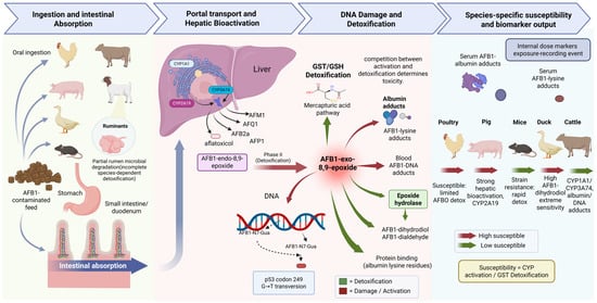
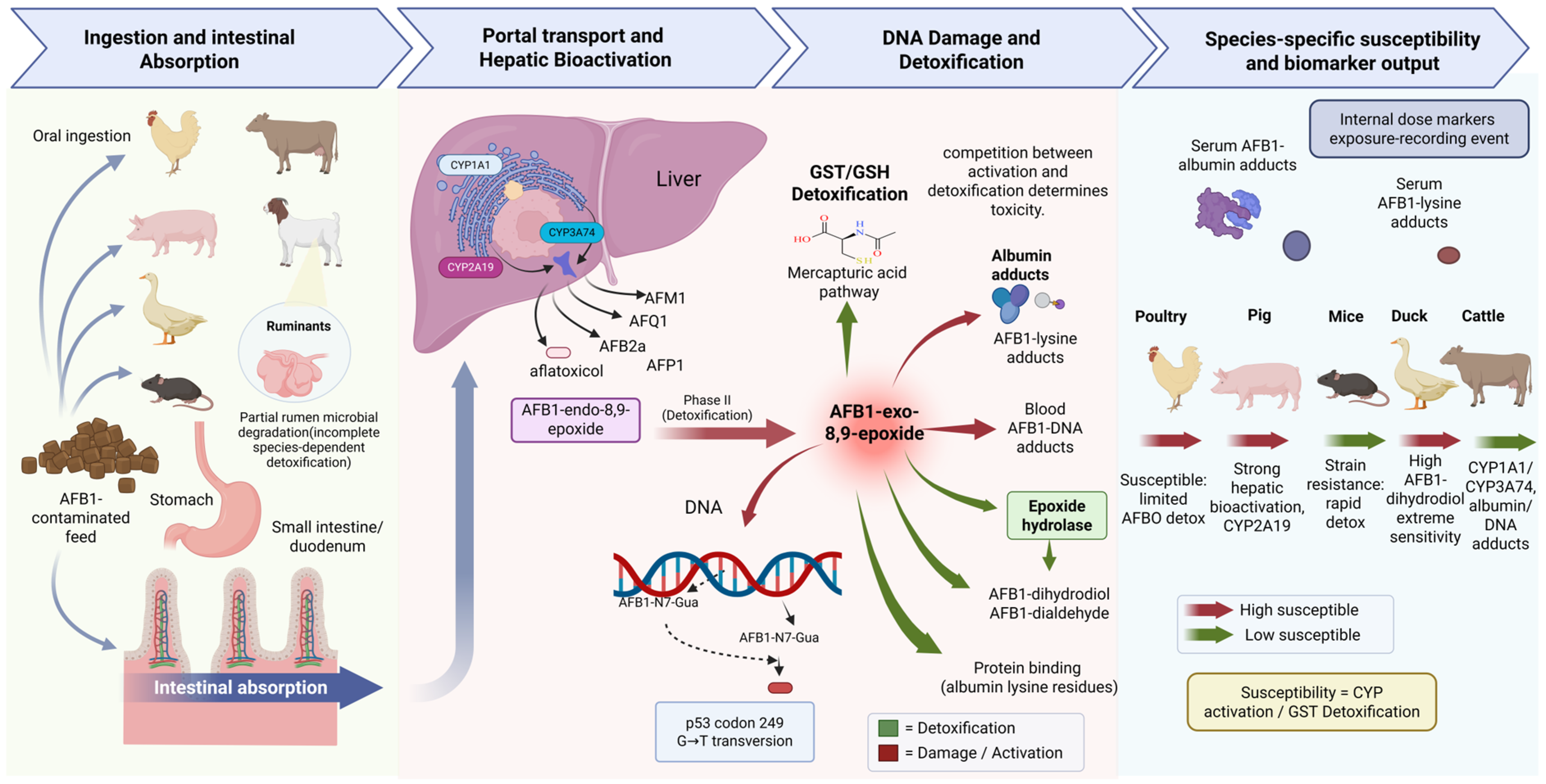
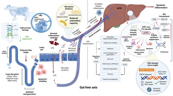
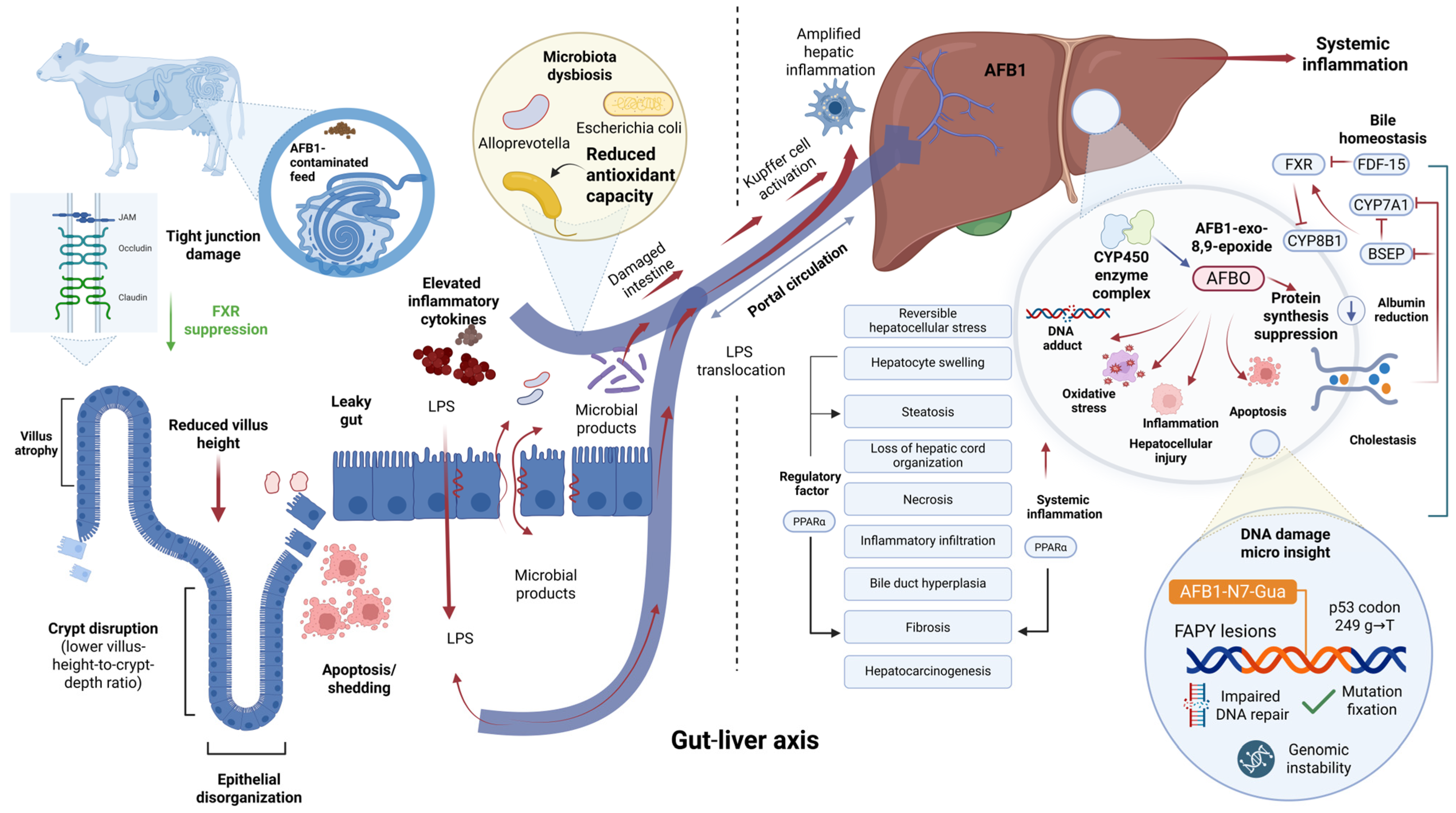
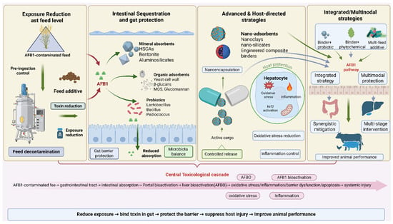
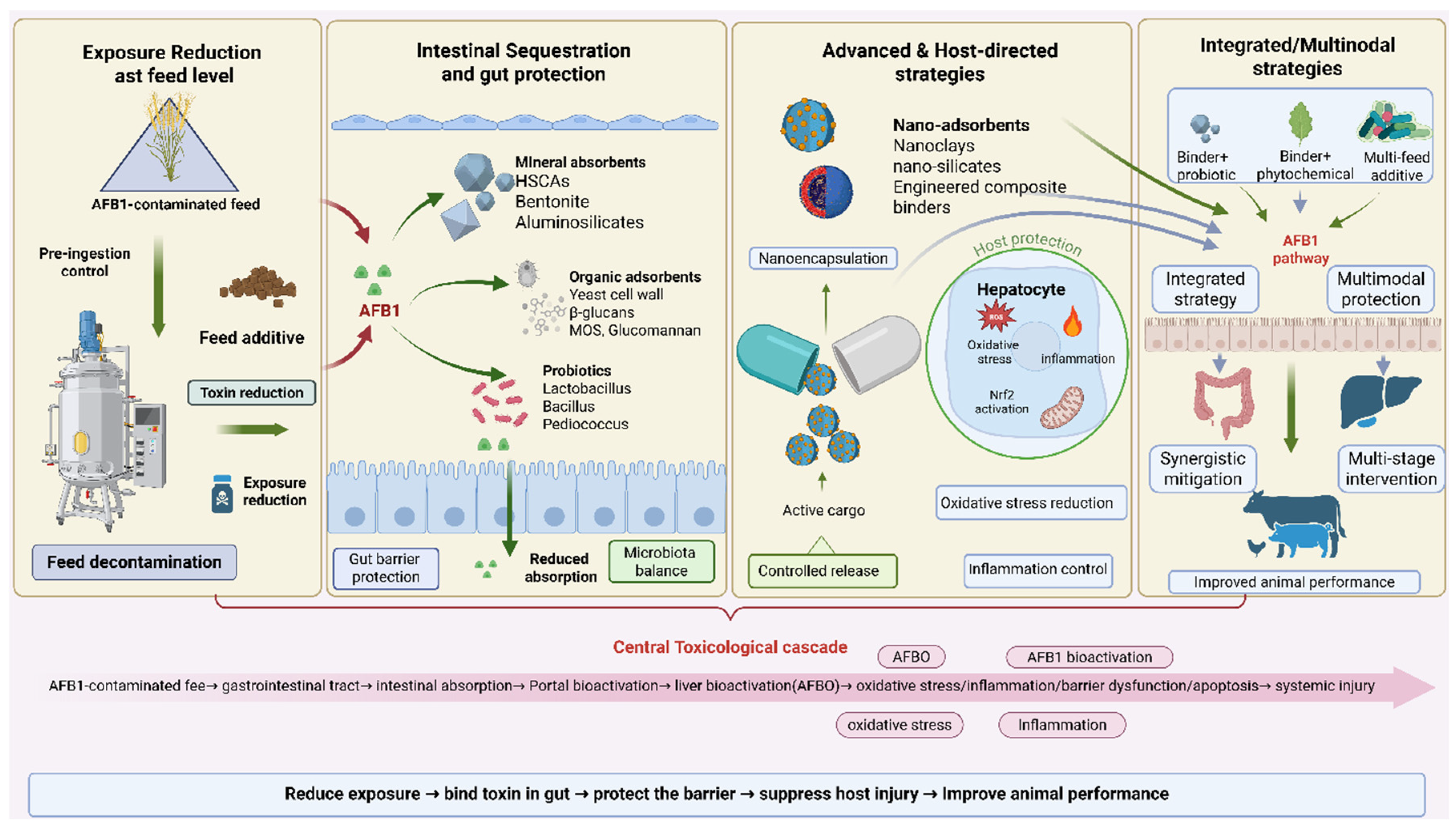

Disclaimer/Publisher’s Note: The statements, opinions and data contained in all publications are solely those of the individual author(s) and contributor(s) and not of MDPI and/or the editor(s). MDPI and/or the editor(s) disclaim responsibility for any injury to people or property resulting from any ideas, methods, instructions or products referred to in the content. |
© 2026 by the authors. Licensee MDPI, Basel, Switzerland. This article is an open access article distributed under the terms and conditions of the Creative Commons Attribution (CC BY) license.