


Abstract
Zearalenone (ZEA) is an estrogenic mycotoxin that poses a potential threat to male reproductive health. This study investigated the age-dependent testicular toxicity of ZEA and its association with autophagy-related changes in male mice. Male C57BL/6J mice at different developmental stages, TM3 cells, and primary ovine Leydig cells were used to evaluate the effects of ZEA on testicular morphology, testosterone secretion, autophagy-related protein expression, and signaling pathways. ZEA induced seminiferous tubule disorganization and spermatogenic cell sloughing in 4–8-week-old mice, whereas no obvious testicular pathological changes were observed in 2-week-old mice. Serum testosterone levels showed a downward trend following ZEA exposure, although the differences were not statistically significant. Western blotting revealed changes in the expression of autophagy-related proteins in testicular tissue, including decreased levels of Beclin1 and LC3, accompanied by increased phosphorylation of PI3K and AKT at 2, 6, and 8 weeks of age and significantly increased mTOR phosphorylation at 6 and 8 weeks of age. In vitro, 40 μM ZEA significantly reduced the viability of TM3 and primary ovine Leydig cells and altered the expression of autophagy-related proteins. These changes were accompanied by increased phosphorylation of PI3K and AKT, whereas the increase in mTOR phosphorylation was not statistically significant in either cell model. Collectively, these findings indicate that susceptibility to ZEA-associated testicular toxicity varies with developmental stage, with more evident histopathological changes observed in 4–8-week-old mice. ZEA exposure was also associated with alterations in autophagy-related protein expression and PI3K/AKT/mTOR pathway phosphorylation. Further studies are needed to determine whether ZEA alters autophagic flux and whether these signaling changes causally contribute to testicular toxicity and Leydig cell dysfunction.
Key Contribution:
Our findings reveal age-dependent differences in susceptibility to ZEA-associated testicular injury in male mice. ZEA exposure was associated with changes in autophagy-related protein expression and PI3K/AKT/mTOR pathway phosphorylation in testicular tissue and Leydig cell models. These findings provide a basis for further investigation of the relationships among developmental stage, autophagy-related processes, and ZEA-induced male reproductive toxicity.
1. Introduction
Zearalenone (ZEA) is a non-steroidal mycotoxin synthesized by Fusarium species. Recognized as a prevalent environmental endocrine disruptor, it is commonly present in grains and animal feed [1,2,3]. Because contaminated feed can serve as an important route of exposure for livestock and poultry, ZEA may enter the food and feed chain and subsequently undergo biotransformation in exposed animals. ZEA exhibits estrogenic activity, and its major metabolites, particularly α-zearalenol (α-ZOL) and β-zearalenol (β-ZOL), also possess estrogenic activity, although their relative estrogenic potencies differ from that of the parent compound. In addition, ZEA and its metabolites may occur in modified or conjugated forms, which are relevant to the overall assessment of exposure.
Autophagy is an evolutionarily conserved degradation mechanism in eukaryotes. During spermatogenesis, one of the functions of autophagy is to degrade and recycle excess proteins or organelles in cells for reuse under conditions of nutrient deprivation or stimulation [4,5].
The PI3K/AKT/mTOR signaling cascade constitutes a primary inhibitory regulatory axis governing autophagic activity [6]. This signaling cascade is abundantly expressed within testicular parenchymal tissue and exerts precise modulatory control over three key testicular biological processes: spermatogenic progression, Leydig cell function, and Leydig cell viability maintenance [7]. When the pathway is activated, signals such as growth factors activate PI3K through receptor tyrosine kinases, catalyzing the conversion of PIP2 into PIP3, which in turn recruits and activates AKT [8]. Activated AKT phosphorylates TSC1/TSC2, relieving its inhibition of Rheb, thereby activating mTORC1. mTORC1 inhibits the kinase activity of the ULK1 complex through phosphorylation, preventing the initiation of autophagy [9]. Conversely, when the pathway is inhibited, ULK1 is dephosphorylated and activated, initiating [6].
Multiple environmental toxicants can inhibit this pathway and induce testicular autophagy. For example, ZEA exposure in primary dairy goat Leydig cells suppresses PI3K/AKT/mTOR signaling [7] and induces oxidative stress-associated autophagy. in TM3 cells, ZEA upregulates PTEN, inhibits PI3K/AKT, and decreases AKT phosphorylation [7,10]. Fluoride exposure also significantly inhibits this pathway, upregulates autophagy markers, and damages mitochondria [11]. Reproductive toxicants such as di-n-butyl phthalate (DBP) and valproic acid (VPA) also affect autophagy through this pathway, but the regulation is complex (e.g., VPA upregulates mTOR yet causes autophagic dysfunction) [12]. It should be noted that pathway inhibition is not always harmful; moderate autophagy activation can protect cells (e.g., in silica nanoparticle-induced Leydig cell injury, autophagy alleviates apoptosis) [13]. In summary, PI3K/AKT/mTOR is a key node linking external stimuli and testicular autophagy response, with profound implications for male reproductive health [11,12,13].
Postnatal development period is a critical stage for male reproductive system development, during which the testis completes the construction of seminiferous tubules, the proliferation and differentiation of spermatogenic cells, and the functional maturation of Leydig cells [14,15]. Its normal development directly determines reproductive performance in adulthood [15]. Autophagy, as a core mechanism for maintaining cellular homeostasis, is involved in regulating testicular structural establishment and functional maturation, and its physiological dynamic balance is crucial for normal testicular development [16,17]. Previous studies have shown that autophagy in the testes of mice during postnatal development exhibits a developmental pattern of increasing from 1 to 2 weeks and decreasing from 2 to 8 weeks, and the PI3K/AKT/mTOR pathway shows a significant negative correlation with the changes in autophagy levels, providing an important physiological reference basis for subsequent studies on toxin interference [17].
It is important to distinguish toxicological hazard identification from dietary risk assessment when interpreting the effects of ZEA [18]. Hazard identification aims to determine whether and under what experimental conditions ZEA can cause adverse biological effects, whereas dietary risk assessment considers actual dietary exposure levels and their relationship with health-based guidance values [18]. The tolerable daily intake (TDI) of 0.25 μg/kg body weight per day for ZEA and its modified forms provides useful context for dietary exposure assessment but should not be directly compared with the short-term parenteral dose used in the present study [18]. In the present study, the 20 mg/kg ZEA regimen was designed as a short-term, relatively high-dose hazard-identification model to investigate the potential reproductive and testicular effects of ZEA in male mice at different ages under controlled experimental conditions.
Leydig cells are essential functional cells in the testis and are primarily responsible for testosterone synthesis and secretion [19,20]. Previous studies have demonstrated that ZEA can disrupt Leydig cell function through mechanisms involving apoptosis, mitochondrial damage, and endoplasmic reticulum stress [21,22,23]. However, the effects of ZEA on autophagy in testicular Leydig cells and the potential involvement of autophagy-related signaling pathways remain insufficiently characterized [24]. Therefore, the present study investigated the age-related differences in ZEA on testicular injury and autophagy in male mice of different ages, with particular emphasis on the involvement of the PI3K/AKT/mTOR signaling pathway. Primary ovine Leydig cells and TM3 cells were further used to examine the effects of ZEA on Leydig cell viability, autophagy-related proteins, and the PI3K/AKT/mTOR pathway under in vitro conditions.
2. Results
2.1. Body Weight, Testis Weight, and Relative Testis Weight in Mice
The body weight, testis weight, and relative testis weight of mice after ZEA administration are shown in Table 1. Compared with the control group, body weight in the ZEA group increased significantly at 4 weeks of age (p < 0.001) and 6 weeks of age (p < 0.05), while no significant differences were observed at 2 weeks and 8 weeks of age. Compared with the control group, there were no significant differences in testis weight in the ZEA group at any time point. Compared with the control group, there were no significant differences in relative testis weight in the ZEA group at any time point.
Table 1.
The body weight, testes weight and relative testes weight.
2.2. Determination of Serum Testosterone
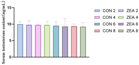
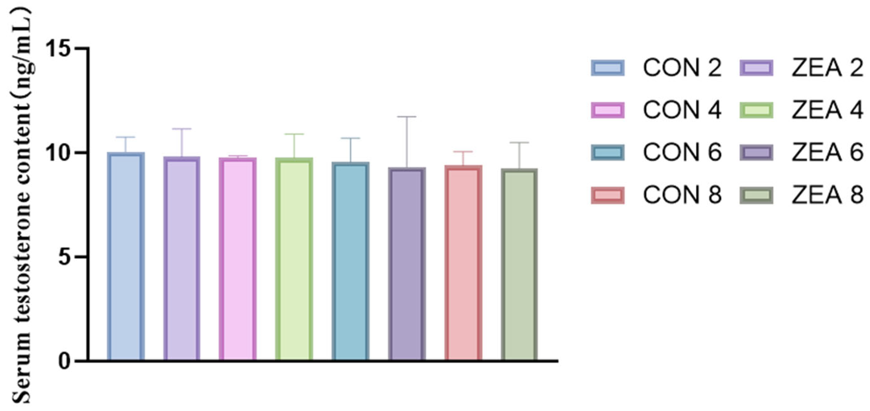
Serum testosterone was measured using an ELISA kit. Compared with the control group, the mean serum testosterone levels in the ZEA group at 2, 4, 6, and 8 weeks of age were all lower, but the differences were not statistically significant overall (Figure 1).
Figure 1.
Measurement of serum testosterone (n = 6). Data are presented as mean ± SD.
2.3. Histological Observations
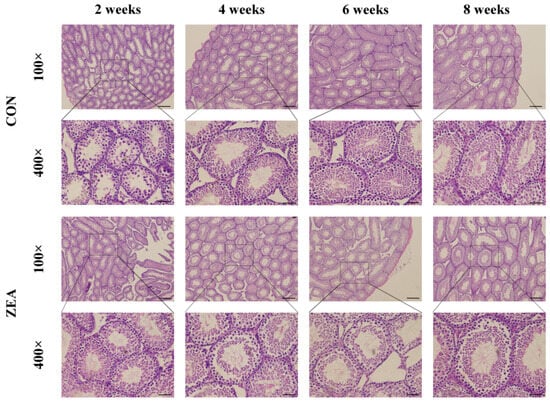
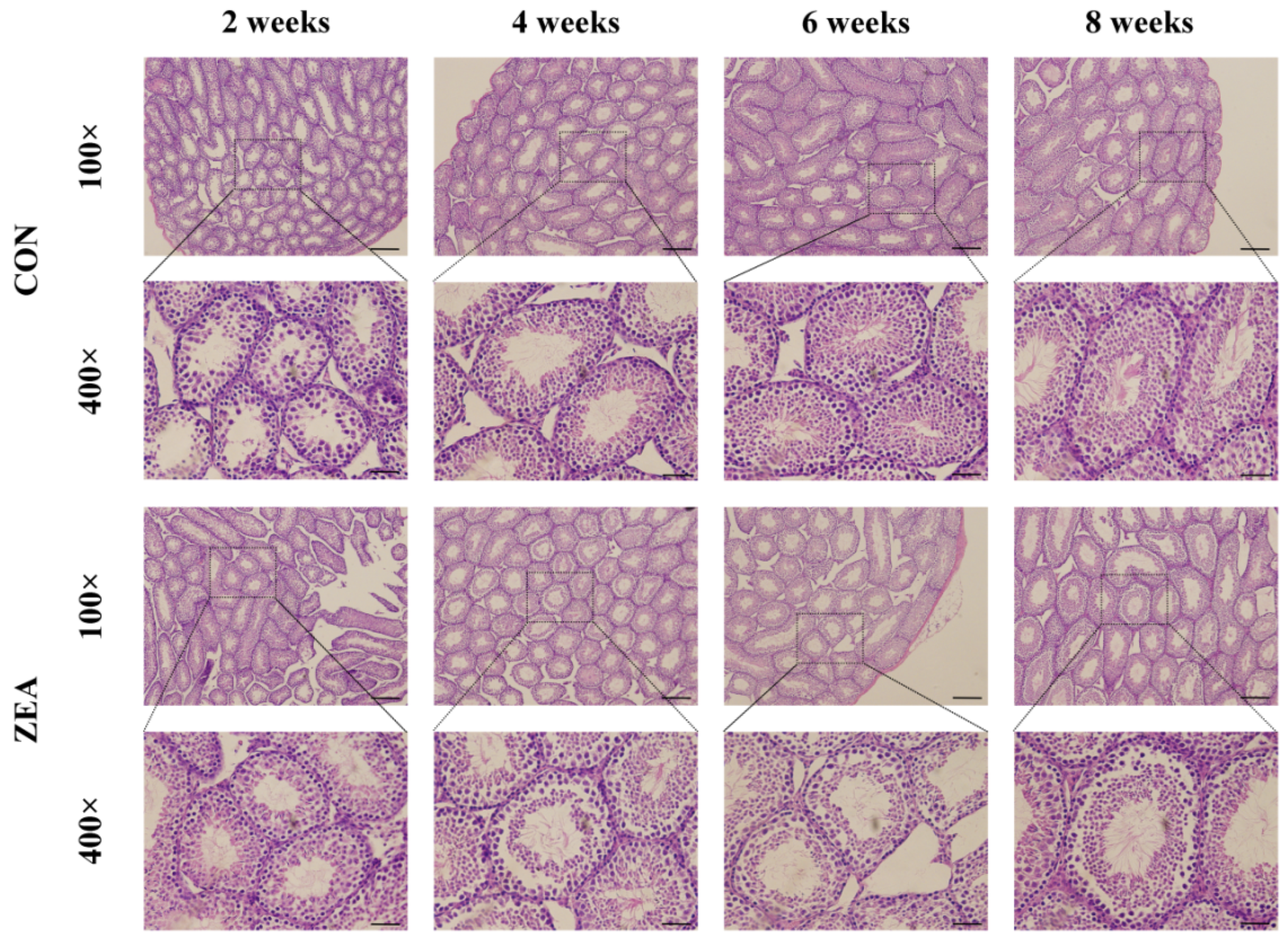
Figure 2 shows the microscopic structural changes in the testes of mice in the control group and the ZEA-treated group. As shown by H&E staining, compared with the control group, the seminiferous tubules in the testes of 2-week-old mice in the ZEA group were more intact; in the testes of 4-, 6-, and 8-week-old mice, spermatogenic cells were observed to have detached from the seminiferous tubules, and their arrangement was more disorganized.
Figure 2.
Microstructural observations of mouse testicular tissue after ZEA treatment (100×, scale bar = 200 μm; 400×, scale bar = 50 μm).
2.4. Expression of Autophagy Marker Proteins
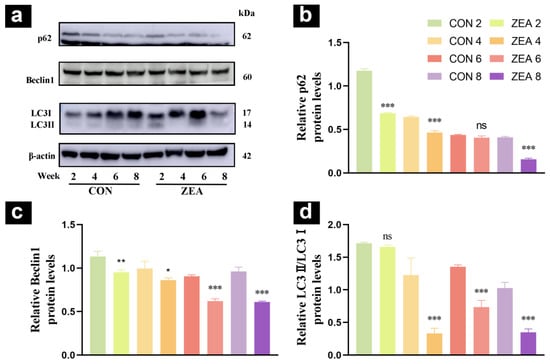
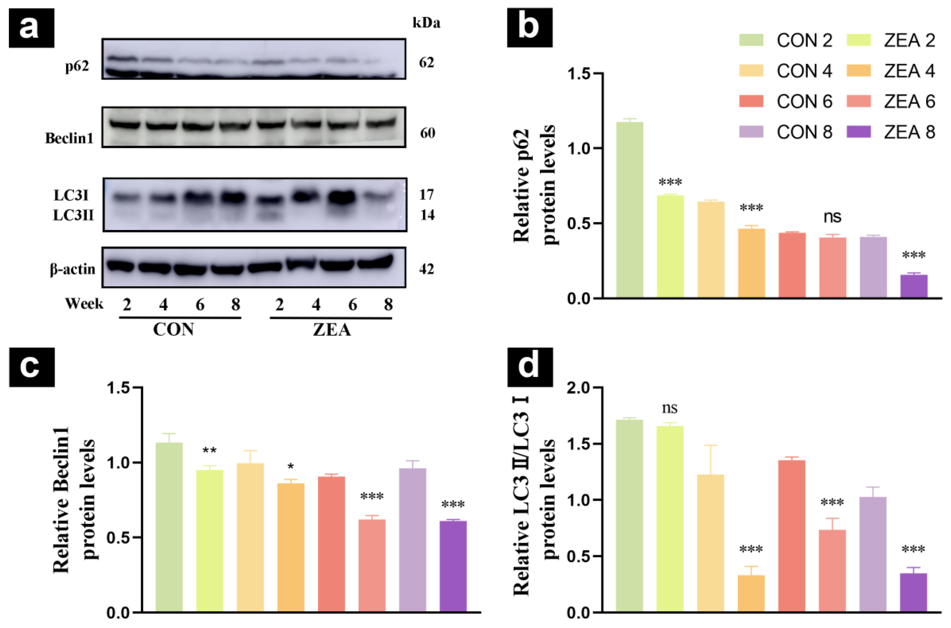
Compared with the control group, p62 expression in the ZEA group was extremely significantly reduced at 2, 4, and 8 weeks of age (p < 0.001, Figure 3b), Beclin1 expression was significantly reduced at 2 weeks of age (p < 0.01), decreased at 4 weeks of age (p < 0.05), and was extremely significantly reduced at 6 and 8 weeks of age (p < 0.001, Figure 3c), LC3 expression was extremely significantly reduced at 4, 6, and 8 weeks of age (p < 0.001, Figure 3d).
Figure 3.
Changes in expression for the autophagy marker proteins. (a) Western blot analysis of the expression levels of autophagy marker proteins p62, Beclin1, and LC3. (b) Quantitative analysis of relative p62 protein levels. (c) Quantitative analysis of relative Beclin1 protein levels. (d) Quantitative analysis of relative LC3II/LC3I protein levels. (* p < 0.05, ** p < 0.01, *** p < 0.001, ns: no significant difference).
2.5. Autophagy Pathway
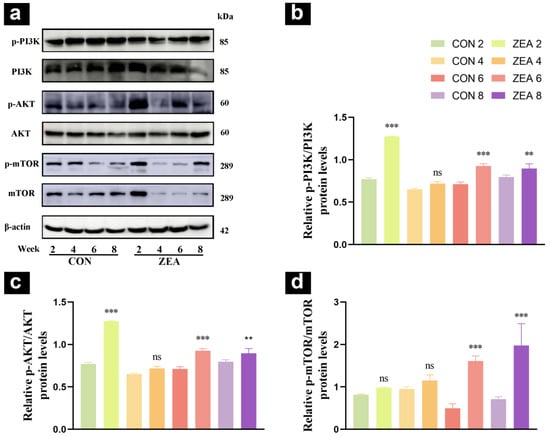
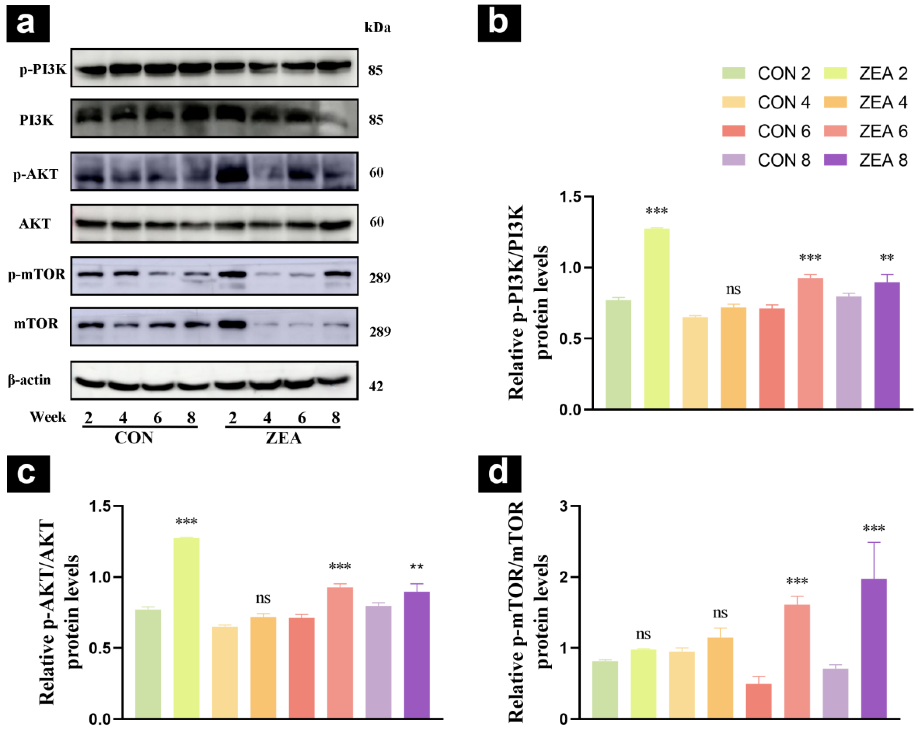
Compared with the control group, PI3K phosphorylation levels in the ZEA group were extremely significantly elevated at 2 and 6 weeks of age (p < 0.001) and significantly elevated at 8 weeks of age (p < 0.01) (Figure 4b), AKT phosphorylation levels were extremely significantly elevated at 2 and 6 weeks of age (p < 0.001) and significantly elevated at 8 weeks of age (p < 0.01) (Figure 4c), and mTOR phosphorylation levels were extremely significantly elevated at 6 and 8 weeks of age (p < 0.001) (Figure 4d).
Figure 4.
Protein expression in the autophagy pathway of mouse testes. (a) Expression levels of PI3K, AKT, mTOR proteins and their phosphorylated forms detected by Western blot analysis. (b) Quantitative analysis of PI3K protein phosphorylation levels. (c) Quantitative analysis of AKT protein phosphorylation levels. (d) Quantitative analysis of mTOR protein phosphorylation levels. (** p < 0.01, *** p < 0.001, ns: not significant).
2.6. Effects of ZEA on Leydig Cell Viability
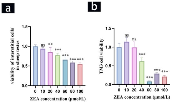
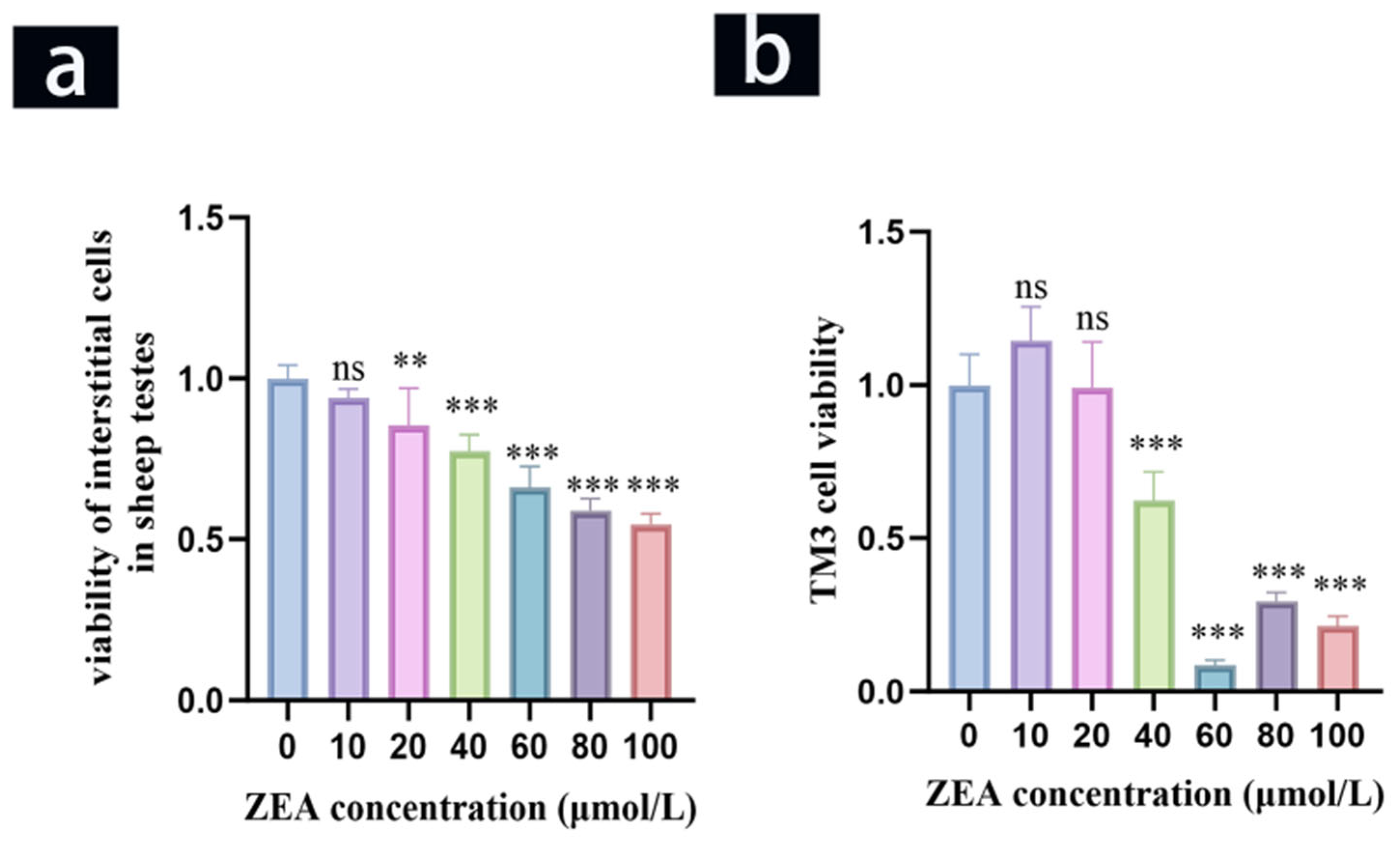
ZEA treatment reduced the viability of primary ovine Leydig cells (Figure 5a). Compared with the control group (0 μM ZEA), cell viability was significantly decreased at 20, 40, 60, 80, and 100 μM ZEA (p < 0.05 or p < 0.001), whereas no significant difference was observed at 10 μM ZEA.
Figure 5.
Effects of ZEA on Leydig cell viability. (a) Viability of ovine Leydig cells. (b) Viability of TM3 cells. (** p < 0.01, *** p < 0.001, ns: not significant).
In TM3 cells, ZEA treatment also reduced cell viability (Figure 5b). Compared with the control group, no significant differences were observed at 10 or 20 μM ZEA, whereas cell viability was significantly decreased at 40, 60, 80, and 100 μM ZEA (p < 0.001).
Based on these results, 40 μM ZEA was selected for subsequent experiments because it produced a highly significant reduction in cell viability in both primary ovine Leydig cells and TM3 cells (p < 0.001).
2.7. Effects of ZEA on Autophagy in Ovine Leydig Cells
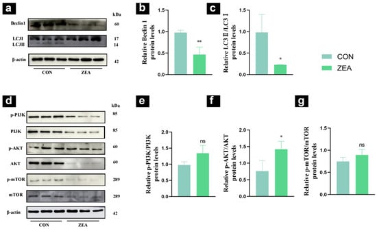
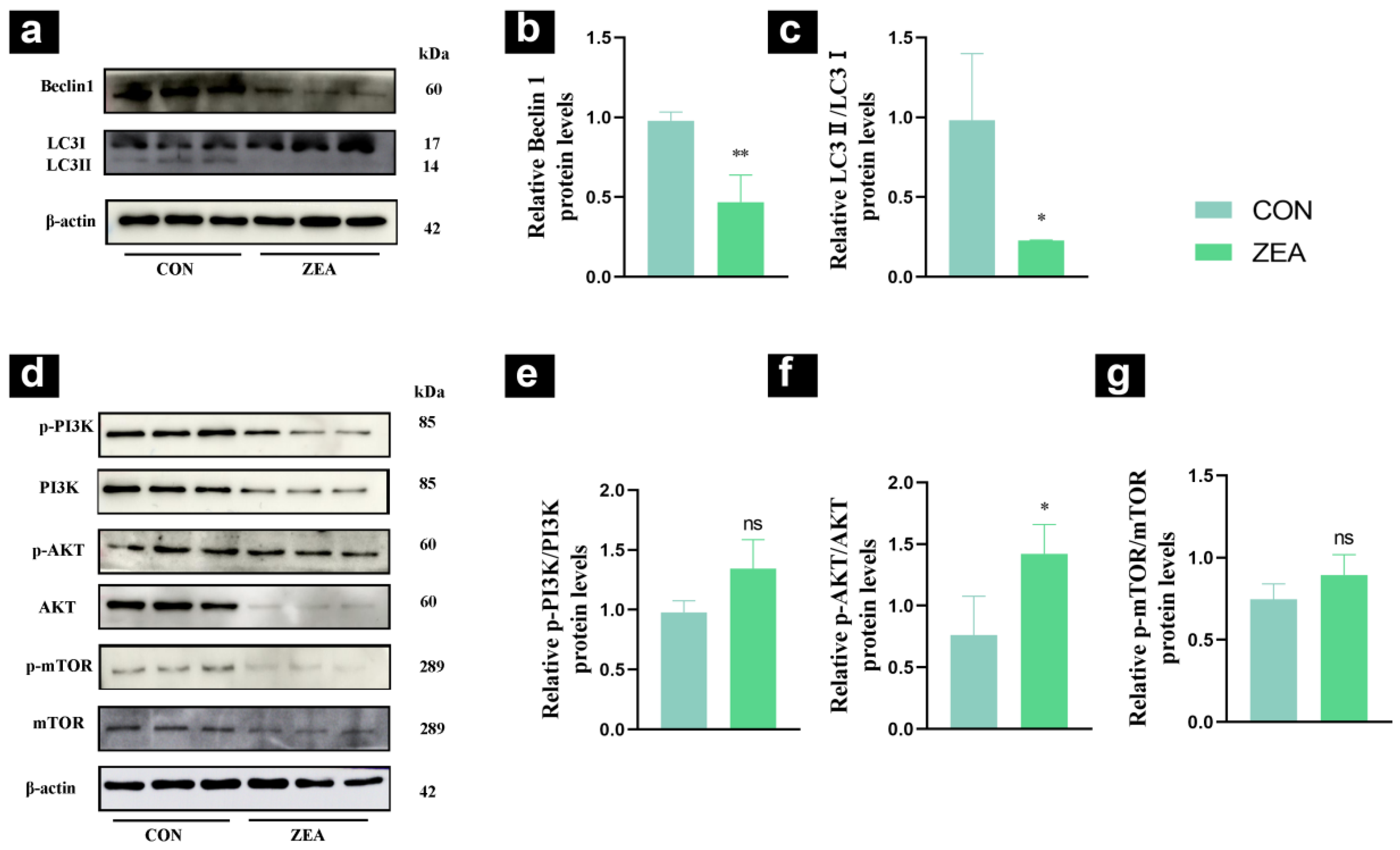
LC3 and Beclin1 are key markers of autophagy and can be used to assess autophagy levels in ovine Leydig cells. To evaluate the effects of ZEA on autophagy levels in ovine Leydig cells, Western blot analysis was performed to detect the expression levels of LC3 and Beclin1. The results showed that, compared with the control group, the ZEA group exhibited a highly significant decrease in Beclin1 expression (p < 0.01, Figure 6b) and a significant decrease in LC3 expression (p < 0.05, Figure 6c).
Figure 6.
Effects of ZEA on ovine Leydig cell autophagy and autophagy pathway. (a) Western blot analysis of the expression levels of autophagy marker proteins Beclin1 and LC3. (b) Quantitative analysis of relative Beclin1 protein levels. (c) Quantitative analysis of relative LC3 protein levels. (d) Expression levels of PI3K, AKT, mTOR proteins and their phosphorylated forms detected by Western blot analysis. (e) Quantitative analysis of PI3K protein phosphorylation levels. (f) Quantitative analysis of AKT protein phosphorylation levels. (g) Quantitative analysis of mTOR protein phosphorylation levels. (* p < 0.05, ** p < 0.01, ns: no significant difference).
The PI3K/AKT/mTOR signaling pathway plays a key role in the regulation of autophagy. To investigate whether this pathway was altered by ZEA, Western blot analysis was used to detect the protein expression levels and phosphorylation status of PI3K, AKT, and mTOR. The results showed that, compared with the control group, the ZEA group exhibited increased PI3K phosphorylation (Figure 6e), significantly increased AKT phosphorylation (p < 0.05, Figure 6f), and increased mTOR phosphorylation, although the difference was not statistically significant (Figure 6g).
2.8. Effects of ZEA on Autophagy in Mouse Testicular Leydig Cells
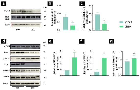
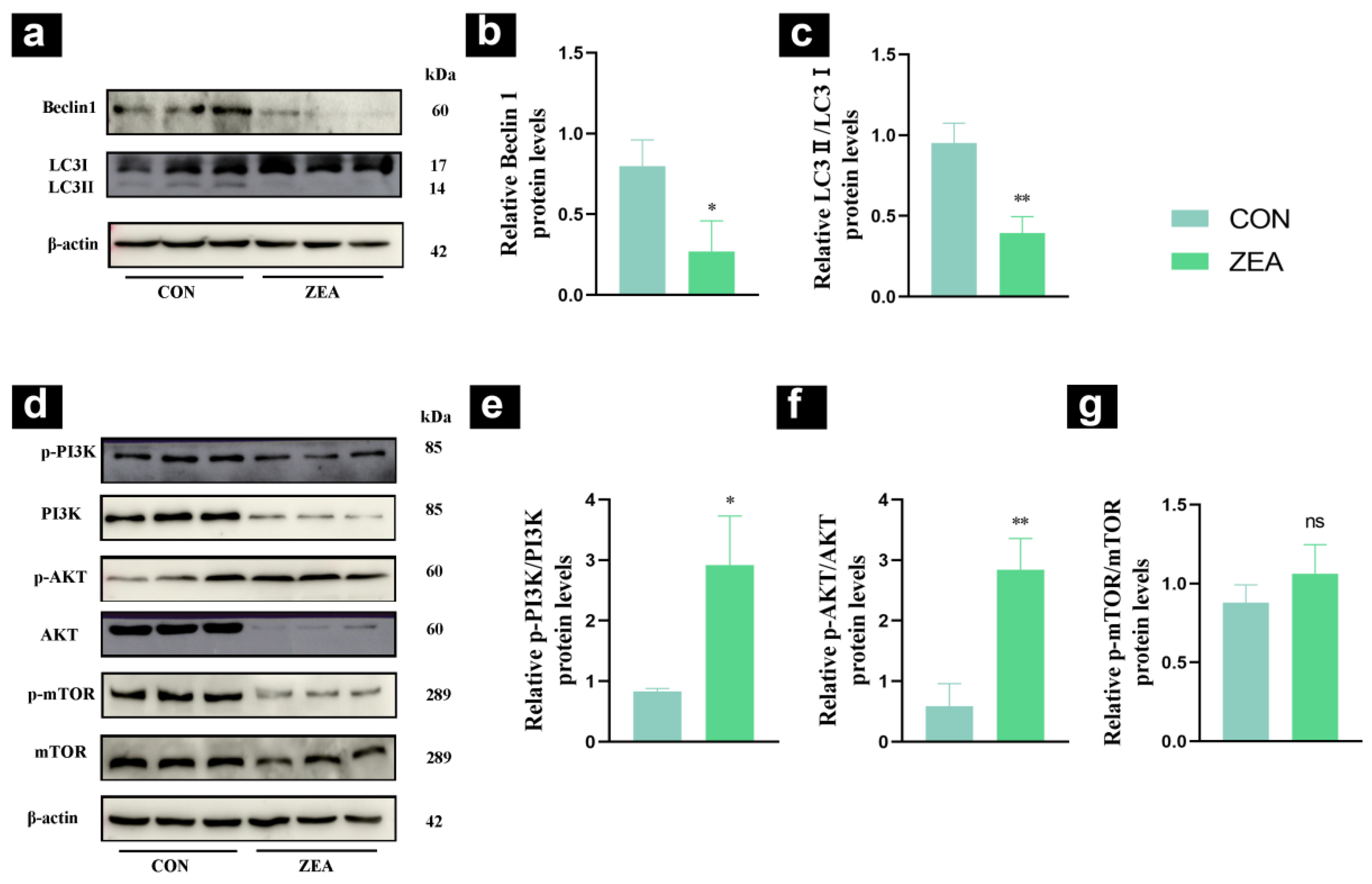
LC3 and Beclin1 are key markers of autophagy and can be used to assess the level of autophagy in TM3 cells. To evaluate the effects of ZEA on autophagy in TM3 cells, Western blot analysis was performed to detect the expression levels of LC3 and Beclin1. The results showed that, compared with the control group, the ZEA group exhibited decreased Beclin1 expression (p < 0.05, Figure 7b) and a significant decrease in LC3 expression (p < 0.01, Figure 7c).
Figure 7.
Effects of ZEA on TM3 autophagy and autophagy pathway. (a) Western blot analysis of the expression levels of autophagy marker proteins Beclin1 and LC3. (b) Quantitative analysis of relative Beclin1 protein levels. (c) Quantitative analysis of relative LC3II protein levels. (d) Expression levels of PI3K, AKT, mTOR proteins and their phosphorylated forms detected by Western blot analysis. (e) Quantitative analysis of PI3K protein phosphorylation levels. (f) Quantitative analysis of AKT protein phosphorylation levels. (g) Quantitative analysis of mTOR protein phosphorylation levels. (* p < 0.05, ** p < 0.01, ns: no significant difference).
The PI3K/AKT/mTOR signaling pathway plays a key role in the regulation of autophagy. To investigate whether this pathway is altered by ZEA, Western blot analysis was used to detect the protein expression levels and phosphorylation status of PI3K, AKT, and mTOR. The results showed that, compared with the control group, the ZEA group exhibited increased PI3K phosphorylation (p < 0.05, Figure 7e), significantly increased AKT phosphorylation (p < 0.01, Figure 7f), and an increased trend in mTOR phosphorylation, although the difference was not statistically significant (Figure 7g).
3. Discussion
The growth parameter analysis revealed that ZEA treatment significantly increased body weight in 4- and 6-week-old mice compared to the control group. However, testicular weight and relative testicular weight remained comparable to control values across all age groups. These findings indicate that ZEA exerts age-dependent interference with overall growth but does not substantially affect testicular tissue mass [15,25]. The 4- and 6-week-old mice were in a phase of rapid growth, during which their metabolic and endocrine regulatory systems were not yet fully mature [15,26]. Consequently, these animals exhibited heightened sensitivity to the estrogenic effects of ZEA, which may have disrupted endocrine homeostasis and led to abnormal body weight gain [25,27]. By contrast, the absence of significant changes in testicular weight across all groups suggests that short-term ZEA exposure does not markedly inhibit overall testicular tissue proliferation. Therefore, ZEA-induced male reproductive toxicity is unlikely to be mediated by blocking testicular tissue growth; instead, it may involve selective disruption of the physiological functions of cells within the testicular interstitium [7,28].
The serum testosterone levels in ZEA-treated mice showed a declining trend across all age groups, although no statistically significant differences were detected between the treatment and control groups. Therefore, the present results do not establish a significant reduction in circulating testosterone following ZEA exposure [25,28]. Although testosterone is essential for male reproductive function and spermatogenesis, the relationship between the observed testosterone trend and the testicular histopathological changes in the present study remains unclear [29]. Further studies are needed to determine whether ZEA exposure affects Leydig cell steroidogenic function and whether such effects contribute to testicular injury [24].
The histological observations revealed that ZEA-induced testicular injury was markedly age-dependent. In 2-week-old mice, the seminiferous tubules retained normal architecture, whereas 4-, 6-, and 8-week-old mice exhibited pronounced histopathological changes, including spermatogenic cell detachment and disorganized tubular arrangement. These findings suggest that 2-week-old mouse testes possess a certain degree of resistance to ZEA toxicity. As the testes mature with advancing age, however, their susceptibility to ZEA-induced damage increases substantially. Disorganization and sloughing of spermatogenic cells are hallmark features of testicular injury, indicating that ZEA disrupts seminiferous tubule integrity, impairs spermatogenic cell adhesion and differentiation, and ultimately poses a potential threat to spermatogenesis [25,30]. Moreover, this injurious effect became progressively more severe with increasing age. Interestingly, further analysis integrating the developmental profile of basal autophagy in mouse testes revealed that 2-week-old mice exhibit peak autophagic activity [17]. We propose that this elevated basal autophagy effectively counteracts ZEA-induced autophagic suppression, thereby preserving cellular homeostasis and tissue structural integrity [31]. This autophagic resilience likely accounts for the absence of overt histopathological changes in the testes of 2-week-old mice. The results of the autophagy marker protein assay showed that ZEA treatment led to a significant overall downward trend in the expression levels of autophagy-related proteins in the testicular tissue of mice at all ages. Specifically, p62 protein levels were extremely significantly reduced at 2, 4, and 8 weeks of age; Beclin1 levels were significantly reduced at all ages, with a more pronounced decrease at 6 and 8 weeks of age; and the LC3II/LC3I ratio was extremely significantly reduced at 4, 6, and 8 weeks of age. As an autophagy substrate protein, p62 is involved in autophagic degradation; however, its decreased expression does not necessarily indicate impaired autophagic degradation. Similarly, reduced expression of Beclin1 and LC3 is consistent with alterations in autophagy-related processes but does not directly establish inhibition of autophagy initiation or autophagosome formation. The combined changes in these proteins indicate that ZEA exposure was associated with alterations in autophagy-related protein expression in mouse testicular tissue. These changes occurred alongside the histopathological alterations observed following ZEA exposure, particularly in older mice. However, because autophagic flux was not directly assessed, the present findings do not establish whether autophagic activity was inhibited. Autophagy is the central pathway by which testicular cells clear damaged organelles and abnormal proteins. When autophagy is inhibited, damaged substances accumulate within cells, thereby disrupting the structural integrity of the seminiferous tubules and impairing the physiological functions of spermatogenic cells; this is also one of the key molecular mechanisms by which ZEA induces pathological damage to testicular tissue [17,31]. In contrast, the level of physiological autophagy in the testes of 4–8-week-old mice gradually declines, making it difficult to compensate for the autophagy inhibition induced by ZEA. At this stage, the decline in autophagy-related proteins is more pronounced, and the corresponding histopathological damage is more evident; thus, the baseline level of physiological autophagy in testicular tissue influences the sensitivity of mice to ZEA-induced toxic damage.
Analysis of PI3K/AKT/mTOR pathway-related proteins revealed that ZEA treatment significantly increased the phosphorylation levels of PI3K, AKT, and mTOR in the testicular tissue across all age groups. Specifically, phosphorylation levels of PI3K and AKT were markedly elevated in 2- and 6-week-old mice, whereas mTOR phosphorylation was significantly increased in 6- and 8-week-old mice. The PI3K/AKT/mTOR pathway is a well-established negative regulator of autophagy; its activation suppresses autophagy by downregulating key factors involved in autophagic initiation [32,33]. In the present study, alterations in PI3K/AKT/mTOR pathway phosphorylation were observed alongside changes in autophagy-related protein expression [32]. These findings suggest an association between pathway phosphorylation and changes in autophagy-related proteins; however, they do not establish a causal relationship between pathway activation and autophagic regulation. Notably, the phosphorylation patterns of PI3K, AKT, and mTOR differed across developmental stages. The functional significance of these age-dependent differences remains to be determined. Although the 2-week-old mice showed no obvious histopathological changes, the present data do not establish that higher basal autophagic activity protected the testes from ZEA-induced injury. Further experiments are needed to investigate this possibility.
The CCK-8 assay demonstrated concentration-dependent cytotoxicity on both primary ovine Leydig cells and TM3 cells, with 40 μM significantly reducing cell viability [18]. This concentration was selected because it allowed for the observation of autophagic changes while avoiding massive cell death [34]. The in vitro data obtained at this concentration enabled further characterization of ZEA-associated changes in autophagy-related proteins in Leydig cell models. Following exposure to 40 μM ZEA, the expression of Beclin1 and LC3 was significantly downregulated in both cell types, indicating changes in autophagy-related protein expression. These findings were consistent with the changes in autophagy-related protein expression observed in testicular tissues from ZEA-treated animals. However, these protein expression changes alone do not establish whether autophagic flux was inhibited. Importantly, this suppressive effect was observed in both mouse and ovine cells, suggesting that similar responses may occur in the two cell models, although further investigation is required to establish whether these effects are conserved across species. These in vitro findings are consistent with the downregulation of autophagy-related proteins observed in testicular tissues from ZEA-treated animals, supporting the relevance of Leydig cell models for investigating ZEA-associated changes in autophagy-related proteins [24].
The pathway analysis revealed that PI3K and AKT phosphorylation increased in both cell models following ZEA exposure, whereas the increases in mTOR phosphorylation were not statistically significant. These findings indicate that changes in pathway phosphorylation occurred alongside changes in autophagy-related protein expression, but do not establish that pathway activation directly suppressed autophagic activity. Although the PI3K/AKT/mTOR pathway is known to regulate autophagy [17], the present findings do not establish that activation of this pathway directly inhibits autophagy or causes testicular injury. Further studies are needed to determine whether the observed changes in pathway phosphorylation contribute to ZEA-associated alterations in autophagy-related protein expression and testicular injury [24].
The impairment of autophagy in Leydig cells may be associated with the accumulation of damaged organelles and disruption of mitochondrial energy metabolism [20]. Concurrently, changes in Leydig cell function may affect steroidogenesis, although this relationship was not directly investigated in the present study [19]. Changes in autophagy-related processes may be associated with alterations in cellular homeostasis and mitochondrial function in Leydig cells [28]. However, the relationship between these changes and Leydig cell steroidogenic function was not directly investigated in the present study. Although serum testosterone levels showed a downward trend in ZEA-treated mice, the differences were not statistically significant. Therefore, the present findings do not establish that ZEA-associated changes in autophagy-related proteins caused impaired testosterone synthesis or secretion. Further studies are needed to clarify the relationships among autophagy-related processes, Leydig cell steroidogenic function, and testicular injury.
The Autophagic homeostasis in Leydig cells is essential for testosterone synthesis, cell survival, and the maintenance of normal physiological functions [19]. However, the extent to which autophagy-related changes affect Leydig cell function following ZEA exposure remains unclear. In the present study, ZEA exposure was associated with changes in autophagy-related protein expression and PI3K/AKT/mTOR pathway phosphorylation in Leydig cell models [33]. Although the PI3K/AKT/mTOR pathway is involved in autophagy regulation, the available data do not establish that pathway activation caused autophagy inhibition. Autophagic flux was not directly assessed, and further studies are needed to clarify the functional significance of these molecular changes and their potential relationships with Leydig cell function and male reproductive toxicity.
In vitro cellular assays provided additional evidence of ZEA-associated changes in autophagy-related protein expression in Leydig cell models. These findings were consistent with the changes observed in testicular tissues from ZEA-treated animals. However, further studies are required to determine the functional significance of these changes and whether Leydig cells are a primary cellular target of ZEA-associated testicular toxicity [34]. These in vitro findings were consistent with both the developmental profile of physiological autophagy in mouse testes and the in vivo toxicity patterns observed in ZEA-treated animals. Together, the findings support an association between ZEA exposure, altered autophagy-related protein expression, and changes in PI3K/AKT/mTOR pathway phosphorylation. Further studies are required to determine the functional significance of these associations. In this study, we employed both primary ovine Leydig cells and TM3 cells for in vitro validation. The consistent results obtained from these two mammalian cell types substantially enhanced the robustness and cross-species applicability of our conclusions, thereby strengthening the mechanistic evidence for ZEA-induced reproductive toxicity [18].
In summary, the in vivo and in vitro findings show that ZEA exposure was associated with changes in autophagy-related protein expression and PI3K/AKT/mTOR pathway phosphorylation in testicular tissue and Leydig cell models [19,32]. These molecular changes were observed alongside testicular histopathological alterations in mice, which varied with age. However, the present study did not directly assess autophagic flux or establish a causal relationship between pathway phosphorylation, autophagy-related changes, and testicular injury. In addition, serum testosterone levels showed a downward trend, but the differences were not statistically significant. Further studies are needed to clarify the functional significance of these molecular changes and their potential relationships with Leydig cell function and testicular injury [35]. Moreover, the findings should be interpreted within the context of the animal and cellular models, exposure regimen, and administration route used in this study. Further research is required to determine the relevance of these findings to dietary ZEA exposure, livestock health, and potential human health risks [34].
A limitation of the present study is that the experimental design represents a short-term, relatively high-dose hazard-identification model rather than a direct simulation of real-world dietary exposure to ZEA. In addition, internal exposure to ZEA was not quantitatively verified. ZEA and its major metabolites, including α-zearalenol (α-ZOL), β-zearalenol (β-ZOL), and their conjugates, were not measured in serum or testicular tissue [36]. Therefore, the administered dose of 20 mg/kg represents the nominal administered dose and should not be considered equivalent to the actual ZEA exposure concentration in serum or target tissues. Moreover, ZEA biotransformation and metabolite formation may vary among species and tissues, which may influence internal exposure and biological effects. Future studies incorporating quantitative toxicokinetic analysis of ZEA and its metabolites in serum and testicular tissue, together with exposure scenarios more representative of dietary exposure, are warranted.
4. Conclusions
ZEA-induced testicular injury exhibited age-dependent characteristics, with more pronounced histopathological changes observed in 4- to 8-week-old mice, as characterized by seminiferous tubule structural disruption and spermatogenic cell sloughing. Both in vivo and in vitro experiments revealed alterations in the expression of autophagy-related proteins, accompanied by changes in PI3K/AKT/mTOR pathway phosphorylation. However, autophagic flux was not directly assessed, and a causal role of this signaling pathway in the observed changes remains to be established. Serum testosterone levels showed a downward trend in ZEA-treated mice, but the differences were not statistically significant. Collectively, these findings provide a basis for further investigating the molecular mechanisms underlying ZEA-induced male reproductive toxicity.
5. Materials and Methods
5.1. Reagents and Chemicals
LC3 polyclonal antibody (GTX127375), SQSTM1/p62 polyclonal antibody (GTX109683), and mTOR polyclonal antibody (GTX101557) were purchased from GeneTex (Irvine, CA, USA). Beclin1 polyclonal antibody (AP0768), AKT polyclonal antibody (BS2987), and p-AKT polyclonal antibody (Ser473 and/or Thr308) (BS4007) were purchased from BioWorld (Dublin, OH, USA). PI3K monoclonal antibody (4257T), p-PI3K monoclonal antibody (Tyr1007/1008 on the p110 subunit) (4228T), and p-mTOR monoclonal antibody (Ser2448) (5536T) were purchased from Cell Signaling Technology (Danvers, MA, USA). The β-actin polyclonal antibody (bs-0061R) was purchased from Bioss (Beijing, China). The immunoglobulin IgG (H + L) secondary antibodies (SA00001-2 and SA00001-1) were purchased from Proteintech (Wuhan, Hubei, China). The antibody diluent was a TBST solution containing 5% skim milk powder. ZEA (purity ≥ 98.22%, HY-103447) was purchased from Med Chem Express (Junction, NJ, USA). The Mouse Testosterone (T) ELISA Research Kit (MM-0569M1) was purchased from Meimian (Yancheng, Jiangsu, China).
The mouse Leydig cell line (TM3) was purchased from BeNa Culture Collection (BNCC, Suzhou, China), with cell accession number BNCC100249.
5.2. Laboratory Animals
All animal experiments were approved by the Animal Care Committee of Gansu Agricultural University (No. GSAU-Eth-VMC-2023-021) and conducted in accordance with the Guiding Opinions on Treating Laboratory Animals Well issued by the Ministry of Science and Technology of the People’s Republic of China.
Specific-pathogen-free-grade male C57BL/6 mice (1, 3, 5, and 7 weeks old) were purchased from the Lanzhou Veterinary Research Institute of the Chinese Academy of Agricultural Sciences. All mice were acclimated for 1 week before treatment. At the start of treatment, male C57BL/6J mice were 2, 4, 6, or 8 weeks of age. Under controlled conditions (temperature: 23 ± 2 °C), the mice were housed in plastic cages (29.00 × 17.80 × 16.00 cm) under a 12/12-h light/dark cycle and had free access to deionized water and standard laboratory rodent chow. The mouse strain used was the C57BL/6J subline obtained from the National Strain Bank; the mice used in this study were fifth-generation. All mice were in good health, as verified by monitoring their food intake, water consumption, and body weight.
5.3. Development of the ZEA Model
The control group received intraperitoneal injections of the vehicle (DMSO:corn oil = 1:9, v/v), while the ZEA group received ZEA dissolved in the same vehicle. The ZEA dosing solution was freshly prepared at a final concentration of 20 mg/mL immediately before administration. The injection volume was individually adjusted according to the body weight of each mouse to achieve a dose of 20 mg/kg body weight (1 mL/kg body weight). The three-day exposure regimen was selected with reference to previous in vivo studies of ZEA [37,38]. The intraperitoneal route of administration was selected according to the recommended experimental application information provided by the ZEA supplier. ZEA was administered intraperitoneally once daily for three consecutive days, beginning when the mice were 2, 4, 6, or 8 weeks of age, depending on the assigned age group. Control mice received the corresponding vehicle according to the same schedule.
5.4. Sample Collection
Twelve mice were included in each age group (2, 4, 6, and 8 weeks of age at the start of treatment), with six mice assigned to the control (CON) group and six to the ZEA group (n = 6 per group). Each mouse was considered an independent biological replicate. After weighing, mice were anesthetized via inhalation of isoflurane. Once adequate anesthetic depth was confirmed, eyeball removal and blood collection were performed. Subsequently, the mice were euthanized by cervical dislocation. The two testes from each mouse were immediately excised and weighed. The left testes from three mice in each group were fixed in 4% paraformaldehyde and stored at room temperature. The left testes from the remaining mice and the right testes from all mice were stored in a −80 °C ultra-low-temperature freezer for subsequent analysis.
5.5. Testosterone
The ELISA method was used to determine testosterone levels in mouse serum. Briefly, collected and centrifuged serum samples were added to a microplate along with a series of concentration standards provided in the kit, following the operating procedures. After incubation at 37 °C and washing, the enzyme-labeled reagent and color developer were added sequentially to allow the reaction to proceed. Finally, the reaction was stopped with a stopping solution, and the absorbance was immediately measured at a wavelength of 450 nm using a microplate reader. The testosterone concentration in the serum was then calculated based on the standard curve.
5.6. Histological Observations
After fixation in paraformaldehyde, the testes were processed using a series of ethanol and xylene solutions, embedded in paraffin, and sectioned into 4-μm-thick sections. Hematoxylin and eosin (H&E) staining was performed, and images were captured and analyzed using a digital microscope (Olympus, Olympus DP-71, Tokyo, Japan) at magnifications of 100× and 400×.
5.7. Protein Extraction and Western Blotting
Total protein was extracted from testicular tissue using RIPA lysis buffer (Solarbio, Beijing, China) containing PMSF (Servicebio, Wuhan, China). Sodium dodecyl sulfate-polyacrylamide gel electrophoresis (SDS-PAGE) buffer was added, and the samples were incubated in a metal bath (TiGen Biotechnology, OSE-DB-01, Beijing, China) at 98 °C, then stored at −20 °C. Proteins were separated by SDS-PAGE and transferred to a polyvinylidene fluoride (PVDF) membrane (Immobilon, IPVH00010, Wicklow, Ireland). After blocking with 5% skim milk at room temperature for 40–60 min, the membrane was incubated with the primary antibody at 4 °C overnight. The following day, the membrane was incubated with horseradish peroxidase-conjugated secondary antibody at 37 °C for 1 h, and protein bands were visualized using a chemiluminescence imager (General Electric, Amersham Imager 600, Boston, MA, USA). To ensure the validity and reliability of the results, all Western blot experiments were performed in three independent experimental runs.
5.8. Culture of Testicular LeydigCells
Primary ovine Leydig cells and TM3 cells were cultured in DMEM/F12 medium supplemented with 10% serum and 1% antibiotic-antibody mixture. The cells were maintained at 37 °C in a humidified atmosphere containing 5% CO2. The culture medium was replaced regularly, and the cells were subcultured when they reached the appropriate confluence. Both cell types were maintained under the same culture conditions before subsequent experiments.
5.9. Assessment of ZEA-Induced Cell Viability and Cytotoxicity
Cells were digested with 0.25% trypsin, and the cell suspension was collected. Cells were seeded into 96-well plates at a density of 1 × 10⁴ cells/mL, with 100 μL per well, and cultured for 24 h in a 37 °C, 5% CO2 incubator. The experiment was divided into a blank group (basic medium), a control group (basic medium + cells), and ZEA treatment groups (10, 20, 40, 60, 80, 100 μM), with six technical replicate wells per group within the same experiment. 24 h after adding ZEA, 10 μL of CCK-8 solution was added to each well. After continuing the culture for 2 h, the 96-well plate was placed in a fully automated microplate reader (Model 680, Bio-Rad Laboratories, Hercules, CA, USA), and the optical absorbance of each well was measured at a wavelength of 450 nm.
5.10. ZEA Treatment
Primary ovine Leydig cells and TM3 cells were cultured under the conditions described above and treated with 40 μM ZEA for 24 h. The corresponding control groups received the vehicle. Following treatment, the cells were collected for subsequent Western blotting of autophagy-related proteins and components of the PI3K/AKT/mTOR signaling pathway.
5.11. Data Analysis
All data were analyzed using ImageJ 1.44p (Java 1.6.0-20, 32-bit) for quantitative analysis of experimental images (National Institutes of Health, Bethesda, MD, USA), OriginPro 9 (32-bit) for statistical calculations (OriginLab Corporation, Northampton, MA, USA), and GraphPad Prism 8.0.2 for data plotting (GraphPad Software, Inc., San Diego, CA, USA). Quantitative data are presented as the mean ± standard deviation (SD). Data normality was assessed using the Shapiro–Wilk test, and homogeneity of variance was assessed before parametric analyses. For normally distributed data with equal variances, comparisons between two groups were performed using the independent-samples t-test. Comparisons among multiple groups were performed using one-way analysis of variance (ANOVA), followed by Tukey’s multiple-comparisons test. For data that did not follow a normal distribution, comparisons among multiple groups were performed using the Kruskal–Wallis test. Statistical significance was defined as p < 0.05.
For the animal experiments, each mouse was considered an independent biological replicate. For cell-based assays and Western blot analyses, the numbers of biological and technical replicates are specified separately according to the experimental design. Statistical significance was set at p < 0.05.
Author Contributions
R.L. (Rui Luo), Conceptualization, Investigation, Writing—Original Draft, Writing—Review and Editing, D.Z., Methodology, Supervision, T.L., Conceptualization, Data Curation, Formal Analysis, R.L. (Rong Li), Data Curation, L.X., Formal Analysis, X.M., Visualization, X.D., Validation, Y.H., Conceptualization, Writing—Review and Editing, Supervision, Funding Acquisition. All authors have read and agreed to the published version of the manuscript.
Funding
This research was supported by the Youth Talent Program of “Fuxi” [Gaufx-03Y02], the Scientific Research Start-up Funds for Openly-Recruited Doctors of Gansu Agricultural University [2017RCZX-13], the National Natural Science Foundation of China [31902339], the Foundation for Youth Doctor of Gansu Province [2021QB-026], and the Special Funds for Discipline Construction of Gansu Agricultural University [GAU-XKJS-2018-067].
Institutional Review Board Statement
The study was conducted in accordance with the Declaration of Helsinki, and approved by the Animal Care Committee of Gansu Agricultural University (protocol code: GSAU-Eth-VMC-2023-021 and 9 March 2023 of approval).
Informed Consent Statement
Not applicable.
Data Availability Statement
The original contributions presented in this study are included in the article. Further inquiries can be directed to the corresponding author.
Conflicts of Interest
The authors declare no conflicts of interest.
References
- Zhang, G.; Feng, Y.; Song, J.; Zhou, X. Zearalenone: A Mycotoxin With Different Toxic Effect in Domestic and Laboratory Animals’ Granulosa Cells. Front. Genet. 2018, 9, 667. [Google Scholar] [CrossRef] [Scilit] [PubMed]
- Wang, M.; Zeng, L.; Su, P.; Ma, L.; Zhang, M.; Zhang, Y.Z. Autophagy: A multifaceted player in the fate of sperm. Hum. Reprod. Update 2021, 28, 200–231. [Google Scholar] [CrossRef] [Scilit] [PubMed]
- Zinedine, A.; Soriano, J.M.; Moltó, J.C.; Mañes, J. Review on the toxicity, occurrence, metabolism, detoxification, regulations and intake of zearalenone: An oestrogenic mycotoxin. Food Chem. Toxicol. 2007, 45, 1–18. [Google Scholar] [CrossRef] [Scilit] [PubMed]
- Ropejko, K.; Twarużek, M. Zearalenone and Its Metabolites-General Overview, Occurrence, and Toxicity. Toxins 2021, 13, 35. [Google Scholar] [CrossRef] [Scilit] [PubMed]
- Shang, Y.; Wang, H.; Jia, P.; Zhao, H.; Liu, C.; Liu, W.; Song, Z.; Xu, Z.; Yang, L.; Wang, Y.; et al. Autophagy regulates spermatid differentiation via degradation of PDLIM1. Autophagy 2016, 12, 1575–1592. [Google Scholar] [CrossRef] [Scilit] [PubMed]
- Long, C.; Zhou, Y.; Shen, L.; Yu, Y.; Hu, D.; Liu, X.; Lin, T.; He, D.; Xu, T.; Zhang, D.; et al. Retinoic acid can improve autophagy through depression of the PI3K-Akt-mTOR signaling pathway via RARα to restore spermatogenesis in cryptorchid infertile rats. Genes DIS. 2021, 9, 1368–1377. [Google Scholar] [CrossRef] [Scilit] [PubMed]
- Chen, J.; Wang, M.; Wang, H.; Long, M. Zearalenone promotes apoptosis of mouse Leydig cells by targeting phosphatase and tensin homolog and thus inhibiting the PI3K/AKT signal pathway. Environ. Sci. Pollut. Res. 2021, 28, 67779–67787. [Google Scholar] [CrossRef] [Scilit] [PubMed]
- Glaviano, A.; Foo, A.S.C.; Lam, H.Y.; Yap, K.C.H.; Jacot, W.; Jones, R.H.; Eng, H.; Nair, M.G.; Makvandi, P.; Geoerger, B.; et al. PI3K/AKT/mTOR signaling transduction pathway and targeted therapies in cancer. Mol. Cancer 2023, 22, 138. [Google Scholar] [CrossRef] [Scilit] [PubMed]
- Hajdú, B.; Holczer, M.; Horváth, G.; Szederkényi, G.; Kapuy, O. Fine-Tuning of mTORC1-ULK1-PP2A Regulatory Triangle Is Crucial for Robust Autophagic Response upon Cellular Stress. Biomolecules 2022, 12, 1587. [Google Scholar] [CrossRef] [Scilit] [PubMed]
- Cheng, A.; Wu, Y.; Luo, H.; Song, X.; Li, X.; Fan, B.; Zhang, X.; Liu, S.; Zhuang, C.; Zhao, Y.; et al. Environmental Fluoride Compromises Male Fertility: Differentially Modulated miR-34a-5p Targets REST to Regulate Autophagy in Testicular Somatic Cells; Research: Washington, DC, USA, 2026; Volume 9, p. 1113. [Google Scholar]
- Zhang, J.; Zhu, Y.; Shi, Y.; Han, Y.; Liang, C.; Feng, Z.; Zheng, H.; Eng, M.; Wang, J. Fluoride-Induced Autophagy via the Regulation of Phosphorylation of Mammalian Targets of Rapamycin in Mice Leydig Cells. J. Agric. Food Chem. 2017, 65, 8966–8976. [Google Scholar] [CrossRef] [Scilit] [PubMed]
- Li, X.; Li, J.; Zhang, Y.; Zhou, Y. Di-n-butyl phthalate induced hypospadias relates to autophagy in genital tubercle via the PI3K/Akt/mTOR pathway. J. Occup. Health 2017, 59, 8–16. [Google Scholar] [CrossRef] [Scilit] [PubMed]
- Zhang, Q.; Grunberger, J.W.; Khurana, N.; Zhou, X.; Xu, X.; Ghandehari, H.; Chen, F. BECLIN-1-Mediated Autophagy Suppresses Silica Nanoparticle-Induced Testicular Toxicity via the Inhibition of Caspase 8-Mediated Cell Apoptosis in Leydig Cells. Cells 2022, 11, 1863. [Google Scholar] [CrossRef] [Scilit] [PubMed]
- Voigt, A.L.; de Lima, E.; Martins Lara, N.; Dobrinski, I. Comparing the adult and pre-pubertal testis: Metabolic transitions and the change in the spermatogonial stem cell metabolic microenvironment. Andrology 2023, 11, 1132–1146. [Google Scholar] [CrossRef] [Scilit] [PubMed]
- Bhattacharya, I.; Dey, S. Emerging concepts on Leydig cell development in fetal and adult testis. Front. Endocrinol. 2023, 13, 1086276. [Google Scholar] [CrossRef] [Scilit] [PubMed]
- Yan, Q.; Zhang, Y.; Wang, Q.; Yuan, L. Autophagy: A Double-Edged Sword in Male Reproduction. Int. J. Mol. Sci. 2022, 23, 15273. [Google Scholar] [CrossRef] [Scilit] [PubMed]
- Zhang, D.; Pang, X.; Yan, Z.; Dong, W.; Fang, Z.; Yang, J.; Wang, Y.; Xue, L.; Zhang, J.; Xue, C.; et al. Dynamic Alterations in Testicular Autophagy in Prepubertal Mice. J. Dev. Biol. 2025, 13, 42. [Google Scholar] [CrossRef] [Scilit] [PubMed]
- Ning, C.; Sun, J.; Zhao, Y.; Xu, H.; Wu, W.; Yang, Y. Integrated Targeted and Untargeted Metabolomics Reveals the Toxic Mechanisms of Zearalenone in Goat Leydig Cells. Animals 2026, 16, 283. [Google Scholar] [CrossRef] [Scilit] [PubMed]
- Gao, F.; Li, G.; Liu, C.; Gao, H.; Wang, H.; Liu, W.; Chen, M.; Shang, Y.; Wang, L.; Shi, J.; et al. Autophagy regulates testosterone synthesis by facilitating cholesterol uptake in Leydig cells. J. Cell Biol. 2018, 217, 2103–2119. [Google Scholar] [CrossRef] [Scilit] [PubMed]
- Afzal, A.; Zhang, Y.; Afzal, H.; Saddozai, U.A.K.; Zhang, L.; Ji, X.; Khawar, M.B. Functional role of autophagy in testicular and ovarian steroidogenesis. Front. Cell Dev. Biol. 2024, 12, 1384047. [Google Scholar] [CrossRef] [Scilit] [PubMed]
- Wang, Y.; Zheng, W.; Bian, X.; Yuan, Y.; Gu, J.; Liu, X.; Liu, Z.; Bian, J. Zearalenone induces apoptosis and cytoprotective autophagy in primary Leydig cells. Toxicol. Lett. 2014, 226, 182–191. [Google Scholar] [CrossRef] [Scilit] [PubMed]
- Chen, S.; Yang, S.; Wang, M.; Chen, J.; Huang, S.; Wei, Z.; Cheng, Z.; Wang, H.; Long, M.; Li, P. Curcumin inhibits zearalenone-induced apoptosis and oxidative stress in Leydig cells via modulation of the PTEN/Nrf2/Bip signaling pathway. Food Chem. Toxicol. 2020, 141, 111385. [Google Scholar] [CrossRef] [Scilit] [PubMed]
- Shahidi, M.; Moradi, A.; Dayati, P. Zingerone attenuates zearalenone-induced steroidogenesis impairment and apoptosis in TM3 Leydig cell line. Toxicon 2022, 211, 50–60. [Google Scholar] [CrossRef] [Scilit] [PubMed]
- Lv, Q.; Xu, W.; Yang, F.; Wei, W.; Chen, X.; Zhang, Z.; Liu, Y. Reproductive Toxicity of Zearalenone and Its Molecular Mechanisms: A Review. Molecules 2025, 30, 505. [Google Scholar] [CrossRef] [Scilit] [PubMed]
- Li, L.; Zhang, T.; Ren, X.; Li, B.; Wang, S. Male reproductive toxicity of zearalenone-meta-analysis with mechanism review. Ecotoxicol. Environ. Saf. 2021, 221, 112457. [Google Scholar] [CrossRef] [Scilit] [PubMed]
- Schipper, L.; van Heijningen, S.; Karapetsas, G.; van der Beek, E.M.; van Dijk, G. Individual housing of male C57BL/6J mice after weaning impairs growth and predisposes for obesity. PLoS ONE 2020, 15, e225488. [Google Scholar] [CrossRef] [Scilit] [PubMed]
- Balló, A.; Busznyákné Székvári, K.; Czétány, P.; Márk, L.; Török, A.; Szántó, Á.; Máté, G. Estrogenic and Non-Estrogenic Disruptor Effect of Zearalenone on Male Reproduction: A Review. Int. J. Mol. Sci. 2023, 24, 1578. [Google Scholar] [CrossRef] [Scilit] [PubMed]
- Zhao, L.; Xiao, Y.; Li, C.; Zhang, J.; Zhang, Y.; Wu, M.; Ma, T.; Yang, L.; Wang, X.; Jiang, H.; et al. Zearalenone perturbs the circadian clock and inhibits testosterone synthesis in mouse Leydig cells. J. Toxicol. Environ. Health Part A 2021, 84, 112–124. [Google Scholar] [CrossRef] [Scilit] [PubMed]
- Witherspoon, L.; Flannigan, R. It puts the T’s in fertility: Testosterone and spermatogenesis. Int. J. Impot. Res. 2022, 34, 669–672. [Google Scholar] [CrossRef] [Scilit] [PubMed]
- She, J.; Feng, N.; Zheng, W.; Zheng, H.; Cai, P.; Zou, H.; Yuan, Y.; Gu, J.; Liu, Z.; Bian, J. Zearalenone Exposure Disrupts Blood—Testis Barrier Integrity through Excessive Ca2+-Mediated Autophagy. Toxins 2021, 13, 875. [Google Scholar] [CrossRef] [Scilit] [PubMed]
- Kirat, D.; Alahwany, A.M.; Arisha, A.H.; Abdelkhalek, A.; Miyasho, T. Role of Macroautophagy in Mammalian Male Reproductive Physiology. Cells 2023, 12, 1322. [Google Scholar] [CrossRef] [Scilit] [PubMed]
- Panwar, V.; Singh, A.; Bhatt, M.; Tonk, R.K.; Azizov, S.; Raza, A.S.; Sengupta, S.; Kumar, D.; Garg, M. Multifaceted role of mTOR (mammalian target of rapamycin) signaling pathway in human health and disease. Signal Transduct. Target. Ther. 2023, 8, 375. [Google Scholar] [CrossRef] [Scilit] [PubMed]
- Deleyto-Seldas, N.; Efeyan, A. The mTOR-Autophagy Axis and the Control of Metabolism. Front. Cell Dev. Biol. 2021, 9, 655731. [Google Scholar] [CrossRef] [Scilit] [PubMed]
- Xu, X.; Xu, X.; Zheng, Y.; Xu, L. Downregulation of microRNA-96-5p protects TM3 cells against zearalenone toxicity via targeting ATG9A. Exp. Ther. Med. 2021, 22, 1209. [Google Scholar] [CrossRef] [Scilit] [PubMed]
- Cai, P.; Feng, N.; Zou, H.; Gu, J.; Liu, X.; Liu, Z.; Yuan, Y.; Bian, J. Zearalenone damages the male reproductive system of rats by destroying testicular focal adhesion. Environ. Toxicol. 2023, 38, 278–288. [Google Scholar] [CrossRef] [Scilit] [PubMed]
- Döll, S.; Dänicke, S. The Fusarium toxins deoxynivalenol (DON) and zearalenone (ZON) in animal feeding. Prev. Vet. Med. 2011, 102, 132–145. [Google Scholar] [CrossRef] [Scilit] [PubMed]
- Wang, P.; Chen, Y.; Jiao, Y.; Zhu, Y.; Wang, M.; Ji, D.; Zou, W.; Cao, Y.; Liu, Y.; Liang, D. Preconception paternal exposure to zearalenone impairs male fertility and IVF embryo survival in mice. Chem.-Biol. Interact. 2025, 420, 111646. [Google Scholar] [CrossRef] [Scilit] [PubMed]
- Takemura, H.; Shim, J.; Sayama, K.; Tsubura, A.; Zhu, B.T.; Shimoi, K. Characterization of the estrogenic activities of zearalenone and zeranol in vivo and in vitro. J. Steroid Biochem. Mol. Biol. 2007, 103, 170–177. [Google Scholar] [CrossRef] [Scilit] [PubMed]

Disclaimer/Publisher’s Note: The statements, opinions and data contained in all publications are solely those of the individual author(s) and contributor(s) and not of MDPI and/or the editor(s). MDPI and/or the editor(s) disclaim responsibility for any injury to people or property resulting from any ideas, methods, instructions or products referred to in the content. |
© 2026 by the authors. Licensee MDPI, Basel, Switzerland. This article is an open access article distributed under the terms and conditions of the Creative Commons Attribution (CC BY) license.