


Abstract
Menopause is a major life transition characterized by declining estrogen levels and increased risk of central adiposity, metabolic syndrome, and cardiovascular disease. Beyond these well-recognized metabolic consequences, menopause may also influence appetite regulation, food reward, sensory perception, and dietary behavior. This narrative review examines the interplay between estrogen decline, eating behavior, food perception, and the gut and oral microbiota during the menopausal transition. Evidence from human longitudinal studies indicates that menopause is associated with central fat redistribution and loss of lean mass, partly independently of chronological aging. Experimental studies in ovariectomized animals suggest that estradiol regulates homeostatic and hedonic feeding pathways and modulates sensitivity to gastrointestinal signals involved in appetite control. However, direct evidence on these mechanisms in menopausal women remains limited. Menopause-related changes in salivary secretion, taste perception, and olfactory function may also influence food preferences and eating patterns. In parallel, emerging but heterogeneous studies suggest that menopause may be associated with alterations in the gut microbiome which might indirectly participate in metabolism and dietary behavior, although causal relationships have not been demonstrated. Changes in the oral microbiome and salivary metabolome may further contribute to sensory alterations, although this area remains poorly characterized and hypothetical. Overall, menopause should be considered a multidimensional transition in which hormonal, metabolic, sensory, behavioral, and microbial factors interact. Better understanding these interactions may help us to identify targets for nutritional strategies that support healthy eating, metabolic health, and quality of life during midlife.
1. Introduction to Menopausal Transition
Natural menopause is defined as the absence of menses for twelve consecutive months due to the loss of ovarian follicular activity [1]. Median age at natural menopause is 50 years. It is defined as premature when it occurs before the age of 40 and early when it occurs between 40 and 45 years. In countries where life expectancy reaches 81 years, women are estimated to spend up to 40% of their lifespan in the postmenopausal stage [2]. During menopausal transition, women show menstrual irregularities characterized by irregular length of menstrual periods and hormonal variability, which appear approximately four years before their final menses [3]. This transition is driven by progressive changes in the hypothalamus-pituitary-reproductive axis. Regarding neuroendocrine changes, it has been described that, some years before any clinical manifestation of menopausal transition, women show higher secretion of pituitary follicle-stimulating hormone (FSH) in the early follicular phase of the menstrual cycle [3] as a result of decline in levels of inhibin B by the ovarian follicles [4], which normally supresses FSH. Higher levels of FSH in plasma can initially cause irregular and sometimes excessive 17β estradiol production (the main active estrogen) [5], the primary circulating estrogen in premenopausal women produced in the ovarian follicles. This would facilitate the achievement of the pituitary luteinizing hormone (LH) surge [6]. With menopausal progression, changes in the hypothalamus reduce its sensitivity to estrogen, making the LH surge less regular. At the same time, ovarian follicles become less responsive to FSH and LH ultimately producing anovulatory cycles, and the first menstrual irregularities [7]. During late perimenopause 17β estradiol drops significantly following at least three consecutive months without menses [3]. Although this decline primarily reflects reduced ovarian function, systemic estrogen availability may also be modulated by the gut microbiota through the estrobolome, whose microbial enzymes contribute to estrogen deconjugation and enterohepatic recirculation [8].
As sex hormones are involved in a wide range of physiological processes, their decline during menopause is associated with multiple health problems that can substantially impair quality of life in middle-aged women. A recent meta-analysis of 321 studies involving 482,067 women across multiple countries assessed the prevalence of 19 menopausal symptoms in premenopausal, perimenopausal, and postmenopausal women [9]. The analysis showed that vasomotor symptom, namely hot flashes and night sweats, as well as sleep disturbances become increasingly prevalent as women progress through the transition, affecting up to 79% and 43% of women, respectively [9], as a result of the brain adaptations during the transition. Menopause is also associated with mood changes. Anxiety symptoms increase across the menopausal transition [10]. In middle-aged women, moderate-to-severe vasomotor symptoms are associated with higher odds of moderate-to-severe depressive symptoms [11]. Furthermore, in the long term, menopause is associated with systemic symptoms such as alterations in adipose tissue distribution and elevated risk of metabolic syndrome and cardiovascular disease [12].
Treatment of menopausal symptoms may include menopausal hormone therapy, consisting of estrogen or estrogenic compounds, together with lifestyle optimisation. Healthy dietary patterns are a key component of this approach [13] and may promote favourable changes in gut microbial composition and function [14], potentially contributing to cardiometabolic health during the menopausal transition. Indeed, growing evidence highlights the benefits of a healthy diet in managing menopause-related health outcomes [15,16,17]. However, longitudinal studies exploring energy intake during menopausal transition are still scarce. The adherence to dietary recommendations and the adoption of healthy food choices are not determined by nutritional knowledge alone. Sensory and perceptual characteristics of foods, previous experiences and habits, expectations, attitudes and beliefs, as well as physiological, psychological, social and environmental factors are all influencing food choices [18,19,20,21]. Consequently, changes in sensory perception may extend beyond the sensory experience itself and potentially influence food preferences, food choice and overall dietary patterns. The mechanisms underlying these sensory changes, their potential dietary consequences, and their contribution to changes in food choice remain insufficiently understood. Therefore, this review synthesises the current evidence on the potential mechanisms underlying sensory changes during menopause, their consequences for food perception and choice, and the dietary and sensory changes reported by women during this life stage. In addition, this review examines intestinal neuroendocrine adaptations and microbiota changes associated with menopause that may influence eating patterns and food perception. The scientific evidence reviewed here, linking appetite-related intestinal signals, food perception and choice, and the microbiome, could help identify knowledge gaps towards which future research efforts should be directed, in order to facilitate the adoption of healthy eating habits during menopause and improve health at this life stage.
This narrative review was conducted between June and September 2026. The literature search was performed mainly in PubMed, using the terms “menopause”, “gut hormones”, “food intake”, “food motivation”, “microbiome”, “intestinal microbiota” and “oral microbiota”, alone or in combination. Additional relevant articles were identified from the reference lists of the selected papers. The authors selected the studies according to their relevance to the relationship between menopause and the intestinal and oral microbiota, giving priority to original studies in humans and animal models and to recent reviews. As this is a narrative review, no systematic search protocol, formal quality assessment or quantitative synthesis was applied. Therefore, some relevant studies may not have been included, and selection bias cannot be excluded.
2. Hormonal Changes in Menopause: Effects on Cardiometabolic Health
Sexual dimorphism in adiposity is well established. Men display greater visceral adiposity, whereas premenopausal women predominantly accumulate subcutaneous fat [22]. Consequently, men exhibit a higher prevalence of metabolic dysfunction, given that subcutaneous adipose tissue confers metabolic protection [22]. Estrogen plays a critical role in regulating body fat distribution. Following menopause, the hormonal profile shifts toward relative androgen excess due to the estrogen decline, thereby altering regional fat storage patterns.
In women entering menopause and in ovariectomized female rats, reduction in estrogen levels leads to changes in adiposity [23,24]. The 2019 meta-analysis by Ambikairajah et al. (nearly 1 million women) demonstrated that total fat mass gain between pre- and postmenopausal women is attributable mainly to aging and not to menopause per se [23]. Nevertheless, central fat redistribution, characterized by higher waist circumference and visceral fat alongside reduced leg fat, is suggestive of a menopause-specific effect, consistent with estrogen decline rather than age alone [23]. These findings are complemented data from the Study of Women’s Health Across the Nation longitudinal study (SWAN) showing that the menopausal transition itself is characterized by a two-fold increase in the rate of fat mass gain and a decline in lean mass, without a parallel increase in total weight, indicating that this fat-lean turnover, rather than total fat gain, is a menopause-specific effect [24]. A 4-year longitudinal study of healthy women undergoing the menopausal transition revealed that menopause onset is associated with lower energy expenditure, as measured by whole-room calorimetry, which may contribute to the observed accelerated fat mass gain [25]. Studies in rodents also provide mechanistic outcomes showing that estrogen receptor alpha (ERα) signalling in adipose tissue regulates fat distribution. Ovariectomy in wild-type female mice, as well as adipocyte-specific knockdown of ERα in genetically modified female mice (AdipoERα), increases visceral (gonadal) adipose tissue [26]. Complementary experiments showed that estradiol replacement largely restored a favourable fat distribution in ovariectomized wild-type females, but this beneficial effect was largely absent in ovariectomized AdipoERα females [26].
In humans, there are consistent evidences that the menopause-associated shift toward visceral fat depots leads independent metabolic disturbances, largely without changes in body weight or body mass index. Indeed, menopause is associated with the appearance of metabolic syndrome, characterized by accumulation of intraabdominal visceral fat, insulin resistance and dyslipidemia, which in turn represent a combination of risk factors of cardiovascular disease [27]. The SWAN study followed the natural menopausal transition demonstrated that, among those participants without metabolic syndrome at baseline, 13.7% of the women developed this condition by the final menstrual period [28].
Although numerous observational studies report a higher prevalence of cardiovascular complications after menopause, most fail to disentangle the effects of the menopausal transition per se from those of chronological aging [29]. In this regard, an observational study derived from the SWAN cohort has reported adverse vascular artery remodelling during menopausal transition independently of aging and metabolic risk factors [30]. Very recently, the cross-sectional REACT study used vascular ultrasound and coronary computed tomography angiography in 16,808 adults and concluded that women showed a steeper increase in subclinical atherosclerosis between 40 and 60 years of age, coinciding with the menopausal transition [31]. Samargandy et al., using data from 3302 racially and ethnically diverse women in the SWAN cohort, demonstrated that blood pressure trajectories vary substantially across women over the menopausal transition [32]. Notably, they identified a subgroup whose trajectory was associated with the menopausal transition independently of chronological age, characterized by a slow rise in blood pressure followed by a marked acceleration beginning one year after the final menstrual period. These alterations, together with metabolic disturbances during menopausal transition and chronological aging could contribute to increase the prevalence of cardiovascular disease in midlife women.
As a non-pharmacological strategy, the adoption of a healthy dietary lifestyle during the menopausal transition is commonly recommended. Numerous studies have demonstrated significant benefits of such interventions in preventing cardiometabolic complications in midlife women, a topic that has been extensively reviewed elsewhere [33]. Specifically, regarding the menopausal transition, the Women’s Healthy Lifestyle Project (WHLP) randomized clinical trial enrolled 535 premenopausal women in an intervention combining increased physical activity with dietary guidelines restricting total fat to 25% of daily calories, saturated fat to 7% of daily calories, and dietary cholesterol to a maximum of 100 mg per day, alongside a total daily caloric intake of 1300 kcal [34]. During perimenopause to postmenopause, the intervention reached to attenuate the increase in LDL cholesterol increase, body weight and waist circumference [34].
3. Menopause and Changes in the Homeostatic, Hedonic, and Sensory Dimensions of Food Intake
Although it is well established that improving diet quality and eating patterns provides clear benefits for cardiometabolic health in midlife women, adherence to a healthy dietary lifestyle may be low, owing to the strong influence of motivation, hedonism, and sensory perception during food consumption. Below, we review main molecular mechanisms that can influence eating behaviour due to the decline of estrogen in menopause, as well as changes in food perception and food choice during this life stage.
3.1. Preclinical Evidence for Role of Estrogen in Eating Behaviour: Central Signalling and Gut Hormone-Mediated Pathways
Estrogen regulates both homeostatic and hedonic aspects of food intake. Although this role has been extensively characterised in animal models, evidence in humans remains limited.
From a homeostatic perspective, estradiol exerts both tonic and phasic inhibitory effects on feeding behaviour. This is evidenced by the hyperphagia observed following ovariectomy in rats, which is reversed by estradiol replacement [35], and by the cyclic fluctuations in food intake throughout the estrous cycle, where feeding is reduced during the high estradiol ovulatory phase [36]. Mechanistically, estradiol binds to ERα and ERβ that, as nuclear transcription factors, mediate slow, long-lasting biological effects [37]. Experiments in ovariectomized rats peripherally receiving ERα or ERβ agonists revealed that, ERα is sufficient to induce the anorexigenic effects of 17β estradiol [38]. Consistent with this, ERα-null mice display deficits in estradiol-dependent control of feeding, weight gain, and cholecystokinin (CCK)-induced satiation, confirming a main role of ERα in feeding [39].
In the brain, the medial preoptic area, arcuate nucleus and dorsal raphe nucleus are key hypothalamic sites of estrogenic anorexigenic action, as demonstrated by decreased food intake in ovariectomized rats after site-specific 17β estradiol microinfusions in these areas [40]. These regions, together with the nucleus of the solitary tract (NTS), show high ERα expression and are considered key brain areas mediating the anorexic effect of 17β estradiol. In the arcuate nucleus, ERα-expressing POMC neurons, which are anorexigenic, are activated while orexigenic AgRP/NPY neurons are suppressed; in the dorsal raphe, ERα acts mainly on serotonergic neurons; and in the NTS, 17β estradiol potentiates the satiating action of CCK [41].
Beyond homeostatic regulation, estrogen also modulates the hedonic and motivational feeding through the mesolimbic dopaminergic pathway. 17β estradiol treatment in ovariectomized rats reduces motivation for palatable sucrose reward, both when administered systemically and when infused directly into the ventral tegmental area, a key brain area of the mesolimbic dopaminergic system [42]. Animal studies confirm that this hormone also influences hedonic taste rewards. In particular, Kosugi et al. demonstrated that ovariectomized rats receiving estradiol replacement showed higher intake of sweetened solutions possibly through μ-opioid receptor signaling in the nucleus accumbens shell [43]. Animal studies also suggest that estradiol can influence food choice by promoting the selection of familiar foods. In particular, Ganesan et al. showed that ovariectomized rats exposed to a novel diet as a conditioned stimulus before implantation of an estradiol pellet subsequently developed a robust conditioned taste aversion, resulting in a marked reduction in their preference for the novel food [44]. Together, these preclinical findings suggest that the decline in estrogen level, a defining feature of menopause, and the resulting reduction in estrogen signalling in the brain may affect eating behaviour in menopausal women. However, clinical evidence remains scarce and findings from animal models cannot be directly extrapolated to humans. Further studies in menopausal women are therefore needed to establish the clinical relevance of these mechanisms.
In addition, estrogen levels indirectly influence feeding behaviour by modulating peripheral signals involved in appetite regulation such as gastrointestinal signals. The gut is a major sensory organ that responds to luminal stimuli, including nutrients, the gut microbiota, and their derived metabolites, to appropriately modulate eating behaviour and ultimately meet energy requirements [45]. Throughout the gastrointestinal tract, enteroendocrine cells, that secrete gastrointestinal hormones, and vagal afferents, are sensory effectors that modulate food intake via gut–brain communication [45]. Beyond the homeostatic control of the energy metabolism, these gut-to brain sensory signals are also involved in motivational and hedonic aspects of food intake [46].
Diverse studies have investigated whether menopause alters gut hormone levels. However, evidence of gut hormone changes driven by estrogen deficiency remains far more consistent in animal models than in human studies. Ovariectomized rats exhibited altered sensitivity to gut hormones compared to sham-operated controls, showing increased sensitivity to the orexigenic hormone ghrelin [47] and decreased sensitivity to the anorexigenic hormones glucagon-like peptide-1 (GLP-1) [48] and CCK [49]. These alterations may account for the increased food intake observed in this animal model. In addition, these studies reveal that 17β estradiol replacement in ovariectomized rats normalized the observed alterations suggesting that estradiol inhibits ghrelin action while stimulates GLP-1 and CCK effects to maintain an equilibrate feeding behaviour in female rats.
Collectively, these preclinical findings suggest that intestinal neuroendocrine adaptations during the menopausal transition may drive changes in feeding behavior and energy metabolism. Whether comparable adaptations occur during the menopausal transition in women remains insufficiently established. Nevertheless, the association between menopause and gut hormone alterations in women are limited and sometimes inconsistent due to methodological differences. Studies comparing ghrelin levels between pre- and postmenopausal states have described no effects [50] or a tendency to have higher ghrelin sensitivity [51], while higher levels have been observed specifically in menopausal transition [52]. The sensitivity of GLP-1 and peptide YY (PYY) in menopause has not been specifically explored in humans, but some studies have shown differences in GLP-1 or PYY levels in plasma in pre- versus post-menopausal women. Specifically, during the menopausal transition, one study reported reduced circulating GLP-1 levels in postmenopausal compared with premenopausal women [53], whereas another found a positive correlation between menopause and PYY levels [54].
Concerning vagal afferents, preclinical studies have shown that 17β estradiol administration to ovariectomized rats increases vagal afferent axonal projections toward the brainstem [55]. In addition, studies in mice have revealed that ERα expression in vagal afferents is higher during estrus than during diestrus, coinciding with the phase of highest plasma estrogen levels [56]. These preclinical findings suggest a potential role for estrogen in modulating vagal afferent sensitivity, although the impact of estrogen loss on food intake during the menopausal transition remains completely unexplored.
3.2. Changes in Food Perception in Menopause: Alterations in Saliva and Odor Perception
Food perception relies on the integration of adequate sensory input, particularly from the olfactory and gustatory systems, together with sufficient salivary secretion. Odours and flavours provide essential sensory information that contributes to the perception and identification of foods, while saliva supports taste perception, dissolves and transports flavour compounds to sensory receptors, and facilitates the formation and swallowing of the food bolus. When there is an alteration in saliva secretion and composition it leads to discomfort and difficulty in bolus formation, altered taste (as saliva is essential for dissolving and transporting tastants), increased susceptibility to infections (like candidiasis), and dental caries [57,58,59]. These physiological processes are particularly relevant when examining sensory changes during menopause, as both salivary function and chemosensory abilities may be influenced by hormonal changes and chronological aging.
Chronological age is an important potential confounder when interpreting differences in chemosensory function between pre- and postmenopausal women. Olfactory and gustatory abilities are known to change with age, with olfactory sensitivity and identification generally declining from midlife onwards and gustatory function showing a more gradual decline [60,61]. Furthermore, it is important to note that with age, the medication use increases. This may affect olfactory and gustatory function directly or indirectly through alterations in salivary secretion and oral dryness [62,63]. These age-related changes may overlap with the menopausal transition, making it difficult to determine whether observed differences are attributable to menopause-related hormonal changes, chronological aging, or a combination of both. However, as it will be shown in the following sections, evidence suggests that menopause may influence chemosensory function through mechanisms that extend beyond chronological aging.
3.2.1. Role of Estrogens in Saliva Secretion
Dry mouth feeling, or hyposalivation is often reported in middle-aged women, indicating a relationship with declining estrogen levels during menopause [64].
It is important to distinguish between a sensation (dry mouth) or a measurable parameter as the lack of saliva. Dry mouth feeling or xerostomia is defined as the subjective sensation of oral dryness, whereas hyposalivation refers to an objectively measured reduction in salivary flow. These conditions are related but distinct, as xerostomia may occur in the absence of measurable salivary hypofunction, and hyposalivation may be asymptomatic. Commonly accepted thresholds for hyposalivation are an unstimulated whole salivary flow rate of ≤0.1 mL/min and a stimulated flow rate of ≤0.5–0.7 mL/min [65,66]. It is known that decrease in estrogen hormone concentration impacts the oral mucosa in a similar way to the vaginal mucosa due to the presence of estrogen receptors in both of these structures [67]. In a study that examined oral sensory complaints among 43 perimenopausal women aged 45–55 years, it was found that the number of menopausal symptoms was significantly associated with xerostomia, taste disturbance, burning mouth, and multiple oral sensory complaints [68].
This aligns with findings that estrogen, binding via ERβ in salivary glands, contributes to maintaining adequate salivary secretion [64]. Estrogens exert their effects on saliva primarily through ERβ, which is the predominant estrogen receptor subtype found in human oral epithelium and salivary glands [64]. ERα was not detected in these tissues, indicating that estrogen’s influence on saliva and oral mucosa is mediated specifically via ERβ [69]. This selective receptor expression explains the sensitivity of salivary glands and oral mucosa to estrogen. The presence of ERβ in keratinocytes as well as acinar and ductal cells of salivary glands suggests that estrogens regulate both the structural and functional aspects of the salivary glands, potentially affecting saliva secretion and its biochemical composition [69].
Beyond the changes in the salivary flow, it seems that could also be changes in previous studies have reported alterations in salivary flow rate, pH and electrolyte composition in postmenopausal women, although the findings remain heterogeneous [70,71,72]. Nevertheless, evidence specifically linking menopause with the composition and functional properties of major salivary mucins, salivary rheology, food bolus formation and enzymatic digestion have not been sufficiently explored.
3.2.2. Role of Estrogens in Odor Discrimination
In the study by Doty et al., estrogen replacement therapy (ERT) appeared to influence odor discrimination primarily through changes in the way the brain processes olfactory information, rather than by simply increasing basic smell sensitivity. Postmenopausal women receiving ERT performed differently from those not receiving ERT on an odor memory/discrimination test: the ERT group showed better performance when odors were presented to the left nostril and poorer performance through the right nostril, whereas women not receiving ERT showed no significant difference between the two nostrils. This suggests that estrogen may modify the lateralization of olfactory processing, particularly processes involving odor recognition, short-term memory, and discrimination. Importantly, ERT did not significantly affect the odor detection threshold for phenyl ethyl alcohol, indicating that estrogen did not necessarily make women more sensitive to detecting an odor at low concentrations. Instead, its effect may be more pronounced on higher-order cognitive processing of odors, potentially involving cortical regions responsible for memory and semantic processing [73]. Thus, estrogen replacement after menopause may help maintain aspects of odor discrimination and memory while altering the balance of processing between the cerebral hemispheres, although the exact neural mechanism remains uncertain and the original study was limited by its small ERT sample (14 women).
With a broader approach to understand oral issues (saliva, periodontal and alveolar bone and changes in the microbiome), Labunet et al. in a scoping review of 30 studies, identified altered taste and xerostomia as the most common oral manifestations of menopause and reported reductions in salivary flow, pH and taste sensitivity in postmenopausal women [72]. Finally, regarding estrogen direct effects on flavor perception, so far, there are not explicitly detailed in bibliography, nor the impact of the oral microbiota on sensory perception during menopause. However, understanding flavor as an integrated sensory experience involving taste, smell, and oral somatosensory inputs allows us to extrapolate how estrogen might indirectly influence flavor perception through its known actions on related oral and sensory functions.
3.2.3. Oral Sensory Alterations: Burning Mouth Syndrome and Dysgeusia
Burning mouth syndrome (BMS) represents an important oral sensory disturbance characterized by persistent burning or dysesthetic sensations in the oral cavity in the absence of evident mucosal abnormalities and may be accompanied by pain, tingling, numbness, xerostomia, and alterations in taste [74,75]. The tongue, particularly its anterior region, is the most frequently affected site [76]. BMS shows a marked female predominance and is particularly common among peri- and postmenopausal women, supporting a potential contribution of hormonal changes to oral sensory dysfunction [77,78]. Indeed, climacteric symptoms have been associated with both painful mouth and xerostomia, although hormone replacement therapy does not consistently prevent these oral manifestations [77,79]. The decline in estradiol, progesterone, and ovarian androgens during menopause may contribute to BMS through changes in oral sensory processing, salivary function, and the local oral environment [75,77]. Neuropathic mechanisms involving altered taste–pain interactions and increased sensitivity to sensory stimuli may also contribute to the characteristic burning sensation [80,81]. Dysgeusia is an alteration of taste, often experienced as an unpleasant metallic, bitter, or salty taste. It can occur together with burning mouth and sometimes alongside mouth drying. Age, medications and oral or dental conditions are main factors related to dysgeusia [82] but there are no clear evidences of its relation with menopause.
3.2.4. Changes in Food Choice in Menopause
As it was explained in previous sections, menopause is associated with clinically relevant changes in oral sensory function, particularly in taste and olfaction, which seems to influence food perception and dietary behaviour. Evidence linking sensory changes during menopause to actual food choices remains limited, as most available studies are small, cross-sectional, or qualitative, warranting caution when interpreting their implications for dietary behaviour. To study how perception modulated eating patterns, Gunarathne et al. compared 30 pre-menopausal and 30 post-menopausal women, with an additional 60 age-matched men and performed olfactory sensitivity to four food-related odours (vanillin, butter, banana, and strawberry) and their eating behaviour (7-day food recall to estimate daily food) [83]. They found that postmenopausal women, in comparison with premenopausal women, had different dietary pattern, with an increase in starch intake. Authors attributed this change to lower olfactory sensitivity than premenopausal women [83].
Skoczek-Rubińska et al. studied the individual fat-taste detection thresholds (of oleic acid) in menopausal women (n = 95) and identifying the lowest concentration that each participant could reliably detect [84]. Based on these thresholds, women were classified as hypersensitive or hyposensitive to fat taste, allowing the researchers to compare sensory sensitivity between participants and examine its relationship with eating habits and body mass index (BMI) [84]. They demonstrated that lower oral fat-taste sensitivity was associated with postmenopausal status and higher BMI, while women with reduced fat sensitivity reported more frequent eating occasions and, before adjustment for BMI, greater consumption of meat, eggs, snacks and fast foods [84].
Qualitative research (n = 40 menopausal women) using focus groups with a semi-structured guide on taste/smell, appetite, food choice, and coping reported three main themes in regards of menopause and food [85]. Participants elicited changes different areas. In sensory perception, participants reported reduced taste alongside enhanced smell and increased sensitivity to certain flavours, particularly sweetness [85]. According to their testimony these changes led to compensatory behaviours, such as adding more salt or spices or consuming stronger coffee, as well as the development of new food aversions and preferences [85]. In eating patterns, participants also reported increased snacking, cravings for sugar and salt, reduced feelings of satiety, weight gain, and changes in body shape, which negatively affected food enjoyment and self-confidence [85]. Finally, participants believed to have an increase in sleep disturbances, joint pain, and “brain fog” that further disrupted eating habits and contributed to social withdrawal, including reduced enjoyment of eating out and embarrassment about changes in appetite [85].
All in all, the current evidence is fragmented, with studies varying considerably in the type of outcomes measured. Some studies have relied on objective sensory assessments, such as olfactory sensitivity testing or taste detection thresholds, to quantify sensory function and relate it to eating behaviour [75,76]. Others have explored self-reported sensory symptoms, capturing women’s perceptions of changes in taste, smell, appetite, and food enjoyment through interviews or questionnaires [77]. These perceived sensory alterations are often linked to changes in food preferences and eating behaviours, including increased liking for specific tastes, food aversions, snacking frequency, cravings, or compensatory seasoning practices [77]. Finally, the impact of menopause on actual dietary habits has primarily been assessed through reported dietary intake methods, such as food recalls, food frequency questionnaires, and food diaries, which provide estimates of food and nutrient consumption but remain subject to reporting bias [75,76]. Together, these approaches capture different, yet interconnected, dimensions of the relationship between sensory changes and dietary behaviour during menopause. All of these findings suggest that alterations in chemosensory perception may extend beyond a simple reduction in taste intensity and potentially affect food choice. However, the evidence remains limited and heterogeneous, and further longitudinal research is needed to distinguish menopause-specific sensory changes from those attributable to chronological ageing and associated metabolic or oral-health changes.
Menopause-related changes in oral function and chemosensory perception may influence food intake, but also its influence the different stages of digestion. Beyond the hormonal regulation of appetite and satiety through gut hormones, changes in gastric emptying, intestinal transit and nutrient absorption could theoretically influence digestive efficiency, nutrient bioavailability and overall nutritional status during the menopausal transition.
4. Microbiome and Menopause
4.1. Gut Microbiota Changes During Menopause
Several studies have investigated whether the menopausal transition is associated with changes in the composition and function of the gut microbiota. These studies have employed various experimental approaches, such as direct comparisons between premenopausal and postmenopausal women, or comparisons within postmenopausal cohorts based on different metabolic or clinical conditions (e.g., hormone therapy use, metabolic syndrome, or degree of osteoporosis). Table 1 summarises the most relevant human studies addressing the relationship between menopause and intestinal microbiota.
Table 1.
Most relevant human studies addressing the relationship between menopause and intestinal microbiota.
Focusing on direct comparisons between premenopausal and postmenopausal women, several studies suggest that menopause may be associated with compositional and functional changes in the gut microbiota. In a small shotgun metagenomic study including 48 women, postmenopausal women showed lower relative abundance of Firmicutes and Roseburia spp., together with higher abundance of Bacteroidetes and Tolumonas [86]. Metagenomic functional profiling inferred an enrichment of the pentose-phosphate pathway before menopause and of pathways related to homocysteine synthesis after menopause. These pathways reflect inferred functional potential, not directly measured metabolic activity [86]. In line with these findings, a large study from the Hispanic Community Health Study (n = 2300 subjects) reported that postmenopausal women tended to have lower microbial diversity and an altered overall microbiota composition compared with premenopausal women [91]. In particular, postmenopausal women showed higher abundance of Bacteroides, Prevotella and Sutterella, together with lower abundance of Akkermansia and a lower inferred microbial β-glucuronidase potential [91]. Importantly, β-glucuronidase potential was inferred from sequencing data and does not necessarily reflect enzymatic activity or estrogen deconjugation in vivo. As β-glucuronidase-producing bacteria contribute to the deconjugation and intestinal reabsorption of estrogens, these changes could be significant in the context of the estrobolome and postmenopausal estrogen metabolism, as it will be described in the next section. However, not all studies have found differences between pre- and postmenopausal women. Yoshikata et al. reported no significant differences in overall gut microbiota composition between healthy premenopausal and postmenopausal women [90].
Hormone therapy may also modulate microbiota changes associated with menopause. In a study analysing duodenal aspirates, postmenopausal women not receiving hormone therapy showed increased Proteobacteria, lower Bacteroidetes and lower microbial diversity than reproductive-age controls [89]. In contrast, the duodenal microbiota of postmenopausal hormone therapy users was more similar to that observed in reproductive-age women [89]. This observation suggests that the decline in ovarian hormones may contribute, at least in part, to changes in intestinal microbial composition after menopause.
The studies in Table 1 differ in design, population and methods, with sample sizes from 35 to about 2300 participants. Importantly, comparisons between premenopausal and postmenopausal women address whether menopausal status is associated with the microbiota, whereas studies within postmenopausal women examine how metabolic or bone-related conditions relate to microbiota variation after menopause, and therefore cannot assess the effect of menopause itself. For example, differences in microbiota composition have been associated with obesity, visceral adiposity, metabolic syndrome, vitamin D status, bone mineral density and age at menopause (Table 1). Within postmenopausal women, lower diversity was associated with low vitamin D [94], metabolic syndrome [96] and osteoporosis [99], and Proteobacteria with visceral adiposity. Collectively, these studies indicate that the postmenopausal gut microbiota is strongly influenced by cardiometabolic and bone-related phenotypes. Nevertheless, the predominance of cross-sectional studies and the limited number of adequately matched premenopausal controls make it difficult to distinguish microbiota changes directly attributable to menopause from those related to ageing, adiposity, diet, hormone therapy or associated diseases.
4.2. Mechanism: Alternations in the Estrobolome and Microbial Estrogen Metabolism
In preclinical research, bilateral ovariectomy in female mice is commonly used to model postmenopausal estrogen deficiency. Since the removal of the ovaries induces a rapid decline in circulating estrogens, this model has been widely used to investigate features of menopause such as bone loss, metabolic dysfunction, inflammation, impaired gut barrier function and alterations to the gut microbiota. Most of the evidence on the mechanisms linking estrogen deficiency and gut microbiota comes from ovariectomised mouse models (surgical menopause models), and these findings cannot be directly extrapolated to menopausal women. In this animal model, the intestinal microbiota becomes altered and the epithelial barrier is weaker [103,104]. This process has been linked to several conditions commonly associated with menopause. One example is the incidence of nonalcoholic fatty liver disease (NAFLD), whose prevalence significantly increase in postmenopausal women. Liu et al. used an ovariectomized mouse model to show that estrogen deficiency can promote NAFLD through gut microbiota alterations and reduced butyrate production [103]. Importantly, both fecal microbiota transplantation and butyrate supplementation improved NAFLD, supporting a mechanistic role for the gut microbiota–short-chain fatty acids (SCFA) axis in estrogen-deficiency-related metabolic dysfunction [103]. More broadly, SCFAs, including butyrate, have been associated with protection against obesity and insulin resistance in preclinical studies [105,106]. However, the translation of these findings to humans remains limited, and evidence on the effects of SCFAs on glucose homeostasis in clinical studies is still inconsistent [107]. Therefore, the role of butyrate in menopause-related metabolic dysfunction should be interpreted with caution until it is confirmed in clinical studies. Other studies have shown that the loss of intestinal homeostasis was linked to increased osteoclast activity and bone loss, whereas treatment with Prevotella histicola helped preserve bone mass by improving gut barrier function and reducing inflammatory signals [108]. Similarly, in ovariectomized mice, estrogen replacement or fecal microbiota transplantation improved microbial composition, hyperlipidemia, and atherosclerotic damage, suggesting that changes in the gut microbiota may contribute to altered lipid metabolism and cardiovascular risk during menopause [104].
Findings in postmenopausal women are limited to associations. For example, visceral adiposity has been associated with a pro-inflammatory gut microbial profile, including increased Proteobacteria abundance and a lower Firmicutes/Bacteroidetes ratio, together with markers of metabolic endotoxemia such as elevated circulating lipopolysaccharide-binding protein (LBP) and anti-LPS IgA [100]. While ovariectomized models show that estrogen deficiency can disrupt gut microbial homeostasis and contribute to menopause-related alterations, the relationship between estrogens and the gut microbiota is bidirectional. In addition to responding to declining estrogen levels, specific gut bacteria can metabolize, transform, or reactivate estrogens, thereby potentially influencing their systemic availability. For instance, in vitro studies have shown that Eggerthella lenta strain C592 expresses the 17β-hydroxysteroid dehydrogenases activity which catalyzes the oxidative conversion of biologically active estradiol to less active estrone [109]. More recent studies have revealed that some gut microbiota members, specifically Klebsiella aerogenes, degrades estradiol through the expression of 3β-hydroxysteroid dehydrogenases [110]. Conversely, experimental studies have shown that the β-glucuronidase (GUS) activity, present in Clostridium perfringens, has the capability to reactivate estrone-3-glucuronide and estradiol-17-glucuronide into estrone and estradiol [98]. This process increases the proportion of estradiol activity in the gut, facilitating the reabsorption into the bloodstream. Collectively, the intestinal bacteria that harbour genes encoding estrogen deconjugating enzymes, particularly GUS, constitute the estrobolome. By limiting fecal estrogen excretion and enabling enterohepatic recirculation, they could influence systemic exposure to free estrogen [8]. However, most of this evidence comes from in vitro and animal studies, and the clinical relevance of the estrobolome in menopausal women has not yet been established.
The health implications of microbial GUS activity are likely context-dependent: In estrogen-deficient states such as menopause, microbial GUS activity may help sustain circulating estrogen availability. Conversely, the same mechanism could increase cumulative exposure to active estrogens in hormone-sensitive tissues, like the breast. An altered gut metagenome has been described in postmenopausal women with breast cancer, although causality remains unproven [111]. Microbial GUS enzymes are structurally diverse, which suggests that only a subset may be relevant to estrogen reactivation and that selective targeting is conceivable [112]. Indeed, selective inhibitors of bacterial GUS have been developed in other contexts, making modulation of the estrobolome a plausible but still unexplored research target in menopause [113].
4.3. Oral Microbiota and Taste Perception Across Menopause
Emerging evidence suggests that menopause may also influence the oral microbiome and salivary metabolome, although this area remains less explored than the gut microbiota. In a small Italian study including women of reproductive age and menopausal women, salivary microbial composition was broadly stable across groups, whereas the salivary metabolomic profile differed after menopause, indicating that hormonal changes may affect oral microbial function more than taxonomic composition [114]. Large prospective studies from the OsteoPerio cohort have further linked the subgingival microbiome to clinically relevant outcomes in postmenopausal women, including alveolar bone loss preceded by the dominance of specific subgingival bacteria [115] and differences associated with menopausal hormone therapy [116]. In the same cohort, dietary quality and specific dietary components were associated with oral microbial diversity and composition in postmenopausal women, suggesting that food preferences and dietary choices during menopause may represent modifiable factors influencing oral microbial ecosystems [117].
Given the menopause-associated changes in the salivary metabolome and oral microbial ecosystem, the reciprocal relationship between taste perception, food intake, and microbiota may be particularly relevant during this life stage. Although this interaction has not yet been specifically characterised in menopausal women, evidence from young adults indicates the existence of differences in taste responsiveness associated with both habitual dietary intake and tongue-dorsum microbial composition [118]. In particular, several Clostridia-affiliated taxa, including Selenomonas, Veillonella and members of the Ruminococcaceae, were positively associated with energy, protein and fat intake and inversely associated with carbohydrate and fibre intake, while Prevotella was linked to vegetable-rich dietary patterns [104]. These associations were observed in young adults and cannot be directly extrapolated to menopausal women. Preclinical studies comparing germ-free and conventional mice have reported differences in intestinal taste receptor expression [119]. Whether oral microorganisms directly influence taste receptors remains to be demonstrated.
A similar bidirectional relationship has been described in the gut: although diet is a major determinant of gut microbiota composition, growing evidence suggests that gut microbes may in turn influence appetite, satiety, food intake and food preferences through the microbiota–gut–brain axis, including vagal, endocrine, immune and microbial metabolite-mediated pathways [120]. Future research should determine whether menopause-related hormonal and microbial changes (oral and intestinal) contribute to altered taste perception and food choices, and whether these changes subsequently influence metabolic health.
5. Conclusions and Future Directions
This work reviews the physiological and behavioral changes occurring during menopause, with particular emphasis on the role of estrogen decline in cardiometabolic health, eating behavior, food perception, gut microbiota and the oral microbiome (Figure 1). As is known, the menopausal transition is accompanied by profound hormonal changes that may contribute to alterations in energy homeostasis, body composition, glucose and lipid metabolism, and cardiovascular risk. Given the limited experimental evidence directly investigating the role of estrogens in eating behavior in menopausal women, findings from animal models, particularly rats, provide important mechanistic insights into how estrogen signaling may influence appetite, food intake, and reward-related processes, although their translation to humans should be interpreted with caution. In humans, emerging evidence suggests that menopause may also influence the sensory experience of food through estrogen-dependent changes in salivary secretion and olfactory function. Alterations in saliva composition and secretion, together with changes in odor discrimination and sensory perception, may modify flavor perception and, consequently, food preferences and dietary choices. These findings highlight that changes in food intake during menopause cannot be understood solely in terms of metabolic or hormonal regulation, but should also be considered within a broader sensory and behavioral framework. Finally, preliminary evidence suggests that menopause may be associated with changes in oral microbial function and the salivary metabolome. Whether these changes influence taste perception and the sensory experience of eating remains hypothetical and has not yet been tested in menopausal women. Menopause-related changes in the gut microbiota may also be relevant, as intestinal microbes can influence estrogen metabolism through the estrobolome and may affect appetite, energy balance, and food-related reward via microbiota–gut–brain signalling. However, an important limitation of the current literature is that most available studies have examined differences in gut microbiota composition among postmenopausal women rather than isolating the direct effects of the menopausal transition itself. Consequently, the causal relationships between estrogen decline, menopause, microbiota alterations (oral and intestinal), taste perception, and dietary behavior remain insufficiently understood.
Figure 1.
Cardiometabolic, feeding-related, sensory, and microbiota findings relevant to the menopausal transition. Human findings are shown alongside female silhouettes, whereas findings from ovariectomized rodents are presented in separate boxes. (A) Human studies associate the menopausal transition with increased visceral adiposity and cardiometabolic risk, including changes in lipid metabolism, insulin sensitivity, vascular structure, and blood pressure. (B) Experimental studies in ovariectomized rodents support a role for estrogen in feeding regulation, including modulation of responses to appetite-related hormones. Corresponding mechanisms remain insufficiently established in menopausal women, and human findings on circulating appetite-related hormones are limited and inconsistent. Changes in salivary secretion, taste, and olfactory function have been reported in women, but their relationship with food choice is supported mainly by observational and qualitative evidence and does not establish causality. Differences in the salivary metabolome have also been reported, whereas their implications for sensory perception remain uncertain. (C) Human studies report heterogeneous findings concerning menopause-associated differences in gut microbial composition, diversity, and estrogen-metabolizing potential. Experimental findings concerning gut microbiota alterations, impaired intestinal barrier function, and hepatic outcomes following ovariectomy are shown. Solid arrows indicate experimentally supported causal relationships, solid lines without arrowheads indicate observed associations, and dashed lines indicate proposed relationships. ↑ indicates an increase; ↓ indicates a decrease.
Overall, the works reviewed support a multidimensional view of menopause in which estrogen depletion may simultaneously affect metabolic regulation, eating behavior, sensory perception, and the oral microbial environment. But there are still many gaps to explain and to be understood. First, the direct contribution of estrogen decline to changes in homeostatic, hedonic, and sensory regulation of food intake remains insufficiently characterized in humans, as much of the mechanistic evidence is derived from animal models. Second, the predominance of cross-sectional studies limits the ability to distinguish the effects of menopause from those of chronological aging and other menopause-related factors. In addition, although alterations in olfactory function, salivary secretion, taste perception, and oral microbiota have been reported, their relationships with actual food choice, dietary intake, and nutritional status remain poorly understood. In particular, the potential interaction between estrogen signalling, salivary function, the oral microbiome, and sensory perception represents an emerging area that remains largely hypothetical at present. Future longitudinal and mechanistic studies integrating hormonal, metabolic, behavioral, sensory, salivary, dietary, and microbiome data are therefore needed to clarify causal pathways and identify potential targets for personalized nutritional and preventive interventions during the menopausal transition.
Author Contributions
Conceptualization, M.R.-P., A.T., M.O. and L.L.; Writing—original draft, M.R.-P., A.T., M.O. and L.L.; Writing—review and editing, M.R.-P., A.T., M.O. and L.L. All authors have read and agreed to the published version of the manuscript.
Funding
The authors thank the Spanish government, MCIU/AEI, for the Center of Excellence Accreditation Severo Ochoa (CEX2021- 001189- S/MCIU/MCIU/AEI/10.13039/501100011033).
Institutional Review Board Statement
Not applicable.
Informed Consent Statement
Not applicable.
Data Availability Statement
No new data were created or analyzed in this study. Data sharing is not applicable to this article.
Conflicts of Interest
The authors declare no conflicts of interest.
Abbreviations
| BMS | Burning mouth syndrome |
| BMI | body mass index |
| CCK | cholecystokinin |
| ERC | estrogen replacement therapy |
| GLP-1 | glucagon-like peptide-1 |
| GUS | β-glucuronidase |
| FSH | follicle-stimulating hormone |
| LBP | lipopolysaccharide-binding protein |
| NAFLD | nonalcoholic fatty liver disease |
| PYY | peptide YY |
| SCFA | short-chain fatty acids |
References
- Harlow, S.D.; Gass, M.; Hall, J.E.; Lobo, R.; Maki, P.; Rebar, R.W.; Sherman, S.; Sluss, P.M.; de Villiers, T.J. STRAW + 10 Collaborative Group Executive summary of the Stages of Reproductive Aging Workshop + 10: Addressing the unfinished agenda of staging reproductive aging. J. Clin. Endocrinol. Metab. 2012, 97, 1159–1168. [Google Scholar] [CrossRef] [Scilit] [PubMed]
- El Khoudary, S.R.; Aggarwal, B.; Beckie, T.M.; Hodis, H.N.; Johnson, A.E.; Langer, R.D.; Limacher, M.C.; Manson, J.E.; Stefanick, M.L.; Allison, M.A.; et al. Menopause Transition and Cardiovascular Disease Risk: Implications for Timing of Early Prevention: A Scientific Statement From the American Heart Association. Circulation 2020, 142, e506–e532. [Google Scholar] [CrossRef] [Scilit] [PubMed]
- Burger, H.G.; Dudley, E.C.; Robertson, D.M.; Dennerstein, L. Hormonal changes in the menopause transition. Recent Prog. Horm. Res. 2002, 57, 257–275. [Google Scholar] [CrossRef] [Scilit] [PubMed]
- Burger, H.G.; Hale, G.E.; Robertson, D.M.; Dennerstein, L. A review of hormonal changes during the menopausal transition: Focus on findings from the Melbourne Women’s Midlife Health Project. Hum. Reprod. Update 2007, 13, 559–565. [Google Scholar] [CrossRef] [Scilit] [PubMed]
- Hale, G.E.; Hughes, C.L.; Burger, H.G.; Robertson, D.M.; Fraser, I.S. Atypical estradiol secretion and ovulation patterns caused by luteal out-of-phase (LOOP) events underlying irregular ovulatory menstrual cycles in the menopausal transition. Menopause 2009, 16, 50–59. [Google Scholar] [CrossRef] [Scilit] [PubMed]
- Santoro, N.; Isaac, B.; Neal-Perry, G.; Adel, T.; Weingart, L.; Nussbaum, A.; Thakur, S.; Jinnai, H.; Khosla, N.; Barad, D. Impaired folliculogenesis and ovulation in older reproductive aged women. J. Clin. Endocrinol. Metab. 2003, 88, 5502–5509. [Google Scholar] [CrossRef] [Scilit] [PubMed]
- Davis, S.R.; Lambrinoudaki, I.; Lumsden, M.; Mishra, G.D.; Pal, L.; Rees, M.; Santoro, N.; Simoncini, T. Menopause. Nat. Rev. Dis. Primer 2015, 1, 15004. [Google Scholar] [CrossRef] [Scilit] [PubMed]
- Plottel, C.S.; Blaser, M.J. Microbiome and malignancy. Cell Host Microbe 2011, 10, 324–335. [Google Scholar] [CrossRef] [Scilit] [PubMed]
- Fang, Y.; Liu, F.; Zhang, X.; Chen, L.; Liu, Y.; Yang, L.; Zheng, X.; Liu, J.; Li, K.; Li, Z. Mapping global prevalence of menopausal symptoms among middle-aged women: A systematic review and meta-analysis. BMC Public Health 2024, 24, 1767. [Google Scholar] [CrossRef] [Scilit] [PubMed]
- Bromberger, J.T.; Kravitz, H.M.; Chang, Y.; Randolph, J.F.; Avis, N.E.; Gold, E.B.; Matthews, K.A. Does risk for anxiety increase during the menopausal transition? Study of women’s health across the nation. Menopause 2013, 20, 488–495. [Google Scholar] [CrossRef] [Scilit] [PubMed]
- Worsley, R.; Bell, R.J.; Gartoulla, P.; Robinson, P.J.; Davis, S.R. Moderate-Severe Vasomotor Symptoms Are Associated with Moderate-Severe Depressive Symptoms. J. Women’s Health 2017, 26, 712–718. [Google Scholar] [CrossRef] [Scilit] [PubMed]
- Nappi, R.E.; Chedraui, P.; Lambrinoudaki, I.; Simoncini, T. Menopause: A cardiometabolic transition. Lancet Diabetes Endocrinol. 2022, 10, 442–456. [Google Scholar] [CrossRef] [Scilit] [PubMed]
- Davis, S.R.; Baber, R.J. Treating menopause—MHT and beyond. Nat. Rev. Endocrinol. 2022, 18, 490–502. [Google Scholar] [CrossRef] [Scilit] [PubMed]
- Cuozzo, M.; O’Connor, C.; Power, E.; Gleeson, E.M.; O’Mahony, S.M. Gut-brain communication in menopause: Insights into neuroendocrine and microbiome interactions. Proc. Nutr. Soc. 2026, 1–11. [Google Scholar] [CrossRef] [Scilit] [PubMed]
- Best, N. Dietary patterns and their associations with body composition, cardiometabolic and bone health in postmenopausal women. Post Reprod. Health 2025, 31, 87–95. [Google Scholar] [CrossRef] [Scilit] [PubMed]
- Flor-Alemany, M.; Marín-Jiménez, N.; Coll-Risco, I.; Aranda, P.; Aparicio, V.A. Influence of dietary habits and Mediterranean diet adherence on menopausal symptoms. The FLAMENCO project. Menopause 2020, 27, 1015–1021. [Google Scholar] [CrossRef] [Scilit] [PubMed]
- Cabre, H.E.; Woolf, E.K.; Redman, L.M. Precision Nutrition for Management of Cardiovascular Disease Risk during Menopause. Lifestyle Genom. 2024, 17, 93–101. [Google Scholar] [CrossRef] [Scilit] [PubMed]
- Chen, P.-J.; Antonelli, M. Conceptual Models of Food Choice: Influential Factors Related to Foods, Individual Differences, and Society. Foods 2020, 9, 1898. [Google Scholar] [CrossRef] [Scilit] [PubMed]
- Fernqvist, F.; Spendrup, S.; Tellström, R. Understanding food choice: A systematic review of reviews. Heliyon 2024, 10, e32492. [Google Scholar] [CrossRef] [Scilit] [PubMed]
- Sobal, J.; Bisogni, C.A. Constructing food choice decisions. Ann. Behav. Med. Publ. Soc. Behav. Med. 2009, 38, S37–S46. [Google Scholar] [CrossRef] [Scilit] [PubMed]
- Leng, G.; Adan, R.A.H.; Belot, M.; Brunstrom, J.M.; de Graaf, K.; Dickson, S.L.; Hare, T.; Maier, S.; Menzies, J.; Preissl, H.; et al. The determinants of food choice. Proc. Nutr. Soc. 2017, 76, 316–327. [Google Scholar] [CrossRef] [Scilit] [PubMed]
- Karastergiou, K.; Smith, S.R.; Greenberg, A.S.; Fried, S.K. Sex differences in human adipose tissues—The biology of pear shape. Biol. Sex Differ. 2012, 3, 13. [Google Scholar] [CrossRef] [Scilit] [PubMed]
- Ambikairajah, A.; Walsh, E.; Tabatabaei-Jafari, H.; Cherbuin, N. Fat mass changes during menopause: A metaanalysis. Am. J. Obstet. Gynecol. 2019, 221, 393–409.e50. [Google Scholar] [CrossRef] [Scilit] [PubMed]
- Greendale, G.A.; Sternfeld, B.; Huang, M.; Han, W.; Karvonen-Gutierrez, C.; Ruppert, K.; Cauley, J.A.; Finkelstein, J.S.; Jiang, S.-F.; Karlamangla, A.S. Changes in body composition and weight during the menopause transition. JCI Insight 2019, 4, e124865. [Google Scholar] [CrossRef] [Scilit]
- Lovejoy, J.C.; Champagne, C.M.; de Jonge, L.; Xie, H.; Smith, S.R. Increased visceral fat and decreased energy expenditure during the menopausal transition. Int. J. Obes. 2008, 32, 949–958. [Google Scholar] [CrossRef] [Scilit] [PubMed]
- Davis, K.E.; Neinast, M.D.; Sun, K.; Skiles, W.M.; Bills, J.D.; Zehr, J.A.; Zeve, D.; Hahner, L.D.; Cox, D.W.; Gent, L.M.; et al. The sexually dimorphic role of adipose and adipocyte estrogen receptors in modulating adipose tissue expansion, inflammation, and fibrosis. Mol. Metab. 2013, 2, 227–242. [Google Scholar] [CrossRef] [Scilit] [PubMed]
- Carr, M.C. The emergence of the metabolic syndrome with menopause. J. Clin. Endocrinol. Metab. 2003, 88, 2404–2411. [Google Scholar] [CrossRef] [Scilit] [PubMed]
- Janssen, I.; Powell, L.H.; Crawford, S.; Lasley, B.; Sutton-Tyrrell, K. Menopause and the metabolic syndrome: The Study of Women’s Health Across the Nation. Arch. Intern. Med. 2008, 168, 1568–1575. [Google Scholar] [CrossRef] [Scilit] [PubMed]
- Marlatt, K.L.; Pitynski-Miller, D.R.; Gavin, K.M.; Moreau, K.L.; Melanson, E.L.; Santoro, N.; Kohrt, W.M. Body composition and cardiometabolic health across the menopause transition. Obesity 2022, 30, 14–27. [Google Scholar] [CrossRef] [Scilit] [PubMed]
- El Khoudary, S.R.; Wildman, R.P.; Matthews, K.; Thurston, R.C.; Bromberger, J.T.; Sutton-Tyrrell, K. Progression rates of carotid intima-media thickness and adventitial diameter during the menopausal transition. Menopause 2013, 20, 8–14. [Google Scholar] [CrossRef] [Scilit] [PubMed]
- Bundgaard, H.; García-Lunar, I.; Kofoed, K.F.; Hasselbalch, R.; Pérez-García, C.N.; Kunkel, J.B.; Stæger, F.F.; Sánchez-Cabo, F.; Fuster, J.J.; Mass, V.; et al. Prevalence of Silent Atherosclerosis across Adult Life. N. Engl. J. Med. 2026, 395, 1818–1829. [Google Scholar] [CrossRef] [Scilit] [PubMed]
- Samargandy, S.; Matthews, K.A.; Brooks, M.M.; Barinas-Mitchell, E.; Magnani, J.W.; Thurston, R.C.; El Khoudary, S.R. Trajectories of Blood Pressure in Midlife Women: Does Menopause Matter? Circ. Res. 2022, 130, 312–322. [Google Scholar] [CrossRef] [Scilit] [PubMed]
- Amiri, M.; Karabegović, I.; van Westing, A.C.; Verkaar, A.J.C.F.; Beigrezaei, S.; Lara, M.; Bramer, W.M.; Voortman, T. Whole-diet interventions and cardiovascular risk factors in postmenopausal women: A systematic review of controlled clinical trials. Maturitas 2022, 155, 40–53. [Google Scholar] [CrossRef] [Scilit] [PubMed]
- Kuller, L.H.; Simkin-Silverman, L.R.; Wing, R.R.; Meilahn, E.N.; Ives, D.G. Women’s Healthy Lifestyle Project: A randomized clinical trial: Results at 54 months. Circulation 2001, 103, 32–37. [Google Scholar] [CrossRef] [Scilit] [PubMed]
- Asarian, L.; Geary, N. Cyclic estradiol treatment normalizes body weight and restores physiological patterns of spontaneous feeding and sexual receptivity in ovariectomized rats. Horm. Behav. 2002, 42, 461–471. [Google Scholar] [CrossRef] [Scilit] [PubMed]
- Eckel, L.A.; Houpt, T.A.; Geary, N. Spontaneous meal patterns in female rats with and without access to running wheels. Physiol. Behav. 2000, 70, 397–405. [Google Scholar] [CrossRef] [Scilit] [PubMed]
- Chen, P.; Li, B.; Ou-Yang, L. Role of estrogen receptors in health and disease. Front. Endocrinol. 2022, 13, 839005. [Google Scholar] [CrossRef] [Scilit] [PubMed]
- Santollo, J.; Wiley, M.D.; Eckel, L.A. Acute activation of ER alpha decreases food intake, meal size, and body weight in ovariectomized rats. Am. J. Physiol. Regul. Integr. Comp. Physiol. 2007, 293, R2194–R2201. [Google Scholar] [CrossRef] [Scilit] [PubMed]
- Geary, N.; Asarian, L.; Korach, K.S.; Pfaff, D.W.; Ogawa, S. Deficits in E2-dependent control of feeding, weight gain, and cholecystokinin satiation in ER-alpha null mice. Endocrinology 2001, 142, 4751–4757. [Google Scholar] [CrossRef] [PubMed]
- Santollo, J.; Torregrossa, A.-M.; Eckel, L.A. Estradiol acts in the medial preoptic area, arcuate nucleus, and dorsal raphe nucleus to reduce food intake in ovariectomized rats. Horm. Behav. 2011, 60, 86–93. [Google Scholar] [CrossRef] [Scilit] [PubMed]
- Xu, Y.; López, M. Central regulation of energy metabolism by estrogens. Mol. Metab. 2018, 15, 104–115. [Google Scholar] [CrossRef] [Scilit] [PubMed]
- Richard, J.E.; López-Ferreras, L.; Anderberg, R.H.; Olandersson, K.; Skibicka, K.P. Estradiol is a critical regulator of food-reward behavior. Psychoneuroendocrinology 2017, 78, 193–202. [Google Scholar] [CrossRef] [Scilit] [PubMed]
- Kosugi, N.; Kanamori, K.; Kondo, S.; Takahashi, A.; Sakagawa, N.; Nishimaki, M.; Morimoto, K.; Takamata, A. Estradiol replacement enhances sweet taste preference in ovariectomized Rats: Interaction with energy intake regulation. Eur. J. Pharmacol. 2025, 1000, 177719. [Google Scholar] [CrossRef] [Scilit] [PubMed]
- Ganesan, R.; Simpkins, J.W. Conditioned taste aversion induced by estradiol pellets. Physiol. Behav. 1991, 50, 849–852. [Google Scholar] [CrossRef] [Scilit] [PubMed]
- Romaní-Pérez, M.; Bullich-Vilarrubias, C.; López-Almela, I.; Liébana-García, R.; Olivares, M.; Sanz, Y. The Microbiota and the Gut-Brain Axis in Controlling Food Intake and Energy Homeostasis. Int. J. Mol. Sci. 2021, 22, 5830. [Google Scholar] [CrossRef] [Scilit] [PubMed]
- de Lartigue, G.; Brierley, D.I.; Choi, H.J. The critical role of gut-brain signalling in eating behaviour and obesity. Nat. Rev. Gastroenterol. Hepatol. 2026, 23, 608–627. [Google Scholar] [CrossRef] [Scilit] [PubMed]
- Clegg, D.J.; Brown, L.M.; Zigman, J.M.; Kemp, C.J.; Strader, A.D.; Benoit, S.C.; Woods, S.C.; Mangiaracina, M.; Geary, N. Estradiol-dependent decrease in the orexigenic potency of ghrelin in female rats. Diabetes 2007, 56, 1051–1058. [Google Scholar] [CrossRef] [Scilit] [PubMed]
- Maske, C.B.; Jackson, C.M.; Terrill, S.J.; Eckel, L.A.; Williams, D.L. Estradiol modulates the anorexic response to central glucagon-like peptide 1. Horm. Behav. 2017, 93, 109–117. [Google Scholar] [CrossRef] [Scilit] [PubMed]
- Geary, N.; Trace, D.; McEwen, B.; Smith, G.P. Cyclic estradiol replacement increases the satiety effect of CCK-8 in ovariectomized rats. Physiol. Behav. 1994, 56, 281–289. [Google Scholar] [CrossRef] [Scilit] [PubMed]
- Purnell, J.Q.; Weigle, D.S.; Breen, P.; Cummings, D.E. Ghrelin levels correlate with insulin levels, insulin resistance, and high-density lipoprotein cholesterol, but not with gender, menopausal status, or cortisol levels in humans. J. Clin. Endocrinol. Metab. 2003, 88, 5747–5752. [Google Scholar] [CrossRef] [Scilit] [PubMed]
- Stojiljkovic-Drobnjak, S.; Fischer, S.; Arnold, M.; Langhans, W.; Ehlert, U. Menopause is associated with decreased postprandial ghrelin, whereas a history of anorexia nervosa is associated with increased total ghrelin. J. Neuroendocrinol. 2019, 31, e12661. [Google Scholar] [CrossRef] [Scilit] [PubMed]
- Sowers, M.R.; Wildman, R.P.; Mancuso, P.; Eyvazzadeh, A.D.; Karvonen-Gutierrez, C.A.; Rillamas-Sun, E.; Jannausch, M.L. Change in adipocytokines and ghrelin with menopause. Maturitas 2008, 59, 149–157. [Google Scholar] [CrossRef] [Scilit] [PubMed]
- Santos-Marcos, J.A.; Rangel-Zuñiga, O.A.; Jimenez-Lucena, R.; Quintana-Navarro, G.M.; Garcia-Carpintero, S.; Malagon, M.M.; Landa, B.B.; Tena-Sempere, M.; Perez-Martinez, P.; Lopez-Miranda, J.; et al. Influence of gender and menopausal status on gut microbiota. Maturitas 2018, 116, 43–53. [Google Scholar] [CrossRef] [Scilit] [PubMed]
- Cahill, F.; Ji, Y.; Wadden, D.; Amini, P.; Randell, E.; Vasdev, S.; Gulliver, W.; Sun, G. The Association of Serum Total Peptide YY (PYY) with Obesity and Body Fat Measures in the CODING Study. PLoS ONE 2014, 9, e95235. [Google Scholar] [CrossRef] [Scilit] [PubMed]
- Ciriello, J.; Caverson, M.M. Effect of estrogen on vagal afferent projections to the brainstem in the female. Brain Res. 2016, 1636, 21–42. [Google Scholar] [CrossRef] [Scilit] [PubMed]
- Huang, K.-P.; Ronveaux, C.C.; de Lartigue, G.; Geary, N.; Asarian, L.; Raybould, H.E. Deletion of leptin receptors in vagal afferent neurons disrupts estrogen signaling, body weight, food intake and hormonal controls of feeding in female mice. Am. J. Physiol. Endocrinol. Metab. 2019, 316, E568–E577. [Google Scholar] [CrossRef] [Scilit] [PubMed]
- Viadel, A.; Laguna, L.; Tárrega, A. Digging into the roots of discomfort and difficult eating: Understanding elderly oral capabilities. Food Res. Int. 2025, 213, 116544. [Google Scholar] [CrossRef] [Scilit] [PubMed]
- Birkhed, D.; Heintze, U. Salivary Secretion Rate, Buffer Capacity, and pH. In Human Saliva, Volume I; CRC Press: Boca Raton, FL, USA, 1989. [Google Scholar]
- Mese, H.; Matsuo, R. Salivary secretion, taste and hyposalivation. J. Oral Rehabil. 2007, 34, 711–723. [Google Scholar] [CrossRef] [Scilit] [PubMed]
- Seiberling, K.A.; Conley, D.B. Aging and olfactory and taste function. Otolaryngol. Clin. N. Am. 2004, 37, 1209–1228. [Google Scholar] [CrossRef] [Scilit] [PubMed]
- Olofsson, J.K.; Ekström, I.; Larsson, M.; Nordin, S. Olfaction and Aging: A Review of the Current State of Research and Future Directions. i-Perception 2021, 12, 20416695211020331. [Google Scholar] [CrossRef] [Scilit] [PubMed]
- Schiffman, S.S. Influence of medications on taste and smell. World J. Otorhinolaryngol.—Head Neck Surg. 2018, 4, 84–91. [Google Scholar] [CrossRef] [Scilit] [PubMed]
- Wolff, A.; Joshi, R.K.; Ekström, J.; Aframian, D.; Pedersen, A.M.L.; Proctor, G.; Narayana, N.; Villa, A.; Sia, Y.W.; Aliko, A.; et al. A Guide to Medications Inducing Salivary Gland Dysfunction, Xerostomia, and Subjective Sialorrhea: A Systematic Review Sponsored by the World Workshop on Oral Medicine VI. Drugs R&D 2017, 17, 1–28. [Google Scholar] [CrossRef] [Scilit] [PubMed]
- Ryo, K.; Takahashi, A.; Tamaki, Y.; Ohnishi-Kameyama, M.; Inoue, H.; Saito, I. Therapeutic effects of isoflavones on impaired salivary secretion. J. Clin. Biochem. Nutr. 2014, 55, 168–173. [Google Scholar] [CrossRef] [Scilit] [PubMed]
- Villa, A.; Connell, C.L.; Abati, S. Diagnosis and management of xerostomia and hyposalivation. Ther. Clin. Risk Manag. 2014, 11, 45–51. [Google Scholar] [CrossRef] [Scilit] [PubMed]
- Navazesh, M.; Kumar, S.K.S. Measuring salivary flow: Challenges and opportunities. J. Am. Dent. Assoc. 2008, 139, 35S–40S. [Google Scholar] [CrossRef] [Scilit] [PubMed]
- Ciesielska, A.; Kusiak, A.; Ossowska, A.; Grzybowska, M.E. Changes in the Oral Cavity in Menopausal Women-A Narrative Review. Int. J. Environ. Res. Public Health 2021, 19, 253. [Google Scholar] [CrossRef] [Scilit] [PubMed]
- Taga, T.; Ito, K.; Takamatsu, K.; Ogawa, M.; Funayama, S.; Inoue, M. Menopausal symptoms are associated with oral sensory complaints in perimenopausal women: An observational study. BMC Women’s Health 2021, 21, 262. [Google Scholar] [CrossRef] [Scilit] [PubMed]
- Valimaa, H.; Savolainen, S.; Soukka, T.; Silvoniemi, P.; Makela, S.; Kujari, H.; Gustafsson, J.; Laine, M. Estrogen receptor-beta is the predominant estrogen receptor subtype in human oral epithelium and salivary glands. J. Endocrinol. 2004, 180, 55–62. [Google Scholar] [CrossRef] [Scilit] [PubMed]
- Yalçin, F.; Gurgan, S.; Gurgan, T. The effect of menopause, hormone replacement therapy (HRT), alendronate (ALN), and calcium supplements on saliva. J. Contemp. Dent. Pract. 2005, 6, 10–17. [Google Scholar] [CrossRef] [Scilit]
- Rukmini, J.N.; Sachan, R.; Thottumkal, N.; Meghana, A.; Malar, C.I. Effect of menopause on saliva and dental health. J. Int. Soc. Prev. Community Dent. 2018, 8, 529. [Google Scholar] [CrossRef] [Scilit] [PubMed]
- Labunet, A.; Objelean, A.; Kui, A.; Rusu, L.; Vigu, A.; Sava, S. Oral Manifestations in Menopause-A Scoping Review. Medicina 2025, 61, 837. [Google Scholar] [CrossRef] [Scilit] [PubMed]
- Doty, R.L.; Tourbier, I.; Ng, V.; Neff, J.; Armstrong, D.; Battistini, M.; Sammel, M.D.; Gettes, D.; Evans, D.L.; Mirza, N.; et al. Influences of hormone replacement therapy on olfactory and cognitive function in postmenopausal women. Neurobiol. Aging 2015, 36, 2053–2059. [Google Scholar] [CrossRef] [Scilit] [PubMed]
- Braud, A.; Touré, B.; Agbo-Godeau, S.; Descroix, V.; Boucher, Y. Characteristics of pain assessed with visual analog scale and questionnaire in burning mouth syndrome patients: A pilot study. J. Oral Facial Pain Headache 2013, 27, 235–242. [Google Scholar] [CrossRef] [Scilit] [PubMed]
- Coculescu, E.C.; Tovaru, S.; Coculescu, B.I. Epidemiological and etiological aspects of burning mouth syndrome. J. Med. Life 2014, 7, 305–309. [Google Scholar] [PubMed]
- Russo, M.; Crafa, P.; Guglielmetti, S.; Franzoni, L.; Fiore, W.; Di Mario, F. Burning Mouth Syndrome Etiology: A Narrative Review. J. Gastrointest. Liver Dis. 2022, 31, 223–228. [Google Scholar] [CrossRef] [Scilit] [PubMed]
- Qaderi, K.; Eghdampour, F.; Mallah, M.A.; Kalhor, M.; Ghasemi, H.; Fallahnia, N.; Sohrabi, Z. Exploring the association between menopause and burning mouth syndrome: An updated review. BMC Oral Health 2026, 26, 1517. [Google Scholar] [CrossRef] [Scilit] [PubMed]
- Wu, S.; Zhang, W.; Yan, J.; Noma, N.; Young, A.; Yan, Z. Worldwide prevalence estimates of burning mouth syndrome: A systematic review and meta-analysis. Oral Dis. 2022, 28, 1431–1440. [Google Scholar] [CrossRef] [Scilit] [PubMed]
- Tarkkila, L.; Linna, M.; Tiitinen, A.; Lindqvist, C.; Meurman, J.H. Oral symptoms at menopause—The role of hormone replacement therapy. Oral Surg. Oral Med. Oral Pathol. Oral Radiol. Endodontol. 2001, 92, 276–280. [Google Scholar] [CrossRef] [Scilit] [PubMed]
- Femiano, F.; Gombos, F.; Esposito, V.; Nunziata, M.; Scully, C. Burning mouth syndrome (BMS): Evaluation of thyroid and taste. Med. Oral Patol. Oral Cir. Bucal 2006, 11, E22–E25. [Google Scholar] [PubMed]
- Payano Sosa, J.S.; Da Silva, J.T.; Burrowes, S.A.B.; Yoo, S.Y.; Keaser, M.L.; Meiller, T.F.; Seminowicz, D.A. Time of Day Influences Psychophysical Measures in Women With Burning Mouth Syndrome. Front. Neurosci. 2021, 15, 698164. [Google Scholar] [CrossRef] [Scilit] [PubMed]
- Jafari, A.; Alaee, A.; Ghods, K. The etiologies and considerations of dysgeusia: A review of literature. J. Oral Biosci. 2021, 63, 319–326. [Google Scholar] [CrossRef] [Scilit] [PubMed]
- Gunarathne, K.; Ambegoda, G.; Palihakkara, P.; Wasalathanthri, S.; Peng, M.; Abeywickrema, S. Sensing less, eating differently: Menopause is associated with lower olfactory sensitivity and higher starch intake. Food Qual. Prefer. 2026, 141, 105894. [Google Scholar] [CrossRef] [Scilit]
- Skoczek-Rubińska, A.; Chmurzynska, A.; Muzsik-Kazimierska, A.; Bajerska, J. The Association between Fat Taste Sensitivity, Eating Habits, and Metabolic Health in Menopausal Women. Nutrients 2021, 13, 4506. [Google Scholar] [CrossRef] [Scilit] [PubMed]
- O’Donovan, S.; Monaghan, S.; Murphy, A.; Conroy, P.M. From Disruption to Control: Insights from Focus Groups Exploring Nutrition and Chemosensory Changes During Menopause. Nutrients 2025, 17, 3411. [Google Scholar] [CrossRef] [Scilit] [PubMed]
- Zhao, H.; Chen, J.; Li, X.; Sun, Q.; Qin, P.; Wang, Q. Compositional and functional features of the female premenopausal and postmenopausal gut microbiota. FEBS Lett. 2019, 593, 2655–2664. [Google Scholar] [CrossRef] [Scilit] [PubMed]
- Peters, B.A.; Xue, X.; Wang, Z.; Usyk, M.; Santoro, N.; Sharma, A.; Anastos, K.; Tien, P.C.; Golub, E.T.; Weber, K.M.; et al. Menopausal status and observed differences in the gut microbiome in women with and without HIV infection. Menopause 2021, 28, 491–501. [Google Scholar] [CrossRef] [Scilit] [PubMed]
- Hou, M.-F.; Ou-Yang, F.; Li, C.-L.; Chen, F.-M.; Chuang, C.-H.; Kan, J.-Y.; Wu, C.-C.; Shih, S.-L.; Shiau, J.-P.; Kao, L.-C.; et al. Comprehensive profiles and diagnostic value of menopausal-specific gut microbiota in premenopausal breast cancer. Exp. Mol. Med. 2021, 53, 1636–1646. [Google Scholar] [CrossRef] [Scilit] [PubMed]
- Leite, G.; Barlow, G.M.; Parodi, G.; Pimentel, M.L.; Chang, C.; Hosseini, A.; Wang, J.; Pimentel, M.; Mathur, R. Duodenal microbiome changes in postmenopausal women: Effects of hormone therapy and implications for cardiovascular risk. Menopause 2022, 29, 264–275. [Google Scholar] [CrossRef] [Scilit] [PubMed]
- Yoshikata, R.; Yamaguchi, M.; Mase, Y.; Tatsuzuki, A.; Myint, K.Z.Y.; Ohta, H. Age-Related Changes, Influencing Factors, and Crosstalk Between Vaginal and Gut Microbiota: A Cross-Sectional Comparative Study of Pre- and Postmenopausal Women. J. Women’s Health 2022, 31, 1763–1772. [Google Scholar] [CrossRef] [Scilit] [PubMed]
- Peters, B.A.; Lin, J.; Qi, Q.; Usyk, M.; Isasi, C.R.; Mossavar-Rahmani, Y.; Derby, C.A.; Santoro, N.; Perreira, K.M.; Daviglus, M.L.; et al. Menopause Is Associated with an Altered Gut Microbiome and Estrobolome, with Implications for Adverse Cardiometabolic Risk in the Hispanic Community Health Study/Study of Latinos. mSystems 2022, 7, e0027322. [Google Scholar] [CrossRef] [Scilit] [PubMed]
- Wang, Y.; Sharma, A.; Weber, K.M.; Topper, E.; Appleton, A.A.; Gustafson, D.; Clish, C.B.; Kaplan, R.C.; Burk, R.D.; Qi, Q.; et al. The menopause-related gut microbiome: Associations with metabolomics, inflammatory protein markers, and cardiometabolic health in women with HIV. Menopause 2024, 31, 52–64. [Google Scholar] [CrossRef] [Scilit] [PubMed]
- Łoniewski, I.; Szulińska, M.; Kaczmarczyk, M.; Podsiadło, K.; Styburski, D.; Skonieczna-Żydecka, K.; Bogdański, P. Analysis of correlations between gut microbiota, stool short chain fatty acids, calprotectin and cardiometabolic risk factors in postmenopausal women with obesity: A cross-sectional study. J. Transl. Med. 2022, 20, 585. [Google Scholar] [CrossRef] [Scilit] [PubMed]
- Gong, J.; He, L.; Zou, Q.; Zhao, Y.; Zhang, B.; Xia, R.; Chen, B.; Cao, M.; Gong, W.; Lin, L.; et al. Association of serum 25-hydroxyvitamin D (25(OH)D) levels with the gut microbiota and metabolites in postmenopausal women in China. Microb. Cell Fact. 2022, 21, 137. [Google Scholar] [CrossRef] [Scilit] [PubMed]
- Liu, Y.; Zhou, Y.; Mao, T.; Huang, Y.; Liang, J.; Zhu, M.; Yao, P.; Zong, Y.; Lang, J.; Zhang, Y. The relationship between menopausal syndrome and gut microbes. BMC Women’s Health 2022, 22, 437. [Google Scholar] [CrossRef] [Scilit] [PubMed]
- López-Montoya, P.; Rivera-Paredez, B.; Palacios-González, B.; Morán-Ramos, S.; López-Contreras, B.E.; Canizales-Quinteros, S.; Salmerón, J.; Velázquez-Cruz, R. Dietary Patterns Are Associated with the Gut Microbiome and Metabolic Syndrome in Mexican Postmenopausal Women. Nutrients 2023, 15, 4704. [Google Scholar] [CrossRef] [Scilit] [PubMed]
- Wang, H.; Liu, J.; Wu, Z.; Zhao, Y.; Cao, M.; Shi, B.; Chen, B.; Chen, N.; Guo, H.; Li, N.; et al. Gut microbiota signatures and fecal metabolites in postmenopausal women with osteoporosis. Gut Pathog. 2023, 15, 33. [Google Scholar] [CrossRef] [Scilit] [PubMed]
- Xie, X.; Song, J.; Wu, Y.; Li, M.; Guo, W.; Li, S.; Li, Y. Study on gut microbiota and metabolomics in postmenopausal women. BMC Women’s Health 2024, 24, 608. [Google Scholar] [CrossRef] [Scilit] [PubMed]
- Ma, Z.; Liu, Y.; Shen, W.; Yang, J.; Wang, T.; Li, Y.; Ma, J.; Zhang, X.; Wang, H. Osteoporosis in postmenopausal women is associated with disturbances in gut microbiota and migration of peripheral immune cells. BMC Musculoskelet. Disord. 2024, 25, 791. [Google Scholar] [CrossRef] [Scilit] [PubMed]
- Gaber, M.; Wilson, A.S.; Millen, A.E.; Hovey, K.M.; LaMonte, M.J.; Wactawski-Wende, J.; Ochs-Balcom, H.M.; Cook, K.L. Visceral adiposity in postmenopausal women is associated with a pro-inflammatory gut microbiome and immunogenic metabolic endotoxemia. Microbiome 2024, 12, 192. [Google Scholar] [CrossRef] [Scilit] [PubMed]
- Becerra-Cervera, A.; Jiménez-Ortega, R.F.; Aparicio-Bautista, D.I.; Palacios-González, B.; Vela-Amieva, M.; Ibarra-González, I.; Canizales-Quinteros, S.; Salmerón, J.; Rivera-Paredez, B.; Velázquez-Cruz, R. Gut microbiota changes in postmenopausal women with low bone density linked to serum amino acid metabolism. Front. Cell. Infect. Microbiol. 2025, 15, 1627519. [Google Scholar] [CrossRef] [Scilit] [PubMed]
- Maffei, V.J.; Bertoni, A.G.; Wood, A.C.; Rotter, J.I.; Crago, O.; Chen, Y.-D.I.; Petrosino, J.F.; Hoffman, K.L.; Goodarzi, M.O.; Jensen, E.T. Menopause factors and alterations in gut microbiota and insulin homeostasis: A cross-sectional analysis of the microbiome and insulin longitudinal evaluation study (MILES). Diabetes Obes. Metab. 2025, 27, 5444–5454. [Google Scholar] [CrossRef] [Scilit] [PubMed]
- Liu, L.; Fu, Q.; Li, T.; Shao, K.; Zhu, X.; Cong, Y.; Zhao, X. Gut microbiota and butyrate contribute to nonalcoholic fatty liver disease in premenopause due to estrogen deficiency. PLoS ONE 2022, 17, e0262855. [Google Scholar] [CrossRef] [Scilit] [PubMed]
- Meng, Q.; Ma, M.; Zhang, W.; Bi, Y.; Cheng, P.; Yu, X.; Fu, Y.; Chao, Y.; Ji, T.; Li, J.; et al. The gut microbiota during the progression of atherosclerosis in the perimenopausal period shows specific compositional changes and significant correlations with circulating lipid metabolites. Gut Microbes 2021, 13, 1–27. [Google Scholar] [CrossRef] [Scilit] [PubMed]
- Canfora, E.E.; Meex, R.C.R.; Venema, K.; Blaak, E.E. Gut microbial metabolites in obesity, NAFLD and T2DM. Nat. Rev. Endocrinol. 2019, 15, 261–273. [Google Scholar] [CrossRef] [Scilit] [PubMed]
- Flegiel, E.; Piotrowska, M.; Ptasznik, M.; Baran, A.; Lenart, J.; Podrażka, M.; Mazurek, J.; Stachowicz, H.; Bartos, W.; Adamczyk, M. Review of the effects of sodium butyrate on obesity, inflammatory bowel disease, pregnancy and colorectal cancer. Prospects Pharm. Sci. 2024, 22, 7–15. [Google Scholar] [CrossRef] [Scilit]
- Bouter, K.; Bakker, G.J.; Levin, E.; Hartstra, A.V.; Kootte, R.S.; Udayappan, S.D.; Katiraei, S.; Bahler, L.; Gilijamse, P.W.; Tremaroli, V.; et al. Differential metabolic effects of oral butyrate treatment in lean versus metabolic syndrome subjects. Clin. Transl. Gastroenterol. 2018, 9, 155. [Google Scholar] [CrossRef] [Scilit] [PubMed]
- Wang, Z.; Chen, K.; Wu, C.; Chen, J.; Pan, H.; Liu, Y.; Wu, P.; Yuan, J.; Huang, F.; Lang, J.; et al. An emerging role of Prevotella histicola on estrogen deficiency-induced bone loss through the gut microbiota-bone axis in postmenopausal women and in ovariectomized mice. Am. J. Clin. Nutr. 2021, 114, 1304–1313. [Google Scholar] [CrossRef] [Scilit] [PubMed]
- Harris, S.C.; Devendran, S.; Méndez- García, C.; Mythen, S.M.; Wright, C.L.; Fields, C.J.; Hernandez, A.G.; Cann, I.; Hylemon, P.B.; Ridlon, J.M. Bile acid oxidation by Eggerthella lenta strains C592 and DSM 2243T. Gut Microbes 2018, 9, 523–539. [Google Scholar] [CrossRef] [Scilit] [PubMed]
- Li, D.; Sun, T.; Tong, Y.; Le, J.; Yao, Q.; Tao, J.; Liu, H.; Jiao, W.; Mei, Y.; Chen, J.; et al. Gut-microbiome-expressed 3β-hydroxysteroid dehydrogenase degrades estradiol and is linked to depression in premenopausal females. Cell Metab. 2023, 35, 685–694.e5. [Google Scholar] [CrossRef] [Scilit] [PubMed]
- Zhu, J.; Liao, M.; Yao, Z.; Liang, W.; Li, Q.; Liu, J.; Yang, H.; Ji, Y.; Wei, W.; Tan, A.; et al. Breast cancer in postmenopausal women is associated with an altered gut metagenome. Microbiome 2018, 6, 136. [Google Scholar] [CrossRef] [Scilit] [PubMed]
- Pollet, R.M.; D’Agostino, E.H.; Walton, W.G.; Xu, Y.; Little, M.S.; Biernat, K.A.; Pellock, S.J.; Patterson, L.M.; Creekmore, B.C.; Isenberg, H.N.; et al. An Atlas of β-Glucuronidases in the Human Intestinal Microbiome. Structure 2017, 25, 967–977.e5. [Google Scholar] [CrossRef] [Scilit] [PubMed]
- Wallace, B.D.; Wang, H.; Lane, K.T.; Scott, J.E.; Orans, J.; Koo, J.S.; Venkatesh, M.; Jobin, C.; Yeh, L.-A.; Mani, S.; et al. Alleviating cancer drug toxicity by inhibiting a bacterial enzyme. Science 2010, 330, 831–835. [Google Scholar] [CrossRef] [Scilit] [PubMed]
- Tramice, A.; Paris, D.; Manca, A.; Guevara Agudelo, F.A.; Petrosino, S.; Siracusa, L.; Carbone, M.; Melck, D.; Raymond, F.; Piscitelli, F. Analysis of the oral microbiome during hormonal cycle and its alterations in menopausal women: The “AMICA” project. Sci. Rep. 2022, 12, 22086. [Google Scholar] [CrossRef] [Scilit] [PubMed]
- LaMonte, M.J.; Andrews, C.A.; Hovey, K.M.; Buck, M.J.; Li, L.; McSkimming, D.I.; Banack, H.R.; Rotterman, J.; Sun, Y.; Kirkwood, K.L.; et al. Subgingival microbiome is associated with alveolar bone loss measured 5 years later in postmenopausal women. J. Periodontol. 2021, 92, 648–661. [Google Scholar] [CrossRef] [Scilit] [PubMed]
- Soliman, A.I.; LaMonte, M.J.; Hovey, K.M.; McSkimming, D.I.; Andrews, C.A.; Diaz, P.I.; Buck, M.J.; Sun, Y.; Millen, A.E.; Wactawski-Wende, J. Relationship between the subgingival microbiome and menopausal hormone therapy use: The Buffalo OsteoPerio study. J. Periodontol. 2022, 93, 1635–1648. [Google Scholar] [CrossRef] [Scilit] [PubMed]
- Yue, Y.; Hovey, K.M.; Wactawski-Wende, J.; LaMonte, M.J.; Andrews, C.A.; Diaz, P.I.; McSkimming, D.I.; Buck, M.; Sun, Y.; Millen, A.E. Association Between Healthy Eating Index-2020 and Oral Microbiome Among Postmenopausal Women. J. Nutr. 2025, 155, 66–77. [Google Scholar] [CrossRef] [Scilit] [PubMed]
- Cattaneo, C.; Riso, P.; Laureati, M.; Gargari, G.; Pagliarini, E. Exploring Associations between Interindividual Differences in Taste Perception, Oral Microbiota Composition, and Reported Food Intake. Nutrients 2019, 11, 1167. [Google Scholar] [CrossRef] [Scilit] [PubMed]
- Swartz, T.D.; Duca, F.A.; de Wouters, T.; Sakar, Y.; Covasa, M. Up-regulation of intestinal type 1 taste receptor 3 and sodium glucose luminal transporter-1 expression and increased sucrose intake in mice lacking gut microbiota. Br. J. Nutr. 2012, 107, 621–630. [Google Scholar] [CrossRef] [Scilit] [PubMed]
- Torres-Fuentes, C.; Schellekens, H.; Dinan, T.G.; Cryan, J.F. The microbiota-gut-brain axis in obesity. Lancet Gastroenterol. Hepatol. 2017, 2, 747–756. [Google Scholar] [CrossRef] [Scilit] [PubMed]
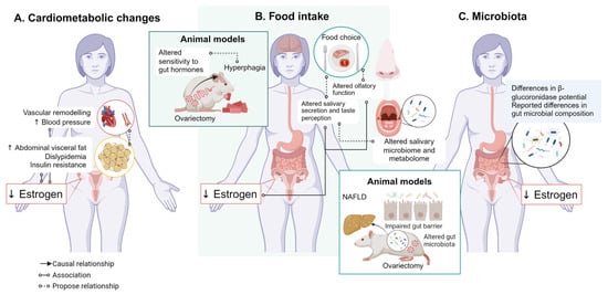
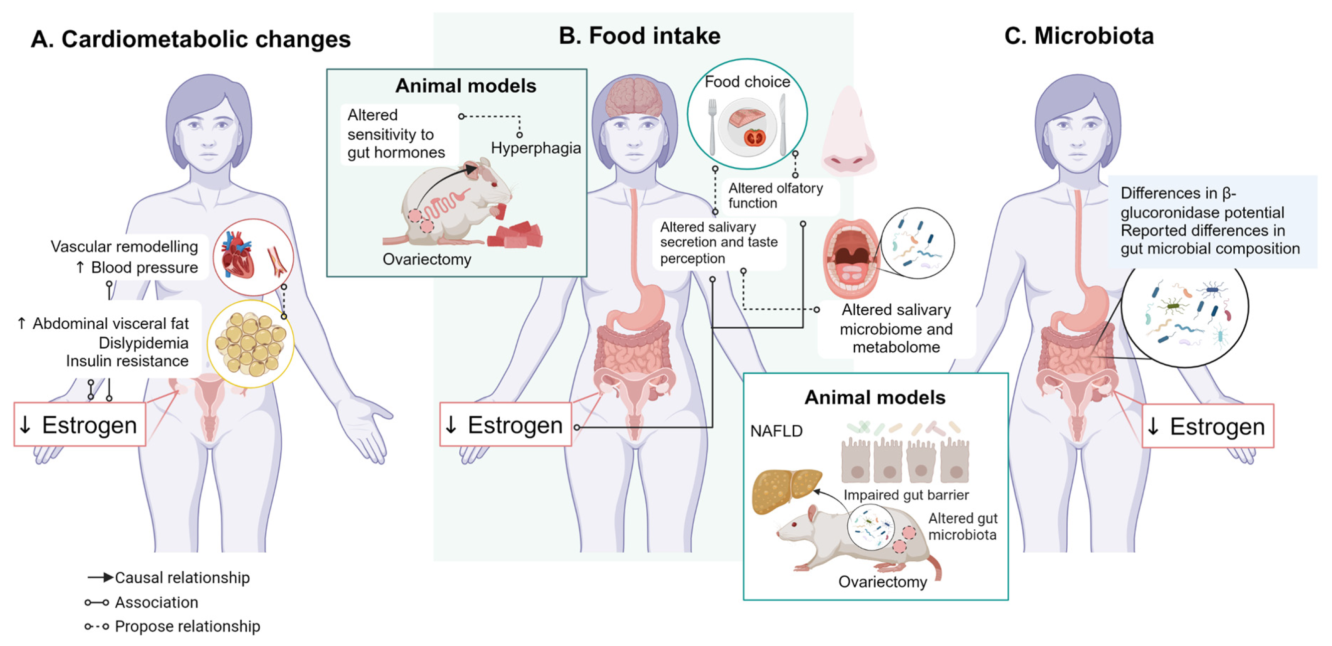

Disclaimer/Publisher’s Note: The statements, opinions and data contained in all publications are solely those of the individual author(s) and contributor(s) and not of MDPI and/or the editor(s). MDPI and/or the editor(s) disclaim responsibility for any injury to people or property resulting from any ideas, methods, instructions or products referred to in the content. |
© 2026 by the authors. Licensee MDPI, Basel, Switzerland. This article is an open access article distributed under the terms and conditions of the Creative Commons Attribution (CC BY) license.