


Abstract
Background/Objectives: Protein Energy Wasting (PEW) is common among patients undergoing maintenance hemodialysis (HD), particularly older adults, and is associated with increased frailty, susceptibility to infections, hospitalization, and mortality. Nutritional counseling alone may be insufficient to improve nutritional status in some patients. Supplementation with essential amino acids and ketoanalogues (EAA/KA) may support nutritional status in patients undergoing HD. This study aimed to evaluate changes in nutritional and bioimpedance parameters in older patients undergoing maintenance HD and receiving EAA/KA supplementation. Methods: This retrospective observational study evaluated changes over six months in 10 patients undergoing maintenance HD with a baseline Malnutrition–Inflammation Score (MIS) greater than 7 who received EAA/KA supplementation. Results: After six months, serum albumin levels increased significantly (p < 0.001), while extracellular water (ECW) decreased (p = 0.024). Bioelectrical impedance analysis (BIA) also showed a significant increase in phase angle (PA; p = 0.029) and significant decreases in the extracellular mass-to-body cell mass ratio (ECM/BCM; p = 0.034) and the sodium-to-potassium ratio (Na/K; p = 0.025). The MIS also decreased significantly (p = 0.003). Conclusions: In this small retrospective study, six months of EAA/KA supplementation were associated with improvements in serum albumin, MIS, and several bioimpedance parameters in patients undergoing maintenance HD. These preliminary findings suggest a potential role for EAA/KA supplementation in the nutritional management of patients at nutritional risk. However, the small sample size, retrospective design, and absence of a control group limit the interpretation of these findings. Further prospective controlled studies involving larger populations are needed to confirm these observations.
1. Introduction
PEW is characterized by a reduction in body protein and energy stores and is often associated with impaired functional capacity in patients with end-stage kidney disease [1,2]. In patients with chronic kidney disease (CKD), particularly those undergoing HD, PEW is associated with adverse clinical outcomes, increased hospitalization rates, and higher mortality [3].
A meta-analysis by Carrero et al. [4] reported a prevalence of PEW ranging from 28% to 54% among patients receiving maintenance HD.
The pathogenesis of PEW in CKD is multifactorial and closely related to the metabolic disturbances associated with uremia [5]. Chronic inflammation, metabolic abnormalities, and metabolic acidosis may promote protein catabolism and increase energy expenditure. These processes, together with reduced dietary intake, protein losses during dialysis, and physical inactivity, contribute to the development and persistence of PEW [6,7,8,9].
PEW is associated with increased morbidity and mortality, loss of muscle mass, frailty, and impaired quality of life. Early identification and appropriate nutritional management are therefore essential. The KDIGO 2024 Clinical Practice Guideline emphasizes the role of renal dietitians and qualified nutrition professionals in providing individualized dietary counselling for patients with CKD [10].
According to the KDOQI 2020 Clinical Practice Guidelines, metabolically stable adults undergoing maintenance HD should generally receive a daily protein intake of 1.0–1.2 g/kg body weight and an energy intake of 25–35 kcal/kg body weight, individualized according to age, physical activity, body composition, clinical condition, and nutritional goals [11].
MIS [12] is a validated, simple, and cost-effective tool for assessing nutritional and inflammatory status in HD patients. It comprises 10 components covering medical history, dietary intake, anthropometric measurements, laboratory parameters, and functional capacity. Several studies have investigated the associations between MIS and quality of life, comorbidities, and mortality in patients with CKD [13,14,15,16]. In particular, an MIS score above 7 has been associated with an increased risk of mortality [13]. Based on this evidence, we adopted an MIS cut-off of >7 to identify patients at high nutritional risk.
EAA/KA formulations are primarily used to support nutritional management in selected patients with advanced CKD following low- or very-low-protein diets [17,18,19]. Ketoanalogues, commonly administered as calcium salts, provide nitrogen-free precursors of essential amino acids, while essential amino acid supplementation supports protein synthesis. Following absorption, ketoanalogues can undergo transamination by accepting amino groups from non-essential amino acids, thereby facilitating nitrogen reutilization for amino acid synthesis and potentially limiting nitrogenous waste production.
Although EAA/KA supplementation is traditionally associated with protein-restricted diets in patients with advanced CKD who are not yet receiving dialysis, this approach may also have a role in selected HD patients with PEW. Studies investigating EAA/KA supplementation in HD patients [20,21,22,23,24,25] have reported improvements in serum albumin levels, Subjective Global Assessment (SGA) scores, and appetite. Potential benefits have also been described in anemia management [26] and in calcium–phosphorus metabolism and parathyroid hormone (PTH) regulation [27,28]. A reduction in peripheral edema has also been reported [23].
Given the clinical burden of PEW in HD patients and the limited evidence regarding the nutritional effects of EAA/KA supplementation in this population, the present study aimed to evaluate changes in nutritional status in elderly patients undergoing maintenance HD and treated with EAA/KA over a six-month observation period.
2. Materials and Methods
Study design and population. This retrospective observational study evaluated patients undergoing maintenance HD at our dialysis center who had a MIS > 7 at baseline [13]. Based on their nutritional status, EAA/KA supplementation was prescribed. No exclusion criteria were applied. Fourteen patients initiated EAA/KA supplementation during the study period. Of these, 10 completed the six-month follow-up and had measurements available at both baseline (T0) and six months (T6); these 10 patients constituted the population included in the primary paired analysis.
The resulting study population was predominantly elderly and characterized by a substantial burden of comorbidities. As shown in Table 1, the high Charlson Comorbidity Index (CCI) [29] scores reflect the considerable clinical complexity of the study population.
According to the KDOQI 2020 Clinical Practice Guideline, patients were prescribed a diet providing 1.2 g of protein/kg body weight/day and 30 kcal/kg body weight/day.
Nutritional status was assessed at baseline and after 3 and 6 months, in accordance with the nutritional monitoring protocol established by the Lazio Regional Health Authority for patients undergoing maintenance HD.
During the observation period, patients also received erythropoietin alfa, iron supplementation with ferric carboxymaltose (FCM), a vitamin D analogue (paricalcitol), a calcimimetic agent (etelcalcetide), a phosphate binder (sucroferric oxyhydroxide, SFO), and statin therapy (simvastatin). The medications and their respective dosages at baseline and at the end of the observation period are reported in Table 2.
Based on previously published studies [20,21,22,23,24,25,26,27,28], EAA/KA supplementation was prescribed at a dose of one tablet per 10 kg of body weight, corresponding to 60 mg/kg body weight. This dose was lower than that commonly used in patients with CKD receiving conservative treatment and following low- or very-low-protein diets, owing to the calcium content of the tablets (Appendix A, Table A3).
The EAA/KA supplements were dispensed directly by the dialysis center upon patients’ request, and each dispensing was recorded. Treatment adherence was assessed by pill count.
Nutritional Assessment and Data Collection. The nutritional assessments included in this study were those prescribed by the Lazio Regional Health Authority for monitoring the nutritional status of patients undergoing maintenance HD. Assessments were performed every three months.
Blood sampling and dialysis adequacy. Blood samples were collected before the first dialysis session following the longest interdialytic interval. The timing of blood sampling followed standard clinical practice and is described in Appendix A. Laboratory assessments included hemoglobin, complete blood count, iron metabolism parameters (serum iron, transferrin, and ferritin), and serum creatinine. Pre- and post-dialysis blood urea levels were measured to calculate single-pool Kt/V (spKt/V), an index of dialysis adequacy, and the normalized protein catabolic rate (nPCR), an indirect indicator of dietary protein intake, using Daugirdas’ single-pool equations.
Nutritional and biochemical parameters. Nutritional indicators included serum albumin, measured by protein electrophoresis, transferrin, total cholesterol, and triglyceride levels.
Mineral metabolism. Serum calcium, phosphorus, and parathyroid hormone (PTH) levels were measured to assess mineral metabolism.
Anthropometric measurements. Body mass index (BMI) was calculated using post-dialysis body weight measured at the midweek hemodialysis session.
Bioelectrical impedance analysis. Body composition was assessed using a BIA 101 device (Akern®, RJL Systems, Florence, Italy). Measurements were performed 20 min after the end of the midweek hemodialysis session. The timing of BIA measurements followed standard clinical practice and is described in Appendix A. Impedance data were processed using Akern® software Bodygram PLUS 1.2.2 and Jamovi 2.6.44. In addition to standard impedance-derived parameters, extracellular mass-to-body cell mass ratio (ECM/BCM) was evaluated. Previous studies have reported that ECM/BCM values greater than 1.2 in HD patients are associated with malnutrition and correlate significantly with MIS and mortality [30,31,32,33,34].
Malnutrition–Inflammation Score. The MIS was calculated according to the method described in the literature [12]. The criteria used for score calculation are presented in Appendix A (Table A1).
Statistical analysis: Statistical analyses were performed using Jamovi 2.6.44 software [35]. Descriptive statistics are used to summarize the study variables. Nutritional assessments were performed at baseline (T0), at three months (T3), and at six months (T6) in all 10 patients who completed the study. The primary analysis focused on within-patient changes in continuous variables between baseline and the end of the six-month observation period. These changes were assessed using the paired Student’s t-test. The normality of the within-subject differences (T6–T0) was assessed using Q–Q plots and the Shapiro–Wilk test. Mean differences (T6–T0) and their corresponding 95% confidence intervals (CIs) were calculated to quantify the magnitude and precision of the observed changes and to provide preliminary estimates to inform sample size calculations for future studies [36]. The longitudinal evolution of selected parameters across T0, T3, and T6 is presented graphically using box plots. Changes in individual MIS components between baseline and six months were explored using the Wilcoxon signed-rank test for paired observations, given the ordinal nature of the item scores and the small sample size. All tests were two-sided, and p values < 0.05 were considered statistically significant.
3. Results
The observation period extended from March 2024 to June 2026. During this period, 14 patients initiated treatment, of whom 10 completed the 6-month follow-up. Four patients discontinued the study: two were hospitalized, one due to cognitive decline and the other due to metastatic cancer; one patient died from an unspecified cause while travelling; and one patient was excluded because of poor treatment adherence.
All patients underwent bicarbonate hemodialysis three times per week and were anuric, with no residual renal function (RRF).
Baseline patient characteristics are presented in Table 1. EAA/KA supplementation was prescribed on the basis of nutritional status, defined as an MIS score > 7, irrespective of dialysis vintage or the presence of severe comorbidities, including cognitive impairment (n = 2) and cancer (n = 3). CCI was also calculated to assess the overall comorbidity burden [29]. All patients had a history of hypertension. However, the primary etiology of kidney failure could not be definitively established, as no patient had undergone a kidney biopsy.
Table 1.
Baseline patient characteristics.
Table 2 summarizes the pharmacological treatments and their respective dosages at baseline and at the end of the 6-month follow-up.
Table 2.
Pharmacological Therapy.
Table 3 summarizes the nutritional, anthropometric, biochemical, dialysis-related, and body composition parameters measured at baseline (T0) and after 6 months of treatment with EAA/KA (T6).
Table 3.
Comparison of nutritional, anthropometric, dialysis, biochemical and body composition parameters between baseline (T0) and 6 months (T6).
Due to the small sample size, mean differences between baseline (T0) and the 6-month follow-up (T6) were also calculated to quantify the magnitude of the observed changes, as reported in Table 4.
Table 4.
Mean differences and 95% confidence intervals for clinical, biochemical and body composition parameters from T0 to T6.
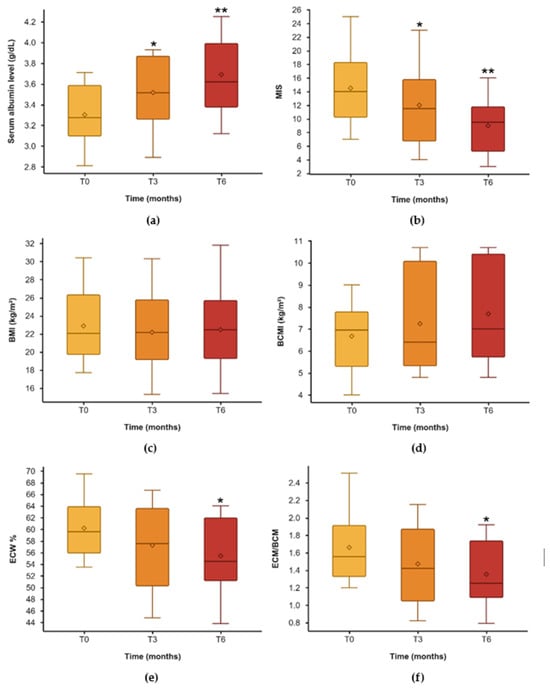
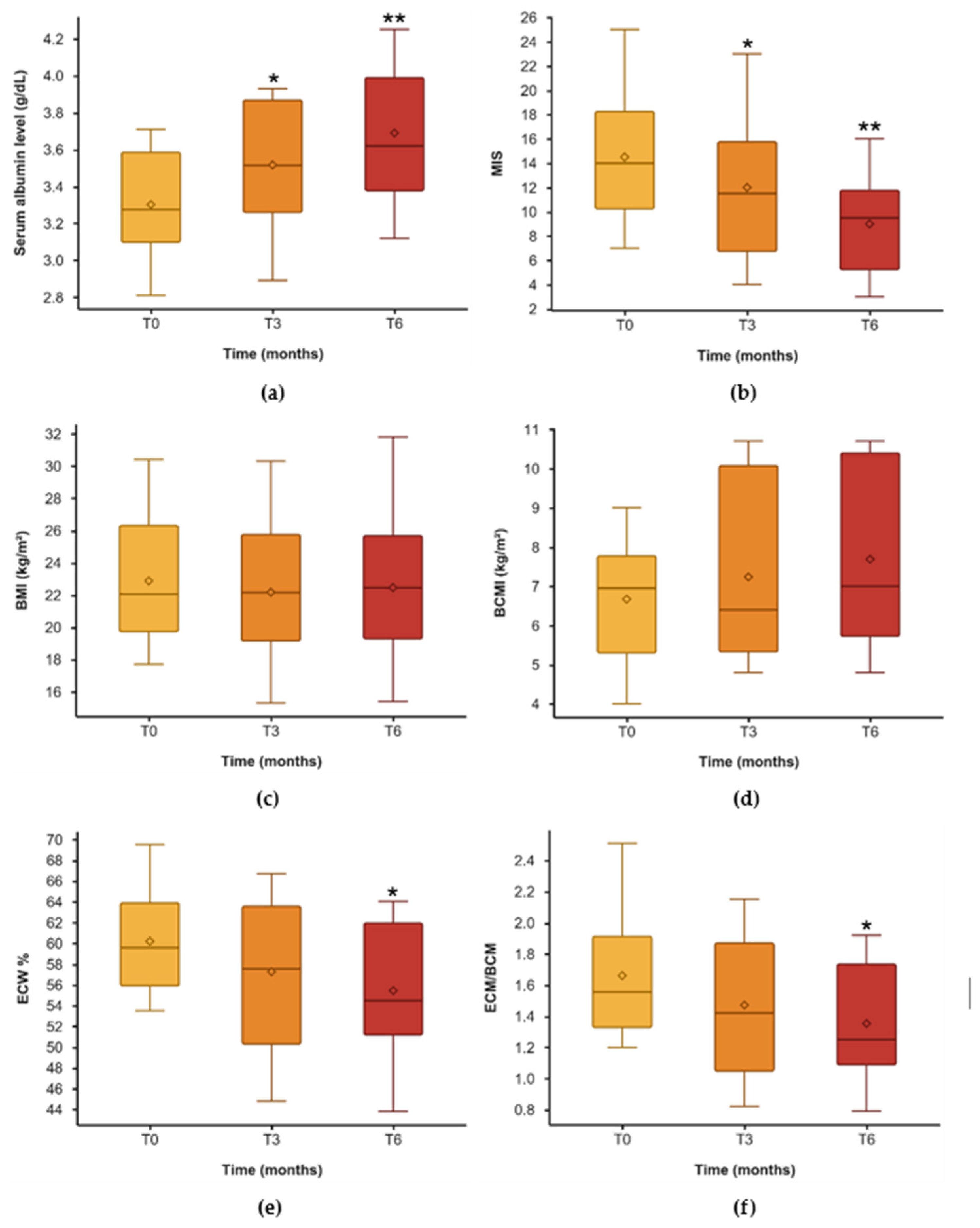
The main findings were an increase in serum albumin levels (p < 0.001; mean difference, +0.39 g/dL) and a reduction in MIS (p = 0.003; mean difference, −5.50). Both changes may be clinically relevant. Significant reductions were also observed in the ECM/BCM (p = 0.034; mean difference, −0.31) and extracellular water expressed as a percentage of total body water (ECW%; p = 0.025). In contrast, body weight and BMI remained substantially unchanged. Bold type indicates statistically significant confidence intervals.
Regarding biochemical parameters, serum calcium levels increased significantly (p = 0.024), while remaining within the normal range. Serum total cholesterol levels also increased, although the change did not reach statistical significance (p = 0.084). The corresponding mean differences were small (+0.37 mg/dL for serum calcium and +7 mg/dL for total cholesterol). No significant changes were observed in complete blood count parameters. Mean serum phosphorus levels increased, whereas PTH levels decreased; neither change was statistically significant.
Dialysis-related parameters, including predialysis serum urea levels, spKt/V, and nPCR, showed no statistically significant changes over the observation period.
BIA showed a significant increase in phase angle (PA; p = 0.029), together with significant decreases in ECM/BCM (p = 0.034) and the Na/K (p = 0.025). These findings, alongside the reduction in extracellular water (ECW) in absolute terms (p = 0.024) and as a percentage of total body water (ECW%; p = 0.025), are consistent with an improvement in hydration status and may indicate a favorable change in body composition. Increases in body cell mass (BCM; p = 0.070) and body cell mass index (BCMI; p = 0.078) were also observed, although these changes did not reach statistical significance. No statistically significant changes were observed in muscle mass (MM) or appendicular skeletal muscle mass (ASMM).
The box plots in Figure 1 illustrate the three-monthly trends in serum albumin, MIS, BMI, BCMI, ECW%, and the ECM/BCM ratio over the six-month observation period.
Figure 1.
Longitudinal changes in nutritional status, anthropometric parameters and body composition from baseline (T0) to 3 months (T3) and 6 months (T6). (a) Serum albumin level, (b) MIS, (c) BMI, (d) BCMI, (e) ECW%, (f) ECM/BCM. Box plots represent the distribution of values, while dots indicate mean values. Statistical significance of changes over time is reported as p-values. * p < 0.05; ** p < 0.001.
4. Discussion
The main limitation of this study is the small sample size. The study population consisted of elderly patients undergoing maintenance HD, with a substantial burden of comorbidities. Hospitalizations, clinical complications, and difficulties with treatment adherence made it challenging to obtain complete six-month follow-up data. Indeed, only 10 of the 14 patients who initiated EAA/KA supplementation completed the observation period.
Another important limitation is the absence of a control group. Therefore, it is not possible to determine whether the observed changes in nutritional and bioimpedance parameters were directly attributable to EAA/KA supplementation or were influenced by other factors, including changes in dietary intake, concomitant treatments, or variations in patients’ clinical condition during follow-up.
The retrospective design represents a further limitation, as some potentially relevant variables, including C-reactive protein and interleukin-6, were not available. Inflammation may have acted as a confounding factor, particularly when interpreting changes in serum albumin and nutritional status. Ferritin was the only available indirect marker related to inflammation; however, its interpretation is complicated by its role as an iron-storage protein and its response to iron supplementation.
Another limitation of this study was the lack of a detailed assessment of dietary intake, such as a three-day dietary record, which would have allowed a more accurate quantification of patients’ energy and protein intake. Nevertheless, the MIS incorporates qualitative assessments of dietary intake and gastrointestinal symptoms in items 2 and 3, respectively. These components provide information on changes in food intake and appetite, as well as on the presence of gastrointestinal symptoms such as nausea and vomiting. Although this approach does not allow a precise quantification of actual dietary intake, changes in these MIS components suggested improvements in appetite and a reduction in gastrointestinal symptoms in some patients during the observation period. These findings may help contextualize the observed improvement in overall nutritional status, although they cannot establish an increase in energy or protein intake (Appendix A, Table A2).
The main findings after six months of treatment were a significant increase in serum albumin levels (p < 0.001; mean difference, +0.39 g/dL) and a significant reduction in the MIS (p = 0.003; mean difference, −5.50). These findings suggest a possible improvement in nutritional status during EAA/KA supplementation.
An increase in serum albumin levels was also reported by Sook et al. [21] in HD patients with hypoalbuminemia (serum albumin < 3.8 g/dL) treated with EAA/KA, with a mean increase of 0.2 g/dL. Similarly, Zakar et al. [23], in a cohort of 42 HD patients receiving EAA/KA at a dose of 0.1 g/kg body weight/day, reported a significant improvement in serum albumin levels among patients with baseline values below 35 g/L. PTH levels also showed a trend towards improvement during supplementation, while nutritional status improved, as indicated by better SGAscores.
The increase in serum albumin observed in our study may be clinically relevant. In addition to its role as a nutritional marker, albumin contributes substantially to plasma oncotic pressure and therefore plays a role in maintaining intravascular fluid balance. Improved albumin levels could potentially support plasma refilling during HD; however, this mechanism cannot be established from the present data. The observed reductions in ECW, both in absolute terms and as a percentage of total body water (ECW%), are consistent with a change in hydration status, although they do not directly demonstrate improved vascular refilling or hemodynamic stability.
Body weight and total body water (TBW) remained essentially unchanged over the observation period, with small, non-significant decreases. In conjunction with the reduction in ECW and ECW%, these findings may indicate a change in fluid distribution rather than a substantial reduction in total body water. The significant decrease in the Na/K and the ECM/BCM provides additional information on changes in body composition and hydration status. Nevertheless, these impedance-derived parameters should be interpreted cautiously, as they do not independently establish a redistribution of water into the intracellular compartment or an improvement in muscle quality.
Regarding calcium–phosphate metabolism, serum calcium levels increased significantly but remained within the normal range. As EAA/KA formulations are administered as calcium salts, their calcium content should be considered when interpreting this finding. Ketoanalogues may also be relevant to phosphate management in patients with CKD, but the increase in serum phosphorus observed at T6, although not statistically significant, cannot be attributed specifically to increased dietary phosphate intake without additional dietary data. Calcium, phosphorus, and PTH should therefore be monitored during supplementation, particularly in patients receiving other treatments that affect mineral metabolism.
Other nutritional interventions have also shown potential benefits in HD patients with PEW. Renal-specific oral nutritional supplements (RS-ONS) have been associated with increased serum albumin levels and reduced MIS [37]. In-center administration of oral nutritional supplements during HD has also been associated with lower hospital readmission rates among patients with severe hypoalbuminemia following hospital discharge [38]. Furthermore, intradialytic administration has been reported to be well tolerated, despite concerns about potential hypotension [39]. Although RS-ONS were used in our dialysis center to support malnourished patients until a few years ago, they are currently no longer available at our center. Consequently, patients who may benefit from this nutritional intervention may need to purchase these products independently, which could limit access for those unable to afford the associated costs.
Intradialytic parenteral nutrition (IDPN), when combined with oral nutritional supplementation, has been associated with improvements in prealbumin, serum albumin, body weight, spontaneous oral intake, and MIS, as well as with favorable clinical outcomes in some studies [40,41,42]. However, IDPN is generally intended to complement, rather than replace, oral nutritional intake. Its administration requires consideration of the additional fluid volume, which may be particularly relevant in patients with hypertension or fluid overload. Blood glucose monitoring is also important in patients with diabetes.
Overall, nutritional management and exercise interventions based on the concept of rehabilitation nutrition are important components of the management of malnutrition in HD patients [43,44]. Oral or intravenous protein supplementation may also help address inadequate nutrient intake and interrupt the cycle of PEW [45,46]. Within this context, our findings suggest that EAA/KA supplementation may be associated with improvements in selected nutritional and bioimpedance parameters in elderly patients undergoing maintenance HD. However, given the retrospective design, small sample size, absence of a control group, and potential confounding factors, these findings should be considered preliminary and require confirmation in larger prospective controlled studies.
5. Conclusions
In this small retrospective observational study of patients undergoing maintenance HD, six months of EAA/KA supplementation was associated with a significant increase in serum albumin levels and significant reductions in the MIS and the ECM/BCM. A significant decrease in ECW% was also observed, suggesting a possible improvement in hydration status.
These findings suggest that EAA/KA supplementation may be a promising nutritional strategy for patients undergoing maintenance hemodialysis who are at nutritional risk. However, given the small sample size, the presence of substantial comorbidities, the retrospective design, and the absence of a control group, these results should be interpreted with caution. Further prospective controlled studies involving larger patient populations are needed to confirm these preliminary findings and clarify the potential role of EAA/KA supplementation in the nutritional management of patients undergoing maintenance hemodialysis.
Author Contributions
Conceptualization, methodology, software, validation, formal analysis, investigation, resources, data curation, writing—original draft preparation, S.P.; writing—review and editing, S.P., A.C., E.A., F.C., L.P.; supervision, E.E. All authors have read and agreed to the published version of the manuscript.
Funding
This research received no external funding.
Institutional Review Board Statement
The study was conducted in accordance with the Declaration of Helsinki. Ethical review and approval were waived for this study because the Italian Ministerial Decree of 26 January 2023 stipulates that ethics committees “are responsible exclusively for the evaluation of Phase I, II, III, and IV clinical trials involving medical devices and medicinal products for human use”. Our study is a non-interventional, non-prospective study; patients were managed in accordance with best clinical practices, and the study is exclusively a retrospective observational study for which the mentioned Italian regulation does not require any ethical review.
Informed Consent Statement
Written informed consent has been obtained from the patients to publish this paper.
Data Availability Statement
Data is unavailable due to privacy.
Acknowledgments
During the preparation of this manuscript, the authors used Jamovi for the statistical analysis. The authors have reviewed and edited the output and take full responsibility for the content of this publication.
Conflicts of Interest
The authors declare no conflicts of interest.
Abbreviations
The following abbreviations are used in this manuscript:
| ASMM | Appendicular Skeletal Muscle Mass |
| BCM | Body Cell Mass |
| BCMI | Body Cell Mass Index |
| BMI | Body Mass Index |
| CCI | Charlson Comorbidity Index |
| CKD | Chronic Kidney Disease |
| EAA-KA | Essential Amino Acids and Keto-Analogues |
| ECW | Extracellular Water |
| ECW% | Extracellular Water percentage of Total Body Water |
| EPO-α | Erythropoietin-α |
| ESKD | End-Stage Kidney Disease |
| FCM | Ferric Carboxymaltose |
| FFM | Fat-Free Mass |
| FM | Fat Mass |
| HD | Hemodialysis |
| IDPN | Intradialytic Parenteral Nutrition |
| KDIGO | Kidney Disease Improving Global Outcome |
| Kt/V sp | Dialytic clearance single pool |
| MIS | Malnutrition–Inflammation Score |
| MM | Muscle Mass |
| Na/K | Sodiumto–Potassium Ratio |
| nPCR sp | Normalized Protein Catabolic Rate single pool |
| ONS | Oral Nutrition Supplements |
| PA | Phase Angle |
| PEW | Protein Energy Wasting |
| PTH | Parathormone |
| RRF | Residual Renal Function |
| Rz | Resistance |
| SFO | Sucroferric oxyhydroxide |
| SGA | Subjective Global Assessment |
| TBW | Total Body Water |
| TIBC | Total Iron-Binding Capacity |
| Xc | Reactance |
Appendix A. Management of HD Patients—Clinical Practice and Nutritional Considerations
HD is generally initiated in patients with end-stage kidney disease (ESKD) when kidney function is no longer sufficient to maintain adequate metabolic, fluid, and electrolyte homeostasis. Upon initiating HD at our Dialysis Centre, patients undergo a nutritional assessment by a nutritionist, who prescribes an individualized dietary plan. According to the KDOQI 2020 Clinical Practice Guidelines, the recommended targets for metabolically stable adults undergoing maintenance HD are a protein intake of 1.0–1.2 g/kg body weight/day and an energy intake of 25–35 kcal/kg body weight/day. In ourcenter, dietary plans are generally designed to provide 1.2 g protein/kg body weight/day and 30 kcal/kg body weight/day [11].
Dietary phosphorus intake is managed according to patients’ serum phosphorus levels and individual nutritional requirements. A daily phosphorus intake of approximately 800 mg may be recommended when clinically appropriate. In addition to high-biological-value protein sources, plant-based proteins are encouraged where suitable, as phosphorus from plant foods generally has lower bioavailability than phosphorus from animal-derived foods and phosphate additives.
Nutritional assessment includes laboratory investigations, BIA, and completion of the MIS form (Table A1). This assessment is performed at the initiation of HD and subsequently every three months.
Laboratory parameters. Blood samples are collected before the first HD session following the longest interdialytic interval. Pre- and post-dialysis serumurea measurements obtained during this session are used to calculate single-pool Kt/V (spKt/V), an index of dialysis adequacy, and the normalized protein catabolic rate (nPCR), an indirect estimate of protein intake under conditions of metabolic stability, using the single-pool equations described by Daugirdas.
The timing of blood sampling also allows assessment of biochemical changes occurring over the interdialytic interval, including potentially elevated serum potassium and phosphorus levels before dialysis. These measurements support the nephrologist in evaluating electrolyte control and tailoring dietary advice and dialysis management to the individual patient.
Bioelectrical impedance analysis (BIA). BIA is performed approximately 20 min after the end of the midweek hemodialysis session, using a standardized post-dialysis time interval to improve the consistency of measurements. This timing was chosen because excessive fluid accumulation during the long interdialytic interval may prevent patients from reaching their prescribed dry weight by the end of the subsequent dialysis session. Performing BIA after the midweek session allows assessment of patients’ hydration and nutritional status under conditions in which the prescribed dry weight is more likely to have been achieved, thereby reducing the potential influence of interdialytic fluid overload on the measurements. Standardizing the time between the end of dialysis and BIA is also important because post-dialysis fluid redistribution may affect bioelectrical impedance measurements.
Table A1.
MIS questionnaire.
The analysis of individual MIS components showed statistically significant reductions in scores for changes in post-dialysis dry weight, dietary intake, gastrointestinal symptoms, signs of muscle wasting, and serum albumin after six months of follow-up. No statistically significant changes were observed in functional capacity, comorbidity, fat stores, BMI, or total iron-binding capacity/serum transferrin. The results for all individual MIS components are presented in Table A2.
Table A2.
Changes in individual MIS components between baseline (T0) and six months (T6).
Table A3.
Contents of one EAA/KA tablet and intake per kgBW/day at the administered dose.
References
- Fouque, D.; Kalantar-Zadeh, K.; Kopple, J.; Cano, N.; Chauveau, P.; Cuppari, L.; Franch, H.; Guarnieri, G.; Ikizler, T.A.; Kaysen, G.; et al. A Proposed Nomenclature and Diagnostic Criteria for Protein–Energy Wasting in Acute and Chronic Kidney Disease. Kidney Int. 2008, 73, 391–398. [Google Scholar] [CrossRef] [Scilit] [PubMed]
- Cupisti, A.; D’Alessandro, C.; Caselli, G.M.; Group M.I.TO.—DP; Egidi, M.F.; Bottai, A.; Onnis, F.E.; Mecacci, A.; Bernardi, M.; Mencherini, A.; et al. Nutritional and Functional assessment of peritoneal dialysis patients in the clinical practice: Report from MITO-DP Group. G. Ital. Nefrol. 2016, 33, gin/33.4.6. [Google Scholar]
- Alp Ikizler, T.; Cano, N.J.; Franch, H.; Fouque, D.; Himmelfarb, J.; Kalantar-Zadeh, K.; Kuhlmann, M.K.; Stenvinkel, P.; TerWee, P.; Teta, D.; et al. Prevention and Treatment of Protein Energy Wasting in Chronic Kidney Disease Patients: A Consensus Statement by the International Society of Renal Nutrition and Metabolism. Kidney Int. 2013, 84, 1096–1107. [Google Scholar] [CrossRef] [Scilit] [PubMed]
- Carrero, J.J.; Thomas, F.; Nagy, K.; Arogundade, F.; Avesani, C.M.; Chan, M.; Chmielewski, M.; Cordeiro, A.C.; Espinosa-Cuevas, A.; Fiaccadori, E.; et al. Global Prevalence of Protein-Energy Wasting in Kidney Disease: A Meta-Analysis of Contemporary Observational Studies From the International Society of Renal Nutrition and Metabolism. J. Ren. Nutr. 2018, 28, 380–392. [Google Scholar] [CrossRef] [Scilit] [PubMed]
- Carrero, J.J.; Stenvinkel, P.; Cuppari, L.; Ikizler, T.A.; Kalantar-Zadeh, K.; Kaysen, G.; Mitch, W.E.; Price, S.R.; Wanner, C.; Wang, A.Y.M.; et al. Etiology of the Protein-Energy Wasting Syndrome in Chronic Kidney Disease: A Consensus Statement From the International Society of Renal Nutrition and Metabolism (ISRNM). J. Ren. Nutr. 2013, 23, 77–90. [Google Scholar] [CrossRef] [Scilit] [PubMed]
- Hanna, R.M.; Ghobry, L.; Wassef, O.; Rhee, C.M.; Kalantar-Zadeh, K. A Practical Approach to Nutrition, Protein-Energy Wasting, Sarcopenia, and Cachexia in Patients with Chronic Kidney Disease. Blood Purif. 2020, 49, 202–211. [Google Scholar] [CrossRef] [Scilit] [PubMed]
- Wang, X.H.; Mitch, W.E. Mechanisms of Muscle Wasting in Chronic Kidney Disease. Nat. Rev. Nephrol. 2014, 10, 504–516. [Google Scholar] [CrossRef] [Scilit] [PubMed]
- Sahathevan, S.; Khor, B.-H.; Ng, H.-M.; Abdul Gafor, A.H.; Mat Daud, Z.A.; Mafra, D.; Karupaiah, T. Understanding Development of Malnutrition in Hemodialysis Patients: A Narrative Review. Nutrients 2020, 12, 3147. [Google Scholar] [CrossRef] [Scilit] [PubMed]
- Bozzoli, L.; Sabatino, A.; Regolisti, G.; Morabito, S.; Donadio, C.; Cupisti, A.; Piotti, G.; Fiaccadori, E. Protein-energy wasting and nutritional supplementation in chronic hemodialysis. G. Ital. Nefrol. 2015, 32, gin/32.5.1. [Google Scholar] [PubMed]
- Stevens, P.E.; Ahmed, S.B.; Carrero, J.J.; Foster, B.; Francis, A.; Hall, R.K.; Herrington, W.G.; Hill, G.; Inker, L.A.; Kazancıoğlu, R.; et al. KDIGO 2024 Clinical Practice Guideline for the Evaluation and Management of Chronic Kidney Disease. Kidney Int. 2024, 105, S117–S314. [Google Scholar] [CrossRef] [Scilit] [PubMed]
- Ikizler, T.A.; Burrowes, J.D.; Byham-Gray, L.D.; Campbell, K.L.; Carrero, J.-J.; Chan, W.; Fouque, D.; Friedman, A.N.; Ghaddar, S.; Goldstein-Fuchs, D.J.; et al. KDOQI Clinical Practice Guideline for Nutrition in CKD: 2020 Update. Am. J. Kidney Dis. 2020, 76, S1–S107. [Google Scholar] [CrossRef] [Scilit] [PubMed]
- Kalantar-Zadeh, K.; Kopple, J.D.; Block, G.; Humphreys, M.H. A Malnutrition-Inflammation Score Is Correlated with Morbidity and Mortality in Maintenance Hemodialysis Patients. Am. J. Kidney Dis. 2001, 38, 1251–1263. [Google Scholar] [CrossRef] [Scilit] [PubMed]
- Borges, M.C.C.; Vogt, B.P.; Martin, L.C.; Caramori, J.C.T. Malnutrition Inflammation Score Cut-off Predicting Mortality in Maintenance Hemodialysis Patients. Clin. Nutr. ESPEN 2017, 17, 63–67. [Google Scholar] [CrossRef] [Scilit] [PubMed]
- Amparo, F.C.; Kamimura, M.A.; Molnar, M.Z.; Cuppari, L.; Lindholm, B.; Amodeo, C.; Carrero, J.J.; Cordeiro, A.C. Diagnostic Validation and Prognostic Significance of the Malnutrition-Inflammation Score in Nondialyzed Chronic Kidney Disease Patients. Nephrol. Dial. Transplant. 2015, 30, 821–828. [Google Scholar] [CrossRef] [Scilit] [PubMed]
- Rambod, M.; Bross, R.; Zitterkoph, J.; Benner, D.; Pithia, J.; Colman, S.; Kovesdy, C.P.; Kopple, J.D.; Kalantar-Zadeh, K. Association of Malnutrition-Inflammation Score with Quality of Life and Mortality in Hemodialysis Patients: A 5-Year Prospective Cohort Study. Am. J. Kidney Dis. 2009, 53, 298–309. [Google Scholar] [CrossRef] [Scilit] [PubMed]
- Molnar, M.Z.; Czira, M.E.; Rudas, A.; Ujszaszi, A.; Lindner, A.; Fornadi, K.; Kiss, I.; Remport, A.; Novak, M.; Kennedy, S.H.; et al. Association of the Malnutrition-Inflammation Score with Clinical Outcomes in Kidney Transplant Recipients. Am. J. Kidney Dis. 2011, 58, 101–108. [Google Scholar] [CrossRef] [Scilit] [PubMed]
- Barsotti, G.; Guiducci, A.; Ciardella, F.; Giovannetti, S. Effects on Renal Function of a Low-Nitrogen Diet Supplemented with Essential Amino Acids and Ketoanalogues and of Hemodialysis and Free Protein Supply in Patients with Chronic Renal Failure. Nephron 1981, 27, 113–117. [Google Scholar] [CrossRef] [Scilit] [PubMed]
- Garneata, L.; Stancu, A.; Dragomir, D.; Stefan, G.; Mircescu, G. Ketoanalogue-Supplemented Vegetarian Very Low–Protein Diet and CKD Progression. JASN 2016, 27, 2164–2176. [Google Scholar] [CrossRef] [Scilit] [PubMed]
- Aparicio, M.; Bellizzi, V.; Chauveau, P.; Cupisti, A.; Ecder, T.; Fouque, D.; Garneata, L.; Lin, S.; Mitch, W.; Teplan, V.; et al. Do Ketoanalogues Still Have a Role in Delaying Dialysis Initiation in CKD Predialysis Patients? Semin. Dial. 2013, 26, 714–719. [Google Scholar] [CrossRef] [Scilit] [PubMed]
- Li, H.-L.; Li, H.; Cao, Y.-F.; Qi, Y.; Wang, W.-Q.; Liu, S.-Q.; Yang, C.-D.; Yu, X.-Y.; Xu, T.; Zhu, Y.; et al. Effects of Keto Acid Supplements on Chinese Patients Receiving Maintenance Hemodialysis: A Prospective, Randomized, Controlled, Single-Center Clinical Study. Chin. Med. J. 2020, 133, 9–16. [Google Scholar] [CrossRef] [Scilit] [PubMed]
- Oh, S.E.; Lee, Y.-K.; Kim, J.K.; Lee, D.H.; Kim, S.-J.; Kim, S.G.; Oh, J.E.; Seo, J.W.; Yoon, J.-W.; Koo, J.-R.; et al. Effects of Keto Acids on Serum Albumin Level in Hemodialysis Patients with Hypoalbuminemia. Korean J. Nephrol. 2010, 29, 82–88. [Google Scholar]
- Chen, J. Nutrition, Phosphorus, and Keto-Analogues in Hemodialysis Patients: A Chinese Perspective. J. Ren. Nutr. 2013, 23, 214–217. [Google Scholar] [CrossRef] [Scilit] [PubMed]
- Zakar, G. The Effect of a Keto Acid Supplement on the Course of Chronic Renal Failure and Nutritional Parameters in Predialysis Patients and Patients on Regular Hemodialysis Therapy: The Hungarian Ketosteril Cohort Study. Wien. Klin. Wochenschr. 2001, 113, 688–694. [Google Scholar] [PubMed]
- Bulla, M.; Bremer, H.J.; Ronda-Vildozola, R.; Roth, B. The Effect of Oral Essential Amino Acids and Their Ketoanalogues on Children Receiving Regular Haemodialysis. Int. J. Pediatr. Nephrol. 1986, 7, 73–80. [Google Scholar] [PubMed]
- Riedel, E.; Hampl, H.; Nündel, M.; Farshidfar, G. Essential Branched-Chain Amino Acids and α-Ketoanalogues in Haemodialysis Patients. Nephrol. Dial. Transplant. 1992, 7, 117–120. [Google Scholar] [CrossRef] [Scilit] [PubMed]
- Neuhäuser, M.; Ulm, A.; Leber, H.W.; Schütterle, G. Influence of Essential Amino Acids and Keto Acids on Protein Metabolism and the Anaemia of Patients on Chronic Intermittent Haemodialysis. Proc. Eur. Dial. Transpl. Assoc. 1977, 14, 557–562. [Google Scholar]
- Fioretti, P.; Melis, G.B.; Ciardella, F.; Barsotti, G.; Orlandi, M.C.; Paoletti, A.M.; Giovannetti, S. Parathyroid Function and Pituitary-Gonadal Axis in Male Uremics; Effects of Dietary Treatment and of Maintenance Hemodialysis. Clin. Nephrol. 1986, 25, 155–158. [Google Scholar] [PubMed]
- Mahmoud, M.D.; Bouche, V.; Collin, P.; Girault, A. Effects of Keto-Analogues on Phosphocalcic and Aminoacid Metabolism in Dialysed Patients: A Crossover Study. Int. J. Artif. Organs 1989, 12, 692–696. [Google Scholar] [CrossRef] [Scilit]
- Charlson, M.E.; Pompei, P.; Ales, K.L.; MacKenzie, C.R. A New Method of Classifying Prognostic Comorbidity in Longitudinal Studies: Development and Validation. J. Chronic Dis. 1987, 40, 373–383. [Google Scholar] [CrossRef] [Scilit] [PubMed]
- Ruperto, M.; Sánchez-Muniz, F.J.; Barril, G. Predictors of Protein-energy Wasting in Haemodialysis Patients: A Cross-sectional Study. J. Hum. Nutr. Diet. 2016, 29, 38–47. [Google Scholar] [CrossRef] [Scilit] [PubMed]
- Papalini, S.; Londrino, F.; De Rocchis, D.; Criscione, A.; Soldo, M.; Espinosa, E. Bioimpedance Analysis (Extracellular Mass to Body Cell Mass Ratio) as Complementary Assessment for Malnutrition in Hemodialysis Patients. Mediterr. J. Nutr. Metab. 2024, 17, 348–356. [Google Scholar] [CrossRef] [Scilit]
- Ruperto, M.; Sánchez-Muniz, F.J.; Barril, G. Extracellular Mass to Body Cell Mass Ratio as a Potential Index of Wasting and Fluid Overload in Hemodialysis Patients. A Case-Control Study. Clin. Nutr. 2020, 39, 1117–1123. [Google Scholar] [CrossRef] [Scilit] [PubMed]
- Ruperto, M.; Barril, G. The Extracellular Mass to Body Cell Mass Ratio as a Predictor of Mortality Risk in Hemodialysis Patients. Nutrients 2022, 14, 1659. [Google Scholar] [CrossRef] [Scilit] [PubMed]
- Avram, M.M.; Fein, P.A.; Borawski, C.; Chattopadhyay, J.; Matza, B. Extracellular Mass/Body Cell Mass Ratio Is an Independent Predictor of Survival in Peritoneal Dialysis Patients. Kidney Int. 2010, 78, S37–S40. [Google Scholar] [CrossRef] [Scilit] [PubMed]
- Jamovi—Open Statistical Software. Available online: https://www.jamovi.org/index.html (accessed on 4 August 2026).
- Bonett, D.G. Confidence Intervals for Standardized Linear Contrasts of Means. Psychol. Methods 2008, 13, 99–109. [Google Scholar] [CrossRef] [Scilit] [PubMed]
- Sezer, S.; Bal, Z.; Tutal, E.; Uyar, M.E.; Acar, N.O. Long-Term Oral Nutrition Supplementation Improves Outcomes in Malnourished Patients with Chronic Kidney Disease on Hemodialysis. J. Parenter. Enter. Nutr. 2014, 38, 960–965. [Google Scholar] [CrossRef] [Scilit] [PubMed]
- Leonberg-Yoo, A.K.; Wang, W.; Weiner, D.E.; Lacson, E. Oral Nutritional Supplements and 30-day Readmission Rate in Hypoalbuminemic Maintenance Hemodialysis Patients. Hemodial. Int. 2019, 23, 93–100. [Google Scholar] [CrossRef] [Scilit] [PubMed]
- López-Cisneros, S.; Ramos-Acevedo, S.; Reyes-Ramírez, L.M.; Rodríguez-Gómez, L.; Serralde-Zúñiga, A.E.; González-Ortiz, A.; Espinosa-Cuevas, Á. Does Intradialytic Oral Nutrition Impact Hemodialysis Patients’ Quality of Life, Appetite, and Safety? A Pilot Study of a Crossover Clinical Trial. Nutr. Hosp. 2024, 41, 315–325. [Google Scholar] [CrossRef] [Scilit] [PubMed]
- Kittiskulnam, P.; Banjongjit, A.; Metta, K.; Tiranathanagul, K.; Avihingsanon, Y.; Praditpornsilpa, K.; Tungsanga, K.; Eiam-Ong, S. The Beneficial Effects of Intradialytic Parenteral Nutrition in Hemodialysis Patients with Protein Energy Wasting: A Prospective Randomized Controlled Trial. Sci. Rep. 2022, 12, 4529. [Google Scholar] [CrossRef] [Scilit] [PubMed]
- Cano, N.D.J.M.; Fouque, D.; Roth, H.; Aparicio, M.; Azar, R.; Canaud, B.; Chauveau, P.; Combe, C.; Laville, M.; Leverve, X.M. Intradialytic Parenteral Nutrition Does Not Improve Survival in Malnourished Hemodialysis Patients: A 2-Year Multicenter, Prospective, Randomized Study. J. Am. Soc. Nephrol. 2007, 18, 2583–2591. [Google Scholar] [CrossRef] [Scilit] [PubMed]
- Carrero, J.J.; Severs, D.; Aguilera, D.; Fiaccadori, E.; Gonzalez, M.G.; Haufe, C.C.; Teta, D.; Molina, P.; Visser, W. Intradialytic Parenteral Nutrition for Patients on Hemodialysis: When, How and to Whom? Clin. Kidney J. 2023, 16, 5–18. [Google Scholar] [CrossRef] [Scilit] [PubMed]
- Cupisti, A.; Giannese, D.; Cozzolino, M.; Panichi, V.; D’Alessandro, C.; Gallieni, M. Dietary Phosphorus and Metabolic Health in CKD and ESKD. CJASN 2025, 20, 1289–1298. [Google Scholar] [CrossRef] [Scilit] [PubMed]
- Okamura, M.; Inoue, T.; Ogawa, M.; Shirado, K.; Shirai, N.; Yagi, T.; Momosaki, R.; Kokura, Y. Rehabilitation Nutrition in Patients with Chronic Kidney Disease and Cachexia. Nutrients 2022, 14, 4722. [Google Scholar] [CrossRef] [Scilit] [PubMed]
- Bossola, M.; Muscaritoli, M.; Tazza, L.; Giungi, S.; Tortorelli, A.; Fanelli, F.R.; Luciani, G. Malnutrition in Hemodialysis Patients: What Therapy? Am. J. Kidney Dis. 2005, 46, 371–386. [Google Scholar] [CrossRef] [Scilit] [PubMed]
- Sabatino, A.; Regolisti, G.; Karupaiah, T.; Sahathevan, S.; Sadu Singh, B.K.; Khor, B.H.; Salhab, N.; Karavetian, M.; Cupisti, A.; Fiaccadori, E. Protein-Energy Wasting and Nutritional Supplementation in Patients with End-Stage Renal Disease on Hemodialysis. Clin. Nutr. 2017, 36, 663–671. [Google Scholar] [CrossRef] [Scilit] [PubMed]

Disclaimer/Publisher’s Note: The statements, opinions and data contained in all publications are solely those of the individual author(s) and contributor(s) and not of MDPI and/or the editor(s). MDPI and/or the editor(s) disclaim responsibility for any injury to people or property resulting from any ideas, methods, instructions or products referred to in the content. |
© 2026 by the authors. Licensee MDPI, Basel, Switzerland. This article is an open access article distributed under the terms and conditions of the Creative Commons Attribution (CC BY) license.