


Abstract
Microplastics and nanoplastics (MNPs) have emerged as widespread environmental contaminants, with increasing evidence suggesting that chronic exposure may adversely affect human health. Experimental and emerging human studies indicate that MNPs can interact with multiple biological systems and trigger a range of mechanisms, including oxidative stress, mitochondrial dysfunction, cellular injury, barrier disruption, and immune and inflammatory responses. These effects may involve direct interactions with tissues and cells as well as indirect pathways, including alterations in gut microbial and intestinal homeostasis. In turn, persistent inflammatory and metabolic disturbances may contribute to tissue dysfunction and the development or progression of chronic diseases. This narrative review summarizes current evidence on the biological effects of MNP exposure, focusing on the mechanisms linking environmental exposure to chronic inflammation and disease. We discuss their potential involvement in cardiovascular, liver, kidney, respiratory, and neurological diseases, while highlighting the emerging role of the gut microbiota and intestinal barrier as potential modulators of these effects. Given the limitations of current human evidence and the heterogeneity of experimental models, we also discuss major knowledge gaps and the need for further mechanistic and epidemiological studies to clarify the clinical relevance of chronic MNPs exposure.
1. Introduction
Few inventions have transformed human civilization as profoundly as plastics. From their discovery in the late 19th century to their widespread industrial use after 1907, plastics have revolutionized manufacturing, medicine, and daily life because of their versatility, durability, low cost, and accessibility [1]. Almost half of all plastics ever created have been produced in the last 15 years. It grew from about 2 million tons in 1950 to 368 million tons in 2019, an increase of almost 200 times. It is estimated that, by 2050, global production will double or triple, reaching between 900 and 1100 million tons [2,3].
Most plastics are derived from petrochemical sources, such as crude oil, natural gas, and coal, and account for approximately 4% of global fossil fuel production. They are synthetic polymers produced by polymerizing monomers, primarily composed of carbon and hydrogen, derived from petrochemicals such as ethylene and propylene. During manufacturing, various additives, including stabilizers, pigments, and flame retardants, are added to increase flexibility, durability, and performance [4,5,6].
The same versatility that once symbolized progress has become a global environmental challenge [1]. When released into the environment, plastic debris undergoes mechanical abrasion, thermal degradation, and exposure to ultraviolet (UV) radiation, forming small particles called microplastics (MPs) or nanoplastics (NPs) that vary in color, size, and shape. MPs are highly persistent contaminants that can accumulate in various environmental matrices, including soil, sediments, air, and aquatic ecosystems. Furthermore, in food chains, they can pose risks to ecosystem integrity and human health, as has already been detected in air, food, and drinking water [6,7].
Since the gut microbiota is highly sensitive to dietary and environmental factors, any ingested or inhaled substance, including MNPs, can affect its composition and function. This dysbiosis can disrupt host–microbe interactions, promoting intestinal inflammation and contributing to the development and progression of chronic diseases.
This narrative review examines the emerging interactions among MNPs, the gut microbiota, and inflammation, highlighting the underlying biological pathways, clinical consequences, and current research gaps. We further discuss how these interactions may contribute to the emergence and progression of chronic diseases. A deeper understanding of these interconnected mechanisms may pave the way for new preventive and therapeutic strategies to mitigate the potential health risks associated with exposure to MNPs.
2. Human Exposure and Biodistribution of MNPs
The accumulation and improper disposal of plastic waste in the environment have become major global concerns, as they lead to the formation of micro- and nanoplastics (MNPs) that can affect ecosystems and human health [8]. These polymers can form plastic particles that are generally classified as MPs when they are ≤5 mm in size, whereas particles smaller than 1 μm are typically classified as nanoplastics (NPs) [9]. Among the different types of plastic, the most widely used are polyethylene (PE) (high and low density, HDPE and LDPE), followed by polyvinyl chloride (PVC), polypropylene (PP), polyethylene terephthalate (PET), polystyrene (PS), and polyurethane (PU), which together account for the majority of global plastic production [5].
MNPs have been increasingly detected in the human body, raising concerns about their widespread contamination and potential health consequences. In addition, some additives, such as triclosan, bisphenols, phthalates, and brominated flame retardants, can bind to plastic particles, be carried throughout the body, and induce toxicity [10].
Studies indicate that commonly consumed foods may be contaminated with plastic particles, and that inhalation of MNPs suspended in the air can also occur [11,12,13]. Although dermal exposure to MNPs remains poorly documented, recent evidence suggests it may represent a plausible route of human exposure. Potential sources include cosmetics containing microbeads, contact with MNP-contaminated dust, and exposure involving damaged skin, which can facilitate particle penetration [14].
Recent evidence indicates that MNPs can enter the body through multiple exposure routes and subsequently disseminate across various tissues and organs, including the respiratory, digestive, renal, hepatic, cardiovascular, placenta, and central nervous systems, as well as in biological samples such as breast milk, meconium, feces, semen, sputum, and urine [15], reinforcing their capacity for systemic distribution and raising concerns about their potential role in organ dysfunction and fetal development [16,17,18].
The shape and size of MNPs can determine their trajectory within the organism, as smaller, irregular particles are more likely to enter the intracellular environment and cause damage. MP particles smaller than 100 μm and NP particles smaller than 1 μm appear to have a greater probability of crossing organ barriers such as the intestinal, placental, and blood–brain barriers. Larger particles, in turn, tend to be eliminated or remain in the intestinal lumen [15,19,20].
Intestinal translocation of plastic particles is poorly understood but is thought to occur mainly via cellular uptake mechanisms, such as endocytosis, while paracellular transport is unlikely due to strict size limitations (~1.5 nm). Nanoplastics seem to cross the intestinal barrier through both passive diffusion and active endocytic pathways, including clathrin-mediated endocytosis and phagocytosis [21,22]. However, further studies are needed to elucidate the behavior of plastic particles in the human body with greater precision. Although the extent of intestinal translocation remains uncertain, MNPs can already exert biologically relevant effects within the intestine. Ingested MNPs have been shown to alter the composition, functional capacity, and alpha diversity of the gut microbiota, raising concerns regarding subsequent metabolic and inflammatory consequences [23].
A pioneering study observed that, among 22 healthy volunteers, the average concentration of MPs detected in the bloodstream was 1.6 µg/mL, with fragments corresponding to PET, styrene-derived polymers, and poly(methyl methacrylate) [24]. Subsequently, the use of advanced analytical techniques, such as Fourier transform infrared spectroscopy (µ-FTIR), expanded the range of polymers detected in blood, enabling the identification of PP, PE, PS, and PET. The analyses also revealed that the blood concentrations of these particles ranged from 1.84 to 4.65 µg/mL and indicated that higher circulating loads of MPs were associated with alterations in coagulation markers, including fibrinogen, activated partial thromboplastin time (aPTT), and C-reactive protein [25,26].
By directing the investigation of MPs in blood to the placenta, Ragusa et al. (2021) reported, for the first time, the presence of MPs in human placentas obtained from healthy pregnant women using Raman microspectroscopy [18]. Twelve particles with dimensions of 5–10 µm were identified and classified as pigmented PP, a type of thermoplastic polymer [18]. Braun et al. (2021) further corroborated these findings in a pilot study that established a protocol for detecting MPs larger than 50 µm in placental tissue and meconium collected during cesarean deliveries and identified PE, PP, and PS particles [27].
With advances in research on MPs in the maternal context, attention has also turned to human milk, the baby’s first source of food. Studies have detected MPs in approximately 40–75% of human milk samples analyzed, with particles ranging from 2 to 50 µm and composed mainly of PE, PP, PS, PVC, PA, and PU [28,29]. Furthermore, it has been observed that the presence of MPs in breast milk can alter its microbiota, with increased abundances of genera such as Staphylococcus and Streptococcus, whereas women without MP detection show a higher prevalence of Enterobacter, Escherichia, Pseudomonas, and Acinetobacter [30].
In human lung tissue, MPs have been detected in both autopsy analyses and clinical studies. The identified particles ranged from a few to tens of micrometers (5–100 µm), with PP, PE, PS, and PET being the most frequent [31,32,33]. Research has also demonstrated the presence of MPs in human feces, indicating ingestion as an important entry point and the gastrointestinal tract as the route of elimination of these particles. Fecal concentrations vary widely (from 1 to approximately 36 particles/g), and the most commonly detected polymers are PE and PP. A statistically significant relationship was found between higher concentrations of MPs in feces and the use of plastics for storage and food preparation, as well as the consumption of ultra-processed foods [34,35]. More recently, Xu et al. (2025) found that individuals with colorectal cancer have significantly higher amounts of MPs in their feces compared to healthy individuals [36].
Although increasing numbers of studies have reported the presence of MNPs in human tissues, their detection remains technically challenging. Most studies rely on highly specialized analytical techniques, including μFTIR spectroscopy, Raman microspectroscopy, pyrolysis–gas chromatography/mass spectrometry (Py-GC/MS), laser direct infrared imaging, and scanning electron microscopy. These methods are not routinely available in clinical laboratories, and differences in sample preparation, contamination control, polymer identification thresholds, and particle size detection limits contribute to considerable methodological heterogeneity among studies. Therefore, comparisons across studies should be interpreted with caution [37].
3. MNPs and Chronic Diseases
Different analytical techniques confirmed the presence of polymers, including PET, PS, and polyacrylonitrile, as well as NPs capable of crossing biological barriers and accumulating in the kidneys, thyroid, and brain [38]. Their presence in these tissues highlights the potential for systemic distribution; however, evidence linking MPs to cardiovascular, metabolic, respiratory, neurological, intestinal, and reproductive disorders as well as tumorigenesis derives primarily from experimental studies investigating mechanisms such as oxidative stress, inflammation, mitochondrial dysfunction, barrier disruption, and cellular senescence [39]. Table 1 summarizes the main findings of in vitro and in vivo studies published between 2021 and 2026 that analyzed the effects of MPs on various tissues, including liver, kidneys, and brain, as well as on cancer.
Table 1.
In vitro and in vivo studies analyzing the effects of MNPs on various tissues and cancer.
Kidney Diseases—Exposure to MNPs has been associated with potential renal toxicity in experimental in vivo and in vitro models, indicating their capacity for systemic translocation and renal accumulation. In urine samples obtained from six volunteers from different cities in southern Italy, MP fragments of PE, PVC, and PP (4–15 µm) were identified using Raman microspectroscopy [68]. Importantly, MPs and inorganic fragments were identified for the first time in human renal tissue using μ-Raman spectroscopy, providing direct evidence of their presence in the kidney and urine [69].
Compared to the liver and heart, the kidneys exhibit greater deposition of plastic particles due to their high blood flow and highly specialized filtration system. This exacerbates cellular damage, promotes oxidative stress, activates inflammatory responses, induces apoptosis and autophagy, and dysregulates the gut–kidney axis, compromising renal metabolism and contributing to tissue fibrosis [70,71]. Indeed, studies in human kidney-derived cells show that MPs are readily internalized, leading to excessive ROS production, downregulation of antioxidant defenses, mitochondrial dysfunction, metabolic impairment, and activation of inflammatory and autophagic pathways [40,58]. In vivo evidence further indicates that MPs and NPs accumulate in renal tissue, causing structural damage to glomeruli and tubules, immune cell infiltration, oxidative stress, and increased expression of pro-inflammatory cytokines, including TNF-α, IL-6, IL-1β, and MCP-1 [44,49,59].
Recently, researchers evaluated MPs in dialysis solution samples and found predominantly polyethylene, polyvinyl chloride, and ethylene-vinyl acetate fibers, with peritoneal dialysis fluids showing slightly higher concentrations than hemodialysis fluids, highlighting dialysis as a potential route of chronic exposure [72].
It is hypothesized that MPs impair intestinal barrier integrity, increasing the circulation of endotoxins, including lipopolysaccharide (LPS), that activate the complement system, leading to C5a generation and the subsequent recruitment of pro-inflammatory and pro-fibrotic mediators, thereby contributing to kidney injury. However, it is still necessary to expand the human evidence base through longitudinal preclinical studies and refine more sensitive and standardized detection techniques to clarify the biological and clinical relevance of these findings for renal health [73].
Lung disease— Some authors indicate that the inhalation of airborne MNPs is a significant route of MNP absorption. Even with the difficulty of fully accessing the lungs due to the mucus lining them, the presence of MNPs in human lung tissues and respiratory fluids has already been demonstrated. Fragments smaller than 10 µm, in particular, can reach the bronchi and even the alveoli, making removal by the body’s respiratory defenses difficult [74,75].
Experimental studies point to several mechanisms responsible for biological damage caused by MP in the lungs. One important mechanism is inflammation induced by the infiltration of inflammatory cells and increased production of cytokines (TNF-α, IL-6), which can be potentiated by the activation of pathways such as TLR2 and NF-κB [60,76].
Studies have suggested a possible association with the development and progression of respiratory diseases, such as Chronic Obstructive Pulmonary Disease (COPD), asthma, and lung cancer. Regarding COPD, MPs can trigger significant airway inflammation characterized by oxidative stress and alveolar destruction, thereby increasing the likelihood of ferroptosis and cellular injury [53,77]. In relation to asthma, this accentuated inflammation is accompanied by eosinophil infiltration and increased mucus synthesis and reactivity, leading to morphological changes in airway tissues [77,78]. Regarding lung cancer, direct evidence in humans is still incipient, but recent studies reveal hypotheses about the collaboration of MPs in pulmonary carcinogenesis through inflammation and oxidative stress [79,80]. Although recent evidence consistently indicates potential risks of plastic microparticles (MPs) to lung health, more robust epidemiological studies are still needed to determine whether a causal relationship exists between environmental exposure to plastic particles and the occurrence of lung diseases in humans.
Neurological disease—MPs and NPs can also interact with the central nervous system, highlighting a new and relevant area of concern for neurological health. Experimental evidence indicates that NPs can act on the central nervous system through different complementary pathways. Experimental studies consistently show that MNPs accumulate in the brain and disrupt the blood–brain barrier, triggering neuroinflammation, oxidative stress, and synaptic dysfunction, ultimately impairing cognition. At the molecular level, NPs promote the aggregation of neurodegeneration-related proteins (e.g., α-synuclein and β-amyloid) and impair neuronal function. In parallel, gut dysbiosis and increased intestinal permeability appear to contribute to these effects via the gut–brain axis, reinforcing the role of systemic inflammation in neurotoxicity [45,51,62].
Recently, a human study provided the first evidence of MPs in cerebrospinal fluid (CSF) and explored the association between MPs and biomarkers related to Alzheimer’s disease (AD) [67]. PE and PVC concentrations were significantly higher in the CSF of amyloid-positive individuals; meanwhile, PE levels were inversely correlated with CSF Aβ42 concentrations and Mini-Mental State Examination (MMSE) scores, and positively associated with cognitive decline over the course of a year. Although these findings do not establish causality, they suggest a potential relationship between microplastic accumulation in the CSF and AD-related changes [67].
Taken together, the available experimental and clinical evidence suggests that exposure to MNPs may act as a potentiating factor in neurodegenerative processes. These effects appear to occur through both direct mechanisms, such as interaction with proteins implicated in diseases like Parkinson’s and Alzheimer’s, and indirect mechanisms involving intestinal dysfunction, systemic inflammation, and cellular impairment. Although the mechanisms are still being consolidated, the evidence reinforces the need for in-depth investigations into the neurological impact of these particles amid increasing environmental exposure.
Liver disease—The interaction between MNPs and the liver has emerged as one of the most important pathophysiological processes in current environmental toxicology. MNPs can be absorbed in the intestine and subsequently reach the liver via the portal vein, causing liver inflammation, metabolic disorders, and oxidative stress in liver cells, which can result in inflammation and cell death [81,82]. Also, a disruption of the intestinal barrier caused by MNPs allows endotoxins to enter the bloodstream, triggering inflammatory responses in the liver. Exposure to MNPs is associated with elevated levels of triglycerides and cholesterol in this organ, contributing to conditions such as non-alcoholic fatty liver disease (NAFLD) [83]. Indeed, research with mice and zebrafish has demonstrated that MPs contribute to NAFLD symptoms, including lipid accumulation and liver inflammation, highlighting the relevance of these findings for understanding the risks to human health [41,84,85]. Researchers have also shown that MPs can positively regulate genes associated with lipid accumulation [55]. In addition, the correlation between MPs and human hepatocellular carcinoma has been observed, emphasizing the need for further research [54].
Cancer Development—MNPs are increasingly recognized as significant contributors to cancer development through mechanisms involving chronic inflammation and oxidative stress, leading to cellular damage and DNA mutations, which are crucial in cancer progression. These tiny plastic particles can trigger pro-inflammatory responses that sustain chronic inflammation, creating a microenvironment conducive to tumor initiation [86,87]. Indeed, recent research has demonstrated that MNP particles can activate signaling pathways associated with tumor development, such as MAPK/ERK and PI3K/AKT, leading to increased DNA damage and enhanced cell survival [54,55].
A study collected human colorectal cancer (CRC) tumor tissues and blood samples and analyzed them for MPs. In parallel, mice chronically exposed to these MPs were used to assess the effects on the gut microbiota and the efficacy of CRC immunotherapy. MPs induced gut microbiota dysbiosis, impaired antitumor immunity, and promoted colorectal cancer progression and resistance to immunotherapy [88].
Another critical point is the transport of carcinogenic additives, such as bisphenol A (BPA), alpha-zeranol (αZAL), and di(2-ethylhexyl) phthalate (DEHP), which frequently adhere to MPs and act as endocrine disruptors, modulating hormonal pathways involved in cancers such as breast cancer. Studies show that these disruptors carried by MPs can stimulate breast cell proliferation, increase hormone receptor expression, and promote genomic instability [89].
Therefore, exposure to MPs can act at multiple levels (inflammation, oxidative damage, genotoxicity, dysbiosis, metabolism, and cell signaling), creating a biologically plausible scenario for tumor initiation and promotion in exposed tissues.
Cardiovascular disease—Experimental and clinical evidence suggests that MNPs may contribute to cardiovascular disease (CVD) through multiple mechanisms, including oxidative stress, endothelial dysfunction, chronic inflammation, metabolic dysregulation, and thrombogenic processes [66]. Preclinical studies have demonstrated that ingestion of polystyrene MPs induces metabolic alterations associated with CVD. In mouse models, chronic exposure to MPs promotes adiposity, obesity, and insulin resistance through gut microbiota remodeling and dysregulated lipid metabolism, which are recognized as determinants of cardiometabolic diseases and atherosclerosis [47,50]. Experimental evidence also shows that MPs can impair endothelial function by increasing ROS production, triggering mitochondrial dysfunction, and activating inflammatory pathways via the ROS-driven NF-κB-NLRP3-GSDMD and AMPK-PGC-1α axes, as well as intestinal barrier dysfunction via the NF-κB/NLRP3/IL-1β/MCLK pathway [90,91,92].
Recent evidence from human induced pluripotent stem cell-derived cardiomyocytes (hiPSC-CMs) further supports the cardiotoxic potential of MPs. Chronic exposure to low-dose polystyrene MNPs reduced cell viability, impaired contractility and calcium homeostasis, exacerbated cardiomyocyte hypertrophy, and promoted mitochondrial dysfunction and oxidative stress, highlighting direct cellular mechanisms that may contribute to cardiovascular injury [93]. MNPs can also interfere with coagulation pathways and platelet activation, potentially increasing the risk of thrombotic events [94].
In a prospective multicenter observational study of patients undergoing carotid endarterectomy, polyethylene and polyvinyl chloride were detected in carotid atherosclerotic plaques using advanced analytical techniques. Importantly, patients with detectable MNPs within the atheroma had a significantly higher risk of major adverse cardiovascular events, including myocardial infarction, stroke, and all-cause mortality, during a mean follow-up of 34 months [66].
MNPs were detected more frequently and at higher concentrations in the coronary blood of patients with ST-segment elevation myocardial infarction (STEMI) than in those with chronic coronary syndromes or normal coronary arteries. Furthermore, MNP detection was associated with higher PM2.5 exposure, suggesting that environmental exposure to MNPs may be linked to established cardiovascular risk factors and a more pronounced inflammatory profile. Although the cross-sectional design precludes inferring causality, these findings reinforce the biological plausibility of a relationship between MNP exposure and cardiovascular disease [95].
Taken together, the available evidence indicates that MNPs may influence CVD through a complex network of metabolic, inflammatory, and vascular mechanisms. Although human data remain limited, the growing body of experimental research highlights the need for further epidemiological studies to clarify the clinical relevance of chronic exposure to MPs.
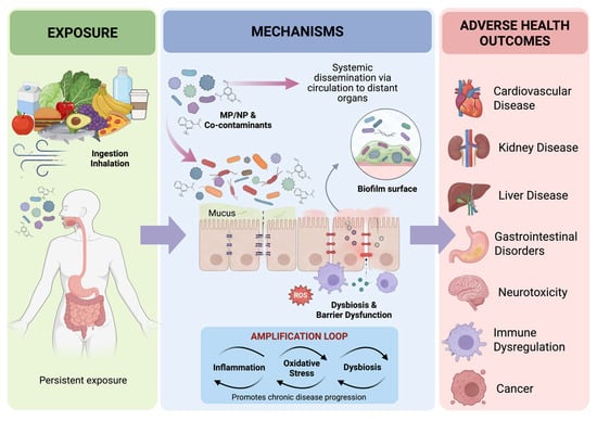
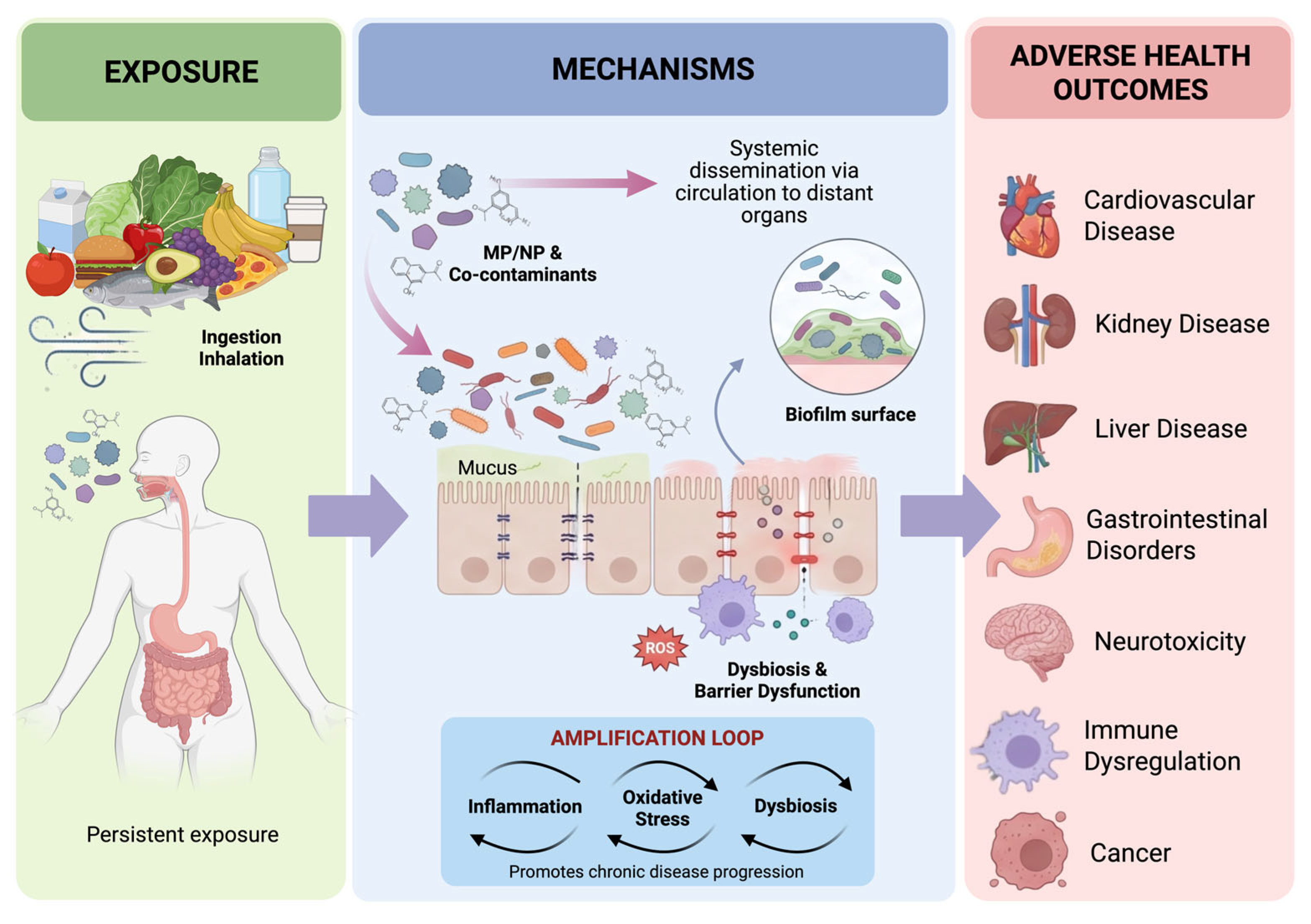
Figure 1 summarizes the proposed biological pathway linking chronic exposure to micro- and nanoplastics (MNPs) with adverse health outcomes. MNPs may enter the body primarily through ingestion and inhalation and, depending on their size, physicochemical properties, and associated contaminants, may interact with the intestinal and respiratory barriers and potentially reach the systemic circulation. Their biological effects may involve direct cellular interactions, oxidative stress, immune activation, barrier dysfunction, and interactions with the gut microbiota. In the intestine, dysbiosis and disruption of the epithelial barrier may further promote the translocation of microbial-derived products and amplify inflammatory responses. In parallel, MNP-associated biofilms and co-contaminants may contribute to additional biological effects. The resulting interplay among inflammation, oxidative stress, and microbial dysbiosis may establish a self-reinforcing cycle that contributes to chronic tissue injury and disease progression across multiple organ systems.
Figure 1.
Proposed pathway linking MNP exposure to chronic inflammation and adverse health outcomes. Created by BioRender.com.
Beyond their direct toxic effects on individual organs, MNPs may also influence host physiology indirectly by disrupting the intestinal ecosystem. Increasing evidence indicates that gut microbial dysbiosis and impaired intestinal homeostasis could contribute to the systemic consequences of chronic MP exposure, providing an additional mechanistic framework for understanding their association with chronic diseases.
4. MNPs as Disruptors of Gut Microbial and Intestinal Homeostasis
Experimental studies indicate that even short-term exposure to MPs can lead to significant alterations in gut microbiota composition and diversity [96]. Complementarily, in vitro models have demonstrated that exposure to PET reshapes the colonic microbial community by directly interacting with resident microorganisms, favoring microbial aggregation and the formation of biofilm-like structures. Certain bacterial species commonly found in the human intestinal tract, such as Escherichia coli, have demonstrated the ability to adhere to and form biofilm-like structures on plastic surfaces, which may facilitate the colonization and protection of microbial communities [97].
Consequently, MPs can act as ecological niches for microbial colonization and as vectors, facilitating the transport of microorganisms, antibiotic resistance determinants, and adsorbed environmental contaminants [98]. Furthermore, these plastic materials can contribute to gut microbiota dysbiosis and the generation of systemic inflammatory responses, as they can also adsorb a wide range of environmental contaminants, such as heavy metals and persistent organic pollutants, thereby exacerbating issues like oxidative stress and inflammatory signaling that intensify intestinal dysfunction [99].
The deposition of MNPs in the lumen can compromise the integrity of the epithelium and mucus layer, leading to wear, reduced mucus thickness, and altered microvillar morphology. Experimental studies indicate that exposure of epithelium to MNPs, particularly PS, increases reactive oxygen species (ROS) production, thereby activating pro-inflammatory signaling pathways, including NF-κB, MAPK, and the NLRP3 inflammasome. This activation stimulates the release of cytokines, including tumor necrosis factor-α (TNF-α), interleukin-1β (IL-1β), and interleukin-6 (IL-6), thereby establishing a local pro-oxidative and pro-inflammatory milieu within the intestinal environment [91].
Inflammatory activation leads to loss of tight junction proteins (occludin, claudins, and ZO-1), increased intestinal permeability, and impaired intestinal barrier integrity. Particle size and morphology critically influence cellular adhesion and internalization, with smaller particles (particularly NPs) being more readily taken up by intestinal epithelial cells and eliciting more pronounced inflammatory responses. Particle shape, including spheres, fibers, and irregular fragments, further affects intestinal toxicity through differences in surface charge and the presence of associated chemical additives. In addition, exposure dose and duration, as well as the organism’s age, substantially modulate the inflammatory outcome, with prolonged or repeated exposures leading to more persistent inflammatory states [100,101]. Host-related factors, including immune status, dietary patterns, gut microbiota composition, and pre-existing intestinal conditions, may further increase susceptibility to the pro-inflammatory effects of MP exposure [99]. Table 2 presents studies involving MNPs and gut microbial composition and intestinal barrier function.
Table 2.
Experimental and clinical evidence on the effects of MNPs on gut microbial composition, intestinal barrier function, and intestinal homeostasis.
A recent systematic review reported that experimental animals exposed to polystyrene exhibit gut dysbiosis, characterized by reduced diversity, depletion of beneficial taxa such as Lactobacillus, Bifidobacterium, and Ruminococcaceae, and enrichment of pro-inflammatory bacteria, including Proteobacteria, Helicobacter, and Staphylococcus [119].
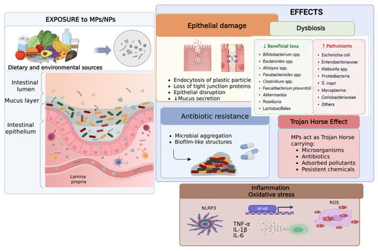
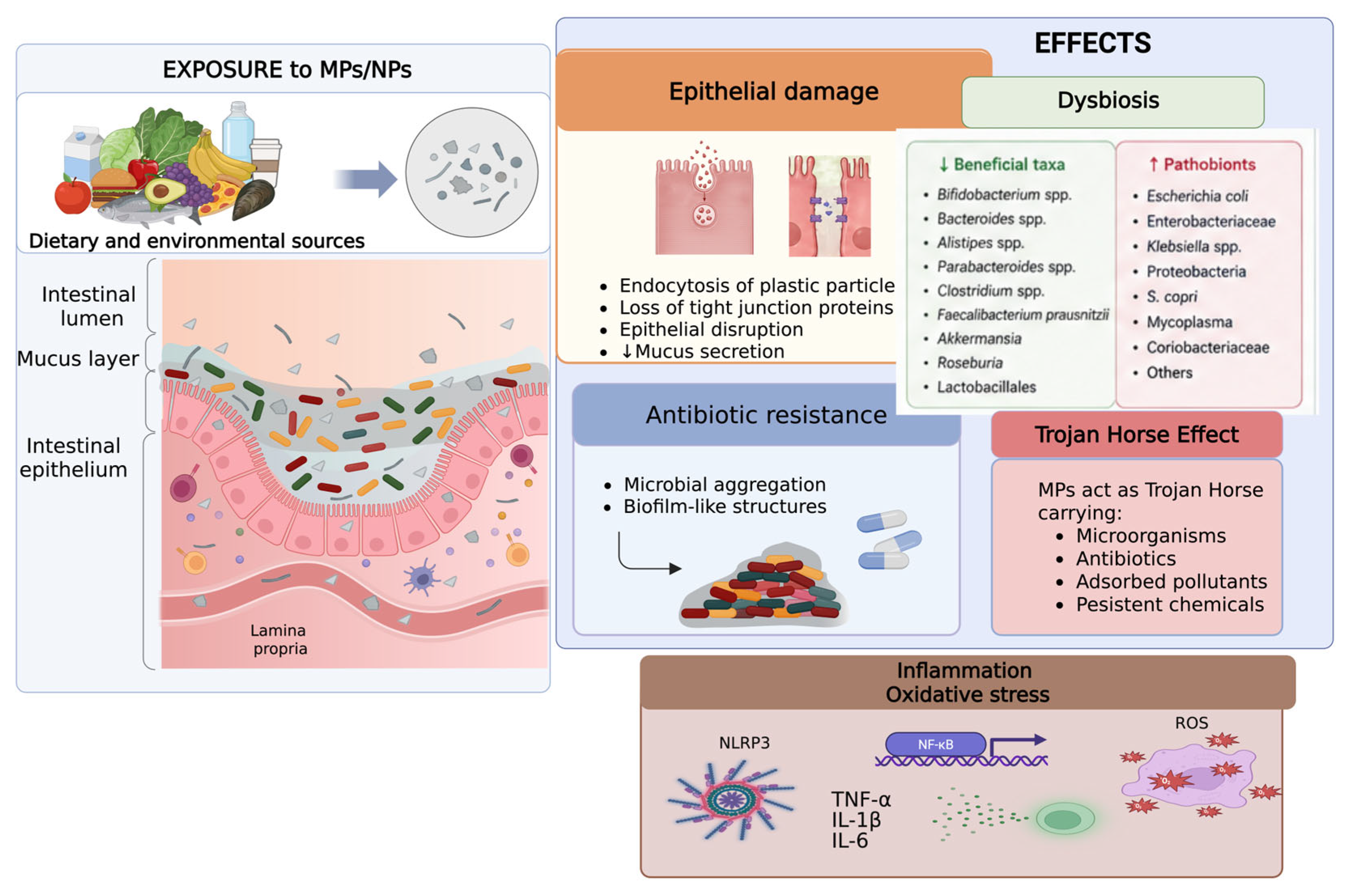
Compromised intestinal barrier function facilitates the translocation of MNPs and microbial products into the systemic circulation. Together with gut microbiota dysbiosis, these alterations promote sustained inflammatory responses and establish a self-reinforcing cycle of intestinal dysfunction. In this context, exposure to MNPs can remodel the gut microbial ecosystem, contributing to the development of inflammatory bowel diseases, chronic diseases, and even neurological disorders [99]. Despite growing experimental evidence, critical knowledge gaps remain, as human epidemiological data are limited and the consequences of long-term, low-dose exposure are still poorly characterized [100]. The proposed mechanisms linking MP and NP exposure to intestinal dysfunction are summarized in Figure 2.
Figure 2.
Proposed mechanisms linking microplastic and nanoplastic exposure to intestinal dysfunction. Microplastics and nanoplastics (MNPs) derived from dietary and environmental sources can interact with the intestinal ecosystem, including the mucus layer, gut microbiota, and epithelial barrier. Experimental evidence suggests that MNPs may induce gut dysbiosis, disrupt epithelial integrity by causing loss of tight junction proteins and altering the mucus barrier, and increase intestinal permeability. Furthermore, MNPs may facilitate microbial aggregation and biofilm formation, potentially favoring antimicrobial resistance. These disturbances can activate inflammatory and oxidative stress pathways, including NLRP3 inflammasome activation, NF-κB signaling, increased production of cytokines (TNF-α, IL-1β, and IL-6), and reactive oxygen species generation. Together, these effects may compromise intestinal homeostasis and contribute to local and systemic inflammation. Created by BioRender.com.
5. Research Gaps and Future Perspectives
A recent review analyzing methods for extracting and quantifying MNPs in biological tissues outlined a range of protocols, including reagent-based and chemical digestion methods, as well as distinct analytical techniques. This diversity hinders the reproducibility and comparison of study results, making it difficult to obtain a more reliable estimate of the actual number of MPs in organs and tissues [37]. Therefore, standardized, validated protocols focused on biological samples with clear definitions of size ranges and MP formats, comparable analyses, digestion techniques adapted to the samples, and minimum quality parameters are becoming increasingly necessary. This standardization would enable the integration of studies into meta-analyses, yielding more robust results and, consequently, advancing understanding of the potential risks of MPs to human health.
Another gap in research on MNPs’ influence on the microbiota and health is the scarcity of longitudinal human studies, as most evidence comes from cross-sectional studies, in vitro digestion simulation models, or animal studies. This context highlights the urgency of prospective cohorts capable of evaluating MNP exposure over time and its correlation with changes in the microbiota, inflammation, and disease emergence [23].
The multiomics approach encompasses strategies such as epigenomics, transcriptomics, and lipidomics, which are important for understanding numerous diseases and dysbiosis resulting from the ingestion of contaminated food. These techniques favor the understanding of the mechanisms of alteration of the intestinal microbiota due to the influence of MPs, which requires both the inclusion of taxonomic (metagenomics), metabolic (metabolomics/lipidomics), and functional (proteomics) data, as well as a detailed description of human exposure to these particles (exposomics). Thus, multiomics approaches that address the issues raised in the previous paragraphs can identify compromised metabolic and immunological pathways and propose more effective solutions to mitigate damage to the microbiome [120].
6. Conclusions
MNPs are emerging as novel environmental stressors that can alter the gut microbiota, promoting inflammation and various diseases, including cancer, lung, cardiovascular, and even neurological problems. More research is essential to clarify dose–response relationships, identify vulnerable populations, and guide public health policies to reduce plastic exposure.
Author Contributions
Conceptualization, G.d.O.F. and D.M.; methodology, D.M.; validation, G.d.O.F., L.F.M.F.C. and D.M.; formal analysis, D.M.; investigation, G.d.O.F., M.J.F.A., P.O.d.P.C. and T.R.; data curation, L.F.M.F.C. and D.M.; writing—original draft preparation, G.d.O.F., M.J.F.A., P.O.d.P.C., T.R. and D.M.; writing—review and editing, N.A.B., L.F.M.F.C. and D.M.; visualization, preparation, creation and/or presentation of the published work, specifically visualization/data presentation, D.M.; supervision, D.M.; project administration, D.M. All authors have read and agreed to the published version of the manuscript.
Funding
Conselho Nacional de Desenvolvimento Científico e Tecnológico (CNPq—No 407814/2024-8), Coordenação de Aperfeiçoamento de Pessoal de Nível Superior (CAPES), and Fundação de Amparo à Pesquisa do Estado do Rio de Janeiro (FAPERJ—No E-26/200.063/2026) support Denise Mafra’s work.
Data Availability Statement
No new data were created or analyzed in this study. Data sharing is not applicable to this article.
Acknowledgments
During the preparation of this manuscript/study, the authors acknowledge the use of BioRender (BioRender.com) to create the graphical illustrations included in this manuscript. The figures were created and customized by the authors using the BioRender platform. The authors have reviewed and edited the output and take full responsibility for the content of this publication.
Conflicts of Interest
The authors declare no conflicts of interest.
Abbreviations
The following abbreviations are used in this manuscript:
| MPs. | Microplastics |
| PA | Polyamide |
| PVC | Polyvinyl chloride |
| PET | Polyethylene terephthalate |
| PU | Polyurethane |
| LDPE | Low-density polyethylene |
| PET-TiO2 | Polyethylene terephthalate with titanium dioxide |
| aPTT | Activated partial thromboplastin time |
| ROS | Reactive oxygen species |
| TNF-α | Tumor necrosis factor-α |
| IL-6 | Interleukin-6 |
| NLRP3 | NLRP3 inflammasome |
| MAPK | Mitogen-activated protein kinases |
| SOD | Superoxide dismutase |
| GSH-Px | Glutathione peroxidase |
| ZO-1 | Zonula occludens-1 |
| MDI | Microbial dysbiosis index |
| COPD | Chronic obstructive pulmonary disease |
| CSF | Cerebrospinal fluid |
| BPA | Bisphenol A |
| GLP-1 | Glucagon-like peptide-1 |
| AMH | Anti-Müllerian hormone |
| AMPK | AMP-activated protein kinase |
| EMT | Epithelial–mesenchymal transition |
| Aβ42 | Beta-amyloid 42 |
| μ-FTIR | Fourier-transform infrared spectroscopy |
| NPs | Nanoplastics |
| PE | Polyethylene |
| PP | Polypropylene |
| PS | Polystyrene |
| HDPE | High-density polyethylene |
| PA66 | Polyamide 66 |
| PP-talc | Polypropylene–talc |
| PCBs | Polychlorinated biphenyls |
| MNPs | Micro- and nanoplastics |
| IL-1β | Interleukin-1β |
| NF-κB | Nuclear factor kappa B |
| LPS | Lipopolysaccharide |
| MDA | Malondialdehyde |
| CAT | Catalase |
| T-AOC | Total antioxidant capacity |
| SCFA | Short-chain fatty acids |
| DSS | Dextran sulfate sodium |
| CVD | Cardiovascular disease |
| NAFLD | Non-alcoholic fatty liver disease |
| PPARγ | Peroxisome proliferator-activated receptor γ |
| PYY | Peptide YY |
| EGFR | Epidermal growth factor receptor |
| ER | Endoplasmic reticulum |
| AREG | Amphiregulin |
| Aβ40 | Beta-amyloid 40 |
References
- Pilapitiya, P.G.C.N.T.; Ratnayake, A.S. The world of plastic waste: A review. Clean. Mater. 2024, 11, 100220. [Google Scholar] [CrossRef]
- Dokl, M.; Copot, A.; Krajnc, D.; Van Fan, Y.; Vujanović, A.; Aviso, K.B.; Tan, R.R.; Kravanja, Z.; Čuček, L. Global projections of plastic use, end-of-life fate and potential changes in consumption, reduction, recycling and replacement with bioplastics to 2050. Sustain. Prod. Consum. 2024, 51, 498–518. [Google Scholar] [CrossRef]
- Saguban, R.; AlAbd, A.M.A.; Rondilla, E.; Buta, J.; Marzouk, S.A.; Maestrado, R.; Sankarapandian, C.; Alkubati, S.A.; Mostoles, R.; Alshammari, S.A.; et al. Investigating the Interplay Between Sleep, Anxiety, and Depression in Chronic Kidney Disease Patients: Implications for Mental Health. Healthcare 2025, 13, 294. [Google Scholar] [CrossRef]
- Soliz, D.L.; Paniagua González, G.; Muñoz-Arnanz, J.; Bravo-Yagüe, J.C.; Fernández Hernando, P.; Garcinuño Martínez, R.M. Identification and morphological characterization of different types of plastic microparticles. Heliyon 2024, 10, e30749. [Google Scholar] [CrossRef]
- Chen, X.; Yan, N. A brief overview of renewable plastics. Mater. Today Sustain. 2020, 7–8, 100031. [Google Scholar] [CrossRef]
- Amponsah, A.K.; Afrifa, E.A.; Essandoh, P.K.; Enyoh, C.E. Evidence of microplastics accumulation in the gills and gastrointestinal tract of fishes from an estuarine system in Ghana. Heliyon 2024, 10, e25608. [Google Scholar] [CrossRef]
- Kye, H.; Kim, J.; Ju, S.; Lee, J.; Lim, C.; Yoon, Y. Microplastics in water systems: A review of their impacts on the environment and their potential hazards. Heliyon 2023, 9, E14359. [Google Scholar] [CrossRef]
- Xu, J.-L.; Lin, X.; Wang, J.J.; Gowen, A.A. A review of potential human health impacts of micro- and nanoplastics exposure. Sci. Total Environ. 2022, 851, 158111. [Google Scholar] [CrossRef]
- Sharma, G.C.; Sharma, A.; Debnath, S.; Chaturvedi, B.; Joshi, R.M.; Singh, M.; Nathiya, D.; Tomar, B.S. A systematic review of the presence and potential health impacts of microplastics in the human body. Environ. Sci. Pollut. Res. 2026, 33, 12175–12194. [Google Scholar] [CrossRef]
- Li, Y.; Tao, L.; Wang, Q.; Wang, F.; Li, G.; Song, M. Potential Health Impact of Microplastics: A Review of Environmental Distribution, Human Exposure, and Toxic Effects. Environ. Health 2023, 1, 249–257. [Google Scholar] [CrossRef]
- Prata, J.C.; da Costa, J.P.; Lopes, I.; Duarte, A.C.; Rocha-Santos, T. Environmental exposure to microplastics: An overview on possible human health effects. Sci. Total Environ. 2020, 702, 134455. [Google Scholar] [CrossRef]
- Zhang, Q.; Xu, E.G.; Li, J.; Chen, Q.; Ma, L.; Zeng, E.Y.; Shi, H. A Review of Microplastics in Table Salt, Drinking Water, and Air: Direct Human Exposure. Environ. Sci. Technol. 2020, 54, 3740–3751. [Google Scholar] [CrossRef]
- Chakraborty, S.; Chakraborty, A.; Ghosh, A.; Bishwanath Singh, N.; Ghosh, D.; Kundu, T.; Das, A. Microplastics and nanoplastics in human toxicity: ROS-mediated mechanisms, cellular damage and systemic effects. Mol. Biol. Rep. 2026, 53, 1391. [Google Scholar] [CrossRef]
- Ageel, H.K.; Harrad, S.; Abdallah, M.A.-E. Occurrence, human exposure, and risk of microplastics in the indoor environment. Environ. Sci. Process. Impacts 2022, 24, 17–31. [Google Scholar] [CrossRef]
- Roslan, N.S.; Lee, Y.Y.; Ibrahim, Y.S.; Anuar, S.T.; Yusof, K.M.K.K.; Lai, L.A.; Brentnall, T. Detection of microplastics in human tissues and organs: A scoping review. J. Glob. Health 2024, 14, 04179. [Google Scholar] [CrossRef]
- Nihart, A.J.; Garcia, M.A.; El Hayek, E.; Liu, R.; Olewine, M.; Kingston, J.D.; Castillo, E.F.; Gullapalli, R.R.; Howard, T.; Bleske, B.; et al. Bioaccumulation of microplastics in decedent human brains. Nat. Med. 2025, 31, 1114–1119. [Google Scholar] [CrossRef]
- Obeng, E.M.; Hodge, C.; You, J. Microplastic pollution: A review of specific blood-tissue barrier breaches and health effects. Environ. Pollut. 2025, 376, 126416. [Google Scholar] [CrossRef]
- Ragusa, A.; Svelato, A.; Santacroce, C.; Catalano, P.; Notarstefano, V.; Carnevali, O.; Papa, F.; Rongioletti, M.C.A.; Baiocco, F.; Draghi, S.; et al. Plasticenta: First evidence of microplastics in human placenta. Environ. Int. 2021, 146, 106274. [Google Scholar] [CrossRef] [PubMed]
- Khan, A.; Jia, Z. Recent insights into uptake, toxicity, and molecular targets of microplastics and nanoplastics relevant to human health impacts. iScience 2023, 26, 106061. [Google Scholar] [CrossRef]
- Meng, X.; Zheng, X.; Mai, W.; Gao, J.; Fan, Y.; Fu, J.; Xu, J. Micro- and nanoplastics differ in particle-mucus interactions: The sight on rheological properties, barrier dysfunction and microbiota dysbiosis. J. Hazard. Mater. 2025, 492, 138130. [Google Scholar] [CrossRef]
- Porfiryeva, N.N.; Zlotver, I.; Sosnik, A. Combined Effect of Size and Charge on the Interaction of Nanoparticles with Mucus-Mimicking Mucin Hydrogels. Pharmaceuticals 2025, 18, 1498. [Google Scholar] [CrossRef]
- DeLoid, G.M.; Yang, Z.; Bazina, L.; Kharaghani, D.; Sadrieh, F.; Demokritou, P. Mechanisms of ingested polystyrene micro-nanoplastics (MNPs) uptake and translocation in an in vitro tri-culture small intestinal epithelium. J. Hazard. Mater. 2024, 473, 134706. [Google Scholar] [CrossRef]
- Thin, Z.S.; Chew, J.; Ong, T.Y.Y.; Raja Ali, R.A.; Gew, L.T. Impact of microplastics on the human gut microbiome: A systematic review of microbial composition, diversity, and metabolic disruptions. BMC Gastroenterol. 2025, 25, 583. [Google Scholar] [CrossRef]
- Leslie, H.A.; van Velzen, M.J.; Brandsma, S.H.; Vethaak, A.D.; Garcia-Vallejo, J.J.; Lamoree, M.H. Discovery and quantification of plastic particle pollution in human blood. Environ. Int. 2022, 163, 107199. [Google Scholar] [CrossRef]
- Lee, D.-W.; Jung, J.; Park, S.-A.; Lee, Y.; Kim, J.; Han, C.; Kim, H.-C.; Lee, J.H.; Hong, Y.-C. Microplastic particles in human blood and their association with coagulation markers. Sci. Rep. 2024, 14, 30419. [Google Scholar] [CrossRef]
- Leonard, S.V.L.; Liddle, C.R.; Atherall, C.A.; Chapman, E.; Watkins, M.; Calaminus, S.D.J.; Rotchell, J.M. Microplastics in human blood: Polymer types, concentrations and characterisation using μFTIR. Environ. Int. 2024, 188, 108751. [Google Scholar] [CrossRef]
- Braun, T.; Ehrlich, L.; Henrich, W.; Koeppel, S.; Lomako, I.; Schwabl, P.; Liebmann, B. Detection of Microplastic in Human Placenta and Meconium in a Clinical Setting. Pharmaceutics 2021, 13, 921. [Google Scholar] [CrossRef]
- Caba-Flores, M.D.; Martínez-Valenzuela, C.; Cárdenas-Tueme, M.; Camacho-Morales, A. Micro problems with macro consequences: Accumulation of persistent organic pollutants and microplastics in human breast milk and in human milk substitutes. Environ. Sci. Pollut. Res. 2023, 30, 95139–95154. [Google Scholar] [CrossRef]
- Liu, S.; Guo, J.; Liu, X.; Yang, R.; Wang, H.; Sun, Y.; Chen, B.; Dong, R. Detection of various microplastics in placentas, meconium, infant feces, breastmilk and infant formula: A pilot prospective study. Sci. Total Environ. 2022, 854, 158699. [Google Scholar] [CrossRef]
- Saraluck, A.; Techarang, T.; Bunyapipat, P.; Boonchuwong, K.; Pullaput, Y.; Mordmuang, A. Detection of Microplastics in Human Breast Milk and Its Association with Changes in Human Milk Bacterial Microbiota. J. Clin. Med. 2024, 13, 4029. [Google Scholar] [CrossRef]
- Amato-Lourenço, L.F.; Carvalho-Oliveira, R.; Júnior, G.R.; dos Santos Galvão, L.; Ando, R.A.; Mauad, T. Presence of airborne microplastics in human lung tissue. J. Hazard. Mater. 2021, 416, 126124. [Google Scholar] [CrossRef]
- Jenner, L.C.; Rotchell, J.M.; Bennett, R.T.; Cowen, M.; Tentzeris, V.; Sadofsky, L.R. Detection of microplastics in human lung tissue using μFTIR spectroscopy. Sci. Total Environ. 2022, 831, 154907. [Google Scholar] [CrossRef]
- Wang, S.; Lu, W.; Cao, Q.; Tu, C.; Zhong, C.; Qiu, L.; Li, S.; Zhang, H.; Lan, M.; Qiu, L.; et al. Microplastics in the Lung Tissues Associated with Blood Test Index. Toxics 2023, 11, 759. [Google Scholar] [CrossRef]
- Hartmann, C.; Lomako, I.; Schachner, C.; El Said, E.; Abert, J.; Satrapa, V.; Kaiser, A.-M.; Walch, H.; Köppel, S. Assessment of microplastics in human stool: A pilot study investigating the potential impact of diet-associated scenarios on oral microplastics exposure. Sci. Total Environ. 2024, 951, 175825. [Google Scholar] [CrossRef]
- Zhang, N.; Bin Li, Y.; He, H.R.; Zhang, J.F.; Ma, G.S. You are what you eat: Microplastics in the feces of young men living in Beijing. Sci. Total Environ. 2021, 767, 144345. [Google Scholar] [CrossRef]
- Xu, J.; Qu, J.; Jin, H.; Mao, W. Associations between microplastics in human feces and colorectal cancer risk. J. Hazard. Mater. 2025, 495, 139099. [Google Scholar] [CrossRef]
- Di Fiore, C.; Ishikawa, Y.; Wright, S.L. A review on methods for extracting and quantifying microplastic in biological tissues. J. Hazard. Mater. 2024, 464, 132991. [Google Scholar] [CrossRef] [PubMed]
- Dzierżyński, E.; Blicharz-Grabias, E.; Komaniecka, I.; Panek, R.; Forma, A.; Gawlik, P.J.; Puźniak, D.; Flieger, W.; Choma, A.; Suśniak, K.; et al. Post-mortem evidence of microplastic bioaccumulation in human organs: Insights from advanced imaging and spectroscopic analysis. Arch. Toxicol. 2025, 99, 4051–4066. [Google Scholar] [CrossRef]
- Lee, S.E.; Kim, D.Y.; Jeong, T.S.; Park, Y.S. Micro- and Nano-Plastic-Induced Adverse Health Effects on Lungs and Kidneys Linked to Oxidative Stress and Inflammation. Life 2025, 15, 392. [Google Scholar] [CrossRef]
- Goodman, K.E.; Hua, T.; Sang, Q.-X.A. Effects of Polystyrene Microplastics on Human Kidney and Liver Cell Morphology, Cellular Proliferation, and Metabolism. ACS Omega 2022, 7, 34136–34153. [Google Scholar] [CrossRef]
- Cheng, W.; Li, X.; Zhou, Y.; Yu, H.; Xie, Y.; Guo, H.; Wang, H.; Li, Y.; Feng, Y.; Wang, Y. Polystyrene microplastics induce hepatotoxicity and disrupt lipid metabolism in the liver organoids. Sci. Total Environ. 2022, 806, 150328. [Google Scholar] [CrossRef]
- Gou, X.; Fu, Y.; Li, J.; Xiang, J.; Yang, M.; Zhang, Y. Impact of nanoplastics on Alzheimer ’s disease: Enhanced amyloid-β peptide aggregation and augmented neurotoxicity. J. Hazard. Mater. 2024, 465, 133518. [Google Scholar] [CrossRef]
- Kim, Y.; Park, S.J.; Oh, J.-M. Polystyrene nanoplastics increase migration in normal lung cells while inducing differential cytotoxicity in lung cancer cells. Toxicology 2025, 519, 154328. [Google Scholar] [CrossRef]
- Gao, Q.; Wang, Y.; Ji, Y.; Zhao, X.; Zhang, P.; Chen, L. Tracking of realistic nanoplastics in complicated matrices by iridium element labeling and inductively coupled plasma mass spectroscopy. J. Hazard. Mater. 2022, 424, 127628. [Google Scholar] [CrossRef]
- Jin, H.; Yang, C.; Jiang, C.; Li, L.; Pan, M.; Li, D.; Han, X.; Ding, J. Evaluation of Neurotoxicity in BALB/c Mice following Chronic Exposure to Polystyrene Microplastics. Environ. Health Perspect. 2022, 130, 107002. [Google Scholar] [CrossRef]
- Zhang, Y.; Yin, K.; Wang, D.; Wang, Y.; Lu, H.; Zhao, H.; Xing, M. Polystyrene microplastics-induced cardiotoxicity in chickens via the ROS-driven NF-κB-NLRP3-GSDMD and AMPK-PGC-1α axes. Sci. Total Environ. 2022, 840, 156727. [Google Scholar] [CrossRef]
- Huang, D.; Zhang, Y.; Long, J.; Yang, X.; Bao, L.; Yang, Z.; Wu, B.; Si, R.; Zhao, W.; Peng, C.; et al. Polystyrene microplastic exposure induces insulin resistance in mice via dysbacteriosis and pro-inflammation. Sci. Total Environ. 2022, 838, 155937. [Google Scholar] [CrossRef]
- Shen, T.; Zhang, W.; Wang, Y.; Li, H.; Wu, J.; Wang, Q.; Qin, L.; Zhang, L.; Liu, C.; Li, R. Effects of Microplastic (MP) Exposure at Environmentally Relevant Doses on the Structure, Function, and Transcriptome of the Kidney in Mice. Molecules 2023, 28, 7104. [Google Scholar] [CrossRef]
- Xiong, X.; Gao, L.; Chen, C.; Zhu, K.; Luo, P.; Li, L. The microplastics exposure induce the kidney injury in mice revealed by RNA-seq. Ecotoxicol. Environ. Saf. 2023, 256, 114821. [Google Scholar] [CrossRef]
- Huang, H.; Wei, F.; Qiu, S.; Xing, B.; Hou, J. Polystyrene microplastics trigger adiposity in mice by remodeling gut microbiota and boosting fatty acid synthesis. Sci. Total Environ. 2023, 890, 164297. [Google Scholar] [CrossRef]
- Liu, Z.; Sokratian, A.; Duda, A.M.; Xu, E.; Stanhope, C.; Fu, A.; Strader, S.; Li, H.; Yuan, Y.; Bobay, B.G.; et al. Anionic nanoplastic contaminants promote Parkinson’s disease–associated α-synuclein aggregation. Sci. Adv. 2023, 9, eadi8716. [Google Scholar] [CrossRef]
- Pan, C.; Wang, X.; Fan, Z.; Mao, W.; Shi, Y.; Wu, Y.; Liu, T.; Xu, Z.; Wang, H.; Chen, H. Polystyrene microplastics facilitate renal fibrosis through accelerating tubular epithelial cell senescence. Food Chem. Toxicol. 2024, 191, 114888. [Google Scholar] [CrossRef]
- Yang, S.; Zhang, T.; Ge, Y.; Yin, L.; Pu, Y.; Liang, G. Inhalation exposure to polystyrene nanoplastics induces chronic obstructive pulmonary disease-like lung injury in mice through multi-dimensional assessment. Environ. Pollut. 2024, 347, 123633. [Google Scholar] [CrossRef]
- Huang, H.; Hou, J.; Yu, C.; Wei, F.; Xi, B. Microplastics exacerbate tissue damage and promote carcinogenesis following liver infection in mice. Ecotoxicol. Environ. Saf. 2024, 286, 117217. [Google Scholar] [CrossRef]
- Chiu, H.-W.; Chu, C.-W.; Huang, C.-C.; Chia, Z.-C.; Wang, Y.-L.; Lee, Y.-H. Polystyrene microplastics induce hepatic lipid metabolism and energy disorder by upregulating the NR4A1-AMPK signaling pathway. Environ. Pollut. 2025, 369, 125850. [Google Scholar] [CrossRef]
- Huang, H.; Hou, J.; Li, M.; Wei, F.; Liao, Y.; Xi, B. Microplastics in the bloodstream can induce cerebral thrombosis by causing cell obstruction and lead to neurobehavioral abnormalities. Sci. Adv. 2025, 11, eadr8243. [Google Scholar] [CrossRef]
- Choi, J.Y.; Hur, J.; Jo, Y.S.; Rhee, C.K. Impact of Microplastic Exposure on Airway Inflammation in an Acute Asthma Murine Model. Tuberc. Respir. Dis. 2026, 89, 29–37. [Google Scholar] [CrossRef]
- Wang, Y.-L.; Lee, Y.-H.; Hsu, Y.-H.; Chiu, I.-J.; Huang, C.C.-Y.; Huang, C.-C.; Chia, Z.-C.; Lee, C.-P.; Lin, Y.-F.; Chiu, H.-W. The kidney-related effects of polystyrene microplastics on human kidney proximal tubular epithelial cells hk-2 and male c57bl/6 mice. Environ. Health Perspect. 2021, 129, 57003. [Google Scholar] [CrossRef]
- Meng, X.; Zhang, J.; Wang, W.; Gonzalez-Gil, G.; Vrouwenvelder, J.S.; Li, Z. Effects of nano- and microplastics on kidney: Physicochemical properties, bioaccumulation, oxidative stress and immunoreaction. Chemosphere 2022, 288, 132631. [Google Scholar] [CrossRef]
- Cao, J.; Xu, R.; Geng, Y.; Xu, S.; Guo, M. Exposure to polystyrene microplastics triggers lung injury via targeting toll-like receptor 2 and activation of the NF-κB signal in mice. Environ. Pollut. 2023, 320, 121068. [Google Scholar] [CrossRef]
- Jin, W.; Zhang, W.; Tang, H.; Wang, P.; Zhang, Y.; Liu, S.; Qiu, J.; Chen, H.; Wang, L.; Wang, R.; et al. Microplastics exposure causes the senescence of human lung epithelial cells and mouse lungs by inducing ROS signaling. Environ. Int. 2024, 185, 108489. [Google Scholar] [CrossRef]
- Jeong, A.; Park, S.J.; Lee, E.J.; Kim, K.W. Nanoplastics exacerbate Parkinson’s disease symptoms in C. elegans and human cells. J. Hazard. Mater. 2024, 465, 133289. [Google Scholar] [CrossRef]
- Yuan, X.; Cui, Z.; Xu, S.; Chen, K.; Wang, Y.; Zheng, F.; Shen, H.-M.; Sun, S.; Wu, Y.; Xia, D. Polystyrene Nanoplastics at an Environmentally Relevant Concentration Promote Ovarian Cancer Progression via CDK4/6-Dependent Signaling. Environ. Health 2025, 4, 488–501. [Google Scholar] [CrossRef]
- Baek, S.-M.; Lee, Y.-J.; Yim, J.-H.; Kim, T.-U.; Kim, W.J.; Lee, S.-W.; Kim, H.-Y.; Kang, K.-K.; Lee, K.-M.; Choi, S.-K.; et al. Korean red ginseng extract inhibits microplastic translocation via the gut−liver axis by ameliorating alcohol-induced intestinal disruption. J. Ginseng Res. 2026, 50, 100922. [Google Scholar] [CrossRef]
- Shanmugiah, J.; Kim, S.; Jeong, H.; Kong, J.; Lee, S.-S.; Kang, D.K.; Kim, J.S. Size-dependent pulmonary toxicity of inhaled micro- and nano-polystyrene and initial identification of microplastics in human lung cancer tissue. J. Hazard. Mater. 2026, 508, 141897. [Google Scholar] [CrossRef]
- Marfella, R.; Prattichizzo, F.; Sardu, C.; Fulgenzi, G.; Graciotti, L.; Spadoni, T.; D’oNofrio, N.; Scisciola, L.; La Grotta, R.; Frigé, C.; et al. Microplastics and Nanoplastics in Atheromas and Cardiovascular Events. N. Engl. J. Med. 2024, 390, 900–910. [Google Scholar] [CrossRef]
- He, P.; Wang, F.; Xi, G.; Li, Y.; Wang, F.; Wang, H.; Li, L.; Ma, X.; Han, Y.; Shi, Y. Association of microplastics in human cerebrospinal fluid with Alzheimer’s disease-related changes. J. Hazard. Mater. 2025, 494, 138748. [Google Scholar] [CrossRef]
- Pironti, C.; Notarstefano, V.; Ricciardi, M.; Motta, O.; Giorgini, E.; Montano, L. First Evidence of Microplastics in Human Urine, a Preliminary Study of Intake in the Human Body. Toxics 2023, 11, 40. [Google Scholar] [CrossRef]
- Massardo, S.; Verzola, D.; Alberti, S.; Caboni, C.; Santostefano, M.; Eugenio Verrina, E.; Angeletti, A.; Lugani, F.; Ghiggeri, G.M.; Bruschi, M.; et al. MicroRaman spectroscopy detects the presence of microplastics in human urine and kidney tissue. Environ. Int. 2024, 184, 108444. [Google Scholar] [CrossRef]
- de Oliveira, R.B.; Pelepenko, L.E.; Masaro, D.A.; Lustosa, G.M.; de Oliveira, M.C.; Roza, N.A.; Marciano, M.A.; dos Reis, L.M.; Kamel, S.; Louvet, L.; et al. Effects of microplastics on the kidneys: A narrative review. Kidney Int. 2024, 106, 400–407. [Google Scholar] [CrossRef]
- La Porta, E.; Exacoustos, O.; Lugani, F.; Angeletti, A.; Chiarenza, D.S.; Bigatti, C.; Spinelli, S.; Kajana, X.; Garbarino, A.; Bruschi, M.; et al. Microplastics and Kidneys: An Update on the Evidence for Deposition of Plastic Microparticles in Human Organs, Tissues and Fluids and Renal Toxicity Concern. Int. J. Mol. Sci. 2023, 24, 14391. [Google Scholar] [CrossRef]
- Kara, E.; Konur, K.; Koca, Y.Ş.; Aytan, Ü. Unveiling hidden contaminants: A systematic quantification and characterization of microplastics in hemodialysis and peritoneal dialysis fluids. BMC Nephrol. 2025, 26, 359. [Google Scholar] [CrossRef]
- Tan, R.-Y.; She, Q.-Y.; Ma, Y.-C.; Liu, M.-H.; Li, L.-J.; Huang, L.-L.; Zhong, Y.-W.; Bi, H.-X. The threat of microplastics to human kidney health: Mechanisms of nephrotoxicity and future research directions. Environ. Res. 2025, 283, 122124. [Google Scholar] [CrossRef]
- Saha, S.C.; Saha, G. Effect of microplastics deposition on human lung airways: A review with computational benefits and challenges. Heliyon 2024, 10, e24355. [Google Scholar] [CrossRef] [PubMed]
- Borgatta, M.; Breider, F. Inhalation of Microplastics—A Toxicological Complexity. Toxics 2024, 12, 358. [Google Scholar] [CrossRef] [PubMed]
- Huang, Y.; Shang, P.; Li, Y.; Wang, Y. Lung hazards of microplastics and their toxicological mechanisms. Environ. Pollut. 2025, 385, 127149. [Google Scholar] [CrossRef] [PubMed]
- Wei, Y.Y.; Chen, T.T.; Zhang, D.W.; Zhang, Y.; Li, F.; Ding, Y.C.; Wang, M.Y.; Zhang, L.; Chen, K.G.; Fei, G.H. Microplastics exacerbate ferroptosis via mitochondrial reactive oxygen species-mediated autophagy in chronic obstructive pulmonary disease. Autophagy 2025, 21, 1717–1743. [Google Scholar] [CrossRef]
- Wang, Q.; He, W.; Zhou, Y.; Feng, R.; Wang, Y.; Liu, L.; Yuan, Y.; Dai, J.; Liu, Y.; Zhang, X. Polystyrene nanoplastics aggravate house dust mite induced allergic airway inflammation through EGFR/ERK-dependent lung epithelial barrier dysfunction. Ecotoxicol. Environ. Saf. 2025, 298, 118329. [Google Scholar] [CrossRef]
- Epeslidou, E.; Scott, J.S.; de Klein, B.; Cudia, J.T.; Melgert, B.; Prekovic, S. Microplastics as environmental modifiers of lung disease. EMBO Mol. Med. 2025, 18, 381–395. [Google Scholar] [CrossRef]
- Ali, A.; Sonne, C.; Aslam, M.; Ali, I.; Hussain, S.; Qadir, A.; Sukhera, S.; Ali, H.; Ahmad, S.R.; Yang, G. First characterization and risk assessment of microplastics in the endangered Indus River dolphin (Platanista minor): Implications for conservation strategies. PLoS ONE 2025, 20, e0330253. [Google Scholar] [CrossRef]
- Cheng, Y.; Yang, Y.; Bai, L.; Cui, J. Microplastics: An often-overlooked issue in the transition from chronic inflammation to cancer. J. Transl. Med. 2024, 22, 959. [Google Scholar] [CrossRef] [PubMed]
- Wang, X.; Deng, K.; Zhang, P.; Chen, Q.; Magnuson, J.T.; Qiu, W.; Zhou, Y. Microplastic-mediated new mechanism of liver damage: From the perspective of the gut-liver axis. Sci. Total Environ. 2024, 919, 170962. [Google Scholar] [CrossRef]
- Shi, Y.; Hong, R.; Fan, Z.; Huan, R.; Gao, Y.; Ma, M.; Liu, T.; Pan, C. Chronic environmental exposure to polystyrene microplastics increases the risk of nonalcoholic fatty liver disease. Toxicology 2025, 511, 154067. [Google Scholar] [CrossRef]
- Auguet, T.; Bertran, L.; Barrientos-Riosalido, A.; Fabregat, B.; Villar, B.; Aguilar, C.; Sabench, F. Are Ingested or Inhaled Microplastics Involved in Nonalcoholic Fatty Liver Disease? Int. J. Environ. Res. Public Health 2022, 19, 13495. [Google Scholar] [CrossRef]
- Zhou, W.; Shi, W.; Du, X.; Han, Y.; Tang, Y.; Ri, S.; Ju, K.; Kim, T.; Huang, L.; Zhang, W.; et al. Assessment of Nonalcoholic Fatty Liver Disease Symptoms and Gut–Liver Axis Status in Zebrafish after Exposure to Polystyrene Microplastics and Oxytetracycline, Alone and in Combination. Environ. Health Perspect. 2023, 131, 047006. [Google Scholar] [CrossRef]
- Deng, X.; Gui, Y.; Zhao, L. The micro(nano)plastics perspective: Exploring cancer development and therapy. Mol. Cancer 2025, 24, 30. [Google Scholar] [CrossRef]
- Kumar, N.; Shukla, A.; Kumar, A.; Pachar, A.K.; Harman; Sharda, N.; Yadav, S.; Lamba, M.; Acharya, A. The hidden poison—Microplastic: Inflammatory catalyst of cancer development. Int. J. Plant Sci. 2024, 19, 120–138. [Google Scholar] [CrossRef]
- Jiang, Z.; Liu, Y.; Zhang, P.; Fei, J.; Wang, K.; Li, Y.; Zhang, X.; Cai, C.; Chen, Y.; Peng, Y.; et al. Tumour-infiltrating microplastics disrupt the JAK-STAT-microbiota axis to promote immunotherapy resistance in colorectal cancer. Mol. Cancer 2026, 25, 73. [Google Scholar] [CrossRef]
- Winz, C.; Li, E.; Xie, C.; Lee, K.C.; Boguszewski, K.; Rohatagi, S.; Hahn, R.; Rancourt, R.; Furmanski, P.; Suh, N. The Endocrine Disrupting Compounds Bisphenol-A and α-Zeranol Mimic the Estrogen Transcriptional Program to Promote Proliferation and Stemness in Breast Cancer Cells. Mol. Carcinog. 2026, 65, 907–920. [Google Scholar] [CrossRef]
- Das, A. The emerging role of microplastics in systemic toxicity: Involvement of reactive oxygen species (ROS). Sci. Total Environ. 2023, 895, 165076. [Google Scholar] [CrossRef]
- Zeng, G.; Li, J.; Wang, Y.; Su, J.; Lu, Z.; Zhang, F.; Ding, W. Polystyrene microplastic-induced oxidative stress triggers intestinal barrier dysfunction via the NF-κB/NLRP3/IL-1β/MCLK pathway. Environ. Pollut. 2024, 345, 123473. [Google Scholar] [CrossRef]
- Zhang, T.; Liao, Y.; Ling, J.; Zhang, J.; Zhang, D.; Yin, X.; Yu, P.; Liu, X. Tiny trouble: Microplastics, nanoplastics, and their heartfelt impact on cardiovascular health. Cardiovasc. Res. 2025, 121, 992–1010. [Google Scholar] [CrossRef]
- Ma, J.; Ladd, D.M.; Kaval, N.; Wang, H.-S. Toxicity of long term exposure to low dose polystyrene microplastics and nanoplastics in human iPSC-derived cardiomyocytes. Food Chem. Toxicol. 2025, 202, 115489. [Google Scholar] [CrossRef]
- Smyth, E.; Solomon, A.; Vydyanath, A.; Luther, P.K.; Pitchford, S.; Tetley, T.D.; Emerson, M. Induction and enhancement of platelet aggregation in vitro and in vivo by model polystyrene nanoparticles. Nanotoxicology 2015, 9, 356–364. [Google Scholar] [CrossRef]
- Paolisso, P.; Scisciola, L.; Belmonte, M.; Scarsini, R.; Galli, V.; Gallinoro, E.; Casenghi, M.; Ausiello, D.; Vincelli, G.; Policastro, P.; et al. Micro- and nano-plastics in the coronary circulation and air pollution exposure in ischaemic heart disease presentation. Eur. Heart J. 2026, ehag447. [Google Scholar] [CrossRef]
- Xie, L.; Chen, T.; Liu, J.; Hou, Y.; Tan, Q.; Zhang, X.; Li, Z.; Farooq, T.H.; Yan, W.; Li, Y. Intestinal flora variation reflects the short-term damage of microplastic to the intestinal tract in mice. Ecotoxicol. Environ. Saf. 2022, 246, 114194. [Google Scholar] [CrossRef]
- Tamargo, A.; Molinero, N.; Reinosa, J.J.; Alcolea-Rodriguez, V.; Portela, R.; Bañares, M.A.; Fernández, J.F.; Moreno-Arribas, M.V. PET microplastics affect human gut microbiota communities during simulated gastrointestinal digestion, first evidence of plausible polymer biodegradation during human digestion. Sci. Rep. 2022, 12, 528. [Google Scholar] [CrossRef]
- Singh, S.; Tripathi, V.; Srivastava, P.; Pandey, D.; Roy, A.; Sillanpää, M. Chemical and biological cargo on microplastics: Current evidence for the Trojan-horse pathway to human exposure. Environ. Res. 2026, 305, 124996. [Google Scholar] [CrossRef]
- Bora, S.S.; Gogoi, R.; Sharma, M.R.; Anshu; Borah, M.P.; Deka, P.; Bora, J.; Naorem, R.S.; Das, J.; Teli, A.B. Microplastics and human health: Unveiling the gut microbiome disruption and chronic disease risks. Front. Cell. Infect. Microbiol. 2024, 14, 1492759. [Google Scholar] [CrossRef]
- Ravindra, K.; Kaur, M.; Mor, S. Impacts of microplastics on gut health: Current status and future directions. Indian J. Gastroenterol. 2025, 45, 20–39. [Google Scholar] [CrossRef]
- Xu, R.; Cao, J.-W.; Lv, H.-L.; Geng, Y.; Guo, M.-Y. Polyethylene microplastics induced gut microbiota dysbiosis leading to liver injury via the TLR2/NF-κB/NLRP3 pathway in mice. Sci. Total Environ. 2024, 917, 170518. [Google Scholar] [CrossRef]
- de Souza-Silva, T.G.; Oliveira, I.A.; da Silva, G.G.; Giusti, F.C.V.; Novaes, R.D.; Paula, H.A.d.A. Impact of microplastics on the intestinal microbiota: A systematic review of preclinical evidence. Life Sci. 2022, 294, 120366. [Google Scholar] [CrossRef]
- Donkers, J.M.; Höppener, E.M.; Grigoriev, I.; Will, L.; Melgert, B.N.; van der Zaan, B.; van de Steeg, E.; Kooter, I.M. Advanced epithelial lung and gut barrier models demonstrate passage of microplastic particles. Microplastics Nanoplastics 2022, 2, 6. [Google Scholar] [CrossRef]
- Ding, F.; Wang, H.; Li, Y.; Leng, X.; Gao, J.; Huang, D. Polystyrene microplastics with absorbed nonylphenol induce intestinal dysfunction in human Caco-2 cells. Environ. Toxicol. Pharmacol. 2024, 107, 104426. [Google Scholar] [CrossRef]
- Brouwer, H.; Busch, M.; Yang, S.; Venus, T.; Aalderink, G.; Crespo, J.F.F.; Villacorta, A.; Hernández, A.; Estrela-Lopis, I.; Boeren, S.; et al. Toxicity of true-to-life microplastics to human iPSC-derived intestinal epithelia correlates to their protein corona composition. J. Hazard. Mater. 2025, 495, 138908. [Google Scholar] [CrossRef]
- Jin, Y.; Lu, L.; Tu, W.; Luo, T.; Fu, Z. Impacts of polystyrene microplastic on the gut barrier, microbiota and metabolism of mice. Sci. Total Environ. 2019, 649, 308–317. [Google Scholar] [CrossRef]
- Chen, X.; Xuan, Y.; Chen, Y.; Yang, F.; Zhu, M.; Xu, J.; Chen, J. Polystyrene nanoplastics induce intestinal and hepatic inflammation through activation of NF-κB/NLRP3 pathways and related gut-liver axis in mice. Sci. Total Environ. 2024, 935, 173458. [Google Scholar] [CrossRef]
- Liang, B.; Deng, Y.; Huang, Y.; Zhong, Y.; Li, Z.; Du, J.; Ye, R.; Feng, Y.; Bai, R.; Fan, B.; et al. Fragile Guts Make Fragile Brains: Intestinal Epithelial Nrf2 Deficiency Exacerbates Neurotoxicity Induced by Polystyrene Nanoplastics. ACS Nano 2024, 18, 24044–24059. [Google Scholar] [CrossRef]
- Sung, M.; Lee, Y.-J.; Sung, S.-E.; Kang, K.-K.; Park, J.W.; Lee, Y.; Kim, D.; Lee, S.; Choi, J.-H.; Lee, S. Exacerbation of polyethylene microplastics in animal models of DSS-induced colitis through damage to intestinal epithelial cell conjunctions. Curr. Res. Toxicol. 2025, 8, 100217. [Google Scholar] [CrossRef]
- Afridi, R.; Ibrahim, M.; Yaqoob, M.; Ahmad, W. Synergistic Effect of Glyphosate and Polyethylene Microplastics on Culturable Gut Microbiota Alterations in Zebrafish. Environ. Toxicol. 2026, Epub ahead of printing. [Google Scholar] [CrossRef]
- Wu, Y.; Qin, L.; Zhang, Y.; Jia, Y.; Lü, Y.; Wang, N.; Zheng, H.; Li, L.; Zhang, Z. Co-exposure to polystyrene microplastics and glyphosate induces gut microbiota dysbiosis and cognitive impairment in honeybees. J. Hazard. Mater. 2026, 507, 141796. [Google Scholar] [CrossRef]
- Zhai, Z.; Yang, Y.; Xu, Y.; Fu, Q.; Chen, S.; Wu, Z. Polydisperse polystyrene microplastics exacerbate colitis through gut microbiota-butyrate-PPARγ axis disruption in mice. J. Hazard. Mater. 2026, 507, 141722. [Google Scholar] [CrossRef]
- Wang, Q.; Kong, P.; Wang, Q.; Ruan, J.; Yang, W.; Teng, X.; Guo, Y. Polyethylene Microplastic Exposure Disrupts Sex and Gut Hormones via Gut Microbial and Metabolic Pathways. FASEB J. 2026, 40, e71632. [Google Scholar] [CrossRef]
- Qin, X.; Song, S.; Xiang, G.; Luo, S.; Wen, X. Microplastics: A potential threat to gut microbiota and antioxidant capacity of broiler chickens. Front. Microbiol. 2026, 17, 1708036. [Google Scholar] [CrossRef]
- Ke, D.; Zheng, J.; Liu, X.; Xu, X.; Zhao, L.; Gu, Y.; Yang, R.; Liu, S.; Yang, S.; Du, J.; et al. Occurrence of microplastics and disturbance of gut microbiota: A pilot study of preschool children in Xiamen, China. eBioMedicine 2023, 97, 104828. [Google Scholar] [CrossRef]
- Gao, B.; Chen, L.; Wu, L.; Zhang, S.; Zhao, S.; Mo, Z.; Chen, Z.; Tu, P. Association between microplastics and the functionalities of human gut microbiome. Ecotoxicol. Environ. Saf. 2024, 290, 117497. [Google Scholar] [CrossRef]
- Ma, X.; Zhang, J.; Wang, Y.; Shi, L.; Zhu, Y.; Wang, L.; Li, J.; He, S. Association between microplastics exposure and gut microbiota and metabolites in older adults: A cross-sectional study. Environ. Res. 2025, 286, 122735. [Google Scholar] [CrossRef]
- Song, Y.; Zhang, J.; Shen, X.; Yang, L.; Jia, Y.; Song, F.; Huang, Y.; Han, B.; Zhang, N.; Ma, G. Study on the association between microplastic exposure and gut microbiota based on metagenomics: A pilot study on 66 young college students in China. Environ. Res. 2026, 288, 122995. [Google Scholar] [CrossRef]
- Özkan Vardar, D.; Ekmen, B.; Çalı, A. Interactions between polystyrene-derived micro- and nanoplastics and the microbiota: A systematic review of multi-omics mouse studies. J. Environ. Sci. Health Part C 2026, 44, 141–159. [Google Scholar] [CrossRef]
- Kostka, A.; Gruszecka-Kosowska, A.; Ruiz-Rodríguez, A.; Aguilera, M. Multi-omics approach in gut and environmental microbiota research under the One Health concept. EFSA J. 2024, 22, e221104. [Google Scholar] [CrossRef]

Disclaimer/Publisher’s Note: The statements, opinions and data contained in all publications are solely those of the individual author(s) and contributor(s) and not of MDPI and/or the editor(s). MDPI and/or the editor(s) disclaim responsibility for any injury to people or property resulting from any ideas, methods, instructions or products referred to in the content. |
© 2026 by the authors. Licensee MDPI, Basel, Switzerland. This article is an open access article distributed under the terms and conditions of the Creative Commons Attribution (CC BY) license.