


Abstract
Background/Objectives: Endothelial dysfunction is increasingly recognized as a key contributor to vascular complications in β-thalassemia but remains insufficiently integrated into clinical assessment and therapeutic trial design. This review summarizes current evidence on the mechanisms, biomarkers, vascular imaging findings, and therapeutic perspectives of endothelial dysfunction across the clinical spectrum of β-thalassemia. Methods: A focused PubMed search was conducted using predefined terms related to endothelial dysfunction, nitric oxide, oxidative stress, adhesion molecules, extracellular vesicles, vascular imaging, and therapeutics in β-thalassemia. Original studies and systematic reviews evaluating endothelial dysfunction, vascular biomarkers, imaging, or cardiovascular outcomes in β-thalassemia were included. Results: Endothelial dysfunction arises from the interplay of hemolysis-driven nitric oxide depletion, free heme- and iron-mediated oxidative injury, chronic inflammation, extracellular vesicle-mediated vascular activation, and hypercoagulability. Meta-analyses demonstrate significant elevations in intercellular adhesion molecule-1, vascular cell adhesion molecule-1, E-selectin, P-selectin, and endothelin-1. Emerging biomarkers include endothelial activation and stress index, adipocytokines, and serum metabolomics. Vascular imaging demonstrates impaired flow-mediated dilation, increased carotid intima-media thickness, elevated pulse-wave velocity, and pulmonary hypertension, with distinct vascular phenotypes in transfusion-dependent thalassemia (TDT) and non-transfusion-dependent thalassemia (NTDT). Iron chelation, luspatercept, mitapivat, and curative gene-based therapies have strong biological rationale for improving endothelial function, although vascular endpoints have rarely been evaluated. Conclusions: Endothelial dysfunction is a multidimensional and potentially modifiable component of β-thalassemia vasculopathy. Future clinical trials should incorporate standardized endothelial biomarkers and vascular imaging as predefined endpoints and evaluate TDT and NTDT separately to advance mechanism-based vascular therapies.
1. Introduction
β-Thalassemia comprises a heterogeneous group of inherited disorders caused by reduced or absent synthesis of the β-globin chain of hemoglobin. The resulting imbalance between α- and non-α-globin chains leads to ineffective erythropoiesis, chronic hemolytic anemia, marrow expansion, and variable transfusion requirements. Clinically, patients are broadly classified as having transfusion-dependent thalassemia (TDT)—classically β-thalassemia major or severe hemoglobin E (HbE)/β-thalassemia—or non-transfusion-dependent thalassemia (NTDT), which includes β-thalassemia intermedia and milder HbE/β-thalassemia phenotypes [1,2]. Although clinically useful, this classification does not fully capture vascular heterogeneity: TDT is characterized predominantly by transfusional iron overload and long-term chelation, whereas NTDT is associated with sustained ineffective erythropoiesis, chronic hemolysis, hypercoagulability, and splenectomy-related complications [1,2,3].
Cardiovascular disease has become a major determinant of morbidity and mortality in β-thalassemia. Although historically attributed mainly to myocardial iron overload, systolic dysfunction, and arrhythmias [3,4], improved survival has revealed a broader spectrum of chronic vascular complications, including pulmonary hypertension, venous and arterial thromboembolism, arterial stiffening, impaired vascular reactivity, subclinical atherosclerosis, and microvascular dysfunction [3,4,5,6,7]. These complications arise through the interplay of hemolysis, iron toxicity, oxidative stress, endothelial and platelet activation, inflammation, extracellular vesicles, altered erythrocyte rheology, and coagulation imbalance [8,9,10,11].
The vascular endothelium is a dynamic endocrine and paracrine organ that regulates vascular tone, thrombosis, leukocyte trafficking, angiogenesis, barrier integrity, and vascular repair. In β-thalassemia, endothelial cells are chronically exposed to cell-free hemoglobin, free heme, non-transferrin-bound iron, reactive oxygen species, activated platelets, phosphatidylserine-positive erythrocytes, and extracellular vesicles [10,12,13,14,15]. A central consequence is reduced nitric oxide (NO) bioavailability due to hemoglobin-mediated NO scavenging, arginase-mediated L-arginine depletion, and endothelial NO synthase uncoupled under oxidative stress [8,9,16]. Free heme further activates endothelial cells through Toll-like receptor 4 (TLR4) signaling, inducing expression of intercellular adhesion molecule-1 (ICAM-1), vascular cell adhesion molecule-1 (VCAM-1), E-selectin, and P-selectin, while promoting Weibel–Palade body degranulation and release of ultra-large von Willebrand factor (vWF) [12,17,18]. Consequently, endothelial dysfunction should be regarded as an early, systemic process that precedes and drives the vasculopathy of arterial stiffening, pulmonary vascular remodeling, and thrombosis [5,6,19].
Recent studies have considerably advanced the field through complementary biomarker and imaging approaches. Circulating ICAM-1, VCAM-1, E-selectin, tumor necrosis factor-α (TNF-α), interleukin-1β (IL-1β), interleukin-6 (IL-6), endothelin-1, asymmetric dimethylarginine (ADMA), and the endothelial activation and stress index (EASIX) are consistently elevated and reflect endothelial activation, oxidative stress, and impaired NO signaling [20,21,22,23,24]. Extracellular vesicles display altered proteomic signatures and directly activate endothelial cells in vitro [25,26], while a meta-analysis of 41 studies confirmed significant elevations of ICAM-1, VCAM-1, E-selectin, P-selectin, and endothelin-1 [27]. Complementary vascular imaging demonstrates impaired flow-mediated dilation (FMD), increased pulse-wave velocity (PWV), and greater carotid intima-media thickness (cIMT), linking endothelial dysfunction to early vascular remodeling even in adolescents [5,6,28,29].
This review synthesizes current evidence on the mechanisms, biomarkers, vascular imaging, clinical vascular phenotypes, and therapeutic perspectives of endothelial dysfunction in β-thalassemia, with particular emphasis on differences between TDT and NTDT and on incorporating endothelial endpoints into future clinical trials.
2. Literature Search Strategy
A structured literature search was conducted in PubMed/MEDLINE, last updated on 30 June 2026, using the following search construct: (“thalassemia” [MeSH Terms] OR “beta-thalassemia” OR “β-thalassemia”) AND (“endothelial dysfunction” OR “endothelium” OR “nitric oxide” OR “oxidative stress” OR “iron overload” OR “free heme” OR “extracellular vesicles” OR “adhesion molecules” OR “biomarkers” OR “vascular imaging” OR “flow-mediated dilation” OR “carotid intima-media thickness” OR “pulse-wave velocity” OR “pulmonary hypertension” OR “thrombosis” OR “chelation” OR “luspatercept” OR “mitapivat” OR “gene therapy”), combined with the Boolean operators AND/OR as indicated, with no language restriction and no lower date limit. As this is a narrative rather than a systematic review, records were not tracked through a formal PRISMA-style identification/screening/inclusion count; titles and abstracts returned by this search were screened by the authors for relevance, and the 89 publications retained in the final reference list were selected using the eligibility criteria below. Additional relevant publications were identified through manual screening of the reference lists of retrieved articles.
Eligible publications included original clinical and experimental studies, systematic reviews, meta-analyses, and guideline documents addressing endothelial dysfunction, vascular biomarkers, imaging findings, or cardiovascular outcomes in patients with β-thalassemia or hemoglobin E (HbE)/β-thalassemia. Priority was given to meta-analyses and systematic reviews where available, supplemented by original studies providing mechanistic, biomarker, imaging, or therapeutic data not captured by existing syntheses, and by current Thalassaemia International Federation (TIF) and comparable guideline documents. Both clinical and preclinical studies were considered when they provided mechanistic or translational insights into endothelial dysfunction relevant to β-thalassemia. Evidence from sickle cell disease was included only when it elucidated hemolysis-mediated endothelial pathways with established relevance to β-thalassemia. Conference abstracts and preliminary or preprint data were included sparingly and are explicitly identified as such where cited.
3. Pathophysiological Basis of Endothelial Dysfunction in β-Thalassemia
Endothelial dysfunction in β-thalassemia is not driven by a single pathway but emerges from the convergence of hemolysis, NO depletion, free heme and iron-mediated oxidative injury, chronic inflammation, extracellular vesicle (EV) release, platelet activation, hypercoagulability, and impaired vascular repair (Figure 1). These mechanisms are tightly interconnected: hemolysis and iron overload promote oxidative stress and endothelial activation; activated endothelium, in turn, enhances leukocyte and platelet adhesion; and procoagulant extracellular vesicles further amplify thrombin generation and vascular inflammation. The relative contribution of each pathway differs between TDT and NTDT, making phenotype-specific interpretation essential when evaluating biomarkers, vascular imaging findings, and therapeutic responses.
Figure 1.
Integrated pathophysiological framework of endothelial dysfunction in β-thalassemia. The figure summarizes the multifactorial mechanisms contributing to endothelial dysfunction in β-thalassemia. Ineffective erythropoiesis and chronic hemolysis, iron overload, oxidative stress, nitric oxide (NO) pathway dysregulation, extracellular vesicles (EVs), and platelet/monocyte activation converge to disrupt endothelial homeostasis. These processes promote endothelial activation, impaired NO signaling, oxidative endothelial injury, inflammation, and a prothrombotic state, culminating in the major vascular complications of β-thalassemia, including pulmonary hypertension, venous and arterial thrombosis, subclinical atherosclerosis, increased arterial stiffness, and microvascular dysfunction. The relative contribution of these mechanisms differs between transfusion-dependent (TDT) and non–transfusion-dependent β-thalassemia (NTDT), with iron overload predominating in TDT and hemolysis, splenectomy, and hypercoagulability playing a greater role in NTDT.
For clarity and consistency, this review uses the following terms according to their specific, non-interchangeable meanings. “Endothelial activation” denotes an inducible, largely reversible pro-adhesive and pro-inflammatory endothelial phenotype, reflected by upregulated adhesion molecules (e.g., ICAM-1, VCAM-1, E-/P-selectin); “endothelial injury” denotes structural or membrane damage to endothelial cells, reflected by markers such as von Willebrand factor, soluble thrombomodulin, and endothelial microparticles; “endothelial dysfunction” is used as the broader functional term encompassing impaired endothelium-dependent vasodilation and loss of the normal vasoprotective, antithrombotic endothelial phenotype (e.g., reduced flow-mediated dilation), which typically arises downstream of, and can co-exist with, activation and injury; and “vasculopathy” is reserved for the composite structural and clinical vascular phenotype (e.g., increased arterial stiffness, subclinical atherosclerosis, pulmonary vascular disease) that results from sustained endothelial dysfunction. These terms are applied according to these definitions throughout the manuscript, including in Table 1.
Table 1.
Endothelial Biomarkers in β-Thalassemia.
3.1. Hemolysis and Nitric Oxide Depletion
One of the central mechanisms underlying endothelial dysfunction in β-thalassemia is reduced nitric oxide (NO) bioavailability. Nitric oxide, produced by endothelial nitric oxide synthase (eNOS), is a key regulator of vascular homeostasis, promoting vasodilation while inhibiting platelet aggregation, leukocyte adhesion, and thrombosis. In chronic hemolytic states, NO is depleted through several converging mechanisms: cell-free hemoglobin rapidly scavenges NO, forming nitrate and methemoglobin; arginase released from lysed erythrocytes depletes L-arginine, the obligate substrate for eNOS; and oxidative stress induces eNOS uncoupling, diverting the enzyme from NO to superoxide production, thereby promoting vasoconstriction, platelet activation, endothelial inflammation, and impaired vascular relaxation [8,9,16]. Consequently, reduced NO bioavailability contributes to pulmonary vascular remodeling, arterial stiffening, and the prothrombotic vascular phenotype of β-thalassemia.
The arginine–NO axis is particularly relevant in NTDT, where persistent hemolysis and ineffective erythropoiesis are less effectively suppressed by transfusion; hemolysis-associated arginine dysregulation is an established pathway to pulmonary hypertension [8]. In HbE/β-thalassemia, reduced circulating NO metabolites and prostaglandin E2 correlated with impaired FMD, supporting endothelial rather than vascular smooth muscle dysfunction [28]. Similarly, an untransfused murine model demonstrated impaired endothelium-dependent vasodilation despite increased aortic eNOS expression and preserved vascular smooth muscle responsiveness, indicating that reduced NO bioavailability, rather than impaired smooth muscle function, is the primary defect [13].
Although investigated in sickle trait and α-thalassemia rather than β-thalassemia, Brooks et al. showed that disruption of endothelial hemoglobin–eNOS interactions alters NOS-dependent vasodilation in human arteries, identifying intracellular hemoglobin–eNOS coupling as a potentially shared and therapeutically modifiable mechanism across hemoglobinopathies [47]. These observations build on seminal work demonstrating that cell-free hemoglobin and free heme released during intravascular hemolysis avidly scavenge NO and drive systemic vasculopathy across hemolytic disorders [48,49].
3.2. Free Heme, Iron and Oxidative Endothelial Injury
Cell-free hemoglobin, free heme, and non-transferrin-bound iron are complementary mediators of endothelial injury that amplify oxidative stress and vascular inflammation. Free heme directly activates endothelial cells through TLR4 signaling, inducing expression of ICAM-1, VCAM-1, and E-selectin [17]. It also triggers Weibel–Palade body degranulation, promoting the release of ultra-large vWF multimers and P-selectin, thereby enhancing platelet and leukocyte adhesion to the endothelium [12]. Clinical evidence supports these mechanisms: in a prospective study of transfusion-dependent patients with hemolytic anemias, elevated circulating free heme and iron, together with depletion of haptoglobin and hemopexin, were associated with increased biomarkers of endothelial dysfunction, oxidative stress, and systemic inflammation [10].
Iron overload results from chronic transfusion therapy, increased intestinal iron absorption secondary to hepcidin suppression, and ongoing hemolysis. Non-transferrin-bound iron catalyzes Fenton reactions, generating reactive oxygen species (ROS) that damage endothelial lipids, proteins, and mitochondria, reduce nitric oxide (NO) bioavailability, and impair endothelial function [14].
The metabolic consequences of oxidative injury have recently been characterized using ultra-high-performance liquid chromatography coupled with Orbitrap mass spectrometry (UHPLC–Orbitrap MS) [42]. The principal discriminatory metabolites included lysophosphatidylcholines (LysoPCs), polyunsaturated fatty acid (PUFA)-enriched phosphatidylcholines, conjugated bile acids, and bilirubin, with enrichment of linoleic acid and arachidonic acid pathways. Although not establishing causality, these findings are consistent with phospholipase A2 (PLA2)-mediated membrane remodeling, whereby iron- and heme-derived ROS promote LysoPC and arachidonic acid generation, both of which enhance endothelial activation in experimental models. The absence of major metabolic differences between TDT and NTDT suggests that oxidative membrane lipid remodeling is an intrinsic consequence of erythrocyte pathology rather than a transfusion-specific phenomenon [42].
3.3. Chronic Inflammation and Endothelial Activation
Beyond hemolysis and oxidative stress, β-thalassemia is characterized by persistent low-grade systemic inflammation, with increased circulating levels of IL-6, TNF-α, C-reactive protein (CRP), and activation of monocytes, lymphocytes, and endothelial cells [20,21,22]. In a cohort of 67 patients with TDT, impaired forearm reactive hyperemia was accompanied by significantly increased IL-6, soluble VCAM-1 (sVCAM-1), and soluble ICAM-1 (sICAM-1), all of which correlated positively with serum ferritin, supporting a role for iron overload in both systemic inflammation and endothelial dysfunction [21]. Similar elevations of soluble adhesion molecules and inflammatory markers have been reported in NTDT, indicating that chronic endothelial activation is a shared feature across the β-thalassemia spectrum despite differing pathogenic mechanisms [30].
Inflammation also extends to the adipocytokine network, linking iron overload, metabolism, and endothelial activation. Patients with TDT consistently exhibit reduced leptin and increased adiponectin concentrations, both correlating with endothelial activation markers, including sICAM-1, sVCAM-1, endothelin-1, and selectins. In multivariable analysis, serum ferritin and delayed initiation of iron chelation were the principal independent determinants of vascular inflammation, supporting the concept that the adipocytokine–endothelial axis is driven predominantly by iron burden and suboptimal chelation [35].
3.4. Extracellular Vesicles and Microparticles
Extracellular vesicles (EVs) released from damaged erythrocytes, platelets, leukocytes, and endothelial cells are increasingly recognized as active mediators of endothelial dysfunction and vascular pathology. Beyond serving as biomarkers of cellular activation, EVs function as intercellular signaling platforms that propagate inflammation, coagulation, and endothelial activation throughout the vascular system. In vitro, microparticles isolated from patients with β-thalassemia/HbE directly induced endothelial expression of tissue factor, IL-6, IL-8, ICAM-1, VCAM-1, and E-selectin, while enhancing monocyte adhesion to endothelial cells [25].
Splenectomy further amplifies this pathogenic pathway. Splenectomized patients exhibit increased circulating IgG-positive erythrocyte- and platelet-derived EVs, reflecting impaired splenic clearance of damaged blood cells and their membrane-derived vesicles [26]. In young patients with TDT, elevated circulating microparticle levels were associated with pulmonary hypertension risk, previous thrombotic events, serum ferritin, D-dimer, vWF, and increased aortic stiffness [32], supporting a mechanistic link between EVs and clinically relevant vascular complications.
3.5. Hypercoagulability, Platelet Activation and Splenectomy
Endothelial dysfunction in β-thalassemia develops within a broader hypercoagulable milieu characterized by phosphatidylserine exposure on abnormal erythrocytes, platelet activation, tissue factor-bearing microparticles, enhanced thrombin generation, impaired protein C and protein S activity, reduced antithrombin levels, and post-splenectomy thrombocytosis [50]. Activated platelets and tissue factor-bearing EVs further amplify endothelial activation through reciprocal platelet–endothelial interactions, establishing a self-perpetuating cycle of inflammation, coagulation, and vascular injury.
Splenectomy further accentuates this prothrombotic phenotype by impairing the clearance of abnormal erythrocytes, procoagulant microparticles, and IgG-opsonized EVs, thereby increasing the circulating burden of thrombogenic cellular elements. In NTDT, thromboembolic events are strongly associated with splenectomy, elevated nucleated red blood cell and platelet counts, pulmonary hypertension, and the absence of regular transfusion therapy [19]. These observations identify splenectomy as one of the strongest modifiers of thrombotic risk and an essential stratification variable in future studies of endothelial dysfunction and vascular complications.
Recent reviews further emphasize that hypercoagulability in β-thalassemia results from the convergence of multiple disease-specific mechanisms, including oxidative stress, pathological erythroid cells, EVs, free heme, and endothelial activation. These interacting pathways are particularly prominent in NTDT, providing a biological explanation for the greater thrombotic burden observed in this phenotype [51].
4. Biomarkers of Endothelial Dysfunction
The complex pathophysiology of endothelial dysfunction in β-thalassemia is reflected by a broad spectrum of circulating biomarkers, each capturing distinct but complementary aspects of vascular biology. Rather than relying on a single marker, endothelial dysfunction is best assessed using multidomain biomarker panels encompassing endothelial activation, structural injury, nitric oxide dysregulation, oxidative stress, coagulation, inflammation, angiogenesis, and vascular repair (Table 1). The following sections review established and emerging biomarkers, emphasizing their biological significance, clinical utility, and integration with vascular imaging for disease phenotyping, risk stratification, and therapeutic monitoring.
4.1. Adhesion Molecules: ICAM-1, VCAM-1 and Selectins
Soluble ICAM-1 and sVCAM-1 are the most extensively studied biomarkers of endothelial activation in β-thalassemia. A recent systematic review and meta-analysis of 41 studies demonstrated significantly increased circulating levels of ICAM-1 (standardized mean difference [SMD] 2.15), VCAM-1 (SMD 2.50), E-selectin (SMD 1.21), and P-selectin (SMD 1.62) compared with healthy controls [27]. These large effect sizes support a consistent endothelial activation profile across diverse patient populations despite predominantly cross-sectional and heterogeneous studies. Collectively, these adhesion molecules regulate leukocyte rolling, firm adhesion, and transendothelial migration, making them biologically relevant indicators of endothelial activation.
Whether this phenotype is pharmacologically reversible has been explored with recombinant human erythropoietin (rHuEPO). An initial case report described a decline in sICAM-1 accompanying hematologic improvement [52], but a subsequent controlled study in TDT and NTDT found no significant changes in sICAM-1, soluble E-selectin, or endothelin-3 despite elevated baseline concentrations [53]. Thus, whether correction of ineffective erythropoiesis attenuates endothelial activation remains uncertain. Likewise, any beneficial effects of newer erythroid-directed therapies, including luspatercept or mitapivat, on these biomarkers remain hypothetical.
Further evidence links endothelial activation to iron overload. In patients with TDT, circulating sICAM-1 and sVCAM-1 are significantly elevated, with endothelial activation correlating with serum ferritin [35]. In multivariable analysis, serum ferritin and delayed initiation of iron chelation independently predicted adhesion molecule elevation, supporting iron burden and suboptimal chelation as major determinants of endothelial activation in TDT with preserved ventricular function. Although these findings derive from a single-center cohort and require multicenter validation, they strengthen the mechanistic link between iron overload and endothelial activation [35].
4.2. Endothelin-1
Whereas adhesion molecules primarily reflect endothelial activation, endothelin-1 (ET-1) provides complementary information on endothelial vasomotor functions. ET-1, the most potent endogenous vasoconstrictor, is produced predominantly by endothelial cells and is upregulated by free heme, reactive oxygen species, and inflammatory cytokines. Consistent with its pathogenic role, circulating ET-1 concentrations were significantly elevated in a recent systematic review and meta-analysis of endothelial biomarkers in thalassemia [27].
Circulating ET-1 also responds dynamically to transfusion, increasing during and shortly after transfusion before returning toward baseline within approximately seven days, suggesting that it reflects both chronic endothelial activation and acute responses to transfusion-related heme and oxidative stress [31].
Together with reduced NO bioavailability, increased ET-1 activity is a key determinant of endothelial dysfunction in β-thalassemia. Beyond its biomarker role, ET-1 promotes pulmonary vascular remodeling through sustained vasoconstriction and vascular smooth muscle proliferation, making it particularly relevant for identifying patients at risk of pulmonary hypertension [8,16,54].
4.3. Nitric Oxide Metabolites, Arginine, Arginase and ADMA
The NO pathway can be assessed using circulating metabolites and regulatory molecules, including plasma nitrite/nitrate (NOx), L-arginine, ornithine, arginase, and asymmetric dimethylarginine (ADMA), an endogenous inhibitor of endothelial nitric oxide synthase (eNOS). Together, these biomarkers provide complementary information on NO bioavailability. Adults with HbE/β-thalassemia have significantly lower plasma NOx concentrations than healthy controls, with reductions correlating with impaired FMD, supporting reduced NO bioavailability as a key determinant of endothelial dysfunction [28]. Elevated ADMA has also been proposed as an early biomarker in children with TDT [23]. However, a recent systematic review and meta-analysis found no significant pooled differences in circulating NO metabolites or ADMA [27], probably reflecting heterogeneity in sample handling, analytical methods, transfusion timing, and disease severity. Compared with adhesion molecules or ET-1, NO-related biomarkers appear particularly susceptible to biological and analytical variability.
Future studies should therefore assess the arginine–NO pathway using standardized multimarker panels incorporating the arginine-to-ornithine ratio, ADMA, NOx, arginase activity, and concurrent hemolysis markers, with sampling performed at predefined intervals relative to transfusion. Such an approach is likely to provide a more robust assessment of endothelial NO homeostasis than isolated biomarker measurements.
4.4. Von Willebrand Factor, Thrombomodulin and Endothelial Microparticles
Whereas adhesion molecules primarily reflect endothelial activation, vWF, soluble thrombomodulin, and endothelial microparticles (EMPs) provide complementary information on structural endothelial injury. VWF antigen, released from endothelial Weibel–Palade bodies following vascular stimulation, is a well-established marker of endothelial injury. Although a recent systematic review and meta-analysis found no significant pooled difference in circulating vWF concentrations between patients with thalassemia and healthy controls [27], young patients with TDT and echocardiographic evidence of heart disease had significantly higher vWF levels, suggesting that vWF may reflect disease severity rather than disease presence alone [29].
Soluble thrombomodulin, another marker of endothelial injury, is elevated in patients with HbE/β-thalassemia but does not correlate with FMD [28], indicating that structural endothelial injury and functional impairment may represent distinct stages of vascular pathology.
Similarly, circulating CD144-positive EMPs are increased in young patients with β-thalassemia, correlate with vWF concentrations, and are more abundant in those with echocardiographic heart disease [29], supporting their role as biomarkers of ongoing endothelial injury and vascular remodeling.
Markers of endothelial glycocalyx degradation, including syndecan-1 and heparan sulfate, have shown diagnostic and prognostic value in other hemolytic and inflammatory vasculopathies and represent a biologically plausible, though still underexplored, dimension of endothelial injury assessment in β-thalassemia.
4.5. Oxidative Stress Markers and EASIX
Malondialdehyde (MDA) and methemoglobin are established biomarkers of vascular oxidative stress. Both are significantly elevated in TDT and correlate strongly with the EASIX, a composite score derived from lactate dehydrogenase, creatinine, and platelet count [24]. By integrating laboratory indices of hemolysis, endothelial stress, organ dysfunction, and platelet activation, EASIX captures multiple, complementary dimensions of endothelial activation, injury, and stress. In TDT, EASIX was significantly higher in splenectomized patients and correlated positively with serum ferritin and CRP, supporting its potential as a readily available biomarker of iron-related oxidative stress, inflammation, and endothelial dysfunction [24].
The clinical relevance of EASIX extends beyond steady-state vascular assessment. In allogeneic hematopoietic stem cell transplantation, a pre-transplant EASIX > 0.85 independently predicted day-100 transplantation-related mortality in patients with TDT, identifying endothelial stress as an important determinant of early transplant outcomes [33]. More recently, an EASIX-ferritin formula has been proposed to better integrate endothelial stress with iron burden and hemolysis in compound HbS/β-thalassemia, although this approach remains exploratory [34].
Collectively, these findings identify EASIX and its derivatives as promising, readily available tools for vascular and transplant risk stratification across hemoglobinopathies. Prospective multicenter studies are needed to establish their value for predicting vascular complications and monitoring therapeutic response in β-thalassemia.
4.6. Adipocytokines as Integrative Vascular Biomarkers
Adipocytokines are emerging integrative biomarkers linking iron overload, inflammation, metabolism, and endothelial dysfunction. Unlike classical endothelial biomarkers, which primarily reflect endothelial activation or injury, they capture systemic metabolic and inflammatory processes contributing to vascular pathology. β-Thalassemia is characterized by a reproducible profile of reduced leptin and increased adiponectin, with additional elevations of resistin and visfatin in more severe disease [35,36,37,38,39,40,41]. These alterations correlate with iron burden, inflammatory activity, and endothelial activation, supporting their role as complementary rather than standalone biomarkers of vascular dysfunction. Interpreted alongside conventional markers such as sICAM-1, sVCAM-1, ET-1, and the EASIX, adipocytokines may improve multidimensional assessment of endothelial dysfunction. However, current evidence is derived largely from small, cross-sectional, single-center studies, limiting conclusions regarding their diagnostic, prognostic, and therapeutic utility.
Future prospective studies should evaluate adipocytokines within multimarker endothelial panels combined with vascular imaging to determine whether they improve risk stratification, predict vascular complications, and monitor responses to iron chelation, erythroid-targeted therapies, and gene-based treatments.
4.7. Serum Metabolomics as an Emerging Biomarker Modality
Unlike individual biomarkers, untargeted metabolomics provides a systems-level assessment of the biochemical disturbances underlying endothelial dysfunction in β-thalassemia by simultaneously interrogating multiple metabolic pathways. Early serum metabolomic studies identified reproducible alterations in glycerophospholipid, fatty acid, glycolysis, pyruvate, and propanoate metabolism, establishing lipid remodeling and altered energy metabolism as major biochemical features of the disease [43]. Subsequent proof-of-concept studies demonstrated partial normalization of these pathways, particularly linoleic acid metabolism, in responders to disease-modifying therapy, supporting metabolomics as a pharmacodynamic biomarker rather than a static disease marker [44,45]. Urinary metabolomic profiling further identified signatures reflecting both hemolytic iron-loading disease and deferasirox-associated renal tubular effects before conventional renal abnormalities became evident [46].
More recently, high-resolution liquid chromatography–mass spectrometry (LC-HRMS) refined these signatures at the individual lipid-species level. Untargeted UHPLC–Orbitrap MS identified lysophosphatidylcholines, arachidonic acid-enriched phosphatidylcholines, conjugated bile acids, and bilirubin as the principal discriminatory metabolites, with enrichment of linoleic and arachidonic acid pathways [42]. These findings support a mechanistic link between iron-mediated oxidative stress, phospholipase A2 (PLA2)-mediated membrane remodeling, and endothelial activation, while the absence of major differences between TDT and NTDT suggests that these alterations are intrinsic to erythrocyte pathology rather than transfusion-specific.
Current evidence positions metabolomics as a complementary biomarker modality integrating oxidative stress, membrane remodeling, inflammation, and iron metabolism. Future studies should standardize analytical pipelines and prospectively validate metabolomic signatures alongside endothelial biomarkers, hemolysis and iron indices, vascular imaging, and longitudinal therapeutic outcomes.
4.8. Angiogenic and Vascular Repair Markers
Whereas the preceding biomarkers primarily reflect endothelial activation or injury, angiogenic biomarkers provide complementary information on vascular repair. Circulating endothelial progenitor cells (EPCs), identified as CD34+VEGFR2+ or CD133+VEGFR2+ cells, are surrogate markers of endothelial regeneration and neovascularization.
Adly et al. demonstrated significantly increased circulating EPCs in young patients with TDT, with further elevations in those with echocardiographic heart disease, consistent with reactive mobilization in response to ongoing vascular injury [29]. Likewise, Cheung et al. reported altered EPC counts associated with increased arterial stiffness, further linking vascular repair pathways to vascular dysfunction [55].
Soluble fms-like tyrosine kinase-1 (sFlt-1), an anti-angiogenic decoy receptor for vascular endothelial growth factor (VEGF) and placental growth factor (PlGF), is elevated in NTDT and associated with endothelial dysfunction and pulmonary hypertension risk markers [56]. Together, these findings indicate that β-thalassemia is characterized not only by persistent endothelial injury but also by impaired vascular repair. Incorporating angiogenic biomarkers into multimarker endothelial panels may improve vascular phenotyping and risk stratification in future studies.
5. Vascular Imaging and Functional Assessment
Circulating biomarkers provide important insights into the molecular mechanisms of endothelial dysfunction but do not directly quantify its functional or structural vascular consequences. Non-invasive vascular imaging complements biomarker assessment by evaluating endothelial-dependent vasodilation, arterial stiffness, vascular remodeling, and pulmonary vascular involvement, thereby offering a more comprehensive characterization of vascular health in β-thalassemia (Table 2). Because these modalities interrogate different aspects of the vasculature, they should be viewed as complementary rather than interchangeable. Their combined application may improve risk stratification, facilitate longitudinal monitoring, and provide objective vascular endpoints for future therapeutic trials.
Table 2.
Vascular Imaging Modalities and Functional Assessment in β-Thalassemia.
5.1. Flow-Mediated Dilation
Brachial artery FMD is the most widely validated non-invasive measure of endothelium-dependent vasodilation and remains the reference technique for assessing endothelial NO-dependent function in vivo. Cheung et al. demonstrated markedly impaired FMD in young adults with TDT despite preserved glyceryl trinitrate (GTN)-mediated vasodilation, confirming endothelial rather than vascular smooth muscle dysfunction. Reduced FMD was inversely associated with carotid stiffness index and brachioradial PWV, linking endothelial dysfunction with early arterial remodeling [5].
Similarly, Aggeli et al. reported a 43% reduction in forearm reactive hyperemia in TDT, with impaired vascular responses correlating with sICAM-1, sVCAM-1, and serum ferritin, linking endothelial dysfunction to inflammation and iron overload [21]. In HbE/β-thalassemia, FMD correlated positively with circulating NOx and prostaglandin E2, supporting impaired NO bioavailability as a major determinant of endothelial dysfunction [28]. Pediatric studies likewise demonstrate impaired FMD and increased oxidative stress, indicating that vascular injury begins early, before overt cardiovascular complications develop [63].
Because FMD is highly operator-dependent and influenced by physiological variables, standardized protocols are essential. Recommended methodology includes overnight fasting, 15–20 min of supine rest, standardized 5 min suprasystolic cuff inflation, automated edge detection where available, predefined assessment of peak post-hyperemic diameter, and consistent timing of vascular studies relative to transfusion.
5.2. Carotid Intima-Media Thickness
CIMT is a validated marker of subclinical arterial remodeling and vascular aging. Whereas flow-mediated dilation primarily reflects functional endothelial impairment, cIMT represents cumulative structural vascular changes and is therefore particularly useful for assessing chronic vascular remodeling. Increased cIMT has been consistently demonstrated in patients with TDT and is associated with arterial stiffness as well as circulating biomarkers of endothelial injury and repair, including vWF, EMPs, and EPCs [6,29]. These findings indicate that structural vascular remodeling parallels endothelial activation and injury rather than occurring as an isolated process. Importantly, patients with β-thalassemia frequently lack—or even exhibit lower levels of—traditional cardiovascular risk factors. Consequently, increased cIMT provides compelling evidence that the disease-specific vascular milieu, driven by chronic hemolysis, iron overload, oxidative stress, inflammation, and endothelial dysfunction, is sufficient to accelerate arterial remodeling independently of conventional atherosclerotic pathways.
5.3. Arterial Stiffness and Pulse-Wave Velocity
Arterial stiffness integrates structural changes (elastin fragmentation, collagen deposition, iron-mediated medial injury) with functional changes (vasoconstriction, reduced endothelium-dependent relaxation). Cheung et al. documented increased carotid stiffness index and brachioradial PWV in TDT, inversely correlated with FMD—supporting endothelial NO as a mediator of arterial tone and compliance [5]. Microparticle levels correlate with aortic wall stiffness in young TDT patients [32], suggesting that circulating EVs contribute mechanistically to arterial remodeling. PWV and augmentation index, measured by applanation tonometry, are the most robust guideline-endorsed measures of arterial stiffness and should be adopted as standard vascular endpoints in thalassemia therapeutic trials. Recent pediatric data reinforce the early onset of arterial structural change: a 2026 study demonstrated significantly elevated PWV and cIMT in children with β-thalassemia major compared with controls, with these vascular abnormalities closely linked to ADMA levels and accompanied by subclinical myocardial deformation abnormalities on strain echocardiography [64].
5.4. Coronary Microvascular Function
Coronary flow reserve (CFR), measured by transthoracic Doppler echocardiography during pharmacological vasodilator stress, reflects the integrated functional capacity of the coronary macro- and microcirculation. Because coronary microvascular dysfunction frequently precedes overt myocardial disease, CFR may represent one of the earliest imaging markers of cardiovascular involvement in β-thalassemia. Gullu et al. demonstrated significantly impaired CFR together with reduced FMD and increased cIMT in patients with TDT, demonstrating the coexistence of functional and structural vascular abnormalities within the same cohort [59]. These findings suggest that coronary microvascular dysfunction may contribute to myocardial ischemia, impaired contractile reserve, and early cardiac dysfunction despite the absence of obstructive epicardial coronary artery disease.
Nailfold videocapillaroscopy, including assessment of capillary density and morphology, may provide an additional non-invasive approach for evaluating systemic microvascular dysfunction. Although evidence in β-thalassemia remains limited, early studies support its potential as a complementary tool for assessing microvascular involvement and warrant further prospective evaluation alongside established vascular imaging modalities [58,65].
5.5. Pulmonary Hypertension Assessment
Pulmonary hypertension (PH) is the most clinically significant vascular complication of β-thalassemia, occurring predominantly in NTDT, particularly after splenectomy. Echocardiography using a tricuspid regurgitant jet velocity (TRV) ≥ 2.5 m/s is recommended for screening, but right-heart catheterization remains essential to confirm the diagnosis, define the hemodynamic phenotype, and guide therapy. The increased PH risk in NTDT parallels the high burden of thromboembolic disease, with venous thromboembolic events occurring in approximately 29% of splenectomized patients over 10 years [66]. These observations underscore the need for systematic PH surveillance in high-risk patients with NTDT, particularly those with prior splenectomy, chronic hemolysis, or other thromboembolic risk factors [54].
A single-center study from Ioannina in Greece examined predictors of elevated pulmonary artery systolic pressure in 27 consecutive TDT patients with normal biventricular function [60]. Only serum ferritin and delayed chelation onset were independent predictors. Given the small sample, this is best viewed as hypothesis-generating, but it highlights iron overload and delayed chelation as actionable risk factors and supports echocardiographic screening of patients with high cumulative ferritin or delayed chelation; timely, adequate chelation may serve as a pulmonary vascular protection strategy in TDT.
6. Differences Between TDT and NTDT
Endothelial dysfunction is not a uniform process across the clinical spectrum of β-thalassemia. Rather, the dominant mechanisms of vascular injury differ substantially between TDT and NTDT, with important implications for risk stratification, biomarker interpretation, vascular imaging, and therapeutic strategies.
In TDT, regular transfusion therapy suppresses ineffective erythropoiesis and reduces chronic hemolysis but introduces additional vascular stress through transfusional iron overload, non-transferrin-bound iron, alloimmunization, chronic inflammation, and bioactive lipids derived from stored red blood cells. These factors collectively promote endothelial injury. Evidence from single-center studies suggests that iron overload, rather than hemolysis alone, is a major determinant of adhesion molecule elevation and pulmonary vascular abnormalities in patients with preserved ventricular function [35,60]. Accordingly, the vascular phenotype of TDT is characterized predominantly by iron-driven oxidative endothelial injury, with progressive arterial remodeling and cardiovascular complications reflecting cumulative iron burden.
In NTDT, persistent ineffective erythropoiesis, chronic hemolysis, hepcidin suppression with increased intestinal iron absorption, splenectomy, and hypercoagulability constitute the principal drivers of endothelial dysfunction. These mechanisms sustain nitric oxide depletion, platelet and endothelial activation, and the release of procoagulant extracellular vesicles. Splenectomized patients with NTDT exhibit a 10-year prevalence of venous thromboembolism approaching 29% [66] together with increased circulating IgG-opsonized extracellular vesicles [26], placing them at particularly high risk of pulmonary hypertension, leg ulcers, and thromboembolic complications. Consequently, assessment of the arginine–nitric oxide pathway, splenectomy status, and biomarkers of hypercoagulability is likely to be particularly informative in this phenotype.
Future mechanistic studies and clinical trials should prospectively analyze TDT and NTDT as distinct vascular phenotypes, incorporating splenectomy status, hemolytic markers, transfusion intensity, and iron burden as key covariates. Biomarker selection, vascular imaging strategies, and therapeutic interventions should likewise be tailored to the predominant pathogenic mechanisms within each phenotype rather than applied uniformly across the entire β-thalassemia spectrum. This phenotype-specific approach is reflected in current Thalassaemia International Federation (TIF) guidelines, which provide separate recommendations for TDT (5th edition, 2025) and NTDT (3rd edition, 2023), acknowledging the distinct vascular and thrombotic risk profiles of the two populations [67,68].
7. Clinical Vascular Phenotypes
The molecular and functional abnormalities underlying endothelial dysfunction ultimately translate into distinct clinical vascular phenotypes. These manifestations reflect the cumulative effects of chronic hemolysis, iron overload, oxidative stress, inflammation, hypercoagulability, and vascular remodeling, with their relative contributions differing between TDT and NTDT. Recognizing these phenotypes is essential for individualized risk stratification, targeted surveillance, and timely intervention, while also providing clinically meaningful endpoints that link mechanistic insights with patient outcomes.
7.1. Pulmonary Hypertension
Pulmonary hypertension integrates hemolysis-driven NO depletion, elevated ET-1, platelet activation, procoagulant microparticles, splenectomy, and vascular remodeling, occurring more often in NTDT than in optimally transfused TDT [8,19,54,61,62]. In TDT with preserved biventricular function, serum ferritin and delayed chelation were the only independent predictors of elevated pulmonary artery systolic pressure—a small single-center finding suggesting iron overload may predominate over the hemolysis–NO pathway in this phenotype [60]. This carries therapeutic implications: in TDT-associated PH with preserved function, optimizing chelation is primary, whereas in NTDT-associated PH, targeting hemolytic and procoagulant drivers takes priority. Metabolomic data further support a biochemical link between iron-mediated oxidative stress and pulmonary vascular remodeling, with arachidonic acid pathway activation and lysophosphatidylcholine accumulation as potential intermediates [42].
7.2. Thrombosis
Thromboembolic events are particularly associated with NTDT and splenectomy. The hypercoagulable state in β-thalassemia encompasses phosphatidylserine exposure on abnormal erythrocytes, reduced protein C and S activity, elevated tissue factor, increased thrombin generation and impaired fibrinolysis [50]. Endothelial activation contributes through tissue factor upregulation, reduced thrombomodulin, platelet recruitment, leukocyte adhesion and microparticle-mediated thrombin generation. Thrombosis in β-thalassemia is both a coagulation and a vascular-endothelial disorder, and anticoagulation decisions should account for both dimensions. The highest-risk profile is splenectomized NTDT patients with elevated nucleated red blood cells, thrombocytosis, pulmonary hypertension and prior thromboembolic events [19,66].
7.3. Subclinical Atherosclerosis and Early Vascular Aging
Accelerated vascular aging is a recognized vascular phenotype of β-thalassemia, characterized by increased cIMT, arterial stiffness, impaired FMD, and abnormal aortic elastic properties. These abnormalities occur even in children and young adults without traditional cardiovascular risk factors, indicating that chronic hemolysis, iron overload, oxidative stress, and endothelial dysfunction are sufficient to drive premature arterial remodeling [69]. Although sharing structural features with premature atherosclerosis, this phenotype is mechanistically distinct from conventional lipid-driven disease.
The coexistence of endothelial injury, impaired vascular repair, and progressive arterial remodeling supports the concept of a chronic vasculopathy that begins early and evolves throughout adulthood [29,55,56]. Accordingly, conventional cardiovascular risk models may underestimate vascular risk in β-thalassemia, underscoring the need for disease-specific risk assessment incorporating iron burden, hemolytic activity, splenectomy status, endothelial biomarkers, and vascular imaging.
Preliminary data from an aging thalassemia cohort (approximately 235 patients), reported in conference proceedings, identified an ASCVD prevalence of about 16%. ASCVD was associated mainly with conventional cardiovascular risk factors, whereas hemoglobin, iron parameters, transfusion burden, serum ferritin, and markers of ineffective erythropoiesis were not independently associated. Lipid concentrations were largely preserved, and endothelial activation markers did not discriminate ASCVD status [70].
These observations are supported by pediatric studies demonstrating increased cIMT from early childhood [57] and by evidence of impaired microvascular density in adults with hemoglobinopathies [58]. Collectively, they indicate that vascular aging begins early in β-thalassemia, supporting lifelong vascular surveillance and the development of disease-specific cardiovascular risk prediction models.
7.4. Vascular–Cardiac Interactions in β-Thalassemia
Cardiac dysfunction in β-thalassemia has traditionally been attributed to myocardial iron overload, but growing evidence indicates that endothelial dysfunction and arterial stiffening also contribute to ventricular remodeling independently of iron. Increased arterial stiffness augments left ventricular afterload and impairs diastolic filling, chronic anemia produces a high-output state with chamber dilatation, and coronary microvascular dysfunction may limit myocardial perfusion and contractile reserve [59]. Dilated cardiomyopathy with reduced ejection fraction predominates in TDT with significant myocardial iron overload, whereas diastolic dysfunction with preserved ejection fraction may be more closely related to hemolysis-driven endothelial dysfunction and vascular stiffening [7]. Comprehensive cardiovascular assessment should therefore integrate cardiac magnetic resonance imaging, echocardiography including global longitudinal strain, arterial stiffness indices, and endothelial biomarkers to capture both myocardial and vascular components of disease.
8. Therapeutic Perspectives
Although current therapies for β-thalassemia have been developed primarily to improve hematological outcomes, many also target biological pathways implicated in endothelial dysfunction, including ineffective erythropoiesis, chronic hemolysis, iron overload, oxidative stress, inflammation, and hypercoagulability. However, evidence demonstrating direct vascular benefit remains limited because endothelial biomarkers, vascular imaging, and clinical vascular outcomes have rarely been incorporated as predefined study endpoints. Future therapeutic trials should therefore complement conventional hematologic efficacy measures with standardized assessments of endothelial function to determine whether correction of disease biology translates into meaningful vascular protection.
8.1. Optimized Transfusion
Regular transfusion therapy remains the cornerstone of management for TDT and favorably influences endothelial dysfunction by suppressing ineffective erythropoiesis, reducing chronic hemolysis, and limiting the circulation of abnormal erythrocytes. Collectively, these effects are expected to reduce endothelial activation, NO scavenging, and EV-mediated vascular injury. Conversely, chronic transfusion promotes progressive iron accumulation, leading to oxidative endothelial injury through non-transferrin-bound iron, reactive oxygen species generation, and vascular inflammation. Stored red blood cell units also contain bioactive lipids and procoagulant microparticles that may further influence endothelial homeostasis, although their long-term vascular significance remains uncertain [71].
In NTDT, selective transfusion may attenuate hemolysis-driven endothelial dysfunction in patients with pulmonary hypertension, recurrent leg ulcers, severe anemia, or increased thrombotic risk, but these potential vascular benefits must be balanced against transfusional iron overload [1,67,68].
Despite a strong mechanistic rationale, evidence that optimized transfusion improves endothelial function remains limited. Vascular-endpoint trials for transfusion strategies are addressed collectively in Section 9.
8.2. Iron Chelation
Iron chelation directly targets oxidative endothelial injury from non–transferrin-bound and labile plasma iron. Deferoxamine, deferiprone, and deferasirox differ in pharmacology, route, tissue distribution, and antioxidant properties; deferiprone, a lipophilic chelator, is particularly effective for myocardial iron removal and may scavenge ROS. Single-center cohort data identify serum ferritin and delayed chelation as principal independent predictors of both adhesion-molecule elevation and pulmonary hypertension in TDT with preserved ventricular function [35,60], supporting early, adequate chelation as not only an iron-detoxification but also a vascular-protection strategy. However, no large prospective trial has compared chelation regimens using FMD, PWV, cIMT, or integrated endothelial biomarker panels as primary vascular endpoints—one of the most actionable evidence gaps in thalassemia vascular biology.
8.3. Antioxidants
Antioxidant therapies have a strong mechanistic rationale for attenuating endothelial dysfunction by reducing oxidative stress and preserving NO bioavailability. Small clinical studies have shown that vitamin E, N-acetylcysteine, quercetin, and curcumin reduce biomarkers of oxidative stress in β-thalassemia, but none has demonstrated improvements in endothelial function or vascular outcomes. Similarly, ascorbic acid and methylene blue may reduce methemoglobin formation and theoretically enhance NO bioavailability by limiting hemoglobin-mediated NO scavenging; however, evidence supporting clinically meaningful vascular benefit is currently lacking. Consequently, antioxidant therapies remain investigational in β-thalassemia [72,73,74] (see Section 9).
8.4. Erythroid-Targeted Therapies
Hydroxyurea induces fetal hemoglobin, reduces ineffective erythropoiesis in selected patients with NTDT, and decreases abnormal circulating erythrocytes [75], thereby potentially attenuating phosphatidylserine exposure, EV release, and hemolysis-mediated endothelial activation. A controlled study of rHuEPO found no significant changes in sICAM-1, soluble E-selectin, or endothelin-3 despite elevated baseline levels [53], indicating that correction of erythropoiesis alone has not been shown to normalize endothelial biomarkers. However, hydroxyurea partially restored abnormal lipid metabolism, particularly linoleic acid pathways, supporting the concept that erythroid-directed therapy may reduce oxidative membrane remodeling and vascular stress [44].
The oral pyruvate kinase activator mitapivat significantly improved anemia in adults with NTDT α- or β-thalassemia in the phase 3 ENERGIZE trial [76] and reduced transfusion burden in TDT in the phase 3 ENERGIZE-T trial [77]. Although endothelial outcomes were not evaluated, mitigation of hemolysis and erythroid stress provides a strong biological rationale for improving endothelial function. Whether these hematologic benefits translate into measurable vascular protection is an open question addressed in Section 9.
Luspatercept, a transforming growth factor-β (TGF-β) superfamily ligand trap, reduces transfusion burden in adults with TDT [78]. By improving ineffective erythropoiesis [79], it may also reduce hemolytic extracellular vesicle (EV) burden, procoagulant erythroid cell release, and cumulative iron loading. Pharmacodynamic metabolomics showing partial metabolome normalization with erythropoietic therapy provides a rationale for downstream vascular benefit, but endothelial biomarkers and imaging have not been incorporated as formal co-endpoints [44]; future trials should add EASIX, adhesion-molecule profiling, EV analysis, FMD, PWV, and pulmonary pressure screening. Long-term NTDT data confirm sustained hemoglobin improvement for approximately 4.6 years with a consistent safety profile [80], and isolated reports describe improved pulmonary arterial pressure and hemolysis markers during therapy, though controlled vascular-endpoint data remain unavailable [81]. Consequently, luspatercept represents one of the highest-priority therapies for future vascular endpoint studies in β-thalassemia.
8.5. Antiplatelet and Anticoagulant Approaches
Antiplatelet and anticoagulant therapies are biologically justified in selected patients because platelet activation, hypercoagulability, endothelial dysfunction, and extracellular vesicle-mediated thrombin generation are central components of β-thalassemia vasculopathy. Current clinical guidelines recommend anticoagulation for documented thromboembolic events, particularly in patients with NTDT and those who have undergone splenectomy [19,50,66]. Prophylactic antithrombotic therapy in the absence of a prior thromboembolic event is not currently endorsed by an evidence-based guideline recommendation; it may be considered on an individualized, case-by-case basis in carefully selected high-risk patients-especially those with previous thrombosis, splenectomy, marked thrombocytosis, pulmonary hypertension, or additional acquired thrombotic risk factors-but this represents expert opinion and mechanistic extrapolation from the hypercoagulability literature [19,50,66] rather than evidence from prospective randomized trials, which are lacking for primary thromboprophylaxis in β-thalassemia. Future studies should determine whether integrating endothelial biomarkers, extracellular vesicle profiling, and vascular imaging can improve thrombotic risk stratification and guide personalized antithrombotic strategies.
8.6. Pulmonary Hypertension-Directed Therapy
Pulmonary vasodilator therapy with phosphodiesterase-5 (PDE-5) inhibitors, endothelin receptor antagonists (ERAs), or prostacyclin analogues should not be initiated without right-heart catheterization confirming pre-capillary pulmonary arterial hypertension and excluding alternative causes. In most patients with echocardiographic pulmonary pressure elevation, optimization of the underlying disease—including transfusion, iron chelation, avoidance of splenectomy where possible, and anticoagulation when indicated—remains the primary therapeutic priority [54].
Several endothelial-targeted therapies have a sound mechanistic rationale but only limited evidence in β-thalassemia. PDE-5 inhibitors and ERAs address the NO–ET-1 imbalance but have shown inconsistent results in hemolysis-associated pulmonary hypertension, and current guidance from sickle cell disease restricts PAH-specific therapy to carefully selected, catheterization-confirmed cases [82]. Likewise, L-arginine and L-citrulline aim to restore NO substrate availability, whereas statins remain untested despite pleiotropic endothelial effects. These approaches should be regarded as investigational and prioritized for phenotype-stratified clinical trials incorporating predefined endothelial and vascular endpoints [83,84].
8.7. Hematopoietic Stem-Cell Transplantation and Gene Therapy
Allogeneic hematopoietic stem-cell transplantation (HSCT) and gene-based therapies offer the potential to eliminate the principal drivers of endothelial dysfunction by correcting the underlying hemoglobinopathy, thereby resolving ineffective erythropoiesis, chronic hemolysis, transfusion dependence, and progressive iron overload [85]. Gene-addition therapy with betibeglogene autotemcel has achieved transfusion independence in most patients with non-β0/β0 TDT, while CRISPR-Cas9 gene editing with exagamglogene autotemcel has produced similarly high rates of transfusion independence in phase 3 studies [86,87]. Long-term follow-up supports the durability of gene-addition therapy, with sustained transfusion independence for up to 10 years and no evidence of insertional oncogenesis [88]. Comparable efficacy has also been demonstrated in patients with severe β0/β0 and β0-like genotypes despite greater baseline disease severity [89].
These potentially curative therapies raise an important unanswered question: does elimination of ineffective erythropoiesis, hemolysis, and transfusion dependence reverse established vasculopathy or primarily prevent its progression? Experimental studies indicate that hemolysis-induced endothelial dysfunction is at least partially reversible following removal of the initiating stimulus [13]. However, whether long-standing vascular abnormalities—including increased carotid intima-media thickness, arterial stiffness, extracellular vesicle remodeling, and pulmonary vascular disease—regress after curative therapy in humans remains unknown. Long-term prospective studies incorporating serial endothelial biomarkers, extracellular vesicle profiling, flow-mediated dilation, pulse-wave velocity, carotid imaging, and pulmonary pressure assessment will be essential to define the reversibility of vascular injury and identify the optimal timing for curative intervention.
9. Future Directions and Conclusions
Endothelial dysfunction has emerged as a central mechanism linking chronic hemolysis, nitric oxide depletion, iron- and heme-mediated oxidative stress, inflammation, extracellular vesicle release, hypercoagulability, and impaired vascular repair to the major cardiovascular complications of β-thalassemia. These interconnected processes culminate in distinct vascular phenotypes, including pulmonary hypertension, thrombosis, accelerated vascular aging, arterial stiffness, coronary microvascular dysfunction, and ventriculo-vascular remodeling. Circulating biomarkers, extracellular vesicle profiling, metabolomics, and non-invasive vascular imaging provide complementary tools for characterizing vascular injury and monitoring disease progression.
The current evidence is limited by predominantly heterogeneous cross-sectional studies with inconsistent vascular outcome measures. Future investigations should prospectively evaluate TDT and NTDT as distinct vascular phenotypes, incorporating transfusion burden, iron overload, chelation history, splenectomy status, hemolysis, and inflammation into integrated risk models. Standardization of endothelial biomarkers, extracellular vesicle analysis, metabolomic profiling, and vascular imaging will be essential for meaningful comparisons across studies and multicenter collaboration.
Emerging priorities include standardized extracellular vesicle characterization, prospective validation of metabolomic signatures, and confirmation that early, optimized iron chelation provides vascular protection beyond iron detoxification.
Although current therapies have transformed β-thalassemia management, they have been evaluated primarily using hematologic endpoints. Whether optimized transfusion, iron chelation, erythroid-targeted therapies, antithrombotic strategies, hematopoietic stem-cell transplantation, and gene-based therapies improve endothelial function, reverse established vasculopathy, or simply prevent progression remains uncertain. Future clinical trials should therefore incorporate standardized endothelial biomarkers and vascular imaging alongside conventional efficacy measures. Critically, despite strong mechanistic rationale, the vascular benefit of current disease-modifying therapies (transfusion optimization, iron chelation, erythroid-targeted agents, and curative HSCT/gene therapy) remains largely unproven, as none has yet been evaluated in a prospective trial using endothelial biomarkers or vascular imaging as a predefined primary endpoint.
Overall, the available evidence supports a conceptual shift from viewing endothelial dysfunction as a secondary consequence of β-thalassemia to recognizing it as a central determinant of cardiovascular risk and a potentially modifiable therapeutic target. Integrating mechanistic biomarkers with vascular imaging and phenotype-specific clinical assessment may redefine cardiovascular risk stratification, guide individualized management, and establish endothelial protection as a major therapeutic goal in β-thalassemia.
Author Contributions
Conceptualization, A.M.; methodology (literature search strategy), A.M.; writing—original draft preparation, A.M. and I.S.; writing—review and editing, A.M., I.S. and A.V.; supervision, A.M. All authors have read and agreed to the published version of the manuscript.
Funding
This research received no external funding.
Institutional Review Board Statement
Not applicable. This is a narrative literature review that did not involve new studies with human participants or animals performed by any of the authors.
Informed Consent Statement
Not applicable.
Data Availability Statement
No new data were created or analyzed in this study. Data sharing is not applicable to this article.
Conflicts of Interest
A.M. is a member of the Editorial Board of Thalassemia Reports. In accordance with the journal’s policy on editorial independence, this manuscript was handled independently of A.M., who did not participate in the editorial or peer-review decisions concerning this submission. The authors declare no other conflicts of interest.
Abbreviations
The following abbreviations are used in this manuscript:
| ADMA | asymmetric dimethylarginine |
| ASCVD | atherosclerotic cardiovascular disease |
| CFR | coronary flow reserve |
| CIMT | carotid intima–media thickness |
| CRP | C-reactive protein |
| EASIX | endothelial activation and stress index |
| EMPs | endothelial microparticles |
| eNOS | endothelial nitric oxide synthase |
| EPCs | endothelial progenitor cells |
| ERAs | endothelin receptor antagonists |
| ET-1 | endothelin-1 |
| EVs | extracellular vesicles |
| FMD | flow-mediated dilation |
| Hb | hemoglobin |
| HbE | hemoglobin E |
| HSCT | hematopoietic stem-cell transplantation |
| ICAM-1 | intercellular adhesion molecule-1 |
| IL-6 | interleukin-6 |
| LC-HRMS | liquid chromatography–high-resolution mass spectrometry |
| LDH | lactate dehydrogenase |
| LysoPCs | lysophosphatidylcholines |
| MDA | malondialdehyde |
| MetHb | methemoglobin |
| MI | myocardial infarction |
| NO | nitric oxide |
| NOx | nitric oxide metabolites |
| NTBI | non-transferrin-bound iron |
| NTDT | non–transfusion-dependent thalassemia |
| PASP | pulmonary artery systolic pressure |
| PCs | phosphatidylcholines |
| PDE-5 | phosphodiesterase-5 |
| PE | pulmonary embolism |
| PLA2 | phospholipase A2 |
| PlGF | placental growth factor |
| PWV | pulse-wave velocity |
| rHuEPO | recombinant human erythropoietin |
| ROS | reactive oxygen species |
| sFlt-1 | soluble fms-like tyrosine kinase-1 |
| SMD | standardized mean difference |
| sTM | soluble thrombomodulin |
| TDT | transfusion-dependent thalassemia |
| TGF-β | transforming growth factor-β |
| TIF | Thalassaemia International Federation |
| TNF-α | tumor necrosis factor-α |
| TRV | tricuspid regurgitant velocity |
| UHPLC–Orbitrap MS | ultra-high-performance liquid chromatography coupled with Orbitrap mass spectrometry |
| VCAM-1 | vascular cell adhesion molecule-1 |
| VEGF | vascular endothelial growth factor |
| vWF | von Willebrand factor |
References
- Taher, A.T.; Musallam, K.M.; Cappellini, M.D. Beta-Thalassemias. N. Engl. J. Med. 2021, 384, 727–743. [Google Scholar] [CrossRef] [Scilit] [PubMed]
- Farmakis, D.; Porter, J.; Taher, A.; Domenica Cappellini, M.; Angastiniotis, M.; Eleftheriou, A. 2021 Thalassaemia International Federation Guidelines for the Management of Transfusion-dependent Thalassemia. Hemasphere 2022, 6, e732. [Google Scholar] [CrossRef] [Scilit] [PubMed]
- Borgna-Pignatti, C.; Cappellini, M.D.; De Stefano, P.; Del Vecchio, G.C.; Forni, G.L.; Gamberini, M.R.; Ghilardi, R.; Origa, R.; Piga, A.; Romeo, M.A.; et al. Survival and complications in thalassemia. Ann. N. Y. Acad. Sci. 2005, 1054, 40–47. [Google Scholar] [CrossRef] [Scilit] [PubMed]
- Borgna-Pignatti, C.; Rugolotto, S.; De Stefano, P.; Zhao, H.; Cappellini, M.D.; Del Vecchio, G.C.; Romeo, M.A.; Forni, G.L.; Gamberini, M.R.; Ghilardi, R.; et al. Survival and complications in patients with thalassemia major treated with transfusion and deferoxamine. Haematologica 2004, 89, 1187–1193. [Google Scholar] [PubMed]
- Cheung, Y.F.; Chan, G.C.; Ha, S.Y. Arterial stiffness and endothelial function in patients with beta-thalassemia major. Circulation 2002, 106, 2561–2566. [Google Scholar] [CrossRef] [Scilit] [PubMed]
- Cheung, Y.F.; Chow, P.C.; Chan, G.C.; Ha, S.Y. Carotid intima-media thickness is increased and related to arterial stiffening in patients with beta-thalassaemia major. Br. J. Haematol. 2006, 135, 732–734. [Google Scholar] [CrossRef] [Scilit] [PubMed]
- Bradach, D.; Salavarria, K.C.; Diaz-Arribasplata, S.; Ibraheem Almahdi, R.S.; Mavus, N.B.; Sangaraju, S.L.; Sam, A.A.; Mohamed, M.S.; Brutus, C.; Poojary, P.S.; et al. Cardiovascular Complications in Hemoglobinopathies: Pathophysiology, Clinical Spectrum, and Management. Eur. J. Haematol. 2026, 117, 338–350. [Google Scholar] [CrossRef] [Scilit] [PubMed]
- Morris, C.R.; Kuypers, F.A.; Kato, G.J.; Lavrisha, L.; Larkin, S.; Singer, T.; Vichinsky, E.P. Hemolysis-associated pulmonary hypertension in thalassemia. Ann. N. Y. Acad. Sci. 2005, 1054, 481–485. [Google Scholar] [CrossRef] [Scilit] [PubMed]
- Morris, C.R. Mechanisms of vasculopathy in sickle cell disease and thalassemia. Hematol. Am. Soc. Hematol. Educ. Program 2008, 2008, 177–185. [Google Scholar] [CrossRef] [Scilit] [PubMed]
- Vinchi, F.; Sparla, R.; Passos, S.T.; Sharma, R.; Vance, S.Z.; Zreid, H.S.; Juaidi, H.; Manwani, D.; Yazdanbakhsh, K.; Nandi, V.; et al. Vasculo-toxic and pro-inflammatory action of unbound haemoglobin, haem and iron in transfusion-dependent patients with haemolytic anaemias. Br. J. Haematol. 2021, 193, 637–658. [Google Scholar] [CrossRef] [Scilit] [PubMed]
- Lewis, D.A.; Nyska, A.; Potti, A.; Hoke, H.A.; Klemp, K.F.; Ward, S.M.; Peddada, S.D.; Wu, J.; Ortel, T.L. Hemostatic activation in a chemically induced rat model of severe hemolysis and thrombosis. Thromb. Res. 2006, 118, 747–753. [Google Scholar] [CrossRef] [Scilit] [PubMed]
- Roumenina, L.T.; Rayes, J.; Lacroix-Desmazes, S.; Dimitrov, J.D. Heme: Modulator of Plasma Systems in Hemolytic Diseases. Trends Mol. Med. 2016, 22, 200–213. [Google Scholar] [CrossRef] [Scilit] [PubMed]
- Stoyanova, E.; Trudel, M.; Felfly, H.; Lemsaddek, W.; Garcia, D.; Cloutier, G. Vascular endothelial dysfunction in beta-thalassemia occurs despite increased eNOS expression and preserved vascular smooth muscle cell reactivity to NO. PLoS ONE 2012, 7, e38089. [Google Scholar] [CrossRef] [Scilit] [PubMed]
- Fibach, E.; Dana, M. Oxidative Stress in beta-Thalassemia. Mol. Diagn. Ther. 2019, 23, 245–261. [Google Scholar] [CrossRef] [Scilit] [PubMed]
- Hovav, T.; Goldfarb, A.; Artmann, G.; Yedgar, S.; Barshtein, G. Enhanced adherence of beta-thalassaemic erythrocytes to endothelial cells. Br. J. Haematol. 1999, 106, 178–181. [Google Scholar] [CrossRef] [Scilit] [PubMed]
- Morris, C.R.; Gladwin, M.T.; Kato, G.J. Nitric oxide and arginine dysregulation: A novel pathway to pulmonary hypertension in hemolytic disorders. Curr. Mol. Med. 2008, 8, 620–632. [Google Scholar] [CrossRef] [Scilit] [PubMed][Green Version]
- Wagener, F.A.; Feldman, E.; de Witte, T.; Abraham, N.G. Heme induces the expression of adhesion molecules ICAM-1, VCAM-1, and E selectin in vascular endothelial cells. Proc. Soc. Exp. Biol. Med. 1997, 216, 456–463. [Google Scholar] [CrossRef] [Scilit] [PubMed]
- Wagener, F.A.; Eggert, A.; Boerman, O.C.; Oyen, W.J.; Verhofstad, A.; Abraham, N.G.; Adema, G.; van Kooyk, Y.; de Witte, T.; Figdor, C.G. Heme is a potent inducer of inflammation in mice and is counteracted by heme oxygenase. Blood 2001, 98, 1802–1811. [Google Scholar] [CrossRef] [Scilit] [PubMed]
- Taher, A.T.; Musallam, K.M.; Karimi, M.; El-Beshlawy, A.; Belhoul, K.; Daar, S.; Saned, M.; Cesaretti, C.; Cappellini, M.D. Splenectomy and thrombosis: The case of thalassemia intermedia. J. Thromb. Haemost. 2010, 8, 2152–2158. [Google Scholar] [CrossRef] [Scilit] [PubMed]
- Kyriakou, D.S.; Alexandrakis, M.G.; Kyriakou, E.S.; Liapi, D.; Kourelis, T.V.; Passam, F.; Papadakis, A. Activated peripheral blood and endothelial cells in thalassemia patients. Ann. Hematol. 2001, 80, 577–583. [Google Scholar] [CrossRef] [Scilit] [PubMed]
- Aggeli, C.; Antoniades, C.; Cosma, C.; Chrysohoou, C.; Tousoulis, D.; Ladis, V.; Karageorga, M.; Pitsavos, C.; Stefanadis, C. Endothelial dysfunction and inflammatory process in transfusion-dependent patients with beta-thalassemia major. Int. J. Cardiol. 2005, 105, 80–84. [Google Scholar] [CrossRef] [Scilit] [PubMed]
- Butthep, P.; Rummavas, S.; Wisedpanichkij, R.; Jindadamrongwech, S.; Fucharoen, S.; Bunyaratvej, A. Increased circulating activated endothelial cells, vascular endothelial growth factor, and tumor necrosis factor in thalassemia. Am. J. Hematol. 2002, 70, 100–106. [Google Scholar] [CrossRef] [Scilit] [PubMed]
- Gursel, O.; Tapan, S.; Sertoglu, E.; Tascilar, E.; Eker, I.; Ileri, T.; Uysal, Z.; Kurekci, A.E. Elevated plasma asymmetric dimethylarginine levels in children with beta-thalassemia major may be an early marker for endothelial dysfunction. Hematology 2018, 23, 304–308. [Google Scholar] [CrossRef] [Scilit] [PubMed]
- Rabie, M.A.F.; El Benhawy, S.A.; Masoud, I.M.; Arab, A.R.R.; Saleh, S.A.M. Impact of met-haemoglobin and oxidative stress on endothelial function in patients with transfusion dependent beta-thalassemia. Sci. Rep. 2024, 14, 25328. [Google Scholar] [CrossRef] [Scilit] [PubMed]
- Kheansaard, W.; Phongpao, K.; Paiboonsukwong, K.; Pattanapanyasat, K.; Chaichompoo, P.; Svasti, S. Microparticles from beta-thalassaemia/HbE patients induce endothelial cell dysfunction. Sci. Rep. 2018, 8, 13033. [Google Scholar] [CrossRef] [Scilit] [PubMed]
- Phongpao, K.; Pholngam, N.; Chokchaichamnankit, D.; Nuamsee, K.; Praneetponkang, R.; Ounjai, P.; Paiboonsukwong, K.; Siwaponanan, P.; Pattanapanyasat, K.; Svasti, J.; et al. Proteomic profiling of circulating beta-thalassaemia/haemoglobin E extra-cellular vesicles reveals that association with immunoglobulin induces membrane vesiculation. Br. J. Haematol. 2024, 204, 2025–2039. [Google Scholar] [CrossRef] [Scilit] [PubMed]
- Chuljerm, H.; Maneekesorn, S.; Thorup, G.; Nantakool, S.; Charoenkwan, P.; Rerkasem, K. The Relevance of Endothelial Dysfunction Biomarkers in Thalassemia Patients and Healthy Individuals: A Systematic Review and Meta-Analysis. Int. J. Mol. Sci. 2025, 26, 3842. [Google Scholar] [CrossRef] [Scilit] [PubMed]
- Satitthummanid, S.; Uaprasert, N.; Songmuang, S.B.; Rojnuckarin, P.; Tosukhowong, P.; Sutcharitchan, P.; Srimahachota, S. Depleted nitric oxide and prostaglandin E(2) levels are correlated with endothelial dysfunction in beta-thalassemia/HbE patients. Int. J. Hematol. 2017, 106, 366–374. [Google Scholar] [CrossRef] [Scilit] [PubMed]
- Adly, A.A.; El-Sherif, N.H.; Ismail, E.A.; El-Zaher, Y.A.; Farouk, A.; El-Refaey, A.M.; Wahba, M.S. Vascular dysfunction in patients with young beta-thalassemia: Relation to cardiovascular complications and subclinical atherosclerosis. Clin. Appl. Thromb. Hemost. 2015, 21, 733–744. [Google Scholar] [CrossRef] [Scilit] [PubMed]
- Kanavaki, I.; Makrythanasis, P.; Lazaropoulou, C.; Tsironi, M.; Kattamis, A.; Rombos, I.; Papassotiriou, I. Soluble endothelial adhesion molecules and inflammation markers in patients with beta-thalassemia intermedia. Blood Cells Mol. Dis. 2009, 43, 230–234. [Google Scholar] [CrossRef] [Scilit] [PubMed]
- Viprakasit, V.; Kankirawatana, S.; Akarasereenont, P.; Durongpisitkul, K.; Chotewuttakorn, S.; Tanphaichitr, V.S. Baseline levels of plasma endothelin-1 (ET-1) and changes during transfusion in thalassemic patients. Am. J. Hematol. 2002, 70, 260–262. [Google Scholar] [CrossRef] [Scilit] [PubMed]
- Tantawy, A.A.; Adly, A.A.; Ismail, E.A.; Habeeb, N.M. Flow cytometric assessment of circulating platelet and erythrocytes microparticles in young thalassemia major patients: Relation to pulmonary hypertension and aortic wall stiffness. Eur. J. Haematol. 2013, 90, 508–518. [Google Scholar] [CrossRef] [Scilit] [PubMed]
- Kulkarni, U.P.; Pai, A.A.; Kavitha, M.L.; Selvarajan, S.; Lionel, S.; Devasia, A.J.; Korula, A.; Fouzia, N.A.; Sindhuvi, E.; Abraham, A.; et al. Endothelial Activation and Stress Index-Measured Pretransplantation Predicts Transplantation-Related Mortality in Patients with Thalassemia Major Undergoing Transplantation with Thiotepa, Treosulfan, and Fludarabine Conditioning. Transpl. Cell Ther. 2022, 28, 356.e1–356.e6. [Google Scholar] [CrossRef] [Scilit] [PubMed]
- Gavriilaki, E.; Dimopoulou, M.; Evangelidis, P.; Flevari, P.; Mantzou, A.; Margeli, A.; Pergantou, E.; Politou, M.; Poziopoulos, C.; Papassotiriou, I. Evaluation of endothelial activation and stress index (EASIX) formulae in patients with compound heterozygosity for hb s and β-thalassemia: Correlation with specific proteins of disease activity. Blood 2025, 146, 6502. [Google Scholar] [CrossRef] [Scilit]
- Chaliasos, N.; Challa, A.; Hatzimichael, E.; Koutsouka, F.; Bourantas, D.K.; Vlahos, A.P.; Siamopoulou, A.; Bourantas, K.L.; Makis, A. Serum adipocytokine and vascular inflammation marker levels in Beta-thalassaemia major patients. Acta Haematol. 2010, 124, 191–196. [Google Scholar] [CrossRef] [Scilit] [PubMed]
- Karachaliou, F.; Vlachopapadopoulou, E.; Theochari, M.; Konstandellou, E.; Michalados, S. Leptin levels in patients with thalassemia major. Minerva Pediatr. 2006, 58, 373–378. [Google Scholar] [PubMed]
- Elsayh, K.I.; Mohammed, W.S.; Zahran, A.M.; Saad, K. Leukocytes apoptosis and adipocytokines in children with beta thalassemia major. Clin. Exp. Med. 2016, 16, 345–350. [Google Scholar] [CrossRef] [Scilit] [PubMed]
- El-Rasheidy, F.H.; Essa, E.S.; Mahmoud, A.A.; Nada Ael, W. Elevated serum adiponectin is related to elevated serum ferritin and interleukin-6 in beta-thalassaemia major children. J. Pediatr. Endocrinol. Metab. 2016, 29, 953–958. [Google Scholar] [CrossRef] [Scilit] [PubMed]
- Harbi, N.S.; Jawad, A.H.; Alsalman, F.K. Evaluation of Adipokines Concentration in Iraqi Patients with Major and Minor Beta Thalassemia. Rep. Biochem Mol. Biol. 2020, 9, 209–215. [Google Scholar] [CrossRef] [Scilit] [PubMed]
- Enli, Y.; Balci, Y.I.; Gonen, C.; Uzun, E.; Polat, A. Adipocytokine concentrations in children with different types of beta-thalassemia. Scand. J. Clin. Lab Investig. 2014, 74, 306–311. [Google Scholar] [CrossRef] [Scilit] [PubMed]
- Dehkordi, E.H.; Nourbakhsh, S.M.; Rostampour, N.; Boroujeni, F.A. Relationship between Serum Visfatin and Vascular Inflammation Markers Level in Beta Thalassemia Major Patients. Int. J. Prev. Med. 2014, 5, 1269–1273. [Google Scholar] [PubMed]
- Makis, A.; Hatzimichael, E.; Palianopoulos, T.; Papagiannaki, D.; Kapsali, E.; Gikas, E.; Sakkas, V. Serum Metabolomic Signatures Indicate Oxidative Membrane Lipid Remodeling in beta-Thalassemia. Metabolites 2026, 16, 170. [Google Scholar] [CrossRef] [Scilit] [PubMed]
- Musharraf, S.G.; Iqbal, A.; Ansari, S.H.; Parveen, S.; Khan, I.A.; Siddiqui, A.J. Beta-Thalassemia Patients Revealed a Significant Change of Untargeted Metabolites in Comparison to Healthy Individuals. Sci. Rep. 2017, 7, 42249. [Google Scholar] [CrossRef] [Scilit] [PubMed]
- Iqbal, A.; Ansari, S.H.; Parveen, S.; Khan, I.A.; Siddiqui, A.J.; Musharraf, S.G. Hydroxyurea Treated beta-Thalassemia Children Demonstrate a Shift in Metabolism Towards Healthy Pattern. Sci. Rep. 2018, 8, 15152. [Google Scholar] [CrossRef] [Scilit] [PubMed]
- Khalid, A.; Siddiqui, A.J.; Ansari, S.H.; Musharraf, S.G. Reflection of treatment proficiency of hydroxyurea treated beta-thalassemia serum samples through nuclear magnetic resonance based metabonomics. Sci. Rep. 2019, 9, 2041. [Google Scholar] [CrossRef] [Scilit] [PubMed]
- Capolongo, G.; Zacchia, M.; Beneduci, A.; Costantini, S.; Cinque, P.; Spasiano, A.; De Luca, G.; Di Pietro, M.E.; Ricchi, P.; Trepiccione, F.; et al. Urinary Metabolic Profile of Patients with Transfusion-Dependent beta-Thalassemia Major Undergoing Deferasirox Therapy. Kidney Blood Press Res. 2020, 45, 455–466. [Google Scholar] [CrossRef] [Scilit] [PubMed]
- Brooks, S.D.; Ruhl, A.P.; Zeng, X.; Cruz, P.; Hassan, S.A.; Kamenyeva, O.; Hakim, M.A.; Ridley, L.A.; Nagata, B.M.; Kabat, J.; et al. Sickle Trait and Alpha Thalassemia Increase NOS-Dependent Vasodilation of Human Arteries Through Disruption of Endothelial Hemoglobin-eNOS Interactions. Circulation 2025, 151, 8–30. [Google Scholar] [CrossRef] [Scilit] [PubMed]
- Rother, R.P.; Bell, L.; Hillmen, P.; Gladwin, M.T. The clinical sequelae of intravascular hemolysis and extracellular plasma hemoglobin: A novel mechanism of human disease. JAMA 2005, 293, 1653–1662. [Google Scholar] [CrossRef] [Scilit] [PubMed]
- Kato, G.J.; Steinberg, M.H.; Gladwin, M.T. Intravascular hemolysis and the pathophysiology of sickle cell disease. J. Clin. Investig. 2017, 127, 750–760. [Google Scholar] [CrossRef] [Scilit] [PubMed]
- Eldor, A.; Rachmilewitz, E.A. The hypercoagulable state in thalassemia. Blood 2002, 99, 36–43. [Google Scholar] [CrossRef] [Scilit] [PubMed]
- Cappellini, M.D.; Taher, A.T.; Motta, I. Thalassemia and hypercoagulability. Hematol. Am. Soc. Hematol. Educ. Program 2025, 2025, 285–290. [Google Scholar] [CrossRef] [Scilit] [PubMed]
- Makis, A.C.; Chaliasos, N.; Hatzimichael, E.C.; Bourantas, K.L. Recombinant human erythropoietin therapy in a transfusion-dependent beta-thalassemia major patient. Ann. Hematol. 2001, 80, 492–495. [Google Scholar] [CrossRef] [Scilit] [PubMed]
- Chaidos, A.; Makis, A.; Hatzimichael, E.; Tsiara, S.; Gouva, M.; Tzouvara, E.; Bourantas, K.L. Treatment of beta-thalassemia patients with recombinant human erythropoietin: Effect on transfusion requirements and soluble adhesion molecules. Acta Haematol. 2004, 111, 189–195. [Google Scholar] [CrossRef] [Scilit] [PubMed]
- Fraidenburg, D.R.; Machado, R.F. Pulmonary hypertension associated with thalassemia syndromes. Ann. N. Y. Acad. Sci. 2016, 1368, 127–139. [Google Scholar] [CrossRef] [Scilit] [PubMed]
- Cheung, Y.F.; Chan, S.; Yang, M.; Ye, J.Y.; Ha, S.Y.; Wong, S.J.; Chan, G.C. Circulating CD133(+)VEGFR2 (+) and CD34 (+)VEGFR2 (+) cells and arterial function in patients with beta-thalassaemia major. Ann. Hematol. 2012, 91, 345–352. [Google Scholar] [CrossRef] [Scilit] [PubMed][Green Version]
- Tantawy, A.A.G.; Adly, A.A.M.; Ismail, E.A.R.; Youssef, O.I.; Ali, M.E. Soluble fms-Like Tyrosine Kinase 1 as a Link Between Angiogenesis and Endothelial Dysfunction in Pediatric Patients with beta-Thalassemia Intermedia. Clin. Appl. Thromb. Hemost. 2017, 23, 943–950. [Google Scholar] [CrossRef] [Scilit] [PubMed]
- Kumaravel, K.S.; Sampathkumar, D.; Punitha, P.; Anurekha, V.; Hema, M.; Rameshbabu, B. Premature Atherosclerosis in Children with Transfusion-Dependent Thalassemia: A Twin-Center Cross-Sectional Study. Indian Pediatr. 2022, 59, 925–928. [Google Scholar] [CrossRef] [Scilit] [PubMed]
- Rawi, R.; Foad, I.Z.; Gamal El Din, H.A.; Fawzy, M.W.; Hanna, H.W.Z.; Said, M.M.; Yacoub, M.F.Y. Arterial stiffness and microvascular changes in adult Egyptian patients with beta-thalassemia and sickle cell anemia: A case-control study. Egypt. J. Intern. Med. 2026, 38, 60. [Google Scholar] [CrossRef] [Scilit]
- Gullu, H.; Caliskan, M.; Caliskan, Z.; Unler, G.K.; Ermisler, E.; Ciftci, O.; Guven, A.; Muderrisoglu, H. Coronary Microvascular function, Peripheral Endothelial Function and Carotid IMT in beta-thalassemia minor. Thromb. Res. 2013, 131, e247–e252. [Google Scholar] [CrossRef] [Scilit] [PubMed]
- Vlahos, A.P.; Koutsouka, F.P.; Papamichael, N.D.; Makis, A.; Baltogiannis, G.G.; Athanasiou, E.; Chaliasos, N.; Bourantas, K.L.; Kolettis, T.M. Determinants of pulmonary hypertension in patients with Beta-thalassemia major and normal ventricular function. Acta Haematol. 2012, 128, 124–129. [Google Scholar] [CrossRef] [Scilit] [PubMed]
- Singer, S.T.; Kuypers, F.A.; Styles, L.; Vichinsky, E.P.; Foote, D.; Rosenfeld, H. Pulmonary hypertension in thalassemia: Association with platelet activation and hypercoagulable state. Am. J. Hematol. 2006, 81, 670–675. [Google Scholar] [CrossRef] [Scilit] [PubMed]
- Fayed, M.A.; Abdel-Hady, H.E.; Hafez, M.M.; Salama, O.S.; Al-Tonbary, Y.A. Study of platelet activation, hypercoagulable state, and the association with pulmonary hypertension in children with beta-thalassemia. Hematol. Oncol. Stem Cell Ther. 2018, 11, 65–74. [Google Scholar] [CrossRef] [Scilit] [PubMed]
- Kukongviriyapan, V.; Somparn, N.; Senggunprai, L.; Prawan, A.; Kukongviriyapan, U.; Jetsrisuparb, A. Endothelial dysfunction and oxidant status in pediatric patients with hemoglobin E-beta thalassemia. Pediatr. Cardiol. 2008, 29, 130–135. [Google Scholar] [CrossRef] [Scilit] [PubMed]
- Kosger, P.; Ozdemir, Z.C.; Sulu, A.; Bor, O.; Ucar, B. Early Vascular Aging and Subclinical Myocardial Deformation in Children with beta-Thalassemia Major: The Role of Asymmetric Dimethylarginine. Children 2026, 13, 461. [Google Scholar] [CrossRef] [Scilit] [PubMed]
- Sapozhnikov, M.; Rehman, M.; Johnson, C.; Daich, J.; Salciccioli, L.; Gillette, P.; Lazar, J.M. Characterization of microvascular disease in patients with sickle cell disease using nailfold capillaroscopy. Microvasc. Res. 2019, 125, 103877. [Google Scholar] [CrossRef] [Scilit] [PubMed]
- Cappellini, M.D.; Robbiolo, L.; Bottasso, B.M.; Coppola, R.; Fiorelli, G.; Mannucci, A.P. Venous thromboembolism and hypercoagulability in splenectomized patients with thalassaemia intermedia. Br. J. Haematol. 2000, 111, 467–473. [Google Scholar] [CrossRef] [Scilit] [PubMed]
- Taher, A.T.; Farmakis, D.; Porter, J.B.; Cappellini, M.D.; Musallam, K.M. (Eds.) Guidelines for the Management of Transfusion-Dependent Β-Thalassaemia (TDT), 5th ed.; Thalassaemia International Federation: Nicosia, Cyprus, 2025. [Google Scholar]
- Taher, A.T.; Musallam, K.M.; Cappellini, M.D. Guidelines for the Management of Non-Transfusion-Dependent Β-Thalassaemia, 3rd ed.; Thalassaemia International Federation: Nicosia, Cyprus, 2023. [Google Scholar]
- Aessopos, A.; Farmakis, D.; Karagiorga, M.; Voskaridou, E.; Loutradi, A.; Hatziliami, A.; Joussef, J.; Rombos, J.; Loukopoulos, D. Cardiac involvement in thalassemia intermedia: A multicenter study. Blood 2001, 97, 3411–3416. [Google Scholar] [CrossRef] [Scilit] [PubMed]
- Scaramellini, N.; Vismara, S.S.; Vismara, C.; Duca, L.; Granata, F.; Di Stefano, V.; Cassinerio, E.; Graziadei, G.; Colleoni, M.; Panzieri, D.L.; et al. Atherosclerotic Risk Assessment in the Aging Beta-Thalassemia Population. In Proceedings of the European Hematology Association Congress, Stockholm, Sweden, 11–14 June 2026. [Google Scholar]
- Lal, A.; Wong, T.; Keel, S.; Pagano, M.; Chung, J.; Kamdar, A.; Rao, L.; Ikeda, A.; Puthenveetil, G.; Shah, S.; et al. The transfusion management of beta thalassemia in the United States. Transfusion 2021, 61, 3027–3039. [Google Scholar] [CrossRef] [Scilit] [PubMed]
- Yanpanitch, O.U.; Hatairaktham, S.; Charoensakdi, R.; Panichkul, N.; Fucharoen, S.; Srichairatanakool, S.; Siritanaratkul, N.; Kalpravidh, R.W. Treatment of beta-Thalassemia/Hemoglobin E with Antioxidant Cocktails Results in Decreased Oxidative Stress, Increased Hemoglobin Concentration, and Improvement of the Hypercoagulable State. Oxid. Med. Cell Longev. 2015, 2015, 537954. [Google Scholar] [CrossRef] [Scilit] [PubMed]
- Ozdemir, Z.C.; Koc, A.; Aycicek, A.; Kocyigit, A. N-Acetylcysteine supplementation reduces oxidative stress and DNA damage in children with beta-thalassemia. Hemoglobin 2014, 38, 359–364. [Google Scholar] [CrossRef] [Scilit] [PubMed]
- Kalpravidh, R.W.; Siritanaratkul, N.; Insain, P.; Charoensakdi, R.; Panichkul, N.; Hatairaktham, S.; Srichairatanakool, S.; Phisalaphong, C.; Rachmilewitz, E.; Fucharoen, S. Improvement in oxidative stress and antioxidant parameters in beta-thalassemia/Hb E patients treated with curcuminoids. Clin. Biochem. 2010, 43, 424–429. [Google Scholar] [CrossRef] [Scilit] [PubMed]
- Makis, A.; Hatzimichael, E.; Papassotiriou, I.; Voskaridou, E. 2017 Clinical trials update in new treatments of beta-thalassemia. Am. J. Hematol. 2016, 91, 1135–1145. [Google Scholar] [CrossRef] [Scilit] [PubMed]
- Taher, A.T.; Al-Samkari, H.; Aydinok, Y.; Besser, M.; Boscoe, A.N.; Dahlin, J.L.; De Luna, G.; Estepp, J.H.; Gheuens, S.; Gilroy, K.S.; et al. Mitapivat in adults with non-transfusion-dependent alpha-thalassaemia or beta-thalassaemia (ENERGIZE): A phase 3, international, randomised, double-blind, placebo-controlled trial. Lancet 2025, 406, 33–42. [Google Scholar] [CrossRef] [Scilit] [PubMed]
- Cappellini, M.D.; Sheth, S.; Taher, A.T.; Al-Samkari, H.; Antmen, A.B.; Beneitez, D.; Cannas, G.; Coates, T.D.; Czapla, L.; Dahlin, J.L.; et al. ENERGIZE-T: A Global, Phase 3, Double-Blind, Randomized, Placebo-Controlled Study of Mitapivat in Adults with Transfusion-Dependent Alpha- or Beta-Thalassemia. Blood 2024, 144, 409. [Google Scholar] [CrossRef] [Scilit]
- Cappellini, M.D.; Viprakasit, V.; Taher, A.T.; Georgiev, P.; Kuo, K.H.M.; Coates, T.; Voskaridou, E.; Liew, H.K.; Pazgal-Kobrowski, I.; Forni, G.L.; et al. A Phase 3 Trial of Luspatercept in Patients with Transfusion-Dependent beta-Thalassemia. N. Engl. J. Med. 2020, 382, 1219–1231. [Google Scholar] [CrossRef] [Scilit] [PubMed]
- Hatzimichael, E.; Timotheatou, D.; Koumpis, E.; Benetatos, L.; Makis, A. Luspatercept: A New Tool for the Treatment of Anemia Related to beta-Thalassemia, Myelodysplastic Syndromes and Primary Myelofibrosis. Diseases 2022, 10, 85. [Google Scholar] [CrossRef] [Scilit] [PubMed]
- Taher, A.T.; Viprakasit, V.; Kattamis, A.; Perrotta, S.; Ricchi, P.; Porter, J.B.; Coates, T.D.; Forni, G.L.; Musallam, K.M.; Esposito, O.; et al. Efficacy and safety of luspatercept in non-transfusion-dependent beta-thalassemia: Long-term results from the BEYOND study. Blood Adv. 2025, 9, 6108–6119. [Google Scholar] [CrossRef] [Scilit] [PubMed]
- Yang, B.; Liu, D.; Yang, C.; Zhou, Y.; Liao, G.; Huang, J.; Li, Y.; Tang, Y.; Yin, X. Luspatercept Treatment in a beta-Thalassemia Patient with Pulmonary Arterial Hypertension: A Case Report. Mediterr. J. Hematol. Infect. Dis. 2025, 17, e2025059. [Google Scholar] [CrossRef] [Scilit] [PubMed]
- Klings, E.S.; Machado, R.F.; Barst, R.J.; Morris, C.R.; Mubarak, K.K.; Gordeuk, V.R.; Kato, G.J.; Ataga, K.I.; Gibbs, J.S.; Castro, O.; et al. An official American Thoracic Society clinical practice guideline: Diagnosis, risk stratification, and management of pulmonary hypertension of sickle cell disease. Am. J. Respir. Crit. Care Med. 2014, 189, 727–740. [Google Scholar] [CrossRef] [Scilit] [PubMed]
- Machado, R.F.; Barst, R.J.; Yovetich, N.A.; Hassell, K.L.; Kato, G.J.; Gordeuk, V.R.; Gibbs, J.S.; Little, J.A.; Schraufnagel, D.E.; Krishnamurti, L.; et al. Hospitalization for pain in patients with sickle cell disease treated with sildenafil for elevated TRV and low exercise capacity. Blood 2011, 118, 855–864. [Google Scholar] [CrossRef] [Scilit] [PubMed]
- Barst, R.J.; Mubarak, K.K.; Machado, R.F.; Ataga, K.I.; Benza, R.L.; Castro, O.; Naeije, R.; Sood, N.; Swerdlow, P.S.; Hildesheim, M.; et al. Exercise capacity and haemodynamics in patients with sickle cell disease with pulmonary hypertension treated with bosentan: Results of the ASSET studies. Br. J. Haematol. 2010, 149, 426–435. [Google Scholar] [CrossRef] [Scilit] [PubMed]
- Makis, A.; Voskaridou, E.; Papassotiriou, I.; Hatzimichael, E. Novel Therapeutic Advances in beta-Thalassemia. Biology 2021, 10, 546. [Google Scholar] [CrossRef] [Scilit] [PubMed]
- Locatelli, F.; Thompson, A.A.; Kwiatkowski, J.L.; Porter, J.B.; Thrasher, A.J.; Hongeng, S.; Sauer, M.G.; Thuret, I.; Lal, A.; Algeri, M.; et al. Betibeglogene Autotemcel Gene Therapy for Non-beta(0)/beta(0) Genotype beta-Thalassemia. N. Engl. J. Med. 2022, 386, 415–427. [Google Scholar] [CrossRef] [Scilit] [PubMed]
- Locatelli, F.; Lang, P.; Wall, D.; Meisel, R.; Corbacioglu, S.; Li, A.M.; de la Fuente, J.; Shah, A.J.; Carpenter, B.; Kwiatkowski, J.L.; et al. Exagamglogene Autotemcel for Transfusion-Dependent beta-Thalassemia. N. Engl. J. Med. 2024, 390, 1663–1676. [Google Scholar] [CrossRef] [Scilit]
- Kwiatkowski, J.L.; Thompson, A.A.; Schneiderman, J.; Thuret, I.; Kulozik, A.E.; Yannaki, E.; Cavazzana, M.; Hongeng, S.; Olson, T.S.; Sauer, M.G.; et al. Long-term efficacy and safety results of betibeglogene autotemcel gene therapy for transfusion-dependent beta-thalassemia. Blood 2026, 147, 2203–2214. [Google Scholar] [CrossRef] [Scilit] [PubMed]
- Kwiatkowski, J.L.; Walters, M.C.; Hongeng, S.; Yannaki, E.; Kulozik, A.E.; Kunz, J.B.; Sauer, M.G.; Thrasher, A.J.; Thuret, I.; Lal, A.; et al. Betibeglogene autotemcel gene therapy in patients with transfusion-dependent, severe genotype beta-thalassaemia (HGB-212): A non-randomised, multicentre, single-arm, open-label, single-dose, phase 3 trial. Lancet 2024, 404, 2175–2186. [Google Scholar] [CrossRef] [Scilit] [PubMed]
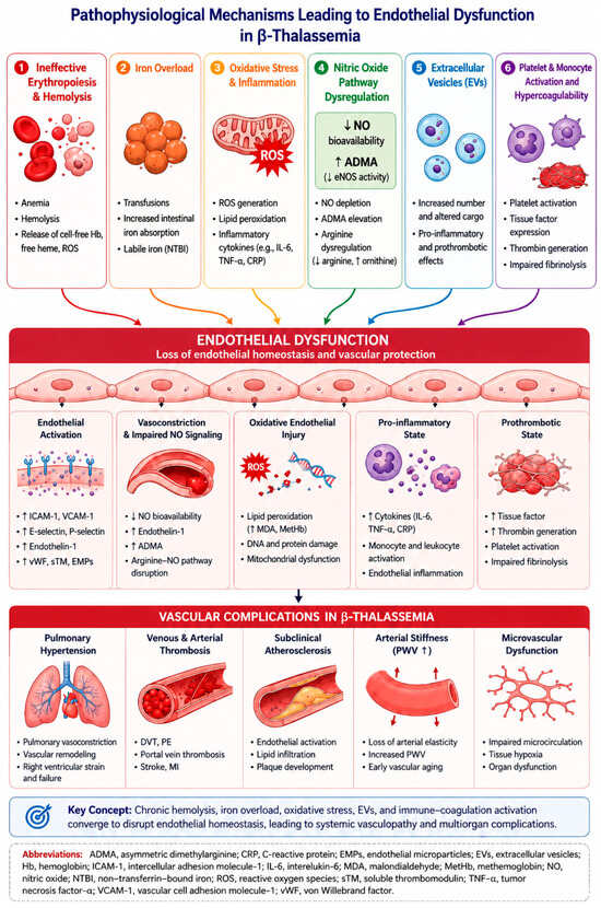
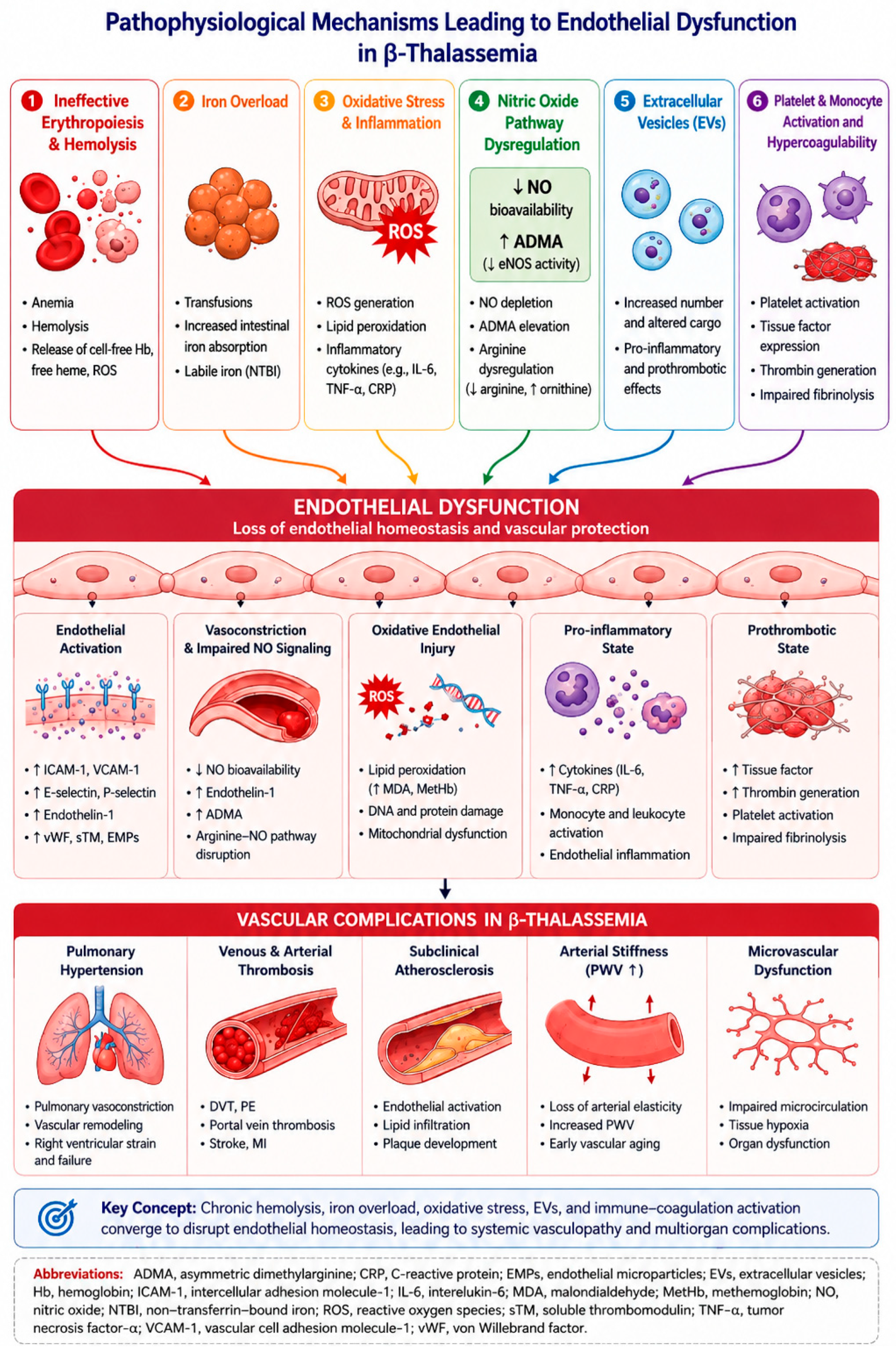

Disclaimer/Publisher’s Note: The statements, opinions and data contained in all publications are solely those of the individual author(s) and contributor(s) and not of MDPI and/or the editor(s). MDPI and/or the editor(s) disclaim responsibility for any injury to people or property resulting from any ideas, methods, instructions or products referred to in the content. |
© 2026 by the authors. Licensee MDPI, Basel, Switzerland. This article is an open access article distributed under the terms and conditions of the Creative Commons Attribution (CC BY) license.