Next Issue
Volume 17, June
Previous Issue
Volume 17, February


Hematol. Rep., Volume 17, Issue 2 (April 2025) – 12 articles

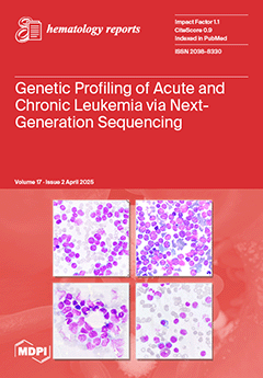
Cover Story (view full-size image): Leukemia is a heterogeneous group of hematologic malignancies characterized by distinct genetic and molecular abnormalities. Advancements in genomic technologies have significantly transformed the diagnosis, prognosis, and treatment strategies for leukemia. Among these, next-generation sequencing (NGS) has emerged as a powerful tool, enabling high-resolution genomic profiling that surpasses the conventional diagnostic approaches. This review highlights the impact of NGS on leukemia research and clinical applications, discussing its advantages over the traditional diagnostic techniques, key sequencing approaches, and emerging challenges. As precision oncology continues to evolve, NGS is expected to play an increasingly central role in the diagnosis and management of leukemia, driving innovations in personalized medicine and therapeutic interventions. View this paper
  • Issues are regarded as officially published after their release is announced to the table of contents alert mailing list.
  • You may sign up for e-mail alerts to receive table of contents of newly released issues.
  • PDF is the official format for papers published in both, html and pdf forms. To view the papers in pdf format, click on the "PDF Full-text" link, and use the free Adobe Reader to open them.
Order results
Result details
Select all
Export citation of selected articles as:
6 pages, 652 KiB  
Case Report
Successful Management of Acquired von Willebrand Syndrome Associated with Monoclonal Gammopathy of Undetermined Significance After Sotorasib Treatment in a Patient with Non-Small-Cell Lung Carcinoma
by Mélissa Julien, Léa Pierre, Anne-Cécile Gérout, Laurent Sattler, Olivier Feugeas and Dominique Desprez
Hematol. Rep. 2025, 17(2), 21; https://doi.org/10.3390/hematolrep17020021 - 16 Apr 2025
Viewed by 209
Abstract
Background: This case report investigates the effects of sotorasib treatment in a patient with acquired von Willebrand syndrome (AVWS) associated with monoclonal gammopathy of undetermined significance (MGUS), who subsequently developed non-small-cell lung carcinoma (NSCLC) with a KRAS G12C mutation. Case Presentation: The patient, [...] Read more.
Background: This case report investigates the effects of sotorasib treatment in a patient with acquired von Willebrand syndrome (AVWS) associated with monoclonal gammopathy of undetermined significance (MGUS), who subsequently developed non-small-cell lung carcinoma (NSCLC) with a KRAS G12C mutation. Case Presentation: The patient, a 79-year-old male, presented with a prolonged history of recurrent lower gastrointestinal bleeding attributed to digestive angiodysplasia, which had persisted for over 30 years. AVWS was suspected based on a qualitative deficiency in von Willebrand factor (VWF), with abnormal results for factor VIII activity (FVIII:C), VWF antigen (VWF:Ag), and VWF ristocetin cofactor activity (VWF:Rco) (40%, 20%, and <2.4%, respectively). Further evaluation revealed the presence of an IgM kappa monoclonal spike, suggesting MGUS. In 2022, the patient was diagnosed with NSCLC harboring the KRAS G12C mutation and initiated second-line treatment with sotorasib. Notably, one year after the initiation of sotorasib therapy, the patient’s hemostasis had normalized, accompanied by significant improvements in VWF levels. VWF multimer electrophoresis demonstrated the restoration of high-molecular-weight multimers (HMWMs), and serum protein electrophoresis no longer detected MGUS. Conclusion: These improvements were likely attributable to the indirect effects of sotorasib on the bone marrow microenvironment. By inhibiting KRAS in stromal cells and osteoclasts, sotorasib may have disrupted the supportive niche necessary for malignant plasma cell survival, resulting in a reduction in the monoclonal spike. Unfortunately, the patient eventually succumbed to carcinogenic pleurisy. Full article
Show Figures

Figure 1

8 pages, 822 KiB  
Case Report
Hemolytic Anemia Due to Gamma-Glutamylcysteine Synthetase Deficiency: A Rare Novel Case in an Arab-Muslim Israeli Child
by Motti Haimi and Jamal Mahamid
Hematol. Rep. 2025, 17(2), 20; https://doi.org/10.3390/hematolrep17020020 - 15 Apr 2025
Viewed by 361
Abstract
Background: Gamma-glutamylcysteine synthetase catalyzes the first and rate-limiting step in the synthesis of glutathione. Gamma-glutamylcysteine synthetase deficiency is a very rare condition that has so far been detected so far in nine patients from seven families worldwide. The inheritance of this disorder is [...] Read more.
Background: Gamma-glutamylcysteine synthetase catalyzes the first and rate-limiting step in the synthesis of glutathione. Gamma-glutamylcysteine synthetase deficiency is a very rare condition that has so far been detected so far in nine patients from seven families worldwide. The inheritance of this disorder is autosomal recessive. Methods: We report a case of 4.11-year-old boy, of Arab-Muslim origin, living in an Arab town in Israel who presented at the age of 2 days with severe anemia, reticulocytosis, and leukocytosis. Investigation for common causes of hemolytic anemia was negative (peripheral blood smear was normal, and he had a negative Coombs test, normal G6PD, and normal flow cytometry spherocytosis). The anemia worsened during the following days (hemoglobin (Hb): 7.2 g/dL) and he needed several blood transfusions. NGS (next-generation sequencing) gene panel analysis was performed. Results: In an NGS gene panel analysis for hereditary hemolytic anemias, we found a homozygotic change in the GCLC gene—G53.385.643c379C > T(homo)pArg127Cys—which confirms the diagnosis of gamma-glutamylcysteine synthetase deficiency. An additional rare change was found in this case in the GCLC gene, with unknown clinical significance: g.53373917, c 828 + 3A > G. Except for chronic anemia (Hb levels around 8 g/dL), the child has normal physical and neurological development. Conclusions: This study reports a rare case of gamma-glutamylcysteine synthetase deficiency in a 4.11-year-old Arab-Muslim boy from Israel who presented with severe anemia at 2 days old, aiming to document the first such case in the Middle East and contribute to the medical literature on this extremely rare condition that has only been detected in nine patients worldwide. Genetic analysis revealed a homozygotic change in the GCLC gene, confirming the diagnosis, and while the patient experiences chronic anemia, he maintains normal physical and neurological development, adding valuable insights to the understanding of this rare genetic disorder. An additional rare change was found in this case in the GCLC gene, with unknown clinical significance: g.53373917, c 828 + 3A > G. Full article
(This article belongs to the Special Issue Anaemia in Focus: Challenges and Solutions in Haematology)
Show Figures

Figure 1

8 pages, 1004 KiB  
Article
Blood Coagulation Favors Anti-Inflammatory Immune Responses in Whole Blood
by Victor I. Seledtsov, Anatoly A. Pyshenko, Tatyana Ya. Lyubavskaya, Irina A. Seledtsova and Alexei A. von Delwig
Hematol. Rep. 2025, 17(2), 19; https://doi.org/10.3390/hematolrep17020019 - 11 Apr 2025
Viewed by 558
Abstract
Background: We studied the effects of human blood coagulation on antioxidant activity and the cellular secretion of immunoregulatory molecules in vitro. Methods: Reactive oxygen species (ROS) activity and cytokine content were determined in plasma and serum blood samples incubated with lipopolysaccharide (LPS) for [...] Read more.
Background: We studied the effects of human blood coagulation on antioxidant activity and the cellular secretion of immunoregulatory molecules in vitro. Methods: Reactive oxygen species (ROS) activity and cytokine content were determined in plasma and serum blood samples incubated with lipopolysaccharide (LPS) for 3 h or 18 h. Results: Coagulation process significantly decreased ROS activity induced by LPS in blood samples from healthy donors. Human serum was found to have significantly higher antioxidant activity than plasma. Blood coagulation markedly reduced LPS-induced secretion of TNF-α by cells, without significantly affecting the secretion of interleukin-1 (IL-1), IL-6, IL-8, or C-reactive protein (CRP). Blood clotting led to an increase in LPS-induced release of vascular endothelial growth factor (VEGF) by blood cells. A significant increase in procalcitonin levels was also observed in serum samples. Conclusions: Blood clotting enhances the antioxidant and anti-inflammatory functions of immunoreactive blood cells. Full article
Show Figures

Graphical abstract

47 pages, 3413 KiB  
Review
Genetic Profiling of Acute and Chronic Leukemia via Next-Generation Sequencing: Current Insights and Future Perspectives
by Laras Pratiwi, Fawzia Hanum Mashudi, Mukti Citra Ningtyas, Henry Sutanto and Pradana Zaky Romadhon
Hematol. Rep. 2025, 17(2), 18; https://doi.org/10.3390/hematolrep17020018 - 28 Mar 2025
Viewed by 494
Abstract
Leukemia is a heterogeneous group of hematologic malignancies characterized by distinct genetic and molecular abnormalities. Advancements in genomic technologies have significantly transformed the diagnosis, prognosis, and treatment strategies for leukemia. Among these, next-generation sequencing (NGS) has emerged as a powerful tool, enabling high-resolution [...] Read more.
Leukemia is a heterogeneous group of hematologic malignancies characterized by distinct genetic and molecular abnormalities. Advancements in genomic technologies have significantly transformed the diagnosis, prognosis, and treatment strategies for leukemia. Among these, next-generation sequencing (NGS) has emerged as a powerful tool, enabling high-resolution genomic profiling that surpasses conventional diagnostic approaches. By providing comprehensive insights into genetic mutations, clonal evolution, and resistance mechanisms, NGS has revolutionized precision medicine in leukemia management. Despite its transformative potential, the clinical integration of NGS presents challenges, including data interpretation complexities, standardization issues, and cost considerations. However, continuous advancements in sequencing platforms and bioinformatics pipelines are enhancing the reliability and accessibility of NGS in routine clinical practice. The expanding role of NGS in leukemia is paving the way for improved risk stratification, targeted therapies, and real-time disease monitoring, ultimately leading to better patient outcomes. This review highlights the impact of NGS on leukemia research and clinical applications, discussing its advantages over traditional diagnostic techniques, key sequencing approaches, and emerging challenges. As precision oncology continues to evolve, NGS is expected to play an increasingly central role in the diagnosis and management of leukemia, driving innovations in personalized medicine and therapeutic interventions. Full article
Show Figures

Figure 1

13 pages, 245 KiB  
Article
Primary Palatine Tonsil Non-Hodgkin Lymphoma in Western Romania: A Comparison of Lower-Stage and Advanced-Stage Disease
by Raluca Morar, Norberth-Istvan Varga, Delia Ioana Horhat, Ion Cristian Mot, Nicolae Constantin Balica, Alina-Andree Tischer, Monica Susan, Razvan Susan, Diana Luisa Lighezan and Rodica Anamaria Negrean
Hematol. Rep. 2025, 17(2), 17; https://doi.org/10.3390/hematolrep17020017 - 28 Mar 2025
Viewed by 211
Abstract
Background: Limited data exist on primary palatine tonsil Non-Hodgkin lymphoma (NHL) from regions with constrained healthcare access. This study investigated this malignancy in Western and South-Western Romania, comparing lower-stage (Ann-Arbor I-III) and advanced-stage (IV) disease. Methods: A retrospective cohort study (2010–2019) [...] Read more.
Background: Limited data exist on primary palatine tonsil Non-Hodgkin lymphoma (NHL) from regions with constrained healthcare access. This study investigated this malignancy in Western and South-Western Romania, comparing lower-stage (Ann-Arbor I-III) and advanced-stage (IV) disease. Methods: A retrospective cohort study (2010–2019) at a tertiary referral hospital included 59 patients with primary palatine tonsil NHL. Data on demographics, clinical presentation, comorbidities (including viral hepatitis B/C), histology, International Prognostic Index (IPI) score, treatment, and outcomes were collected. Statistical comparisons between lower-stage (n = 26) and advanced-stage (n = 33) groups were performed. Results: A high proportion presented with advanced-stage disease (55.9%). The advanced-stage group had significantly more B symptoms (90.9% vs. 69.2%, p = 0.038) and elevated LDH levels (93.9% vs. 57.7%, p = 0.013). Viral hepatitis B and/or C infection was more frequent in advanced-stage disease (30.3% vs. 15.4%, p = 0.44). Combined chemoradiotherapy was more commonly used in lower-stage disease (38.46% vs. 12.12%, p = 0.019). There was no statistically significant difference in relapse rates between the groups. Conclusions: This study highlights the substantial burden of advanced-stage primary palatine tonsil NHL in Western Romania, suggesting a need for improved early detection. The association between viral hepatitis and advanced-stage, although not statistically significant, warrants further investigation. These findings may inform tailored management approaches in resource-constrained settings. Full article
10 pages, 966 KiB  
Article
Treatment of Non-Hodgkin Lymphoma Involving Head and Neck Sites with a 1.5 T MR-Linac: Preliminary Results from a Prospective Observational Study
by Andrea Emanuele Guerini, Stefania Nici, Stefano Riga, Ludovica Pegurri, Paolo Borghetti, Eneida Mataj, Jacopo Balduzzi, Mirsada Katica, Gianluca Cossali, Giorgio Facheris, Luca Triggiani, Albert Sakiri, Luigi Spiazzi, Stefano Maria Magrini and Michela Buglione
Hematol. Rep. 2025, 17(2), 16; https://doi.org/10.3390/hematolrep17020016 - 27 Mar 2025
Viewed by 189
Abstract
Purpose: Lymphomas are generally radiosensitive; therefore, disease volume tends to shrink during radiotherapy courses. As MRI-linac provides excellent soft tissue definition and allows daily re-contouring of gross tumor volume and clinical target volume, its adoption could be beneficial for the treatment of [...] Read more.
Purpose: Lymphomas are generally radiosensitive; therefore, disease volume tends to shrink during radiotherapy courses. As MRI-linac provides excellent soft tissue definition and allows daily re-contouring of gross tumor volume and clinical target volume, its adoption could be beneficial for the treatment of lymphomas. Nonetheless, at this time there is a lack of literature regarding the use of MR-linac in this context. Methods: A prospective observational study was conducted on patients affected by non-Hodgkin lymphoma (NHL) involving head and neck (H&N) sites and treated with Elekta Unity® MR-Linac. The clinical and dosimetric data of the first eight patients were collected and integrated with relevant data from medical records. Results: Seven patients had B-cell lymphoma (three DLBCL, two MALT, one follicular, and one mantle-cell) and one T-cell/NK lymphoma. The intent of RT was radical for four patients, salvage treatment for three, and CAR-T bridging for one. Two patients presented orbital localizations and six cervical lymphonodal sites. Median GTV was 5.74 cc, median CTV 127.01 cc, and median PTV 210.37 cc. The prescribed dose was 24–50 Gy in 2 Gy fractions for seven patients and 24 Gy in 3 Gy fractions for one patient. All the patients experienced acute toxicity, the maximum grade was G1 for five patients and G2 for three at the end of RT. One month after radiotherapy seven patients still experienced G1 toxicity, but no toxicity grade ≥ 2 was reported. First radiological assessment was performed for all the patients after a median of 101.5 days, reporting complete response in all the cases. After a median follow up of 330 days, no patient experienced local disease progression, while one patient developed distant progression. Conclusions: radiotherapy for NHL with H&N localization using a 1.5 T MR-linac was feasible, with no >G2 toxicity and optimal response rate and disease control. Full article
Show Figures

Figure 1

8 pages, 204 KiB  
Communication
Avascular Necrosis of the Femoral Head in Patients with Antiphospholipid Syndrome: A Case Series
by Paschalis Evangelidis, Eleni Gavriilaki, Nikolaos Kotsiou, Zacharo Ntova, Panagiotis Kalmoukos, Theodosia Papadopoulou, Sofia Chissan and Sofia Vakalopoulou
Hematol. Rep. 2025, 17(2), 15; https://doi.org/10.3390/hematolrep17020015 - 21 Mar 2025
Viewed by 307
Abstract
Background/Objectives: Antiphospholipid syndrome (APS) is a systemic autoimmune disease characterized by thrombosis or obstetric complications and the laboratory detection of antiphospholipid antibodies. Although vascular thrombosis is the main manifestation of the disease, other rarer complications have also been described. Avascular necrosis (AN) [...] Read more.
Background/Objectives: Antiphospholipid syndrome (APS) is a systemic autoimmune disease characterized by thrombosis or obstetric complications and the laboratory detection of antiphospholipid antibodies. Although vascular thrombosis is the main manifestation of the disease, other rarer complications have also been described. Avascular necrosis (AN) is considered a rare manifestation of APS. The aim of our case series is to study patients with APS and AN. Methods: A retrospective study was performed on 80 patients diagnosed with APS. Results: AN was observed in 3 patients out of 80 diagnosed with APS. AN of the femoral head was observed in all cases. Case (1): A 54-year-old woman presented due to multiple ischemic infarctions in the brain, as detected in magnetic resonance imaging of the brain, Raynaud’s phenomenon, and AN of the femoral head. In laboratory testing, a prolongation of activated partial thromboplastin time was recorded. A heterozygous mutation was also found in the gene MTHFR C677T, and the patients was positive for lupus anticoagulant (LA). The patient was given clopidogrel and acenocoumarol. Case (2): A 52-year-old man was diagnosed with APS, based on the clinical presentation (stroke) and positivity for LA and anti-β2GPI (anti-β2 glycoprotein I antibody). In his medical history, episodes of vertigo and an episode of AN of the femoral head 2 years ago were described. Case (3): A woman aged 43 years presented due to AN of the femoral head. Due to suspected APS, immunological testing was performed, and positivity for LA and IgM anticardiolipin antibodies was detected. She was treated with acenocoumarol. Conclusions: AN is a rare clinical manifestation of APS, which may precede the diagnosis of APS for many years. Full article
7 pages, 1804 KiB  
Case Report
Pure White Cell Aplasia, an Exceedingly Rare Complication of a Thymoma, and Immune Reconstitution Following Bone Marrow Recovery
by Giby V. George, J. C. Uy, John L. Mariano, Marisa Jacob-Leonce and Chauncey R. Syposs
Hematol. Rep. 2025, 17(2), 14; https://doi.org/10.3390/hematolrep17020014 - 21 Mar 2025
Viewed by 208
Abstract
Background: Thymoma-associated pure white cell aplasia (PWCA), characterized by agranulocytosis with absent myeloid precursors in the bone marrow in the setting of preserved erythropoiesis and megakaryopoiesis, is exceedingly rare, with only a few cases reported in the literature. We present a case of [...] Read more.
Background: Thymoma-associated pure white cell aplasia (PWCA), characterized by agranulocytosis with absent myeloid precursors in the bone marrow in the setting of preserved erythropoiesis and megakaryopoiesis, is exceedingly rare, with only a few cases reported in the literature. We present a case of type-B2-thymoma-associated PWCA and immune reconstitution following marrow recovery. Case Presentation: A 75-year-old woman was incidentally found to have a concomitant mediastinal mass and peripheral leukopenia with absent granulocytes and monocytes. Bone marrow assessment was notable for a hypocellular marrow (<10%) with absent granulopoiesis and monopoiesis. Chest CT demonstrated a large lobulated anterior mediastinal mass, for which the patient underwent a video-assisted thoracoscopic thymectomy. Pathological evaluation of the mediastinal mass specimen revealed a type B2 thymoma. A tentative diagnosis of thymoma-associated PWCA was made, and the patient was started on cyclosporine/granulocyte-colony stimulating factor (G-CSF)/filgrastim therapy. Despite promising marrow recovery, she developed several comorbidities and had a leukemoid reaction, provoking concern for immune reconstitution following prolonged neutropenia and subsequent treatment. She passed away on post-operative day 15, and the results of a post-mortem bone marrow examination were consistent with granulocytic hyperplasia. Conclusions: This case of thymoma-associated PWCA heightens awareness regarding this entity, providing a note of caution regarding the possibility of immune reconstitution following treatment and marrow recovery. Full article
Show Figures

Figure 1

9 pages, 1146 KiB  
Case Report
Hairy Cell Leukemia: A Differential Diagnosis of Hepatitis B-Associated Aplastic Anemia and Syphilis
by I. Kindekov, E. Beleva, M. Kadish, I. Ionchev and N. Semerdzhieva
Hematol. Rep. 2025, 17(2), 13; https://doi.org/10.3390/hematolrep17020013 - 15 Mar 2025
Viewed by 386
Abstract
Aplastic anemia occurs with an incidence of 2–5: 1 million people worldwide. However, the frequency of newly diagnosed cases of bone marrow aplasia is greater, and some of these patients present to emergency departments initially. Description of Case: We present the case of [...] Read more.
Aplastic anemia occurs with an incidence of 2–5: 1 million people worldwide. However, the frequency of newly diagnosed cases of bone marrow aplasia is greater, and some of these patients present to emergency departments initially. Description of Case: We present the case of a middle-aged man with pancytopenia. In this case, aplastic anemia associated with hepatitis B and syphilis was only the initial diagnosis. An indolent hematologic malignancy—hairy cell leukemia—was diagnosed as the real cause of the bone marrow failure in a clinic of hematology. Conclusions: This clinical case allows us to make a conclusion, albeit not definitively, about the contribution of hepatitis B and syphilis to the clinical manifestation of hairy cell leukemia. A detailed and consistent diagnostic plan is also required in patients presenting with pancytopenia. Failure to diagnose a hepatitis B infection in a patient with malignant hematologic disease would lead to fatal therapeutic errors. Full article
Show Figures

Figure 1

7 pages, 1839 KiB  
Case Report
Acute Intracerebral Hemorrhage Associated with Extensive Venous Thrombosis Due to Spontaneous Heparin-Induced Thrombocytopenia After Total Knee Replacement: A Case Report
by Mehdi Kashani, Meghan Brown and Juan Pablo Domecq Graces
Hematol. Rep. 2025, 17(2), 12; https://doi.org/10.3390/hematolrep17020012 - 13 Mar 2025
Viewed by 412
Abstract
Introduction: Heparin-induced thrombocytopenia (HIT) is an autoimmune life-threatening prothrombotic syndrome associated with low platelet count after heparin exposure. Spontaneous heparin-induced thrombocytopenia (S-HIT) is an even less frequent variant of HIT, with only a handful of reports available in the literature, where unexplained thrombocytopenia [...] Read more.
Introduction: Heparin-induced thrombocytopenia (HIT) is an autoimmune life-threatening prothrombotic syndrome associated with low platelet count after heparin exposure. Spontaneous heparin-induced thrombocytopenia (S-HIT) is an even less frequent variant of HIT, with only a handful of reports available in the literature, where unexplained thrombocytopenia and/or thrombosis without recent heparin exposure occurs in the setting of positive anti-PF4 antibodies. Case Presentation: We report a case of S-HIT associated with pulmonary artery embolism, left internal jugular vein, and cerebral vein sinus thrombosis complicated with ipsilateral acute intracerebral hemorrhage. Discussion: It is important to highlight that in patients with otherwise unexplained thrombocytopenia and prior exposure to an inflammatory process, S-HIT should be on the differential. Conclusions: Recognition and avoidance of heparin exposure is the most important aspect of S-HIT, as the management is otherwise similar to HIT. Full article
Show Figures

Figure 1

6 pages, 559 KiB  
Case Report
Feasibility of Intensive Chemotherapy in Hereditary Spherocytosis
by Carrai Valentina, Giubbilei Cristina, Ciceri Manuel, D’Angelo Simona, Nassi Luca, Sordi Benedetta, Vannucchi Alessandro Maria and Puccini Benedetta
Hematol. Rep. 2025, 17(2), 11; https://doi.org/10.3390/hematolrep17020011 - 24 Feb 2025
Viewed by 295
Abstract
Background: This study presents a young man with hereditary spherocytosis (HS) who underwent intensive chemotherapy for newly diagnosed diffuse large B-cell lymphoma (DLBCL) and achieved complete remission. This case challenges the idea of HS as a barrier to standard DLBCL treatment. Discussion: By [...] Read more.
Background: This study presents a young man with hereditary spherocytosis (HS) who underwent intensive chemotherapy for newly diagnosed diffuse large B-cell lymphoma (DLBCL) and achieved complete remission. This case challenges the idea of HS as a barrier to standard DLBCL treatment. Discussion: By meticulously monitoring blood counts and providing timely transfusions, the team successfully mitigated potential complications associated with chemotherapy-induced stress on red blood cells. Conclusions: This experience underscores the importance of a multidisciplinary approach and tailored treatment plans for patients with co-existing conditions, suggesting that HS should not automatically disqualify them from potentially curative therapies for aggressive lymphomas. Full article
Show Figures

Figure 1

5 pages, 875 KiB  
Case Report
Factor X Concentrate Treatment Schedule and Dosing in Acquired FX Deficiency
by Andrew Ross, Rebecca J. Shaw, Louise Garth and Cathy Farrelly
Hematol. Rep. 2025, 17(2), 10; https://doi.org/10.3390/hematolrep17020010 - 21 Feb 2025
Viewed by 363
Abstract
Background: Acquired factor X (FX) deficiency is a rare condition that can cause life threatening bleeding. Here we outline a successful management strategy for gastrointestinal bleeding (GI) using human FX concentrate. Case description: A 61-year-old male presented with upper GI bleeding and a [...] Read more.
Background: Acquired factor X (FX) deficiency is a rare condition that can cause life threatening bleeding. Here we outline a successful management strategy for gastrointestinal bleeding (GI) using human FX concentrate. Case description: A 61-year-old male presented with upper GI bleeding and a prolonged prothrombin time. Investigations demonstrated an acquired FX deficiency (determined to be secondary to AL amyloidosis). Results: Treatment with FX concentrate to maintain trough FX levels >20% resulted in successful cessation of bleeding symptoms, and levels >50% facilitated urgent invasive procedures. Conclusions: This case report adds valuable insight into the management of this rare condition, and how best to utilize FX concentrates in acquired FX deficiency. Full article
Show Figures

Figure 1

Previous Issue
Next Issue
Back to TopTop