
Treatment of Non-Hodgkin Lymphoma Involving Head and Neck Sites with a 1.5 T MR-Linac: Preliminary Results from a Prospective Observational Study
 , , , and
Abstract
1. Introduction
2. Methods
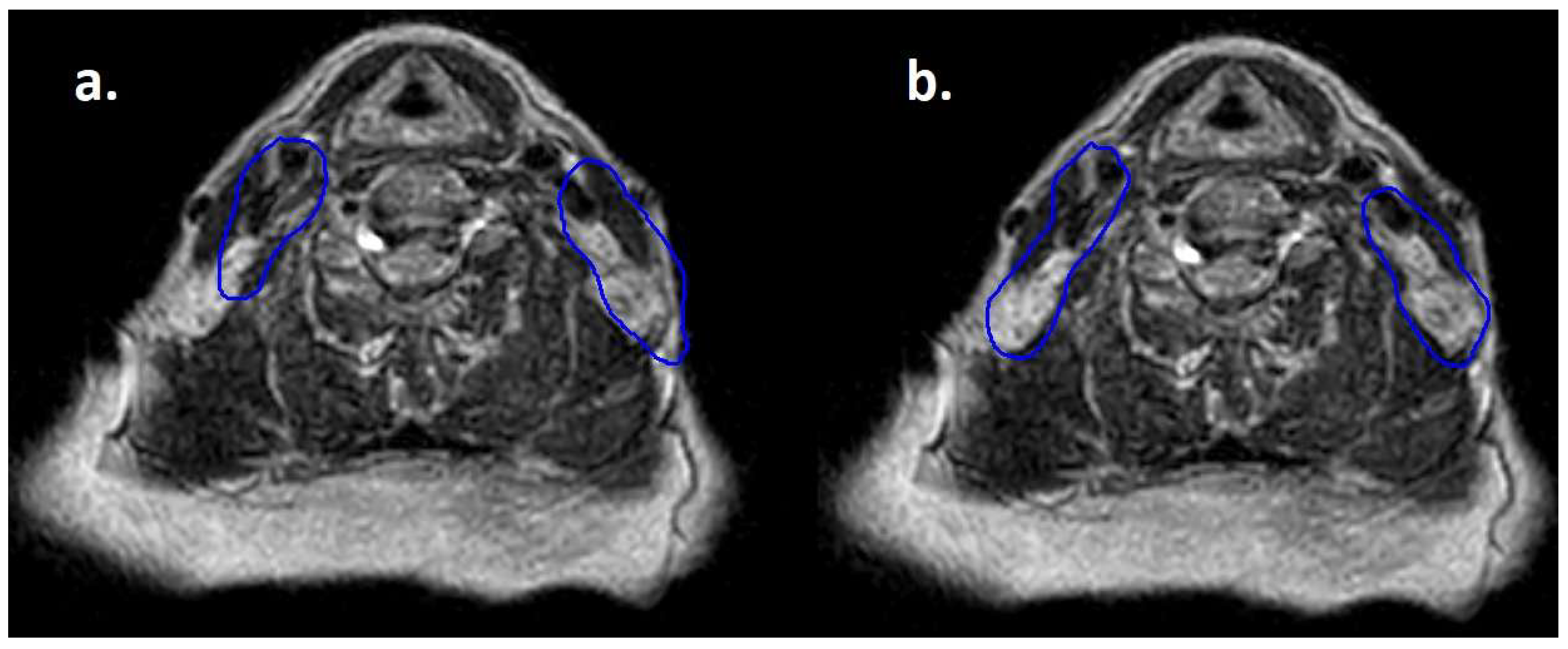
3. Results
4. Discussion
5. Conclusions
Author Contributions
Funding
Institutional Review Board Statement
Informed Consent Statement
Data Availability Statement
Conflicts of Interest
References
- Pepper, N.B.; Oertel, M.; Rehn, S.; Kobe, C.; Eich, H.T. Modern PET-Guided Radiotherapy Planning and Treatment for Malignant Lymphoma. Semin. Nucl. Med. 2023, 53, 389–399. [Google Scholar] [CrossRef] [PubMed]
- Yahalom, J.; Illidge, T.; Specht, L.; Hoppe, R.T.; Li, Y.X.; Tsang, R.; Wirth, A.; International Lymphoma Radiation Oncology Group. Modern radiation therapy for extranodal lymphomas: Field and dose guidelines from the International Lymphoma Radiation Oncology Group. Int. J. Radiat. Oncol. Biol. Phys. 2015, 92, 11–31. [Google Scholar] [CrossRef] [PubMed]
- Specht, L.; Yahalom, J.; Illidge, T.; Berthelsen, A.K.; Constine, L.S.; Eich, H.T.; Girinsky, T.; Hoppe, R.T.; Mauch, P.; Mikhaeel, N.G.; et al. Modern radiation therapy for Hodgkin lymphoma: Field and dose guidelines from the international lymphoma radiation oncology group (ILROG). Int. J. Radiat. Oncol. Biol. Phys. 2014, 89, 854–862. [Google Scholar] [CrossRef] [PubMed]
- Zhan, Z.; Guo, W.; Wan, X.; Bai, O. Second primary malignancies in non-Hodgkin lymphoma: Epidemiology and risk factors. Ann. Hematol. 2023, 102, 249–259. [Google Scholar] [CrossRef] [PubMed]
- Hodgson, D.C. Long-term toxicity of chemotherapy and radiotherapy in lymphoma survivors: Optimizing treatment for individual patients. Clin. Adv. Hematol. Oncol. 2015, 13, 103–112. [Google Scholar] [PubMed]
- Milgrom, S.A.; Bakst, R.L.; Campbell, B.A. Clinical Outcomes Confirm Conjecture: Modern Radiation Therapy Reduces the Risk of Late Toxicity in Survivors of Hodgkin Lymphoma. Int. J. Radiat. Oncol. Biol. Phys. 2021, 111, 841–850. [Google Scholar] [CrossRef] [PubMed]
- Buglione, M.; Guerini, A.E.; Filippi, A.R.; Spiazzi, L.; Pasinetti, N.; Magli, A.; Toraci, C.; Borghetti, P.; Triggiani, L.; Alghisi, A.; et al. A Systematic Review on Intensity Modulated Radiation Therapy for Mediastinal Hodgkin’s Lymphoma. Crit. Rev. Oncol. Hematol. 2021, 167, 103437. [Google Scholar] [CrossRef] [PubMed]
- Mikhaeel, N.G.; Milgrom, S.A.; Terezakis, S.; Berthelsen, A.K.; Hodgson, D.; Eich, H.T.; Dieckmann, K.; Qi, S.N.; Yahalom, J.; Specht, L. The Optimal Use of Imaging in Radiation Therapy for Lymphoma: Guidelines from the International Lymphoma Radiation Oncology Group (ILROG). Int. J. Radiat. Oncol. Biol. Phys. 2019, 104, 501–512. [Google Scholar] [CrossRef] [PubMed]
- Di Russo, A.; Simontacchi, G.; Guerini, A.E.; Filippi, A.R.; Levis, M.; Ciammella, P.; De Sanctis, V.; Vagge, S.; Meregalli, S.; De Marco, G.; et al. Advanced Radiotherapy Techniques for Mediastinal Lymphomas: Results from an Italian Survey. Hemato 2021, 2, 496–504. [Google Scholar] [CrossRef]
- Wippold, F.J., 2nd. Head and neck imaging: The role of CT and MRI. J. Magn. Reson. Imaging 2007, 25, 453–465. [Google Scholar] [CrossRef] [PubMed]
- Tijssen, R.H.N.; Philippens, M.E.P.; Paulson, E.S.; Glitzner, M.; Chugh, B.; Wetscherek, A.; Dubec, M.; Wang, J.; van der Heide, U.A. MRI commissioning of 1.5T MR-linac systems—A multi-institutional study. Radiother. Oncol. 2019, 132, 114–120. [Google Scholar] [CrossRef] [PubMed]
- Guerini, A.E.; Nici, S.; Magrini, S.M.; Riga, S.; Toraci, C.; Pegurri, L.; Facheris, G.; Cozzaglio, C.; Farina, D.; Liserre, R.; et al. Adoption of Hybrid MRI-Linac Systems for the Treatment of Brain Tumors: A Systematic Review of the Current Literature Regarding Clinical and Technical Features. Technol. Cancer Res. Treat. 2023, 22, 15330338231199286. [Google Scholar] [CrossRef] [PubMed] [PubMed Central]
- Habrich, J.; Boeke, S.; Fritz, V.; Koerner, E.; Nikolaou, K.; Schick, F.; Gani, C.; Zips, D.; Thorwarth, D. Reproducibility of diffusion-weighted magnetic resonance imaging in head and neck cancer assessed on a 1.5 T MR-Linac and comparison to parallel measurements on a 3 T diagnostic scanner. Radiother. Oncol. 2024, 191, 110046. [Google Scholar] [CrossRef] [PubMed]
- McDonald, B.A.; Dal Bello, R.; Fuller, C.D.; Balermpas, P. The Use of MR-Guided Radiation Therapy for Head and Neck Cancer and Recommended Reporting Guidance. Semin. Radiat. Oncol. 2024, 34, 69–83, Erratum in Semin. Radiat. Oncol. 2024, 34, 365. [Google Scholar] [CrossRef] [PubMed] [PubMed Central]
- Ababneh, H.S.; Connor Johnson, P.; Pursley, J.; Patel, C.G. Adaptive bridging radiation therapy for relapsed/refractory B-cell lymphoma patient undergoing CAR T-cell therapy: Case report. Clin. Transl. Radiat. Oncol. 2024, 48, 100832. [Google Scholar] [CrossRef] [PubMed] [PubMed Central]
- de Mol van Otterloo, S.R.; Christodouleas, J.P.; Blezer, E.L.A.; Akhiat, H.; Brown, K.; Choudhury, A.; Eggert, D.; Erickson, B.A.; Daamen, L.A.; Faivre-Finn, C.; et al. Patterns of Care, Tolerability, and Safety of the First Cohort of Patients Treated on a Novel High-Field MR-Linac Within the MOMENTUM Study: Initial Results From a Prospective Multi-Institutional Registry. Int. J. Radiat. Oncol. Biol. Phys. 2021, 111, 867–875. [Google Scholar] [CrossRef]
- Dincer, N.; Ugurluer, G.; Gungor, G.; Zoto Mustafayev, T.; Atalar, B.; Ozyar, E. Magnetic Resonance Imaging-Guided Radiation Therapy for Early-Stage Gastric Mucosa-Associated Lymphoid Tissue Lymphoma. Cureus 2022, 14, e29035. [Google Scholar] [CrossRef]
- Song, Y.; Li, Z.; Wang, H.; Zhang, Y.; Yue, J. MR-LINAC-Guided Adaptive Radiotherapy for Gastric MALT: Two Case Reports and a Literature Review. Radiation 2022, 2, 259–267. [Google Scholar] [CrossRef]
- Eismann, J.; Elsayad, K.; Rolf, D.; Sarif, I.; Wardelmann, E.; Berssenbrügge, H.; Lenz, G.; Eich, H.T. Intensity-modulated Radiotherapy in Patients With Aggressive Extranodal Non-Hodgkin Lymphoma of the Head and Neck. Anticancer. Res. 2021, 41, 5131–5135. [Google Scholar] [CrossRef] [PubMed]
- Zukauskaite, R.; Rumley, C.N.; Hansen, C.R.; Jameson, M.G.; Trada, Y.; Johansen, J.; Gyldenkerne, N.; Eriksen, J.G.; Aly, F.; Christensen, R.L.; et al. Delineation uncertainties of tumour volumes on MRI of head and neck cancer patients. Clin. Transl. Radiat. Oncol. 2022, 36, 121–126. [Google Scholar] [CrossRef] [PubMed] [PubMed Central]
- Mulder, S.L.; Heukelom, J.; McDonald, B.A.; Van Dijk, L.; Wahid, K.A.; Sanders, K.; Salzillo, T.C.; Hemmati, M.; Schaefer, A.; Fuller, C.D. MR-Guided Adaptive Radiotherapy for OAR Sparing in Head and Neck Cancers. Cancers 2022, 14, 1909. [Google Scholar] [CrossRef] [PubMed] [PubMed Central]
- Gupta, A.; Dunlop, A.; Mitchell, A.; McQuaid, D.; Nill, S.; Barnes, H.; Newbold, K.; Nutting, C.; Bhide, S.; Oelfke, U.; et al. Online adaptive radiotherapy for head and neck cancers on the MR linear Accelerator: Introducing a novel modified Adapt-to-Shape approach. Clin. Transl. Radiat. Oncol. 2021, 32, 48–51. [Google Scholar] [CrossRef] [PubMed] [PubMed Central]
- Guo, J.; Liu, Z.; Shen, C.; Li, Z.; Yan, F.; Tian, J.; Xian, J. MR-based radiomics signature in differentiating ocular adnexal lymphoma from idiopathic orbital inflammation. Eur. Radiol. 2018, 28, 3872–3881. [Google Scholar] [CrossRef] [PubMed]
- Albano, D.; Cuocolo, R.; Patti, C.; Ugga, L.; Chianca, V.; Tarantino, V.; Faraone, R.; Albano, S.; Micci, G.; Costa, A.; et al. Whole-body MRI radiomics model to predict relapsed/refractory Hodgkin Lymphoma: A preliminary study. Magn. Reson. Imaging 2022, 86, 55–60. [Google Scholar] [CrossRef] [PubMed]
- Salvestrini, V.; Greco, C.; Guerini, A.E.; Longo, S.; Nardone, V.; Boldrini, L.; Desideri, I.; De Felice, F. The role of feature-based radiomics for predicting response and radiation injury after stereotactic radiation therapy for brain metastases: A critical review by the Young Group of the Italian Association of Radiotherapy and Clinical Oncology (yAIRO). Transl. Oncol. 2022, 15, 101275. [Google Scholar] [CrossRef] [PubMed] [PubMed Central]
- Guerini, A.E.; Filippi, A.R.; Tucci, A.; Simontacchi, G.; Re, A.; Guaineri, A.; Morelli, V.; Borghetti, P.; Triggiani, L.; Pegurri, L.; et al. ‘Le Roi est mort, vive le Roi’: New Roles of Radiotherapy in the Treatment of Lymphomas in Combination With Immunotherapy. Clin. Lymphoma Myeloma Leuk. 2022, 22, e135–e148. [Google Scholar] [CrossRef] [PubMed]
- Umemura, Y.; Wang, D.; Peck, K.K.; Flynn, J.; Zhang, Z.; Fatovic, R.; Anderson, E.S.; Beal, K.; Shoushtari, A.N.; Kaley, T.; et al. DCE-MRI perfusion predicts pseudoprogression in metastatic melanoma treated with immunotherapy. J. Neurooncol. 2020, 146, 339–346. [Google Scholar] [CrossRef] [PubMed] [PubMed Central]
- Li, Y.; Ma, Y.; Wu, Z.; Xie, R.; Zeng, F.; Cai, H.; Lui, S.; Song, B.; Chen, L.; Wu, M. Advanced Imaging Techniques for Differentiating Pseudoprogression and Tumor Recurrence After Immunotherapy for Glioblastoma. Front. Immunol. 2021, 12, 790674. [Google Scholar] [CrossRef] [PubMed] [PubMed Central]
- Barnes, H.; Alexander, S.; Bower, L.; Ehlers, J.; Gani, C.; Herbert, T.; Lawes, R.; Møller, P.K.; Morgan, T.; Nowee, M.E.; et al. Development and results of a patient-reported treatment experience questionnaire on a 1.5 T MR-Linac. Clin. Transl. Radiat. Oncol. 2021, 30, 31–37. [Google Scholar] [CrossRef] [PubMed] [PubMed Central]
- Ng-Cheng-Hin, B.; Nutting, C.; Newbold, K.; Bhide, S.; McQuaid, D.; Dunlop, A.; Harrington, K.; Wong, K.H. The impact of restricted length of treatment field and anthropometric factors on selection of head and neck cancer patients for treatment on the MR-Linac. Br. J. Radiol. 2020, 93, 20200023. [Google Scholar] [CrossRef] [PubMed] [PubMed Central]
- Nachbar, M.; Mönnich, D.; Boeke, S.; Gani, C.; Weidner, N.; Heinrich, V.; Lo Russo, M.; Livi, L.; Winter, J.; Tsitsekidis, S.; et al. Partial breast irradiation with the 1.5 T MR-Linac: First patient treatment and analysis of electron return and stream effects. Radiother. Oncol. 2020, 145, 30–35. [Google Scholar] [CrossRef] [PubMed]
- Wang, M.H.; Kim, A.; Ruschin, M.; Tan, H.; Soliman, H.; Myrehaug, S.; Detsky, J.; Husain, Z.; Atenafu, E.G.; Keller, B.; et al. Comparison of Prospectively Generated Glioma Treatment Plans Clinically Delivered on Magnetic Resonance Imaging (MRI)-Linear Accelerator (MR-Linac) Versus Conventional Linac: Predicted and Measured Skin Dose. Technol. Cancer Res. Treat. 2022, 21, 15330338221124695. [Google Scholar] [CrossRef] [PubMed] [PubMed Central]


| Patients’ Characteristics | |
|---|---|
| Age | median 60.3 years (range 48.5–83.2 years) |
| Histology | Diffuse large B-cell lymphoma 3 patients Mucosa-associated lymphoid tissue lymphoma 2 patients Follicular lymphoma 1 patient Mantle cell lymphoma 1 patient T-cell/natural killer cell lymphoma 1 patient |
| Stage | Stage I 6 patients Stage II 1 patient Stage IV 1 patient |
| Setting | First diagnosis 5 patients Relapse 3 patients |
| Treatment modality | Exclusive radiotherapy 4 patients Sequential treatment with chemotherapy 4 patients |
| Time from diagnosis to RT | median 5.45 months after diagnosis (range 1.5–30.7 months) |
| ECOG performance status | PS 0 4 patients PS 1 4 patients |
| Lymphoma Type | RT Intent | Stage | Setting | Line of Treatment (n) | Systemic Treatment Before RT (n of Cycles) |
|---|---|---|---|---|---|
| DLBCL MYC-BCL2 “double expressor” | CAR-T bridging | IVA | Relapse | 3 | R-CHOP (2) + R-COMP (2) + R-miniDHAP (2) |
| Classic mantle cell lymphoma | Radical RT | IA | First diagnosis | 1 | no |
| Natural killer (NK)/T-cell lymphoma, nasal type | radical RT (+ adjuvant CHT) | II | First diagnosis | 1 | post RT |
| MALT lymphoma | Radical RT | IA | First diagnosis | 1 | no |
| Follicle center lymphoma | Salvage RT | IVA | Relapse | 1 | no |
| MALT lymphoma | Radical RT | IA | First diagnosis | 1 | no |
| Germinal center DLBCL | PR after I line CIT | IA | Refractory after I line | 1 | R-CHOP (4) + R (2) |
| DLBCL MYC-BCL2 “double expressor” | PR after I line CIT | IA | Refractory after I line | 1 | R-mini CHOP (2) + R (2) |
Disclaimer/Publisher’s Note: The statements, opinions and data contained in all publications are solely those of the individual author(s) and contributor(s) and not of MDPI and/or the editor(s). MDPI and/or the editor(s) disclaim responsibility for any injury to people or property resulting from any ideas, methods, instructions or products referred to in the content. |
© 2025 by the authors. Licensee MDPI, Basel, Switzerland. This article is an open access article distributed under the terms and conditions of the Creative Commons Attribution (CC BY) license (https://creativecommons.org/licenses/by/4.0/).
Share and Cite
Guerini, A.E.; Nici, S.; Riga, S.; Pegurri, L.; Borghetti, P.; Mataj, E.; Balduzzi, J.; Katica, M.; Cossali, G.; Facheris, G.; et al. Treatment of Non-Hodgkin Lymphoma Involving Head and Neck Sites with a 1.5 T MR-Linac: Preliminary Results from a Prospective Observational Study. Hematol. Rep. 2025, 17, 16. https://doi.org/10.3390/hematolrep17020016
Guerini AE, Nici S, Riga S, Pegurri L, Borghetti P, Mataj E, Balduzzi J, Katica M, Cossali G, Facheris G, et al. Treatment of Non-Hodgkin Lymphoma Involving Head and Neck Sites with a 1.5 T MR-Linac: Preliminary Results from a Prospective Observational Study. Hematology Reports. 2025; 17(2):16. https://doi.org/10.3390/hematolrep17020016
Chicago/Turabian StyleGuerini, Andrea Emanuele, Stefania Nici, Stefano Riga, Ludovica Pegurri, Paolo Borghetti, Eneida Mataj, Jacopo Balduzzi, Mirsada Katica, Gianluca Cossali, Giorgio Facheris, and et al. 2025. "Treatment of Non-Hodgkin Lymphoma Involving Head and Neck Sites with a 1.5 T MR-Linac: Preliminary Results from a Prospective Observational Study" Hematology Reports 17, no. 2: 16. https://doi.org/10.3390/hematolrep17020016
APA StyleGuerini, A. E., Nici, S., Riga, S., Pegurri, L., Borghetti, P., Mataj, E., Balduzzi, J., Katica, M., Cossali, G., Facheris, G., Triggiani, L., Sakiri, A., Spiazzi, L., Magrini, S. M., & Buglione, M. (2025). Treatment of Non-Hodgkin Lymphoma Involving Head and Neck Sites with a 1.5 T MR-Linac: Preliminary Results from a Prospective Observational Study. Hematology Reports, 17(2), 16. https://doi.org/10.3390/hematolrep17020016