


Abstract
Self-limited focal epilepsies in childhood (SELFEs), formerly referred to as “benign epilepsies in childhood”, constitute a heterogeneous group of epileptic conditions with onset predominantly in the neonatal, infantile, and childhood periods. A defining feature of these syndromes is that seizures arise without underlying structural, metabolic, or other demonstrable cerebral pathology, and the overall clinical trajectory is expected to be favorable, with seizures resolving spontaneously over time. Current nosological frameworks divide SELFEs into two broad categories according to age at onset: (a) neonatal and infantile forms, encompassing self-limited familial and non-familial neonatal, neonatal-infantile, and infantile epilepsies, genetic epilepsy with febrile seizures plus (GEFS+), and myoclonic epilepsy of infancy (MEI); and (b) childhood-onset forms, including self-limited epilepsy with centrotemporal spikes (SeLECTS), self-limited epilepsy with autonomic seizures (SeLEAS), childhood occipital visual epilepsy (COVE), and photosensitive occipital lobe epilepsy (POLE). Despite their historically “benign” label, there is no general agreement to include GEFS + and MEI among the group of SELFEs as both these conditions have been not classified as focal epilepsy in general. Accumulating evidence shows that a subset of affected children subsequently develop additional seizure types, cognitive deterioration, and behavioral or neuropsychiatric difficulties—outcomes that the word “benign” does not adequately communicate. Advances in molecular genetics have identified pathogenic variants affecting ion channels, synaptic transmission, and neuronal excitability, reshaping current understanding of disease mechanisms and phenotypic variability across these syndromes. This review highlights clinically relevant challenges in the diagnosis and management of SELFEs, critically examines emerging genotype–phenotype correlations, and provides evidence-based recommendations for antiseizure medication initiation and withdrawal tailored to individual syndrome characteristics and risk profiles.
1. Introduction
The earliest accounts of epilepsies in infants and young children running a benign clinical course are attributed to Fukuyama et al. [1], who provided detailed clinical and electroclinical observations, and to Rett and Teubel [2], who documented neonatal convulsions with a favorable outcome within a single family. Over the subsequent years, in striking contrast to the generally grave prognosis associated with infantile seizure disorders, a growing body of case reports described newborns and infants whose seizures resolved without lasting neurological sequelae [3,4]. Pavone et al. [5] contributed observations on an extended family followed longitudinally over many years, in which four members experienced seizures during the first week of life with no identifiable neurological cause. Treatment with phenobarbital was administered, and after gradual tapering, seizures did not recur beyond four years of age. In a related context, Watanabe et al. [6] described nine infants in whom “benign partial complex epilepsy” was diagnosed through simultaneous EEG-video monitoring. These infants presented with clustered episodes characterized by behavioral arrest, reduced responsiveness, simple automatisms, and mild convulsive activity, with focal paroxysmal discharges on EEG. Both carbamazepine and phenobarbital produced an excellent response, and all children remained seizure-free for over three years. Relatives were noted to have had infantile seizures, yet all achieved normal psychomotor milestones and normal interictal EEGs. This entity was termed “benign partial epilepsy in infancy,” drawing an analogy with the same authors’ previously described “benign partial epilepsy with secondarily generalized seizures” [7].
Renewed clinical interest in childhood seizures with a favorable course emerged in the early 1990s. Vigevano et al. [8] reported a six-month-old infant with benign familial convulsions, and shortly thereafter the same group [9] described five infants aged four to six months whose partial seizures featured head and eye deviation, generalized hypertonus, and bilateral limb jerking with secondary generalization. Interictal EEGs were unremarkable, while ictal recordings showed diffuse discharges originating from the central-occipital region. A positive family history was present, and no further seizures or EEG abnormalities were observed at follow-up. Based on these findings, the authors [9,10] introduced the designation “benign familial infantile seizures” for this clinical entity. Multiple classification systems for neonatal and infantile epilepsies with a favorable course have been advanced over time [11,12]. Building on the preceding International League Against Epilepsy (ILAE) framework, Zuberi et al. [13] stratified neonatal and infantile epilepsy syndromes into two principal groups: (a) self-limited epilepsy syndromes, where spontaneous remission is anticipated alongside normal developmental progression; and (b) epileptic encephalopathies, in which developmental impairment is tied to both the underlying etiology and the ongoing epileptic burden [13]. Syndromes classified within the self-limited group include self-limited (familial) neonatal epilepsy (SeLNE), self-limited (familial) neonatal-infantile epilepsy (SeLFNIE), self-limited (familial) infantile epilepsy (SeLIE), genetic epilepsy with febrile seizures plus (GEFS+), and myoclonic epilepsy of infancy (MEI) [13].
For epilepsy syndromes beginning in childhood, the ILAE classification identifies three major categories; within the self-limited focal epilepsy group, the following syndromes are recognized: self-limited epilepsy with centrotemporal spikes (SeLECTS), self-limited epilepsy with autonomic seizures (SeLEAS), childhood occipital visual epilepsy (COVE), and photosensitive occipital lobe epilepsy (POLE) [11]. This nosological framework was similarly adopted by Manokaran et al. [14] in a recent pediatric-focused review. Menon and Cross [15] enumerate the pediatric self-limited focal epilepsies as including self-limited familial and non-familial neonatal-infantile convulsions, self-limited epilepsy with autonomic seizures, self-limited epilepsy with centrotemporal spikes, childhood occipital visual epilepsy, and photosensitive occipital lobe epilepsy, noting that these conditions share a presumed genetic substrate, age-dependent expression, and typical pharmacoresponsiveness by adolescence. Based on current evidence, the term “self-limited focal epilepsies” (SELFEs) describes a cluster of disorders whose seizures predominate in infancy and childhood, displaying distinctive age-dependent onset, high pharmaco-responsiveness, and a generally favorable natural history with seizure remission over a relatively short interval. A genetic underpinning is presumed, and this group accounts for approximately 25% of all pediatric epilepsies [15,16]. The shift in terminology from “benign” to “self-limited” is justified by the observation that, in 10–20% of cases, the seizures may transition to other epilepsy types, and that cognitive difficulties, neurodevelopmental disturbances, and neuropsychiatric disabilities may emerge over time [17,18]—implications poorly captured by the term “benign” in communication with both families and clinicians. Seizures in childhood serve as clinical indicators of a wide range of disorders, often severe, and carry a substantial psychological burden for affected families. Multiple specialists—neonatologists, pediatricians, neuropediatricians, and neuroradiologists—typically participate in the evaluation of these children. Although the prognosis of childhood seizures, whether favorable or not, is difficult to predict a priori—especially when onset occurs in early infancy—a thorough family and personal history combined with neurological, electrophysiological, with the presence of the generalized discharges and pathogenetic variants including GRIN2A mutations, and neuroimaging investigations can provide a reasonable forecast of the clinical course. The objectives of this study were: to individually examine the disorders within the SELFE group; to describe established and emerging genetic variants documented in selected syndromes; and to outline treatment approaches tailored to the differing presentations of each disorder.
The clinical profile of SELFEs (Table 1) has been well characterized and may be summarized under several key features: (a) autosomal dominant transmission, (b) predominant onset in the neonatal, infantile, or childhood period, (c) normal neurological examination, neuroimaging, and laboratory investigations, (d) focal seizures that may secondarily generalize, (e) a positive family history of SELFE or other epilepsy types, (f) prompt response to antiseizure therapy, (g) phenotypic variability across individual syndromes, and (h) a tendency for seizures to remit spontaneously with or without pharmacological intervention [14]. Several epileptic syndromes beginning in childhood and following a favorable trajectory have been identified: SeLFNIE, SeLECTS, SeLEAS, COVE, POLE, GEFS+, and MEI. For GEFS+ and MEI specifically, there is no wide consensus on their inclusion within the SELFE framework, as recent data have documented diverse focal seizure types and neurological complications during these conditions [13]. POLE has been proposed for reclassification within the reflex epilepsy group.
Table 1.
Summary of the main clinical features of Self-Limited Focal Epilepsies in Childhood.
Accordingly, the present PRISMA-guided review aims to map and synthesize the available evidence regarding neonatal-, infantile-, and childhood-onset SELFEs, with particular emphasis on clinical presentation, electroencephalographic characteristics, genetic architecture, natural history, and therapeutic management. Specifically, this review seeks to: (i) summarize the defining clinical and electroclinical features of individual SELFE syndromes; (ii) examine established and emerging genetic variants associated with these disorders and their genotype–phenotype correlations; (iii) discuss current controversies in classification and prognosis, including the debated role of GEFS+ and MEI within the SELFE framework; and (iv) provide an updated overview of treatment approaches and antiseizure medication management tailored to the distinct characteristics of each syndrome.
2. Materials and Methods
This review was conducted in accordance with the Preferred Reporting Items for Systematic and Meta-Analyses (PRISMA) guidelines to provide a structured overview of the current clinical, electrophysiological, and genetic evidence regarding self-limited focal epilepsies in childhood (SELFEs). Given the heterogeneity of the included syndromes and study designs, the objective was to map and synthesize the available literature rather than to perform a formal systematic review or meta-analysis. A comprehensive literature search was performed across multiple electronic databases, including MEDLINE, Embase, PubMed, Cochrane Central Register of Controlled Trials, and Scopus, covering the period from January 1996 to January 2025. The search strategy combined controlled vocabulary (e.g., MeSH terms) and free-text keywords related to “self-limited focal epilepsies,” “benign childhood epilepsies,” “SeLECTS,” “SeLEAS,” “COVE,” “POLE,” “GEFS+,” and “MEI.”
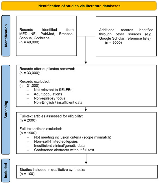
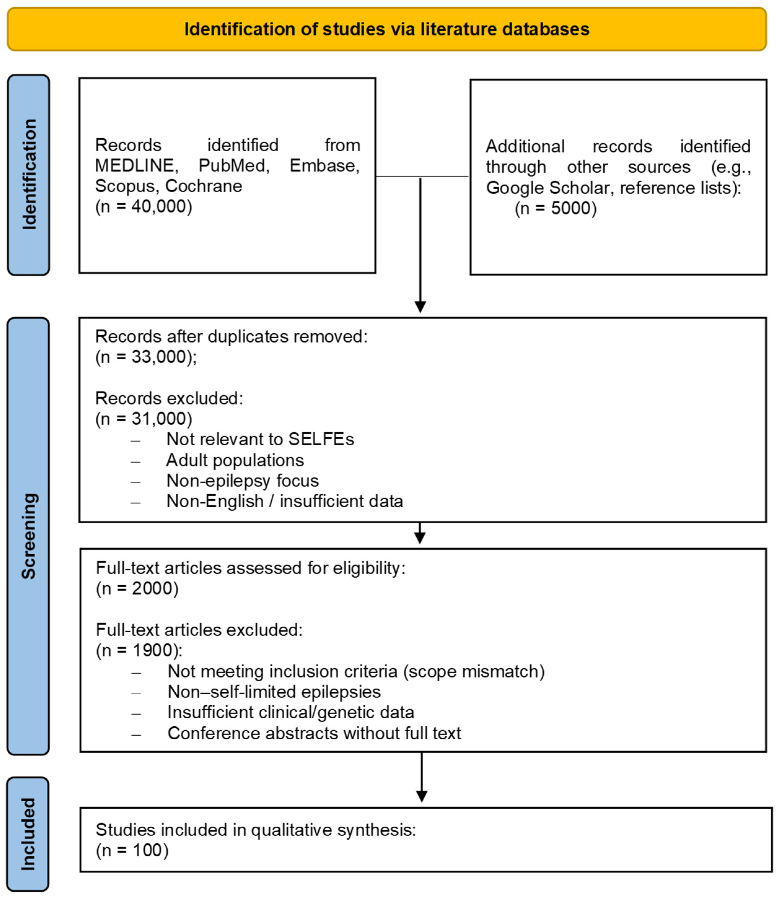
All retrieved records were exported into a reference management software, and duplicates were removed prior to screening. The study selection process followed a two-step approach: (i) title and abstract screening and (ii) full-text review. Inclusion criteria were defined as studies involving pediatric populations (<18 years) diagnosed with self-limited focal epilepsies or related syndromes, including neonatal, infantile, and childhood-onset forms. Both clinical and genetic studies were considered. Exclusion criteria included non-English articles, studies not focused on SELFEs, adult-only populations, conference abstracts without full text, and articles lacking sufficient clinical or methodological detail. A total of 40,000 records were initially identified across databases and additional sources. After removal of duplicates (n = 12,000) and exclusion based on title/abstract screening (n = 15,000), around 2000 articles were deemed eligible for full-text assessment. Following detailed evaluation, 100 studies met the predefined inclusion criteria and were included in the final qualitative synthesis, with a particular focus on childhood-onset SELFEs. Data extraction was performed independently by multiple reviewers using a standardized form, capturing study characteristics (author, year, study design), population features, clinical phenotypes, EEG findings, genetic data, and treatment approaches. Discrepancies were resolved through discussion and consensus. Given the nature of the review, no formal risk-of-bias assessment or meta-analysis was conducted; instead, results were synthesized descriptively to map the breadth of available evidence. The study selection process is summarized in a PRISMA flow diagram (Figure 1; Supplementary File).
Figure 1.
PRISMA 2020 flow diagram illustrating the study selection process for the review.
3. Discussion
The clinical characteristics of the SELFEs has been well defined and may be synthetized in several points: (a) an autosomic dominant transmission, (b) prevalent neonatal, infantile and childhood appearance, (c) normal clinical examination, neuroradiologic and laboratory analyses, (d) focal seizures which may evolve to tonic–clonic generalization, (e) positive familial history for SeLFE episodes or other types of epilepsies, (f) rapid response to antiseizure treatment, (g) variability of clinical presentation for each of these disorders, (h) seizures that might tend to disappear with and without the contribution of the antiseizure treatment [14]. Various epileptic syndromes starting in childhood and evolving with a favorable course have been recognized and include SeLFNIE, SeLECTS, SeLEAS, COVE, POLE, GEFS+ and MEI. For the last two, GEFS+ and MEI, there has been no general agreement as to whether they should be inserted into the group of SELFEs, as recent observations have reported new types of focal seizures—neurological complications in the clinical course of the disorders. POLE has been included in the group of the Reflex Epilepsy.
3.1. SeLFNIE
In this condition, the terms “convulsions,” “epilepsies,” and “seizures” have been used interchangeably. The word “convulsions” may be more apt for neonates and young infants, since abnormal movements such as jerking, pedaling, and stepping observed in the first weeks of life are not always considered to be epileptic in nature [15]. Zuberi et al. [13] further subdivided the early-onset SELFE group based on timing of seizure onset, distinguishing self-limited (familial) neonatal, neonatal-infantile, and infantile epilepsy syndromes. Although these subtypes differ in their precise age of onset, they share enough clinical characteristics to be discussed collectively here. In the neonatal subgroup, seizure onset typically occurs between the second and fifth day of life, whereas in the infantile subgroup it falls within the first weeks to months. Episodes are focal, consisting of motor tonic or clonic activity alternating between sides, and are sometimes accompanied by non-motor features such as cyanosis and apnea. Episodes are generally brief (lasting only a few minutes), and tend to occur in clusters, occasionally recurring after a brief apparent remission. Between episodes, affected infants are alert and behaviorally normal. The interictal EEG background is reported as normal, while ictal recordings may reveal focal discharges predominantly arising from temporal or posterior regions. Seizure remission typically occurs between 6 weeks and 24 months of age [13,17]. Family histories in these children often reveal neonatal-infantile SELFEs as well as diverse seizure types among relatives. Phenobarbital is considered the first-line antiseizure medication (ASM) regardless of etiology, with phenytoin or carbamazepine serving as first-line options in confirmed channelopathies [19]. Gene variants associated with this group include KCNQ2, KCNQ3, SCN2A, SCN8A, and PRRT2 [17,20,21].
A SeLFNIE family was the subject of extended clinical observation spanning many years [5]. We recently had the opportunity to re-contact one of the originally affected members, who was a neonate at the time of the initial report and is now 43 years old. He reported being in good health, and neither he nor his two children (aged 8 and 11 years) have experienced any neurological problems to date. KCNQ2 mutations have been identified in children with a wide range of clinical severity, from otherwise healthy neonates belonging to the self-limited (familial) neonatal epilepsy group (KCNQ2-SeLNE) to the severe end of the spectrum represented by neonatal-onset developmental and epileptic encephalopathy (KCNQ2-NEO-DEE) [22]. The severe phenotype manifests in the first week of life with frequent focal motor tonic and clonic seizures, interspersed with autonomic signs, and showing poor pharmacological control. EEG typically demonstrates burst-suppression or multifocal epileptiform activity. Pavone et al. [5] described a child who presented on day 3 of life with multifocal clonic seizures in clusters, migrating alternately across both body sides and lasting only briefly. Autonomic features were also present, including cyanosis, ocular deviation, and vomiting. The ictal EEG on day 3 showed a chaotic background, whereas the interictal EEG on day 7 was normal, as was the clinical examination and laboratory workup. Genetic analysis identified a pathogenic variant c.1508C>G in heterozygous state in KCNQ2. The infant responded well to phenobarbital, with seizure remission achieved; treatment was discontinued at four months of age. Different epilepsy phenotypes were documented among other family members.
3.2. SeLECTS
This syndrome is also referred to as “benign epilepsy with centrotemporal spikes” (BECTs) and benign Rolandic epilepsy (BRE). It occurs in children with otherwise normal cognitive and neurological development and is widely regarded as the most prevalent epileptic syndrome of childhood, with an estimated incidence of 10–20 cases per 100,000 children under 15 years of age [23,24]. The seizure focus characteristically lies within the Rolandic region, which spans the frontal (motor) and parietal (sensory) cortices, producing facial, oral, and pharyngeal motor and sensory manifestations in affected children. Seizure onset spans ages 3 to 13 years, with a peak at 9–10 years [24]. Clinically, episodes are brief (typically 2–3 min), infrequent, and focal, presenting as hemifacial motor activity often accompanied by somatosensory symptoms. Facial twitching and stiffness, along with numbness or tingling affecting one side of the face, throat, tongue, lips, gums, inner cheek, and teeth, are frequently reported, giving rise to drooling, gurgling, and hypersalivation. Focal seizures can spread to bilateral tonic–clonic convulsions. Symptoms predominantly emerge shortly after sleep onset or in the peri-waking period. Children with onset before the age of 5 years appear more likely to experience a longer duration before seizure and EEG resolution. The EEG background is normal, with typically unilateral high-amplitude centrotemporal sharp waves occurring in repetitive bursts, sometimes followed by a slow component; these discharges are enhanced during drowsiness and sleep. In children experiencing only isolated nocturnal seizures, antiseizure therapy is generally not considered necessary [25]. When treatment is warranted, seizures typically respond well [26], and a single bedtime dose of levetiracetam has been demonstrated to be effective in a randomized clinical trial by Fan et al. [27]. Neuropsychiatric comorbidities have been documented in SeLECTS: Ross et al. [28] identified an elevated risk of neuropsychiatric symptoms in affected children over the course of the disease. Academic underachievement and behavioral problems [29], as well as dyslexia and dyscalculia [30], have also been reported. Lacey et al. [31] found a higher prevalence of neurodevelopmental comorbidities and school difficulties in Welsh children with SeLECTS compared to the general pediatric population, attributing this to disruption of functional brain networks mediated by centrotemporal spiking activity [31]. Persistent increased functional connectivity has been observed during sleep, with further elevation during and around spike events [32]. Atypical presentations of SeLECTS—including status epilepticus, electrical status epilepticus during slow-wave sleep, and Landau–Kleffner syndrome—have been described [33]. Certain features, such as comparable age at onset, an overall favorable prognosis, and occasional co-occurrence in the same child, are shared between childhood absence epilepsy and SeLECTS [34]. Gene variants documented in SeLECTS include those in KCNQ2 and KCNQ3 [35], ELP4 [36], and GRIN2A [37].
3.3. SeLEAS
This disorder was first delineated as a distinct syndrome by Panayiotopoulos in 1989 [38]. Previously designated as Panayiotopoulos syndrome and early-onset benign occipital epilepsy, it is an idiopathic, age- and self-limited condition that ranks as the second most common focal epilepsy of childhood. The disorder predominantly affects children between 3 and 6 years of age, with a broader range of 1 to 14 years [38,39]. A history of febrile seizures is present in 5–7% of cases. Its clinical signature consists of three cardinal ictal features: nocturnal onset, tonic ocular deviation, and emesis [38], reflecting the prominence of focal autonomic manifestations. Seizures are often preceded by a transient alteration of consciousness and consist of prolonged episodes of nausea and vomiting, pallor, urinary incontinence, hypersalivation, pupillary dilatation, and unresponsive flaccid loss of postural tone (ictal syncope), lasting up to 30 min. Although they can arise at any point during the day, they occur most frequently during the initial phase of sleep [40,41]. Episodes may extend from several minutes to more than two hours, occasionally evolving into autonomic status epilepticus, with spontaneous remission typically occurring within a few years of onset [40,41]. Ictal events can closely mimic non-epileptic conditions such as syncope, migraine, cyclic vomiting syndrome, motion sickness, sleep disorders, gastroenteritis, and acute encephalitis [41]. Caraballo et al. [42] reported on 192 subjects, noting a peak age of onset at 5 years. Autonomic features, including eye deviation, dominated the ictal semiology. In virtually all patients, seizures occurred during sleep, though in approximately one-third they were also observed during wakefulness. The overall prognosis was good: 84 patients (44.2%) had only a single seizure, 79 (41.2%) had 2–3 episodes, and 28 (14.6%) had multiple events. Generalized evolution of seizures was common, and roughly one-third experienced partial status epilepticus. In the series of Durá-Travé et al. [43], which included 37 patients, the mean age at diagnosis was 5.4 years and a female-to-male ratio of 2:1 was observed. Autonomic features or vomiting were reported in 70.1% of patients, and ictal eye and/or head deviation was frequently noted, as was progression to partial or generalized seizures. Seizure recurrence occurred primarily within the first six months after diagnosis, with 82.9% of patients remaining seizure-free beyond two years from onset. Syncope-like epileptic seizures were documented by Koutroumanidis et al. [44] in at least one episode among 17 of 33 children (51.5%).
As reported by Panayiotopoulos et al. [38], the EEG background is normal. Interictal recording typically reveals multifocal high-voltage repetitive spikes and slow waves with a predilection for the occipital regions, though anterior, central, temporal, and midline involvement may also occur. Ictal EEG is generally unilateral and frequently shows posterior onset with rhythmic slow activity intermixed with small spikes [38]. Oguni [45] emphasized that the morphology, distribution, and appearance of interictal EEG foci may vary with the child’s age at the time of recording; additionally, the author proposed that the absence of frontoparietal EEG foci may serve as a specific marker supporting the diagnosis [45]. Psychomotor development is usually preserved, and the long-term outlook for both seizure frequency and evolution is generally favorable. Caraballo et al. [46] described atypical outcomes in two children: a 3-year-old who developed frequent inhibitory seizures causing pseudoataxic gait, behavioral disturbances, and aphasia, and a 9-year-old girl who, after 18 months of seizure freedom, developed mild intellectual disability. Mild deficits in language and executive functioning, as well as poor academic performance, have also been documented in this disorder [47].
To our knowledge, no gene mutations specifically linked to this disorder have been identified. However, familial clustering in siblings and across generations suggests a polygenic basis. There is general consensus that treatment should be guided by seizure frequency and severity, with oxcarbazepine, carbamazepine, levetiracetam, lamotrigine, and sulthiame being the most frequently employed antiseizure agents [15].
3.4. COVE
This syndrome was previously designated as idiopathic childhood occipital epilepsy—Gastaut type, and as late-onset (benign) childhood occipital epilepsy. It is distinguished from other forms of benign occipital epilepsy by its distinct clinical presentation. COVE is a relatively uncommon syndrome manifesting with frequent and brief focal sensory episodes—typically lasting from a few seconds to fewer than three minutes—characterized predominantly by visual phenomena occurring in the awake state and often followed by headache with migrainous features [48]. Elementary visual hallucinations, ictal blindness, and post-ictal headache are among the most commonly reported symptoms [49]. Onset can occur between ages 1 and 18 years, with the condition most commonly presenting in late childhood or early adolescence, peaking at 8–9 years. The ictal semiology includes visual hallucinations, transient blindness, orbital pain, and occasionally focal sensory or motor disturbances [50]. Visual phenomena are characteristically described as small, multicolored circular shapes appearing in the peripheral visual field, which progressively expand toward the center and may spread horizontally, sometimes followed by eye deviation. Caraballo et al. [51] reported on 33 patients with COVE and identified a mean age at onset of 8.5 years. Visual manifestations were the predominant ictal feature, with eye deviation being the most frequent symptom; episodes occurred predominantly in the awake state, though less commonly during sleep as well. A subsequent publication [52] provided further differentiation of COVE from migraine and other epilepsy types. Wakamoto et al. [53] reported on 12 affected children (three boys, nine girls) with a median age of onset of 10.3 years. The most frequent ictal manifestations included elementary visual hallucinations (75%), blindness or blurring of vision (50%), and secondary generalized tonic–clonic seizures (58.3%). A frequent association with generalized tonic–clonic seizures and atypical evolution from childhood absence epilepsy was also documented [53]. Interictal EEG showed occipital spike-wave paroxysms responsive to eye closure and opening in all patients; extra-occipital spike-wave activity was present in four patients (33.3%) and generalized spike-wave discharges in two (16.7%). In a retrospective analysis of 129 patients with childhood occipital epilepsy of Gastaut (COE-G), Verrotti et al. [54] reported visual hallucinations as the initial symptom in 62% and the sole manifestation in 38.8% of cases, with an overall favorable prognosis and good response to antiepileptic therapy. A more recent study [55] of 30 patients diagnosed with COVE between 1988 and 2023 found elementary visual hallucinations, such as colored lights, in 93% of cases; orofacial seizures were recorded in 7%, and nocturnal seizures in 37%. EEG abnormalities were primarily occipital and resolved over time in 85% of patients. At final follow-up, 77% had achieved seizure freedom and 47% had discontinued medication [55]. Shu et al. [56] observed a correlation between occipital spike-wave discharges and the state of eye closure, noting that EEG abnormalities were present with eyes closed and suppressed upon eye opening. In their cohort of seven patients, two continued to experience frequent seizures despite treatment, with associated learning difficulties and behavioral disturbances. A distinctive EEG feature in COVE, as described by Rots et al. [57], is fixation-off sensitivity—the elicitation of epileptiform discharges when visual fixation is removed. The long-term prognosis of COVE is generally favorable, with seizure remission and EEG normalization expected by the late teenage years [49], and an excellent outcome reported in approximately 80% of cases [51]. The genetic architecture is considered complex and polygenic.
Fonte et al. [48] recently described a mother-son pair displaying clinical and EEG features of COVE/POLE in whom a large deletion at 5q34 encompassing the entire GABRA1 gene and a portion of GABRG2 was identified. Antiepileptic treatment is indicated for children with frequent ictal events; carbamazepine has demonstrated efficacy in approximately 90% of treated cases [50].
3.5. POLE
POLE is an uncommon condition defined by focal visual seizures triggered by photic stimuli. It was previously termed idiopathic photosensitive occipital lobe epilepsy and belongs to the broader category of reflex epilepsies (RE)—a group of epileptic events consistently and reproducibly provoked by specific stimuli such as flickering light or reading [58]. Seizure onset in POLE spans ages 4 to 17 years (mean 11 years, range 1–50 years) and shows a marked predominance in females. Light sources such as television and video games are the principal seizure triggers. Episodes are brief (fewer than 3 min) and variable in frequency, featuring visual sensations including colored spots, formed visual hallucinations, or visual blurring and loss of vision that migrate across the visual field. Symptoms may progress to include autonomic features, impaired consciousness, and evolution to generalized tonic–clonic seizures. Sirin et al. [59] reported on 29 patients with POLE (mean age 20.1 ± 7.6 years) and noted overlapping features with genetic generalized epilepsy in approximately one-third; this overlapping subgroup showed higher rates of febrile seizure history and self-induction behavior. Guerrini et al. [60] studied 10 patients (8 female, 2 male) with idiopathic photosensitive occipital lobe epilepsy, ranging in age from 8 to 30 years (mean 17 years). Seizure onset was reported between ages 5 and 17 years (mean 11 years); stimulus-related episodes began with elementary visual symptoms and progressed to a gradual clustering of headache, epigastric discomfort, and vomiting, with either preserved or mildly impaired responsiveness. EEG features included normal background activity, occipital spikes and waves, and a photoparoxysmal response appearing in the occipital area, in a generalized distribution, or both. Politi-Elishkevich et al. [61] described 16 children and adolescents, including two sets of siblings and seven patients with a positive family history of seizures. All patients had occipital-onset seizures, and 15 showed secondary generalization to tonic–clonic seizures; seizure frequency was relatively low in all but one patient. Two patients subsequently developed myoclonic seizures [61]. Remission is observed in most cases by puberty. Trigger avoidance and use of tinted glasses may be of benefit.
Despite the electroclinical differences among individual syndromes, several unifying themes emerge across the SELFEs spectrum. Most disorders share an age-dependent onset, focal seizure semiology with possible secondary generalization, normal neurological examination and neuroimaging, characteristic but often evolving EEG abnormalities, and an overall tendency toward spontaneous remission during childhood or adolescence. However, increasing evidence demonstrates that seizure remission does not invariably correspond to a completely benign neurodevelopmental outcome. Cognitive, behavioral, language, attentional, and psychiatric comorbidities have been documented in multiple SELFEs, particularly in SeLECTS, SeLEAS, GEFS+, and MEI, suggesting that these disorders may reflect transient dysfunction of developing cortical networks rather than isolated epileptic phenomena alone. This evolving perspective has important clinical implications because treatment decisions should not rely exclusively on seizure frequency but also consider neuropsychological profile, sleep-related EEG abnormalities, school performance, and quality of life.
3.6. Genetic Epilepsy with Febrile Seizures Plus (GEFS+)
GEFS+, formerly termed “generalized epilepsy with febrile seizures plus,” is a genetic syndrome with a broad phenotypic spectrum affecting multiple family members and encompassing a variety of presentations ranging from typical to atypical febrile seizures and/or other seizure types, including absences, myoclonic seizures, atonic seizures, myoclonic-astatic epilepsy, and, uncommonly, severe epileptic encephalopathy [62,63]. The qualifier “generalized” was subsequently removed from the syndrome name when it became apparent that focal features were also prevalent among affected family members [62]. GEFS+ was formally incorporated into the ILAE syndrome classification as a recognized entity in 2001 [62]. The original description is attributed to Scheffer and Berkovic [64], who characterized a large family in which related members across four generations exhibited varied seizure phenotypes: some presented with atypical febrile seizures (FS+) with either unusually early onset (median age 1 year) or late persistence beyond 6 years of age and multiple recurrences, while others displayed combined phenotypes such as FS+ with absence, FS+ with myoclonic seizures, FS+ with atonic seizures, and myoclonic-astatic epilepsy [64]. The etiology is complex, reflecting a heterogeneous genetic predisposition that interacts with febrile stimuli, including specific viral infections [65]. Autosomal dominant inheritance with incomplete penetrance is the most observed pattern, although other inheritance modes have been reported [66]. Thomas et al. [67] studied 80 recruited families with the aim of better characterizing both definite and borderline GEFS+ phenotypes, identifying four broad subtypes: classical GEFS+, borderline GEFS+, unclassified epilepsy, and alternative syndromic diagnoses. The borderline GEFS+ group shared many characteristics with classical GEFS+ but included a higher proportion of adults with focal rather than generalized seizures and a twofold excess of migraine [67]. To refine the phenotypic spectrum further, Zhang et al. [68] analyzed 409 affected individuals from 60 families and identified focal seizures without preceding febrile seizures in 16/409 (4%), classic genetic generalized epilepsies in 22/409 (5%), and afebrile generalized tonic–clonic seizures in 9/409 (2%). Pathogenic variants in established GEFS+ genes were identified in approximately one-third of tested families. A detailed clinical and genetic analysis of GEFS+ was conducted by Chen et al. [69] in 392 affected individuals from 133 families. The predominant phenotypes were febrile seizures (FS) (288/392, 73.5%) and FS+ (70/392, 17.9%); other phenotypes included FS/FS+ with generalized seizures (21/392, 5.4%), FS/FS+ with focal seizures (8/392, 2.0%), Dravet syndrome (8/392, 2.0%), afebrile generalized tonic–clonic seizures (17/392, 4.3%), complex phenotypes (5/392, 1.3%), and unclassified seizures (35/392, 8.9%). Regarding the role of fever sensitivity, patients with lower thermal sensitivity were more likely to exhibit focal, myoclonic, and tonic seizures and to have multiple seizure types, whereas those with high thermal sensitivity more often had a single seizure type. Genetic testing of GEFS+ patients most frequently yielded variants in voltage-gated channel genes SCN1A, SCN1B, KCNT1, CACNA1A, CACNAIH, and GABA receptor-related genes GABRA1, GABRB2, GABRB3, and GABRG2 [69]. Baso et al. [70] proposed predictive criteria for evolution to epilepsy or GEFS+ in children presenting with FS, FS with status epilepticus, or recurrent FS: in a cohort of 231 children, a family history of epilepsy, higher total number of seizure episodes, older age at FS onset, and the occurrence of afebrile seizures were each independently associated with an increased risk of progression. Genetic variants associated with GEFS+ have additionally been reported in KCNAB3 [71], GABRG2 (Q390X) [72,73], and SCN1A (A2336G, exon 13) [74]. Diagnosis rests on the seizure type and a comprehensive family history. EEG shows a normal background with generalized discharges. Seizures tend to remit by late childhood or puberty. A significantly elevated rate of auditory phoneme discrimination impairment, articulation difficulties, and language disorders has been reported in patients with GEFS+ [75].
3.7. MEI
Myoclonic epilepsy of infancy (MEI), originally termed “myoclonic epilepsy of childhood” by Jeavons [76] and subsequently described as “benign myoclonic epilepsy in infants” by Dravet et al. [77,78], is an uncommon idiopathic generalized epilepsy presenting with brief myoclonic seizures in previously healthy and normally developing children, with onset confined to the first three years of life. The term “benign” has been abandoned given the recognition that the course is not invariably uncomplicated; the disorder is now classified within the idiopathic generalized epilepsies with age-related onset [11]. The myoclonic jerks are brief (1–3 s) and primarily involve the upper extremities and head, with less frequent involvement of the lower limbs. Additional manifestations include head nodding, upward eye rolling, and eyelid blinking. No other seizure type co-occurs in MEI, although the myoclonic events may be preceded by prior febrile seizures [79]. The jerks may be isolated or clustered, and tend to predominate during drowsiness or slow-wave sleep, though they may also arise spontaneously [80]. Recognized MEI variants include: (a) reflex myoclonic epilepsy of infancy (RMEI), triggered by unexpected tactile or auditory stimuli [81]; (b) photosensitive MEI, induced by photic stimulation; (c) nocturnal MEI, occurring exclusively during sleep [82]; and (d) familial infantile myoclonic epilepsy with autosomal recessive inheritance. No consensus exists on whether RMEI should be classified separately from classical MEI [80,83]. Caraballo et al. [84] recruited 102 infants with BMEI: clinical features encompassed myoclonus, spasms, brief tonic contractions, shuddering, atonia or negative myoclonus, and multiple motor phenomena. Episodes were predominantly observed during wakefulness, were recurrent several times daily, and occurred in clusters in nearly half of the patients. Interictal EEG is typically normal during both wakefulness and sleep. Paroxysmal slowing in central areas has been reported. Under photic stimulation, generalized spike-wave or polyspike-wave discharges may be recorded during drowsiness or sleep in approximately 20% of patients. In a subsequent series of 38 infants with MEI reported by Caraballo et al. [85], 12 had reflex myoclonus—10 triggered by tactile stimuli and 2 by noise and light; interictal EEG showed generalized spike-wave, polyspike, and polyspike-wave paroxysms, and was normal in 12 patients. At last follow-up, 32 patients had normal neurological and neuropsychological evaluations, two had significant cognitive impairment (IQ 60 and 63), and four had significant learning impairment, including two with ADHD.
Zuberi and O’Regan [86] reviewed the existing literature and six additional cases, concluding that long-term seizure outcomes are generally favorable with sodium valproate; however, cognitive difficulties may be present in approximately one-third of children with MEI at long-term follow-up, while no cognitive impairment was found in RMEI cases. Among seven patients with extended follow-up reported by Prats-Viñas et al. [87], three showed unfavorable intellectual and behavioral trajectories, and three developed generalized seizures during follow-up. Long-term neuropsychological outcomes in seven patients with BMEI were assessed by Mangano et al. [88]: 86% presented with neuropsychological and intellectual difficulties, including fine motor skill deficits, attention deficits, language impairment, and learning disorders.
A follow-up study by Domínguez-Carral et al. [89] in ten BMEI patients found that 60% achieved seizure freedom with valproate; IQ scores ranged from 74 to 93, with three patients in the borderline intelligence range and six in the medium-to-low range. ADHD was present in nearly all patients assessed. Gene mutations specifically associated with MEI are infrequent. Using next-generation sequencing for idiopathic epilepsy screening, Campostrini et al. [90] identified the heterozygous variant p.Arg550Cys (c.1648C>T) in HCN4 in two brothers affected by BMEI. In a large epilepsy gene study, Wang et al. [91] found TBCID24 involvement in a family with infantile myoclonic epilepsy. Valproic acid remains the treatment of choice for MEI, with clonazepam and levetiracetam also showing favorable results.
3.8. Benign Genetic Epilepsies: From Molecular Pathogenesis to Clinical Phenotypes
Benign genetic epilepsies represent a continuum of neurological disorders in which the genetic landscape shapes both fundamental neuronal function and the dynamics of cortical network development [92,93]. This interplay determines a wide spectrum of phenotypes, ranging from clinically self-limited forms to severe encephalopathic presentations associated with significant neurological disability. From a pathogenetic standpoint, most monogenic epilepsies involve ion channels, synaptic receptors, or transporters that directly influence neuronal excitability, the excitatory/inhibitory (E/I) balance, and the maturation of cortical circuits. Genes most commonly implicated include SCN1A, SCN2A, KCNQ2, GRIN2A, SLC2A1, and SLC6A1, alongside variants participating in metabolic pathways or synaptic transmission—such as GABRA1, GABRG2, and STXBP1 [15,20,92].
In self-limited focal epilepsies, the causative genetic variants typically produce transient functional perturbations in neuronal excitability without permanently disrupting brain maturation [94,95]. For example, KCNQ2 and SCN2A mutations have been found in self-limited neonatal-infantile forms; in these cases, the potassium channel Kv7.2 or the sodium channel Nav1.2 undergoes functional shifts that heighten neuronal excitability during a critical developmental window but do not interfere with long-term cortical organization, thereby allowing spontaneous seizure remission in many patients [92]. GRIN2A, encoding an NMDA receptor subunit, is associated with benign focal phenotypes, likely through subtle alterations in glutamatergic transmission that affect synaptic plasticity without inducing widespread cortical network dysfunction [96]. SCN1A variants may manifest either as benign focal epilepsy or within the broader GEFS+ spectrum, reflecting phenotypic complexity dependent on the specific nature and location of the mutation within the gene [97,98]. The identification of underlying channelopathies has progressively redirected the therapeutic paradigm toward precision medicine. In patients carrying KCNQ2 or SCN2A gain-of-function mutations, sodium channel blockers such as oxcarbazepine, carbamazepine, and phenytoin have demonstrated superior efficacy over phenobarbital, achieving faster seizure control and, in some cases, improved neurodevelopmental outcomes. Early genetic testing is therefore recommended in neonates with refractory seizures, as antiseizure medication selection should ideally be guided by the underlying molecular mechanism rather than by empirical drug escalation.
Taken together, the evidence reviewed here highlights the substantial heterogeneity underlying the SELFEs spectrum and the limitations of considering these syndromes as uniformly “benign.” Although most patients experience favorable seizure outcomes, prognostic variability remains considerable, particularly regarding cognitive and neuropsychiatric trajectories. The distinction between self-limited epilepsies and developmental and epileptic encephalopathies may therefore represent a continuum rather than a strict categorical separation, especially in disorders associated with ion-channel dysfunction. Furthermore, unresolved classification issues persist, including the debated inclusion of GEFS+ and MEI within the SELFE group and the positioning of POLE within reflex epilepsies. Current literature also underscores the need for longitudinal studies integrating electroclinical, neuropsychological, and genetic data in order to better identify predictors of atypical evolution and treatment response. Future advances in molecular diagnostics and precision medicine may further refine syndrome classification and support individualized therapeutic strategies aimed not only at seizure control but also at preservation of neurodevelopmental outcomes.
3.9. Evolution and Treatment
Vikin et al. [99] analyzed a birth cohort of 114,000 children born between 1999 and 2009 with epilepsy syndromes classified according to epilepsy type, with the aim of characterizing the course of epileptic episodes. Overall, 78% of the cohort achieved seizure freedom. Outcomes were particularly favorable in children with focal epilepsy (84%), while the lowest rates of seizure freedom were observed in combined generalized and focal epilepsy (26%). The best outcomes were documented in self-limited focal epilepsies, childhood absence epilepsy, and self-limited infantile syndromes.
The appropriate management of children with self-limited focal epilepsy remains a subject of debate. Given that these disorders are generally characterized by a favorable clinical course, antiseizure treatment is not always considered necessary, and the potential adverse effects of medications on the developing brain must be carefully weighed. Conversely, withholding therapy carries the risk of ongoing seizure activity with negative psychosocial consequences for the child in both school and social settings. For these reasons, treatment decisions in SELFE should be individualized, considering both the syndrome type and the specific child’s circumstances. Key factors to consider include the age of the patient—since neonates and infants are particularly vulnerable to drug-related adverse effects—as well as EEG findings, seizure type, and the duration, severity, and frequency of episodes, along with the clinical and EEG response to therapy. Seizure suppression can have a meaningful positive psychological impact on both the child and the family, including at school and in peer relationships. For SELFEs, following a careful assessment of the indications for pharmacological treatment, the following antiseizure management (ASM) strategies are recommended: for SeLFNIE, phenobarbital as first-line [19], with phenytoin, levetiracetam, midazolam, or lidocaine as second-line options; for broader SELFEs, lamotrigine or levetiracetam as first-line agents, though carbamazepine, oxcarbazepine, valproate, and zonisamide are also in use.
4. Limitations
This review has several limitations that should be acknowledged. In addition, given the nature of the review, no formal quality or risk-of-bias assessment of the included studies was performed, limiting the ability to weigh the strength of the evidence. The included literature is highly heterogeneous, encompassing case reports, retrospective studies, and cohort analyses with variable diagnostic criteria and evolving classifications of SELFEs, which may affect the comparability of findings. Furthermore, the ongoing refinement of the International League Against Epilepsy framework and the lack of consensus regarding the inclusion of certain syndromes (e.g., GEFS+ and MEI) introduce additional variability in interpretation. The genetic landscape of SELFEs is also rapidly evolving, and current knowledge may not fully capture emerging variants or genotype–phenotype correlations. Finally, the absence of quantitative synthesis precludes definitive conclusions regarding treatment efficacy or prognostic outcomes, and the results should therefore be interpreted as a qualitative mapping of the existing evidence rather than as a basis for clinical standardization.
5. Conclusions
SELFEs constitute a clinically relevant group of disorders on account of their notable frequency within the spectrum of pediatric neurological conditions, their generally favorable prognosis in the majority of cases, and the considerable psychological burden they place on both affected children and their families. Several unresolved issues remain within this group: (a) a subset of disorders classically considered to have a favorable course have been shown in a small proportion of patients to carry additional negative clinical implications; (b) there is no universal agreement on which conditions should be classified as SELFEs, as some previously included syndromes have demonstrated complicated clinical courses; the most recent consensus recognizes the following disorders as SELFEs: self-limited familial and non-familial neonatal-infantile convulsions, self-limited epilepsy with autonomic seizures, self-limited epilepsy with centrotemporal spikes, childhood occipital visual epilepsy, and photosensitive occipital lobe epilepsy [15]; and (c) treatment modalities must be individualized according to the specific syndrome, with primary consideration given to the duration, intensity, and frequency of seizures, as well as the broader impact on the child and family.
This review has examined several key clinical dimensions pertaining to this group of disorders. Emerging clinical observations and expanding genetic data continue to enhance our understanding of SELFEs and hold promise for further improvements in diagnosis, counseling, and personalized management.
Supplementary Materials
The following supporting information can be downloaded at: https://www.mdpi.com/article/10.3390/pediatric18030074/s1, Supplementary File. The PRISMA 2020 Checklist [100].
Author Contributions
Conceptualization, P.P., E.P., A.P. and R.F.; methodology, P.P., F.S., C.C., A.C. and X.G.P.; validation, P.P., F.S., C.C., A.C., G.B.D. and X.G.P.; formal analysis, E.P.; investigation, P.P., F.S., C.C., A.C., G.B.D. and X.G.P.; resources, P.P., F.S., C.C., A.C., G.B.D. and X.G.P.; data curation, P.P., F.S., C.C., A.C., G.B.D. and X.G.P.; writing—original draft preparation, P.P., F.S., C.C., A.C. and X.G.P.; writing—review and editing, P.P., F.S., C.C., A.C., X.G.P., E.P., A.P. and R.F.; visualization, E.P., A.P. and R.F.; supervision, P.P., E.P., A.P., R.F., A.C. and X.G.P.; project administration, P.P., A.C. and X.G.P.; funding acquisition, P.P. All authors have read and agreed to the published version of the manuscript.
Funding
This research received no external funding.
Institutional Review Board Statement
Not applicable.
Informed Consent Statement
Not applicable.
Data Availability Statement
No new data were created or analyzed in this study.
Conflicts of Interest
The authors declare no conflicts of interest.
Abbreviations
The following abbreviations are used in this manuscript:
| BFIS | Benign familial infantile seizures |
| ILAE | International League Against Epilepsy |
| SeLE | Self-limited epilepsy syndrome |
| SeLFNE | Self-limited (familial) neonatal epilepsy |
| SeLFNIE | Self-limited neonatal-infantile epilepsy |
| SeLIE | Self-limited (familial) infantile epilepsy |
| GEFS+ | Genetic epilepsy with febrile seizures plus |
| MEI | Myoclonic epilepsy of infancy |
| SeLECTS | Self-limited epilepsy with centrotemporal spikes |
| SeLEAS | Self-limited epilepsy with autonomic seizures |
| COVE | Childhood occipital visual epilepsy |
| POLE | Photosensitive occipital lobe epilepsy |
| SeLFE | Self-limited focal epilepsies |
| BECT | Benign epilepsy with centrotemporal spikes |
| BRE | Benign rolandic epilepsy |
| COE-G | Childhood occipital epilepsy-Gastaut |
| RMEI | Reflex myoclonic epilepsy of infancy |
| PMEI | Photosensitive myoclonic epilepsy |
| FMEI | Familial infantile myoclonic epilepsy |
| ASM | Antiseizure management |
References
- Fukuyama, Y.; Arima, M.; Okada, R.; Suzuki, M.; Komiya, H.; Komiya, K.; Kamoshita, S. Electro-clinical characteristics of epilepsies in infancy and childhood. Paediatr. Univ. Tokyo 1963, 9, 11–16. [Google Scholar] [PubMed]
- Rett, A.; Teubel, R. Neugeborenen Krampfe im Rahmen einer epileptisch belasten Familie. Wien. Klin. Wochenschr. 1964, 76, 609–613. [Google Scholar]
- Bjerre, I.; Corelius, E. Benign familial neonatal convulsions. Acta Paediatr. Scand. 1968, 57, 557–561. [Google Scholar] [CrossRef] [Scilit] [PubMed]
- Carton, D. Benign familial neonatal convulsions. Neuropadiatrie 1978, 9, 167–171. [Google Scholar] [CrossRef] [Scilit] [PubMed]
- Pavone, P.; Praticò, A.D.; Falsaperla, R.; Striano, P.; Ruggieri, M. Is benign familial neonatal KCNQ2-related epilepsy always familially benign? J. Pediatr. Neonatal Individ. Med. (JPNIM) 2018, 7, e070221. [Google Scholar] [CrossRef] [Scilit]
- Watanabe, K.; Yamamoto, N.; Negoro, T.; Takaesu, E.; Aso, K.; Furune, S.; Takahashi, I. Benign complex partial epilepsies in infancy. Pediatr. Neurol. 1987, 3, 208–211. [Google Scholar] [CrossRef] [Scilit] [PubMed]
- Watanabe, K.; Negoro, T.; Aso, K. Benign partial epilepsy with secondarily generalized seizures in infancy. Epilepsia 1993, 34, 635–638. [Google Scholar] [CrossRef] [Scilit] [PubMed]
- Vigevano, F.; Di Capua, M.; Fusco, L.; Ricci, S.; Sebastianelli, R.; Lucchini, P. Sixth-month benign familial convulsions. Epilepsy Res. Suppl. 1992, 6, 127–129. [Google Scholar] [PubMed]
- Vigevano, F.; Fusco, L.; Di Capua, M.; Ricci, S.; Sebastianelli, R.; Lucchini, P. Benign infantile familial convulsions. Eur. J. Pediatr. 1992, 151, 608–612. [Google Scholar] [CrossRef] [Scilit] [PubMed]
- Specchio, N.; Vigevano, F. The spectrum of benign infantile seizures. Epilepsy Res. 2006, 70, S156–S167. [Google Scholar] [CrossRef] [Scilit] [PubMed]
- Specchio, N.; Wirrell, E.C.; Scheffer, I.E.; Nabbout, R.; Riney, K.; Samia, P.; Guerreiro, M.; Gwer, S.; Zuberi, S.M.; Wilmshurst, J.M.; et al. International League Against Epilepsy classification and definition of epilepsy syndromes with onset in childhood: Position paper by the ILAE Task Force on Nosology and Definitions. Epilepsia 2022, 63, 1398–1442. [Google Scholar] [CrossRef] [Scilit] [PubMed]
- Mulley, J.C.; Heron, S.E.; Dibbens, L.M. Proposed genetic classification of the ‘benign’ familial neonatal and infantile epilepsies. Epilepsia 2011, 52, 649–650. [Google Scholar] [CrossRef] [Scilit] [PubMed]
- Zuberi, S.M.; Wirrell, E.; Yozawitz, E.; Wilmshurst, J.M.; Specchio, N.; Riney, K.; Pressler, R.; Auvin, S.; Samia, P.; Hirsch, E.; et al. ILAE classification and definition of epilepsy syndromes with onset in neonates and infants: Position statement by the ILAE Task Force on Nosology and Definitions. Epilepsia 2022, 63, 1349–1397. [Google Scholar] [CrossRef] [Scilit] [PubMed]
- Manokaran, R.K.; Sharma, S.; Ramachandrannair, R. The 2022 International League Against Epilepsy Classification and Definition of Childhood Epilepsy Syndromes: An Update for Pediatricians. Indian Pediatr. 2024, 61, 179–183. [Google Scholar] [CrossRef] [Scilit] [PubMed]
- Menon, R.N.; Helen Cross, J. Childhood epilepsy. Lancet 2025, 406, 636–649. [Google Scholar] [CrossRef] [Scilit] [PubMed]
- Quito-Betancourt, B.F.; Reyes Valenzuela, G. Self-limited focal epilepsies of childhood. Medicina 2023, 83, 57–62. [Google Scholar] [PubMed]
- Gunasekaran, V.; Ighodaro, E.T.; Govil-Dalela, T. Neonatal Seizures and Neonatal Epilepsy. In StatPearls; StatPearls Publishing: Treasure Island, FL, USA, 2025. Available online: http://www.ncbi.nlm.nih.gov/books/NBK609108/ (accessed on 15 February 2026). [PubMed]
- Deonna, T.; Ziegler, A.L. So-called benign epilepsies in children. Rev. Med. Suisse Rom. 1994, 114, 861–867. [Google Scholar] [PubMed]
- Pressler, R.M.; Abend, N.S.; Auvin, S.; Boylan, G.; Brigo, F.; Cilio, M.R.; De Vries, L.S.; Elia, M.; Espeche, A.; Hahn, C.D.; et al. Treatment of seizures in the neonate: Guidelines and consensus-based recommendations-Special report from the ILAE Task Force on Neonatal Seizures. Epilepsia 2023, 64, 2550–2570. [Google Scholar] [CrossRef] [Scilit] [PubMed]
- Bayat, A.; Bayat, M.; Rubboli, G.; Møller, R.S. Epilepsy Syndromes in the First Year of Life and Usefulness of Genetic Testing for Precision Therapy. Genes 2021, 12, 1051. [Google Scholar] [CrossRef] [Scilit] [PubMed] [PubMed Central]
- Vikin, T.; Lossius, M.I.; Brandlistuen, R.E.; Chin, R.F.; Aaberg, K.M. Incidence of childhood and youth epilepsy: A population-based prospective cohort study utilizing current International League Against Epilepsy classifications for seizures, syndromes, and etiologies. Epilepsia 2025, 66, 776–789. [Google Scholar] [CrossRef] [Scilit] [PubMed] [PubMed Central]
- Miceli, F.; Soldovieri, M.V.; Weckhuysen, S.; Cooper, E.C.; Taglialatela, M. KCNQ3-Related Disorders. In GeneReviews®; Adam, M.P., Bick, S., Mirzaa, G.M., Pagon, R.A., Wallace, S.E., Amemiya, A., Eds.; University of Washington: Seattle, WA, USA, 1993. Available online: http://www.ncbi.nlm.nih.gov/books/NBK201978/ (accessed on 15 February 2026). [PubMed]
- Benign Rolandic Epilepsy (BRE): Seizure Semiology and the Role of an EEG Study. Available online: https://search.emarefa.net/en/detail/BIM-1440125-benign-rolandic-epilepsy-bre-lrm-seizure-semiology-and-the-r (accessed on 15 February 2026).
- Amrutkar, C.V.; Riel-Romero, R.M. Rolandic Epilepsy Seizure. In StatPearls; StatPearls Publishing: Treasure Island, FL, USA, 2025. Available online: http://www.ncbi.nlm.nih.gov/books/NBK534845/ (accessed on 15 February 2026). [PubMed]
- Hamada, Y.; Okuno, T.; Hattori, H.; Mikawa, H. Indication for anti-epileptic drug treatment of benign childhood epilepsy with centro-temporal spikes. Brain Dev. 1994, 16, 159–161. [Google Scholar] [CrossRef] [Scilit] [PubMed]
- Vargas, R.; Beltrán, L.; Lizama, R.; Valenzuela, G.R.; Caraballo, R. Benign rolandic epilepsy and generalized paroxysms: A study of 13 patients. Seizure 2018, 57, 27–31. [Google Scholar] [CrossRef] [Scilit] [PubMed]
- Fan, L.; Shen, Y.; Zhang, J.; Wang, X.; Li, Y.; Rao, X.; Liu, H.; Chen, J.; Wang, J.; Gong, X.; et al. Safety and efficacy of one-dose nocturnal levetiracetam for the treatment of self-limited epilepsy with centrotemporal spikes: A randomized clinical trial. Sci. Rep. 2025, 15, 34302. [Google Scholar] [CrossRef] [Scilit] [PubMed] [PubMed Central]
- Ross, E.E.; Stoyell, S.M.; Kramer, M.A.; Berg, A.T.; Chu, C.J. The natural history of seizures and neuropsychiatric symptoms in childhood epilepsy with centrotemporal spikes (CECTS). Epilepsy Behav. EB 2020, 103, 106437. [Google Scholar] [CrossRef] [Scilit] [PubMed] [PubMed Central]
- Pérez-Villena, A.; López-Marín, L.; García-Peñas, J.J.; Calleja-Gero, M.L.; Castaño-De la Mota, C.; Losada-Del Pozo, R.; Castillo, M.C.F.-D. Benign childhood epilepsies: Academic difficulties and behavioural disorders. Rev. Neurol. 2012, 54, 17–23. [Google Scholar] [PubMed]
- Canavese, C.; Rigardetto, R.; Viano, V.; Vittorini, R.; Bassi, B.; Pieri, I.; Capizzi, G. Are dyslexia and dyscalculia associated with Rolandic epilepsy? A short report on ten Italian patients. Epileptic Disord. Int. Epilepsy J. Videotape 2007, 9, 432–436. [Google Scholar] [CrossRef] [Scilit] [PubMed]
- Lacey, A.S.; Jones, C.B.; Ryoo, S.G.; Stephen, J.; Weir, C.J.; Pickrell, W.O.; Chin, R.F. Epidemiology of self-limited epilepsy with centrotemporal spikes (SeLECTS): A population study using primary care records. Seizure 2024, 122, 52–57. [Google Scholar] [CrossRef] [Scilit] [PubMed]
- Goad, B.S.; Lee-Messer, C.; He, Z.; Porter, B.E.; Baumer, F.M. Connectivity increases during spikes and spike-free periods in self-limited epilepsy with centrotemporal spikes. Clin. Neurophysiol. 2022, 144, 123–134. [Google Scholar] [CrossRef] [Scilit] [PubMed] [PubMed Central]
- Kramer, U. Atypical presentations of benign childhood epilepsy with centrotemporal spikes: A review. J. Child Neurol. 2008, 23, 785–790. [Google Scholar] [CrossRef] [Scilit] [PubMed]
- Verrotti, A.; Casciato, S.; Spalice, A.; Carotenuto, M.; Striano, P.; Parisi, P.; Zamponi, N.; Savasta, S.; Rinaldi, V.E.; D’Alonzo, R.; et al. Coexistence of childhood absence epilepsy and benign epilepsy with centrotemporal spikes: A case series. Eur. J. Paediatr. Neurol. 2017, 21, 570–575. [Google Scholar] [CrossRef] [Scilit] [PubMed]
- Neubauer, B.A.; Waldegger, S.; Heinzinger, J.; Hahn, A.; Kurlemann, G.; Fiedler, B.; Eberhard, F.; Muhle, H.; Stephani, U.; Garkisch, S.; et al. KCNQ2 and KCNQ3 mutations contribute to different idiopathic epilepsy syndromes. Neurology 2008, 71, 177–183. [Google Scholar] [CrossRef] [Scilit] [PubMed]
- Strug, L.J.; Clarke, T.; Chiang, T.; Chien, M.; Baskurt, Z.; Li, W.; Dorfman, R.; Bali, B.; Wirrell, E.; Kugler, S.L.; et al. Centrotemporal sharp wave EEG trait in rolandic epilepsy maps to Elongator Protein Complex 4 (ELP4). Eur. J. Hum. Genet. 2009, 17, 1171–1181. [Google Scholar] [CrossRef] [Scilit] [PubMed] [PubMed Central]
- Lemke, J.R.; Lal, D.; Reinthaler, E.M.; Steiner, I.; Nothnagel, M.; Alber, M.; Geider, K.; Laube, B.; Schwake, M.; Finsterwalder, K.; et al. Mutations in GRIN2A cause idiopathic focal epilepsy with rolandic spikes. Nat. Genet. 2013, 45, 1067–1072. [Google Scholar] [CrossRef] [Scilit]
- Panayiotopoulos, C.P. Benign nocturnal childhood occipital epilepsy: A new syndrome with nocturnal seizures, tonic deviation of the eyes, and vomiting. J. Child Neurol. 1989, 4, 43–49. [Google Scholar] [CrossRef] [Scilit] [PubMed]
- Covanis, A. Panayiotopoulos syndrome: A benign childhood autonomic epilepsy frequently imitating encephalitis, syncope, migraine, sleep disorder, or gastroenteritis. Pediatrics 2006, 118, e1237–e1243. [Google Scholar] [CrossRef] [Scilit] [PubMed]
- Koukounas, C. Panayiotopoulos Syndrome: A Clinical Perspective. Available online: https://www.hilarispublisher.com/open-access/panayiotopoulos-syndrome-a-clinical-perspective-105460.html (accessed on 16 February 2026).
- Felgueiras, A.; Bolas, S.; Ferreira, D.M.; Raimundo, D. Self-Limited Epilepsy with Autonomic Seizures: A Case Report. Cureus 2024, 16, e66355. [Google Scholar] [CrossRef] [Scilit] [PubMed] [PubMed Central]
- Caraballo, R.; Cersósimo, R.; Fejerman, N. Panayiotopoulos syndrome: A prospective study of 192 patients. Epilepsia 2007, 48, 1054–1061. [Google Scholar] [CrossRef] [Scilit] [PubMed]
- Durá-Travé, T.; Yoldi-Petri, M.E.; Gallinas-Victoriano, F. Panayiotopoulos syndrome: Epidemiological and clinical characteristics and outcome. Eur. J. Neurol. 2008, 15, 336–341. [Google Scholar] [CrossRef] [Scilit] [PubMed]
- Koutroumanidis, M.; Ferrie, C.D.; Valeta, T.; Sanders, S.; Michael, M.; Panayiotopoulos, C.P. Syncope-like epileptic seizures in Panayiotopoulos syndrome. Neurology 2012, 79, 463–467. [Google Scholar] [CrossRef] [Scilit] [PubMed]
- Oguni, H. A reappraisal of interictal EEG characteristics in self-limited epilepsy with autonomic seizures, formerly known as Panayiotopoulos syndrome or early-onset benign occipital epilepsy. Eur. J. Paediatr. Neurol. 2023, 47, 18–24. [Google Scholar] [CrossRef] [Scilit] [PubMed]
- Caraballo, R.H.; Astorino, F.; Cersósimo, R.; Soprano, A.M.; Fejerman, N. Atypical evolution in childhood epilepsy with occipital paroxysms (Panayiotopoulos type). Epileptic Disord. Int. Epilepsy J. Videotape 2001, 3, 157–162. [Google Scholar] [CrossRef] [Scilit] [PubMed]
- Hodges, S.L.; Gabriel, M.T.; Perry, M.S. Neuropsychological findings associated with Panayiotopoulos syndrome in three children. Epilepsy Behav. EB 2016, 54, 158–162. [Google Scholar] [CrossRef] [Scilit] [PubMed]
- Fonte, J.; Videira, G.; Chorão, R.; Freitas, J.; Carrilho, I.; Freixo, J.P.; Oliveira, J.; Chaves, J. Familial occipital lobe epilepsy associated with GABAA receptor variants. Seizure 2023, 112, 139–142. [Google Scholar] [CrossRef] [Scilit] [PubMed]
- Beal, J.C. Childhood Occipital Visual Epilepsy; Lewis, S.L., Ed.; Chief MedLink Neurol MedLink LLC.: San Diego, CA, USA, 2024; Available online: www.medlink.com (accessed on 3 January 2025).
- Emmady, P.D.; Das, J.M. Benign Occipital Seizure. In StatPearls; StatPearls Publishing: Treasure Island, FL, USA, 2025. Available online: http://www.ncbi.nlm.nih.gov/books/NBK557470/ (accessed on 16 February 2026). [PubMed]
- Caraballo, R.H.; Cersósimo, R.O.; Fejerman, N. Childhood occipital epilepsy of Gastaut: A study of 33 patients. Epilepsia 2008, 49, 288–297. [Google Scholar] [CrossRef] [Scilit] [PubMed]
- Caraballo, R.; Koutroumanidis, M.; Panayiotopoulos, C.P.; Fejerman, N. Idiopathic childhood occipital epilepsy of Gastaut: A review and differentiation from migraine and other epilepsies. J. Child Neurol. 2009, 24, 1536–1542. [Google Scholar] [CrossRef] [Scilit] [PubMed]
- Wakamoto, H.; Nagao, H.; Fukuda, M.; Watanabe, S.; Motoki, T.; Ohmori, H.; Ishii, E. Idiopathic childhood occipital epilepsy of Gastaut: Report of 12 patients. Pediatr. Neurol. 2011, 44, 183–186. [Google Scholar] [CrossRef] [Scilit] [PubMed]
- Verrotti, A.; Laino, D.; Rinaldi, V.E.; Suppiej, A.; Giordano, L.; Toldo, I.; Margari, L.; Parisi, P.; Rizzo, R.; Matricardi, S.; et al. Clinical dissection of childhood occipital epilepsy of Gastaut and prognostic implication. Eur. J. Neurol. 2016, 23, 241–246. [Google Scholar] [CrossRef] [Scilit] [PubMed]
- Atacan Yaşgüçlükal, M.; Güleç, B.; Emre, D.H.; Elmalı, A.D.; Ertürk Çetin, Ö.; Demirbilek, A.V. Clinical and Electrophysiological Characteristics and Prognosis of Childhood Occipital Visual Epilepsy in Light of Current ILAE Criteria. Clin. EEG Neurosci. 2025, 57, 293–298. [Google Scholar] [CrossRef] [Scilit] [PubMed]
- Shu, X.M.; Zhang, G.P.; Yang, B.Z.; Li, J. The characterization of childhood occipital epilepsy of Gastaut: A study of seven patients. Cell Biochem. Biophys. 2013, 67, 991–995. [Google Scholar] [CrossRef] [Scilit] [PubMed]
- Rots, M.L.; De Vos, C.C.; Smeets-Schouten, J.S.; Portier, R.; van Putten MJa, M. Suppressors of interictal discharges in idiopathic childhood occipital epilepsy of Gastaut. Epilepsy Behav. EB 2012, 25, 189–191. [Google Scholar] [CrossRef] [Scilit] [PubMed]
- Hanif, S.; Musick, S.T. Reflex Epilepsy. Aging Dis. 2021, 12, 1010–1020. [Google Scholar] [CrossRef] [Scilit]
- Cerrahoğlu Şirin, T.; Yılmaz, T.; Elmalı, A.D.; Bebek, N.; Demirbilek, V.; Baykan, B. Photosensitive occipital lobe epilepsy: Delineation of an under-recognized reflex epilepsy syndrome according to the new ILAE criteria and long-term follow-up. Epileptic Disord. Int. Epilepsy J. Videotape 2023, 25, 187–199. [Google Scholar] [CrossRef] [Scilit] [PubMed]
- Guerrini, R.; Dravet, C.; Genton, P.; Bureau, M.; Bonanni, P.; Ferrari, A.R.; Roger, J. Idiopathic photosensitive occipital lobe epilepsy. Epilepsia 1995, 36, 883–891. [Google Scholar] [CrossRef] [Scilit] [PubMed]
- Politi-Elishkevich, K.; Kivity, S.; Shuper, A.; Levine, H.; Goldberg-Stern, H. Idiopathic photosensitive occipital epilepsy: Clinical and electroencephalographic (EEG) features. J. Child Neurol. 2014, 29, 307–311. [Google Scholar] [CrossRef] [Scilit] [PubMed]
- Li, X.; Guo, S.; Sun, Y.; Ding, J.; Chen, C.; Wu, Y.; Li, P.; Sun, T.; Wang, X. GABRG2 mutations in genetic epilepsy with febrile seizures plus: Structure, roles, and molecular genetics. J. Transl. Med. 2024, 22, 767. [Google Scholar] [CrossRef] [Scilit] [PubMed] [PubMed Central]
- Ding, J.; Wang, C.; Huang, G.Q.; Zhang, J.W.; Zhai, Q.X.; Chen, Z.H.; Zhang, Y.-X.; Guo, Y.-X. Functional characterization of a KCNAB3 genetic epilepsy with febrile seizures plus adult mouse model. Transl. Pediatr. 2022, 11, 1615–1623. [Google Scholar] [CrossRef] [Scilit] [PubMed] [PubMed Central]
- Scheffer, I.E.; Berkovic, S.F. Generalized epilepsy with febrile seizures plus. A genetic disorder with heterogeneous clinical phenotypes. Brain J. Neurol. 1997, 120, 479–490. [Google Scholar] [CrossRef] [Scilit] [PubMed]
- Camfield, P.; Camfield, C. Febrile seizures and genetic epilepsy with febrile seizures plus (GEFS+). Epileptic Disord. Int. Epilepsy J. Videotape 2015, 17, 124–133. [Google Scholar] [CrossRef] [Scilit] [PubMed]
- Panagiotakaki, E.; Papadopoulou, M.; Karanikola, F.; Arzimanoglou, A. Genetic Epilepsy with Febrile Seizures Plus; Lewis, S.L., Ed.; Chief MedLink Neurol LLC.: San Diego, CA, USA, 2024; Available online: www.medlink.com (accessed on 11 November 2024).
- Zhang, Y.H.; Burgess, R.; Malone, J.P.; Glubb, G.C.; Helbig, K.L.; Vadlamudi, L.; Kivity, S.; Afawi, Z.; Bleasel, A.; Grattan-Smith, P.; et al. Genetic epilepsy with febrile seizures plus: Refining the spectrum. Neurology 2017, 89, 1210–1219. [Google Scholar] [CrossRef] [Scilit] [PubMed]
- Thomas, R.H.; Johnston, J.A.; Hammond, C.L.; Bagguley, S.; White, C.; Smith, P.E.; Rees, M.I. Genetic epilepsy with febrile seizures plus: Definite and borderline phenotypes. J. Neurol. Neurosurg. Psychiatry 2012, 83, 336–338. [Google Scholar] [CrossRef] [Scilit] [PubMed]
- Chen, S.; Quan, Y.; Yin, F.; Peng, J. Clinical and genetics spectrum of 392 Chinese patients with genetic epilepsy with febrile seizures plus. J. Neurol. 2025, 273, 18. [Google Scholar] [CrossRef] [Scilit] [PubMed]
- Baso, P.; Masnada, S.; Lodi, M.M.; Teutonico, F.; Vignoli, A.; Ricci, E.; Canevini, M.P.; Brustia, F.; Viri, M.; Cereda, C.; et al. Prediction of evolution to epilepsy or genetic epilepsy with febrile seizures plus (GEFS+) in children presenting with febrile seizures: A retrospective multicenter longitudinal study. Seizure 2025, 132, 147–153. [Google Scholar] [CrossRef] [Scilit] [PubMed]
- Ding, J.; Miao, Q.F.; Zhang, J.W.; Guo, Y.X.; Zhang, Y.X.; Zhai, Q.X.; Chen, Z. H258R mutation in KCNAB3 gene in a family with genetic epilepsy and febrile seizures plus. Brain Behav. 2020, 10, e01859. [Google Scholar] [CrossRef] [Scilit]
- Poliquin, S.; Nwosu, G.; Randhave, K.; Shen, W.; Flamm, C.; Kang, J.Q. Modulating Endoplasmic Reticulum Chaperones and Mutant Protein Degradation in GABRG2(Q390X) Associated with Genetic Epilepsy with Febrile Seizures Plus and Dravet Syndrome. Int. J. Mol. Sci. 2024, 25, 4601. [Google Scholar] [CrossRef] [Scilit] [PubMed] [PubMed Central]
- Li, X.; Guo, S.; Liu, K.; Zhang, C.; Chang, H.; Yang, W.; Rong, S.; Hu, Q.; Cui, J.; Wang, F.; et al. GABRG2 Deletion Linked to Genetic Epilepsy with Febrile Seizures Plus Affects the Expression of GABAA Receptor Subunits and Other Genes at Different Temperatures. Neuroscience 2020, 438, 116–136. [Google Scholar] [CrossRef] [Scilit]
- Polizzi, A.; Incorpora, G.; Pavone, P.; Ruggieri, M.; Annesi, G.; Gambardella, A.; Pavone, L.; Quattrone, A. Generalised epilepsy with febrile seizures plus (GEFS+): Molecular analysis in a restricted area. Childs Nerv. Syst. 2012, 28, 141–145. [Google Scholar] [CrossRef] [Scilit] [PubMed]
- Sager, G.; Cetin, B.S.; Cag, Y.; Pinar, Z.V.; Akin, Y. Auditory phoneme discrimination, articulation, and language disorders in patients with genetic epilepsy with febrile seizures plus: A case-control study. Epilepsy Behav. EB 2022, 129, 108626. [Google Scholar] [CrossRef] [Scilit] [PubMed]
- Jeavons, P.M. Nosological problems of myoclonic epilepsies in childhood and adolescence. Dev. Med. Child Neurol. 1977, 19, 3–8. [Google Scholar] [CrossRef] [Scilit]
- Dravet, C.; Bureau, M. Benign myoclonic epilepsy in infancy. Adv. Neurol. 2005, 95, 127–137. [Google Scholar] [PubMed]
- Dravet, C.; Giraud, N.; Bureau, M.; Roger, J.; Gobbi, G.; Dalla Bernardina, B. Benign myoclonus of early infancy or benign non-epileptic infantile spasms. Neuropediatrics 1986, 17, 33–38. [Google Scholar] [CrossRef] [Scilit]
- Ito, S.; Oguni, H.; Osawa, M. Benign myoclonic epilepsy in infancy with preceding afebrile generalized tonic-clonic seizures in Japan. Brain Dev. 2012, 34, 829–833. [Google Scholar] [CrossRef] [Scilit] [PubMed]
- Balasundaram, P.; Anilkumar, A.C. Myoclonic Epilepsy of Infancy. In StatPearls; StatPearls Publishing: Treasure Island, FL, USA, 2025. Available online: http://www.ncbi.nlm.nih.gov/books/NBK570566/ (accessed on 18 February 2026). [PubMed]
- Zafeiriou, D.; Vargiami, E.; Kontopoulos, E. Reflex myoclonic epilepsy in infancy: A benign age-dependent idiopathic startle epilepsy. Epileptic Disord. Int. Epilepsy J. Videotape 2003, 5, 121–122. [Google Scholar] [CrossRef] [Scilit] [PubMed]
- Prabhu, A.M.; Pathak, S.; Khurana, D.; Legido, A.; Carvalho, K.; Valencia, I. Nocturnal variant of benign myoclonic epilepsy of infancy: A case series. Epileptic Disord. Int. Epilepsy J. Videotape 2014, 16, 45–49. [Google Scholar] [CrossRef] [Scilit] [PubMed]
- de Falco, F.A.; Majello, L.; Santangelo, R.; Stabile, M.; Bricarelli, F.D.; Zara, F. Familial infantile myoclonic epilepsy: Clinical features in a large kindred with autosomal recessive inheritance. Epilepsia 2001, 42, 1541–1548. [Google Scholar] [CrossRef] [Scilit] [PubMed]
- Caraballo, R.H.; Capovilla, G.; Vigevano, F.; Beccaria, F.; Specchio, N.; Fejerman, N. The spectrum of benign myoclonus of early infancy: Clinical and neurophysiologic features in 102 patients. Epilepsia 2009, 50, 1176–1183. [Google Scholar] [CrossRef] [Scilit] [PubMed]
- Caraballo, R.H.; Flesler, S.; Pasteris, M.C.; Lopez Avaria, M.F.; Fortini, S.; Vilte, C. Myoclonic epilepsy in infancy: An electroclinical study and long-term follow-up of 38 patients. Epilepsia 2013, 54, 1605–1612. [Google Scholar] [CrossRef] [Scilit] [PubMed]
- Zuberi, S.M.; O’Regan, M.E. Developmental outcome in benign myoclonic epilepsy in infancy and reflex myoclonic epilepsy in infancy: A literature review and six new cases. Epilepsy Res. 2006, 70, S110–S115. [Google Scholar] [CrossRef] [Scilit] [PubMed]
- Prats-Viñas, J.M.; Garaizar, C.; Ruiz-Espinoza, C. Benign myoclonic epilepsy in infant. Rev. Neurol. 2002, 34, 201–204. [Google Scholar] [PubMed]
- Mangano, S.; Fontana, A.; Cusumano, L. Benign myoclonic epilepsy in infancy: Neuropsychological and behavioural outcome. Brain Dev. 2005, 27, 218–223. [Google Scholar] [CrossRef] [Scilit] [PubMed]
- Domínguez-Carral, J.; García-Peñas, J.J.; Pérez-Jiménez, M.Á.; Fournier-Del Castillo, M.C.; Carreras-Sáez, I.; Jiménez-Echevarría, S. Benign myoclonic epilepsy in infancy: Natural history and behavioral and cognitive outcome. Rev. Neurol. 2014, 58, 97–102. [Google Scholar] [PubMed]
- Campostrini, G.; Difrancesco, J.; Castellotti, B.; Milanesi, R.; Gnecchi-Ruscone, T.; Bonzanni, M.; Bucchi, A.; Baruscotti, M.; Ferrarese, C.; Franceschetti, S.; et al. A loss-of-function HCN4 mutation associated with familial benign myoclonic epilepsy in infancy causes increased neuronal excitability. Front. Mol. Neurosci. 2018, 11, 269. [Google Scholar] [CrossRef] [Scilit]
- Wang, J.; Lin, Z.J.; Liu, L.; Xu, H.Q.; Shi, Y.W.; Yi, Y.H.; He, N.; Liao, W.-P. Epilepsy-associated genes. Seizure 2017, 44, 11–20. [Google Scholar] [CrossRef] [Scilit] [PubMed]
- Millevert, C.; Weckhuysen, S.; ILAE Genetics Commission. ILAE Genetic Literacy Series: Self-limited familial epilepsy syndromes with onset in neonatal age and infancy. Epileptic Disord. Int. Epilepsy J. Videotape 2023, 25, 445–453. [Google Scholar] [CrossRef] [Scilit] [PubMed]
- Balestrini, S.; Arzimanoglou, A.; Blümcke, I.; Scheffer, I.E.; Wiebe, S.; Zelano, J.; Walker, M.C. The aetiologies of epilepsy. Epileptic Disord. Int. Epilepsy J. Videotape 2021, 23, 1–16. [Google Scholar] [CrossRef] [Scilit] [PubMed]
- Myers, K.A. Genetic Epilepsy Syndromes. Continuum 2022, 28, 339–362. [Google Scholar] [CrossRef] [Scilit] [PubMed]
- Kovačević, M.; Milićević, O.; Branković, M.; Janković, M.; Novaković, I.; Sokić, D.; Ristić, A.; Shamsani, J.; Vojvodić, N. Novel variants in established epilepsy genes in focal epilepsy. Seizure 2023, 110, 146–152. [Google Scholar] [CrossRef] [Scilit] [PubMed]
- Talarico, M.; Procopio, R.; Gagliardi, M.; Sarubbi, M.C.; Fortunato, F.; Sammarra, I.; Lesca, G.; Malanga, D.; Annesi, G.; Gambardella, A. Novel KCNQ2 Variants Related to a Variable Phenotypic Spectrum Ranging from Epilepsy with Auditory Features to Severe Developmental and Epileptic Encephalopathies. Int. J. Mol. Sci. 2024, 26, 295. [Google Scholar] [CrossRef] [Scilit]
- de Sainte Agathe, J.M.; Monin, P.; Riccardi, F.; Nava, C.; Arnaud, L.; Mignot, C.; Ville, D.; Auvin, S.; Tardieu, S.; Larcher, K.; et al. The Clinical and Genetic Landscape of a French Multicenter Cohort of 2563 Epilepsy Patients Referred for Genetic Diagnosis. Eur. J. Neurol. 2025, 32, e70324. [Google Scholar] [CrossRef] [Scilit] [PubMed] [PubMed Central]
- Bigay, D. Frequency of genetic variants associated with epilepsy in pediatric patients. Genet. Clin. Genom. 2023, 1, 12–21. [Google Scholar] [CrossRef] [Scilit]
- Vikin, T.; Chin, R.F.; Lossius, M.I.; Hofoss, D.; Brandlistuen, R.E.; Aaberg, K.M. Syndromic and etiological classification predicts seizure freedom in childhood and youth onset epilepsy: A population-based study from the Norwegian Mother, Father, and Child Cohort Study. Epilepsia 2025, 67, 726–740. [Google Scholar] [CrossRef] [Scilit] [PubMed]
- Page, M.J.; McKenzie, J.E.; Bossuyt, P.M.; Boutron, I.; Hoffmann, T.C.; Mulrow, C.D.; Shamseer, L.; Tetzlaff, J.M.; Akl, E.A.; Brennan, S.E.; et al. The PRISMA 2020 statement: An updated guideline for reporting systematic reviews. BMJ 2021, 372, n71. [Google Scholar] [CrossRef] [Scilit] [PubMed]

Disclaimer/Publisher’s Note: The statements, opinions and data contained in all publications are solely those of the individual author(s) and contributor(s) and not of MDPI and/or the editor(s). MDPI and/or the editor(s) disclaim responsibility for any injury to people or property resulting from any ideas, methods, instructions or products referred to in the content. |
© 2026 by the authors. Licensee MDPI, Basel, Switzerland. This article is an open access article distributed under the terms and conditions of the Creative Commons Attribution (CC BY) license.