Communication

Protective Effects of Laktera Nature Probiotic in Experimentally Induced Gastric Ulcers in Rats

by
Kaloyan D. Georgiev
1,*,
Deyan Dzhenkov
2,
Marieta Georgieva
3 and
Antoaneta Tsvetkova
3
1
Department of Pharmacology, Toxicology and Pharmacotherapy, Faculty of Pharmacy, Medical University—Varna, 9000 Varna, Bulgaria
2
Department of General and Clinical Pathology, Forensic Medicine and Deontology, Division of General and Clinical Pathology, Faculty of Medicine, Medical University—Varna, 9000 Varna, Bulgaria
3
Medical College, Medical University—Varna, 9000 Varna, Bulgaria
*
Author to whom correspondence should be addressed.
Microbiol. Res. 2026, 17(3), 59; https://doi.org/10.3390/microbiolres17030059

Submission received: 30 January 2026 / Revised: 13 March 2026 / Accepted: 16 March 2026 / Published: 17 March 2026

Abstract

Probiotics are live microorganisms that, when administered in adequate amounts, confer health benefits to the host. The present study aimed to evaluate the protective effects of a probiotic against experimentally induced gastric ulcers in rats. Male Wistar rats were allocated into five groups (n = 6 per group): Control, Model (indomethacin-induced ulcers, IND), Positive control (IND + famotidine), IND + Laktera Nature (LN) probiotic (800 mg/kg), and IND + LN probiotic (1600 mg/kg). The study duration was 14 days. On day 15, rats in groups 2–5 received 1 mL of indomethacin (40 mg/kg) orally. All rats were euthanized four hours later via cervical dislocation under diethyl ether anesthesia. LN probiotics have demonstrated dose-dependent protection against indomethacin-induced gastric ulcers, as assessed by both macroscopic and microscopic evaluation. Therefore, LN probiotics may represent a valuable adjunctive strategy in the management of gastric ulcers.

1. Introduction

Probiotics are live microorganisms that, when administered in adequate amounts, provide health benefits to the host [1]. Probiotics are used across a wide range of medical fields, including gastroenterology, pediatrics, immunology, dermatology, and even neurology, due to their ability to restore gut microbiota balance, enhance mucosal barrier function, and modulate immune and inflammatory responses [2,3].
Over the past decades, a growing body of scientific evidence has highlighted their beneficial role in the prevention and management of various gastrointestinal tract (GIT) disorders. These include functional disorders such as irritable bowel syndrome, inflammatory diseases like Crohn’s disease and ulcerative colitis, and infectious conditions such as enteritis. Moreover, an imbalance in the intestinal microbiota—commonly referred to as dysbiosis—has been associated with a wide range of gastrointestinal pathologies, including gastritis, peptic ulcer disease, and even certain neoplastic processes [4,5]. It is precisely for this reason that, in recent years, there has been an increase in research into the antineoplastic effects of probiotics on a wide variety of neoplasms in the gastrointestinal tract [6,7,8].
Probiotics exert their beneficial effects through multiple mechanisms. These include restoring microbial balance in the gut, competitively inhibiting the colonization of pathogenic bacteria, strengthening the integrity of the mucosal barrier, and modulating both local and systemic immune responses [9,10]. Additionally, certain probiotic strains have been shown to exert anti-inflammatory, antioxidant, and anti-apoptotic effects, further contributing to gastrointestinal protection [11,12]. One promising area of current research is the use of probiotics as adjunctive therapy in drug-induced gastrointestinal injuries. Non-steroidal anti-inflammatory drugs (NSAIDs), such as indomethacin, are widely used for their analgesic and anti-inflammatory properties but are known to cause significant gastric mucosal damage. NSAID-induced gastric ulcers occur due to inhibition of prostaglandin synthesis, oxidative stress, increased gastric acid secretion, and compromised mucosal defense mechanisms [13,14]. These side effects limit the long-term use of NSAIDs and necessitate the development of effective gastroprotective strategies. Recent experimental studies have suggested that probiotics may attenuate NSAID-induced gastric injury by modulating inflammatory responses, enhancing mucosal defense, and promoting tissue repair [15]. However, the efficacy of different probiotic strains and formulations remains an area of active investigation.
The aim of the present study was to evaluate the gastroprotective effects of the Laktera Nature probiotic formulation against indomethacin-induced gastric ulcers in rats. This study sought to determine whether Laktera Nature can mitigate the extent of mucosal damage through macroscopic and histopathological assessment, as well as to explore the potential dose-dependent nature of its protective effects.

2. Materials and Methods

The bacterial cell culture LDB-ST (Lactobacillus delbrueckii subsp. bulgaricus and Streptococcus thermophiles), which are the basis of the commercial product Lactera Nature, was kindly provided by Daflorn Ltd. Each milligram of the formulation contained 25 × 106 viable and dormant colony-forming units (CFUs), comprising the following strains: Lactobacillus delbrueckii subsp. bulgaricus DWT1, Lactobacillus helveticus DWT2, Lactobacillus lactis DWT3, and Streptococcus thermophilus strains DWT4–DWT8. All additional reagents, including indomethacin, famotidine, 10% neutral buffered formalin, and 0.9% saline (NaCl), were obtained from Merck (Darmstadt, Germany).

2.1. Animals

The study involved 30 male albino Wistar healthy rats, weighing 200–220 g, obtained from the animal facility of the Medical University of Varna. The animals were housed under standard laboratory conditions, with a controlled ambient temperature of 22 ± 2 °C, a 12 h light/dark cycle, and ad libitum access to food and water. Animals were allowed to acclimatize to the housing conditions for 7 days prior to the initiation of the experiment. All 30 animals were randomly assigned to four groups (n = 6 per group, per cage) to ensure the statistical validity of the results. All experimental procedures complied with Directive 2010/63/EU and were approved by the Bulgarian Food Safety Agency (Approval Document No. 175).

2.2. Experimental Design

The study followed the same experimental design as our previous publication [16], with a general overview provided below. All experimental procedures—including the allocation of animals to experimental groups, treatment administration, macroscopic assessment of lesion size, and histological examination—were performed under blinded conditions to minimize bias and to reduce potential confounding factors they were treated daily in the same order and at the same time. The first three groups were identical in both studies: Control group (Group I), IND group (indomethacin-induced ulcer model, Group II), and IND + FM group (indomethacin + famotidine, Group III). The famotidine dose (20 mg/kg) was selected based on previous experimental studies in which famotidine at 20 mg/kg significantly reduced indomethacin-induced gastric mucosal lesion count and area in rats [17]. Two additional groups received LN at 800 mg/kg (Group IV) and 1600 mg/kg (Group V). Details are provided in Table 1.
Throughout the experiment, all animals completed the study, and no adverse events or exclusions occurred. All treatments were delivered orally via gavage. Indomethacin and famotidine were prepared in 0.5% sodium carboxymethylcellulose (NaCMC) as the vehicle. Rats were fasted for 24 h prior to indomethacin administration, with unrestricted access to water. On day 15, four hours after indomethacin administration, the animals were euthanized by cervical dislocation under diethyl ether anesthesia. The stomachs were then removed, opened along the greater curvature, rinsed with saline, and assessed macroscopically. Subsequently, the tissues were fixed in 10% neutral buffered formalin for histopathological examination.

2.3. Macroscopic Evaluation of Gastric Ulcers

Gross inspection of the gastric mucosa was performed to evaluate both the incidence and intensity of ulcerative damage. Lesions were scored using the following numerical scale: 0 = intact mucosa; 1 = presence of blood in the gastric lumen; 2 = punctate erosions; 3 = 1–5 small erosions (<2 mm); 4 = >5 small erosions (<2 mm); 5 = 1–3 large erosions (>2 mm); 6 = >3 large erosions (>2 mm) [18]. The ulcer index (UI) for each experimental group was calculated by dividing the cumulative lesion score by the number of animals in that group. Gastroprotective efficacy was expressed as percentage protection (PP), calculated using the formula [19]:
P P ( % ) = U I   i n d u c e d   b y   I N D U I   w h e n   t r e a t i n g U I   i n d u c e d   b y   I N D × 100

2.4. Histopathological Evaluation of Gastric Mucosal Injury

Gastric tissue samples were fixed in 10% neutral buffered formalin, processed using standard paraffin-embedding techniques, and cut into 4 μm sections with a rotary microtome. The sections were stained with hematoxylin and eosin (H&E) for histological evaluation. For each sample, a minimum of six microscopic fields were examined using a Leica DM 1000 LED light microscope equipped with a Leica MC170 HD digital camera and accompanying software (Leica Microsystems AG, Wetzlar, Germany). Images were captured at 200× magnification. Histological evaluation of mucosal injury was descriptive and performed without a formal scoring system by a qualified histologist blinded to treatment groups.

2.5. Statistical Analysis

Data from the macroscopic evaluations are expressed as mean ± standard deviation (SD). Each value represents the mean of two independent calculations, each performed in triplicate. Statistical analyses were performed using GraphPad Prism 8.0.1 (GraphPad Software, San Diego, CA, USA). Differences among multiple groups were evaluated by one-way ANOVA with Tukey’s post hoc test. p < 0.05 was considered statistically significant.

3. Results

3.1. Macroscopic Evaluation of Gastric Ulcers

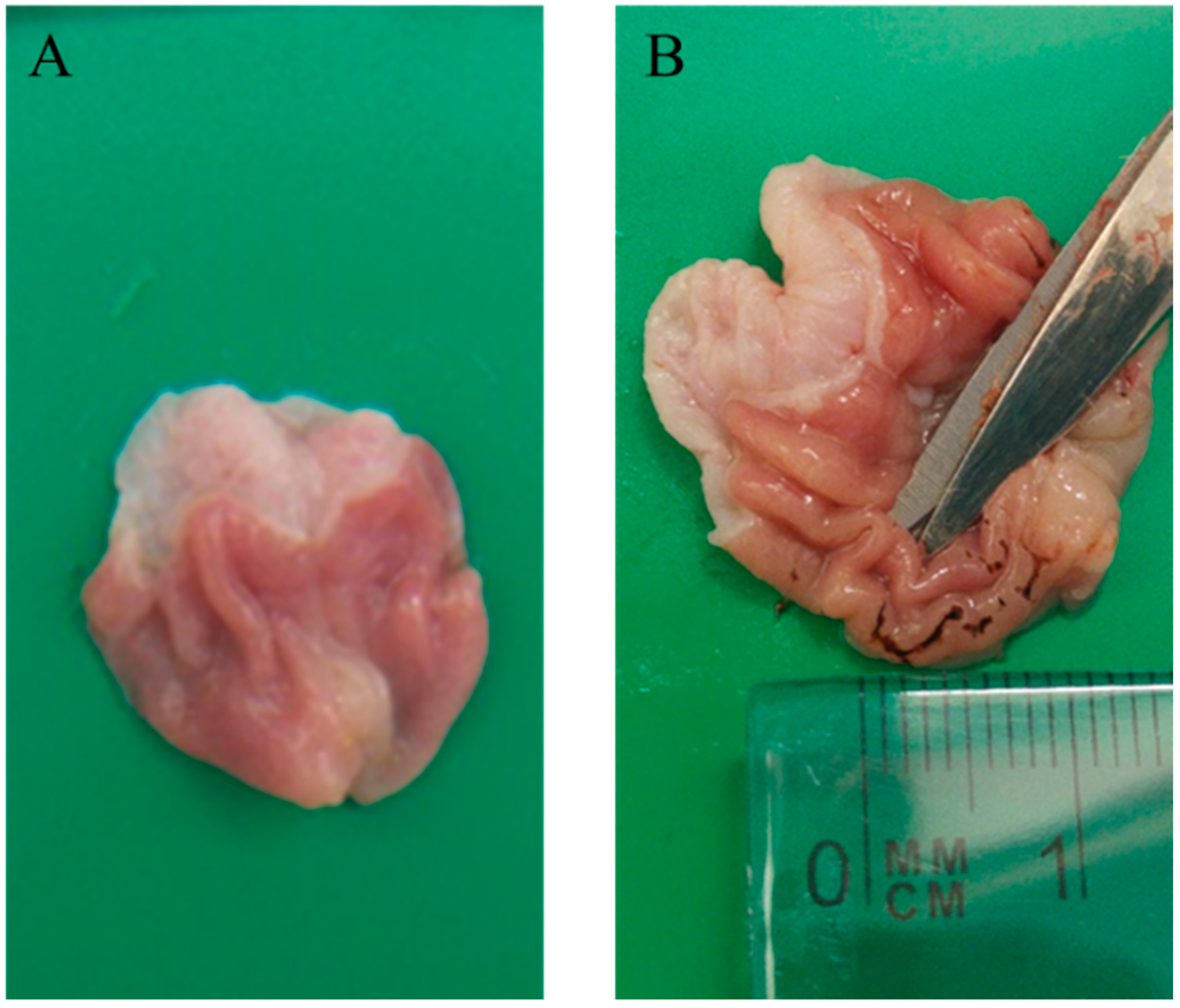
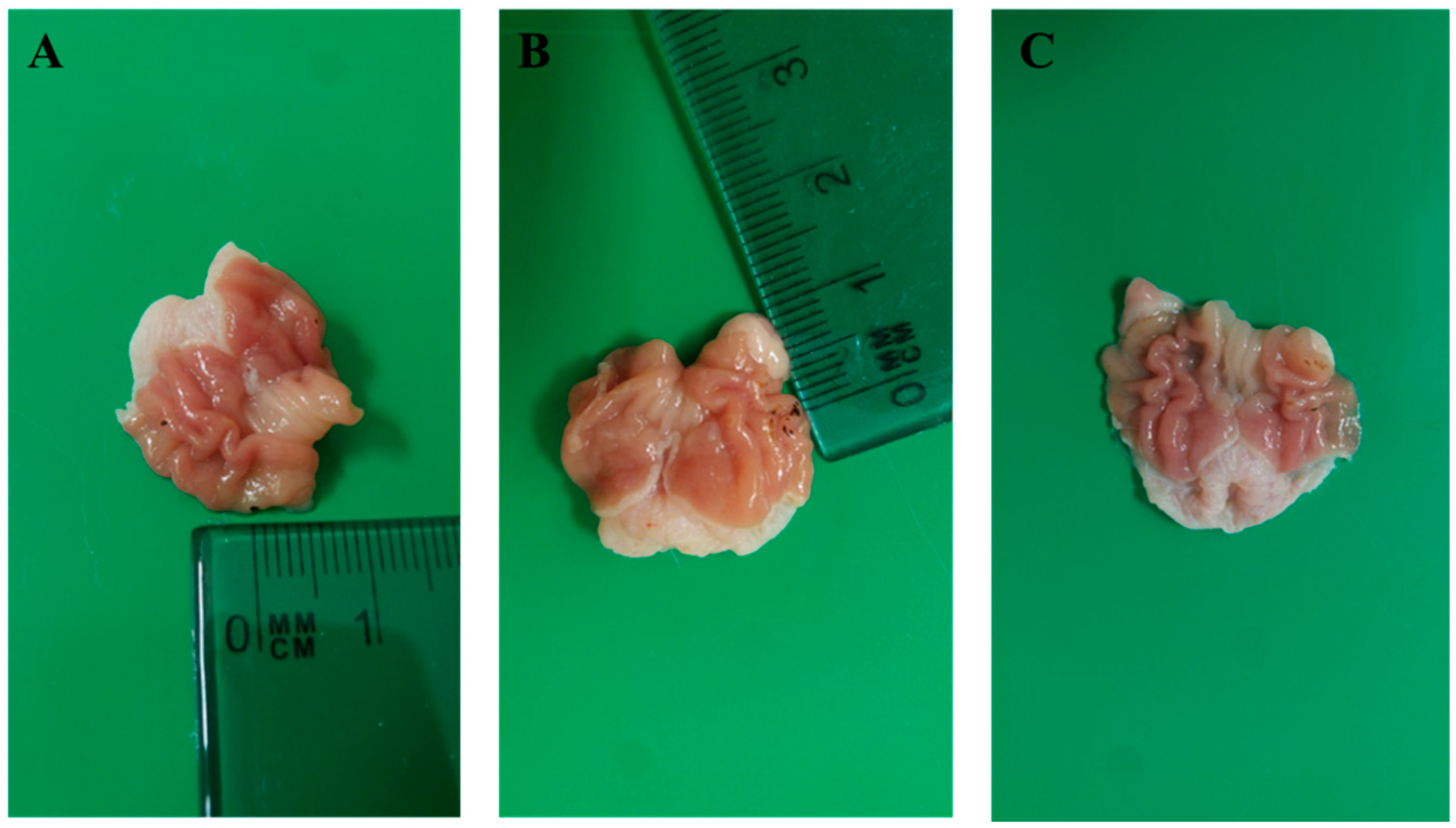
No ulcerative lesions were detected in the control group, whereas the indomethacin-treated group exhibited pronounced gastric ulceration (Figure 1). Macroscopic assessment demonstrated that the LN probiotic produced a significant, dose-dependent protective effect against ulcer formation in rats, comparable to the effect observed with famotidine (Figure 2).
The ulcer index (UI) for all groups and the percentage of protection (PP) for pretreated groups (III, IV, and V) were calculated to assess the severity of mucosal lesions. The results are shown in Table 2.

3.2. Histological Analysis of Gastric Ulcers

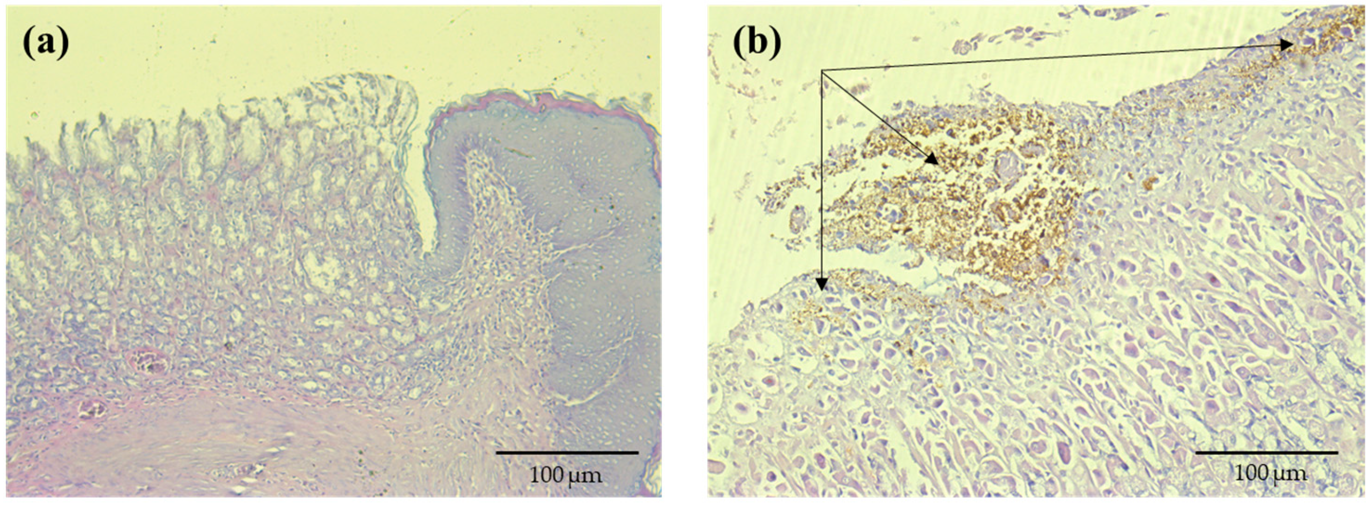
In the control group (Group I), H&E-stained gastric sections displayed normal mucosal architecture with intact epithelium, well-preserved foveolar and glandular zones, and no signs of inflammation or hemorrhage. In contrast, the indomethacin-treated group (Group II) showed pronounced mucosal damage, including epithelial degeneration, surface erosions, and focal hemorrhages with hemosiderin deposits, indicative of acute inflammation (Figure 3).
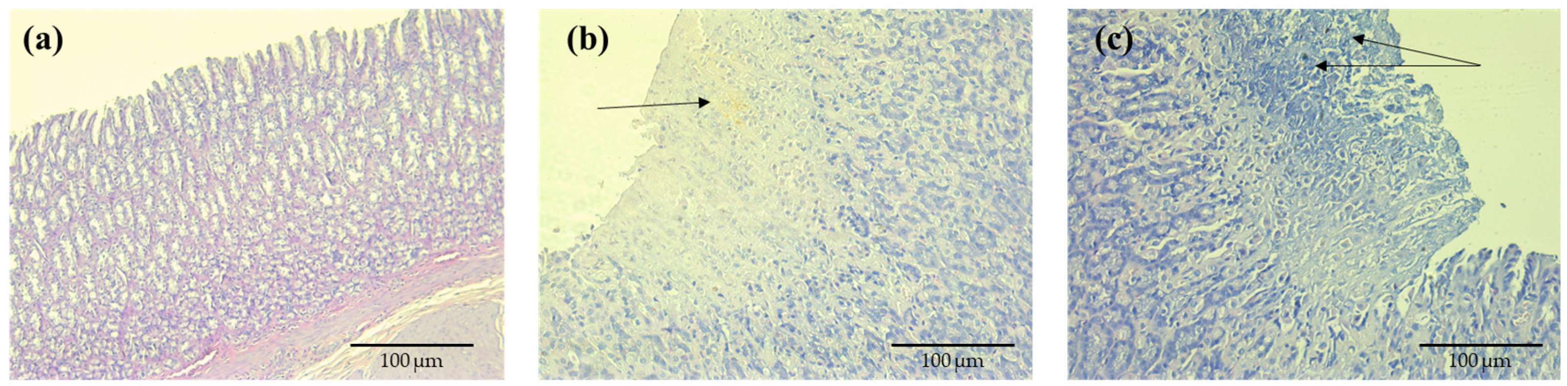
Rats pretreated with famotidine (20 mg/kg) for 14 days prior to ulcer induction showed well-preserved gastric mucosa, with intact epithelium and no evidence of inflammation or hemorrhage. Groups IV and V, receiving LN at 800 mg/kg and 1600 mg/kg, respectively (Figure 4b,c), demonstrated a dose-dependent gastroprotective effect. LN treatment substantially reduced indomethacin-induced histopathological damage, maintaining mucosal integrity comparable to the famotidine-treated group.

4. Discussion

To date, numerous scientific articles on probiotics have been indexed in PubMed, with more than 10,000 published in 2024 alone. While many of these studies highlight the beneficial effects of probiotics on the gastrointestinal tract through in vitro experiments, animal models, and investigations in both healthy and diseased volunteers, research specifically addressing their impact on gastric ulcers remains relatively scarce. This limited availability is probably due to the stomach’s harsh conditions—its acidity, digestive enzymes, bile acids, and mechanical forces—which hinder probiotic survival and colonization. To counter these obstacles, high doses of the LN probiotic were used in an effort to strengthen mucosal defenses and promote ulcer repair. The most widely studied probiotics for the treatment and/or prevention of gastrointestinal diseases are lactic acid bacteria, specifically the species Lactobacillus and Bifidobacterium. They can withstand the harsh luminal environment of the gastrointestinal tract [20].
Indomethacin-induced damage to the gastrointestinal mucosa results from complex interactions among multiple pathophysiological mechanisms. A significant factor is the inhibition of cyclooxygenase and the subsequent reduction in prostaglandins, which leads to impaired secretion of mucin and bicarbonates, reduced mucosal blood flow, and compromised barrier function [21,22]. These alterations are intensified by heightened oxidative stress, greater neutrophil infiltration, and reduced microcirculatory function; when gastric acid is present, the injury becomes even more severe. Consequently, the mucosal barrier is compromised, creating favorable conditions for erosion and ulcer formation.
Pre-treatment with the LN probiotic has been shown to exert significant dose-dependent protective effects against indomethacin-induced gastric ulcers, reducing damage to the gastric mucosa by approximately 42–52%. Other studies have reported even greater protection in certain cases. For example, Aliouche et al. (2025) demonstrated that Lactiplantibacillus plantarum E1K2R2 reduced ulcer formation by ~88.4%, which is comparable to the effect of omeprazole (~89.9%) in an ibuprofen-induced rat ulcer model [23]. The gastroprotective effects of probiotics are multifactorial, involving enhancement of mucosal defense, modulation of inflammation, and promotion of mucosal regeneration. These include increased mucus production, which reinforces the gastric mucosal barrier, enhanced synthesis of prostaglandin E2—a key mediator of mucosal protection—and modulation of the gut microbiota, promoting a favorable microecological balance [24,25]. Additionally, probiotics reduce oxidative stress and enhance the activity of antioxidant enzymes, compete for receptors, suppress inflammatory mediators such as TNF-α and IL-6, inhibit apoptosis through regulation of Bax and Bcl-2, and exert immunomodulatory effects within intestinal lymphoid tissue. Together, these mechanisms contribute to maintaining mucosal integrity and preventing pathogenic colonization [26]. Lactobacilli not only act locally in the gut but also contribute more broadly to gastrointestinal health. They can suppress harmful microbes, moderate inflammatory activity, and limit allergic or neoplastic changes, indicating benefits that extend beyond the immediate site of action. Probiotics also influence epithelial turnover by shifting the balance toward greater cell proliferation relative to apoptosis, a process that supports the repair of injured epithelial surfaces, especially along the borders of gastric ulcers [27]. The LN probiotic further exhibits notable antioxidant activity, including the neutralization of free radicals and a reduction in lipid peroxidation—effects that likely strengthen its protective role by lowering oxidative stress in the gastric mucosa. These antioxidant properties appear closely linked to the number of viable Lactobacillus bulgaricus cells in the product, emphasizing the importance of cell viability for achieving therapeutic benefits [28].
The limitations of this study include the relatively small number of animals used, which may reduce the statistical power and generalizability of the findings. Additionally, the mechanisms underlying NSAID-induced ulcerogenesis differ between animals and humans. Experimental models typically involve acute administration of high doses over a short period, whereas in humans, ulcers generally develop after chronic exposure to therapeutic doses. Furthermore, species-specific anatomical and physiological differences—such as variations in gastric mucosal structure, acid secretion, mucosal blood flow, and regenerative capacity—can influence both susceptibilities to injury and protective or healing responses. These factors limit the direct extrapolation of the results to clinical settings.

5. Conclusions

In conclusion, gastric ulcers result from a disruption between injurious factors and the gastric mucosa’s defense mechanisms. The present findings suggest that LN probiotic exerts a protective and healing effect on gastric mucosa, likely through multiple mechanisms. Notwithstanding the above limitations, these results support the potential of LN probiotic as an adjunctive strategy for gastric ulcer management and highlight the need for further research to optimize probiotic-based therapies in both health and disease.

Author Contributions

Conceptualization, methodology and research: K.D.G. and M.G.; literature search and analysis: K.D.G. and M.G.; data acquisition and analysis: K.D.G., M.G. and A.T.; statistical analysis: K.D.G., M.G. and A.T.; histopathological analysis: D.D.; writing of the manuscript-draft: K.D.G. and M.G.; review and editing of the original manuscript: K.D.G. and M.G.; final review and approval of the manuscript: K.D.G., M.G., D.D. and A.T. All authors have read and agreed to the published version of the manuscript.

Funding

This study was financed by the European Union—NextGenerationEU—through the National Recovery and Resilience Plan of the Republic of Bulgaria, Scientific Group 3.1.6., Project № BG-RRP-2.004-0009-C02.

Institutional Review Board Statement

All experimental procedures complied with Directive 2010/63/EU and were approved by the Bulgarian Food Safety Agency (Approval Document No. 175 issued on 7 July 2017).

Informed Consent Statement

Not applicable.

Data Availability Statement

All of the data that support the findings of this study are available in the main text.

Conflicts of Interest

The authors declare no conflicts of interest.

Abbreviations

The following abbreviations are used in this manuscript:
INDIndomethacin
LNLaktera Nature
GITGastrointestinal tract
NSAIDsNon-steroidal anti-inflammatory drugs
NaCMCSodium carboxymethyl cellulose
UIUlcer index

References

  1. Hill, C.; Guarner, F.; Reid, G.; Gibson, G.R.; Merenstein, D.J.; Pot, B.; Morelli, L.; Canani, R.B.; Flint, H.J.; Salminen, S.; et al. Expert Consensus Document: The International Scientific Association for Probiotics and Prebiotics Consensus Statement on the Scope and Appropriate Use of the Term Probiotic. Nat. Rev. Gastroenterol. Hepatol. 2014, 11, 506–514. [Google Scholar] [CrossRef] [PubMed]
  2. Georgieva, M.; Georgiev, K.D.; Hvarchanova, N. Probiotics: Past, Present, and Future Challenges. In Advances in Food and Nutrition Research; Watson, R.R., Preedy, V.R., Eds.; Academic Press: London, UK, 2022; pp. 431–448. [Google Scholar] [CrossRef]
  3. Georgiev, K.; Georgieva, M.; Iliev, I.; Peneva, M.; Alexandrov, G. Antiproliferative Effect of Bulgarian Spring Water Probiotics (Lakter a Nature Probiotics®) against Human Colon Carcinoma Cell Line. World J. Pharm. Pharm. Sci. 2015, 4, 130–136. [Google Scholar]
  4. Quigley, E.M.M. Gut Microbiota and the Role of Probiotics in Therapy. Curr. Opin. Pharmacol. 2013, 13, 893–896. [Google Scholar] [CrossRef] [PubMed]
  5. Tilg, H.; Moschen, A.R. Microbiota and Diabetes: An Evolving Relationship. Gut 2014, 63, 1513–1521. [Google Scholar] [CrossRef]
  6. Georgieva, M.; Georgiev, K.; Dobromirov, P. Probiotics and Immunity. In Immunopathology and Immunomodulation; IntechOpen: London, UK, 2015. [Google Scholar] [CrossRef][Green Version]
  7. Davoodvandi, A.; Fallahi, F.; Tamtaji, O.R.; Faraji-Rad, M.; Akbari, H.; Rezaei, N.; Jafarinia, M. An Update on the Effects of Probiotics on Gastrointestinal Cancers. Front. Pharmacol. 2021, 12, 680400. [Google Scholar] [CrossRef]
  8. Dikeocha, I.J.; Al-Kabsi, A.M.; Eid, E.E.M.; Hussin, S.; Alshawsh, M.A. Probiotics Supplementation in Patients with Colorectal Cancer: A Systematic Review of Randomized Controlled Trials. Nutr. Rev. 2022, 80, 22–49. [Google Scholar] [CrossRef]
  9. Bron, P.A.; van Baarlen, P.; Kleerebezem, M. Emerging Molecular Insights into the Interaction between Probiotics and the Host Intestinal Mucosa. Nat. Rev. Microbiol. 2012, 10, 66–78. [Google Scholar] [CrossRef]
  10. Hemarajata, P.; Versalovic, J. Effects of Probiotics on Gut Microbiota: Mechanisms of Intestinal Immunomodulation and Neuromodulation. Ther. Adv. Gastroenterol. 2013, 6, 39–51. [Google Scholar] [CrossRef]
  11. Gareau, M.G.; Sherman, P.M.; Walker, W.A. Probiotics and the Gut Microbiota in Intestinal Health and Disease. Nat. Rev. Gastroenterol. Hepatol. 2010, 7, 503–514. [Google Scholar] [CrossRef]
  12. Wang, Y.; Wu, Y.; Wang, Y.; Xu, H.; Mei, X.; Yu, D.; Li, L.; Li, X. Antioxidant Properties of Probiotic Bacteria. Nutrients 2017, 9, 521. [Google Scholar] [CrossRef]
  13. Cryer, B.; Feldman, M. Cyclooxygenase-1 and cyclooxygenase-2 selectivity of widely used nonsteroidal anti-inflammatory drugs. Am. J. Med. 1998, 104, 413–421. [Google Scholar] [CrossRef]
  14. Wallace, J.L. Prostaglandins, NSAIDs, and Gastric Mucosal Protection: Why Doesn’t the Stomach Digest Itself? Physiol. Rev. 2008, 88, 1547–1565. [Google Scholar] [CrossRef] [PubMed]
  15. Ukena, S.N.; Singh, A.; Dringenberg, U.; Engelhardt, R.; Seidler, U.; Hansen, W.; Bleich, A.; Stallmach, A.; Schmidt, M.A.; Schäfer, K.H.; et al. Probiotic Escherichia coli Nissle 1917 Inhibits Leaky Gut by Enhancing Mucosal Integrity. PLoS ONE 2007, 2, e1308. [Google Scholar] [CrossRef]
  16. Slavov, I.; Ivanova, N.; Radeva-Ilieva, M.; Stoeva-Grigorova, S.; Dzhenkov, D.; Georgiev, K.D. Phytochemical Analysis and Anti-Ulcer Potential of Phenolic Compounds of Inonotus nidus-pici Pilát. Pharmaceuticals 2025, 18, 1265. [Google Scholar] [CrossRef] [PubMed]
  17. Karakaya, K.; Hanci, V.; Bektas, S.; Can, M.; Ucan, H.B.; Emre, A.U.; Tascilar, O.; Ozkocak Turan, I.; Comert, M.; Irkorucu, O.; et al. Mitigation of indomethacin-induced gastric mucosal lesions by a potent specific type V phosphodiesterase inhibitor. World J. Gastroenterol. 2009, 15, 5091–5096. [Google Scholar] [CrossRef] [PubMed]
  18. Dekanski, J.B.; Macdonald, A.; Sacra, P. Effects of Fasting, Stress and Drugs on Gastric Glycoprotein Synthesis in the Rat. Br. J. Pharmacol. 1975, 55, 387–392. [Google Scholar] [CrossRef]
  19. Adinortey, M.B.; Ansah, C.; Galyuon, I.; Nyarko, A. In Vivo Models Used for Evaluation of Potential Antigastroduodenal Ulcer Agents. Ulcers 2013, 2013, 796405. [Google Scholar] [CrossRef]
  20. Bezkorovainy, A. Probiotics: Determinants of Survival and Growth in the Gut. Am. J. Clin. Nutr. 2001, 73, 399S–405S. [Google Scholar] [CrossRef]
  21. Elliott, S.L.; Ferris, R.J.; Giraud, A.S.; Cook, G.A.; Skeljo, M.V.; Yeomans, N.D. Indomethacin Damage to Rat Gastric Mucosa Is Markedly Dependent on Luminal pH. Clin. Exp. Pharmacol. Physiol. 1996, 23, 432–434. [Google Scholar] [CrossRef]
  22. Takeuchi, K. Pathogenesis of NSAID-Induced Gastric Damage: Importance of Cyclooxygenase Inhibition and Gastric Hypermotility. World J. Gastroenterol. 2012, 18, 2147–2160. [Google Scholar] [CrossRef]
  23. Aliouche, N.; Sifour, M.; Kebsa, W.; Khennouf, T.; Ercan, F.; Ouled-Haddar, H. Prophylactic Effect and Antiulcerogenic Potential of Probiotic Lactiplantibacillus plantarum E1K2R2 and Its Exopolysaccharide against Ibuprofen-Induced Acute Gastric Ulcer. Probiotics Antimicrob. Proteins 2025, 17, 3887–3902. [Google Scholar] [CrossRef]
  24. Lam, E.K.Y.; Tai, E.K.K.; Koo, M.W.L.; Wong, B.C.Y.; Cho, C.H. Enhancement of Gastric Mucosal Integrity by Lactobacillus rhamnosus GG. Life Sci. 2007, 80, 2128–2136. [Google Scholar] [CrossRef]
  25. Khoder, G.; Al-Menhali, A.A.; Al-Yassir, F.; Karam, S.M. Potential Role of Probiotics in the Management of Gastric Ulcer. Exp. Ther. Med. 2016, 12, 3–17. [Google Scholar] [CrossRef]
  26. Gelen, V.; Gedikli, S.; Gelen, S.U.; Şengül, E.; Makav, M. Probiotic Bacteria Protect against Indomethacin-Induced Gastric Ulcers through Modulation of Oxidative Stress, Inflammation, and Apoptosis. Mol. Biol. Rep. 2024, 51, 684. [Google Scholar] [CrossRef]
  27. Yan, F.; Cao, H.; Cover, T.L.; Whitehead, R.; Washington, M.K.; Polk, D.B. Soluble Proteins Produced by Probiotic Bacteria Regulate Intestinal Epithelial Cell Survival and Growth. Gastroenterology 2007, 132, 562–575. [Google Scholar] [CrossRef]
  28. Saide, J.A.; Gilliland, S.E. Antioxidative Activity of Lactobacilli Measured by Oxygen Radical Absorbance Capacity. J. Dairy Sci. 2005, 88, 1352–1357. [Google Scholar] [CrossRef] [PubMed]
Figure 1. Macroscopic evaluation of control group (I, (A)) and Indomethacin model of gastric ulcer (II, (B)).
Microbiolres 17 00059 g001
Figure 2. Macroscopic evaluation of famotidine-pretreatment group (III, (A)), LN probiotic 800 mg/kg (IV, (B)) and LN probiotic 1600 mg/kg (V, (C)).
Microbiolres 17 00059 g002
Figure 3. Representative histological images of gastric tissue stained with H&E (original magnification ×200; scale bar = 100 μm). (a) Group I, control group; (b) Group II, Indomethacin-treated group (40 mg/kg induction dose), showing mucosal damage and areas of erosion with hemosiderin deposition (indicated by arrows).
Microbiolres 17 00059 g003
Figure 4. Gross and microscopic images of gastric tissue stained with H&E (original magnification ×200; scale bar = 100 μm). (a) Group III, pretreated with famotidine (20 mg/kg) for 14 consecutive days before ulcer induction, showing intact gastric mucosa with no observable lesions, both macroscopically and histologically. (b,c) Groups IV and V, pretreated with the LN probiotic at doses of 800 mg/kg and 1600 mg/kg, respectively, for 14 consecutive days prior to ulcer induction. These groups exhibited dose-dependent gastroprotective effects on histological examination, with markedly reduced mucosal erosion and fewer haemosiderin deposits (some indicated by arrows).
Microbiolres 17 00059 g004
Table 1. Experimental Design.
GroupTreatmentDoseDurationNumber of Animals
IDistilled water1 mL14 days6
IIDistilled water1 mL14 days6
Indomethacin (IND)40 mg/kg, 1 mLon 15th day
IIIFamotidine (FM)1.25 mL (20 mg/kg)14 days6
Indomethacin (IND)40 mg/kg, 1 mLon 15th day
IVLN probiotic 800 mg/kg, 1 mL14 days6
Indomethacin40 mg/kg, 1 mLon 15th day
VLN probiotic1600 mg/kg, 1 mL14 days6
Indomethacin40 mg/kg, 1 mLon 15th day
Table 2. Data are shown as mean ± SD (n = 6). * p < 0.05, p < 0.005 vs. indomethacin group (Group II); ** p < 0.01 vs. indomethacin group (Group II).
Calculated Ulcer Index (UI)Calculated Percentage of Protection (PP), %
Group I (control group)0-
Group II (IND)4.67 ± 0.45-
Group III (FAM + IND)2.17 ± 0.61 **53.53
Group IV (LN, 800 mg/kg + IND)2.74 ± 0.40 *41.32
Group V (LN, 1600 mg/kg + IND)2.27 ± 0.43 *51.39
Disclaimer/Publisher’s Note: The statements, opinions and data contained in all publications are solely those of the individual author(s) and contributor(s) and not of MDPI and/or the editor(s). MDPI and/or the editor(s) disclaim responsibility for any injury to people or property resulting from any ideas, methods, instructions or products referred to in the content.