


Abstract
Antimicrobial resistance among multidrug-resistant enteric pathogens, such as Salmonella enterica and Shigella flexneri, poses a critical global health challenge, necessitating novel therapeutic strategies from natural sources. This study evaluated the preliminary phytochemical profile and dual antibacterial and antibiofilm activities of methanolic leaf extract from Platanus orientalis Linn. Qualitative and quantitative phytochemical analyses were conducted to assess phenolic and flavonoid contents, while antibacterial activity against multidrug-resistant isolates was determined using broth microdilution to obtain minimum inhibitory concentrations (MICs). Antibiofilm effects were examined at sub-MIC concentrations via the crystal violet assay. The extract was found to be rich in phenolics and flavonoids and exhibited concentration-dependent antibacterial activity, with MICs of 64 µg/mL for S. enterica and 32 µg/mL for S. flexneri. Sub-MIC concentrations significantly reduced biofilm biomass by 60–80%, likely through interference with quorum sensing and extracellular polymeric substance production. These results highlight the extract’s potential as a phytotherapeutic agent targeting both planktonic cells and biofilms, providing insights for the development of natural antimicrobials against resistant enteric pathogens. Overall, the findings underscore the potential of Platanus orientalis methanolic leaf extract as a promising natural antibacterial and antibiofilm agent against multidrug-resistant enteric pathogens and support its further development as an alternative or complementary therapeutic strategy. It should be noted that the phytochemical results are qualitative and indicative, and further LC–MS or NMR analyses are required for definitive compound identification.
1. Introduction
The global antimicrobial resistance (AMR) crisis represents one of the most severe threats to modern medicine. The increasing prevalence of multidrug-resistant (MDR) and extensively drug-resistant (XDR) organisms has critically undermined the effectiveness of conventional antibiotics, compromising the management of infectious diseases, surgical interventions, organ transplants, and chemotherapy [1]. Enteric pathogens, such as Salmonella enterica and Shigella flexneri, are major contributors to AMR-related morbidity and mortality worldwide, with global surveillance systems reporting nearly 5 million deaths in 2023 attributable to resistant infections [2]. These alarming trends underscore the urgent need for enhanced surveillance, antibiotic stewardship, and the development of novel antimicrobial agents, particularly given the substantial public health burden imposed by these pathogens.
Various resistance mechanisms have been identified in Enterobacteriaceae, including Salmonella and Shigella, which significantly increase their tolerance to antimicrobial compounds [3]. Notably, Salmonella and Shigella biofilms form complex physical and biochemical barriers that enhance bacterial resistance to antibiotics and host immune responses. These biofilms consist of extracellular polymeric substance (EPS) matrices that can confer up to a 1000-fold increase in antibiotic tolerance compared to planktonic cells [4,5,6]. Bacteria within these biofilms communicate via quorum sensing (QS), a regulatory mechanism that controls gene expression related to virulence factors and facilitates the horizontal transfer of resistance genes, including plasmid-mediated ones [7]. This coordinated gene regulation via QS contributes to the persistence of biofilm-associated infections, often linked to chronic carrier states and recurrent enteric diseases caused by Salmonella and Shigella [8].
Among medicinal plants, Platanus orientalis Linn., native to Europe, Asia, and the Middle East, has attracted considerable attention due to its diverse pharmacological activities. The leaves, bark, and fruits of P. orientalis exhibit antibacterial effects, primarily attributed to flavonoids, tannins, terpenoids, saponins, and polyphenols, which act synergistically to inhibit bacterial growth and block biofilm formation. Flavonoids in P. orientalis extracts function by interacting with bacterial signaling molecules to reduce EPS production, while polyphenols target microbial cell membranes or chelate metal ions essential for enzymatic function [9]. Recent studies have explored its inhibitory activity against biofilm-producing bacteria such as Klebsiella pneumoniae and Pseudomonas aeruginosa. However, its efficacy against clinically relevant enteric pathogens, including S. enterica and S. flexneri, which pose a growing threat in sanitation-challenged regions, remains insufficiently investigated.
Plant-derived phytochemicals, particularly those from P. orientalis, therefore present a promising and eco-friendly alternative. Their multifaceted properties in modulating both planktonic and biofilm-encased bacteria offer a strategic approach to addressing multidrug resistance in pathogenic bacteria. By elucidating the antimicrobial and antibiofilm effects of P. orientalis, this study contributes valuable evidence supporting plant-based therapeutics as potential alternatives to conventional antibiotics. The anticipated outcomes aim to promote the development of natural medicines or microbial coatings capable of inhibiting bacterial colonization and enhancing infection control in healthcare and environmental settings.
Given the pressing molecular challenges posed by AMR in Enterobacteriaceae—including horizontal gene transfer, efflux pump activation, and QS-regulated biofilms—targeting these mechanisms using phytochemicals that suppress key biofilm-related genes such as bssS, csgD, and luxS is critically important [10,11,12]. Although several medicinal plants have been investigated for their antimicrobial properties, limited data are available regarding the antibiofilm potential of P. orientalis against multidrug-resistant enteric pathogens. Furthermore, few studies have combined phytochemical analysis with antibacterial and antibiofilm activity assessment. This study, therefore, provides new insights by correlating the phytochemical diversity of P. orientalis methanolic leaf extract with its antimicrobial and antibiofilm potential.
The objectives of the current study are to investigate the antimicrobial resistance patterns of clinical and environmental strains of S. enterica and S. flexneri, and to assess the antibacterial and antibiofilm activities of P. orientalis methanolic leaf extract. Specifically, the study aims to: (i) identify the phytochemical constituents of the extract, (ii) evaluate its antibacterial activity against multidrug-resistant strains, (iii) assess its antibiofilm activity, and (iv) establish correlations between the observed biological activities and the phytochemical composition of the extract.
2. Materials and Methods
2.1. Microbial Stains Collection and Ethical Considerations
No human participants, human data, or human tissue were involved in this study. All experiments were conducted using standard microbiological techniques with bacterial cultures and plant extracts. No ethical approval or informed consent was required for this purely in vitro research. A total of 250 clinical samples (wound swabs, stool, burn swabs, body fluids, blood, and urine) from inpatients, using anonymized IDs to prevent duplication and 50 soil and water samples from the premises of various hospitals in Baghdad were collected.
2.2. Molecular Conformation Using 16S rRNA
Isolates of Salmonella enterica and Shigella flexneri were identified and confirmed using biochemical tests and 16S rRNA gene sequencing at AMS Research Laboratory, Baghdad. Genomic DNA was extracted using a commercial kit (Promega, Madison, WI, USA). The 16S rRNA PCR method involved specific primers (Table 1) designed by Primer3Plus (https://www.primer3plus.com, 3 February 2026) accessed in November 2022 and amplification with an initial denaturation at 94 °C for 3 min followed by 34 PCR cycles. The 25 µL reaction mix contained 12.5 µL of PCR master mix (Promega, Madison, WI, USA), 4 µL of each primer (10 pmol/µL), 4.5 µL of nuclease-free water, and 4 µL of DNA template [13]. Isolates were stored at −80 °C in trypticase soy broth with 20% glycerol and sub-cultured twice before testing. Purity and identity were verified by 16S rRNA sequencing and confirmed with BLAST (https://blast.ncbi.nlm.nih.gov/Blast.cgi, 3 February 2026) alignment showing >98% similarity. For susceptibility testing, bacterial inoculum density was standardized to 1.5 × 10⁸ CFU/mL using McFarland standards to ensure consistent conditions. This procedure provided reliable microbial identification and standardized material for further testing.
Table 1.
PCR primers designed for molecular identification of Salmonella enterica and Shigella flexneri.
2.3. Antimicrobial Susceptibility Testing
Antimicrobial susceptibility testing was performed using the Kirby-Bauer disc diffusion method on Mueller-Hinton agar plates inoculated with bacterial suspensions adjusted to 0.5 McFarland turbidity (1.5 × 10⁸ CFU/mL). The antibiotic panel (Bioanalyse, Istanbul, Turkey) included representatives from major clinical classes: β-lactams (cefotaxime [CTX], 30 µg; ceftriaxone [CTR], 30 µg; amoxicillin [AX], 25 µg; and augmentin [AMC], 30 µg), carbapenems (meropenem [MRP], 10 µg; imipenem [IPM], 10 µg), aminoglycosides (amikacin [AK], 30 µg; gentamicin [GEN], 10 µg), and fluoroquinolones (ciprofloxacin [CIP], 10 µg; levofloxacin [LEV], 5 µg). Each disc contained the indicated concentration of the respective antibiotic. Plates were incubated at 35 ± 2 °C for 16–18 h under ambient conditions, following Clinical and Laboratory Standards Institute (CLSI) 2023 recommendations. All susceptibility tests were performed in triplicate. After incubation, inhibition zones were measured in millimeters according to the CLSI 2023 guidelines to determine bacterial susceptibility or resistance. This standardized protocol ensures reliable and reproducible antibiotic sensitivity profiles for the tested bacterial isolates [14].
2.4. Plant Material Collection and Preparation
Platanus orientalis Linn leaves were collected from the Amedi region, Duhok Governorate, in the Kurdistan Region of Iraq (37.09° N, 43.48° E, elevation 1200 m). The climate is Mediterranean mountain type, with moderately acidic to neutral soil (pH 6.5–7.2) favorable for the species. The leaves were washed, air-dried, and ground into powder.
2.5. Extraction Procedure
Extraction was performed using a Soxhlet apparatus with 500 mL absolute methanol at 65 °C for 4 h. The extract was filtered and concentrated under reduced pressure at 40 °C using rotary evaporation. The yield percentage was calculated based on initial plant powder weight [15,16].
2.6. Preliminary Qualitative Phytochemical Screening
The phytochemical screening of the methanolic extract of P. orientalis Linn was carried out in an effort to test for the presence of prominent secondary compounds such as tannins, polysaccharides, terpenoids, glycosides, alkaloids, phenolic compounds, and flavonoids. Tannins were tested for by combining 2 mL of the extract with 5% ferric chloride solution (FeCl3), and a blue-black to greenish coloration was observed. Carbohydrates were detected using Molisch’s test, which involved combining the extract with Molisch’s reagent and then concentrated sulfuric acid (H2SO4), forming a violet ring at the interface. Glycosides were detected by combining the extract with glacial acetic acid and 2% ferric chloride solution, and then adding concentrated (H2SO4) slowly, forming a brown ring at the interface. Alkaloids were detected by combining the extract with 2N hydrochloric acid (HCl), and then testing with Mayer’s reagent, forming a cream or white precipitate, which confirmed their presence. Phenolic compounds were detected by adding 2% ferric chloride solution, which gave a bluish-black coloration. Flavonoids were detected by heating the powdered plant material in ethyl acetate, filtering the mixture, and then testing the filtrate with 10% ammonia solution, which gave a yellow fluorescence in an alkaline solution. All assays were performed in triplicate to ensure reliability [17,18]. It is important to note that these qualitative phytochemical assays are non-specific and may yield false-positive reactions, particularly for alkaloid detection, due to cross-reactivity with nitrogen-containing phenolic or weakly basic compounds. Therefore, the results obtained from these tests are indicative rather than confirmatory.
2.7. Antimicrobial Properties of Platanus orientalis Linn
The antibacterial properties of Platanus orientalis Linn were evaluated using the agar well diffusion method with extract concentrations of 125, 250, and 500 mg/mL in Mueller-Hinton agar inoculated with bacterial suspensions standardized to 0.5 McFarland (1.5 × 10⁸ CFU/mL) [19]. Sub-minimum inhibitory concentrations (½-MICs) were determined by broth microdilution in microtiter plates, using resazurin dye as an indicator; following incubation at 37 °C for 18–20 h, a color change from blue to pink indicated bacterial growth [20,21]. This assay provided a clear evaluation of antibacterial activity through zone of inhibition measurement and determination of minimal inhibitory concentration values, confirming the bioactive potential of Platanus orientalis leaf extracts [22]. All assays were performed in triplicate to ensure reliability.
2.8. Biofilm Formation and Inhibition Assay
Biofilm formation by Salmonella enterica and Shigella flexneri was induced by inoculating bacterial cultures in brain heart infusion (BHI) broth supplemented with 2% sucrose and incubating at 37 °C for 24 h. For quantification, 20 µL of bacterial suspension was added to microtiter wells containing 180 µL of BHI. The plates were then rinsed with normal saline, treated with 99% methanol, air-dried, and stained with 1% crystal violet. The dye was solubilized using 96% ethanol, and optical density (OD) was measured at 630 nm. Biofilm production was classified as non (OD ≤ ODc), weak (ODc < OD ≤ 2 × ODc), moderate (2 × ODc < OD ≤ 4 × ODc), or strong (OD > 4 × ODc) based on OD values relative to a cutoff (ODc).
2.9. Statistical Analysis
All experiments were performed in triplicate, and results are presented as mean ± standard deviation (SD). Data were compiled and analyzed using Microsoft Excel. Descriptive statistical analysis was applied to summarize antibacterial and antibiofilm activity outcomes.
3. Results
3.1. Bacterial Isolate Characteristics
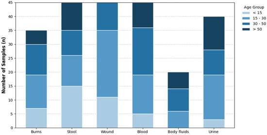
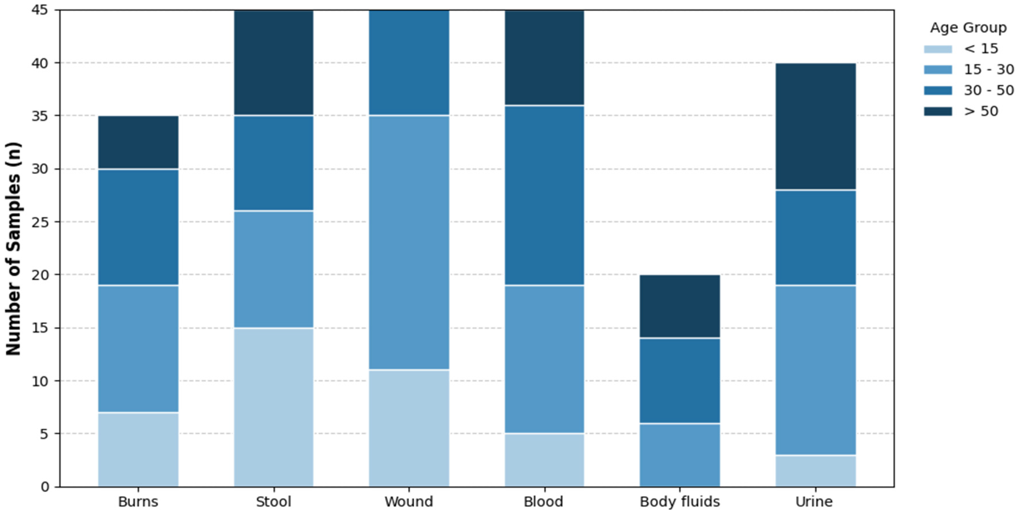
A total of 250 clinical specimens were collected, including 124 (49.6%) from male inpatients and 126 (50.4%) from female inpatients (Table 2). In the 15–30 years age group, wound specimens were the most common (n = 24/65, 36.9%), followed by burn and urine specimens (n = 12/35, 34.2% and n = 16/40, respectively). Stool specimens were most common among patients older than 15 years (n = 15/45, 33.3%). In the 30–50 years age group, body fluid specimens were the most frequent (n = 8/20, 40%), with blood specimens also prominent (n = 17/45, 37.7%) (Figure 1). Salmonella enterica was isolated from 37.5% of water samples and 25% of soil samples, while Shigella flexneri was isolated from 12.5% of water samples and 12.5% of soil samples. These observations clearly demonstrate that a significant burden of pathogenic bacteria is present in these sources, underscoring the need for proper surveillance and control measures to prevent contamination and associated health hazards. The analysis of 250 clinical samples indicated that pathogen isolation was restricted to stool and blood specimens. Salmonella enterica was isolated from three stool samples (6.7%) and one blood sample (2.2%), whereas Shigella flexneri was isolated exclusively from five stool samples (11.1%). All remaining clinical specimens, including burns, wounds, urine, and body fluids, showed no growth (0%).
Table 2.
Distribution of clinical and environmental isolates of Salmonella enterica and Shigella flexneri by source, gender, and age group.
Figure 1.
Demographic Distribution and Clinical Source Profiles of Specimens Collected Across Different Patient Age Groups. The stacked bar chart illustrates the number of samples ($n$) collected from various clinical sources, including burns, stool, wounds, blood, body fluids, and urine. Data is categorized into four age demographics: <15 years (light blue), 15–30 years (medium blue), 30–50 years (dark blue), and >50 years (navy).
3.2. Antibiotic Susceptibility Profiles
Salmonella enterica isolates showed a high level of susceptibility to most β-lactams, carbapenems, and azithromycin, with only limited resistance and strong responsiveness to these agents (Table 3). In contrast, S. flexneri exhibited a more complex and pronounced resistance profile, particularly against ampicillin, polymyxin B, chloramphenicol, and fluoroquinolones, suggesting a stronger multidrug-resistant phenotype. However, S. flexneri was relatively more susceptible to azithromycin, meropenem, and third-generation cephalosporins. Taken together, these findings indicate that S. enterica has a more favorable susceptibility profile, whereas S. flexneri presents greater treatment challenges due to its broader resistance pattern.
Table 3.
Antibiotic susceptibility profiles of Salmonella enterica and Shigella flexneri isolates.
The susceptibility patterns revealed that all isolates were completely resistant to vancomycin, while all were susceptible to meropenem. Salmonella enterica exhibited full susceptibility (100%) to ceftriaxone, augmentin, levofloxacin, and imipenem, but displayed complete resistance to amikacin. In contrast, Shigella flexneri was fully susceptible to azithromycin (AZM, 15 µg), yet demonstrated high resistance to colistin (CL, 10 µg; 100%), amoxicillin (75%), and polymyxin B (200 IU), highlighting significant interspecies differences in antimicrobial resistance profiles.
3.3. Preliminary Phytochemical Screening and Extract Yield of Platanus orientalis Linn
The methanolic extract of Platanus orientalis Linn yielded 10% (w/w) from the aerial parts. Preliminary phytochemical screening revealed the presence of phenolic compounds, tannins, glycosides, carbohydrates, and terpenes, along with weak non-confirmatory reactions with general alkaloid-detecting reagents (Table 4). Phytochemical analyses indicated a complex mixture of bioactive compounds, with a total phenolic content of 159.40 ± 4.34 mg gallic acid equivalents (GAE)/g and a total flavonoid content of 36.25 ± 0.45 mg quercetin equivalents (QE)/g. The phenolic compounds chlorogenic acid, rutin, and quercetin derivatives are well known for their ability to interfere with bacterial cell wall biosynthesis, quorum sensing, and metal ion chelation, which are essential for enzymatic function. Representative images of the qualitative phytochemical screening tests are provided in Supplementary Figure S1.
Table 4.
Qualitative phytochemical constituents of Platanus orientalis Linn methanolic leaf extract.
3.4. Antibacterial Activity of P. orientalis Linn Extracts
The antibacterial potential of the methanolic extract of P. orientalis Linn. was investigated using the well diffusion assay, demonstrating concentration-dependent activity against multidrug-resistant Salmonella enterica and Shigella flexneri. The zone of inhibition was directly proportional to the extract concentration, ranging from 12 mm at 125 mg/mL to 20 mm at 500 mg/mL. The minimum inhibitory concentration (MIC) was determined to be 64 µg/mL for S. enterica and 32 µg/mL for S. flexneri, indicating notable antibacterial efficacy. The MIC values established in this study served as the basis for evaluating antibiofilm activity, highlighting the potential use of the extract in infection prevention.
3.5. Biofilm Formation and Inhibition
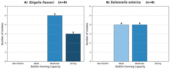
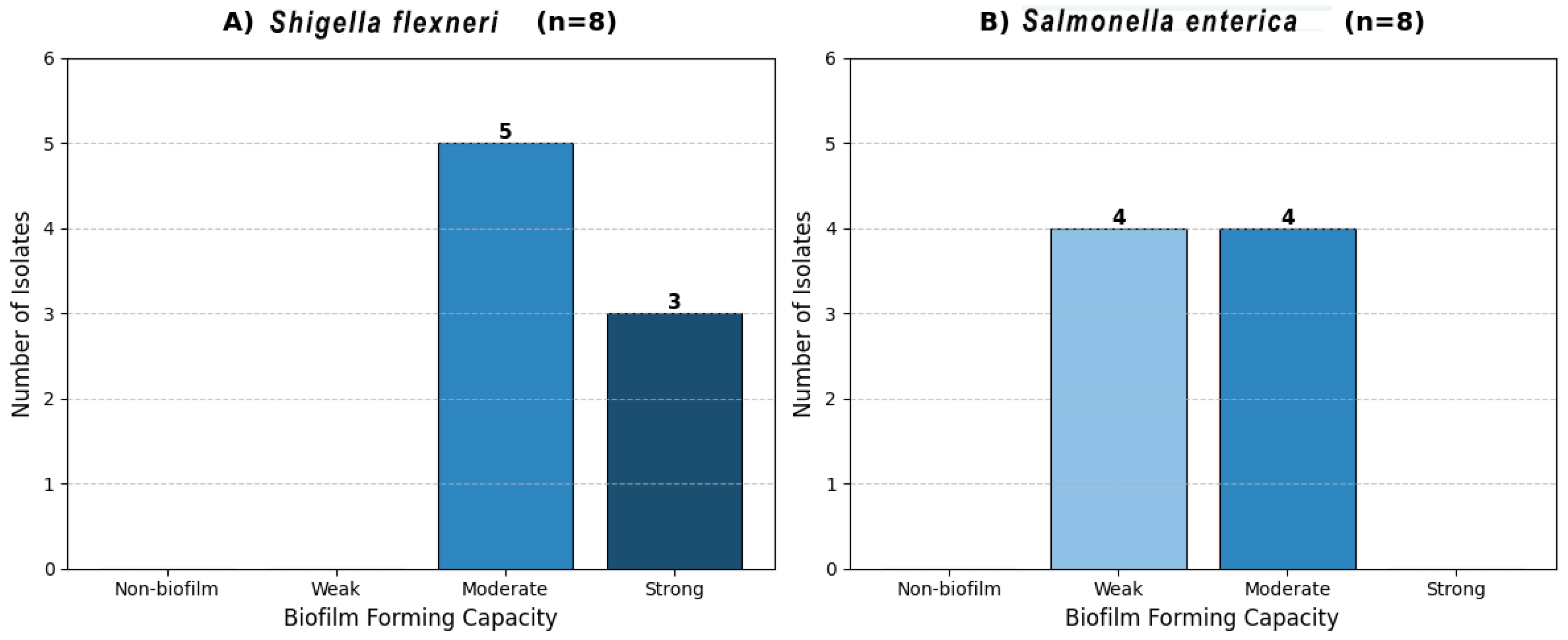
Biofilm-forming species analysis revealed species-specific variability in the degree of biofilm-forming ability. Among Shigella flexneri strains, moderate (62.5%) to strong (37.5%) biofilm-forming capacity predominated, whereas Salmonella enterica strains exhibited moderate (50%) and weak (50%) biofilm-forming abilities (Figure 2). Sub-minimum inhibitory concentration (½-MIC) levels of the methanolic extract of Platanus orientalis significantly inhibited biofilm formation in both species (Table 5). In S. flexneri strains, strong biofilm-forming ability was completely abolished, with strains shifting to weak or non-biofilm-forming phenotypes. In S. enterica strains, 75% of isolates lost their biofilm-forming ability and became non-biofilm formers, while the remaining strains lost their moderate or strong biofilm-forming capacities. Overall, biofilm biomass was reduced by 60–80%, as indicated by optical density measurements at 630 nm (OD630) (Table 4).
Figure 2.
Classification of Shigella flexneri and Salmonella enterica isolates based on their specific biofilm-forming capacity at OD630. The bar charts represent the distribution of $n = 8$ isolates for each species across four phenotypic categories: non-biofilm, weak, moderate, and strong.
Table 5.
Effect of Platanus orientalis Linn methanolic extract on biofilm formation of Salmonella enterica and Shigella flexneri.
4. Discussion
This study investigated the antimicrobial resistance profiles of Salmonella enterica and Shigella flexneri isolates and evaluated the antibacterial and antibiofilm potential of Platanus orientalis methanolic leaf extract. The findings demonstrated that the extract exhibited significant inhibitory effects against both planktonic cells and biofilm formation in multidrug-resistant isolates, highlighting its potential as a natural antimicrobial agent. Although weak positive reactions were observed with general alkaloid-detecting reagents, Platanus orientalis is not widely reported as a classical alkaloid-producing species in phytochemical databases such as Reaxys. Therefore, these reactions are interpreted as indicative of alkaloid-like or nitrogen-containing metabolites rather than confirmed alkaloids. Consequently, the observed antibacterial and antibiofilm activities are primarily attributed to phenolic and flavonoid compounds, which are well documented for their antimicrobial and quorum-sensing-inhibitory properties. The findings for Shigella flexneri revealed concerning trends in antibiotic resistance, including resistance to antibiotics such as ampicillin, amoxicillin–clavulanate, and fluoroquinolones. These observations are consistent with previous reports documenting the increasing trend of antimicrobial resistance in Shigella spp. [23]. The methanolic leaf extract of Platanus orientalis Linn. contained 159.40 ± 4.34 mg GAE/g of total phenolic compounds, reflecting a high phenolic content equivalent to that amount of gallic acid per gram of extract. These observations are consistent with previous reports [24]. These compounds are known to inhibit bacterial cell wall synthesis, interfere with quorum-sensing mechanisms, and chelate metal ions crucial for enzymatic functions. Such properties are likely responsible for the strong antimicrobial activities observed against MDR bacterial strains, consistent with recent metabolomic studies reporting high abundances of these compounds in Platanus orientalis extracts [25]. In addition, the total flavonoid content (TFC) of the methanolic leaf extract was 36.25 ± 0.45 mg QE/g, representing the flavonoid content expressed as quercetin equivalents per gram of sample. These results highlight the abundant presence of bioactive secondary metabolites, such as phenolic and flavonoid compounds, which are commonly associated with strong antioxidant properties and other beneficial biological activities in the material under investigation [26]. Given the rising antimicrobial resistance observed in clinical isolates of Salmonella enterica and Shigella flexneri, as corroborated by our susceptibility profiles, the need to identify novel bioactive molecules from natural sources becomes imperative. Plant-derived phytochemicals, such as those from Platanus orientalis Linn. characterized in this study, offer promising alternatives because of their multitarget antimicrobial and antibiofilm activities. Developing such bioactive agents could play a crucial role in mitigating the spread and impact of these pathogens by targeting both planktonic bacteria and their biofilms, thus addressing a critical gap in current therapeutic strategies [27].
The methanolic extract of Platanus orientalis Linn. demonstrated significant antibacterial and antibiofilm activities against multidrug-resistant (MDR) Salmonella enterica and Shigella flexneri, corroborating the hypothesis that polyphenol-rich plant extracts possess multitarget inhibitory effects on resistant bacteria. A direct correlation was observed between the methanolic extract of Platanus orientalis and its antibacterial and antibiofilm activities against MDR Salmonella enterica and Shigella flexneri. This relationship was evidenced by the concentration-dependent increase in inhibition zones and the corresponding reduction in biofilm formation following extract treatment. Higher extract concentrations resulted in greater antibacterial efficacy, accompanied by a marked decrease in biofilm biomass, indicating that suppression of planktonic bacterial growth directly contributed to impaired biofilm development. Furthermore, the antibiofilm activity was particularly pronounced among strong and moderate biofilm-forming isolates, suggesting that the extract not only inhibits bacterial proliferation but also interferes with biofilm establishment and stability. This dual effect supports a functional link between the antibacterial potency of the extract and its capacity to disrupt biofilm-associated resistance mechanisms in MDR enteric pathogens. Collectively, these findings demonstrate that the methanolic extract of Platanus orientalis exerts a coordinated antibacterial and antibiofilm effect, reinforcing its potential as a natural therapeutic agent against biofilm-mediated multidrug resistance. The observed minimum inhibitory concentrations (MICs) of 64 μg/mL for S. enterica and 32 μg/mL for S. flexneri indicate strong bacteriostatic efficacy that aligns with reports of plant-derived antimicrobial compounds targeting MDR pathogens. The concentration-dependent increase in inhibition zone diameters (12–20 mm) highlights the potent antimicrobial effects attributable to the extract’s rich flavonoid and phenolic contents, which likely act by compromising bacterial membrane integrity and increasing permeability, consistent with enhanced damage to lipopolysaccharide layers as documented in resistant Gram-negative bacteria [25,28,29]. Importantly, sub-inhibitory concentrations of the P. orientalis extract markedly reduced biofilm formation in both pathogens. This antibiofilm effect primarily arises from interference with early adhesion events and suppression of extracellular polymeric substance (EPS) synthesis, which are critical for biofilm maturation. The observed shift from strong biofilm producers to weak or non-producers suggests disruption of quorum-sensing pathways that regulate gene expression during biofilm formation. This conclusion is supported by significant reductions in optical density readings at 630 nm following treatment. Key bioactive compounds, such as quercetin and kaempferol derivatives present in the extract, are known quorum-sensing inhibitors targeting AI-2 and LuxR receptors, which may explain the inhibition of biofilm regulatory genes and signaling cascades [30,31]. These findings are consistent with existing studies demonstrating that plant polyphenols can suppress the expression of fimbrial adhesins (fimA, fimH), curli regulators (csgD), and biofilm stability factors (bssS), thereby reducing biofilm biomass in Enterobacteriaceae and other pathogens [11]. The multitargeted mechanism of P. orientalis methanolic extract positions it as a promising candidate for phytotherapeutic development to combat biofilm-associated antimicrobial resistance, which remains a significant clinical hurdle. However, the current study has limitations, including the use of relatively low-resolution techniques, such as HPLC, for phytochemical profiling without complementary transcriptomic or molecular docking analyses to confirm and elucidate gene suppression mechanisms. Future investigations incorporating multi-omics approaches, such as metabolomics, proteomics, and genomics, will be essential to fully characterize active components, their precise molecular targets, and safety profiles. Cytocompatibility studies will also be required to assess clinical applicability and potential toxicity.
5. Conclusions
In conclusion, the methanolic leaf extract of Platanus orientalis demonstrated eco-friendly potential for managing multidrug-resistant (MDR) bacterial infections by exhibiting notable antibacterial and antibiofilm activities against MDR isolates of Salmonella enterica and Shigella flexneri. This activity is likely due to its polyphenol- and flavonoid-rich phytochemical composition, as indicated by preliminary screening of the plant, particularly its phenolic and flavonoid components. These findings support the therapeutic potential of Platanus orientalis as a natural antimicrobial agent and contribute to addressing the urgent global challenge of antibiotic resistance through the exploration of novel plant-derived products with multifaceted antimicrobial properties. However, further research is required to validate its clinical applicability.
Supplementary Materials
The following supporting information can be downloaded at https://www.mdpi.com/article/10.3390/microbiolres17020036/s1, Figure S1: The inhibition zones of plant extract against multi-drug resistant bacterial (Shigella flexneri Isolate 5 and isolate 8); Figure S2: The inhibition zones of plant extract against multi-drug resistant bacterial (Salmonella enterica Isolates and isolate 5); Figure S3. Gel electrophoresis of PCR product of 16s rRNA gene among Salmonella enterica isolates; Figure S4. Gel electrophoresis of PCR product of 16s rRNA gene among Shigella flexneri isolates; Figure S5: Bio film and Antibiofilm activity; Figure S6. Representative images of preliminary qualitative phytochemical screening tests performed on Platanus orientalis methanolic leaf extract, including alkaloid-detecting reagent, terpene, glycoside, flavonoid, carbohydrate, tannin, and phenol tests. These assays are indicative and do not provide definitive identification of individual compounds. Table S1: The Inhibition zone of different concentration of crude Plant methanolic extract against the Salmonella entrica isolates; Table S2: The Inhibition zone of different concentration of crude Plant methanolic extract against the Shigella flexneri isolates.
Author Contributions
A.N.A.: Conceptualization, Data curation, Formal analysis, Methodology, Software writing—original draft. M.G.B.A.: Conceptualization, Formal analysis, Supervision, writing—original draft. R.G.: Conceptualization, Project administration, Writing—review and editing. All authors have read and agreed to the published version of the manuscript.
Funding
This research received no external funding.
Institutional Review Board Statement
This research has been conducted in vitro using bacterial isolates, without the involvement of human beings or animals. No ethical approval was required for this purely in vitro research.
Informed Consent Statement
This research has been conducted in vitro using bacterial isolates, without the involvement of human beings or animals. No informed consent was required for this purely in vitro research.
Data Availability Statement
The raw data supporting the results of this study are available in the public repository at https://doi.org/10.5281/zenodo.18474906.
Conflicts of Interest
The authors declare no conflicts of interest.
Abbreviations
| AK | Amikacin |
| AMC | Amoxicillin-Clavulanic Acid |
| AX | Amoxicillin |
| AZM | Azithromycin |
| BHI | Brain heart infusion |
| CAZ | Ceftazidime |
| CIP | Ciprofloxacin |
| CL | Colistin |
| CN | Gentamicin |
| CTX | Cefotaxime |
| DMSO | Dimethyl Sulfoxide |
| EPS | Extracellular Polymeric Substances |
| GAE | Gallic Acid Equivalent |
| MDR | Multidrug-Resistant |
| MeOH | Methanol |
| MIC | Minimum Inhibitory Concentration |
| n | Number |
| OD | Optical Density |
| QE | Quercetin Equivalent |
| QS | Quorum Sensing |
| S. enterica | Salmonella enterica |
| S. flexneri | Shigella flexneri |
| SD | Standard deviation |
| TFC | Total Flavonoid Content |
| TPC | Total Phenolic Content |
References
- World Health Organization. Global Antimicrobial Resistance and Use Surveillance Report 2024; WHO Press: Geneva, Switzerland, 2024. [Google Scholar]
- Centers for Disease Control Prevention. Antibiotic Resistance Threats Report, 2023; U.S. Department of Health and Human Services: Washington, DC, USA, 2023. [Google Scholar]
- Shahid, A.; Arshad, A.; Aslam, B.; Ali, M.A.; Munir, S.; Aslam, S.; Khurshid, M. Antimicrobial Resistance: Causes, Mechanisms, Origins and Characterization. In Handbook of Molecular Biotechnology; CRC Press: Boca Raton, FL, USA, 2024; pp. 382–396. [Google Scholar]
- Elshobary, M.E.; Badawy, N.K.; Ashraf, Y.; Zatioun, A.A.; Masriya, H.H.; Ammar, M.M.; Mohamed, N.A.; Mourad, S.; Assy, A.M. Combating Antibiotic Resistance: Mechanisms, Multidrug-Resistant Pathogens, and Novel Therapeutic Approaches: An Updated Review. Pharmaceuticals 2025, 18, 402. [Google Scholar] [CrossRef] [Scilit]
- Zhao, A.; Sun, J.; Liu, Y. Understanding bacterial biofilms: From definition to treatment strategies. Front. Cell. Infect. Microbiol. 2023, 13, 1137947. [Google Scholar] [CrossRef] [Scilit]
- Ahmed, A.; Khan, A.K. Microbial Biofilms-Aided Resistance and Remedies to Overcome It. In Microbial Biofilms; CRC Press: Boca Raton, FL, USA, 2020; pp. 331–371. [Google Scholar] [CrossRef] [Scilit]
- Nahum, Y.; Muhvich, J.; Morones-Ramirez, J.R.; Casillas-Vega, N.G.; Zaman, M.H. Biofilms as potential reservoirs of antimicrobial resistance in vulnerable settings. Front. Public Health 2025, 13, 1568463. [Google Scholar] [CrossRef] [Scilit] [PubMed]
- Sharma, S.; Mohler, J.; Mahajan, S.D.; Schwartz, S.A.; Bruggemann, L.; Aalinkeel, R. Microbial Biofilm: A Review on Formation, Infection, Antibiotic Resistance, Control Measures, and Innovative Treatment. Microorganisms 2023, 11, 1614. [Google Scholar] [CrossRef] [Scilit] [PubMed]
- Harrell, J.E.; Hahn, M.M.; D’Souza, S.J.; Vasicek, E.M.; Sandala, J.L.; Gunn, J.S.; McLachlan, J.B. Salmonella Biofilm Formation, Chronic Infection, and Immunity Within the Intestine and Hepatobiliary Tract. Front. Cell. Infect. Microbiol. 2021, 10, 624622. [Google Scholar] [CrossRef] [Scilit]
- D’Aquila, P.; De Rose, E.; Sena, G.; Scorza, A.; Cretella, B.; Passarino, G.; Bellizzi, D. Quorum Quenching Approaches against Bacterial-Biofilm-Induced Antibiotic Resistance. Antibiotics 2024, 13, 619. [Google Scholar] [CrossRef] [Scilit]
- Dávila-Aviña, J.; Gil-Solís, C.; Merino-Mascorro, J.; García, S.; Heredia, N. Phenolics with bactericidal activity alter motility and biofilm formation in enterotoxigenic, enteropathogenic, and enterohemorrhagic Escherichia coli. Foodborne Pathog. Dis. 2020, 17, 568–575. [Google Scholar] [CrossRef] [Scilit] [PubMed]
- Xu, Z.; Wang, C.; Wang, M.; Liang, J.; Li, C.; Chen, W.; Wei, P. Unveiling the Hidden Mechanism: How LuxS/AI-2 quorum sensing system drives antimicrobial resistance in Salmonella pullorum through activation of the efflux pump AcrAB-TolC. Poult. Sci. 2025, 104, 104972. [Google Scholar] [CrossRef] [Scilit]
- Paulin-Curlee, G.G.; Singer, R.S.; Sreevatsan, S.; Isaacson, R.; Reneau, J.; Foster, D.; Bey, R. Genetic Diversity of Mastitis-Associated Klebsiella pneumoniae in Dairy Cows. J. Dairy Sci. 2007, 90, 3681–3689. [Google Scholar] [CrossRef] [Scilit]
- CLSI. M100: Performance Standards for Antimicrobial Susceptibility Testing, 33rd ed.; Clinical and Laboratory Standards Institute: Berwyn, PA, USA, 2023. [Google Scholar]
- López-Bascón, M.A.; De Castro, M.D.L. Soxhlet Extraction. In Liquid-Phase Extraction; Elsevier: Amsterdam, The Netherlands, 2019; pp. 327–354. [Google Scholar] [CrossRef] [Scilit]
- Loustau, E.; Rols, J.-L.; Leflaive, J.; Marcato-Romain, C.-E.; Girbal-Neuhauser, E. Comparison of extraction methods for the characterization of extracellular polymeric substances from aggregates of three biofilm-forming phototrophic microorganisms. Can. J. Microbiol. 2018, 64, 887–899. [Google Scholar] [CrossRef] [Scilit]
- Gokhale, M.S. Pharmacognosy; Pragati Books Pvt. Ltd.: Pune, India, 2008. [Google Scholar]
- Nortjie, E.; Basitere, M.; Moyo, D.; Nyamukamba, P. Extraction Methods, Quantitative and Qualitative Phytochemical Screening of Medicinal Plants for Antimicrobial Textiles: A Review. Plants 2022, 11, 2011. [Google Scholar] [CrossRef] [Scilit]
- Atef, N.M.; Shanab, S.M.; Negm, S.I.; Abbas, Y.A. Evaluation of antimicrobial activity of some plant extracts against antibiotic susceptible and resistant bacterial strains causing wound infection. Bull. Natl. Res. Cent. 2019, 43, 144. [Google Scholar] [CrossRef] [Scilit]
- Chen, W.; Xu, Z.; Li, C.; Wang, C.; Wang, M.; Liang, J.; Wei, P. Investigation of biofilm formation and the associated genes in multidrug-resistant Salmonella pullorum in China (2018–2022). Front. Vet. Sci. 2023, 10, 1248584. [Google Scholar] [CrossRef] [Scilit]
- Adeosun, I.J.; Baloyi, I.T.; Cosa, S. Anti-Biofilm and Associated Anti-Virulence Activities of Selected Phytochemical Compounds against Klebsiella pneumoniae. Plants 2022, 11, 1429. [Google Scholar] [CrossRef] [Scilit]
- Ohikhena, F.U.; Wintola, O.A.; Afolayan, A.J. Evaluation of the Antibacterial and Antifungal Properties of Phragmanthera capitata (Sprengel) Balle (Loranthaceae), a Mistletoe Growing on Rubber Tree, Using the Dilution Techniques. Sci. World J. 2017, 2017, 9658598. [Google Scholar] [CrossRef] [Scilit]
- Pakbin, B.; Didban, A.; Monfared, Y.K.; Mahmoudi, R.; Peymani, A.; Modabber, M.R. Antibiotic susceptibility and genetic relatedness of Shigella species isolated from food and human stool samples in Qazvin, Iran. BMC Res. Notes 2021, 14, 144. Available online: https://link.springer.com/article/10.1186/s13104-021-05554-3 (accessed on 3 February 2026). [CrossRef] [Scilit] [PubMed]
- Fazeli, S.T.; Bimakr, M. Optimization of Bioactive Compounds Extraction from Platanus Orientalis Leaves using Microwave-Assisted Technique. J. Hum. Environ. Health Promot. 2024, 10, 209–216. [Google Scholar] [CrossRef] [Scilit]
- Lobiuc, A.; Pavăl, N.E.; Mangalagiu, I.I.; Gheorghiță, R.; Teliban, G.C.; Amăriucăi-Mantu, D.; Stoleru, V. Future antimicrobials: Natural and functionalized phenolics. Molecules 2023, 28, 1114. [Google Scholar] [CrossRef] [Scilit] [PubMed]
- Othman, L.; Sleiman, A.; Abdel-Massih, R.M. Antimicrobial activity of polyphenols and alkaloids in middle eastern plants. Front. Microbiol. 2019, 10, 911. [Google Scholar] [CrossRef] [Scilit]
- Samrot, A.V.; Abubakar Mohamed, A.; Faradjeva, E.; Si Jie, L.; Hooi Sze, C.; Arif, A.; Chuan Sean, T.; Norbert Michael, E.; Yeok Mun, C.; Xiao Qi, N.; et al. Mechanisms and impact of biofilms and targeting of biofilms using bioactive compounds—A review. Medicina 2021, 57, 839. [Google Scholar] [CrossRef] [Scilit] [PubMed]
- Sokoudjou, J.B.; Atolani, O.; Njateng, G.S.S.; Khan, A.; Tagousop, C.N.; Bitombo, A.N.; Kodjio, N.; Gatsing, D. Isolation, characterization and in vitro anti-salmonellal activity of compounds from stem bark extract of Canarium schweinfurthii. BMC Complement. Med. Ther. 2020, 20, 316. [Google Scholar] [CrossRef] [Scilit] [PubMed]
- Kengne, I.C.; Feugap, L.D.T.; Njouendou, A.J.; Ngnokam, C.D.J.; Djamalladine, M.D.; Ngnokam, D.; Voutquenne-Nazabadioko, L.; Tamokou, J.D.D. Antibacterial, antifungal and antioxidant activities of whole plant chemical constituents of Rumex abyssinicus. BMC Complement. Med. Ther. 2021, 21, 164. [Google Scholar] [CrossRef] [Scilit] [PubMed]
- Pandit, S.; Fazilati, M.; Gaska, K.; Derouiche, A.; Nypelö, T.; Mijakovic, I.; Kádár, R. The exo-polysaccharide component of extracellular matrix is essential for the viscoelastic properties of Bacillus subtilis biofilms. Int. J. Mol. Sci. 2020, 21, 6755. [Google Scholar] [CrossRef] [Scilit]
- Paczkowski, J.E.; Mukherjee, S.; McCready, A.R.; Cong, J.P.; Aquino, C.J.; Kim, H.; Henke, B.R.; Smith, C.D.; Bassler, B.L. Flavonoids suppress Pseudomonas aeruginosa virulence through allosteric inhibition of quorum-sensing receptors. J. Biol. Chem. 2017, 292, 4064–4076. [Google Scholar] [CrossRef] [Scilit] [PubMed]

Disclaimer/Publisher’s Note: The statements, opinions and data contained in all publications are solely those of the individual author(s) and contributor(s) and not of MDPI and/or the editor(s). MDPI and/or the editor(s) disclaim responsibility for any injury to people or property resulting from any ideas, methods, instructions or products referred to in the content. |
© 2026 by the authors. Licensee MDPI, Basel, Switzerland. This article is an open access article distributed under the terms and conditions of the Creative Commons Attribution (CC BY) license.