


Abstract
The highly destructive pathogenic processes in patients with periodontitis are attributed to the presence of subgingival biofilms comprising key periodontal pathogens, such as Porphyromonas gingivalis, Treponema denticola, and Tannerella forsythia, as well as other periodontopathogens including Fusobacterium nucleatum, Selenomonas spp., Centipeda spp., and Campylobacter spp. Considering the vast microbial diversity in periodontitis, we aimed to analyze the presence of various bacterial species in subgingival dental plaque, with a special focus on Selenomonas spp. We first developed a phylogenetic tree for Selenomonas–Veillonella clusters, using in silico analysis followed by fluorescence in situ hybridization (FISH) on subgingival dental plaque samples from 22 patients with a history of chronic periodontitis, by using specific 16S rRNA oligonucleotide probes. These oligonucleotide probes’ specificity and hybridization conditions were determined on previously characterized bacterial strains. Qualitative and quantitative analysis of FISH slides was carried out by using an epifluorescence microscope. The majority of the patient samples showed high fluorescence signals with the oligonucleotide probes SEL1150, Sspu439, and SEL1469 (specific for Selenomonas spp.), ACI623 (identifying Selenomonas, Veillonella, and Dialister spp.), Tfor127 (T. forsythia) and L-Pgin1006-23 (P. gingivalis). SEL1150 showed specificity for bacterial species in the subclusters A, B, and C, namely S. dianae, S. infelix, S. flueggei, C. periodontii, S. artemidis, S. noxia, and S. sputigena; Sspu439 for S. sputigena; SEL1469 for subclusters A and B, and for S. sputigena; ACI623 for bacterial species in subclusters C and F, namely the S. sputigena and Veillonella species. The experimentally observed specificities of the oligonucleotide probes corresponded with our in silico analysis. Selenomonas spp. may play a role in the subgingival microbiome of periodontitis and contribute to the disease process. Targeting Selenomonas spp. with specific therapeutic strategies could offer new insights into the management of periodontitis. However, further studies are needed to determine a definite functional significance.
1. Introduction
Selenomonas are Gram-negative anaerobic, rod-shaped bacteria that contain flagella on their concave surface for motility [1,2]. Selenomonas spp. are found in the rumen and ceca of mammals, where they are suggested to play a role in the fermentation of saccharose [3]. The presence of Selenomonas bacteria in the oral cavity of humans has also been extensively described, in which case they are found to mainly be associated with gingivitis [4], periodontitis [1,5], and endodontic infections [6]. Recently, Selenomonas sputigena has been discovered as a pathobiont cooperating with Streptococcus mutans in early childhood caries [7].
The very first descriptions of the crescent-shaped bacteria date back to 1886 in a study by Fluegge et al. [8], who described the motile bacteria in the oral cavity as “Spirillum sputigenum” [8]. The name Selenomonas was proposed by von Prowazek in 1913 for bacteria that showed a similar morphology [9]. Later, the name S. sputigena was formally adopted [2,9,10,11]. Novel Selenomonas spp.—S. noxia, S. flueggei, S. infelix, S. dianae, and S. artemidis—were subsequently identified by Moore et al. [1].
The dental and gingival surfaces in the oral cavity are covered with a biofilm comprising bacteria, their endotoxins, and breakdown products and supported by an extracellular matrix [12,13,14,15]. Interestingly, the composition of this biofilm varies not only between healthy and infected tissue but also between patients with either gingivitis or periodontitis [4,16,17]. A detailed analysis of the diversity of the oral microbiota has been undertaken by Paster et al., using 16S rRNA sequencing in periodontitis and acute necrotizing ulcerative gingivitis patients [18]. This hallmark study presented a detailed phylogenetic analysis of the phyla Fusobacteria, Actinobacteria, Firmicutes, and Proteobacteria and the genus Treponema, providing an initial overview of the disease-associated oral bacteria.
The highly destructive pathogenic processes in patients with periodontitis are attributed to the presence of subgingival biofilms comprising key periodontal pathogens, such as Porphyromonas gingivalis, Treponema denticola, and Tannerella forsythia. Other periodontopathogens include Fusobacterium nucleatum, Selenomonas spp., and Campylobacter spp. [19,20]. In this aspect, Selenomonas spp., owing to the presence of flagella, plays an essential role in the pathogenesis of periodontal diseases such as gingivitis and periodontitis, enabling invasion into deep layers of the connective tissues [2,19]. In a study with patients with generalized aggressive periodontitis, it has been found that the levels of P. gingivalis and S. sputigena are higher in sites with probing depths ≥ 5 mm compared to sites with depths ≤ 3 mm [21], as these niches are more anaerobic and better support the growth of these strict anaerobes. Similarly, Selenomonas noxia showed higher levels in the periodontal pockets of periodontitis patients than that of healthy controls [22]. Moreover, in addition to subgingival pockets, S. noxia is present in high concentrations in saliva and gingival crevicular fluid [23]. There is a correlation between Selenomonas species and specific periodontal pathogens, particularly members of the red complex, such as Porphyromonas gingivalis and Treponema denticola. Additionally, several studies have identified Selenomonas species in conjunction with orange complex pathogens, further supporting their potential role in periodontal disease pathology [24]. Traditionally overshadowed by Streptococcus mutans, recent studies suggest that S. sputigena may have a synergistic role in dental caries, particularly in forming biofilms associated with this condition. However, to date, direct evidence of S. sputigena enhancing acid production through metabolic cooperation with S. mutans is limited. There is evidence that S. sputigena contributes to the biofilm structure, which may bolster the protective ability of acidogenic bacteria, but specific mechanisms of cooperation need further elucidation. Additionally, S. sputigena has been implicated in systemic health issues, with some studies indicating associations with increased inflammatory cytokines and pregnancy complications, such as preterm birth and low birth weight, although more research is needed to establish direct causative links. The implications for both oral and systemic health are thus significant but require further investigation.
A study by Shi et al. (2018) found distinct microbial community structures associated with varying pocket depths, indicating that deeper pockets tend to harbor a more diverse and pathogenic microbiome, often dominated by species such as Porphyromonas gingivalis and Tannerella forsythia [25]. Additionally, research by Califf et al. highlighted a positive correlation between microbial biodiversity and probing depth, emphasizing the evolutionary diversity of subgingival samples in relation to disease severity, with a reported Spearman’s correlation coefficient indicating this positive relationship [26].
Several studies have pointed towards a correlation between periodontitis and other systemic diseases. The link to diabetes is well established and forms a strong basis for diagnosis and therapy [27,28,29]. In this regard, the analysis of the microbiota in periodontitis patients is of high clinical significance, as growing evidence suggests a correlation of periodontitis with cardiovascular diseases, gastrointestinal and colorectal cancers, and Azheimer’s disease [30]. One of the early reports of a systemic disease associated with oral bacteria was presented in 1981, in which the patient presented with septicemia and the blood cultures showed the presence of S. sputigena and Veillonella parvula [31]. Very interestingly, similar cases of septicemia originating from a lung abscess [32,33] and bacteremia in patients with cecal adenocarcinoma [33] and pancreatic cancer [34] showed the presence of Selenomonas spp. in blood cultures; all of these reports show evidence that the native microflora can become pathogenic in immunocompromised patients. While the specific data on bacteriocins produced by Selenomonas spp. remains limited, research has shown that bacteriocins from various bacterial strains can inhibit the growth of pathogenic bacteria, which suggests that similar mechanisms may eventually be discovered in Selenomonas species [35].
In a previous study, Drescher et al. described the spatial distribution of Selenomonas spp. in subgingival biofilms [36]. However, the study was limited to only one genus-specific oligonucleotide probe. In the present study, we aim to describe the various Selenomonas species that are present in subgingival dental plaque, using fluorescence in situ hybridization (FISH) analysis with several specific 16S rRNA oligonucleotide probes. Although the importance of Selenomonas spp. in periodontal diseases is well established, their precise role within the microbial ecosystem and their contribution to disease progression remain poorly understood. FISH stands out as a powerful tool for understanding the complex dynamics of periodontal biofilms. FISH provides several advantages, including the ability to visualize and localize specific bacterial populations directly within the biofilm context, thereby offering insights into their spatial relationships and community structure that other methods may not reveal. Recent studies show that FISH enhances the identification of pathogens in various environments, allowing for direct observation, which supports its utility in this context [37]. While 16S rRNA sequencing is valuable for detailed microbial community profiling, it can be cost-prohibitive and may lead to misinterpretations regarding species localization and interaction within biofilms, as it does not provide real-time visual confirmation of microbial presence [38]. Furthermore, while qPCR allows for the quantification of specific targets, it often does so at the expense of capturing the broader ecological context, potentially overlooking non-target organisms, and failing to provide data on spatial distribution [39,40].
We aimed to first analyze the specificity of the oligonucleotide probes on previously characterized reference strains, adjust the hybridization conditions for FISH, and then determine the proportions of certain periodontopathogenic bacteria in subgingival dental plaque samples from periodontitis patients.
2. Materials and Methods
2.1. Dental Plaque Samples and Bacterial Species
The study was approved by the Ethics Committee of the University of Zurich, Switzerland (BASEC-Number Req-2022-01146 (Business Administration System for Ethics Committees)). A total of 10 Selenomonas species, 2 Centipeda species, 2 Eubacterium species, 3 Veillonella species, and 2 Eikenella species were used for testing the specificity of 16S rRNA probes. All the aforementioned bacterial species had already been stored in the liquid nitrogen tank in the Division of Clinical Oral Microbiology and Immunology (COMI), Center of Dental Medicine, University of Zurich. All bacterial species, as well as their specificity, are listed in Appendix A.2. Subgingival dental plaque samples were collected from 22 patients, and a tongue smear was taken from one patient. The samples were partly collected at the Clinic of Conservative and Preventive Dentistry, University Center of Dental Medicine, University of Zurich, and partly in a private dental practice in Zurich, Switzerland. The samples collected in the private dental practice were from patients with a history of chronic periodontitis and were in their recall phase at the time of sample collection. In our study, periodontitis was defined based on the established criteria: specifically, the presence of at least two interproximal sites with a clinical attachment loss (CAL) of 6 mm or greater, and at least one additional site with a probing pocket depth (PD) of 5 mm or greater. For all patients, it was ensured that no treatment with antibiotics or other antimicrobial agents, e.g., chlorhexidine (CHX), was performed three months before sampling. During the recall appointments, the reduction in bacterial counts was performed mainly using photodynamic laser therapy (PDL) [41]. The samples were collected before PDL treatment. Briefly, the region for the collection of subgingival plaque was dried using cotton pads; supragingival plaque was removed, if necessary, with dental scalers, and samples of subgingival plaque were collected by submerging three sterile paper points in the gingival sulcus for 10 s. The paper points were immersed in 600 µL sterile 0.9% NaCl and frozen at −80 °C until further use. The tongue smear was collected using a tongue cleaner and the samples were likewise frozen in 0.9% NaCl. All samples were stored at −80 °C and subsequently analyzed at the same time after collection.
2.2. Design of 16S rRNA Oligonucleotide Probes
A total of 8 rRNA oligonucleotide probes were designed, using the software ARB (http://www.arb-home.de, accessed on 19 January 2025) [42], and confirmed using the “Ribosomal-Database-Project II”, while 6 oligonucleotide probes had already been tested in earlier reports [43,44,45,46,47]. The details of the oligonucleotide probes are described in Appendix A.1. The probes were marked either with the fluorescence marker Cy3 (indocarbocyanine) or FAM (6-carboxyfluorescein). Synthesis of the oligonucleotide probes was carried out by Microsynth AG, Balgach, Switzerland. The lyophilized probes were resuspended in 100 μl TE buffer, diluted either to 50 ng/μL (probes marked with Cy3) or 150 ng/μL (probes marked with FAM), aliquoted, and stored at −20 °C until use. All the 16S rRNA oligonucleotide probes were tested against the various bacterial species to determine the optimal reaction conditions. Once the hybridization specificity of the probes was established, these optimal conditions were then used to test the patient samples.
2.3. Coating of Bacterial Cells and Patient Dental Plaque Samples on Glass Slides
The aforementioned bacterial species were thawed and cultured anaerobically (85% N2, 10% H2, 5% CO2) at 37 °C in a liquid universal medium enriched with 5% horse serum. To initiate biofilm formation, the disks were covered with 1 ml of growth medium containing saliva and modified fluid universal medium (mFUM), and 200 μL of a microbial suspension prepared from equal volumes and densities of each strain, corresponding to OD550 = 1.0. mFUM is a well–established tryptone–yeast–based broth medium designated as FUM [48] and modified by supplementing 67 mM Sørensen’s buffer (final pH 7.2). Additionally, microorganisms were cultivated on blood agar plates. The strains could be harvested from the exponential growth phase after 12 to 24 h. The cultures were pelleted and washed in 0.9% NaCl and the pellets were fixed using a 4% paraformaldehyde (PFA) solution. The fixed bacterial cells were resuspended and stored in a 1:1 ethanol/water solution until use. For FISH, the bacterial suspension was further diluted (1:50) in coating buffer (0.9% NaCl, 0.02% NaN3, 2.5 × 10⁻⁴% hexadecyltrimethylammonium bromide; CTAB) and 10 μl of this suspension was divided and fixed on 24-well epoxy-coated slides with a well diameter of 4 mm (Cel-Line, Erie Scientific Company, Portsmouth, NH, USA). Blocking of unspecific binding included treatment with Denhardt’s solution (Sigma-Aldrich; diluted 1:50 in 0.9% NaCl) in the presence of ProtectRNA RNase inhibitor (1:500, Sigma-Aldrich GmbH, Buchs, Switzerland) and incubation for 30 min at 37 °C.
The dental plaque samples were thawed and analyzed for the presence of erythrocytes. If erythrocytes were detected visually, the samples were centrifuged in 0.83% NH4Cl for 4 min at 4 °C. A 2–10-fold dilution of the samples was performed in coating buffer and coated onto the glass slide (10 µL suspension per well). Thereafter, the glass slides were fixed in 4% PFA for 20 min at 4 °C, rinsed in ELISA wash buffer (0.9% NaCl, 0.01% NaN3, 0.05% tween 20) and H2O, and dried for further use.
2.4. Fluorescence In Situ Hybridization (FISH)
The 16S rRNA oligonucleotide probes were mixed with the hybridization buffer (containing different concentrations of formamide) to reach a final concentration of 5 ng/µL for probes conjugated with Cy3 or 15 ng/µL for probes conjugated with FAM. The formamide concentration was adjusted between 30 and 60% to determine the ideal concentration for each probe and bacterial species. Two oligonucleotide probes labeled with different fluorophores and similar hybridization conditions were added to each well of the glass slide in a volume of 4 μL hybridization buffer. The glass slides were incubated at 46 °C for two hours in a humid 50 mL tube containing the hybridization buffer, followed by incubation in a NaCl washing buffer at 48 °C for 15–30 min. The concentration of NaCl was adjusted according to the formamide concentration. Afterwards, the slides were briefly immersed in ELISA wash buffer and distilled H20 and air-dried in the dark. Finally, 45 µL of mounting fluid (VECTASHIELD® mounting medium, Vector Laboratories Ltd., Peterborough, UK) was applied dropwise to each slide in the middle row between the wells; the slide was covered with a coverslip (24 × 60 mm) and analyzed with microscopy.
2.5. Qualitative and Quantitative Microscopy Analysis of FISH Data
Following FISH, the glass slides were analyzed using an Olympus BX60-epifluorescence microscope (Olympus Optical, Volketswil, Switzerland). Images were captured with an Olympus Camedia 3030 digital camera. Both quantitative and qualitative analysis of the fluorescence intensity was performed. For qualitative analysis, fluorescence intensity was arbitrarily defined on a scale of 0–4, with 0 (also represented as “-” in Appendix A.2 and Appendix A.3) being “no fluorescence” and 4 being the “highest fluorescence intensity”. The intermediate fluorescence intensities were defined as 1 = weak, 2 = moderate, and 3 = strong. The fluorescence intensities 2, 3, and 4 were defined as positive [49]. The quantitative analysis of the fluorescent cells was performed by counting up to 30 microscopy fields (or the complete glass slide for diluted samples). The area of a counting field was 1/330th that of the glass slide. Positive bacterial cells in 1 mL of cell suspension were calculated as follows: x × 330 × v × 1000/10 (x = cell count per field, v = reciprocal of the dilution of the plaque suspension).
2.6. Generation of Phylogenetic Tree
The phylogenetic tree for the Selenomonas–Veillonella clusters was generated using the “tree builder” function of the Ribosomal Data Project release 10. The details on the oligonucleotide probes and their specificity for bacterial species are presented in Appendix A.1 Table A1.
2.7. Scanning Electron Microscopy
The bacterial cultures were fixed with 2.5% glutaraldehyde in the growth media for 2 h, washed twice by gentle centrifugation (1500 rpm, 30 min) in cacodylate buffer, re-suspended in 1% OsO4 in cacodylate buffer for 1 h, washed twice in distilled water, and dehydrated in a graded series of acetone. After critical point drying, they were applied to stubs, sputtered with platinum, and viewed with a Tescan VEGA scanning electron microscope (Tescan Group a.s., Brno, Czech Republic).
2.8. Statistical Analysis
Fluorescence intensity scores from the FISH evaluations were compared across groups, using the non-parametric Kruskal–Wallis test. Fisher’s exact test was used to assess the prevalence of the subclusters in the studied patient cohort, based on their fluorescence intensity–defined classifications. Statistical significance was set at p < 0.05.
3. Results
3.1. Phylogenetic Tree for Selenomonas–Veillonella clusters and In Silico Analysis for the 16S rRNA Oligonucleotide Probes
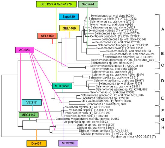
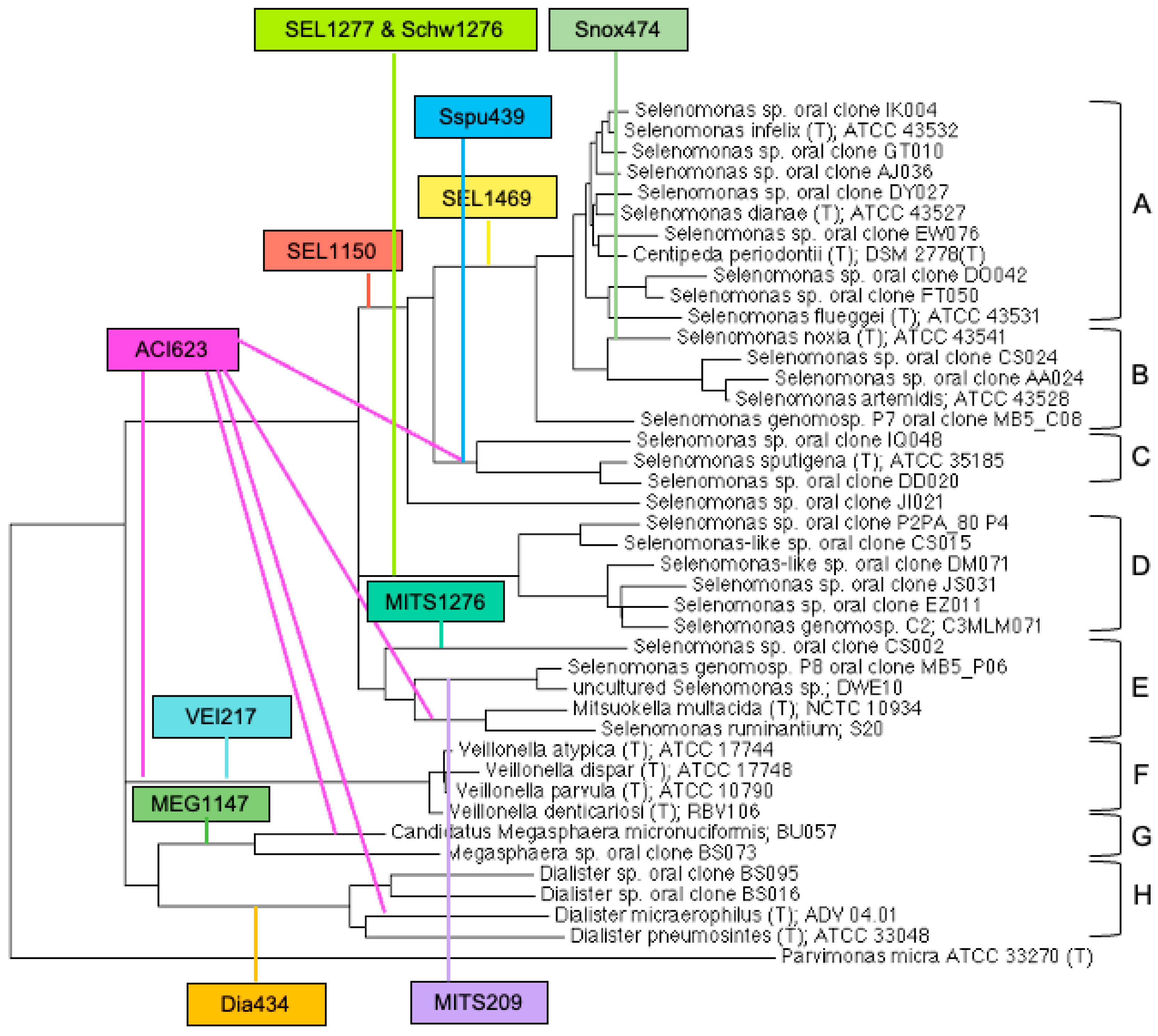
The phylogenetic tree, generated using the “tree builder” function of the Ribosomal Data Project release 10, is depicted in Figure 1. The bacterial species were grouped into subclusters A-H, based on the bacterial diversity, and were assigned as follows: A: S. dianae and its closely associated species S. infelix, S. flueggei, and Centipeda periodontii; B: S. artemidis and S. noxia; C: S. sputigena; D: not culturable Selenomonas oral type strains; E: Selenomonas and Mitsuokella species; F: Veillonella subcluster; G: Megasphaera subcluster; and H: Dialister subcluster.
Figure 1.
Phylogenetic tree for Selenomonas–Veillonella clusters and in silico analysis for the 16S rRNA oligonucleotide probes. The details on the oligonucleotide probes are presented in Appendix A.1 and in Materials and Methods. Colored boxes represent 16S rRNA probes; the colored lines depict the subclusters which the oligonucleotide probes detect, and are as follows: SEL1150: A, B, C; SEL1277: D; SEL1469: A, B; Sspu439: C; Snox474: B; MITS1276: E; MITS209: E; ACI623: C, E, F, G, H; SCHW1276: D; VEI217: Veillonella spp.; MEG1147: Megasphaera spp.; and Dia434: Dialister spp. The subclusters A–H are depicted on the right. Details on probe specificity for bacterial species are presented in Appendix A.1 and described in detail in the text as well. The probes VEI217, MEG1147, and DIA434 are included to demonstrate their broad reactivity across clusters, but they lack specificity.
In the in silico FISH analysis, SEL1150 showed specificity for the subclusters A, B, and C; Sspu439 for subcluster C; SEL1469 for subclusters A and B; ACI623 for subclusters C, E, F, G, and H; SEL1277/SCHW1276 for subcluster D; and MITS1276 and MITS209 for subcluster E. The oligonucleotide probes VEI217, MEG1147, and DIA434 were specific for the Veillonella, Megasphaera, and Dialister subclusters, respectively; however, these probes were not further analyzed as part of this study. An additional oligonucleotide probe, EUB338, was used as a positive control in parallel to each probe and bacterial/patient sample [49].
3.2. FISH Analysis for the Bacterial Reference Strains
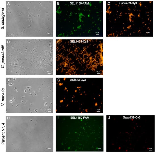
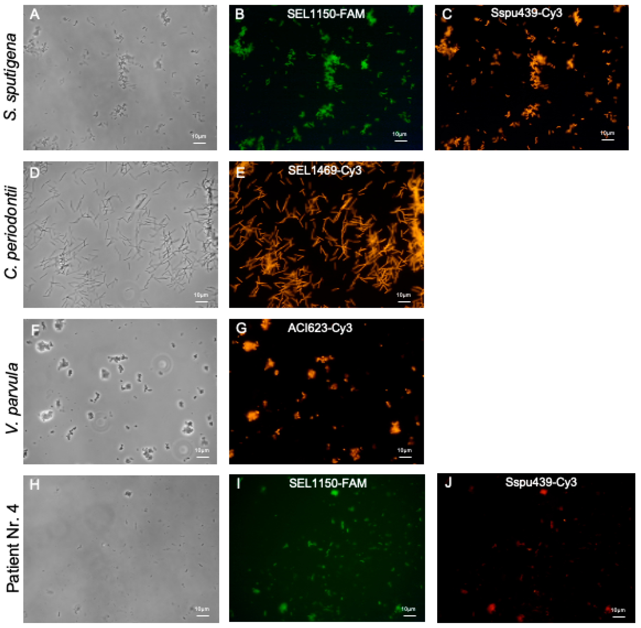
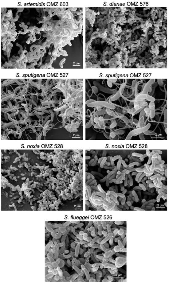
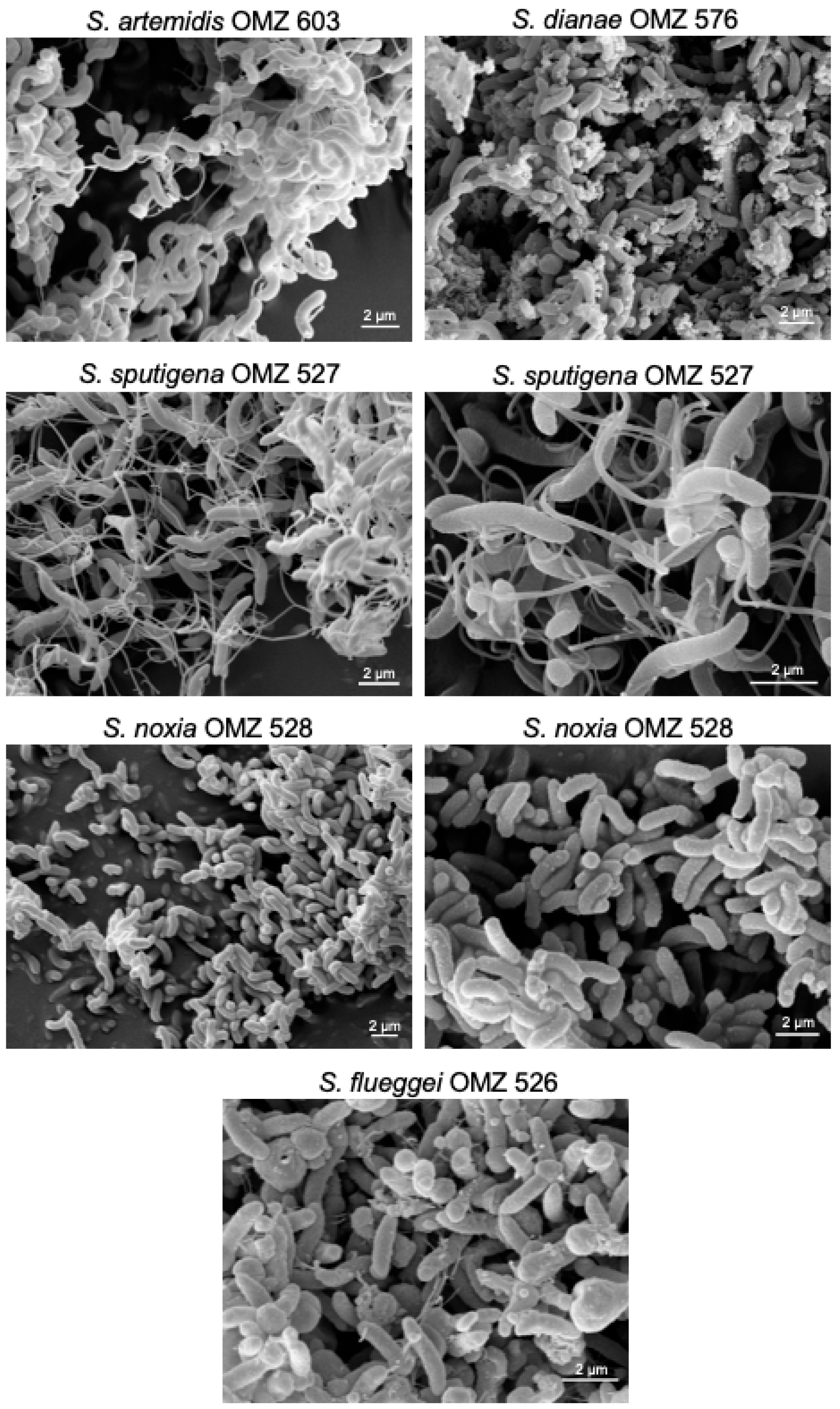
Representative microscopy images for S. sputigena OMZ 527 probed with SEL1150-FAM and Sspu439-Cy3, C. periodontii OMZ 614 probed with SEL1469-Cy3, and V. parvula OMZ 687 probed with ACI623-Cy3 are depicted in Figure 2A–G. The microscopy images show typical rod-shaped cells that are characteristic of Selenomonas spp. Additionally, scanning electron microscopy performed on S. artemidis, S. dianae, S. sputigena, S. noxia, and S. flueggei confirmed the crescent-shaped, flagellated morphology, as described earlier by Lessel, 1954 [2] (Figure 3). The C. periodontii culture shows rod-shaped, long, “serpentine-like” cells, as described by Lai et al. in 1983 [50]. The name was proposed by Lai et al. due to its centipede-like appearance, owing to the presence of multiple flagella arising from both sides of the cell. V. parvula cells appear as typical cocci in groups or short chains, as defined by Gronow et al., 2010, using electron microscopy [51].
Figure 2.
Representative microscopy images post-FISH, probed using specific 16S rRNA oligonucleotide probes. FISH was performed on the selected bacterial species or the subgingival plaque samples with the specific 16S rRNA oligonucleotide probes, as described in Appendix A.1, Appendix A.2 and Appendix A.3. (B,C) S. sputigena OMZ 527 probed with (B) SEL1150-FAM (green) and (C) Sspu439-Cy3 (orange). (E) C. periodontii OMZ 614 probed with SEL1469-Cy3 (orange). (G) V. parvula OMZ 687 probed with ACI623-Cy3 (orange). (I,J) Plaque from patient Nr. 4 probed with (I) SEL1150-FAM (green) and (J) Sspu439-Cy3 (red). (A,D,F,H) depict the corresponding phase contrast images. Scale bar = 10 µm.
Figure 3.
Scanning electron microscopy images depicting the morphology of Selenomonas bacterial species. Scale bar = 2 µm. The OMZ numbers depict the bacterial strains in the repository of the Division of Clinical Oral Microbiology and Immunology (COMI), Center of Dental Medicine, University of Zurich.
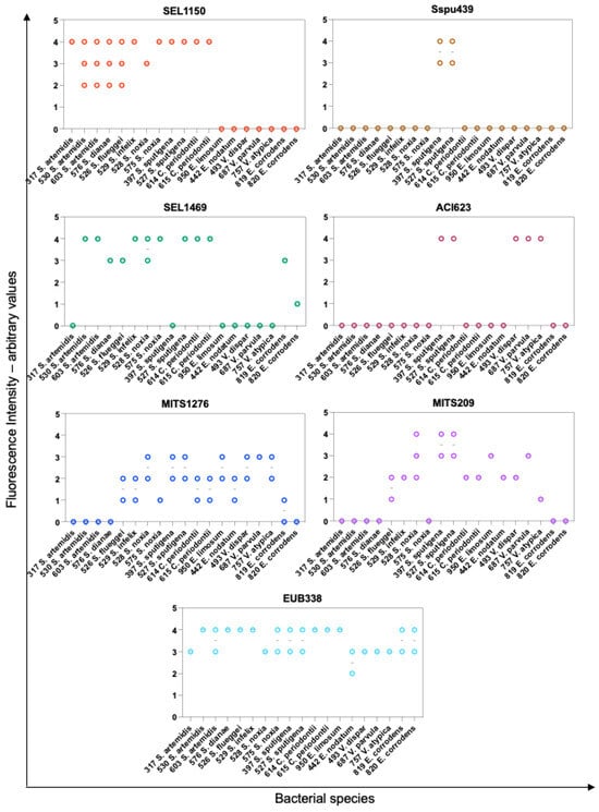
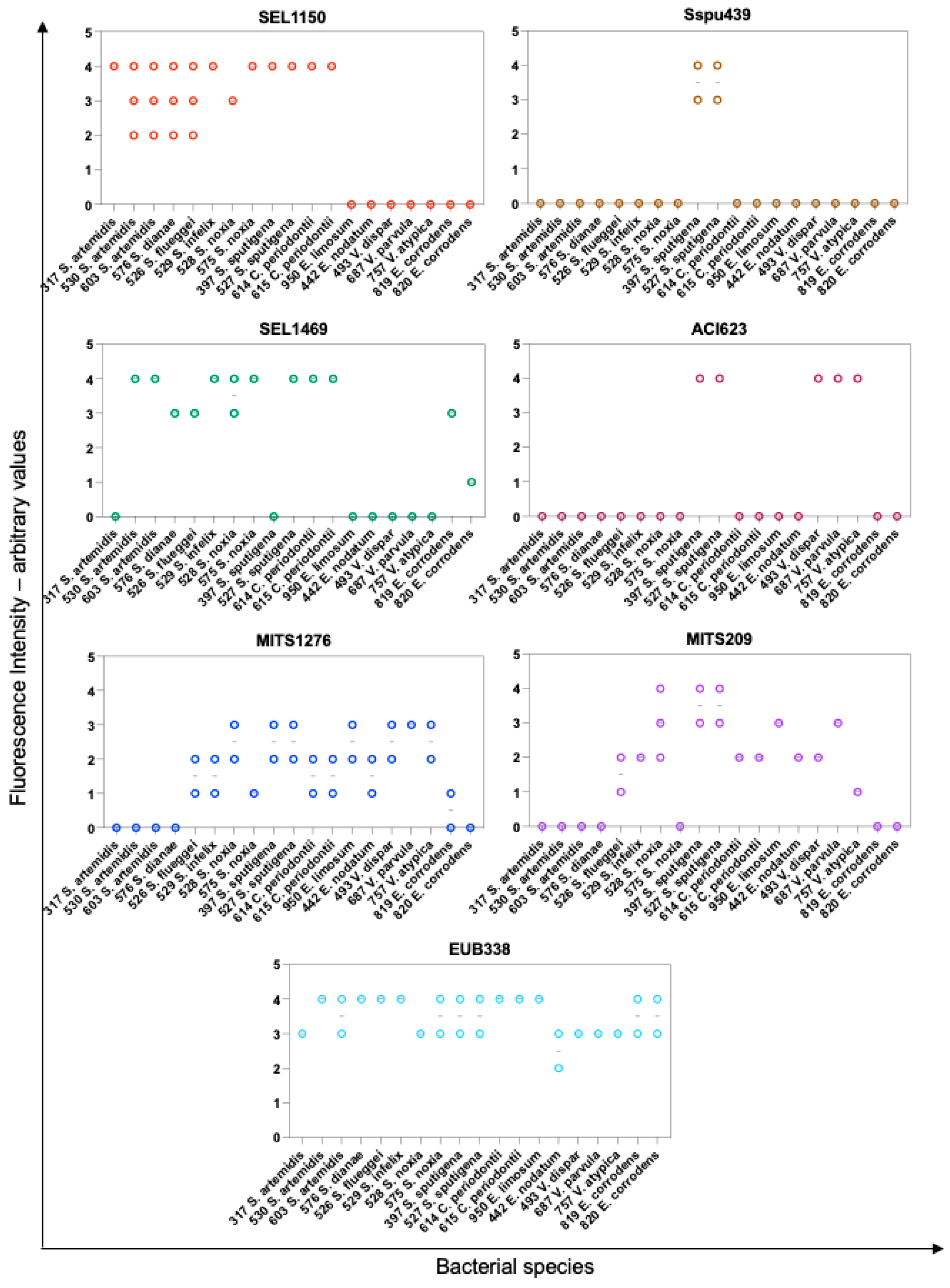
As predicted, using the in silico FISH analysis, a strong specificity was observed for the applied oligonucleotide probes. However, the oligonucleotide probe targeting one of the tested subclusters did not exhibit statistical significance (p > 0.99). The oligonucleotide probe EUB338, which was used as a reference, showed high fluorescence intensities in the range of 3–4 for most of the bacterial species tested. SEL1150 exhibited high fluorescence intensity for the bacterial species in subclusters A, B, and C, namely S. dianae, S. infelix, S. flueggei, C. periodontii, S. artemidis, S. noxia, and S. sputigena. The probe Sspu439 showed a strong fluorescence only for the specific bacterial species S. sputigena in subcluster C, highlighting the high specificity of the probe. Similar results were obtained with the probe SEL1469 (Microsynth AG, Balgach, Switzerland), which exhibited a strong fluorescence in the range of 3–4 for the bacterial species in the subclusters A and B, and for one of the S. sputigena culture. This difference might be due to variations in the expression levels or accessibility of the target molecule recognized by SEL1469 (Microsynth AG, Balgach, Switzerland), between the two strains. Strain-specific differences in cell wall composition, surface structures, or metabolic activity could also play a role. As predicted for the probe ACI623 (Microsynth AG, Balgach, Switzerland), a high specificity was observed for the bacterial species belonging to the subclusters C and F, namely for S. sputigena and various Veillonella spp. Although this probe ACI623 is predicted to detect the additional subclusters G and H, corresponding to the Megasphaera and Dialister spp., we focused mainly on Selenomonas and Veillonella spp. The probes MITS1276 and MITS209 (Microsynth AG, Balgach, Switzerland) were designed to detect the Mitsuokella species, which represent the subcluster E. However, both of the probes react with various bacterial species, not limited to the Mitsuokella species, and present with a wide range of fluorescence intensities, leading to non-conclusive outcomes (Figure 4).
Figure 4.
FISH reaction depicted as fluorescence intensities for the selected bacterial species, probed with various 16S rRNA oligonucleotide probes. For the depicted bacterial species, FISH was performed with SEL1150, Sspu439, SEL1469, ACI623, MITS1276, MITS209 and EUB338 oligonucleotide probes. The fluorescence intensities are depicted as arbitrary values, ranging from 0 to 4, as described in the Materials and Methods. Additional results and further details for the analysis of the fluorescence intensities are described in Appendix A.2. The dots represent results obtained from separate assays.
3.3. FISH Analysis for the Subgingival Dental Plaque Samples Using Specific 16S rRNA Oligonucleotide Probes
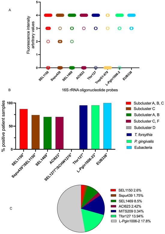
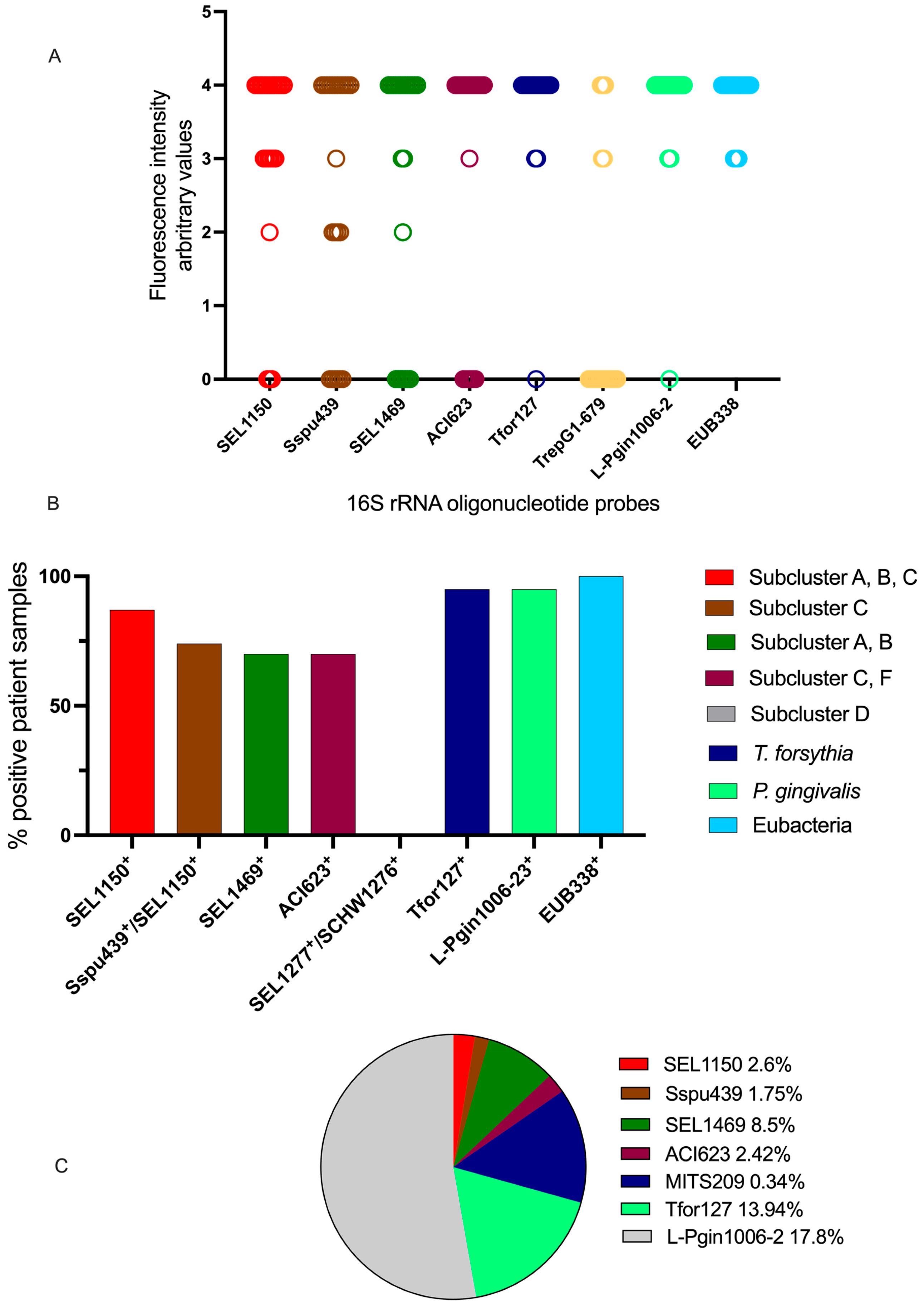
The subgingival dental plaque samples were subjected to FISH and microscopy analysis, using the various oligonucleotide probes that were previously tested on cultured bacterial reference strains. Additionally, oligonucleotide probes were used for the detection of T. forsythia, P gingivalis, and Treponema spp. Representative microscopy images of FISH analysis on the plaque sample from patient Nr. 4 probed with SEL1150-FAM and Sspu439-Cy3 are depicted in Figure 2H–J. As seen in Figure 5A, the majority of the dental plaque samples showed high fluorescence after application of the oligonucleotide probes SEL1150 (p = 0.012), Sspu439 (p < 0.005), ACI623 (p < 0.027), Tfor127 (T. forsythia, p < 0.001), and L-Pgin1006-23 (P. gingivalis, p = 0.035). All samples were highly fluorescent for the universal probe EUB338, which acts as a positive control. The patient samples showed only a weak–medium fluorescence intensity following the application of the probes MITS1276 and MITS209.
Figure 5.
FISH analysis of the bacterial species in patient plaque samples. FISH was performed on patient plaque samples, using the specific 16S rRNA oligonucleotide probes and the fluorescence intensities depicted as arbitrary values (A), ranging from 0 to 4, as described in the Materials and Methods. (B) Prevalence of the subclusters in the studied patient cohort, based on the fluorescence intensities: percentage of FISH-positive patient samples (as shown in (A)), for the indicated oligonucleotide probes and the corresponding subclusters (as depicted in Figure 1). (C) Proportions of bacterial populations in the studied patient cohort with respect to the EUB338 probe: the positively stained bacterial cells in the patient plaque samples for the oligonucleotide probes, which showed a medium–strong positive signal, namely for SEL1150, Sspu439, SEL1469, ACI623, Tfor127, and L-Pgin1006-23, as well as for MITS209 (for which very few patients showed a positive fluorescence signal), were quantified and depicted as a percentage relative to cells that were positive for EUB338. “Other bacteria/probes” represent other bacterial populations, which are present in the oral milieu but were not specifically analyzed as a part of this study. Additional results and further details on the fluorescence intensities are described in Appendix A.2 and Appendix A.3.
The percentage of dental plaque samples that were positive for each oligonucleotide probe is plotted in Figure 5B, wherein the dental plaque samples that were positive for the universal probe EUB338 were set to 100%. The prevalence of the different subclusters and bacterial populations could be identified, namely, subclusters A, B, and C, as well as high populations of T. forsythia and P. gingivalis, predominated in the dental plaque samples from our patient cohort. Interestingly, the probe TrepG1-679 showed positive signals in only 5 of the 19 patient dental plaque samples tested (Figure 5A). The FISH data following the application of the probes MITS1276 and MITS209 (Microsynth AG, Balgach, Switzerland) exhibited positive results for only five patients, with low/moderate fluorescence intensity (arbitrary values 1–2) for MITS1276 and moderate/high fluorescence intensity (arbitrary values 2–3) for MITS209. Thus, these probes resulted in inconclusive results regarding the prevalence of the subcluster E in the patient samples.
Quantification of the positively stained cells was performed and depicted relative to the positive control EUB338 (Figure 5C). This analysis revealed a high prevalence of the two bacterial species, which are highly associated with periodontitis: T. forsythia (13.94%) and P. gingivalis (17.8%). The prevalence of the bacterial populations detected using the four specific oligonucleotide probes is as follows: SEL1150: 2.6%, Sspu439: 1.75%, SEL1469: 8.5%, and ACI623: 2.42%. The cell counts determined after using the probe MITS209 are below 1%, which can again be attributed to the inconclusive specificity of this probe.
4. Discussion
Our study aimed to determine the bacterial diversity of Selenomonas species in the subgingival samples from periodontitis patients, using FISH based on 16S rRNA sequences. The first step was to perform an in silico analysis of the oligonucleotide probes, based on the specificity prediction for the bacterial clusters, as represented in the phylogenetic tree (Figure 1). The phylogeny analysis was carried out based on the 16S rRNA sequence analysis, on the basis of the analysis conducted by Hespell et al. [3,52]. In the analysis by Hespell et al., two branches were described: Selenomonas and Veillonella. Based on advances in the current knowledge, our analysis revealed three branches: Selenomonas, Veillonella, and a third branch comprising Megasphaera and Dialister spp. Moreover, the analysis by Dewhirst et al. defined the bacteria S. infelix, S. dianae, S. flueggi, C. periodontii, S. noxia, and S. artemidis as a very tight cluster with a homology range of 96–99%. Likewise, we found these bacterial species to be closely related. However, in our current in silico analysis, precise differences place the species S. noxia and S. artemidis in the subcluster B. Selenomonas and Veillonella spp. showed an 85% 16S rRNA sequence homology with the Gram-positive bacterial species of Clostridium, Enterococcus, and Bacillus genera [3,53]. Enterococcus faecalis has been associated with the failure of endodontic therapy, owing to the presence of virulence factors such as the production of extracellular superoxide, lytic enzymes, and bacterial surface adhesins, leading to a massive stimulation of inflammation markers [54,55]. In a recent systematic review on bacterial taxa, which persisted after chemo-mechanical root canal treatment, the presence of various pathogenic bacterial species, including Selenomonas spp. and E. faecalis [6] was revealed. Considering the close sequence homology and the very important role of E. faecalis in secondary root canal infections, it would be extremely important and interesting to analyze the molecular mechanisms underlying the role of Selenomonas spp. in cases of endodontic treatment failure.
Considering the wide spectrum of detection that can be achieved using the probe ACI623 (subclusters C, E, F, G, and H, Figure 1), the interpretation of results needs to be performed with caution. In the oral samples, we limited our interpretation to subclusters C and F, since these represent S. sputigena and Veillonella spp., which are part of the microbiota of the oral cavity. Megasphaera spp. usually reside in the gastro urinary tract [56] and have been described as a commensal bacteria coaggregating with Streptococcus and Lactobacillus spp. [57], whereas Dialister spp. has been implicated in gingivitis, periodontitis [58,59], dental root canal infections [60,61], respiratory tract infections [62], and brain abscesses [18,63,64]. However, since we did not analyze pure bacterial reference strains of Megasphaera and Dialister spp. in detail concerning ACI623, we did not comment on the specificity and diversity of these two species in the oral cavity in our patient cohort. ACI623 recognized various subclusters, making it useful to identify multiple species in a single experiment. However, at the same time, it was less sensitive for any individual bacterial cluster. It should be noted that ACI623-positive signals in patient samples may theoretically include Dialister or Megasphaera species and, thus, should be interpreted with caution.
As seen in Figure 2A–C, FISH staining on S. sputigena using the 16S rRNA oligonucleotide probes SEL1150 and Sspu439 highlighted the presence of the characteristic rod-shaped bacterial cells. Similarly, FISH staining of subgingival dental plaque samples likewise detected rod-shaped cells of Selenomonas spp. This is consistent with previous reports on the identification of Selenomonas spp. and their visualization using electron microscopy [8,33,34]. In addition, FISH analysis using probes that are specifically for Veillonella spp. exhibited small clumps of closely adhering bacterial cells, which is consistent with the previously defined morphology of V. parvula, using scanning electron microscopy, as cocci growing in pairs or short chains [51]. Whole-genome deep sequencing (WGS) has been shown to provide detailed insights into the complex microbial communities associated with periodontal disease, as distinct microbiome clusters were identified within periodontitis biofilms [65]. Our results align with the published studies that utilize WGS, highlighting the importance of these advanced sequencing methods in elucidating the specific bacterial populations present in different periodontal conditions. Although we utilized fluorescence in situ hybridization (FISH) for our analysis, which offers complementary insights into microbial community dynamics, WGS remains a powerful tool in characterizing these communities.
The genera Veillonella, Megasphaera, and Dialister belong to the family Veillonellaceae and the order Selenomonadales. Interestingly, this class of Negativicutes contains a Gram-negative cell wall structure; however, it has been grouped in the phylum Firmicutes of Gram-positive bacteria [66]. The Gram-negative S. ruminantium and Megasphaera elsdenii share cell membrane characteristics with the Gram-positive Sporomusa spp., such as the presence of putrescin or cadaverin in the membrane-peptidoglycan [53]. The oligonucleotide probe ACI623 was predicted to detect the subclusters C, E, F, G, and H (Figure 1) and indeed showed a high fluorescence intensity for S. sputigena and V. parvula (Figure 4). Similarly, high fluorescence intensity was observed in the subgingival plaque samples (Figure 5A) and 70% of the patients were positive for subclusters C and F (Figure 5B). Consistent with the hypothesis based on similar membrane and sequence properties that they might have evolved from a common Gram-positive bacterium [53], it explains the close association of S. sputigena in the oral milieu and their importance as periodontopathogenic bacteria, as reflected in our dental plaque samples. As expected, 95% of the dental plaque samples in our study were positive for the presence of T. forsythia and P. gingivalis (Figure 5B), which is consistent with their established involvement in periodontitis [67]. The high specificity of the oligonucleotide probes SEL1150, Sspu439, and SEL 1469 for the Selenomonas spp. can be recognized by the strong fluorescence signals in Figure 5A, with which Selenomonas spp. of the subclusters A, B, and C were found to be present in 70–90% of the patients (Figure 5B). Drescher et al. determined the prevalence of oral Selenomonas spp. by using a single specific oligonucleotide probe with FISH and dot-blot hybridization techniques on subgingival biofilm samples from patients with aggressive periodontitis [36]. Interestingly, the authors found a close spatial organization for Selenomonas spp. and F. nucleatum. Although the oligonucleotide probe used by Drescher et al. detected C. periodontii along with the Selenomonas spp., the authors could not conclude with certainty if the probe detected C. periodontii in the subgingival biofilm samples. Similarly, we could show the specificity, as well as a high fluorescence intensity, obtained with the probes SEL1150 and SEL1469 on our cultures of characterized C. periodontii strains (Figure 2D,E and Figure 4). The hybridization conditions for FISH were tightly controlled and kept consistent in the bacterial cultures and the dental plaque samples, so that conclusive results could be obtained. For quantitative analysis, fluorescent cells with morphology that was specific for Selenomonas and C. periodontii were considered separately. The quantification of the FISH positive cells in the dental plaque samples was performed, and the percentage was determined relative to cells that were positive for the oligonucleotide probe EUB338. The highest proportion was found for P. gingivalis, followed by T. forsythia. The combined percentage of Selenomonas spp., C. periodontii, and Veillonella spp., determined using the four specific probes SEL1150, Sspu439, SEL1469, and ACI623, is depicted in Figure 4. Since the probes detect overlapping subclusters, the exact proportions of each subcluster cannot be determined. Still, it can be concluded that the subgingival dental plaque samples consisted of the abovementioned bacterial species.
The specificity and performance of the utilized probes are crucial for accurate interpretations of microbial community structures in subgingival plaque. The probe ACI623’s ability to recognize multiple genera complicates the definitive conclusions regarding the presence of specific pathogens, necessitating caution in data interpretation. The differences in fluorescence intensities across strains of the same species, such as those detected by probe SEL1469 for S. sputigena, may be related to variations in ribosomal RNA (rRNA) expression, the availability of probe epitopes, or metabolic changes within the bacteria. These factors can significantly influence the efficacy of probe hybridization and, subsequently, the quantitative interpretation of results, highlighting the need for careful consideration when analyzing the data from complex polymicrobial environments. Furthermore, the inconsistent results from probes MITS1276 and MITS209 suggest that their application in future studies should be critically evaluated and potentially omitted to enhance the clarity and reliability of findings related to periodontal disease etiology. In our study, hybridization stringency adjustments using formamide gradients were applied universally across all probes, allowing for optimal hybridization conditions while ensuring specificity. By implementing these adjustments consistently, we aimed to minimize cross-reactivity and enhance the reliability of our findings in polymicrobial biofilms.
Owing to the presence of Selenomonas spp. in periodontitis patients, and in the light of previous case reports on the role of Selenomonas in septicemia, it is important to study the correlation between periodontitis and other systemic diseases in terms of their progression, prognostic aspects, and correlation to the multitude of oral microbial populations. This would be essential to select a patient cohort that might need a very close recall interval, because the elimination of the bacteria and/ or a reduction in the periodontopathogens in these patients could impact their survival. Selenomonas genomosp. refers to unclassified or uncultured members of the genus Selenomonas that have primarily been identified through molecular studies such as 16S rRNA sequencing and metagenomics. However, these members have not yet been formally characterized or named. Due to the limited phenotypic and genomic data available for these phylotypes, as well as the difficulties in distinguishing them from other closely related Selenomonas species, we did not include them in our analysis. Nonetheless, Selenomonas genomosp. may play important ecological roles in various oral environments, including biofilm formation, coaggregation with other bacteria, and potential contributions to oral health and disease. We acknowledge the need for future studies to further elucidate their taxonomy and function, as this will be crucial for understanding their significance within the oral microbiome.
The sensitivity of fluorescence in situ hybridization (FISH) is generally considered to be lower compared to a quantitative polymerase chain reaction (qPCR), which is known for its ability to detect lower abundance levels of target microorganisms more effectively [68]. When certain probes in the study yielded non-significant statistical results (p > 0.99), it could be attributed to their presence falling below the sensitivity threshold of FISH. This illustrates a key limitation of FISH when compared to more sensitive molecular techniques like qPCR that enhance detection accuracy.
Selenomonas was found to be present in high amounts in root canals before endodontic therapy [6], indicating that it might play a role in lesion development and therapy outcome. Additionally, Selenomonas spp. might be relevant to perio–endo combined lesions and possibly patients containing higher levels of Selenomonas bacteria or containing a definite subset of pathogens might have a higher tendency to develop endodontic problems when already suffering from periodontitis. The presence of Selenomonas spp. in systemic infections highlights their potential as opportunistic pathogens, especially in immunocompromised individuals. This underlines the need for further research into their pathogenic mechanisms and systemic effects.
Important information obtained from our cohort of patients is the presence of the subclusters A, B, and C and the presence of P. gingivalis and T. forsythia, confirming the already known periodontopathogens [6]. Although Selenomonas spp. were frequently detected in subgingival plaque, their relative abundance (2.6–8.5%) was lower than that of P. gingivalis (17.8%) and T. forsythia (13.9%), suggesting that their importance may lie in ecological or functional roles, rather than in dominance by biomass.
Interestingly, the patients at the time of sample collection were in the recall phase of periodontitis therapy (6 months after last scaling and root planing). High levels of Selenomonas spp., P. gingivalis, and T. forsythia might be an important reason for the generally high resistance to therapy in certain periodontitis cases, or for the destructive progression of the disease despite a successful initial therapy phase. It is important to analyze whether these periodontopathogens are responsible for an unfavorable disease outcome. Thus, this underlines the importance of analyzing biofilm samples before and during therapy, as well as in the recall phase, and correlates the changes in microbial composition with disease progression. This further reinforces the importance of long-term follow-up of patients with periodontitis, in which the sensitization of patients and regular motivation play a very important role. Additionally, it would be relevant to check the variations in the composition of beneficial microorganisms during therapy [69]. Additionally, the transfer of samples under frozen conditions ensures that any potential degradation of bacterial DNA is minimized, thus supporting the accurate identification and quantification of bacterial populations, including Selenomonas species.
Our primary focus was to investigate the prevalence and spatial distribution of specific Selenomonas subclusters within a defined population of periodontitis patients, which provides valuable insights into the microbial dynamics in diseased states. This approach aligns with similar studies that acknowledged the limitation of not including healthy controls but still highlighted distinct shifts in microbial communities in disease conditions, using relative abundances [7,70]. Research indicates that significant differences in microbial profiles exist between diseased and healthy states, emphasizing the influence of disease on microbiota composition. While we acknowledge the limitations posed by the absence of a healthy control group, our analysis reveals important emerging patterns that warrant further investigation in the context of periodontal disease. Our findings can serve as foundational data for future studies that include healthy individuals for a more comprehensive understanding of the microbial landscape associated with periodontal health. Our study does indeed present further limitations. The samples were collected from periodontal pockets of patients who were in the recall phase of their treatment. An elaborate analysis concerning probing pocket depths and the overall periodontal status of the patients would be important. Additionally, further studies need to incorporate periodontally healthy subjects to confirm the conclusions. Since the majority of oligonucleotide probes for FISH recognize combinations of subclusters, it would be relevant to consider further data verification using whole-genome sequencing. Another limitation of this study is the use of semi-quantitative FISH scoring. Objective image-analysis tools were not applied, but future studies will include software-based quantification for standardized bacterial counts.
5. Conclusions
As predicted, using in silico FISH analysis, a strong specificity for the previously characterized bacterial species was observed with the applied oligonucleotide probes. The majority of the dental plaque samples showed high fluorescence after application of the oligonucleotide probes SEL1150 (subclusters A, B and C), Sspu439 (S. sputigena), SEL 1469 (subclusters A and B), ACI623 (subclusters C and F), Tfor127 (T. forsythia), and L-Pgin1006-23 (P. gingivalis). The prevalence of the bacterial populations detected using the four specific oligonucleotide probes is lower in comparison to that observed for T. forsythia and P. gingivalis. Nevertheless, it can be concluded that Selenomonas spp. constitute an important part of the subgingival microbiome in periodontitis patients and potentially contribute to disease establishment and progression. However, further studies (inclusion of healthy subjects, whole genome sequencing, etc.) are needed for a definite functional significance. Future studies should also aim to clarify the interactions between Selenomonas spp. and the other microbial communities within biofilms. Understanding these relationships could reveal potential synergistic or antagonistic effects that influence the outcomes of periodontal disease.
Author Contributions
M.G. interpreted the data and drafted the manuscript. F.S. contributed to data acquisition and data analysis. L.K. and T.A. contributed to manuscript correction and data interpretation. T.T. conceptualized the study and analyzed the data. All authors have read and agreed to the published version of the manuscript.
Funding
This study was supported by institutional funds of the University of Zurich.
Institutional Review Board Statement
The study was originally conducted in 2012–2013. At that time, verbal informed consent was obtained from all participants prior to sample collection, in accordance with the applicable ethical standards. In 2022, a formal clarification of responsibility regarding the use of these samples was obtained from the Ethics Committee of the University of Zurich (BASEC No. Req-2022-01146, 16 September 2022). All experimental procedures and data handling were performed in compliance with the relevant institutional and national guidelines and regulations.
Informed Consent Statement
The sample collection and initial study were performed before 2013. At that time, there was no necessity to obtain written informed consent from patients in Switzerland. Therefore, verbal consent was obtained from all patients before sample collection. The need to get a written informed consent came into effect on 1 January 2014, under the Human Research Act (https://www.fedlex.admin.ch/eli/cc/2013/617/en, accessed on 11 July 2025).
Data Availability Statement
The datasets used and/or analyzed during the current study are available from the corresponding author upon reasonable request.
Acknowledgments
The authors are grateful to Rudolf Gmür, who designed the study, Figure 1, and all new rRNA FISH probes described in Appendix A.1. The authors thank Helga Lüthi-Schaller for excellent experimental support regarding FISH staining and analysis. The authors thank Steve Reese for excellent technical support with scanning electron microscopy.
Conflicts of Interest
The authors declare that they have no competing interests.
Appendix A
Appendix A.1
Table A1.
The 16S rRNA oligonucleotide probes used for FISH analysis and the recognized bacterial species.
Appendix A.2
Table A2.
Arbitrary fluorescence values 1–4 after FISH on the characterized bacterial species, 16S rRNA oligonucleotide probes and hybridization conditions.
Appendix A.3
Table A3.
Arbitrary fluorescence intensity after FISH on dental plaque samples. The patient sample, 16S rRNA probe, and the hybridization conditions are mentioned.
References
- Moore, L.V.H.; Johnson, J.L.; Moore, W.E.C. Selenomonas noxia sp. nov., Selenomonas flueggei sp. nov., Selenomonas infelix sp. nov., Selenomonas dianae sp. nov., and Selenomonas artemidis sp. nov., from the Human Gingival Crevice. Int. J. Syst. Evol. Microbiol. 1987, 37, 271–280. [Google Scholar] [CrossRef] [Scilit]
- Lessel, E.F., Jr.; Breed, R.S. Selenomonas Boskamp, 1922; a genus that includes species showing an unusual type of flagellation. Bacteriol. Rev. 1954, 18, 165–168. [Google Scholar] [CrossRef]
- Hespell, R.B.; Paster, B.J.; Dewhirst, F.E. The Genus Selenomonas. In The Prokaryotes: Volume 4: Bacteria: Firmicutes, Cyanobacteria; Dworkin, M., Falkow, S., Rosenberg, E., Schleifer, K.-H., Stackebrandt, E., Eds.; Springer: New York, NY, USA, 2006; pp. 982–990. [Google Scholar] [CrossRef] [Scilit]
- Moore, L.V.; Moore, W.E.; Cato, E.P.; Smibert, R.M.; Burmeister, J.A.; Best, A.M.; Ranney, R.R. Bacteriology of human gingivitis. J. Dent. Res. 1987, 66, 989–995. [Google Scholar] [CrossRef] [Scilit]
- Tanner, A.; Bouldin, H.D.; Maiden, M.F. Newly delineated periodontal pathogens with special reference to selenomonas species. Infection 1989, 17, 182–187. [Google Scholar] [CrossRef] [Scilit]
- Nardello, L.C.L.; Pinheiro, E.T.; Gavini, G.; Prado, L.C.; Romero, R.X.; Gomes, B.; Skelton-Macedo, M.C. Nature and Prevalence of Bacterial Taxa Persisting after Root Canal Chemomechanical Preparation in Permanent Teeth: A Systematic Review and Meta-analysis. J. Endod. 2022, 48, 572–596. [Google Scholar] [CrossRef] [Scilit]
- Cho, H.; Ren, Z.; Divaris, K.; Roach, J.; Lin, B.M.; Liu, C.; Azcarate-Peril, M.A.; Simancas-Pallares, M.A.; Shrestha, P.; Orlenko, A.; et al. Selenomonas sputigena acts as a pathobiont mediating spatial structure and biofilm virulence in early childhood caries. Nat. Commun. 2023, 14, 2919. [Google Scholar] [CrossRef] [Scilit]
- Flügge, C. Die Mikroorganismen; Illus: Leipzig, Germany, 1886; p. 692. [Google Scholar]
- Johnson, J.L.; Holdeman, L.V.; Moore, W.E.C. Replacement of the Type Strain of Selenomonas sputigena under Rule 18: Request for an Opinion. Int. Hournal Syst. Evol. Microbiol. 1985, 35. [Google Scholar] [CrossRef] [Scilit]
- Kingsley, V.V.; Hoeniger, J.F. Growth, structure, and classification of Selenomonas. Bacteriol. Rev. 1973, 37, 479–521. [Google Scholar] [CrossRef] [PubMed]
- Collection, A.T.C. Catalogue of Strains I; American Type Culture Collection: Manassas, VA, USA, 1980; Volume 14. [Google Scholar]
- Hannig, M. Ultrastructural investigation of pellicle morphogenesis at two different intraoral sites during a 24-h period. Clin. Oral Investig. 1999, 3, 88–95. [Google Scholar] [CrossRef] [Scilit] [PubMed]
- Mihai, M.M.; Holban, A.; Giurcăneanu, C.; Popa, L.; Oanea, R.; Lazăr, V.; Chifiriuc, M.; Popa, M.; Popa, M.I. Microbial Biofilms: Impact On Pathogenesis Of Periodontitis, Cystic Fibrosis, Chronic Wounds And Medical Device-Related Infections. Curr. Top. Med. Chem. 2015, 15, 1552–1576. [Google Scholar] [CrossRef] [Scilit]
- Gao, L.; Xu, T.; Huang, G.; Jiang, S.; Gu, Y.; Chen, F. Oral microbiomes: More and more importance in oral cavity and whole body. Protein Cell 2018, 9, 488–500. [Google Scholar] [CrossRef] [Scilit]
- Sedghi, L.; DiMassa, V.; Harrington, A.; Lynch, S.V.; Kapila, Y.L. The oral microbiome: Role of key organisms and complex networks in oral health and disease. Periodontology 2000 2021, 87, 107–131. [Google Scholar] [CrossRef] [Scilit] [PubMed]
- Savitt, E.D.; Socransky, S.S. Distribution of certain subgingival microbial species in selected periodontal conditions. J. Periodontal Res. 1984, 19, 111–123. [Google Scholar] [CrossRef] [Scilit]
- Kunath, B.J.; De Rudder, C.; Laczny, C.C.; Letellier, E.; Wilmes, P. The oral-gut microbiome axis in health and disease. Nat. Rev. Microbiol. 2024, 22, 791–805. [Google Scholar] [CrossRef] [Scilit]
- Paster, B.J.; Boches, S.K.; Galvin, J.L.; Ericson, R.E.; Lau, C.N.; Levanos, V.A.; Sahasrabudhe, A.; Dewhirst, F.E. Bacterial diversity in human subgingival plaque. J. Bacteriol. 2001, 183, 3770–3783. [Google Scholar] [CrossRef] [Scilit]
- Popova, C.; Dosseva-Panova, V.; Panov, V. Microbiology of Periodontal Diseases. A Review. Biotechnol. Biotechnol. Equip. 2013, 27, 3754–3759. [Google Scholar] [CrossRef] [Scilit]
- Baker, J.L.; Mark Welch, J.L.; Kauffman, K.M.; McLean, J.S.; He, X. The oral microbiome: Diversity, biogeography and human health. Nat. Rev. Microbiol. 2024, 22, 89–104. [Google Scholar] [CrossRef] [Scilit] [PubMed]
- Gonçalves, L.F.; Fermiano, D.; Feres, M.; Figueiredo, L.C.; Teles, F.R.; Mayer, M.P.; Faveri, M. Levels of Selenomonas species in generalized aggressive periodontitis. J. Periodontal Res. 2012, 47, 711–718. [Google Scholar] [CrossRef] [Scilit]
- Kodrat, N.R.S.; Sunarto, H.; Soeroso, Y.; Bachtiar, B.M. Quantitative Analysis of Selenomonas noxia in Periodontitis Patients via Two Different Sample Collection Methods. J. Int. Dent. Med. Res. 2020, 13, 1320–1325. [Google Scholar]
- McDaniel, J.; McDaniel, S.; Samiano, B.J.; Marrujo, M.; Kingsley, K.; Howard, K.M. Microbial Screening Reveals Oral Site-Specific Locations of the Periodontal Pathogen Selenomonas noxia. Curr. Issues Mol. Biol. 2021, 43, 353–364. [Google Scholar] [CrossRef] [Scilit]
- Chen, W.P.; Chang, S.H.; Tang, C.Y.; Liou, M.L.; Tsai, S.J.; Lin, Y.L. Composition Analysis and Feature Selection of the Oral Microbiota Associated with Periodontal Disease. Biomed. Res. Int. 2018, 2018, 3130607. [Google Scholar] [CrossRef] [Scilit]
- Shi, M.; Wei, Y.; Hu, W.; Nie, Y.; Wu, X.; Lu, R. The Subgingival Microbiome of Periodontal Pockets With Different Probing Depths in Chronic and Aggressive Periodontitis: A Pilot Study. Front. Cell Infect. Microbiol. 2018, 8, 124. [Google Scholar] [CrossRef] [Scilit]
- Califf, K.J.; Schwarzberg-Lipson, K.; Garg, N.; Gibbons, S.M.; Caporaso, J.G.; Slots, J.; Cohen, C.; Dorrestein, P.C.; Kelley, S.T. Multi-omics Analysis of Periodontal Pocket Microbial Communities Pre- and Posttreatment. mSystems 2017, 2, e00016-17. [Google Scholar] [CrossRef] [Scilit]
- Genco, R.J.; Borgnakke, W.S. Diabetes as a potential risk for periodontitis: Association studies. Periodontology 2000 2020, 83, 40–45. [Google Scholar] [CrossRef] [Scilit] [PubMed]
- Lalla, E.; Papapanou, P.N. Diabetes mellitus and periodontitis: A tale of two common interrelated diseases. Nat. Rev. Endocrinol. 2011, 7, 738–748. [Google Scholar] [CrossRef] [Scilit]
- Luong, A.; Tawfik, A.N.; Islamoglu, H.; Gobriel, H.S.; Ali, N.; Ansari, P.; Shah, R.; Hung, T.; Patel, T.; Henson, B.; et al. Periodontitis and diabetes mellitus co-morbidity: A molecular dialogue. J. Oral Biosci. 2021, 63, 360–369. [Google Scholar] [CrossRef] [Scilit] [PubMed]
- Bui, F.Q.; Almeida-da-Silva, C.L.C.; Huynh, B.; Trinh, A.; Liu, J.; Woodward, J.; Asadi, H.; Ojcius, D.M. Association between periodontal pathogens and systemic disease. Biomed. J. 2019, 42, 27–35. [Google Scholar] [CrossRef] [Scilit] [PubMed]
- McCarthy, L.R.; Carlson, J.R. Selenomonas sputigena septicemia. J. Clin. Microbiol. 1981, 14, 684–685. [Google Scholar] [CrossRef] [Scilit]
- Pinon, G.; Grollier, G.; Romet-Lemonne, J.L.; de Rautlin de la Roy, Y. Fatal Selenomonas sputigena septicemia probably originating from lung abscess. Eur. J. Clin. Microbiol. 1985, 4, 343–344. [Google Scholar] [CrossRef] [Scilit]
- Bisiaux-Salauze, B.; Perez, C.; Sebald, M.; Petit, J.C. Bacteremias caused by Selenomonas artemidis and Selenomonas infelix. J. Clin. Microbiol. 1990, 28, 140–142. [Google Scholar] [CrossRef] [Scilit]
- Pomeroy, C.; Shanholtzer, C.J.; Peterson, L.R. Selenomonas bacteraemia--case report and review of the literature. J. Infect. 1987, 15, 237–242. [Google Scholar] [CrossRef] [Scilit]
- Liang, T.; Liu, F.; Liu, L.; Zhang, Z.; Dong, W.; Bai, S.; Ma, L.; Kang, L. Effects of Helicobacter pylori Infection on the Oral Microbiota of Reflux Esophagitis Patients. Front. Cell Infect. Microbiol. 2021, 11, 732613. [Google Scholar] [CrossRef] [Scilit] [PubMed]
- Drescher, J.; Schlafer, S.; Schaudinn, C.; Riep, B.; Neumann, K.; Friedmann, A.; Petrich, A.; Göbel, U.B.; Moter, A. Molecular epidemiology and spatial distribution of Selenomonas spp. in subgingival biofilms. Eur. J. Oral. Sci. 2010, 118, 466–474. [Google Scholar] [CrossRef] [Scilit]
- Gescher, D.M.; Kovacevic, D.; Schmiedel, D.; Siemoneit, S.; Mallmann, C.; Halle, E.; Göbel, U.B.; Moter, A. Fluorescence in situ hybridisation (FISH) accelerates identification of Gram-positive cocci in positive blood cultures. Int. J. Antimicrob. Agents 2008, 32, S51–S59. [Google Scholar] [CrossRef] [Scilit]
- Zhu, Y.; Wang, Y.; Yan, Y.; Xue, H. Rapid and Sensitive Quantification of Anammox Bacteria by Flow Cytometric Analysis Based on Catalyzed Reporter Deposition Fluorescence In Situ Hybridization. Environ. Sci. Technol. 2019, 53, 6895–6905. [Google Scholar] [CrossRef] [Scilit]
- Dechesne, A.; Musovic, S.; Palomo, A.; Diwan, V.; Smets, B.F. Underestimation of ammonia-oxidizing bacteria abundance by amplification bias in amoA-targeted qPCR. Microb. Biotechnol. 2016, 9, 519–524. [Google Scholar] [CrossRef] [Scilit]
- Baptista, J.D.; Lunn, M.; Davenport, R.J.; Swan, D.L.; Read, L.F.; Brown, M.R.; Morais, C.; Curtis, T.P. Agreement between amoA gene-specific quantitative PCR and fluorescence in situ hybridization in the measurement of ammonia-oxidizing bacteria in activated sludge. Appl. Environ. Microbiol. 2014, 80, 5901–5910. [Google Scholar] [CrossRef] [Scilit]
- Romanos, G.E.; Brink, B. Photodynamic therapy in periodontal therapy: Microbiological observations from a private practice. Gen. Dent. 2010, 58, e68–e73. [Google Scholar] [PubMed]
- Ludwig, W.; Strunk, O.; Westram, R.; Richter, L.; Meier, H.; Yadhukumar; Buchner, A.; Lai, T.; Steppi, S.; Jobb, G.; et al. ARB: A software environment for sequence data. Nucleic Acids Res. 2004, 32, 1363–1371. [Google Scholar] [CrossRef] [Scilit] [PubMed]
- Zijnge, V.; van Leeuwen, M.B.; Degener, J.E.; Abbas, F.; Thurnheer, T.; Gmür, R.; Harmsen, H.J. Oral biofilm architecture on natural teeth. PLoS ONE 2010, 5, e9321. [Google Scholar] [CrossRef] [Scilit]
- Züger, J.; Lüthi-Schaller, H.; Gmür, R. Uncultivated Tannerella BU045 and BU063 are slim segmented filamentous rods of high prevalence but low abundance in inflammatory disease-associated dental plaques. Microbiol. Read. 2007, 153, 3809–3816. [Google Scholar] [CrossRef] [Scilit]
- Gmür, R.; Lüthi-Schaller, H. A combined immunofluorescence and fluorescent in situ hybridization assay for single cell analyses of dental plaque microorganisms. J. Microbiol. Methods 2007, 69, 402–405. [Google Scholar] [CrossRef] [Scilit] [PubMed]
- Guggenheim, B.; Gmür, R.; Galicia, J.C.; Stathopoulou, P.G.; Benakanakere, M.R.; Meier, A.; Thurnheer, T.; Kinane, D.F. In vitro modeling of host-parasite interactions: The ‘subgingival’ biofilm challenge of primary human epithelial cells. BMC Microbiol. 2009, 9, 280. [Google Scholar] [CrossRef] [Scilit]
- Amann, R.I.; Krumholz, L.; Stahl, D.A. Fluorescent-oligonucleotide probing of whole cells for determinative, phylogenetic, and environmental studies in microbiology. J. Bacteriol. 1990, 172, 762–770. [Google Scholar] [CrossRef] [Scilit]
- Gmür, R.; Guggenheim, B. Antigenic heterogeneity of Bacteroides intermedius as recognized by monoclonal antibodies. Infect. Immun. 1983, 42, 459–470. [Google Scholar] [CrossRef] [Scilit]
- Thurnheer, T.; Gmür, R.; Guggenheim, B. Multiplex FISH analysis of a six-species bacterial biofilm. J. Microbiol. Methods 2004, 56, 37–47. [Google Scholar] [CrossRef] [Scilit]
- Lai, C.H.; Males, M.B.; Dougherty, P.A.; Berthold, P.; Listgarten, M.A. Centipeda periodontii gen. nov., sp. nov. from Human Periodontal Lesions. Int. J. Syst. Evol. Microbiol. 1983, 33, 628–635. [Google Scholar] [CrossRef] [Scilit]
- Gronow, S.; Welnitz, S.; Lapidus, A.; Nolan, M.; Ivanova, N.; Glavina Del Rio, T.; Copeland, A.; Chen, F.; Tice, H.; Pitluck, S.; et al. Complete genome sequence of Veillonella parvula type strain (Te3). Stand. Genom. Sci. 2010, 2, 57–65. [Google Scholar] [CrossRef] [Scilit] [PubMed]
- Dewhirst, F.E.; Paster, B.J.; Bright, P.L.; Pelletier, D.A.; Edwards, J.B.; Ago, J.M. Selenomonas, Centipeda and Veillonella: 16S rRNA sequences, probes and phylogeny. J. Dent. Res. 1989, 68, 255–261. [Google Scholar]
- Stackebrandt, E.; Pohla, H.; Kroppenstedt, R.; Hippe, H.; Woese, C.R. 16S rRNA analysis of Sporomusa, selenomonas, and Megasphaera: On the phylogenetic origin of Gram-positive Eubacteria. Arch. Microbiol. 1985, 143, 270–276. [Google Scholar] [CrossRef] [Scilit]
- Kayaoglu, G.; Ørstavik, D. Virulence factors of Enterococcus faecalis: Relationship to endodontic disease. Crit. Rev. Oral. Biol. Med. 2004, 15, 308–320. [Google Scholar] [CrossRef] [Scilit] [PubMed]
- Alghamdi, F.; Shakir, M. The Influence of Enterococcus faecalis as a Dental Root Canal Pathogen on Endodontic Treatment: A Systematic Review. Cureus 2020, 12, e7257. [Google Scholar] [CrossRef] [Scilit]
- Cobo, F. Infections Caused by Anaerobic Microorganisms. In Encyclopedia of Infection and Immunity; Rezaei, N., Ed.; Elsevier: Oxford, UK, 2022; pp. 614–627. [Google Scholar] [CrossRef] [Scilit]
- Nallabelli, N.; Patil, P.P.; Pal, V.K.; Singh, N.; Jain, A.; Patil, P.B.; Grover, V.; Korpole, S. Biochemical and genome sequence analyses of Megasphaera sp. strain DISK18 from dental plaque of a healthy individual reveals commensal lifestyle. Sci. Rep. 2016, 6, 33665. [Google Scholar] [CrossRef] [Scilit] [PubMed]
- Doan, N.; Contreras, A.; Flynn, J.; Slots, J.; Chen, C. Molecular identification of Dialister pneumosintes in subgingival plaque of humans. J. Clin. Microbiol. 2000, 38, 3043–3047. [Google Scholar] [CrossRef] [Scilit]
- Contreras, A.; Doan, N.; Chen, C.; Rusitanonta, T.; Flynn, M.J.; Slots, J. Importance of Dialister pneumosintes in human periodontitis. Oral Microbiol. Immunol. 2000, 15, 269–272. [Google Scholar] [CrossRef] [Scilit]
- Rôças, I.N.; Siqueira, J.F., Jr. Characterization of Dialister species in infected root canals. J. Endod. 2006, 32, 1057–1061. [Google Scholar] [CrossRef] [Scilit] [PubMed]
- Rolph, H.J.; Lennon, A.; Riggio, M.P.; Saunders, W.P.; MacKenzie, D.; Coldero, L.; Bagg, J. Molecular identification of microorganisms from endodontic infections. J. Clin. Microbiol. 2001, 39, 3282–3289. [Google Scholar] [CrossRef] [Scilit]
- Goldstein, E.J.; Citron, D.M.; Finegold, S.M. Role of anaerobic bacteria in bite-wound infections. Rev. Infect. Dis. 1984, 6, S177–S183. [Google Scholar] [CrossRef] [Scilit]
- Morio, F.; Jean-Pierre, H.; Dubreuil, L.; Jumas-Bilak, E.; Calvet, L.; Mercier, G.; Devine, R.; Marchandin, H. Antimicrobial susceptibilities and clinical sources of Dialister species. Antimicrob. Agents Chemother. 2007, 51, 4498–4501. [Google Scholar] [CrossRef] [Scilit]
- Rousée, J.M.; Bermond, D.; Piémont, Y.; Tournoud, C.; Heller, R.; Kehrli, P.; Harlay, M.L.; Monteil, H.; Jaulhac, B. Dialister pneumosintes associated with human brain abscesses. J. Clin. Microbiol. 2002, 40, 3871–3873. [Google Scholar] [CrossRef] [Scilit]
- Wirth, R.; Pap, B.; Maróti, G.; Vályi, P.; Komlósi, L.; Barta, N.; Strang, O.; Minárovits, J.; Kovács, K.L. Toward Personalized Oral Diagnosis: Distinct Microbiome Clusters in Periodontitis Biofilms. Front. Cell. Infect. Microbiol. 2021, 11, 747814. [Google Scholar] [CrossRef] [Scilit] [PubMed]
- Marchandin, H.; Jumas-Bilak, E. The Family Veillonellaceae. In The Prokaryotes: Firmicutes and Tenericutes; Rosenberg, E., DeLong, E.F., Lory, S., Stackebrandt, E., Thompson, F., Eds.; Springer: Berlin/Heidelberg, Germany, 2014; pp. 433–453. [Google Scholar] [CrossRef] [Scilit]
- Gambin, D.J.; Vitali, F.C.; De Carli, J.P.; Mazzon, R.R.; Gomes, B.; Duque, T.M.; Trentin, M.S. Prevalence of red and orange microbial complexes in endodontic-periodontal lesions: A systematic review and meta-analysis. Clin. Oral Investig. 2021, 25, 6533–6546. [Google Scholar] [CrossRef] [Scilit] [PubMed]
- Biedermann, K.; Dandachi, N.; Trattner, M.; Vogl, G.; Doppelmayr, H.; Moré, E.; Staudach, A.; Dietze, O.; Hauser-Kronberger, C. Comparison of real-time PCR signal-amplified in situ hybridization and conventional PCR for detection and quantification of human papillomavirus in archival cervical cancer tissue. J. Clin. Microbiol. 2004, 42, 3758–3765. [Google Scholar] [CrossRef] [Scilit]
- Bhuyan, R.; Bhuyan, S.K.; Mohanty, J.N.; Das, S.; Juliana, N.; Juliana, I.F. Periodontitis and Its Inflammatory Changes Linked to Various Systemic Diseases: A Review of Its Underlying Mechanisms. Biomedicines 2022, 10, 2659. [Google Scholar] [CrossRef] [Scilit]
- Miranda, B.P.; da Silva, A.F.; Ascenção, J.C.; Santos, H.L.C. An overview of the microbiota of the oral cavity of humans and non-human primates with periodontal disease: Current issues and perspectives. Arch. Oral Biol. 2025, 172, 106121. [Google Scholar] [CrossRef] [Scilit]
- Gmür, R.; Wyss, C.; Xue, Y.; Thurnheer, T.; Guggenheim, B. Gingival crevice microbiota from Chinese patients with gingivitis or necrotizing ulcerative gingivitis. Eur. J. Oral Sci. 2004, 112, 33–41. [Google Scholar] [CrossRef] [Scilit] [PubMed]
- Kubota, K.; Ohashi, A.; Imachi, H.; Harada, H. Improved in situ hybridization efficiency with locked-nucleic-acid-incorporated DNA probes. Appl. Environ. Microbiol. 2006, 72, 5311–5317. [Google Scholar] [CrossRef] [Scilit]
- Fuchs, B.M.; Glöckner, F.O.; Wulf, J.; Amann, R. Unlabeled helper oligonucleotides increase the in situ accessibility to 16S rRNA of fluorescently labeled oligonucleotide probes. Appl. Environ. Microbiol. 2000, 66, 3603–3607. [Google Scholar] [CrossRef] [Scilit]
Disclaimer/Publisher’s Note: The statements, opinions and data contained in all publications are solely those of the individual author(s) and contributor(s) and not of MDPI and/or the editor(s). MDPI and/or the editor(s) disclaim responsibility for any injury to people or property resulting from any ideas, methods, instructions or products referred to in the content. |
© 2026 by the authors. Licensee MDPI, Basel, Switzerland. This article is an open access article distributed under the terms and conditions of the Creative Commons Attribution (CC BY) license.