


Abstract
Rapid and reliable molecular detection of Salmonella is important for laboratory investigation and infectious disease surveillance. This study compared six candidate molecular targets (hilA, invA, phoP, fimA, rpoS, and spvA) and developed a hilA-targeting TaqMan real-time PCR assay for Salmonella enterica detection. During conventional PCR screening, the hilA-M1-F/R primer pair demonstrated the most consistent and reproducible amplification across the tested isolate panel under the experimental conditions used and was therefore selected for subsequent assay development. The developed assay showed an amplification efficiency of 93.4% and an R2 of 0.9988 over a linear genomic DNA range from 1 ng/μL to 10 fg/μL. Experimentally determined detection limits under the conditions tested were 102 CFU/mL for bacterial suspensions and 10 fg/μL for genomic DNA. All 24 whole-genome sequencing-confirmed S. enterica isolates representing multiple serovars and sequence types were reproducibly detected, whereas no specific amplification was observed within the expanded panel of 15 non-target bacterial strains or isolates. The assay also detected S. enterica DNA in artificially contaminated milk and carrot matrices in preliminary proof-of-concept experiments. These findings demonstrate promising analytical performance of the hilA-targeting TaqMan assay within the evaluated experimental framework. However, broader validation using geographically diverse S. enterica isolate collections, additional non-target organisms, internal amplification controls, naturally contaminated food products, primary clinical, veterinary, and environmental specimens, and independent laboratories is required before routine diagnostic, food-testing, or epidemiological surveillance applications can be established.
1. Introduction
Salmonellosis remains an important public health problem worldwide and is one of the major bacterial foodborne infections in terms of prevalence and public health impact [1,2,3]. Non-typhoidal Salmonella infections account for a substantial global burden of morbidity and mortality [4,5].
Members of the genus Salmonella are characterized by considerable pathogenic and serological diversity. According to the Kauffmann–White–Le Minor classification, more than 2500 Salmonella serovars have been described, with S. enterica serovars Enteritidis and Typhimurium among the most epidemiologically important [6]. In addition to these predominant serovars, less common serovars are increasingly being identified in sporadic cases and foodborne outbreaks [7]. The emergence and spread of antimicrobial-resistant and multidrug-resistant Salmonella strains further complicate infection control and represent an additional public health concern [8].
In Kazakhstan, salmonellosis continues to represent a relevant epidemiological problem, with S. enterica serovars Enteritidis and Typhimurium remaining among the predominant circulating serovars [9,10,11]. At the same time, cases of acute intestinal infection without an identified etiological agent remain an important diagnostic challenge, particularly among children, emphasizing the need for rapid and reliable laboratory detection methods [12].
Conventional bacteriological culture remains a reference method for the detection and identification of Salmonella but has several limitations, including a relatively long turnaround time, the need to obtain pure cultures followed by biochemical and serological identification, and substantial laboratory requirements [13]. Molecular diagnostic approaches, particularly real-time polymerase chain reaction (real-time PCR), can complement conventional methods by providing rapid, sensitive, and specific detection of bacterial pathogens [13,14].
Several genetic targets have been proposed for the molecular detection of Salmonella, including invA, hilA, phoP, fimA, rpoS, and spvA. The invA gene is among the most widely used targets for PCR-based identification of Salmonella. However, variation in the distribution or detectability of individual molecular markers among genetically diverse isolates may affect the performance of assays based on a single target [15,16]. Therefore, selecting a suitable molecular target capable of detecting a broad range of epidemiologically relevant serovars remains important for the development of reliable diagnostic assays.
The hilA gene represents a promising candidate for this purpose. Located within Salmonella pathogenicity island 1 (SPI-1), hilA encodes a transcriptional regulator involved in controlling the expression of genes required for epithelial cell invasion. Its chromosomal localization, functional importance, and previously reported occurrence across diverse Salmonella serovars support its consideration as a molecular target for PCR-based detection [17,18]. Previous studies have also demonstrated the potential diagnostic utility of hilA-based PCR approaches for detecting Salmonella in clinical and food-related applications.
Although hilA has previously been used as a molecular target for Salmonella detection, to the best of our knowledge, this is the first study in Kazakhstan to comparatively evaluate multiple molecular targets and subsequently develop a hilA-targeting TaqMan real-time PCR assay based on the results of this screening. Six candidate targets (hilA, invA, phoP, fimA, rpoS, and spvA) were evaluated across a serologically diverse panel of Salmonella isolates, including both common and less frequently encountered serovars. This comparative approach enabled the selection of the primer–target combination showing the most consistent amplification within the tested isolate panel and provided the basis for subsequent assay development and analytical evaluation.
Therefore, this study aimed to compare six candidate molecular targets for S. enterica detection, identify the primer–target combination showing the most consistent amplification within the tested isolate panel, and develop and analytically evaluate a TaqMan real-time PCR assay for the detection of S. enterica.
2. Materials and Methods
2.1. Bacterial Isolates and Study Materials
All procedures performed in this study complied with applicable international and national ethical requirements for biomedical research and were approved by the Local Bioethics Committee of the M. Aikimbayev National Scientific Center for Especially Dangerous Infections (Protocol No. 2, dated 12 February 2024).
Bacterial isolates used in this study were selected from the National Collection of Microorganisms (NCM) maintained at the M. Aikimbayev National Scientific Center for Especially Dangerous Infections (NSCEDI), Almaty, Kazakhstan. The collection included isolates of human and animal origin, as well as isolates of unrecorded origin. Human-derived isolates had originally been obtained during routine clinical diagnostic and infectious disease surveillance activities and were subsequently transferred to the NCM for long-term preservation. No isolates were collected specifically for the purposes of the present study. All bacterial strains and associated microbiological records were retrieved from the NCM on 9 February 2026 for retrospective laboratory investigation. Before inclusion in the study, all patient-related information had been de-identified, and analyses were performed exclusively using anonymized materials.
The isolates originated from different years and geographic locations and represented multiple Salmonella enterica serovars, including less common serovars. For bacteriological examination, archived isolates were recovered on Endo and Ploskirev selective and differential media and incubated aerobically at 37 °C for 18–24 h. These media were used for recovery, purity assessment, and conventional phenotypic characterization of the archived bacterial cultures and were not intended for primary isolation of Salmonella from clinical or food specimens. Preliminary identification was based on colony morphology and conventional biochemical characteristics, followed by molecular confirmation as described below. Serological identification was performed by slide agglutination using polyvalent adsorbed O-antiserum against Salmonella serogroups A–E, followed by serovar determination using specific O and H antisera according to the Kauffmann–White–Le Minor scheme [19].
Serovar confirmation, whole-genome sequencing (WGS), and multilocus sequence typing (MLST) of the selected isolates were performed as part of a parallel study reported separately [20]. Information on isolation source, geographic origin, serovar, and sequence type is summarized in Table 1. One isolate (18S), initially assigned to the genus Salmonella based on conventional cultural and biochemical characteristics, was subsequently identified as Proteus mirabilis by WGS and was therefore retained as a non-Salmonella comparator in the analytical evaluation of the assay.
Table 1.
Characteristics of the strains used in this study and the results of their serovar and genetic typing.
Analytical specificity was additionally evaluated using DNA from an expanded panel of 15 non-target strains obtained from the National Collection of Microorganisms (NCM) maintained at the M. Aikimbayev National Scientific Center for Especially Dangerous Infections (NSCEDI), Almaty, Kazakhstan. The panel comprised Shigella flexneri IIa strain 16 (human, Almaty), Shigella sonnei strain 14 (human, Almaty), Yersinia pseudotuberculosis strain 2841 (reference strain), Yersinia pestis strain NP6 (great gerbil, Aral region), Y. enterocolitica strain 5 (human, Almaty), Y. kristensenii strain 148 (human, Almaty), Bacillus anthracis strain 23 (vaccine strain), Vibrio cholerae O1 strain ch166 (human, Almaty), Francisella tularensis strain FT24 (rabbit, Zhambyl region), Escherichia coli strain 75 (human, Almaty), Klebsiella aerogenes strain 11, K. pneumoniae strain 15, Listeria monocytogenes strain 4, V. cholerae non-O1 strain 245 (water, Shymkent region), and Proteus mirabilis strain 18S (human, Almaty). Where historical provenance information was unavailable in the NCM records, it is indicated as not recorded. Internal NCM accession/deposit identifiers are not disclosed because they constitute restricted collection information.
2.2. DNA Extraction
Genomic DNA was extracted from bacterial cell suspensions and artificially contaminated food samples. Overnight bacterial cultures were inactivated by heating at 100 °C for 30 min prior to DNA extraction.
For artificial contamination experiments, milk and carrot matrices were prepared in a final volume of 5 mL and inoculated with S. enterica serovar Enteritidis strain 6S to obtain a nominal initial concentration of approximately 2 × 10² CFU/mL. Following artificial contamination, the samples were incubated at 37 °C for 18 ± 2 h directly in the food matrix prior to DNA extraction. Therefore, the nominal concentration of approximately 200 CFU/mL represents the initial inoculation level and not the bacterial concentration present at the time of DNA extraction. No transfer to a separate selective enrichment medium was performed.
After incubation, 200 μL of each food matrix was used for DNA extraction with the QIAamp DNA Mini Kit (QIAGEN, Hilden, Germany) according to the manufacturer’s instructions [21]. DNA was eluted in 50 μL of AE buffer, and 5 μL of the resulting DNA eluate was used as template in each real-time PCR reaction. No additional sample-concentration step was performed before amplification. For each food matrix, an uninoculated matrix control was processed in parallel through the same DNA extraction and real-time PCR workflow. An internal amplification control (IAC) for monitoring potential PCR inhibition was not included in this preliminary experiment.
Genomic DNA from bacterial cell suspensions was also extracted using the QIAamp DNA Mini Kit (QIAGEN, Hilden, Germany) according to the manufacturer’s instructions [21]. Purified DNA was eluted in AE buffer and stored at −20 °C until PCR analysis.
2.3. Primer Design, Conventional PCR Screening, and Real-Time PCR Conditions
Primers were designed using MPrimer version 1.4 [22]. The in silico specificity of the designed oligonucleotides was assessed using BLASTn (NCBI BLAST web service: https://blast.ncbi.nlm.nih.gov/Blast.cgi; accessed on 20 August 2026) against the NCBI core_nt database [23]. Oligonucleotides were synthesized using the standard phosphoramidite method with an H6 DNA/RNA synthesizer (K&A Laborgeraete GbR, Schaafheim, Germany). Lyophilized primers were reconstituted in deionized water to a stock concentration of 100 μM and stored at −20 °C until use.
For the initial comparative screening of candidate molecular targets, conventional multiplex PCR was performed in a final reaction volume of 25 μL. Each reaction contained 12.5 μL of 2× GoTaq® Green Master Mix (Promega Corporation, Madison, WI, USA), 1.0 μL of each individual forward and reverse primer from a 10 μM stock solution (final concentration, 0.4 μM for each primer), 5.0 μL of template DNA, and nuclease-free water to a final volume of 25 μL. The volume of nuclease-free water was adjusted according to the number of primers included in each multiplex reaction.
Amplification was performed with an initial denaturation at 94 °C for 5 min, followed by 35 cycles of denaturation at 94 °C for 40 s, annealing at 55 or 56 °C for 40 s, and extension at 72 °C for 40 s, followed by a final extension at 72 °C for 10 min. Annealing temperatures of 55 and 56 °C were evaluated during optimization of the different primer combinations.
PCR products were evaluated by agarose gel electrophoresis. Primer combinations were assessed according to the presence of amplicons of the expected size, stability and relative intensity of amplification, and the presence or absence of nonspecific amplification products. Combinations showing weak, inconsistent, or nonspecific amplification were not selected for subsequent assay development.
Real-time PCR amplification was performed using a Rotor-Gene Q real-time PCR system (QIAGEN, Hilden, Germany). Each reaction was prepared in a final volume of 25 μL and contained 5 μL of 5× Genta qPCR Master Mix (GenTerra, Moscow, Russia), 1 μL of the forward primer (10 pmol/μL), 1 μL of the reverse primer (10 pmol/μL), 0.5 μL of the TaqMan probe (10 pmol/μL), 5 μL of template DNA, and nuclease-free water to the final reaction volume. The final concentrations of the forward and reverse primers were 400 nM each, and that of the probe was 200 nM.
Following selection of hilA during conventional PCR screening, a single TaqMan primer–probe system (B-hilA-F, B-hilA-R, and B-hilA-probe) was developed and evaluated. Parallel experimental screening of multiple alternative TaqMan primer–probe combinations was not performed in the present study.
DNA from the reference S. enterica serovar Enteritidis strain 6S carrying the target hilA gene was used as a positive control during initial assay development, while deionized water without template DNA served as the no-template control. A recombinant pTG19-T-hilA plasmid containing the target sequence was subsequently used as an additional positive-control construct.
The amplification protocol consisted of an initial hold at 95 °C for 5 min, followed by 40 amplification cycles at 95 °C for 15 s, 60 °C for 45 s, and 72 °C for 20 s. Fluorescence was acquired in the FAM/Green channel during the 60 °C step of each amplification cycle.
Real-time PCR experiments were performed in three independent runs. Amplification data were analyzed using Rotor-Gene Q Series Software version 2.3 (QIAGEN, Hilden, Germany). For the purposes of this study, Ct ≤ 37 was used as an operational positivity threshold. This threshold was based on the analytical sensitivity experiments, in which the lowest genomic DNA concentration reproducibly detected in all three independent runs (10 fg/μL) yielded Ct values of 36.31, 36.42, and 36.96, whereas 1 fg/μL was not detected in any run. Ct values > 37 were not considered validated for positive classification under the present assay conditions, whereas reactions showing no amplification were classified as negative. The Ct ≤37 threshold should therefore be interpreted as an empirical, study-specific decision threshold rather than as a formally validated diagnostic cutoff. A formal gray zone around this decision threshold was not independently established in the present study.
2.4. Statistical Analysis and Analytical Performance
Quantitative results are presented as mean ± standard deviation (SD). Amplification data and cycle threshold (Ct) values were processed using Rotor-Gene Q Series Software version 2.3 (QIAGEN, Hilden, Germany), and additional statistical analyses were performed using Microsoft Excel 2019 (Microsoft Corp., Redmond, WA, USA).
Assay reproducibility across independent PCR runs was evaluated using Ct values obtained from three separate real-time PCR experiments. Mean Ct values, SDs, and coefficients of variation (CVs) were calculated for each isolate. The CV was calculated as (SD/mean Ct) × 100, with CV values < 5% considered indicative of acceptable inter-run reproducibility.
For determination of analytical sensitivity using bacterial suspensions, the bacterial suspension was initially adjusted to a 0.5 McFarland turbidity standard. The viable bacterial concentration was subsequently verified by plate counting and expressed as colony-forming units per milliliter (CFU/mL). Ten-fold serial dilutions of the quantified bacterial suspension and genomic DNA were prepared for analytical sensitivity testing. For genomic DNA, the experimentally determined detection limit was defined as the lowest tested concentration at which amplification was observed in all three independent real-time PCR runs. This value reflects assay performance under the experimental conditions used and does not represent a probabilistically determined LOD95.
Standard curves were generated by plotting Ct values against the logarithm of bacterial concentration (CFU/mL) or DNA concentration. Amplification efficiency (E) was calculated from the slope of the linear regression according to Equation (1) [24]:
E = (10(−1/slope)–1) × 100.
The coefficient of determination (R2) was used to assess goodness of fit. The concentration interval over which a linear relationship between Ct and the logarithm of template concentration was maintained was defined as the linear dynamic range. Amplification efficiencies of 90–110% and R2 values ≥ 0.98 were considered acceptable quality criteria in accordance with the MIQE guidelines [24].
2.5. Evaluation Using the Bacterial Isolate Panel
The developed TaqMan real-time PCR assay (working name, “NSCEDI Salmonella qPCR”) was further evaluated in a laboratory performance trial at the M. Aikimbayev National Scientific Center for Especially Dangerous Infections in accordance with an internal validation protocol dated 11 June 2026. The evaluation panel included 24 Salmonella strains selected from the National Collection of Microorganisms (NCM), representing multiple serovars and sequence types, as well as the recombinant pTG19-T-hilA plasmid as a positive control and deionized water as a no-template negative control. Strain 18S, initially assigned to the genus Salmonella based on conventional cultural and biochemical characteristics but subsequently identified as Proteus mirabilis ST490 by whole-genome sequencing and genomic typing, was included as a non-Salmonella comparator.
DNA extracted from the bacterial isolates was tested using the B-hilA-F and B-hilA-R primers and the specific B-hilA TaqMan probe under the amplification conditions described above. Each sample was analyzed in three independent real-time PCR runs. Assay performance was assessed based on the presence or absence of specific amplification, Ct values, inter-run reproducibility, and the expected results for positive, negative, and non-Salmonella comparator samples. Results were interpreted using the predefined Ct criteria described in Section 2.3.
3. Results
3.1. Screening of Target Genes and Selection of the Optimal Primer Set
Nucleotide sequences of candidate molecular targets for the detection of Salmonella enterica were retrieved from the GenBank database (National Center for Biotechnology Information, NCBI, USA). Six candidate genes—hilA, invA, phoP, fimA, rpoS, and spvA—were selected for comparative experimental evaluation based on their biological relevance and previously reported potential for molecular detection of Salmonella. The invA gene was included as a widely established molecular target for PCR-based detection of Salmonella [25].
At the initial screening stage, three multiplex primer combinations were evaluated: hilA + invA + phoP, phoP + fimA + hilA, and rpoS + spvA + hilA. Among the six candidate targets, the hilA-M1-F/R and invA-M1-F/R primer pairs showed the most consistent amplification, producing specific amplicons of 151 bp and 234 bp, respectively. The primer pairs targeting phoP, fimA, rpoS, and spvA showed no amplification or inconsistent amplification in one or more isolates under the screening conditions used and were therefore not selected for further assay development. The invA gene was retained as a comparator because of its established use as a molecular target for Salmonella detection [25].
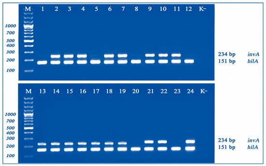
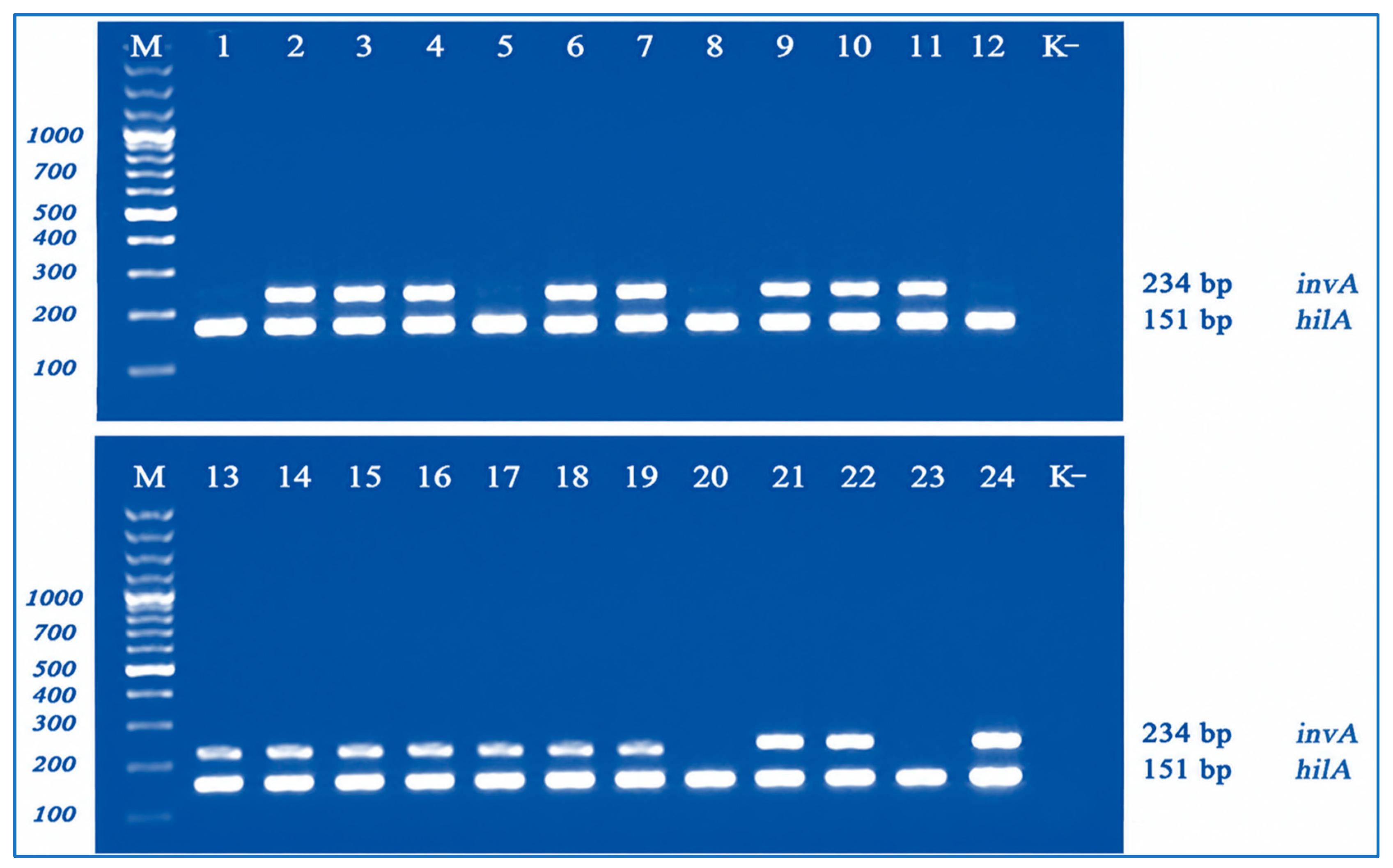
Electrophoretic analysis demonstrated consistent amplification of the expected 151 bp hilA product across the Salmonella isolates included in the initial screening panel. In contrast, the expected 234 bp invA product was not detected in a subset of the tested isolates, including representatives of less common serovars (Figure 1). These differences in amplification were interpreted as reflecting experimental primer pair performance under the screening conditions used and not as evidence of the absence of the corresponding target genes. Based on the stable and reproducible amplification obtained with the hilA-M1-F/R primer pair across the tested isolate panel, hilA was selected as the target for subsequent development of the TaqMan real-time PCR assay.
Figure 1.
Agarose gel electrophoresis of amplification products obtained using the hilA-M1 and invA-M1 primer pairs in duplex PCR. M, DNA molecular weight marker; lane 1, S. enterica serovar Typhi 15S; lane 2, S. enterica serovar Typhimurium 8S; lane 3, S. enterica serovar Enteritidis 6S; lane 4, S. enterica serovar Enteritidis 17S; lane 5, S. enterica serovar Paratyphi B 2S; lane 6, S. enterica serovar Dublin 10S; lane 7, S. enterica serovar Choleraesuis 23S; lane 8, S. enterica serovar Anatum 16S; lanes 9–11, S. enterica serovar Enteritidis 37S, 40S, and 41S; lane 12, S. enterica serovar Infantis 5S; lane 13, S. enterica serovar Muenchen 30S; lane 14, S. enterica serovar Muenchen 31S; lane 15, S. enterica serovar Muenchen 36S; lane 16, S. enterica serovar Typhimurium 20S; lane 17, S. enterica serovar Typhimurium 26S; lane 18, S. enterica serovar Typhimurium 28S; lane 19, S. enterica serovar Typhimurium 29S; lane 20, S. enterica serovar Kottbus 13S; lane 21, S. enterica serovar Choleraesuis 24S; lane 22, S. enterica serovar Dublin 25S; lane 23, S. enterica serovar Litchfield 27S; lane 24, positive control (S. enterica serovar Typhimurium strain 32S); K−, negative control (deionized water). Expected amplicon sizes were 151 bp for hilA and 234 bp for invA.
3.2. Analytical Specificity of the Screening Primer Sets
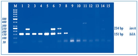
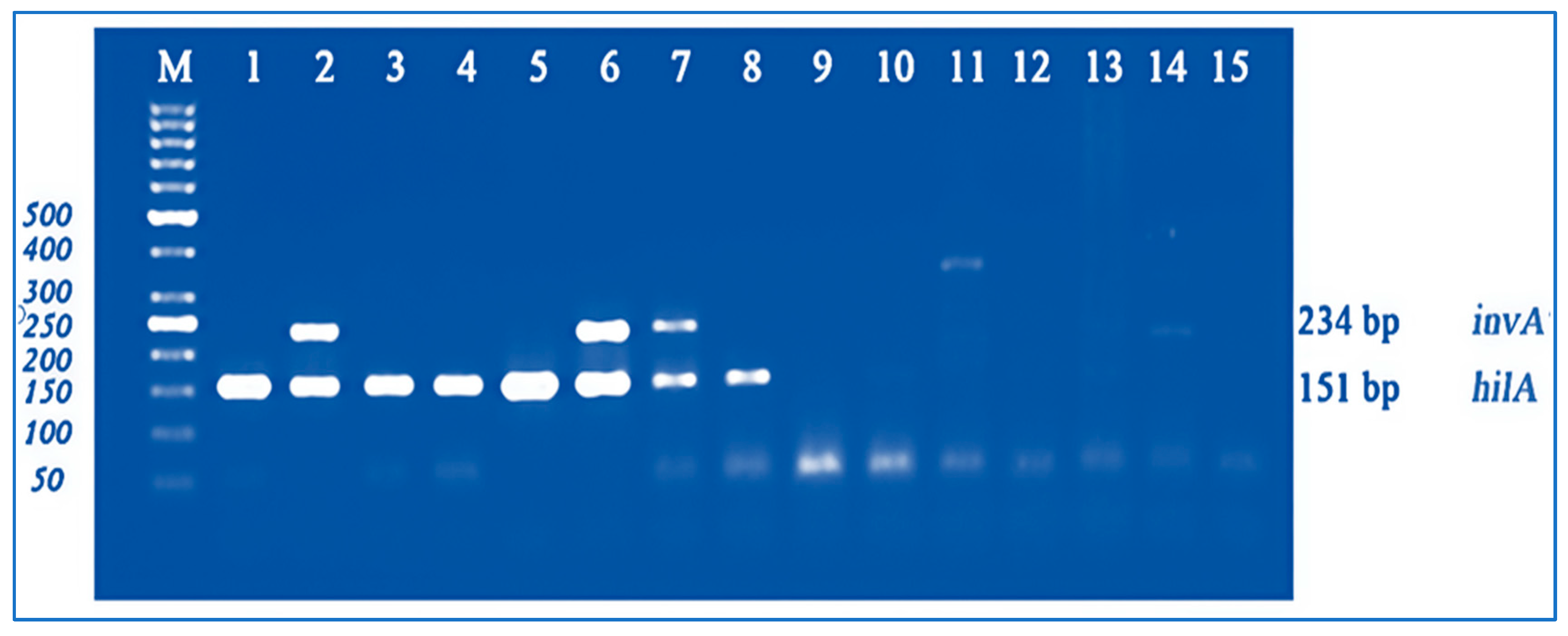
The analytical specificity of the hilA-M1 and invA-M1 primer sets was initially evaluated by conventional PCR followed by agarose gel electrophoresis. The test panel included DNA from selected Salmonella serovars and non-Salmonella bacterial species, including Shigella sonnei, Shigella flexneri, Yersinia pseudotuberculosis, Y. enterocolitica, Y. kristensenii, and Vibrio cholerae (Figure 2).
Figure 2.
Agarose gel electrophoresis of PCR products obtained during the analytical specificity assessment of the hilA-M1 and invA-M1 primer sets. M, DNA molecular weight marker; lane 1, S. enterica serovar Typhi 15S; lane 2, S. enterica serovar Enteritidis 6S; lane 3, S. enterica serovar Infantis 5S; lane 4, S. enterica serovar Kottbus 13S; lane 5, S. enterica serovar Paratyphi B 2S; lane 6, S. enterica serovar Dublin 10S; lane 7, S. enterica serovar Typhimurium 8S; lane 8, S. enterica serovar Anatum 16S; lane 9, Shigella sonnei 14; lane 10, Shigella flexneri 16; lane 11, Yersinia pseudotuberculosis 2841; lane 12, Y. enterocolitica 5; lane 13, Y. kristensenii 148; lane 14, Vibrio cholerae ch166; lane 15, negative control (deionized water).
The expected amplicon sizes were 151 bp for hilA and 234 bp for invA. Target-specific amplification products were observed only in reactions containing Salmonella DNA. The hilA-M1-F/R primer pair generated the expected 151 bp amplicon in the tested Salmonella isolates, whereas the invA-M1-F/R primer pair generated the expected 234 bp amplicon only in responsive Salmonella isolates. No target-specific amplification was observed with DNA from any of the non-Salmonella species tested or in the no-template control, supporting the analytical specificity of both primer sets within the bacterial panel evaluated.
Specific amplification products were observed only in reactions containing Salmonella DNA. The hilA-M1-F/R primer pair generated the expected 151 bp amplicon, whereas the invA-M1-F/R primer pair generated the expected 234 bp amplicon in responsive Salmonella isolates. No target-specific amplification was observed with DNA from the non-Salmonella species tested or in the no-template control, supporting the analytical specificity of the primer sets under the conditions evaluated.
3.3. Development and Analytical Sensitivity of the TaqMan Real-Time PCR Assay
Based on the target-screening results, the hilA gene was selected for development of the TaqMan real-time PCR assay. A shorter inner primer pair, B-hilA-F/R, was designed to amplify an 85 bp region containing the binding site for a FAM/BHQ1-labeled TaqMan hydrolysis probe. A recombinant plasmid, pTG19-T-hilA, containing the corresponding target sequence was used as a positive-control construct (Table 2, Section B).
Table 2.
Primers, TaqMan probe, and positive control construct used in this study. Section A: screening primers used in conventional multiplex PCR for target gene comparison. Section B: TaqMan real-time PCR assay oligonucleotides and positive control.
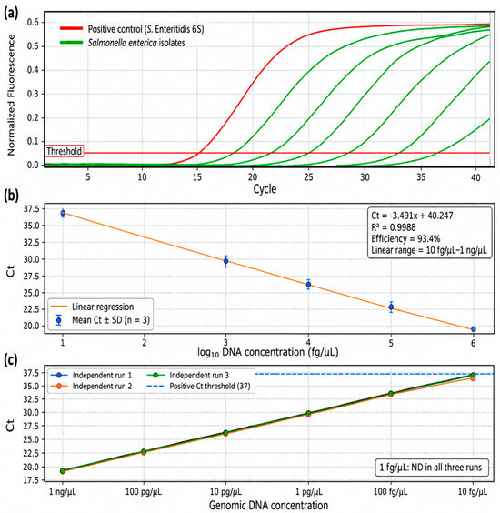
Analytical sensitivity was evaluated using ten-fold serial dilutions of genomic DNA from S. enterica serovar Enteritidis strain 6S. Ct values increased progressively with decreasing DNA concentration, demonstrating the expected concentration-dependent amplification pattern. Linear regression over the range from 1 ng/μL to 10 fg/μL yielded a slope of −3.491, a y-intercept of 40.247, and a coefficient of determination (R2) of 0.9988, corresponding to an amplification efficiency of 93.4%. The lowest genomic DNA concentration reproducibly detected across all three independent real-time PCR runs was 10 fg/μL, whereas 1 fg/μL was not detected in any run. Therefore, 10 fg/μL was considered the experimentally determined detection limit for genomic DNA under the conditions tested.
The Ct values obtained across the three independent real-time PCR runs for each genomic DNA concentration, together with the corresponding mean Ct, standard deviation, and coefficient of variation, are summarized in Table 3.
Table 3.
Analytical sensitivity and inter-run reproducibility of the hilA-targeting TaqMan real-time PCR assay using ten-fold serial dilutions of S. enterica serovar Enteritidis strain 6S genomic DNA.
At the experimentally determined genomic DNA detection limit of 10 fg/μL, Ct values of 36.31, 36.42, and 36.96 were obtained in the three independent runs, respectively, whereas 1 fg/μL was not detected in any run. The corresponding amplification profiles, standard curve, and Ct values obtained across the three independent runs are presented in Figure 3a–c.
Figure 3.
Analytical sensitivity and reproducibility of the developed hilA-targeting TaqMan real-time PCR assay using ten-fold serial dilutions of S. enterica serovar Enteritidis strain 6S genomic DNA. (a) Representative real-time PCR amplification curves showing the concentration-dependent shift in amplification. (b) Standard curve generated by linear regression of mean Ct values against log10-transformed genomic DNA concentrations. Error bars represent the standard deviation of three independent real-time PCR runs. The linear range extended from 1 ng/μL to 10 fg/μL, with a slope of −3.491, a y-intercept of 40.247, R2 = 0.9988, and an amplification efficiency of 93.4%. (c) Ct values obtained across the three independent runs at each genomic DNA concentration, demonstrating consistent inter-run performance. The lowest genomic DNA concentration reproducibly detected in all three runs was 10 fg/μL, whereas 1 fg/μL was not detected.
Using bacterial cell suspensions, the lowest concentration detected under the tested conditions was 102 CFU/mL. Thus, the experimentally determined detection limits under the conditions evaluated were 102 CFU/mL for bacterial suspensions and 10 fg/μL for genomic DNA.
3.4. Analytical Specificity of the TaqMan Real-Time PCR Assay
The analytical specificity of the developed hilA-targeting TaqMan real-time PCR assay was evaluated using an expanded panel of 15 non-target bacterial strains or isolates. The panel included Shigella flexneri 16, Shigella sonnei 14, Yersinia pseudotuberculosis 2841, Y. pestis NP6, Y. enterocolitica 5, Y. kristensenii 148, Bacillus anthracis 23, Vibrio cholerae ch166, Francisella tularensis FT24, Escherichia coli 75, Klebsiella aerogenes 11 (formerly Enterobacter aerogenes), K. pneumoniae 15, Listeria monocytogenes 4, V. cholerae non-O1 245, and Proteus mirabilis 18S. The P. mirabilis 18S isolate was also retained as the WGS-confirmed non-Salmonella comparator described above.
No specific amplification of the hilA target was detected in any organism in the expanded non-target panel; Ct values remained undetermined, whereas the S. enterica serovar Enteritidis positive control produced the expected amplification signal. These findings support the absence of detectable cross-reactivity within the organisms tested. However, because the panel remains limited relative to the full diversity of potentially relevant non-target bacteria, the results should not be interpreted as establishing exhaustive analytical exclusivity.
3.5. Assay Performance in Artificially Contaminated Food Samples
As a preliminary proof-of-concept assessment, the developed hilA-targeting TaqMan real-time PCR assay was evaluated using artificially contaminated milk and carrot samples inoculated with S. enterica serovar Enteritidis strain 6S at a nominal initial concentration of approximately 2 × 10² CFU/mL. Following inoculation, the samples were incubated at 37 °C for 18 ± 2 h directly in the food matrix prior to DNA extraction. Therefore, the stated inoculum concentration represents the initial contamination level and not the bacterial concentration at the time of DNA extraction.
Specific amplification of the hilA target was detected in all artificially contaminated samples. Ct values ranged from 32.59 to 35.10 for the carrot matrix and from 24.45 to 29.28 for the milk matrix. No specific amplification was detected in the corresponding uninoculated matrix controls. Because the artificially contaminated samples were incubated for 18 ± 2 h before DNA extraction, the bacterial concentration present at the time of extraction was not quantitatively determined. Therefore, the Ct values obtained from the food matrices cannot be directly compared with the purified genomic DNA standard curve on the basis of the nominal initial inoculum concentration. These results were interpreted qualitatively as a preliminary proof-of-concept assessment and were not used to determine bacterial load or an analytical detection limit in food matrices. Because an internal amplification control was not included, potential matrix-associated PCR inhibition was not independently assessed.
3.6. Assay Performance Using Bacterial Isolates
The performance of the developed hilA-targeting TaqMan real-time PCR assay was further evaluated using 25 bacterial isolates from the NCM collection, comprising 24 WGS-confirmed Salmonella isolates representing multiple serovars and sequence types and one Proteus mirabilis isolate (18S). Following DNA extraction, all samples were analyzed using the B-hilA-F/R primers and the specific B-hilA TaqMan probe.
Specific amplification of the hilA target was detected in all 24 WGS-confirmed Salmonella isolates (Table 4). For most isolates, mean Ct values were below 24; higher values were observed for strain 29S (S. enterica serovar Typhimurium; mean Ct = 31.47 ± 0.20) and strain 25S (S. enterica serovar Dublin; mean Ct = 36.66 ± 0.14). Strain 25S yielded Ct values of 36.68, 36.51, and 36.79 in the three independent runs, respectively (mean Ct = 36.66 ± 0.14), and amplification was reproducibly detected in all three runs. Although these values were close to the operational Ct threshold of 37, their reproducibility supported classification of this isolate as positive under the experimental conditions used. No specific amplification was detected for strain 18S, which was identified by WGS as P. mirabilis ST490. The recombinant pTG19-T-hilA positive control showed consistent amplification (Ct = 11.91), whereas no amplification was detected in the no-template control.
Table 4.
Performance and inter-run reproducibility of the hilA-targeting TaqMan real-time PCR assay across the bacterial isolate panel.
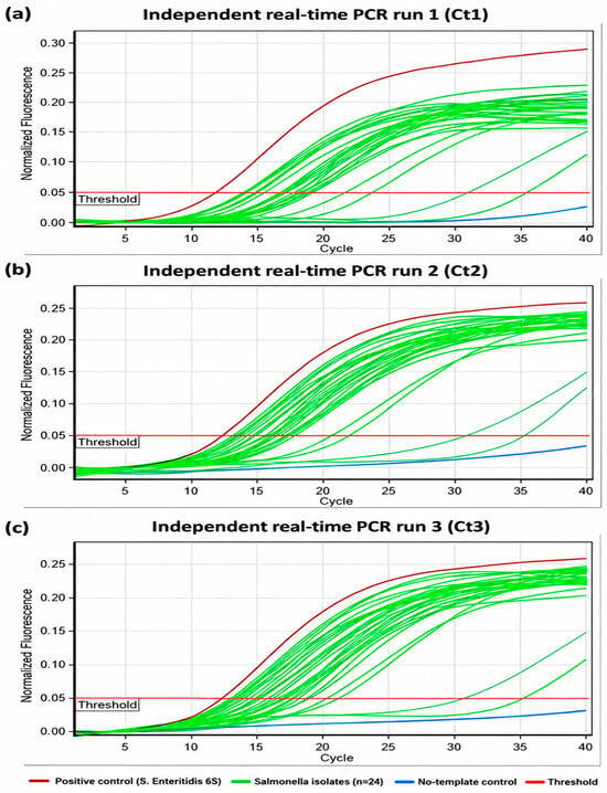
Inter-run reproducibility was high across the 24 Salmonella isolates, with CV values ranging from 0.38% to 2.12%. All CV values were below the predefined 5% threshold, supporting acceptable inter-run reproducibility of the assay under the experimental conditions used. Amplification profiles obtained in the three independent real-time PCR runs are shown in Figure 4a–c and were consistent with the low inter-run variability indicated by the calculated CV values.
Figure 4.
Real-time PCR amplification profiles obtained during evaluation of the developed hilA-targeting TaqMan assay using the bacterial isolate panel. (a–c) Amplification profiles from three independent real-time PCR runs performed under identical experimental conditions. The consistent amplification patterns observed across the three independent runs demonstrate the inter-run reproducibility of the assay. No specific amplification was observed for the Proteus mirabilis comparator or the no-template control.
Amplification profiles obtained in the three independent real-time PCR runs are presented in Figure 4a–c. Consistent amplification patterns were observed across the three runs, in agreement with the low inter-run variability indicated by the calculated CV values.
Overall, the developed hilA-targeting TaqMan real-time PCR assay demonstrated reproducible detection of Salmonella across the tested isolate panel, with experimentally determined detection limits of 102 CFU/mL for bacterial suspensions and 10 fg/μL for genomic DNA under the conditions tested, and no cross-reactivity with the non-target bacterial species examined. Specific amplification of the hilA target was detected in all 24 WGS-confirmed Salmonella isolates within the predefined positive Ct range (≤37), including S. enterica serovar Dublin strain 25S, which showed the highest mean Ct value (36.66 ± 0.14). No specific amplification was observed for the P. mirabilis comparator or the no-template control. The assay also successfully detected Salmonella DNA in both artificially contaminated food matrices tested.
3.7. Overall Assay Development and Analytical Evaluation Workflow
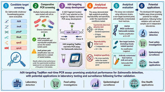
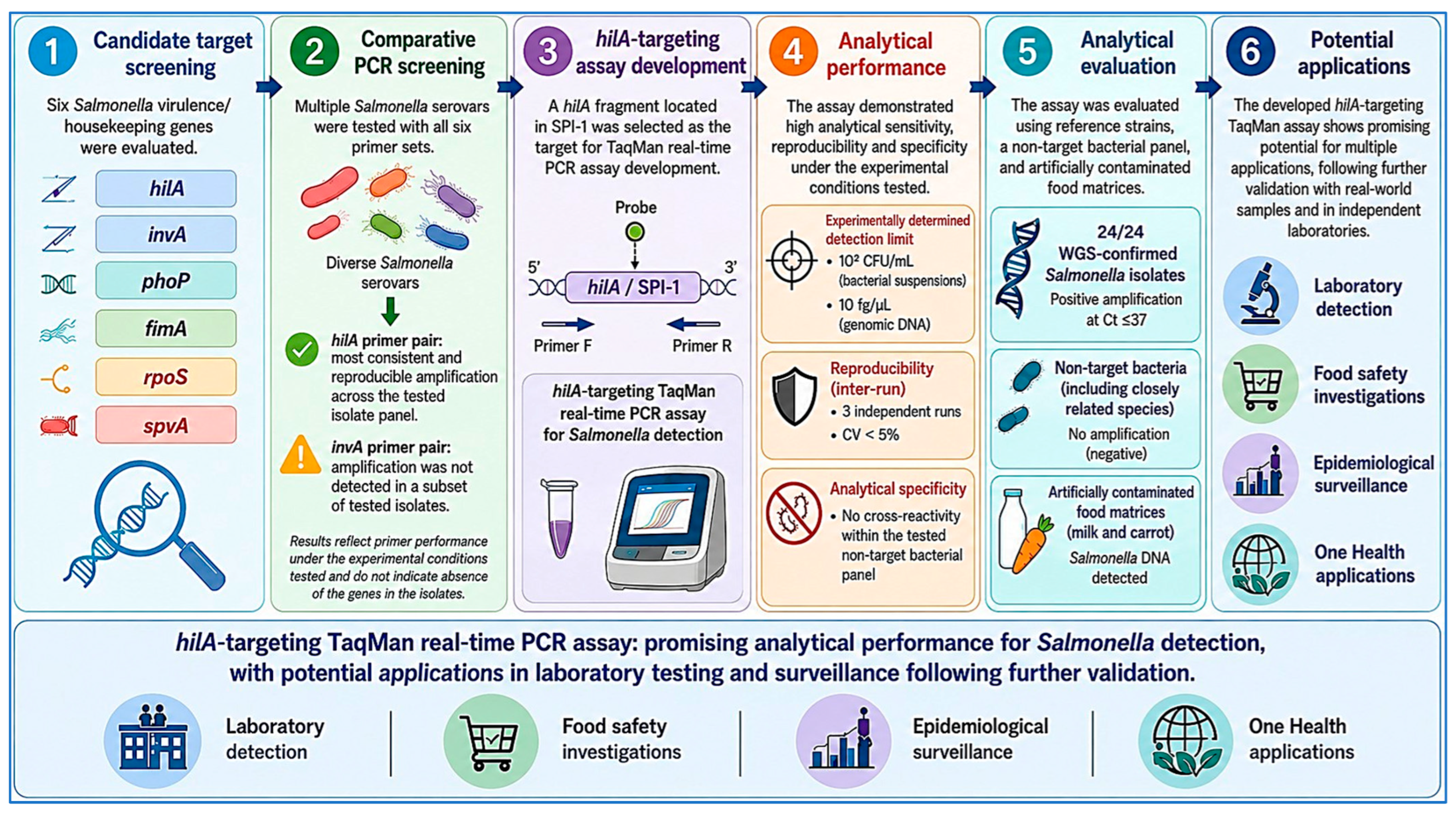
The overall workflow of target selection, assay development, and analytical evaluation is summarized in Figure 5. The study progressed from comparative screening of six candidate molecular targets (hilA, invA, phoP, fimA, rpoS, and spvA) to selection of hilA based on the stable and reproducible amplification obtained with the hilA-M1-F/R primer pair under the experimental conditions used. The selected target was subsequently used to develop the hilA-targeting TaqMan real-time PCR assay, followed by evaluation of analytical sensitivity, specificity, inter-run reproducibility, performance across WGS-confirmed Salmonella isolates, and detection in artificially contaminated food matrices. Together, these experimental stages provide an integrated overview of the assay-development and analytical evaluation strategy used in this study.
Figure 5.
Overall workflow for the development and analytical evaluation of the hilA-targeting TaqMan real-time PCR assay for Salmonella enterica detection. Six candidate molecular targets (hilA, invA, phoP, fimA, rpoS, and spvA) were comparatively screened using the corresponding primer pairs. The hilA primer pair demonstrated the most consistent and reproducible amplification performance across the tested isolate panel under the experimental screening conditions and was therefore selected for subsequent TaqMan assay development. Analytical evaluation included determination of sensitivity under the conditions tested, assessment of inter-run reproducibility and analytical specificity, testing of WGS-confirmed Salmonella isolates and non-target bacteria, and preliminary proof-of-concept testing in artificially contaminated milk and carrot matrices. The workflow summarizes the sequential experimental strategy from candidate-target screening to laboratory evaluation of the developed assay and indicates its potential applications requiring further validation. Numbers 1–6 denote the sequential stages of the workflow; colors and icons are used only to visually distinguish the stages and application categories. Scientific illustrations and graphical elements were created using Mind the Graph and finalized in Adobe Illustrator (Adobe Inc., San Jose, CA, USA).
4. Discussion
The principal contribution of this study is the comparative experimental evaluation of six candidate molecular targets for Salmonella detection in a serologically diverse collection of isolates and the subsequent development of a hilA-targeting TaqMan real-time PCR assay. To the best of our knowledge, this represents the first study in Kazakhstan to apply such a comparative target-selection approach to the development of a hilA-based real-time PCR assay for Salmonella detection. Among the six candidate targets evaluated, the hilA-M1-F/R primer pair showed the most consistent amplification across the tested isolate panel. Differences observed with the other primer pairs may reflect primer performance, primer template sequence variation, or multiplex reaction effects and should not be interpreted as evidence of absence of the corresponding genes. These findings are consistent with previous studies demonstrating the functional importance of hilA as a regulatory gene within Salmonella pathogenicity island 1 (SPI-1) and its utility as a molecular target for Salmonella detection [26,27,28,29].
Genomic characterization of part of the strain collection, performed by whole-genome sequencing in a parallel study [20], provides additional context for interpretation of the candidate molecular targets evaluated. The hilA gene is located within SPI-1, a chromosomal pathogenicity island that plays a central role in regulating epithelial cell invasion by S. enterica. In contrast, spvA, one of the candidate targets included in the initial screening, belongs to the spv virulence locus commonly associated with large virulence plasmids, the distribution of which varies among Salmonella serovars and strains [20,30]. Thus, the chromosomal localization and functional importance of hilA, together with its consistent amplification across the isolate panel examined in the present study, provide a biological rationale for its selection as a molecular target. However, broader genomic and experimental evaluation across the diversity of the genus Salmonella will be required before its universality can be established.
The developed assay produced specific amplification across a genetically and serologically diverse collection of S. enterica, including serovars Enteritidis, Typhimurium, Choleraesuis, Dublin, Paratyphi B, Infantis, Kottbus, Typhi, Anatum, Litchfield, and Muenchen. This diversity is relevant because the epidemiology of salmonellosis involves numerous serovars originating from human, animal, food, and environmental reservoirs. International surveillance studies have demonstrated considerable diversity among circulating non-typhoidal Salmonella. For example, analysis of poultry-associated isolates from 15 countries in the Americas identified 131 Salmonella serovars [31]. In the European Union, in addition to Enteritidis and Typhimurium, monophasic Typhimurium, Infantis, and other serovars remain epidemiologically important [32]. These observations underscore the importance of evaluating molecular assays across genetically and serologically diverse Salmonella collections.
From an analytical perspective, the developed hilA-targeting TaqMan real-time PCR assay demonstrated experimentally determined detection limits of 102 CFU/mL for bacterial cell suspensions and 10 fg/μL for purified genomic DNA under the conditions tested. These values indicate favorable analytical sensitivity under the experimental conditions used and are within the range reported for molecular methods developed for Salmonella detection [29,33,34,35]. Importantly, the genomic DNA dilution series showed a strong linear relationship between Ct and template concentration over the range from 1 ng/μL to 10 fg/μL, with a slope of −3.491, R2 of 0.9988, and an amplification efficiency of 93.4%. The 10 fg/μL concentration was reproducibly detected in all three independent runs, whereas 1 fg/μL was not detected, supporting 10 fg/μL as the experimentally determined genomic DNA detection limit under the conditions tested.
The reproducibility of the assay was further supported by the low inter-run variability observed across the WGS-confirmed Salmonella isolate panel. CV values ranged from 0.38% to 2.12% across the three independent real-time PCR runs and remained below the predefined 5% threshold for all isolates. These findings indicate consistent assay performance under the experimental conditions used. However, because all experiments were conducted within a single laboratory, these data reflect inter-run rather than inter-laboratory reproducibility; multicenter evaluation will be required to assess assay robustness across different laboratories, operators, instruments, and reagent lots.
Analytical specificity was evaluated using the expanded non-target panel. No cross-amplification was observed among the tested Shigella, Yersinia, Bacillus, Vibrio, Francisella, Escherichia, Klebsiella, Listeria, and Proteus organisms. Particularly informative was isolate 18S, which had initially been assigned to the genus Salmonella on the basis of conventional cultural and biochemical characteristics but was subsequently identified as Proteus mirabilis ST490 by whole-genome sequencing and genomic typing. No hilA-specific amplification was observed for this isolate. The expanded panel provides additional evidence of analytical specificity within the organisms tested, including several representatives of Enterobacterales. Nevertheless, the panel does not encompass all phylogenetically related or food-associated microorganisms. Broader testing, including at least 20 closely related non-target strains, together with expanded in silico analysis against representative genome collections, would provide a more comprehensive assessment of assay exclusivity.
Evaluation of the assay using 24 WGS-confirmed Salmonella isolates showed specific hilA amplification in all isolates included in the tested panel. All 24 isolates were reproducibly detected under the study-specific Ct ≤37 criterion. S. enterica serovar Dublin strain 25S showed the highest mean Ct value (36.66 ± 0.14) but was reproducibly detected in all three independent runs. The comparatively high Ct value observed for this isolate may reflect differences in template concentration, DNA quality, or other isolate-specific factors and warrants further investigation. In contrast, no specific amplification was observed for the P. mirabilis comparator.
The potential applicability of the assay to food testing was explored in a preliminary proof-of-concept experiment using artificially contaminated milk and carrot matrices. Samples were inoculated at a nominal initial concentration of approximately 2 × 10² CFU/mL and incubated at 37 °C for 18 ± 2 h prior to DNA extraction. Salmonella DNA was detected in all experimentally contaminated samples, with Ct values ranging from 32.59 to 35.10 for carrot and from 24.45 to 29.28 for milk. Because the bacterial concentration at the time of DNA extraction was not quantitatively determined, these Ct values cannot be directly compared with the purified genomic DNA standard curve on the basis of the nominal initial inoculum concentration. Accordingly, these findings are interpreted as preliminary qualitative proof-of-concept evidence rather than as validation of a detection limit in food matrices. Artificial inoculation may not fully reproduce naturally contaminated foods with respect to bacterial distribution, physiological state, or DNA recovery efficiency.
Taken together, the findings indicate promising analytical performance of the developed hilA-targeting TaqMan real-time PCR assay under the conditions evaluated, including reproducible detection within the tested Salmonella isolate panel, experimentally determined sensitivity, and no detectable cross-reactivity within the expanded non-target panel. However, this single-laboratory study does not establish routine applicability for clinical diagnosis, food testing, epidemiological surveillance, or One Health monitoring. Further validation using larger and geographically diverse isolate collections, naturally contaminated food products, primary clinical, veterinary, and environmental specimens, and independent laboratories is required before such applications can be considered.
Limitations of the Study
The present study has several limitations that should be considered when interpreting the findings. First, the performance of the developed assay was evaluated using a relatively limited collection of Salmonella isolates, predominantly originating from Kazakhstan. Although the collection represented multiple serovars and sequence types, including less common serovars, it does not encompass the full genomic and serological diversity of the genus Salmonella. Consistent amplification within this isolate collection therefore does not establish universal conservation of hilA or of the primer- and probe-binding regions across Salmonella. Broader evaluation using larger and geographically diverse isolate collections together with representative genome-based analyses is required. In addition, after hilA was selected, only one TaqMan primer–probe system was developed; parallel experimental screening of multiple alternative TaqMan primer–probe combinations was not performed.
Second, analytical sensitivity was assessed using three independent runs at each genomic DNA concentration. Therefore, the reported 10 fg/μL value should be interpreted as an experimentally determined detection limit under the conditions tested rather than as a statistically validated LOD95. Formal determination of LOD95 would require a larger number of replicates at concentrations close to the detection limit. The Ct ≤37 threshold was likewise an empirical, study-specific operational threshold and was not established using ROC-based statistical validation. A formal diagnostic cutoff and an independent gray zone around this decision threshold were not established. ROC-supported threshold determination should be performed in future validation studies using larger collections of well-characterized positive and negative samples.
Third, analytical specificity was evaluated using an expanded but still limited non-target panel that included several representatives of Enterobacterales and other clinically or food-relevant bacteria. No cross-reactivity was observed among the organisms tested; however, this does not establish exhaustive exclusivity across all potentially relevant taxa. Further validation should include at least 20 closely related non-target strains and expanded in silico evaluation against a representative genomic dataset.
Fourth, evaluation in food matrices was limited to a preliminary proof-of-concept experiment using artificially contaminated milk and carrot samples. Naturally contaminated food products and a broader range of food matrices were not examined. Although uninoculated matrix controls were included, an internal amplification control was not used; therefore, potential PCR inhibition associated with the food matrices was not independently assessed. Further validation using additional matrices, appropriate inhibition controls, and naturally contaminated samples is required before application of the assay for routine food testing. Artificially inoculated matrices may also differ from naturally contaminated foods in bacterial distribution, physiological state, and DNA recovery efficiency.
Finally, the assay is a single-target qualitative method designed to detect the hilA sequence. A positive result therefore indicates detection of the target sequence but does not by itself establish strain identity or serovar, characterize virulence potential, or determine whether the detected organism is viable. The assay was also not designed for quantitative determination of bacterial load. In addition, the assay underwent single-laboratory analytical evaluation; therefore, inter-laboratory reproducibility and robustness across different laboratory settings, operators, instruments, and reagent lots were not assessed. Multicenter evaluation will be required to confirm transferability and robustness before broader implementation. Accordingly, positive molecular detection using the developed assay should be considered complementary to conventional culture, serotyping, and genomic approaches when detailed characterization or viability assessment is required.
5. Conclusions
In the present study, a TaqMan real-time PCR assay targeting the hilA gene was developed and analytically evaluated for the detection of Salmonella enterica. Comparative screening of six candidate molecular targets (hilA, invA, phoP, fimA, rpoS, and spvA) showed that the hilA-M1-F/R primer pair provided the most stable and reproducible amplification within the tested isolate panel under the experimental conditions used and was therefore selected for subsequent TaqMan assay development.
The developed assay demonstrated experimentally determined detection limits of 102 CFU/mL for bacterial suspensions and 10 fg/μL for genomic DNA under the conditions tested, with an amplification efficiency of 93.4% and an R2 of 0.9988 across the linear range from 1 ng/μL to 10 fg/μL. No cross-reactivity was observed within the tested non-target bacterial panel. All 24 WGS-confirmed Salmonella isolates yielded positive amplification within the predefined Ct threshold (≤37), including S. enterica serovar Dublin strain 25S, which showed the highest mean Ct value (36.66 ± 0.14). No specific amplification was observed for the Proteus mirabilis comparator. The assay also detected Salmonella DNA in artificially contaminated milk and carrot samples.
These findings indicate that the developed hilA-targeting TaqMan real-time PCR assay has promising analytical performance under the experimental conditions evaluated. However, the present study does not establish readiness for routine diagnostic, food-testing, epidemiological surveillance, or One Health applications. Further validation using larger and geographically diverse isolate collections, naturally contaminated food products, primary clinical, veterinary, and environmental specimens, and independent laboratories is required before such applications can be considered.
Author Contributions
Conceptualization, D.Y., B.A. and A.A.; methodology, D.Y., B.A., Z.A., A.R., A.K. and O.R.; software, O.R.; validation, B.A., Z.A., A.K. and O.R.; formal analysis, D.Y., B.A., O.R. and A.A.; investigation, D.Y., B.A., D.K., A.R., A.K., S.I. and N.S.; resources, A.R., D.K., S.I., N.S. and A.A.; data curation, D.Y., B.A., Z.A. and A.K.; writing—original draft preparation, D.Y.; writing—review and editing, D.Y., B.A., O.R. and A.A.; visualization, Z.A., N.S. and O.R.; supervision, B.A., Z.A., O.R. and A.A.; project administration, A.A.; funding acquisition, A.A. All authors have read and agreed to the published version of the manuscript.
Funding
This research was funded by the Ministry of Science and Higher Education of the Republic of Kazakhstan through the Committee of Science, grant IRN AP23490794, “Investigation of the Distribution of Salmonellosis and Enteroviral Infection Pathogens Using PCR-Based Diagnostic Assays Developed to Improve Epidemiological Surveillance” (2024–2026).
Institutional Review Board Statement
The study was conducted in accordance with the principles of the Declaration of Helsinki and was approved by the Local Bioethics Committee of the M. Aikimbayev National Scientific Center for Especially Dangerous Infections (Protocol No. 2, dated 12 February 2024).
Informed Consent Statement
Informed consent was not required because the study used previously collected, archived, and fully de-identified bacterial strains, and no personally identifiable information was accessed.
Data Availability Statement
The data supporting the findings of this study are available from the corresponding author upon reasonable request. The bacterial strains analyzed in this study are preserved in the National Collection of Microorganisms of the M. Aikimbayev National Scientific Center for Especially Dangerous Infections, Almaty, Kazakhstan.
Conflicts of Interest
The authors declare no conflicts of interest.
Abbreviations
The following abbreviations are used in this manuscript:
| PCR | Polymerase chain reaction |
| qPCR | Quantitative polymerase chain reaction |
| DNA | Deoxyribonucleic acid |
| NSCEDI | M. Aikimbayev National Scientific Center for Especially Dangerous Infections |
| NCM | National Collection of Microorganisms |
| WGS | Whole-genome sequencing |
| MLST | Multilocus sequence typing |
| ST | Sequence type |
| SPI-1 | Salmonella pathogenicity island 1 |
| Ct | Cycle threshold |
| CV | Coefficient of variation |
| LOD | Limit of detection |
| SD | Standard deviation |
| MIQE | Minimum Information for Publication of Quantitative Real-Time PCR Experiments |
References
- Teklemariam, A.D.; Al-Hindi, R.R.; Albiheyri, R.S.; Alharbi, M.G.; Alghamdi, M.A.; Filimban, A.A.R.; Al Mutiri, A.S.; Al-Alyani, A.M.; Alseghayer, M.S.; Almaneea, A.M.; et al. Human Salmonellosis: A Continuous Global Threat in the Farm-to-Fork Food Safety Continuum. Foods 2023, 12, 1756. [Google Scholar] [CrossRef] [Scilit] [PubMed]
- Gazezova, S.; Nabirova, D.; Waltenburg, M.; Rakhimzhanova, M.; Smagul, M.; Kasabekova, L.; Horth, R. Salmonellosis outbreak associated with the consumption of food at a wedding in an urban restaurant in Kazakhstan: A retrospective cohort study. BMC Infect. Dis. 2024, 24, 1474. [Google Scholar] [CrossRef] [Scilit] [PubMed]
- Woh, P.Y.; Yeung, M.P.S.; Goggins, W.B.; Lo, N.; Wong, K.T.; Chow, V.; Chau, K.Y.; Fung, K.; Chen, Z.; Ip, M. Genomic epidemiology of multidrug-resistant nontyphoidal Salmonella in young children hospitalized for gastroenteritis. Microbiol. Spectr. 2021, 9, e00248-21. [Google Scholar] [CrossRef] [Scilit] [PubMed]
- Majowicz, S.E.; Musto, J.; Scallan, E.; Angulo, F.J.; Kirk, M.; O’Brien, S.J.; Jones, T.F.; Fazil, A.; Hoekstra, R.M.; International Collaboration on Enteric Disease ‘Burden of Illness’ Studies. The global burden of nontyphoidal Salmonella gastroenteritis. Clin. Infect. Dis. 2010, 50, 882–889. [Google Scholar] [CrossRef] [Scilit] [PubMed]
- Stanaway, J.D.; Parisi, A.; Sarkar, K.; Blacker, B.F.; Reiner, R.C.; Hay, S.I.; Nixon, M.R.; Dolecek, C.; James, S.L.; Mokdad, A.H.; et al. The global burden of non-typhoidal Salmonella invasive disease: A systematic analysis for the Global Burden of Disease Study 2017. Lancet Infect. Dis. 2019, 19, 1312–1324. [Google Scholar] [CrossRef] [Scilit] [PubMed]
- Ranjan, A.; Chandna, M.; Stevens, N.J.; Kandil, J.; Dinh, B.; Kuhn, M.; Mian, N.; Tran, B.; Hamid, A.; Kim, P.; et al. Salmonella infections: Global trends and emerging challenges. Microorganisms 2026, 14, 816. [Google Scholar] [CrossRef] [Scilit] [PubMed]
- Chae, S.J.; Yun, Y.S.; Yoo, C.K.; Chung, G.T.; Lee, D.Y. First report of Salmonella serotype Tilene infection in Korea. Ann. Clin. Microbiol. 2016, 19, 24–27. [Google Scholar] [CrossRef] [Scilit]
- European Food Safety Authority (EFSA); European Centre for Disease Prevention and Control (ECDC). The European Union summary report on antimicrobial resistance in zoonotic and indicator bacteria from humans, animals and food in 2021–2022. EFSA J. 2024, 22, e8583. [Google Scholar] [CrossRef] [Scilit] [PubMed]
- Nogaybek, M.D.; Baigenzheeva, R.K. Analysis of Salmonellosis Incidence in Kazakhstan During 2018–2022. Nauka Mirovozzrenie 2024, 1, 13–15. [Google Scholar]
- Baimuratova, M.; Abdul, B.A.; Ryskulova, A.; Tugulbayeva, A.; Jumatova, U.; Abdusallamova, Z. Microbiological monitoring in the system of epidemiological surveillance of salmonellosis in the children’s population of Almaty city. Nauka Zdr. 2021, 23, 121–130. [Google Scholar] [CrossRef] [Scilit]
- Grjibovski, A.M.; Kosbayeva, A.; Menne, B. The effect of ambient air temperature and precipitation on monthly counts of salmonellosis in four regions of Kazakhstan, Central Asia, in 2000–2010. Epidemiol. Infect. 2014, 142, 608–615. [Google Scholar] [CrossRef] [Scilit] [PubMed]
- Karaaslan, A.; Çetin, C.; Köle, M.T.; Demir Tekol, S.; Söbü, E.; Akın, Y. Salmonella gastroenteritis in children: Six-year experience in İstanbul, Turkey. J. Infect. Dev. Ctries. 2022, 16, 1757–1761. [Google Scholar] [CrossRef] [Scilit] [PubMed]
- Andrews, J.R.; Ryan, E.T. Diagnostics for invasive Salmonella infections: Current challenges and future directions. Vaccine 2015, 33, C8–C15. [Google Scholar] [CrossRef] [Scilit] [PubMed]
- Espy, M.J.; Uhl, J.R.; Sloan, L.M.; Buckwalter, S.P.; Jones, M.F.; Vetter, E.A.; Yao, J.D.C.; Wengenack, N.L.; Rosenblatt, J.E.; Cockerill, F.R., III; et al. Real-time PCR in clinical microbiology: Applications for routine laboratory testing. Clin. Microbiol. Rev. 2006, 19, 165–256, Correction in Clin. Microbiol. Rev. 2006, 19, 595. https://doi.org/10.1128/CMR.00022-06. [Google Scholar] [CrossRef] [Scilit] [PubMed]
- Galán, J.E.; Ginocchio, C.; Costeas, P. Molecular and functional characterization of the Salmonella invasion gene invA: Homology of InvA to members of a new protein family. J. Bacteriol. 1992, 174, 4338–4349. [Google Scholar] [CrossRef] [Scilit] [PubMed]
- Ohl, M.E.; Miller, S.I. Salmonella: A model for bacterial pathogenesis. Annu. Rev. Med. 2001, 52, 259–274. [Google Scholar] [CrossRef] [Scilit] [PubMed]
- Boddicker, J.D.; Knosp, B.M.; Jones, B.D. Transcription of the Salmonella invasion gene activator, hilA, requires HilD activation in the absence of negative regulators. J. Bacteriol. 2003, 185, 525–533. [Google Scholar] [CrossRef] [Scilit] [PubMed]
- Adesiji, Y.O.; Deekshit, V.K.; Odunola, R.A.; Karunasagar, I.; Daodu, O.B.; Ahmad, A.M. Virulence-encoding genes conserved in Salmonella isolated from humans, poultry, and seafood. J. Trop. Med. 2025, 2025, 1139253. [Google Scholar] [CrossRef] [Scilit] [PubMed]
- Grimont, P.A.D.; Weill, F.-X. Antigenic Formulae of the Salmonella Serovars, 9th ed.; WHO Collaborating Centre for Reference and Research on Salmonella, Institut Pasteur: Paris, France, 2007. [Google Scholar]
- Yessimseit, D.T.; Rysbekova, A.K.; Zhumadilova, Z.B.; Abdeliyev, B.Z.; Tukhanova, N.B.; Kassenova, A.K.; Abdrakhmanova, A.K.; Mereke, A.; Agzam, S.D.; Nurpeisova, A.S.; et al. Genetic and epigenetic diversity of Salmonella enterica isolates from Kazakhstan from clinical and veterinary sources. bioRxiv 2026. [Google Scholar] [CrossRef] [Scilit]
- QIAGEN. QIAamp DNA Mini and Blood Mini Handbook; QIAGEN: Hilden, Germany, 2025; Available online: https://www.qiagen.com/us/resources/kithandbook/hb-0329-007-hb-qiaamp-dna-mini-blood-mini-0725-ww (accessed on 20 August 2026).
- Shen, Z.; Qu, W.; Wang, W.; Lu, Y.; Wu, Y.; Li, Z.; Hang, X.; Wang, X.; Zhao, D.; Zhang, C. MPrimer: A program for reliable multiplex PCR primer design. BMC Bioinform. 2010, 11, 143. [Google Scholar] [CrossRef] [Scilit] [PubMed]
- Altschul, S.F.; Gish, W.; Miller, W.; Myers, E.W.; Lipman, D.J. Basic local alignment search tool. J. Mol. Biol. 1990, 215, 403–410. [Google Scholar] [CrossRef] [PubMed]
- Bustin, S.A.; Benes, V.; Garson, J.A.; Hellemans, J.; Huggett, J.; Kubista, M.; Mueller, R.; Nolan, T.; Pfaffl, M.W.; Shipley, G.L.; et al. The MIQE guidelines: Minimum information for publication of quantitative real-time PCR experiments. Clin. Chem. 2009, 55, 611–622. [Google Scholar] [CrossRef] [Scilit] [PubMed]
- Rahn, K.; De Grandis, S.A.; Clarke, R.C.; McEwen, S.A.; Galán, J.E.; Ginocchio, C.; Curtiss, R.; Gyles, C.L. Amplification of an invA gene sequence of Salmonella typhimurium by polymerase chain reaction as a specific method of detection of Salmonella. Mol. Cell. Probes 1992, 6, 271–279. [Google Scholar] [CrossRef] [Scilit] [PubMed]
- Bajaj, V.; Hwang, C.; Lee, C.A. hilA is a novel ompR/toxR family member that activates the expression of Salmonella typhimurium invasion genes. Mol. Microbiol. 1995, 18, 715–727. [Google Scholar] [CrossRef] [Scilit] [PubMed]
- Pathmanathan, S.G.; Cardona-Castro, N.; Sánchez-Jiménez, M.M.; Correa-Ochoa, M.M.; Puthucheary, S.D.; Thong, K.L. Simple and rapid detection of Salmonella strains by direct PCR amplification of the hilA gene. J. Med. Microbiol. 2003, 52, 773–776. [Google Scholar] [CrossRef] [Scilit] [PubMed]
- Guo, X.; Chen, J.; Beuchat, L.R.; Brackett, R.E. PCR detection of Salmonella enterica serotype Montevideo in and on raw tomatoes using primers derived from hilA. Appl. Environ. Microbiol. 2000, 66, 5248–5252. [Google Scholar] [CrossRef] [Scilit] [PubMed]
- Sánchez-Jiménez, M.M.; Cardona-Castro, N. Validation of a PCR for diagnosis of typhoid fever and salmonellosis by amplification of the hilA gene in clinical samples from Colombian patients. J. Med. Microbiol. 2004, 53, 875–878. [Google Scholar] [CrossRef] [Scilit] [PubMed][Green Version]
- Tasmin, R.; Gulig, P.A.; Parveen, S. Detection of virulence plasmid-encoded genes in Salmonella Typhimurium and Salmonella Kentucky isolates recovered from commercially processed chicken carcasses. J. Food Prot. 2019, 82, 1364–1368. [Google Scholar] [CrossRef] [Scilit] [PubMed]
- Diaz, D.; Hernandez-Carreño, P.E.; Velazquez, D.Z.; Chaidez-Ibarra, M.A.; Montero-Pardo, A.; Martinez-Villa, F.A.; Canizalez-Roman, A.; Ortiz-Navarrete, V.F.; Rosiles, R.; Gaxiola, S.M.; et al. Prevalence, main serovars and antimicrobial resistance profiles of non-typhoidal Salmonella in poultry samples from the Americas: A systematic review and meta-analysis. Transbound. Emerg. Dis. 2022, 69, 2544–2558. [Google Scholar] [CrossRef] [Scilit] [PubMed]
- European Food Safety Authority (EFSA); European Centre for Disease Prevention and Control (ECDC). The European Union One Health 2021 Zoonoses Report. EFSA J. 2022, 20, 7666. [Google Scholar] [CrossRef] [Scilit] [PubMed]
- Malorny, B.; Paccassoni, E.; Fach, P.; Bunge, C.; Martin, A.; Helmuth, R. Diagnostic real-time PCR for detection of Salmonella in food. Appl. Environ. Microbiol. 2004, 70, 7046–7052. [Google Scholar] [CrossRef] [Scilit] [PubMed]
- Postollec, F.; Falentin, H.; Pavan, S.; Combrisson, J.; Sohier, D. Recent advances in quantitative PCR (qPCR) applications in food microbiology. Food Microbiol. 2011, 28, 848–861. [Google Scholar] [CrossRef] [Scilit] [PubMed]
- Elizaquível, P.; Aznar, R.; Sánchez, G. Recent developments in the use of viability dyes and quantitative PCR in the food microbiology field. J. Appl. Microbiol. 2014, 116, 1–13. [Google Scholar] [CrossRef] [Scilit] [PubMed]

Disclaimer/Publisher’s Note: The statements, opinions and data contained in all publications are solely those of the individual author(s) and contributor(s) and not of MDPI and/or the editor(s). MDPI and/or the editor(s) disclaim responsibility for any injury to people or property resulting from any ideas, methods, instructions or products referred to in the content. |
© 2026 by the authors. Licensee MDPI, Basel, Switzerland. This article is an open access article distributed under the terms and conditions of the Creative Commons Attribution (CC BY) license.