


Abstract
Streptomyces is a versatile genus widely used in drug production and biotechnological applications. This study aimed to identify and characterize bioactive compounds produced by Streptomyces UP-AC4 and UP-3.2 strains and evaluate their antibacterial and anticancer activities. The strains were identified as Streptomyces californicus and Streptomyces purpurascens via chemotaxonomy, 16S rRNA sequencing, amplified ribosomal DNA restriction analysis, and phylogenetic analysis. Bioactive compounds were extracted using heat treatments at 63 °C for 30 min or 73–110 °C for 10 min. Antibacterial activity against Staphylococcus aureus, Bacillus cereus, and Escherichia coli was assessed by agar disc assay, with MICs of 0.024–0.195 mg/mL and MBCs of 0.098–0.391 mg/mL for the most effective extracts. Anticancer activity against A549, H1299, and Lu99 lung cancer cells was evaluated using the MTT assay, showing IC50 values of 0.23 ± 0.06 to 4.85 ± 0.64 mg/mL, while exhibiting no toxicity to normal fibroblast cells. HPLC analysis indicated that heat-assisted extraction of UP-AC4 at 73 °C for 10 min enriched a dinactin-associated compound as a predominant metabolite with antibiotic and anticancer activities. In conclusion, Streptomyces UP-AC4 and UP-3.2 produce promising low-cost bioactive compounds with strong potential for pharmaceutical and healthcare applications.
1. Introduction
Streptomyces are Gram-positive bacteria found in aquatic and terrestrial ecosystems that form a branching network of filaments and produce spores [1]. Genome sequencing has revealed that Streptomyces species can produce a broader spectrum of secondary metabolites than previously thought [2,3]. Streptomyces-derived secondary metabolites are an environmentally friendly source of bioactive compounds with numerous applications, including in the medical and biotechnology industries [4,5,6]. Therefore, Streptomyces are ideal candidates for industrial-scale fermentation and purification of bioactive compounds due to their various fermentation strategies, high yields, and ability to proliferate on inexpensive substrates [7]. Moreover, Streptomyces species are the source of most clinically used antibiotics and several widely used drugs against common diseases such as cancer [8,9,10]. The antibiotic-anticancer substances produced by Streptomyces have been approved for anticancer drugs, such as anthracyclines (doxorubicin, daunomycin, epirubicin, and idarubicin), glycopeptides (bleomycin), non-ribosomal peptides (dactinomycin), and quinones (mitomycin C) [11,12,13,14].
Various chemical or physical methods have been developed and validated to extract bioactive compounds from Streptomyces. The conventional extraction processes for bioactive compounds use harmful organic solvents, such as methanol, and are restricted due to the toxicity and combustibility of the solvents. Consequently, innovative mechanical cell disruption methods have been developed, such as bead milling, high-pressure homogenization, ultrasonication, pressurized liquid extraction, microwave-assisted extraction, ultrasound extraction, pulsed electric field extraction, and supercritical fluid extraction. However, these procedures could reduce the extraction yield of bioactive compounds due to heat generation, high energy consumption, and increased cost [15,16]. Therefore, new Streptomyces strains that synthesize thermostable bioactive compounds must be discovered.
Previous studies have demonstrated that S. fildesensis (INACH3013), isolated from soil, is a prolific producer of actinomycin-like secondary metabolites with anticancer activity. These compounds induce apoptosis via caspase activation, decrease mitochondrial membrane potential, and alter cell morphology in both tumoral and non-tumoral cell lines, and are typically isolated using ethyl acetate–based solvent extraction followed by multistep purification methods [17]. Moreover, S. cavourensis TN638, isolated from industrial waste soil, produces bioactive compounds—including nonactin, monactin, dinactin, and trinactin—with potent antimicrobial activity, obtained through solid-state fermentation and extensive chromatographic purification [18]. In addition, S. actinomycinicus PJ85, isolated from dry dipterocarp forest soil, produces actinomycin D and dihomo-γ-linolenic acid, which exhibit antibacterial activity against Gram-positive bacteria, including methicillin-resistant Staphylococcus aureus (MRSA), following purification by silica gel column chromatography [19]. While these studies clearly confirm the pharmaceutical potential of Streptomyces-derived metabolites, conventional extraction approaches heavily rely on organic solvents and labor-intensive purification steps, thereby limiting scalability and environmental sustainability. Recently, green extraction technologies, such as heat-assisted extraction, have emerged as simple and environmentally friendly alternatives; however, they remain insufficiently explored for Streptomyces metabolites.
Considering these observations, this research aims to identify and characterize Streptomyces strains that produce thermostable bioactive compounds and evaluate their bioactivity against bacteria and cancer cells. The bioactive compounds were extracted from whole cells fermented broth of candidate Streptomyces strains using heat-assisted extraction at various temperatures and exposure times, including pasteurization and sterilization conditions. The lyophilized crude extracts were screened for antibacterial activity using the agar disc diffusion method, and their minimal inhibitory concentration (MIC) and bactericidal concentration (MBC) were determined. In addition, their cytotoxic activity against cancer cells was measured using the MTT assay and reported as the half-maximal inhibition concentration (IC50). Their bioactive compound was analyzed by HPLC.
2. Materials and Methods
2.1. Actinomycetes Characterization
Actinomycetes designated as UP-AC4 and UP-3.2 strains were isolated from forest soil in an area surrounding the University of Phayao, Phayao Province, Thailand, as part of the Plant Genetic Conservation Project initiated by Her Royal Highness Princess Maha Chakri Sirindhorn [20]. Pure colonies of Actinomycete strains UP-AC4 and UP-3.2 grown on starch casein agar (SCA) were inoculated into 150 mL of starch casein broth (SCB) and incubated at 30 °C for 7 days in a shaker incubator at 150 rpm for subsequent experiments. These strains were characterized and identified using macroscopic and microscopic examination and physiological and biochemical tests, as recommended by Bergey’s Manual of Systematic Bacteriology, Second Edition, Volume. 5, The Actinobacteria, Part A [21]. Macroscopic examinations observed the aerial mycelium, submerged mycelium, color, and diffusible pigments of Actinomycetes using slide culture, Gram staining, and light microscopy. Spore formation was explored using scanning electron microscopy (SEM; JSM 5910 LV; Oxford Instrument, Santa Barbara, CA, USA), as previously described [22,23]. The numerical similarity of the Actinomycetes’ phenotypes to the reference species was determined using simple matching coefficients (SSM) and clustering by unweighted pair group method with arithmetic mean (UPGMA) analysis in SPSS (version 24; IBM, Armonk, NY, USA). The binary data was helpful for the proximity matrix table and was graded visually, where “1” indicated a positive result and “0” indicated a negative result of the phenotypic character.
2.2. Chemotaxonomy Analysis
Chemotaxonomic characteristics of the Actinomycete strains UP-AC4 and UP-3.2 were investigated to support their taxonomic identification. The isomer of diaminopimelic acid (DAP) present in the peptidoglycan layer of the cell wall was determined by whole-cell DAP analysis following the method of Hasegawa et al. [24]. The sugar composition of whole-cell hydrolysates was analyzed by thin-layer chromatography (TLC) according to the procedure of Komagata and Suzuki [25]. The presence or absence of diagnostic sugars, including rhamnose, xylose, mannose, galactose, ribose, madurose, arabinose, and glucose, was recorded to aid in the identification of characteristic cell-wall sugar patterns. In addition, polar lipids were extracted and separated by two-dimensional thin-layer chromatography (2-D TLC) following the standard protocol described by Minnikin et al. [26]. The TLC plates were subsequently sprayed with detection reagents—phosphomolybdic acid (PMA), Dittmer and Lester reagent, ninhydrin, anisaldehyde, and Dragendorff’s reagent—to visualize total lipids, phospholipids, aminolipids, glycolipids, and nitrogen-containing lipids, respectively. The lipid components were identified based on their chromatographic mobility and specific staining characteristics.
2.3. Molecular Identification of Actinomycetes
2.3.1. DNA Extraction and 16S rRNA Gene Amplification
The genomic DNA was extracted using the Genomic DNA Extraction Kit (Real Genomics; RBC Bioscience, New Taipei, Taiwan). The 16S ribosomal RNA (rRNA) gene was amplified using the bacterial universal primers 27F (5′-AGAGTTTGATCCTGGCTCAG-3′) and 1492R (5′-TACCTTGTTACGACTTACCCCA-3′), as previously described but with minor modifications [27]. Amplification was performed in 50 µL polymerase chain reactions (PCR) containing 0.5 mM MgCl, 2U of Taq polymerase, 0.1 mM mix dNTP (RBC Bioscience, Taipei, Taiwan), 10 µM of each primer, and 1 µL of DNA template. The cycling protocol for the PCR amplification was as follows: initial denaturation at 95 °C for 5 min; 30 cycles of 95 °C for 30 s, annealing at 55 °C for 30 s, and extension at 72 °C for 1 min; and a final extension at 72 °C for 10 min. The 1500 bp PCR product was checked using 1.5% agarose gel electrophoresis and the Novel Juice DNA Staining Reagent (Bio-Helix Co., Ltd.; Taipei, Taiwan). The DNA fragment was measured alongside 100 bp and 1.5 kb markers (100 bp DNA ladder RTU; GeneDireX, Inc., Taipei, Taiwan). The appropriated PCR product was then purified using the HiYieldTM Gel/PCR Fragments Extraction Kit (RBC Bioscience, Taipei, Taiwan).
2.3.2. Amplified Ribosomal DNA Restriction Analysis (ARDRA)
An amplified ribosomal DNA (rDNA) restriction analysis (ARDRA) was conducted to identify the genus level of the Actinomycetes. The 1500 bp amplified 16S rDNA was digested using the restriction enzymes Bsp143l (Sau3AI or MboI; /GATC; Thermo Fisher Scientific, San Jose, CA, USA) and KpnI (GGTAC/C; Thermo Fisher Scientific, San Jose, CA, USA) [28]. The restriction fragments were separated by 2.5% agarose gel electrophoresis and stained with ethidium bromide. The size of ARDRA fingerprint profiles was measured by the Gel DocXR+ system (Bio-Rad, Hercules, CA, USA) and Image Lab software version 5.2.1 (Applied Maths, St Martens-Latem, Belgium) and compared with the 100 bp and 1.5 kb markers (100 bp DNA ladder RTU; GeneDireX, Inc., Universal Biotechnology, Tatarpur, Delhi, India). Fragments of <100 bp were not chosen for analysis.
2.3.3. Phylogenetic Analysis
The purified PCR product of 16S rDNA was sequenced using the universal primers of bacteria as follows: 27F 5′-AGAGTTTGATCMTGGCTCAG-3′ and 1492R 5′-CGGTTACCTTGTTACGACTT-3′ [29]. The sequencing was performed using an automated Applied Biosystems 3100 sequencer with the BigDye® Terminator v3.1 Cycle Sequencing Kit (Macrogen Inc., Seoul, Republic of Korea). The nucleotide sequence data were validated and edited using BioEdit software (version 7.2; Informer Technologies Inc., Los Angeles, CA, USA). The similarity between the generated sequences and those of related species in the GenBank database was calculated. The sequences of the Actinomycetes strains were aligned using the ClustalW multiple alignment program implemented in MEGA version 11. The evolutionary distances were computed using the maximum composite likelihood method and reported in units of the number of base substitutions per site. The phylogenetic tree was constructed using the Molecular Evolutionary Genetics Analysis software (version 11.0) using the neighbor-joining (NJ) method with 1000 bootstraps [30].
2.4. Actinomycete Culturing and Crude Heat-Assisted Extraction of Metabolites
A pure colony of Actinomycete strains UP-AC4 and UP-3.2 on SCA was inoculated into 150 mL of SCB as a starter and incubated at 30 °C for 4 days in an incubator shaker (150 rpm). Next, the starter was inoculated into the SCB at a rate of 5% of the final volume and incubated at 30 °C in an incubator shaker (150 rpm) for 14 days. Then, the cultured SCB was heated to various temperatures for pasteurization and sterilization (63 °C for 30 min, 73 °C for 10 min, 89 °C for 10 min, 100 °C for 10 min, or 110 °C for 10 min) using an autoclave (TOMY SX-700; Tomy Seiko, Tokyo, Japan). Next, mycelium pellets were separated by centrifugation at 5000 rpm for 10 min. Then, the supernatant was preserved and powdered by lyophilization (−80 °C at 0.128 mbar; Bio-Rad, Hercules, CA, USA).
2.5. Antibacterial Activity Test
2.5.1. Bacterial Strains
The bacterial reference strains were Staphylococcus aureus DMST 8840 (American Type Culture Collection [ATCC]: 25923), Bacillus cereus DMST 5040 (ATCC: 11778), and Escherichia coli DMST 4212 (ATCC: 25922) from the Department of Medical Sciences Thailand (DMST) Culture Collection, Ministry of Public Health, Nonthaburi, Thailand.
2.5.2. Agar Disc Diffusion Assay
In order to perform the preliminary screening of the antibacterial effect of crude heat-extract powder, each crude sample was dissolved in dimethyl sulfoxide (DMSO) to a final concentration of 1% and then filtrated using 0.45 µm syringe filters. Agar disc diffusion assays were performed according to Clinical and Laboratory Standards Institute (CLSI) guidelines [31]. The discs were filled with 25 µL of crude samples at a concentration of 25 mg/mL. Each disc was examined for the inhibition zone using a vernier caliper after 18 h of incubation at 37 °C. Tetracycline (30 µg/disc) was used as a positive control, while 1% DMSO and Mueller–Hinton broth (MHB) media were used as a negative control [32].
2.5.3. MIC and MBC Determination
The MIC was determined using a 96-well microtiter plate and the CLSI protocol, as previously described [22,31]. The crude extract solution was serially diluted twice with MHB (from 0.0244 mg/mL to 25 mg/mL). Next, all wells were inoculated with the test organisms at a final volume of 100 μL of bacterial inoculum (5 × 10⁵ colony forming units/mL) and incubated for 24 h at 37 °C. Then, 1 mg/mL of resazurin (10 µL) was added to each well, and the plates were incubated for 4 h to observe the color change. After incubation, columns with no color change (the blue resazurin color remained unchanged) were scored as exceeding the MIC value. MHB served as the negative control, while a tetracycline solution (30 µg/mL) served as the qualitative and positive control. The MBC was determined by dropping 10 µL of the test solution with concentrations greater than the MIC onto Mueller–Hinton agar plates and incubating them at 37 °C for 24 h. The MBC was calculated when there was no colony growth on the plate.
2.6. Anticancer Activity Test
2.6.1. Cell Cultures and Reagents
Human non-small cell lung cancer (NSCLC) cell lines A549, H1299, and Lu99 were kindly provided by Dr. Masami Suganuma at Saitama University, Saitama, Japan. A normal human dermal fibroblast (NHDF) cell line was purchased from PromoCell GmbH (Heidelberg, Germany). The cells were cultured in Roswell Park Memorial Institute 1640 media supplemented with 10% fetal bovine serum (Sigma-Aldrich; St. Louis, MO, USA) at 37 °C with 5% CO2 [33].
2.6.2. MTT Assay
Cancer cell cytotoxicity was determined using the 3-(4,5-dimethylthiazol-2-yl)-2,5-diphenyl tetrazolium bromide (MTT) assay, as we described previously [33]. Briefly, 1 × 10³ cells/100 µL were inoculated into 96-well culture plates and incubated overnight. Next, the crude sample solutions were filtered through 0.22 µm syringe filters and used to treat cells at various concentrations (0.0244 mg/mL to 25 mg/mL) for 72 h. Then, the cells were treated with 20 μL of 5 mg/mL of MTT, and the resulting formazan crystals were dissolved in DMSO (200 μL). Finally, absorbance was recorded at 570 nm with a reference at 630 nm as the blank using an ELISA plate reader (ETY-96; Toyosokki Co., Ltd., Tokyo, Japan). The IC50 value for cytotoxicity was calculated using a dose–response model in GraphPad Prism (version 5).
2.7. High-Performance Liquid Chromatography (HPLC) Analysis
Dinactin in crude samples was analyzed using an Agilent 1290 Infinity II UHPLC system (Agilent Technologies, Santa Clara, CA, USA) following a method modified from a previous report [34]. Separation was performed on an Agilent ZORBAX Eclipse Plus C18 column (Agilent Technologies, Santa Clara, CA, USA) (4.6 × 150 mm, 5 µm) using Solvent A (0.1% formic acid in water) and Solvent B (0.1% formic acid in acetonitrile). The gradient was 95% A/5% B from 0 to 5 min, linearly decreasing to 0% A/100% B from 5 to 20 min, and held at 100% B from 20 to 25 min. The flow rate was 0.2 mL/min, the column temperature was 30 °C, and the injection volume was 10 µL. Detection was carried out using a UV detector at 270 and 340 nm, and dinactin-associated peak eluted at approximately 21 min. Sample extracts (5 mg/mL) were analyzed alongside dinactin standards (0–0.5 mM) to support retention time comparison and to assess detector response linearity. Retention times and relative peak areas were recorded, and a calibration curve was constructed solely to evaluate signal linearity (R2 = 0.99, n = 3). Peaks were identified by comparing retention times with the dinactin standards. The HPLC method was used for comparative profiling only and was not subjected to full analytical validation.
2.8. Statistical Analysis
The values are presented as the mean ± standard deviation (SD) of at least three independent experiments. Mean values among experimental and control groups were compared using one-way analysis of variance (ANOVA), followed by Tukey’s multiple comparison post hoc test. A p-value < 0.05 was considered statistically significant. Statistical were performed using GraphPad Prism (version 5).
3. Results
3.1. Hierarchical Taxonomy with Phenotypic and Genotypic Consistency of UP-AC4 and UP-3.2 Strains
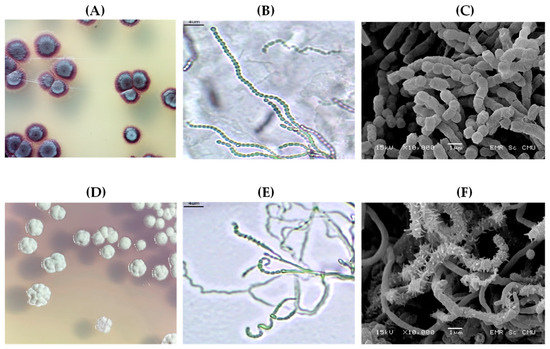
Based on the observations after 7 days of culturing on SCA at 30 °C, the UP-AC4 and UP-3.2 strains showed the typical morphological characteristics of the Streptomyces genus: Gram-positive with white aerial mycelium. Their morphological characteristics, spore arrangement, and ornamentation are shown in Figure 1 and Table 1. The UP-AC4 strain formed a white colony with soluble purple pigment, spores arranged in rectus flexibilis form, and a smooth spore surface. The UP-3.2 strain formed a white colony with soluble purple pigment in the media, spores arranged in a spiral, and a spiny spore surface.
Figure 1.
Morphological characteristics of the UP-AC4 and UP-3.2 strains grown on SCA at 30 °C for 7 days. The UP-AC4 strain formed a white colony with soluble purple pigment (A), spores arranged in rectus flexibilis form (B), and a smooth spore surface (C). The UP-3.2 strain formed a white colony with soluble purple pigment in the media (D), spores arranged in a spiral (E), and a spiny spore surface (F). The images were captured using a light microscope at 100× magnification (B,E), and the SEM micrographs (C,F) were captured at 10,000× magnification.
Table 1.
Differential physiological and biochemical characteristics of the UP-AC4, UP-3.2, and reference strains (S. californicus, S. microflavus, S. purpurascens, and S. thermocarboxydus).
We next investigated the chemotaxonomic characteristics of the UP-AC4 and UP-3.2 strains. Their physiological and biochemical characteristics were compared to reference strains, including S. californicus, S. microflavus, and S. purpurascens (Table 1). Notably, S. californicus, S. microflavus, and S. purpurascens were the reference strains for actinobacterial species listed in Bergey’s Manual of Systematic Bacteriology [21], and S. thermocarboxydus was taken from previous reports [35,36,37].
Moreover, analysis of the cell wall composition revealed that strains UP-AC4 and UP-3.2 contained LL-diaminopimelic acid (LL-DAP) as the diagnostic diamino acid in the peptidoglycan (Figure S1A). The whole-cell sugar analysis indicated the presence of xylose, mannose, and glucose as the major sugars in both strains (Figure S1B). In addition, the lipid staining profiles demonstrated that isolates UP-AC4 and UP-3.2 possessed a complex mixture of phospholipids, aminolipids, and glycolipids, which are consistent with the chemotaxonomic characteristics commonly observed in members of the genus Actinobacteria (Figure S2).
ARDRA is an excellent molecular genome typing method for classifying Actinomycetes at the genus level [28]. Therefore, we next identified the genus of the UP-AC4 and UP-3.2 by analyzing their restriction digestion profiles with the Bsp143l and Kpnl endonucleases. Their ARDRA fingerprint profiles are shown in Supplemental Figure S3. A series of dichotomous keys have been created to help quickly identify genera based on Bsp143l digestion products. The ARDRA fingerprint profile for Bsp143l digestion, in which the largest DNA fragments were <750 bp, correlated with the genera Glycomyces, Gordonia, Kitasatospora, Microbispora, Nocadia, Nocardioides, Nonomuraea, Pseudonocardia, Skermania, Sporichthya, and Streptomyces. The UP-AC4 strain showed three bands (637, 257, and 155 bp), and the UP-3.2 strain showed two bands (644 and 157 bp). In addition, their KpnI digestions produced DNA fragments in two bands (1000–1100 and 410–470 bp), correlating to the genera Kitasatospora, Sporichthya, and Streptomyces. The UP-AC4 strain showed three DNA fragments (1024, 457, and 264 bp), and the UP-3.2 strain showed two DNA fragments (1024 and 457 bp).
While the ARDRA could not clearly differentiate the genus Streptomyces from Kitasatospora and Sporichthya, the morphology of the UP-AC4 and UP-3.2 strains did distinguish them [21]. Therefore, the UP-AC4 and UP-3.2 strains could be classified as in the genus Streptomyces.
The 16S rDNA sequences of the UP-AC4 and UP-3.2 strains were determined and submitted to GenBank (accession numbers ON629791.1 [1343 bp] and ON629763.1 [1339 bp], respectively). The sequences were first analyzed using the Basic Local Alignment Search Tool (BLAST, NCBI web interface, accessed in October 2023) and then aligned with those of Streptomyces-type strains available in the GenBank database, which confirmed their identification at the genus level. The top-hit species of BLAST searches [38] showed that the 1343 bp 16S rDNA sequence of the UP-AC4 strain had 99–100% query coverage with 99.85–100% identity with 39 accession numbers belonging to seven Streptomyces species (Table S1): S. californicus (30/39), S. microflavus (3/39), S. mediolani (2/39), S. violaceoruber (1/39), S. cavourensis (1/39), S. griseus (1/39), and S. vinaceus (1/39). Therefore, S. californicus was the closest species, accounting for 76.92% of the total BLAST hits. The similarity analysis of the 1339 bp sequence of UP-3.2 strain showed 99–100% query coverage with 99.25–100% identity with 38 accession numbers belonging to nine Streptomyces species (Table S2): S. purpurascens (11/34), S. thermocarboxydus (11/34), S. spinoverrucosus (3/34), S. bellus (2/34), S. violaceus (2/34), S. indiaensis (2/34), S. coeruleorubidus (1/34), S. parvulus (1/34), and S. coerulescens (1/34). While the top-hit species, S. purpurascens and S. thermocarboxydus, had the same number of hits (32.35%), their chemotaxonomy differed. Therefore, the UP-3.2 strain could be classified as S. purpurascens.
Notably, the BLAST search generates pairwise alignments between a query and the database sequences that have been searched but does not compute an alignment between the various database sequences explicitly. Therefore, the proposed hierarchy of 16S rDNA sequences may vary along different major branches of the dendrogram and will be heavily influenced by phenotypic consistency at each level. Therefore, we next determined the numerical similarity of the UP-AC4 and UP-3.2 strains’ phenotypes to those of the reference strains using SSM calculated based on 27 units of morphology and biochemical tests and clustering with the UPGMA algorithm (Table 2). The similarity matrix for the UP-AC4 strain was compared to the hypothetical species from the top BLAST hit species (100% similarity). The analysis indicated that the best match species were S. californicus and its derivative data (S. californicus_d) with 90–100% similarity. The UP-AC4 strain showed only 50% phenotypic similarity with S. microflavus but 100% similarity with S. californicus. Therefore, the UP-AC4 strain was concluded to be S. californicus. Similar results were obtained for the UP-3.2 strain, which was phenotypically closer to S. purpurascens than S. thermocarboxydus with 90–100% and 45% phenotypic identity, respectively. Therefore, the UP-3.2 strain was concluded to be S. purpurascens.
Table 2.
The SSM of the UP-AC4 and UP-3.2 strains with the reference strains (S. californicus, S. microflavus, S. purpurascens, and S. thermocarboxydus).
3.2. Phylogenetic Relationships of the UP-AC4 and UP-3.2 Strains Based on 16S rDNA Sequences
Since the 16S rDNA sequence data is important for identifying Streptomyces species, a phylogenetic tree analysis could determine the relationships between S. californicus strain UP-AC4 and S. purpurascens strain UP-3.2 with other Streptomyces species. Therefore, we next constructed a phylogenetic tree using the NJ algorithm.
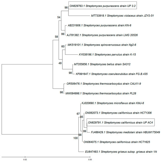
The phylogenetic tree included the closest 11 Streptomyces species represented by 16 nucleotide sequences: S. purpurascens (AB231806.1 and AJ781382), S. spinoverrucosus (MK519101.1), S. thermocarboxydus (OR506476.1 and MW084998.1), S. californicus (ON062073.1), S. parvulus (KY038196.1), S. bellus (MT355856.1), Streptomyces coeluleorubidus (KF991647.1), S. griseus (EU647493.1), S. microflavus (KJ020690.1), S. mediolani (FJ486429.1), and S. violaceus (MT733918.1). The optimal tree is shown in Figure 2. All ambiguous positions were removed for each sequence pair (pairwise deletion option). There were 1505 positions in the final dataset. The evolutionary analysis indicated that S. californicus strain UP-AC4 and S. purpurascens strain UP-3.2 had different taxonomic positions. Notably, S. purpurascens strain UP-3.2 is in a separate clade in the tree with S. thermocarboxydus, with which it shared a 16s rDNA sequence similarity of 99.33% and a top-hit taxon of 100%.
Figure 2.
A phylogenetic tree of the UP-AC4 and UP-3.2 strains with closely related species constructed using the NJ method based on 16S rDNA sequences. Numbers at the nodes represent bootstrap values expressed as percentages of 1000 replicates. The scale bar corresponds to 0.02 substitutions per nucleotide position.
Altogether, these molecular-based identifications indicated that the UP-AC4 and UP-3.2 strains predominantly belonged to the genus Streptomyces and correlated with S. californicus and S. purpurascens, respectively.
3.3. Description of the Streptomyces UP Strains
S. californicus strain UP-AC4 is a Gram-positive, aerobic actinobacteria. Their morphological characteristics showed that the aerial mycelium was colorless or white, and the vegetative mycelium appeared a violet-pink color on the SCA. Each rectus-flexibilis spore chain has over 20 spores, a gray series, and a smooth spore surface. S. californicus strain UP-AC4 produced a violet-soluble pigment on starch casein media but not melanin. Moreover, it produced catalase, amylase, and urease but not gelatinase. It could reduce nitrates, but it could not produce indole and hydrogen sulfide. It could utilize cellobiose, dulcitol, fructose, galactose, glucose, and mannitol carbon sources but not arabinose, inositol, raffinose, rhamnose, sucrose, trehalose, or citrate. Chemotaxonomically, UP-AC4 contains LL-diaminopimelic acid in its peptidoglycan, with xylose, mannose, and glucose as the major whole-cell sugars, and a mixture of phospholipids, aminolipids, and glycolipids.
S. purpurascens strain UP-3.2 showed the typical characteristics of the genus Streptomyces: Gram-positive, aerobic, white aerial mycelium, spiral spore chains, and a spiny spore surface. This strain could produce violet diffusible pigments but did not appear to produce melanoid pigments. It could utilize arabinose, cellobiose, dulcitol, fructose, galactose, glucose, inositol, mannitol, raffinose, rhamnose, sucrose, trehalose, and xylose as carbon sources but not citrate. It produced catalase, amylase, tryptophanase (tryptophan indole-lyase), and urease but did not appear to reduce nitrates. The gelatinase and hydrogen sulfide tests were negative. Chemotaxonomically, UP-3.2 displays the same profile as UP-AC4, containing LL-diaminopimelic acid in its peptidoglycan, with xylose, mannose, and glucose as the major whole-cell sugars, and a mixture of phospholipids, aminolipids, and glycolipids.
3.4. Antibacterial Activity of S. californicus Strain UP-AC4 and S. purpurascens Strain UP-3.2
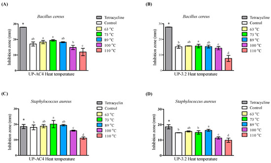
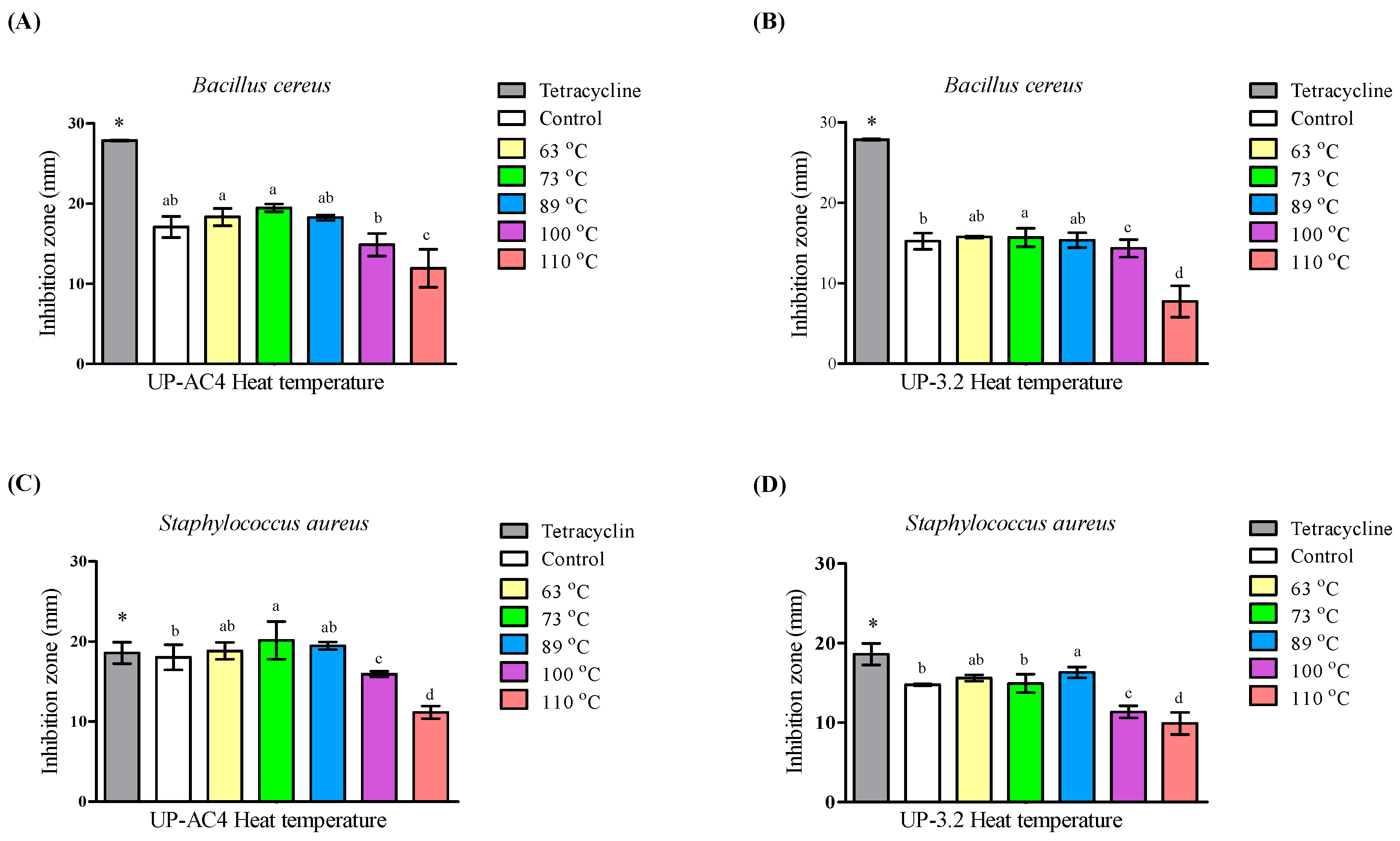
We next investigated the bioactivity of the secondary metabolites of the UP-AC4 and UP-3.2 strains extracted from cell culture broths by heating. Since heat treatment excluded the bioactive compounds from mycelium cells in the suspension, the results demonstrated that pasteurization temperatures could extract secondary metabolites while also stabilizing their antibiotic effect. The agar disc diffusion results of antibacterial activity are shown in Figure 3. Crude extracts of both UP-AC4 and UP-3.2 could inhibit the growth of Gram-positive bacteria with and without endospores (B. cereus and S. aureus, respectively). However, they were ineffective against Gram-negative bacteria (E. coli).
Figure 3.
Antimicrobial activity assessed using the agar disc diffusion assay. Metabolites from S. californicus strain UP-AC4 and S. purpurascens strain UP-3.2 were extracted by heating at various temperatures: 63 °C for 30 min or 73 °C, 89 °C, 100 °C, or 110 °C for 10 min. These crude extracts were applied to the test organisms B. cereus (A,B) and S. aureus (C,D) at 25 mg/mL per disc. The diameter of the inhibition zone (mean ± SD) was measured in triplicate experiments. Tetracycline (30 µg/mL) served as the positive control, and MHB served as the negative control. Different lowercase letters indicate significant differences among treatments (p < 0.05). An asterisk (*) indicates a significant difference compared with the control (p < 0.05).
The crude extract of UP-AC4 prepared by heat treatment at 73 °C for 10 min had the greatest inhibition effect against B. cereus (inhibition zone: 19.47 ± 0.2 mm) and S. aureus (inhibition zone: 20.16 ± 0.95 mm) among the pasteurizations. In contrast, sterilization treatments at 110 °C or 121 °C for 10 min had significantly decreased effects. A similar result was found with the crude extract of UP-3.2; the sterilized temperatures significantly decreased the antibacterial effect, and pasteurization treatments at 63 °C for 30 min or 73 °C or 89 °C for 10 min suppressed bacterial growth, with an inhibition zone between 15.36 ± 0.37 and 16.3 ± 0.27 mm.
We next evaluated the MIC and MBC of each crude extract to confirm the antimicrobial activity (Table 3). The MIC of both Gram-positive strains ranged from 0.024 to 12.5 mg/mL. The crude extract of UP-AC4 prepared by heat treatment at 73 °C for 10 min had the greatest effect against S. aureus, with a MIC of 0.024 mg/mL and an MBC of 0.098 mg/mL. It was also effective against B. cereus, with a MIC of 0.049 mg/mL and an MBC of 0.098 mg/mL. In contrast, the crude extract of UP-3.2 prepared by heat treatment at 89 °C for 10 min had the greatest effect against B. cereus, with a MIC of 0.049 mg/mL and an MBC of 0.098 mg/mL.
Table 3.
The susceptibility of B. cereus and S. aureus to crude heat extracts from S. californicus strain UP-AC4 and S. purpurascens strain UP-3.2.
The marked reduction in antibacterial activity observed at higher temperatures (≥89 °C) suggests thermal sensitivity of the bioactive metabolites, whereas retention of activity under mild pasteurization conditions (73 °C for 10 min) supports the use of this temperature as a practical compromise between extraction efficiency and compound stability. This is the first study to demonstrate that pasteurization conditions commonly used in the food preservation industry, applying a temperature below 100 °C for a short duration, effectively extract potent antibiotic substances from Streptomyces UP strains. Notably, heat extraction at 73 °C for 10 min released the bioactive compounds from the UP-AC4 strain, which showed 4–8-fold higher antibacterial efficiency than non-heat treatment (control), as indicated by the MIC values against B. cereus and S. aureus.
Altogether, these results indicate that Streptomyces strains UP-AC4 and UP-3.2 produce bioactive compounds capable of destroying both the vegetative cells and endospores of B. cereus with easy extraction through pasteurization at 73 °C for 10 min. Therefore, the crude extract from this pasteurization process was further investigated for anticancer activity.
3.5. Anticancer Effect of S. californicus Strain UP-AC4 and S. purpurascens Strain UP-3.2
Next, we screened the anticancer activity of crude heat extracts of the UP-AC4 and UP-3.2 strains against NSCLC cells (A549, H1299, and Lu99) using the MTT assay. Notably, lung cancer is one of the malignant tumors causing the highest mortality worldwide [39]. The effectiveness of the crude extracts varied between cancer cell lines (Table 4). The crude extract from UP-AC4 had the greatest effect on A549 cells (IC50: 0.65 ± 0.21 mg/mL), followed by H1299 (IC50:1.40 ± 0.53 mg/mL) and Lu99 (IC50: 4.85 ± 0.64 mg/mL) cells, whereas it was nontoxic in NHDF cells (IC50: 26.8 ± 1.25 mg/mL). In contrast, the crude extract from UP-3.2 had the greatest effect on Lu99 cells (IC50: 0.23 ± 0.06 mg/mL), followed by H1299 (IC50: 1.30 ± 0.26 mg/mL) and A549 (IC50: 4.13 ± 2.43 mg/mL) cells. Altogether, these results suggest that the crude extracts prepared from S. californicus strain UP-AC4 and S. purpurascens strain UP-3.2 by heating at 73 °C for 10 min could be a potential source of anticancer drugs.
Table 4.
The cytotoxic activity on NSCLC cells of the crude heat-associated extracts prepared at 73 °C for 10 min from S. californicus strain UP-AC4 and S. purpurascens strain UP-3.2.
3.6. HPLC Analysis of Crude Extracts
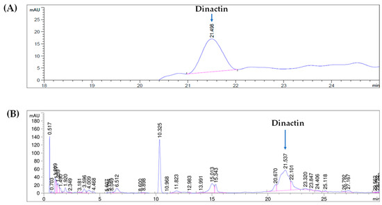
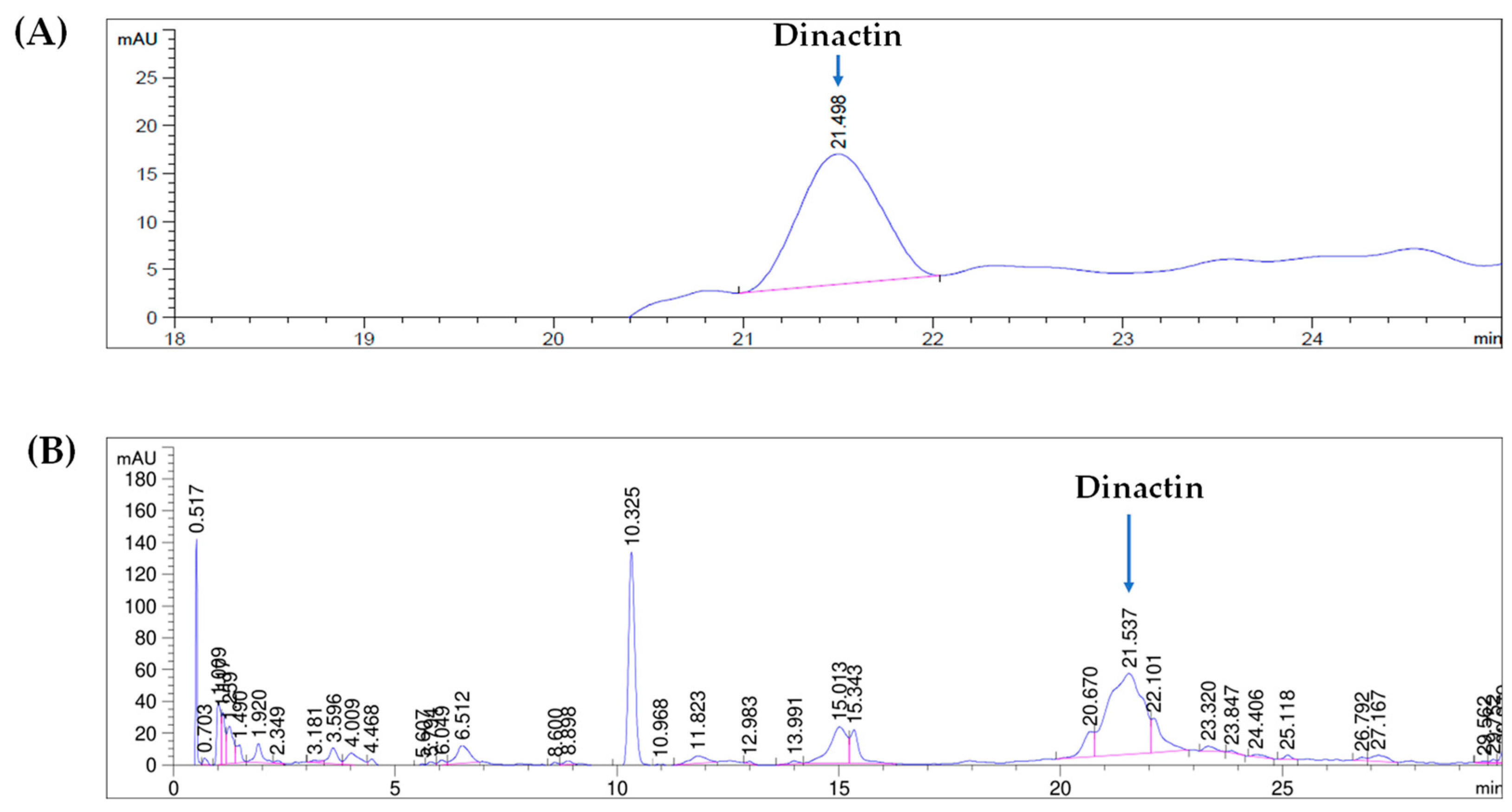
The potent bioactive metabolites present in the crude extracts of Streptomycete UP strains were analyzed using HPLC. Dinactin, a natural secondary metabolite and antitumor antibiotic produced by various Streptomyces species, was tentatively detected in S. californicus strain UP-AC4, which had been extracted by heating at 73 °C for 10 min. The representative HPLC chromatogram of the crude extract from strain UP-AC4 (Figure 4) revealed multiple peaks corresponding to diverse metabolites. The peak associated with dinactin was identified based on its retention time (~21.5 min) by comparison with an authenticated standard and exhibited high signal intensity, suggesting that this compound is a predominant metabolite in the crude extract at an approximate concentration of 0.7 mM. Additional minor peaks were also observed, indicating the presence of other secondary metabolites. Therefore, HPLC profiling suggests that dinactin is a predominant secondary metabolite in S. californicus strain UP-AC4, supporting its potential as a promising bioactive compound with antibiotic and anticancer properties.
Figure 4.
High-performance liquid chromatography (HPLC) chromatograms at 270 nm of dinactin standard (0.5 mM) (A) and crude extract metabolites from S. californicus strain UP-AC4 after heating at 73 °C for 10 min (B).
4. Discussion
Streptomyces is a genus of bacteria widely recognized for its significant benefits to various fields, including biology, medicine, agriculture, and industry. Streptomyces are prolific producers of antibiotics in their secondary metabolites, including well-known antibiotics such as streptomycin, erythromycin, and tetracycline [40]. In the context of industrial production, it is essential to prioritize Streptomyces strains that are easy to cultivate and from which bioactive compounds can be readily extracted [41]. In this study, we identified two new Streptomyces strains—S. californicus strain UP-AC4 and S. purpurascens strain UP-3.2—that produce dinactin, an antibiotic and anticancer compound, which can be efficiently extracted under pasteurization conditions by heating at 73 °C for 10 min.
Indeed, bioactive secondary metabolites are synthesized by Streptomyces and either retained within their cells or released into the culture broth [42]. Heat treatment disrupts cell membranes, releasing the bioactive compounds from the cells [43]. Biotechnological industrial production is primarily conducted through large-scale fermentation in bioreactors. Green extraction methods, such as heating under pasteurization conditions, offer a rapid and cost-effective approach. Therefore, these Streptomyces UP strains may hold promise as sources of antibiotic and anticancer compounds.
S. californicus, also known as Streptomyces puniceus, is known to produce diverse bioactive compounds, including antibiotics, enzymes, and various secondary metabolites. For example, Hussain et al. observed that S. puniceus strain AS13 produced a particularly effective compound called dinactin, which showed both antibiotic and anticancer properties [44]. Notably, dinactin is a novel antitumor compound that operates through various mechanisms, including inhibiting cancer proliferation, reducing clonogenic survival, inhibiting cell migration and invasion, blocking the Wnt/β-catenin signaling pathway in cancer cells, and inducing G0/G1 cell cycle arrest by downregulating cyclins A, B, D3 (CCND3), and cyclin-dependent kinase 2 (CDK2). Dinactin also functions as a cancer stem cell (CSC) inhibitor by suppressing the expression of CSC stemness markers, such as aldehyde dehydrogenase 1 family member A1 (ALDH1A1), Nanog homeobox (NANOG), POU class 5 homeobox 1 (POU5F1/OCT4), and SRY-box transcription factor 2 (SOX2) [32,33,45]. It is important to note that S. californicus strain UP-AC4 can produce dinactin, and this compound can be easily extracted by simple heat treatment at 73 °C for 10 min, offering a low-cost and efficient extraction approach. This heat-based method highlights the Streptomyces UP-strain’s potential for scalable production of dinactin, making it an attractive candidate for future biotechnological and pharmaceutical applications.
S. purpurascens is also recognized as a potent antibiotic producer. Holkar et al. reported that S. purpurascens, isolated from soil, primarily produces bioactive compounds from the rhodomycin group that are effective against Gram-positive bacteria [46]. Rhodomycin A has antitumor activity, inhibiting cancer cell proliferation, migration, invasion, and clonogenicity in vitro and tumor growth in vivo. This inhibition is achieved by suppressing multiple signaling pathways related to SRC proto-oncogene non-receptor tyrosine kinase (SRC), including phosphoinositide 3-kinase (PI3K), Janus kinase (JNK), paxillin (PXN), and BCAR1 scaffold protein Cas family member (BCAR1/p130cas), in lung cancer [47]. Therefore, the antibiotic anticancer activity of S. purpurascens strain UP-3.2 might be attributed to the presence of compounds similar to dinactin and rhodomycin.
Implications for Industrial Applications
Streptomyces UP strains, particularly S. californicus strain UP-AC4, demonstrate strong potential for industrial application through the efficient production of a dinactin-associated compound that can be readily recovered using a simple heat treatment at 73 °C for 10 min. This cost-effective and energy-efficient approach is compatible with existing commercial pasteurization systems and supports the feasibility of scalable upstream processing for pharmaceutical, antimicrobial, and anticancer applications. However, the presence of multiple secondary metabolites in the crude extract underscores the importance of rigorous downstream purification, analytical validation, and product standardization for industrial translation. As fraction collection and purification were not conducted in the present study, future work will require large-scale cultivation, preparative HPLC fractionation, and comprehensive structural elucidation to unequivocally confirm compound identity, including verification using a spike-in (standard addition) method. In addition, advanced analytical techniques such as LC-MS/MS and NMR will be essential to ensure batch-to-batch consistency, establish robust quality control parameters, and validate the correlation between the purified compound and its biological activities. Addressing these analytical and processing challenges will be critical for translating laboratory-scale findings into reliable, scalable, and economically viable industrial applications.
5. Conclusions
S. californicus strain UP-AC4 and S. purpurascens strain UP-3.2 appear to be promising sources of potential antibiotic and anticancer compounds. The optimal heat-assisted extraction conditions were determined to be 73 °C for 10 min, effectively targeting Gram-positive bacterial cells and their spores. The crude extract of UP-AC4 showed the highest anticancer activity against A549 cells, followed by H1299 and Lu99 cells. In contrast, the crude extract of UP-3.2 showed similar effectiveness against Lu99, H1299, and A549 cells. HPLC analysis further confirmed the presence of dinactin-associated compound—particularly as a major metabolite in S. californicus UP-AC4—supporting its role as a key bioactive compound contributing to the observed antibiotic and anticancer effects. Therefore, the antibiotic and anticancer activities of these Streptomyces crude extracts highlight their strong potential for pharmaceutical product development, especially through heat-assisted extraction and commercial sterilization approaches.
Supplementary Materials
The following supporting information can be downloaded at: https://www.mdpi.com/article/10.3390/microbiolres17010016/s1, Figure S1: Chemotaxonomic analysis of cell wall composition in strains UP-AC4 and UP-3.2. Figure S2: Polar lipid profiles of strains UP-AC4 (A) and UP-3.2 (B) analyzed by two-dimensional thin-layer chromatography. Figure S3: ARDRA fingerprint profiles of UP-AC4 and UP-3.2 strains from Bsp143I and KpnI restriction analysis. Table S1: The top-hit species of BLAST searches of the UP-AC4 strain; Table S2: The top-hit species of BLAST searches of the UP-3.2 strain.
Author Contributions
Conceptualization, G.P.-I. and A.R.; methodology, G.P.-I., S.T., C.P., K.S., S.S., T.T. and A.R.; software, G.P.-I., S.T. and A.R.; validation, G.P.-I. and A.R.; formal analysis, G.P.-I. and A.R.; investigation, A.R.; resources, G.P.-I. and A.R.; data curation, G.P.-I. and A.R.; writing—original draft preparation, G.P.-I. and A.R.; writing—review and editing, A.R.; visualization, G.P.-I. and A.R.; supervision, A.R.; project administration, G.P.-I. and A.R.; funding acquisition, G.P.-I. and A.R. All authors have read and agreed to the published version of the manuscript.
Funding
This research was funded by the School of Medical Sciences (grant no. MS221007), the Fundamental Fund (grant no. FF68-5086/2567), the University of Phayao, the Thailand Science Research and Innovation (TSRI) through the Program Management Unit for Competitiveness (PMUC), Naive Innova Co., Ltd. (grant no. C02F660119) that operated by the University of Phayao (grant no. R66095), and the Northern Science Park (NSP), Chiang Mai, through Technology to Industry Convergence (grant no. TIC002/2564), Thailand.
Institutional Review Board Statement
Not applicable.
Informed Consent Statement
Not applicable.
Data Availability Statement
All data generated or analyzed during this study are included in this article and its Supplementary Materials. Further inquiries can be directed to the corresponding author. The partial 16S rDNA sequence of S. californicus strain UP-AC4 has been deposited in GenBank under accession number ON629791.1. The partial 16S rDNA sequence of S. purpurascens strain UP-3.2 has been deposited in GenBank under accession number ON629763.1.
Acknowledgments
We extend our heartfelt appreciation to Masami Suganuma of the Graduate School of Science and Engineering at Saitama University, Saitama, Japan, for the laboratory support.
Conflicts of Interest
The authors declare no conflicts of interest.
Abbreviations
The following abbreviations are used in this manuscript:
| MIC | Minimum inhibitory concentration |
| MBC | Minimum bactericidal concentration |
| IC50 | The half maximal inhibitory concentration |
References
- Barka Essaid, A.; Vatsa, P.; Sanchez, L.; Gaveau-Vaillant, N.; Jacquard, C.; Klenk, H.-P.; Clément, C.; Ouhdouch, Y.; van Wezel Gilles, P. Taxonomy, Physiology, and Natural Products of Actinobacteria. Microbiol. Mol. Biol. Rev. 2015, 80, 1–43. [Google Scholar] [CrossRef] [Scilit] [PubMed]
- Donald, L.; Pipite, A.; Subramani, R.; Owen, J.; Keyzers, R.A.; Taufa, T. Streptomyces: Still the Biggest Producer of New Natural Secondary Metabolites, a Current Perspective. Microbiol. Res. 2022, 13, 418–465. [Google Scholar] [CrossRef] [Scilit]
- Lee, N.; Kim, W.; Hwang, S.; Lee, Y.; Cho, S.; Palsson, B.; Cho, B.-K. Thirty complete Streptomyces genome sequences for mining novel secondary metabolite biosynthetic gene clusters. Sci. Data 2020, 7, 55. [Google Scholar] [CrossRef] [Scilit] [PubMed]
- Narsing Rao, M.P.; Xiao, M.; Li, W.-J. Fungal and Bacterial Pigments: Secondary Metabolites with Wide Applications. Front. Microbiol. 2017, 8, 1113. [Google Scholar] [CrossRef] [Scilit]
- Sarmiento-Tovar, A.A.; Silva, L.; Sánchez-Suárez, J.; Diaz, L. Streptomyces-Derived Bioactive Pigments: Ecofriendly Source of Bioactive Compounds. Coatings 2022, 12, 1858. [Google Scholar] [CrossRef] [Scilit]
- Medema, M.H.; Breitling, R.; Takano, E. Synthetic biology in Streptomyces bacteria. Methods Enzymol. 2011, 497, 485–502. [Google Scholar] [CrossRef] [Scilit]
- Venil, C.K.; Dufossé, L.; Renuka Devi, P. Bacterial Pigments: Sustainable Compounds with Market Potential for Pharma and Food Industry. Front. Sustain. Food Syst. 2020, 4, 100. [Google Scholar] [CrossRef] [Scilit]
- Gao, Y.; Shang, Q.; Li, W.; Guo, W.; Stojadinovic, A.; Mannion, C.; Man, Y.G.; Chen, T. Antibiotics for cancer treatment: A double-edged sword. J. Cancer 2020, 11, 5135–5149. [Google Scholar] [CrossRef] [Scilit]
- Chifiriuc, M.C.; Filip, R.; Constantin, M.; Pircalabioru, G.G.; Bleotu, C.; Burlibasa, L.; Ionica, E.; Corcionivoschi, N.; Mihaescu, G. Common themes in antimicrobial and anticancer drug resistance. Front. Microbiol. 2022, 13, 960693. [Google Scholar] [CrossRef] [Scilit]
- Kingwell, K. Antibiotics switch to anticancer target. Nat. Rev. Drug Discov. 2021, 20, 666. [Google Scholar] [CrossRef] [Scilit]
- Demain, A.L.; Vaishnav, P. Natural products for cancer chemotherapy. Microb. Biotechnol. 2011, 4, 687–699. [Google Scholar] [CrossRef] [Scilit]
- Mann, J. Natural products in cancer chemotherapy: Past, present and future. Nat. Rev. Cancer 2002, 2, 143–148. [Google Scholar] [CrossRef] [Scilit] [PubMed]
- Law, J.W.-F.; Law, L.N.-S.; Letchumanan, V.; Tan, L.T.-H.; Wong, S.H.; Chan, K.-G.; Ab Mutalib, N.-S.; Lee, L.-H. Anticancer Drug Discovery from Microbial Sources: The Unique Mangrove Streptomycetes. Molecules 2020, 25, 5365. [Google Scholar] [CrossRef] [Scilit]
- Khazir, J.; Riley, D.L.; Pilcher, L.A.; De-Maayer, P.; Mir, B.A. Anticancer agents from diverse natural sources. Nat. Prod. Commun. 2014, 9, 1655–1669. [Google Scholar] [CrossRef] [Scilit]
- Nemer, G.; Louka, N.; Vorobiev, E.; Salameh, D.; Nicaud, J.-M.; Maroun, R.G.; Koubaa, M. Mechanical Cell Disruption Technologies for the Extraction of Dyes and Pigments from Microorganisms: A Review. Fermentation 2021, 7, 36. [Google Scholar] [CrossRef] [Scilit]
- Rizkiyah, D.N.; Putra, N.R.; Yunus, M.A.C.; Veza, I.; Irianto, I.; Aziz, A.H.A.; Rahayuningsih, S.; Yuniarti, E.; Ikhwani, I. Insight into Green Extraction for Roselle as a Source of Natural Red Pigments: A Review. Molecules 2023, 28, 1336. [Google Scholar] [CrossRef] [Scilit]
- Astudillo-Barraza, D.; Oses, R.; Henríquez-Castillo, C.; Vui Ling Wong, C.M.; Pérez-Donoso, J.M.; Purcarea, C.; Fukumasu, H.; Fierro-Vásquez, N.; Pérez, P.A.; Lavin, P. Apoptotic Induction in Human Cancer Cell Lines by Antimicrobial Compounds from Antarctic Streptomyces fildesensis (INACH3013). Fermentation 2023, 9, 129. [Google Scholar] [CrossRef] [Scilit]
- Kaaniche, F.; Hamed, A.; Elleuch, L.; Chakchouk-Mtibaa, A.; Smaoui, S.; Karray-Rebai, I.; Koubaa, I.; Arcile, G.; Allouche, N.; Mellouli, L. Purification and characterization of seven bioactive compounds from the newly isolated Streptomyces cavourensis TN638 strain via solid-state fermentation. Microb. Pathog. 2020, 142, 104106. [Google Scholar] [CrossRef] [Scilit]
- Chanthasena, P.; Hua, Y.; Rosyidah, A.l.; Pathom-Aree, W.; Limphirat, W.; Nantapong, N. Isolation and Identification of Bioactive Compounds from Streptomyces actinomycinicus PJ85 and Their In Vitro Antimicrobial Activities against Methicillin-Resistant Staphylococcus aureus. Antibiotics 2022, 11, 1797. [Google Scholar] [CrossRef] [Scilit]
- Pook-In, G.; Seansupa, K.; Upakut, S. Inhibition of Staphylococcus aureus by the cotton fabrics treated with the crude finish produced from Streptomyces sp. strain AC4. J. Food Health Bioenviron. Sci. 2019, 12, 44–53. [Google Scholar]
- Parte, A.; Whitman, W.B.; Goodfellow, M.; Kämpfer, P.; Busse, H.J.; Trujillo, M.E.; Ludwig, W.; Suzuki, K. Bergey’s Manual of Systematic Bacteriology: Volume 5: The Actinobacteria; Springer: New York, NY, USA, 2012. [Google Scholar]
- Yosboonruang, A.; Kiddee, A.; Siriphap, A.; Pook-In, G.; Suwancharoen, C.; Duangjai, A.; Praphasawat, R.; Reuk-ngam, N.; Nawong, S.; Rawangkan, A. Potential of Cannabidiol (CBD) to overcome extensively drug-resistant Acinetobacter baumannii. BMC Complement. Med. Ther. 2025, 25, 308. [Google Scholar] [CrossRef] [Scilit]
- Rawangkan, A.; Yosboonruang, A.; Kiddee, A.; Siriphap, A.; Pook-In, G.; Praphasawat, R.; Saokaew, S.; Duangjai, A. Restoring Ampicillin Sensitivity in Multidrug-Resistant Escherichia coli Following Treatment in Combination with Coffee Pulp Extracts. J. Microbiol. Biotechnol. 2023, 33, 1179–1188. [Google Scholar] [CrossRef] [Scilit]
- Hasegawa, T.; Takizawa, M.; Tanida, S. A rapid analysis for chemical grouping of aerobic actinomycetes. J. Gen. Appl. Microbiol. 1983, 29, 319–322. [Google Scholar] [CrossRef] [Scilit]
- Komagata, K.; Suzuki, K.-I. 4 Lipid and cell-wall analysis in bacterial systematics. In Methods in Microbiology; Elsevier: Amsterdam, The Netherlands, 1988; Volume 19, pp. 161–207. [Google Scholar]
- Minnikin, D.E.; O’Donnell, A.G.; Goodfellow, M.; Alderson, G.; Athalye, M.; Schaal, A.; Parlett, J. An integrated procedure for the extraction of bacterial isoprenoid quinones and polar lipids. J. Microbiol. Methods 1984, 2, 233–241. [Google Scholar] [CrossRef] [Scilit]
- Khadayat, K.; Sherpa, D.D.; Malla, K.P.; Shrestha, S.; Rana, N.; Marasini, B.P.; Khanal, S.; Rayamajhee, B.; Bhattarai, B.R.; Parajuli, N. Molecular Identification and Antimicrobial Potential of Streptomyces Species from Nepalese Soil. Int. J. Microbiol. 2020, 2020, 8817467. [Google Scholar] [CrossRef] [Scilit]
- Cook, A.E.; Meyers, P.R. Rapid identification of filamentous actinomycetes to the genus level using genus-specific 16S rRNA gene restriction fragment patterns. Int. J. Syst. Evol. Microbiol. 2003, 53, 1907–1915. [Google Scholar] [CrossRef] [Scilit]
- Frank, J.A.; Reich, C.I.; Sharma, S.; Weisbaum, J.S.; Wilson, B.A.; Olsen, G.J. Critical evaluation of two primers commonly used for amplification of bacterial 16S rRNA genes. Appl. Environ. Microbiol. 2008, 74, 2461–2470. [Google Scholar] [CrossRef] [Scilit]
- Tamura, K.; Stecher, G.; Kumar, S. MEGA11: Molecular Evolutionary Genetics Analysis Version 11. Mol. Biol. Evol. 2021, 38, 3022–3027. [Google Scholar] [CrossRef] [Scilit] [PubMed]
- CLSI M100; Performance Standards for Antimicrobial Susceptibility Testing. Clinical and Laboratory Standards Institute: Wayne, PA, USA, 2024.
- Balouiri, M.; Sadiki, M.; Ibnsouda, S.K. Methods for in vitro evaluating antimicrobial activity: A review. J. Pharm. Anal. 2016, 6, 71–79. [Google Scholar] [CrossRef] [Scilit]
- Rawangkan, A.; Wongsirisin, P.; Pook-In, G.; Siriphap, A.; Yosboonruang, A.; Kiddee, A.; Chuerduangphui, J.; Reukngam, N.; Duangjai, A.; Saokaew, S.; et al. Dinactin: A New Antitumor Antibiotic with Cell Cycle Progression and Cancer Stemness Inhibiting Activities in Lung Cancer. Antibiotics 2022, 11, 1845. [Google Scholar] [CrossRef] [Scilit]
- Zhou, J.; He, H.; Wang, X.; Lu, J.; Zhou, X.; Cai, M.; Zhang, Y. Optimization of nutrients for dinactin production by a marine Streptomyces sp. from the high latitude Arctic. Biotechnol. Bioprocess Eng. 2015, 20, 725–732. [Google Scholar] [CrossRef] [Scilit]
- Kim, S.B.; Falconer, C.; Williams, E.; Goodfellow, M. Streptomyces thermocarboxydovorans sp. nov. and Streptomyces thermocarboxydus sp. nov., two moderately thermophilic carboxydotrophic species from soil. Int. J. Syst. Bacteriol. 1998, 48, 59–68. [Google Scholar] [CrossRef] [Scilit]
- Kim, B.; Sahin, N.; Minnikin, D.E.; Zakrzewska-Czerwinska, J.; Mordarski, M.; Goodfellow, M. Classification of thermophilic streptomycetes, including the description of Streptomyces thermoalcalitolerans sp. nov. Int. J. Syst. Bacteriol. 1999, 49, 7–17. [Google Scholar] [CrossRef] [Scilit]
- Kämpfer, P.; Kroppenstedt, R.M.; Dott, W. A numerical classification of the genera Streptomyces and Streptoverticillium using miniaturized physiological tests. Microbiology 1991, 137, 1831–1891. [Google Scholar] [CrossRef] [Scilit]
- Bizuye, A.; Gedamu, L.; Bii, C.; Gatebe, E.; Maina, N. Molecular-Based Identification of Actinomycetes Species That Synthesize Antibacterial Silver Nanoparticles. Int. J. Microbiol. 2020, 2020, 8816111. [Google Scholar] [CrossRef] [Scilit]
- Sung, H.; Ferlay, J.; Siegel, R.L.; Laversanne, M.; Soerjomataram, I.; Jemal, A.; Bray, F. Global Cancer Statistics 2020: GLOBOCAN Estimates of Incidence and Mortality Worldwide for 36 Cancers in 185 Countries. CA A Cancer J. Clin. 2021, 71, 209–249. [Google Scholar] [CrossRef] [Scilit]
- Alam, K.; Mazumder, A.; Sikdar, S.; Zhao, Y.-M.; Hao, J.; Song, C.; Wang, Y.; Sarkar, R.; Islam, S.; Zhang, Y.; et al. Streptomyces: The biofactory of secondary metabolites. Front. Microbiol. 2022, 13, 968053. [Google Scholar] [CrossRef] [Scilit]
- Sekurova, O.N.; Schneider, O.; Zotchev, S.B. Novel bioactive natural products from bacteria via bioprospecting, genome mining and metabolic engineering. Microb. Biotechnol. 2019, 12, 828–844. [Google Scholar] [CrossRef] [Scilit] [PubMed]
- Barbuto Ferraiuolo, S.; Cammarota, M.; Schiraldi, C.; Restaino, O.F. Streptomycetes as platform for biotechnological production processes of drugs. Appl. Microbiol. Biotechnol. 2021, 105, 551–568. [Google Scholar] [CrossRef] [Scilit] [PubMed]
- Ebrahimi, A.; Csonka, L.N.; Alam, M.A. Analyzing Thermal Stability of Cell Membrane of Salmonella Using Time-Multiplexed Impedance Sensing. Biophys. J. 2018, 114, 609–618. [Google Scholar] [CrossRef] [Scilit]
- Hussain, A.; Rather, M.A.; Dar, M.S.; Dangroo, N.A.; Aga, M.A.; Qayum, A.; Shah, A.M.; Ahmad, Z.; Dar, M.J.; Hassan, Q.P. Streptomyces puniceus strain AS13., Production, characterization and evaluation of bioactive metabolites: A new face of dinactin as an antitumor antibiotic. Microbiol. Res. 2018, 207, 196–202. [Google Scholar] [CrossRef] [Scilit]
- Hussain, A.; Dar, M.S.; Bano, N.; Hossain, M.M.; Basit, R.; Bhat, A.Q.; Aga, M.A.; Ali, S.; Hassan, Q.P.; Dar, M.J. Identification of dinactin, a macrolide antibiotic, as a natural product-based small molecule targeting Wnt/β-catenin signaling pathway in cancer cells. Cancer Chemother. Pharmacol. 2019, 84, 551–559. [Google Scholar] [CrossRef] [Scilit] [PubMed]
- Holkar, S.; Begde, D.; Nashikkar, N.; Kadam, T.; Upadhyay, A. Rhodomycin analogues from Streptomyces purpurascens: Isolation, characterization and biological activities. Springerplus 2013, 2, 93. [Google Scholar] [CrossRef] [Scilit] [PubMed]
- Lai, Y.H.; Chen, M.H.; Lin, S.Y.; Lin, S.Y.; Wong, Y.H.; Yu, S.L.; Chen, H.W.; Yang, C.H.; Chang, G.C.; Chen, J.J. Rhodomycin A, a novel Src-targeted compound, can suppress lung cancer cell progression via modulating Src-related pathways. Oncotarget 2015, 6, 26252–26265. [Google Scholar] [CrossRef] [Scilit] [PubMed]

Disclaimer/Publisher’s Note: The statements, opinions and data contained in all publications are solely those of the individual author(s) and contributor(s) and not of MDPI and/or the editor(s). MDPI and/or the editor(s) disclaim responsibility for any injury to people or property resulting from any ideas, methods, instructions or products referred to in the content. |
© 2026 by the authors. Licensee MDPI, Basel, Switzerland. This article is an open access article distributed under the terms and conditions of the Creative Commons Attribution (CC BY) license.