


Abstract
Background: Urosepsis is a life-threatening condition accounting for approximately 20–30% of all sepsis cases and typically arises from ascending infection by uropathogenic Escherichia coli (UPEC). Disease progression is mediated by virulence factors, including adhesins, iron acquisition systems, and toxins. Among these, P fimbriae, particularly papGII adhesin subunit, have been implicated in the transition from uncomplicated urinary tract infection (UTI) to severe urosepsis. This study aimed to evaluate whether papGII carriage, alone or in combination with other UPEC virulence determinants and clinical risk factors, can predict urosepsis. Methods: A total of 60 paired Escherichia coli isolates from concurrent blood and urine samples of adults with clinical sepsis were collected between January and June 2024. Control isolates were obtained from patients with cystitis (n = 28) and pyelonephritis (n = 32). Polymerase chain reaction (PCR) assays were used to detect fifteen virulence-associated genes, including the pap operon (with papG allelic variants), the type 1 fimbriae (fimH), S fimbriae (sfaS), curli fimbriae (csgA), afa/Dr adhesin operon genes, cytotoxic necrotizing factor 1 (cnf1), and the aerobactin biosynthesis (iucD) and receptor (iutA) genes. Associations between gene carriage and clinical groups were analyzed using chi-square tests. Results: The incidence of urosepsis increased with age, peaking in the 60–69-year age group. Renal disease and catheterization were identified as significant risk factors (p < 0.05). More than 95% of UPEC isolates carried the csgA gene associated with biofilm formation and the iucD gene. The α- hemolysin toxin (hlyA) was significantly associated with urosepsis [X2(1, N = 120) = 6.62, p = 0.03]. No significant differences were observed in the carriage of papA, papC, or fimH. Although papGII was present in 65% of urosepsis-associated UPEC isolates, it did not demonstrate a statistically significant independent association with urosepsis [p = 0.1]. Conclusion: This study demonstrates that while papGII may contribute to the pathogenic potential of UPEC and facilitate systemic infection, it is not a reliable independent predictor of urosepsis.
1. Introduction
Urosepsis is a complex life-threatening syndrome caused by an inflammatory response to a systemic infection originating in the urogenital tract [1]. It accounts for approximately 25% of all sepsis cases in adults and is associated with a mortality rate of 20–40%, underscoring the need for early diagnosis and timely management [2]. Gram-negative coliforms are the predominant causative agents of urinary tract infections (UTIs), with Escherichia coli (E. coli) responsible for nearly half of all cases. Other commonly implicated organisms include Klebsiella spp., Enterococcus spp., Proteus spp., and Pseudomonas aeruginosa [1].
UTIs most commonly arise from an ascending infection following perineal colonization by intestinal or skin microbiota. Alternatively, direct inoculation of the urinary tract may occur during catheterization, particularly in the setting of suboptimal aseptic techniques [3]. The pathogenic potential of uropathogenic E. coli (UPEC) is largely attributed to the acquisition of virulence genes through horizontal gene transfer, mediated by mobile genetic elements such as pathogenicity islands, transposons, bacteriophages, and plasmids [4,5]. A key step in the pathogenesis of urosepsis is the ability of UPEC to adhere and colonize the uroepithelium. This is facilitated by fimbrial and afimbrial adhesins [6,7]. Additional virulence mechanisms include iron acquisition via siderophores and heme receptors, as well as the production of toxins, such as α-hemolysin and cytotoxic necrolyzing factor 1, and serum resistance ability, which contribute to immune evasion and tissue damage [6,7]. P fimbriae are extensively studied virulence factors in UPEC and are strongly implicated in invasive diseases [7]. P fimbriae are encoded by a pyelonephritis-associated pilus (pap) operon, consisting of subunit proteins papC, papA, papK, papE, papF, and papG. The papG adhesin, located at the tip of the P fimbriae, mediates bacterial attachment to uroepithelial cells. Four allelic variants of papG (I–IV) have been described. Among these, papGI is rarely detected in UTIs, while papGIV remains insufficiently characterized. In contrast, papGII is frequently identified in UPEC isolates associated with urosepsis, with reported prevalence rates of 64–72% [8,9,10]. This adhesin preferentially binds to Gb04 glycosphingolipid receptors on the uroepithelial cells and has been linked to progression from lower UTIs to acute pyelonephritis and urosepsis. The human kidney cells express both globo-series glycosphingolipid Gb04 and Gb03 receptors, with Gb03 present in more abundance than Gb04. In contrast, bladder epithelial cells express Gb04 and Gb05 receptors, with Gb04 again less abundant. While both papGII and papGIII bind to Gb04 receptors, papGIII additionally has the ability to bind to Gb05 receptors. This results in a strong association of papGII with pyelonephritis and bacteremia, and papGIII with cystitis. In 2023, a genome-wide study (bGWAS) identified papGII as a key genetic determinant in the progression of UTIs to urosepsis, suggesting a potential role as a diagnostic or prognostic biomarker [11,12,13,14].
In this context, the present study aimed to evaluate the role of virulence gene carriage, particularly papGII, along with associated clinical risk factors, in the progression of E. coli UTIs to urosepsis. This study further explores whether papGII alone may serve as a predictor of urosepsis.
2. Methods
2.1. Study Design and Setting
This is a cross-sectional observational study conducted in the Department of Clinical Microbiology, Christian Medical College Hospital, Vellore, India.
2.2. Study Population and Isolate Selection
The inclusion criteria for the urosepsis group included E. coli isolated from blood cultures collected within 7 days before or after a urine culture demonstrating significant bacteriuria (>105 CFU/mL) with E. coli of an identical antibiogram in symptomatic patients [9]. These paired E. coli isolates from both the blood and midstream clean-catch urine samples were collected between January 2024 and June 2024, and stored in nutrient agar deeps until further testing.
An equal number of E. coli isolated in urine specimens from patients diagnosed with clinical cystitis and pyelonephritis during the same study period were included as controls for comparison of virulence gene carriage and associated risk factors.
All E. coli were identified by conventional quality-passed biochemicals and MALDI-TOF VITEK® MS, bioMérieux, Marcy-l’Étoile, France, Version 3.2.0.
2.3. Bacterial Processing and DNA Extraction
All isolates were sub-cultured onto quality-passed MacConkey agar plates to ensure purity. Genomic DNA was extracted from pure bacterial growth. A loopful of bacterial growth was suspended in 200 μL of normal saline, and DNA extraction was performed using the QIAmp DNA Mini Kit (Qiagen, Hilden, Germany) according to the manufacturer’s instructions. DNA was eluted in 100 μL of elution buffer and stored at −20 °C until use.
2.4. PCR Amplification of Virulence Genes
Conventional polymerase chain reaction (PCR) was performed using previously validated primers to detect papGII and other virulence-associated genes, including those encoding adhesins, toxins, and siderophores. Adhesin genes included P fimbriae—papGI, II, III, IVa, papA, and papC genes, type 1 fimbriae (fimH), afimbrial adhesin (afaC), S fimbriae—(sfaS), and curli fimbriae—(csgA); toxin genes included hlyA (α-hemolysin) and cnf1, while aerobactin synthesis genes included iutA and iucD [15,16,17].
PCR controls were included in each run. E. coli ATCC 35218 was used as a positive control strain for papA, papC, cnf1, sfaS, hlyA, csgA, and papGIII, while E. coli ATCC 25922 was used as a control for fimH, iutA/iucD, and papGII. In-house DNA controls were used for the detection of other targets.
PCR cycling conditions for papGI–IVa primers included an initial denaturation at 95 °C for 5 min, followed by cycles of denaturation at 94 °C for 60 s, annealing at 60 °C for 90 s, and extension at 72 °C for 90 s, with a final extension at 72 °C for 10 min, and a hold at 4 °C.
The PCR products were analyzed by electrophoresis on 2% agarose gels with a 100 bp DNA ladder to confirm expected amplicon sizes.
2.5. Clinical Data Collection
Clinical data was retrieved from the electronic medical records, including age, gender, residence, presenting symptoms, and comorbidities (diabetes mellitus, hypertension, malignancy, and renal disease), as well as a prior history of UTI, catheterization, and urinary calculi.
2.6. Statistical Analysis
Associations between virulence gene carriage (chiefly pap genes) and clinical risk factors were evaluated using the chi-square test. A p-value < 0.05 was considered statistically significant.
3. Results
Sixty paired isolates of E. coli from the urine and blood cultures submitted within one week of each other from 60 patients with clinical urosepsis were included in this study. Concomitantly, 32 E. coli from patients with pyelonephritis and 28 isolates from cystitis cases were included. The patient demographics and underlying risk factors were compared in the three clinical levels of infection.
3.1. Demographic Results
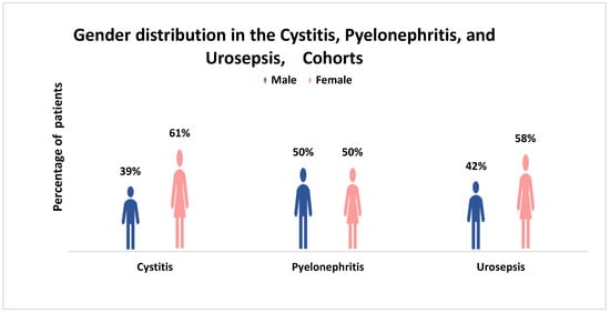
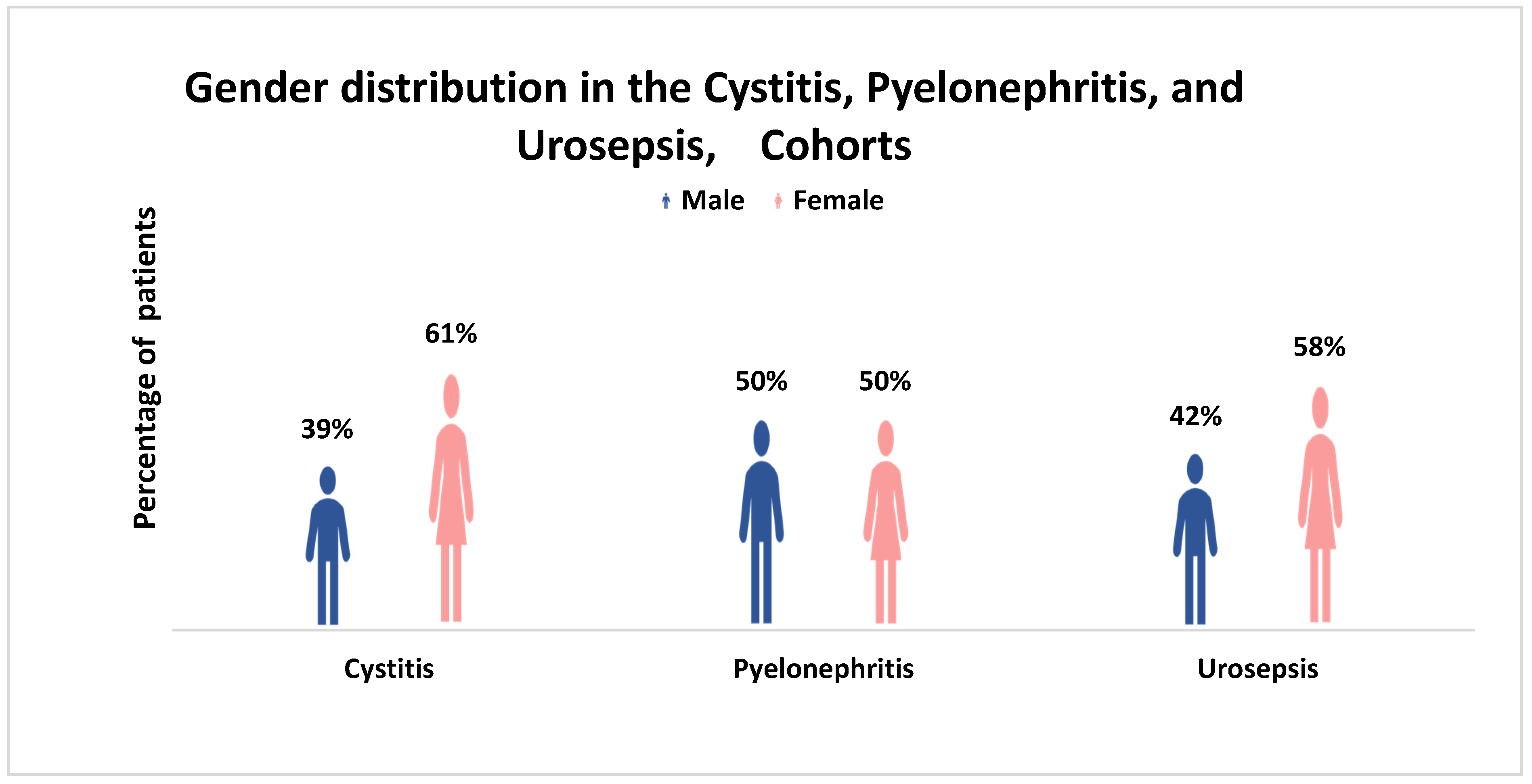
A female predominance was noted in the acquisition of both lower tract (61%) and invasive infections (58%) (Figure 1).
Figure 1.
Cis-Gender Distribution of UPEC-Isolated Patients Across Different Levels of Urinary Tract Infection at CMC, Vellore (January–June 2024).
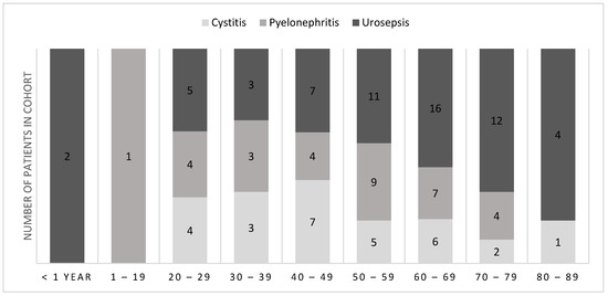
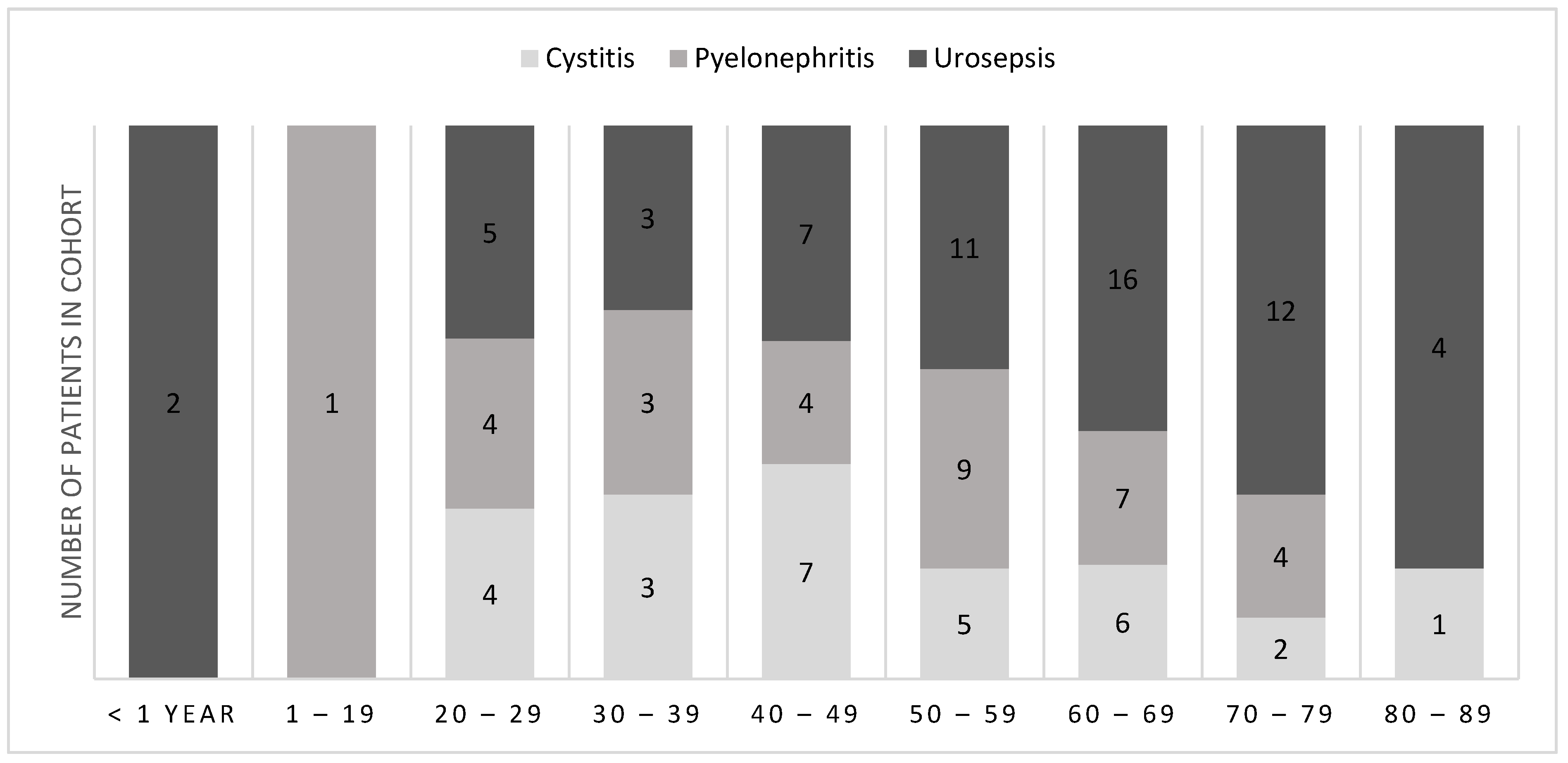
Clinical urosepsis was noted from the neonatal period to the elderly, steadily increasing with advancing age, with the peak occurrence in the 60–69 age group. Pyelonephritis was seen in the second decade and peaked in the sixth decade, while cystitis was seen among the 20–80 age group, peaking in the fifth decade (Figure 2).
Figure 2.
Age Distribution of UPEC-Isolated Patients Across Different Levels of Urinary Tract Infection at CMC, Vellore (January–June 2024).
Underlying risk factors contribute to the ascension of organisms and the development of urinary tract infections.
A chi-square test of independence was performed to examine the causal relation between risk factors and urosepsis. The presence of a catheter and the underlying renal disease were significant risk factors for the development of urosepsis (Table 1).
Table 1.
Risk Factors for Urosepsis, Cystitis, and Pyelonephritis.
3.2. Molecular Characterization of UPEC Virulence Genes by PCR
The adhesion-associated genes: papGII carriage progressively increased from the lower (42.9%) to upper tract (56.3%) to the isolates associated with urosepsis (66.7%). No amplifications were observed for papGI [17]. The pap operon papA and papC also increased with ascending infection and were found in 68.3% and 71.7% among urine isolates associated with urosepsis (Table 2).
Table 2.
Virulence gene carriage comparison in different levels of UTI.
The type 1 fimbriae adhesin gene fimH, with receptors throughout the tract, was found in 67.9% of cystitis-associated infections, 81.3% of pyelonephritis, and 85.0% of urosepsis. The core virulence factor essential for biofilm formation, curli fimbriae subunit csgA, was detected in almost all isolates, 98.7% of urine and 95% of blood urosepsis isolates, while 100% of isolates causing cystitis also carried the gene. Among the urosepsis strains, 66.6% (n = 40/60) possessed papGII, while 28.3% (n = 17/60) were papGII-negative isolates. (Table 3). Both the papGII and the papGII-negative strains isolated from urosepsis had a larger number and a similar type of virulence factors profile, such as iucD, iutA, csgA, and Fim H (Table 4), than those associated with cystitis and pyelonephritis.
Table 3.
The distribution of papG genes among the three levels of UTI.
Table 4.
Distribution of virulence genes in relation to the papG genes carriage.
Toxin genes: The α-hemolysin gene hlyA was identified in 48.3% of urine compared to 40.0% of blood isolates. A chi-square test of independence performed to examine the relation between hlyA and urosepsis, X2(1, n = 120) = 6.62, p = 0.03, was significant.
Iron uptake gene: survival essential genes, the aerobactin receptor iutA, and biosynthesis iucD gene were identified in all levels of infection, 81.7% from urine and blood isolates, 85.7% in lower tract infections, and highest in the upper tract, 90.6%.
4. Discussion
Uropathogenic E. coli express a wide range of virulence factors. Among these, P fimbriae encoded by the pap A-K operon are considered key determinants associated with ascending infection to pyelonephritis and urosepsis [10,18]. The adhesin papG located at the pilus tip mediates bacterial attachment to glycan receptors of the globo-series glycosphingolipids, including the Forssman antigen (Gb5), globotetraosylceramide (Gb4), and globotriaosylceramide (Gb3) [19]. Notably, the papGII allele has been found to be associated with 65% of bacteremia/urosepsis in bacterial genome-wide association studies (GWAS) [9,14].
In the present study, 65% of the UPEC isolates associated with urosepsis carried the papGII gene; however, this association was not statistically significant. These isolates displayed a higher virulence gene content, which is consistent with previous studies [20,21]. Owrangi et al. reported an increased adhesion and invasion among the urosepsis and hospital-acquired UTI isolates [21]. More than 90% of the isolates co-harbored papA, papC, iutA, iucD, and csgA compared with the papG-negative strains (Table 4). Further, more than 95% of papGII-positive strains carried papA and papC genes, reflecting the intact nature of the pap gene operon in these strains in contrast to papGIII or papG-negative isolates [20]. Importantly, 37% of papGII-negative isolates are also associated with urosepsis (Table 3), indicating that papGII carriage alone does not predict progression to urosepsis.
The type 1 fimbriae adhesin gene fimH binds to receptors throughout the urinary tract, including the desmoglein-2, on renal epithelial cells [22]. This interaction facilitates initial attachment, colonization, biofilm formation, and rapid bacterial replication, chiefly in the lower tract, while also contributing to ascending infections [15,16]. During early infection, fimH-dependent invasion of bladder epithelial cells enables formation of intracellular bacterial communities (IBCs), promoting bacterial persistence, immune evasion, and recurrent urinary tract infections [23].
Curli fimbriae encoded by csgA complements type 1 fimbriae (fimH) by promoting robust adherence and biofilm development. Curli fimbriae-associated biofilms improve UPEC survival by providing protection against host immune defenses, including cationic antimicrobial peptides. Consistent with previous studies, csgA-expressing strains demonstrate increased colonization efficiency and tissue damage in both the bladder and kidneys [24]. Although fimH and csgA are individually considered weaker biofilm contributors compared to sfa [25], their co-occurrence in 80% of the isolates suggests a synergistic role in colonization and persistence. Notably, among the papGII-positive isolates, 84% carried fimH, and 97% carried csgA, and among papG-negative isolates, 80.4% and 95.6% carried fimH and csgA, respectively. The comparable prevalence of these adhesins in both groups suggests that fimH and csgA may act as independent contributors to infection, persistence, and ascending spread, irrespective of papGII carriage.
The pore-forming toxin α-hemolysin (hlyA) has been consistently implicated in the UPEC pathogenicity, contributing to epithelial damage, inflammation, and renal injury [26]. In this study, hlyA showed a significant association with urosepsis (p = 0.03) and was present in 31.3% of pyelonephritis isolates, supporting its role as a marker of disease severity. These findings are consistent with prior reports linking hlyA expression to adverse clinical outcomes and progression to systemic disease [27].
The iron acquisition genes iutA and iucD co-existed in 84% of the total isolates, with 56.6% observed among papGII-positive strains and 27.5% among papG-negative strains. Their presence reflects adaptation to iron-limited host environments through carriage of iutA and iucD, siderophore-mediated uptake (aerobactin system) [9]. The widespread distribution of these genes across urosepsis, cystitis, and pyelonephritis groups likely reflects their co-localization on ColV virulence plasmids [28].
Overall, the presence of type 1 and type II adhesin gene carriage, combined with efficient iron acquisition systems and biofilm-forming capacity, highlights the invasive and persistence potential of UPEC across the spectrum of urinary tract infections. These findings suggest that progression to urosepsis is driven by a multifactorial interplay of virulence determinants, with individual strains exhibiting a distinct virulence gene profile [29].
In addition to bacterial factors, host susceptibility plays a critical role in determining infection outcomes [30,31,32]. Even strains lacking classical virulence determinants such as papGII may cause invasive disease in susceptible individuals, highlighting the interplay between host defenses and bacterial potential [10,33,34]. In the present study, the cis-female gender, an underlying renal disease, and catheterization were identified as significant risk factors for urosepsis (p < 0.05). This contrasts with previous reports by Prabhu et al. [17] and Bijou et al. [35], which identified diabetes mellitus as the primary risk factor; however, these studies focused on cases of urosepsis. In our cohort, although diabetes (55%) and renal disease (53.3%) were common, neither showed a statistically significant difference compared to cystitis cases. These observations reinforce the importance of host-related factors in UTI classification and progression [36].
In summary, the findings of this study suggest that while papGII may enhance the likelihood of systemic infection, it is not an independent predictor. Rather, progression to urosepsis appears to result from a complex interplay between bacterial virulence traits and host factors, including gender, renal disease, and indwelling medical devices. The bacterial genome-wide association studies (GWAS) have identified genetic variants linked to disease risk, which represent correlations, but further integrative approaches combining genomic, functional, and clinical data are required to better elucidate the determinants of UPEC pathogenicity and disease progression [37,38].
Study Limitation
The study limitation includes a smaller number of samples in both groups and the selection of 15 key virulence genes among the UPEC virulence factors.
5. Conclusions
papGII is enriched within a more virulent genetic background and is not an independent predictor of urosepsis. Future research must integrate genomic, functional, and clinical data while accounting for host comorbidities to better understand the biological mechanisms underlying urosepsis.
Author Contributions
Conceptualization, N.H., R.D.S., R.V. and B.V.; investigation, methodology, analysis, and visualization, N.H., S.T. and J.S.A.; supervision, R.D.S. and B.V.; writing, N.H., R.D.S., R.V., S.T. and J.S.A. All authors have read and agreed to the published version of the manuscript.
Funding
This research was funded by the institutional grant (The Fluid research Grant), Christian Medical College, Vellore.
Institutional Review Board Statement
This study was conducted in accordance with the Declaration of Helsinki, and the protocol was approved by the Christian Medical College, Vellore, Institutional Review Board and Ethics Committee. The number is IRB Min. No. 2502002 dated 12 February 2025.
Informed Consent Statement
An informed consent form is not applicable since this study did not recruit any subjects. The isolates were included in the study only when they were isolated from the specimens as part of routine diagnostics. The clinical diagnosis and other demographic details of the patients were retrospectively collected from the hospital LIMS system.
Data Availability Statement
The raw data supporting the conclusions of this article will be made available by the authors on request.
Acknowledgments
Selected sentences in this manuscript were reviewed for language clarity and grammatical accuracy with the assistance of the AI-based tool ChatGPT version-5.3. The tool was used solely for editing purposes and did not contribute to the conceptualization, analysis, or interpretation of the research.
Conflicts of Interest
The authors declare that they have no conflicts of interest to disclose.
References
- Guliciuc, M.; Maier, A.C.; Maier, I.M.; Kraft, A.; Cucuruzac, R.R.; Marinescu, M.; Şerban, C.; Rebegea, L.; Constantin, G.B.; Firescu, D. The urosepsis—A literature review. Medicina 2021, 57, 872. [Google Scholar] [CrossRef] [Scilit] [PubMed]
- Subramanian, A.; Bhat, S.; Mookkappan, S.; Anitha, P.; Kandasamy, R.; Kanungo, R. Empiric antibiotic and in-vitro susceptibility of urosepsis pathogens: Do they match? The outcome of a study from south India. J. Infect. Dev. Ctries. 2021, 15, 1346–1350. [Google Scholar] [CrossRef] [Scilit] [PubMed]
- Valiatti, T.B.; Santos, F.F.; Santos, A.C.; Nascimento, J.A.; Silva, R.M.; Carvalho, E.; Sinigaglia, R.; Gomes, T.A. Genetic and virulence characteristics of a hybrid atypical enteropathogenic and uropathogenic Escherichia coli (aEPEC/UPEC) strain. Front. Cell. Infect. Microbiol. 2020, 10, 492. [Google Scholar] [CrossRef] [Scilit] [PubMed]
- Schmidt, H.; Hensel, M. Pathogenicity islands in bacterial pathogenesis. Clin. Microbiol. Rev. 2004, 17, 14–56, Erratum in Clin. Microbiol. Rev. 2006, 19, 257. https://doi.org/10.1128/cmr.19.1.257.2006. [Google Scholar] [CrossRef] [Scilit] [PubMed]
- da Silva, L.C.; de Mello Santos, A.C.; Silva, R.M. Uropathogenic Escherichia coli pathogenicity islands and other ExPEC virulence genes may contribute to the genome variability of enteroinvasive E. coli. BMC Microbiol. 2017, 17, 68. [Google Scholar] [CrossRef] [Scilit] [PubMed]
- Firoozeh, F.; Saffari, M.; Neamati, F.; Zibaei, M. Detection of virulence genes in Escherichia coli isolated from patients with cystitis and pyelonephritis. Int. J. Infect. Dis. 2014, 29, 219–222. [Google Scholar] [CrossRef] [Scilit] [PubMed]
- Lane, M.C.; Mobley, H.L. Role of P-fimbrial-mediated adherence in pyelonephritis and persistence of uropathogenic Escherichia coli (UPEC) in the mammalian kidney. Kidney Int. 2007, 72, 19–25. [Google Scholar] [CrossRef] [Scilit] [PubMed]
- Shetty, A.V.; Kumar, S.H.; Shekar, M.; Shetty, A.K.; Karunasagar, I. Prevalence of adhesive genes among uropathogenic Escherichia coli strains isolated from patients with urinary tract infection in Mangalore. Indian J. Med. Microbiol. 2014, 32, 175–178. [Google Scholar] [CrossRef] [Scilit] [PubMed]
- Cuénod, A.; Agnetti, J.; Seth-Smith, H.M.; Roloff, T.; Wälchli, D.; Shcherbakov, D.; Akbergenov, R.; Tschudin-Sutter, S.; Bassetti, S.; Siegemund, M.; et al. Bacterial genome-wide association study substantiates papGII of Escherichia coli as a major risk factor for urosepsis. Genome Med. 2023, 15, 89. [Google Scholar] [CrossRef] [Scilit] [PubMed]
- Johnson, J.R. papG alleles among Escherichia coli strains causing urosepsis: Associations with other bacterial characteristics and host compromise. Infect. Immun. 1998, 66, 4568–4571. [Google Scholar] [CrossRef] [PubMed]
- Burgaya, J.; Marin, J.; Royer, G.; Condamine, B.; Gachet, B.; Clermont, O.; Jaureguy, F.; Burdet, C.; Lefort, A.; de Lastours, V.; et al. The bacterial genetic determinants of Escherichia coli capacity to cause bloodstream infections in humans. PLoS Genet. 2023, 19, e1010842. [Google Scholar] [CrossRef] [Scilit] [PubMed]
- Bingen-Bidois, M.; Clermont, O.; Bonacorsi, S.; Terki, M.; Brahimi, N.; Loukil, C.; Barraud, D.; Bingen, E. Phylogenetic analysis and prevalence of urosepsis strains of Escherichia coli bearing pathogenicity island-like domains. Infect. Immun. 2002, 70, 3216–3226. [Google Scholar] [CrossRef] [Scilit] [PubMed]
- Hsiao, C.Y.; Lee, Y.C.; Shyu, D.J.; Su, C.P.; Lin, M.Y.; Guo, N.Y.; Chiang, C.L.; Chen, Y.Y. Molecular Characterization of High and Low Virulent Escherichia coli Clinical Strains Isolated from Patients with Urinary Tract Infections with or without Bacteremia in Southern Taiwan. Infect. Drug Resist. 2024, 17, 2389–2399. [Google Scholar] [CrossRef] [Scilit] [PubMed]
- Biggel, M.; Xavier, B.B.; Johnson, J.R.; Nielsen, K.L.; Frimodt-Møller, N.; Matheeussen, V.; Goossens, H.; Moons, P.; Van Puyvelde, S. Horizontally acquired papGII-containing pathogenicity islands underlie the emergence of invasive uropathogenic Escherichia coli lineages. Nat. Commun. 2020, 11, 5968. [Google Scholar] [CrossRef] [Scilit] [PubMed]
- Jahandeh, N.; Ranjbar, R.; Behzadi, P.; Behzadi, E. Uropathogenic Escherichia coli virulence genes: Invaluable approaches for designing DNA microarray probes. Cent. Eur. J. Urol. 2015, 68, 452–458. [Google Scholar] [CrossRef] [Scilit] [PubMed]
- Lillington, J.; Geibel, S.; Waksman, G. Biogenesis and adhesion of type 1 and P pili. Biochim. Biophys. Acta (BBA)-Gen. Subj. 2014, 1840, 2783–2793. [Google Scholar] [CrossRef] [Scilit] [PubMed]
- Bijou, M.R.; Bhat, K.S.; Kanun, R. Characteristics of blood stream isolates in urosepsis from a tertiary care hospital. Int. J. Curr. Microbiol. Appl. Sci. 2016, 5, 424–431. [Google Scholar] [CrossRef] [Scilit]
- Bien, J.; Sokolova, O.; Bozko, P. Role of uropathogenic Escherichia coli virulence factors in development of urinary tract infection and kidney damage. Int. J. Nephrol. 2012, 2012, 681473. [Google Scholar] [CrossRef] [Scilit] [PubMed]
- Legros, N.; Ptascheck, S.; Pohlentz, G.; Karch, H.; Dobrindt, U.; Müthing, J. PapG subtype-specific binding characteristics of Escherichia coli towards globo-series glycosphingolipids of human kidney and bladder uroepithelial cells. Glycobiology 2019, 29, 789–802. [Google Scholar] [CrossRef] [Scilit] [PubMed]
- Kudinha, T.; Kong, F. Distribution of papG alleles among uropathogenic Escherichia coli from reproductive age women. J. Biomed. Sci. 2022, 29, 66. [Google Scholar] [CrossRef] [Scilit] [PubMed]
- Owrangi, B.; Masters, N.; Kuballa, A.; O’Dea, C.; Vollmerhausen, T.L.; Katouli, M. Invasion and translocation of uropathogenic Escherichia coli isolated from urosepsis and patients with community-acquired urinary tract infection. Eur. J. Clin. Microbiol. 2018, 37, 833–839. [Google Scholar] [CrossRef] [Scilit] [PubMed]
- Schwartz, L.; de Dios Ruiz-Rosado, J.; Stonebrook, E.; Becknell, B.; Spencer, J.D. Uropathogen and host responses in pyelonephritis. Nat. Rev. Nephrol. 2023, 19, 658–671. [Google Scholar] [CrossRef] [Scilit] [PubMed]
- Poole, N.M.; Green, S.I.; Rajan, A.; Vela, L.E.; Zeng, X.L.; Estes, M.K.; Maresso, A.W. Role for FimH in extraintestinal pathogenic Escherichia coli invasion and translocation through the intestinal epithelium. Infect. Immun. 2017, 85, 10–128. [Google Scholar] [CrossRef] [Scilit] [PubMed]
- Luna-Pineda, V.M.; Moreno-Fierros, L.; Cázares-Domínguez, V.; Ilhuicatzi-Alvarado, D.; Ochoa, S.A.; Cruz-Córdova, A.; Valencia-Mayoral, P.; Rodríguez-Leviz, A.; Xicohtencatl-Cortes, J. Curli of uropathogenic Escherichia coli enhance urinary tract colonization as a fitness factor. Front. Microbiol. 2019, 10, 2063. [Google Scholar] [CrossRef] [Scilit] [PubMed]
- Naziri, Z.; Kilegolan, J.A.; Moezzi, M.S.; Derakhshandeh, A. Biofilm formation by uropathogenic Escherichia coli: A complicating factor for treatment and recurrence of urinary tract infections. J. Hosp. Infect. 2021, 117, 9–16. [Google Scholar] [CrossRef] [Scilit] [PubMed]
- Wiles, T.J.; Kulesus, R.R.; Mulvey, M.A. Origins and virulence mechanisms of uropathogenic Escherichia coli. Exp. Mol. Pathol. 2008, 85, 11–19. [Google Scholar] [CrossRef] [Scilit] [PubMed]
- Mora-Rillo, M.; Fernández-Romero, N.; Navarro-San Francisco, C.; Díez-Sebastián, J.; Romero-Gómez, M.P.; Arnalich Fernández, F.; Arribas López, J.R.; Mingorance, J. Impact of virulence genes on sepsis severity and survival in Escherichia coli bacteremia. Virulence 2015, 6, 93–100. [Google Scholar] [CrossRef] [Scilit] [PubMed]
- Fonseca-Martínez, S.A.; Martínez-Vega, R.A.; Farfán-García, A.E.; González Rugeles, C.I.; Criado-Guerrero, L.Y. Association between uropathogenic Escherichia coli virulence genes and severity of infection and resistance to antibiotics. Infect. Drug Resist. 2023, 16, 3707–3718. [Google Scholar] [CrossRef] [Scilit] [PubMed]
- Varghese, A.; Saleena, U.V.; Bhat, G.; Ke, V.; Am, C. Comparison of genetic factors of Escherichia coli in patients with urosepsis and urinary tract infections. A systematic review. Rev. Med. Microbiol. 2023, 34, 94–100. [Google Scholar] [CrossRef] [Scilit]
- Edén, C.S.; Jodal, U.; Hanson, L.A.; Lindberg, U.; Åkerlund, A.S. Variable adherence to normal human urinary-tract epithelial cells of Escherichia coli strains associated with various forms of urinary-tract infection. Lancet 1976, 308, 490–492. [Google Scholar] [CrossRef] [Scilit]
- Murugan, P.P.; Iyyan, A.B. Clinical Profile and Outcome Analysis among Patients with Urosepsis at a Tertiary Care Centre: A Retrospective Cohort Study. J. Clin. Diagn. Res. 2022, 16, 10. [Google Scholar] [CrossRef] [Scilit]
- Pednekar, J.L.; Shinde, S.; Pednekar, S. Study of prognostic factors affecting outcome of urosepsis in a tertiary care hospital in Mumbai. Int. Surg. J. 2016, 3, 119. [Google Scholar] [CrossRef] [Scilit]
- Tenaillon, O.; Skurnik, D.; Picard, B.; Denamur, E. The population genetics of commensal Escherichia coli. Nat. Rev. Microbiol. 2010, 8, 207–217. [Google Scholar] [CrossRef] [Scilit] [PubMed]
- Johnson, J.R. Virulence factors in Escherichia coli urinary tract infection. Clin. Microbiol. Rev. 1991, 4, 80–128. [Google Scholar] [CrossRef] [PubMed]
- Prabhu, K.; Bhat, P.N.; Boloor, R.; Shetty, A.K.; Nayak, S.G.; Sundarsingh, V. Clinico-microbiological profile of urosepsis patients in a tertiary care hospital in India: A 1-year study. J. Lab. Physicians 2024, 16, 387–392. [Google Scholar] [CrossRef] [Scilit]
- Smelov, V.; Naber, K.; Johansen, T.E. Improved classification of urinary tract infection: Future considerations. Eur. Urol. Suppl. 2016, 15, 71–80. [Google Scholar] [CrossRef] [Scilit]
- Tam, V.; Patel, N.; Turcotte, M.; Bossé, Y.; Paré, G.; Meyre, D. Benefits and limitations of genome-wide association studies. Nat. Rev. Genet. 2019, 20, 467–484. [Google Scholar] [CrossRef] [Scilit] [PubMed]
- Witte, J.S. Genome-wide association studies and beyond. Annu. Rev. Public Health 2010, 31, 9–20. [Google Scholar] [CrossRef] [Scilit] [PubMed]

Disclaimer/Publisher’s Note: The statements, opinions and data contained in all publications are solely those of the individual author(s) and contributor(s) and not of MDPI and/or the editor(s). MDPI and/or the editor(s) disclaim responsibility for any injury to people or property resulting from any ideas, methods, instructions or products referred to in the content. |
© 2026 by the authors. Licensee MDPI, Basel, Switzerland. This article is an open access article distributed under the terms and conditions of the Creative Commons Attribution (CC BY) license.