


Abstract
Background: Secondary bacterial infections are increasingly recognized after coronavirus disease 2019 (COVID-19); however, bacterial abscess formation remains uncommon, and the simultaneous occurrence of brain and lung abscesses has not been previously reported. We report a rare case of Streptococcus intermedius infection presenting with multiple brain microabscesses and a lung abscess following COVID-19. Case Presentation: A 75-year-old man with no significant medical history except cholelithiasis experienced persistent fever following a diagnosis of COVID-19 and subsequently developed impaired consciousness 17 days later. Because bacterial meningitis was suspected, he was admitted to a neurology-specialized hospital on the same day. Brain MRI revealed more than 80 small enhancing lesions scattered throughout the brain parenchyma, consistent with multiple microabscesses. Chest CT demonstrated a mass-like lesion in the left lower lobe. Although cerebrospinal fluid cultures were negative, blood cultures obtained on admission yielded S. intermedius. Further investigation of the source of infection revealed moderate periodontitis, suggesting the oral cavity as the probable portal of entry. The patient was treated with intravenous antibiotics for eight weeks based on antimicrobial susceptibility testing, resulting in near-complete resolution of the lesions. Conclusions: Although a causal relationship between COVID-19 and abscess formation cannot be established, COVID-19-associated immune and mucosal barrier dysfunction may have contributed to the progression and dissemination of infection in this patient. Clinicians should be aware of the possibility of severe bacterial superinfection when fever or respiratory symptoms related to COVID-19 persist, even in patients without overt immunocompromise, particularly in those with pre-existing oral infections.
1. Introduction
Secondary bacterial infections are increasingly recognized after coronavirus disease 2019 (COVID-19), and several studies have demonstrated an elevated risk of invasive bacterial disease [1]. Although several case reports have described either lung or brain abscesses following COVID-19 [2,3], the simultaneous development of both types of abscesses has not been documented. Streptococcus intermedius is a well-known pathogen that frequently causes brain abscesses; however, such abscesses are typically solitary rather than markedly multiple [4]. Reports describing extensive multiple microabscesses, such as the more than 80 lesions observed in the present case, are exceedingly rare. Here, we report a case of S. intermedius-associated multiple brain microabscesses and a lung abscess that developed 17 days after a diagnosis of COVID-19.
2. Case Presentation
A 75-year-old man with a medical history notable only for cholelithiasis had otherwise been in good health. He had no smoking or drinking habits. Twenty days before admission, he developed low-grade fever, cough, and sore throat, and was diagnosed with COVID-19 at a local clinic. No antiviral medication was prescribed. His symptoms persisted during home recovery. Seventeen days after the COVID-19 diagnosis, he developed impaired consciousness and a high fever of 39 °C. He was transferred to another hospital by ambulance, where a lumbar puncture suggested meningitis, and was subsequently admitted to our neurology-specialized hospital on the same day. He had received vaccinations for COVID-19 several times before admission.
On admission, his body temperature was 40.0 °C, Glasgow Coma Scale score was 12 (E3-V4-M5), blood pressure was 166/77 mmHg, heart rate was 103 bpm, and oxygen saturation by pulse oximetry was 98% on room air. He was 174 cm tall and weighed 66 kg. Cerebrospinal fluid (CSF) analysis at the previous hospital showed a cell count of 4007/mm3, with 15% mononuclear cells and 85% polymorphonuclear cells, a protein level of 257 mg/dL, chloride of 123 mmol/L, and glucose of 37 mg/dL, findings highly suggestive of bacterial meningitis. Initial laboratory examination showed a white blood cell count of 19,190/μL and a C-reactive protein level of 14.9 mg/dL.
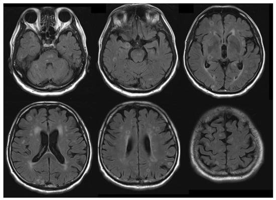
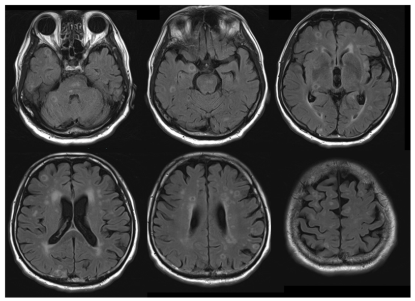
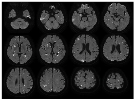
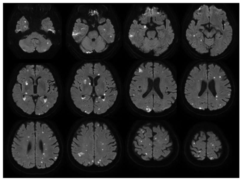
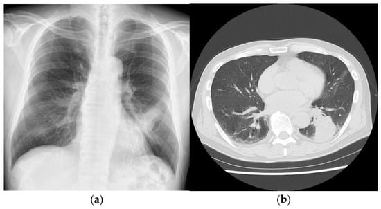
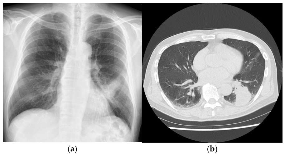
Magnetic resonance imaging (MRI) on admission demonstrated multiple enhancing lesions measuring less than approximately 1 cm scattered throughout the brain parenchyma (Figure 1). On repeat MRI obtained on day 6 of hospitalization, the lesions demonstrated more prominent enhancement, providing clearer visualization (Figure 2). Chest X-ray and computed tomography (CT) obtained on admission showed a mass-like lesion (approximately 5 cm) in the left lower lobe (Figure 3). Blood cultures obtained at the initiation of treatment later yielded S. intermedius. CSF cultures were negative. The multiplex meningitis/encephalitis panel detected human herpesvirus 6 (HHV-6); however, subsequent quantitative polymerase chain reaction testing for HHV-6 DNA was negative. CSF cytology was negative. Sputum culture could not be submitted because of difficulty in expectoration. The QuantiFERON-TB test and HIV-1/2 antibody confirmation test were negative, and serum assays for β-D-glucan, Aspergillus antigen, cryptococcal antigen and pneumococcal antigen were also negative. Oral examination revealed generalized moderate chronic periodontitis with abundant dental plaque accumulation and multiple periodontal pockets greater than 4 mm in depth. An ill-fitting maxillary denture with food impaction was also noted. These findings suggested poor oral hygiene and supported the oral cavity as the probable portal of bacterial entry. Transthoracic echocardiography revealed no findings suggestive of infective endocarditis. Because transthoracic echocardiography showed no findings suggestive of infective endocarditis and no clinical features raising a strong suspicion of endocarditis were present, transesophageal echocardiography was not performed. No other potential source of infection was identified in the cardiovascular or otorhinolaryngological systems. Based on the clinical presentation, bacteriological findings, and imaging results, the final diagnosis was multiple brain abscesses and a lung abscess.
Figure 1.
Axial fluid-attenuated inversion recovery (FLAIR) MRI of the brain on admission. Axial FLAIR images demonstrate numerous small ring-like lesions (<1 cm) distributed throughout both cerebellar hemispheres and both cerebral hemispheres.
Figure 2.
Axial brain diffusion-weighted imaging (DWI) MRI on hospital Day 6. DWI demonstrates numerous microlesions distributed throughout both cerebellar hemispheres and both cerebral hemispheres.
Figure 3.
Initial chest X-ray and computed tomography (CT) on admission. (a) Chest X-ray shows a mass-like lesion in the left lower lobe; (b) Chest CT with lung windows shows a mass-like lesion in the left lower lobe.
Empirical antibiotic therapy was initiated with meropenem (2 g every 8 h) and vancomycin (1 g every 12 h) on the day of admission, considering the possibility of both brain and lung abscesses. On hospital day 7, the organism and its antimicrobial susceptibility were identified from the blood cultures obtained at admission. Subsequently, based on the susceptibility profile of the isolated organism, the antibiotic regimen was switched to ceftriaxone (2 g every 12 h). Fever of approximately 38 °C persisted until the seventh day after admission despite antibiotic therapy. Antimicrobial therapy was continued for a total of eight weeks. At the end of treatment, brain MRI and chest CT showed near-complete resolution of the abscesses. The patient became fully conscious by hospital day 30 and was able to walk independently at discharge. Follow-up MRI performed one month after completion of therapy showed no enhancing lesions in the brain.
3. Discussion
We report a case of multiple brain abscesses and a lung abscess that developed following COVID-19. S. intermedius is a Gram-positive coccus normally inhabiting the oral cavity, nasal passages, pharynx, and gastrointestinal tract. Unlike other oral streptococci, this bacterium has strong virulence and is known to form abscesses in organs such as the brain and liver [5]. In this patient, COVID-19 may have caused impairment of the immune barrier. Under this condition, S. intermedius, a pathogen associated with pre-existing periodontitis, could have entered the bloodstream from the oral cavity. The organism may then have spread hematogenously, resulting in multiple brain abscesses and a lung abscess.
Numerous reports have documented secondary infections following COVID-19. In a study of approximately 190,000 hospitalized COVID-19 patients, Murray and colleagues reported that 6.9% developed secondary infections; of these, 6.0% were bacterial, 0.9% viral, and 0.2% fungal [1]. Risk factors for secondary infection included older age (≥70 years), a history of chronic disease, lack of vaccination, invasive therapies, longer hospital stay, prior antibiotic treatment, and steroid use, among others [1,6,7].
The mechanism of secondary infection after COVID-19 is believed to involve multiple factors, including viral-induced immune dysfunction, tissue injury, and breakdown of mucosal barriers [8,9]. Severe acute respiratory syndrome coronavirus 2 (SARS-CoV-2) enters cells via the angiotensin-converting enzyme 2 (ACE2) [10]. ACE2 is not only an enzyme but also a functional receptor on cell surfaces. It is expressed in various tissues throughout the body, including intestinal epithelial cells, renal tubular cells, the gallbladder, cardiomyocytes, the eyes, vascular endothelial cells, and vascular smooth muscle cells [11]. SARS-CoV-2 triggers inflammation when it infects vascular endothelial cells via ACE2 [12]. In the cerebral vasculature, it has been demonstrated that the invasion of SARS-CoV-2 damages the integrity of tight junctions, the basement membrane, and adhesion molecules, leading to breakdown of the blood–brain barrier (BBB) [13]. Among central nervous system complications of COVID-19, a wide spectrum has been described, such as cerebral vascular disorders, encephalitis, encephalopathy, meningitis, and brain abscess [14,15]. There have been reports that, in COVID-19 cases presenting with neurological symptoms, disruption of the blood–cerebrospinal fluid barrier was observed in more than half of patients, even when cerebrospinal fluid samples were collected more than 30 days after the onset of neurological symptoms [16]. This observation raises the possibility that brain abscesses related to COVID-19 infection might develop after a delayed interval. In addition, delayed secondary bacterial infections and abscess formation have been reported following COVID-19, suggesting that infectious complications may occur beyond the acute phase of SARS-CoV-2 infection [17]. In the present case, brain abscesses developed approximately 20 days after the onset of COVID-19.
Similarly, in the oral cavity, high expression of ACE2 in the gingival sulcus has been reported, suggesting that mucosal barrier breakdown may predispose to secondary infection [18]. Taken together, the SARS-CoV-2-induced breakdown of immune barriers likely increased susceptibility to brain and lung abscess formation in this patient with a history of periodontitis. However, it is possible that the lung abscess had already begun to develop at the time of COVID-19 diagnosis. The patient experienced persistent fever and respiratory symptoms from the onset of COVID-19, and because chest imaging was not performed at that time, the presence of an early lung abscess could neither be confirmed nor excluded. Nevertheless, SARS-CoV-2-associated impairment of mucosal and immune barriers may have facilitated progression of the infection and subsequent hematogenous dissemination to the brain.
In addition, the combination of multiple ring-enhancing brain lesions and a pulmonary lesion may mimic metastatic malignancy, particularly lung cancer with brain metastases, potentially complicating the diagnostic process. Therefore, infectious etiologies should be carefully considered in the differential diagnosis when systemic inflammatory findings are present. In the present case, brain biopsy was not performed because the lesions consisted of numerous small microabscesses distributed throughout the brain parenchyma, making tissue sampling technically difficult and unlikely to provide substantial additional diagnostic benefit. Moreover, the diagnosis was supported by characteristic imaging findings, positive blood cultures for S. intermedius, and the presence of a concomitant lung abscess.
To better contextualize this case, we conducted a narrative literature review using PubMed/MEDLINE. Relevant articles were identified using the keywords “COVID-19”, “SARS-CoV-2”, “brain abscess”, “lung abscess”, and “Streptococcus intermedius”. Articles published in English were included, and the references of selected articles were also screened to identify additional relevant studies. Although several reports have described brain or lung abscesses following COVID-19, no cases involving the simultaneous occurrence of both have been reported. Among documented abscesses caused by S. intermedius after COVID-19, three cases involved the brain and one involved the lungs (Table 1) [3,19,20,21]. When comparing the present case with previously reported cases, notable differences include the younger age of the reported patients, a sinonasal portal of entry, and the presence of only one or two solitary abscesses in those cases. Solitary lesions likely reflected direct extension from adjacent structures such as the paranasal sinuses, whereas hematogenous spread, as suspected in our patient, tends to produce multiple lesions and can involve other organs. Multiple brain abscesses caused by S. intermedius are relatively rare, with only 11 cases reported (Table 2) [22,23,24,25,26,27,28,29,30,31,32]. Although some occurred in healthy young adults, many cases (including ours) had dental comorbidities such as periodontitis, caries, or recent dental treatment, which likely contributed to abscess formation. Another notable feature of our case is the presence of more than 80 tiny abscesses disseminated throughout the brain parenchyma, representing a state of multiple brain microabscesses. Such numerous lesions are exceedingly rare; we identified only two comparable cases in the literature [31,32]. Notably, the report by Virtanen et al. did not provide information on abscess size or brain imaging, preventing definitive assessment [29]. Therefore, the present case represents an exceptionally rare manifestation of S. intermedius infection.
Table 1.
Comparison of reported cases of Streptococcus intermedius brain or lung abscesses following COVID-19.
Table 2.
Comparison of reported cases of multiple brain abscesses caused by Streptococcus intermedius.
Brain abscess formation is classically described in four stages, i.e., early cerebritis, late cerebritis, early capsule formation, and late (mature) capsule formation, during which neutrophils play an essential role in organizing purulent material and forming a capsule [33]. When neutrophils are depleted or functionally impaired, this “consolidation” of pus fails to occur, preventing the development of a typical encapsulated abscess. Consequently, multiple brain microabscesses are more commonly observed in conditions such as neutropenia, neutrophil dysfunction, immunocompromise, or fungal infections [34,35]. Although our patient did not have an immunocompromised background, previous studies have reported impaired leukocyte chemotaxis following SARS-CoV-2 infection [36]. In the present case, COVID-19-related vascular and immunological disturbances in the central nervous system may have facilitated hematogenous dissemination of S. intermedius, potentially contributing to the formation of multiple brain microabscesses.
As the patient had periodontitis, which can directly cause such abscesses, we cannot completely rule out the possibility that the development of brain and lung abscesses was unrelated to the prior COVID-19 infection. Although a causal relationship cannot be established, the temporal association and the reported effects of SARS-CoV-2 on immune and mucosal barriers raise the possibility that COVID-19 contributed to the progression and dissemination of infection in this patient. Further accumulation of similar cases of secondary infection after COVID-19 is warranted.
4. Conclusions
We report a patient who developed multiple brain abscesses and a lung abscess after COVID-19. Although the relationship between COVID-19 and abscess formation remains uncertain, clinicians should consider the possibility of severe secondary bacterial infection when fever or respiratory symptoms persist beyond the typical course of COVID-19, particularly in patients with pre-existing oral disease such as periodontitis.
Author Contributions
Conceptualization, R.T.; methodology, R.T., K.Y., and T.A.; investigation, R.T., K.Y., and T.A.; data curation, R.T., K.Y., and T.A.; writing—original draft preparation, R.T.; writing—review and editing, T.I. and H.N.; supervision, R.T. All authors have read and agreed to the published version of the manuscript.
Funding
This research received no external funding.
Institutional Review Board Statement
This study was approved by the Institutional Review Board of Nakamura Memorial Hospital (approval number: 2025120501, approved on 18 December 2025).
Informed Consent Statement
Written informed consent has been obtained from the patient to publish this paper.
Data Availability Statement
No new data were created or generated in this study. The case report is based on the patient’s clinical course and existing medical records, and the literature review is based on previously published studies. Therefore, data sharing is not applicable to this article.
Conflicts of Interest
The authors declare no conflicts of interest.
Abbreviations
The following abbreviations are used in this manuscript:
| ACE2 | Angiotensin-converting enzyme 2 |
| BBB | Blood–brain barrier |
| COVID-19 | Coronavirus disease 2019 |
| CSF | Cerebrospinal fluid |
| CT | Computed tomography |
| HHV-6 | Human herpesvirus 6 |
| MRI | Magnetic resonance imaging |
| SARS-CoV-2 | Severe acute respiratory syndrome coronavirus 2 |
References
- Murray, H.C.; Muleme, M.; Cooper, D.; McNamara, B.J.; Hussain, M.A.; Bartolo, C.; O’BRien, D.P.; Athan, E. Prevalence, risk factors, and outcomes of secondary infections among hospitalized patients with COVID-19 or post-COVID-19 conditions in Victoria, 2020–2023. Int. J. Infect. Dis. 2024, 145, 107078. [Google Scholar] [PubMed]
- Umamoto, K.; Horiba, M. Lung abscess as a secondary infection of COVID-19: A case report and literature review. J. Infect. Chemother. 2023, 29, 700–702. [Google Scholar] [CrossRef] [Scilit] [PubMed]
- Kunapaisal, T.; Guo, S.; Gomez, C.; Theard, M.A.; Lynch, J.B.; Lele, A.V.; King, M.A.; Buckley, R.; Vavilala, M.S. Bacterial brain abscess and life-threatening intracranial hypertension requiring emergent decompressive craniectomy after SARS-CoV-2 infection in a healthy adolescent. Cureus 2023, 15, e36258. [Google Scholar] [PubMed]
- Darlow, C.A.; McGlashan, N.; Kerr, R.; Oakley, S.; Pretorius, P.; Jones, N.; Matthews, P.C. Microbial aetiology of brain abscess in a UK cohort: Prominent role of Streptococcus intermedius. J. Infect. 2020, 80, 623–629. [Google Scholar] [CrossRef] [Scilit] [PubMed]
- Jacobs, J.A.; Pietersen, H.G.; Stobberingh, E.E. Streptococcus anginosus, Streptococcus constellatus and Streptococcus intermedius: Clinical relevance, hemolytic and serologic characteristics. Am. J. Clin. Pathol. 1995, 104, 547–553. [Google Scholar] [CrossRef] [Scilit] [PubMed]
- Na, Y.S.; Baek, A.-R.; Baek, M.S.; Kim, W.-Y.; Kim, J.H.; Lee, B.Y.; Seong, G.M.; Lee, S.-I. Clinical outcomes of and risk factors for secondary infection in patients with severe COVID-19: A multicenter cohort study in South Korea. Korean J. Intern. Med. 2023, 38, 68–79. [Google Scholar] [CrossRef] [Scilit] [PubMed]
- Wu, G.; Lu, J.; Liu, D.; He, Y. Characteristics and risk factors of secondary bacterial infections in COVID-19 patients. Antimicrob. Steward. Healthc. Epidemiol. 2023, 3, e156. [Google Scholar] [CrossRef] [Scilit] [PubMed]
- Chen, K.; Weng, R.; Li, J.; Wu, H.; Tie, X.; Li, H.; Zhang, Y. Dual threat: Susceptibility mechanisms and treatment strategies for COVID-19 and bacterial co-infections. Comput. Struct. Biotechnol. J. 2025, 27, 2107–2122. [Google Scholar] [CrossRef] [Scilit] [PubMed]
- Smail, S.W.; Albarzinji, N.; Salih, R.H.; Taha, K.O.; Hirmiz, S.M.; Ismael, H.M.; Noori, M.F.; Azeez, S.S.; Janson, C. Microbiome dysbiosis in SARS-CoV-2 infection: Implication for pathophysiology and management strategies of COVID-19. Front. Cell. Infect. Microbiol. 2025, 15, 1537456. [Google Scholar] [PubMed]
- Beyerstedt, S.; Casaro, E.B.; Rangel, E.B. COVID-19: Angiotensin-converting enzyme 2 (ACE2) expression and tissue susceptibility to SARS-CoV-2 infection. Eur. J. Clin. Microbiol. Infect. Dis. 2021, 40, 905–919. [Google Scholar] [PubMed]
- Hikmet, F.; Méar, L.; Edvinsson, Å.; Micke, P.; Uhlén, M.; Lindskog, C. The protein expression profile of ACE2 in human tissues. Mol. Syst. Biol. 2020, 16, e9610. [Google Scholar] [CrossRef] [Scilit] [PubMed]
- Varga, Z.; Flammer, A.J.; Steiger, P.; Haberecker, M.; Andermatt, R.; Zinkernagel, A.S.; Mehra, M.R.; Schuepbach, R.A.; Ruschitzka, F.; Moch, H. Endothelial cell infection and endotheliitis in COVID-19. Lancet 2020, 395, 1417–1418. [Google Scholar] [CrossRef] [Scilit] [PubMed]
- Maliha, S.T.; Fatemi, R.; Araf, Y. COVID-19 and the brain: Understanding the pathogenesis and consequences of neurological damage. Mol. Biol. Rep. 2024, 51, 318. [Google Scholar] [CrossRef] [Scilit] [PubMed]
- Shehata, G.A.; Lord, K.C.; Grudzinski, M.C.; Elsayed, M.; Abdelnaby, R.; Elshabrawy, H.A. Neurological complications of COVID-19: Underlying mechanisms and management. Int. J. Mol. Sci. 2021, 22, 4081. [Google Scholar] [CrossRef] [Scilit] [PubMed]
- Kawachi, M. Posterior reversible encephalopathy syndrome after COVID-19 in a patient with chronic renal failure. Rinsho Shinkeigaku 2025, 65, 32–38. (In Japanese) [Google Scholar] [CrossRef] [Scilit]
- Jarius, S.; Pache, F.; Körtvelyessy, P.; Jelčić, I.; Stettner, M.; Franciotta, D.; Keller, E.; Neumann, B.; Ringelstein, M.; Senel, M.; et al. Cerebrospinal fluid findings in COVID-19: A multicenter study of 150 lumbar punctures in 127 patients. J. Neuroinflamm. 2022, 19, 19. [Google Scholar] [CrossRef] [Scilit]
- Renaud-Picard, B.; Gallais, F.; Riou, M.; Zouzou, A.; Porzio, M.; Kessler, R. Delayed pulmonary abscess following COVID-19 pneumonia: A case report. Respir. Med. Res. 2020, 78, 100776. [Google Scholar] [CrossRef] [Scilit] [PubMed]
- Okui, T.; Matsuda, Y.; Karino, M.; Hideshima, K.; Kanno, T. Oral mucosa could be an infectious target of SARS-CoV-2. Healthcare 2021, 9, 1068. [Google Scholar] [CrossRef] [Scilit] [PubMed]
- Griffin, S.; Cleary, R.T.; Prim, M.; Musgrave, N.; Coppens, J.R.; Kemp, J. Sinusitis complicated by intracranial abscess in 3 patients with coronavirus disease 2019: Illustrative cases. J. Neurosurg. Case Lessons 2023, 5, CASE22423. [Google Scholar] [CrossRef] [Scilit] [PubMed]
- Blitz, S.E.; McMahon, J.T.; Chalif, J.I.; Jarvis, C.A.; Segar, D.J.; Northam, W.T.; Chen, J.A.; Bergmark, R.W.; Davis, J.M.; Yawetz, S.; et al. Intracranial complications of hypercoagulability and superinfection in the setting of COVID-19: Illustrative cases. J. Neurosurg. Case Lessons 2022, 3, CASE22127. [Google Scholar] [CrossRef] [Scilit] [PubMed]
- Bueno, C.O.P.; Trillos, S.J.G.; Rosales, D.J.C.; García, E.A.B. Lung abscess due to Streptococcus intermedius associated with SARS-CoV-2 infection in pregnancy. Clin. Case Rep. 2023, 11, e6763. [Google Scholar] [PubMed]
- Maliyil, J.; Caire, W.; Nair, R.; Bridges, D. Splenic abscess and multiple brain abscesses caused by Streptococcus intermedius in a young healthy man. Bayl. Univ. Med. Cent. Proc. 2011, 24, 195–199. [Google Scholar]
- Reyes, J.V.M.; Dondapati, M.; Ahmad, S.; Song, D.; Lieber, J.J.; Pokhrel, N.B.; Jaiswal, V. A case report of multiple abscesses caused by Streptococcus intermedius. Clin. Case Rep. 2023, 11, e6813. [Google Scholar] [CrossRef] [Scilit] [PubMed]
- Isern, R.D., III; Toth, S.; Goldfarb, M.; Ahmad, F. Multifocal brain abscesses due to Streptococcus intermedius. Cureus 2022, 14, e32797. [Google Scholar] [CrossRef] [Scilit] [PubMed]
- Bhatia, N.S.; Farrell, J.J.; Sampath, R.; Ranken, R.; Rounds, M.A.; Ecker, D.J.; Bonomo, R.A. Identification of Streptococcus intermedius central nervous system infection by use of PCR and electrospray ionization mass spectrometry. J. Clin. Microbiol. 2012, 50, 4160–4162. [Google Scholar] [CrossRef] [Scilit] [PubMed]
- Fransson, M.; Helldén, A.; Balkhed, Å.Ö.; Dernroth, D.N.; Ha, M.; Haglund, M.; Milos, P.; Hanberger, H.; Kågedal, B. Case report: Subtherapeutic vancomycin and meropenem concentrations due to augmented renal clearance in a patient with intracranial infection caused by Streptococcus intermedius. Front. Pharmacol. 2021, 12, 728075. [Google Scholar] [CrossRef] [Scilit] [PubMed]
- Herskovitz, M.Y.; Goldsher, D.; Finkelstein, R.; Bar Lavi, Y.; Constantinescu, M.; Telman, G. Multiple brain abscesses associated with tongue piercing. Arch. Neurol. 2009, 66, 1292. [Google Scholar] [CrossRef] [Scilit] [PubMed]
- Swied, M.Y.; Alom, M.; Daaboul, O.; Azzawi, M.; Swied, A. Esophageal perforation presenting initially as multiple brain abscesses secondary to Streptococcus intermedius. J. Investig. Med. High Impact Case Rep. 2024, 12, 23247096241239572. [Google Scholar] [CrossRef] [Scilit] [PubMed]
- Virtanen, P.S.; Jimenez, M.J.D.; Horak, V.J.; Desai, V.R.; Manaloor, J.J.; Raskin, J.S. Concomitant brain abscess and spinal cord abscess in an immunocompetent teenage male: Illustrative case. J. Neurosurg. Case Lessons 2023, 5, CASE22458. [Google Scholar] [CrossRef] [Scilit] [PubMed]
- Sayyahmelli, S.; Sayyahmelli, S.; Erginoglu, U.; Başkaya, M.K. Neurotoxic effects of ammonia in a patient with ornithine transcarbamylase deficiency and bilateral brain abscesses: Case report. Neurohospitalist 2021, 11, 241–245. [Google Scholar] [PubMed]
- Ohara, N.; Asai, K.; Ohkusu, K.; Wakayama, A. A case of culture-negative brain abscess caused by Streptococcus intermedius infection diagnosed by broad-range PCR of 16S ribosomal RNA. Brain Nerve 2013, 65, 1199–1203. (In Japanese) [Google Scholar] [PubMed]
- Yao, L.; Chen, S.; Yu, Z.; Yu, T. Multifocal brain abscesses caused by invasive Streptococcus intermedius: A case report. Front. Neurol. 2022, 13, 893627. [Google Scholar] [CrossRef] [Scilit] [PubMed]
- Britt, R.H.; Enzmann, D.R. Clinical stages of human brain abscesses on serial CT. J. Neurosurg. 1983, 59, 972–989. [Google Scholar] [CrossRef] [Scilit] [PubMed]
- Sakai, C.; Iuchi, T.; Ishii, A.; Kumagai, K.; Takagi, T. Bacillus cereus brain abscesses occurring in a severely neutropenic patient: Successful treatment with antimicrobial agents, granulocyte colony-stimulating factor and surgical drainage. Intern. Med. 2001, 40, 654–657. [Google Scholar] [CrossRef] [Scilit] [PubMed]
- DeLone, D.R.; Goldstein, R.A.; Petermann, G.; Salamat, M.S.; Miles, J.M.; Knechtle, S.J.; Brown, W.D. Disseminated aspergillosis involving the brain: Distribution and imaging characteristics. AJNR Am. J. Neuroradiol. 1999, 20, 1597–1604. [Google Scholar] [PubMed]
- Wang, G.; Guan, J.; Li, G.; Wu, F.; Yang, Q.; Huang, C.; Shao, J.; Xu, L.; Guo, Z.; Zhou, Q.; et al. Effect of ORF7 of SARS-CoV-2 on the chemotaxis of monocytes and neutrophils in vitro. Dis. Markers 2021, 2021, 6803510. [Google Scholar] [PubMed]

Disclaimer/Publisher’s Note: The statements, opinions and data contained in all publications are solely those of the individual author(s) and contributor(s) and not of MDPI and/or the editor(s). MDPI and/or the editor(s) disclaim responsibility for any injury to people or property resulting from any ideas, methods, instructions or products referred to in the content. |
© 2026 by the authors. Licensee MDPI, Basel, Switzerland. This article is an open access article distributed under the terms and conditions of the Creative Commons Attribution (CC BY) license.