


Abstract
Background: Gastrointestinal (GI) involvement is increasingly recognised in paediatric SARS-CoV-2 infection and multisystem inflammatory syndrome in children (MIS-C), but severe GI bleeding remains rare. Its pathogenesis in this setting is multifactorial: direct viral injury, hyperinflammatory microvascular damage, treatment-related mucosal injury, and coagulopathy may all contribute, and the relative role of each is often difficult to disentangle. Case Presentation: A previously healthy 12-year-old boy underwent appendectomy for perforated appendicitis with peritonitis. On postoperative Day 1, he developed high fever and was found to be SARS-CoV-2 PCR-positive; SARS-CoV-2 IgM and IgG were also positive. He met the positive clinical elements of the CDC 2023 case definition for MIS-C (persistent fever, multisystem involvement—gastrointestinal, hepatic, haematological, and markedly elevated inflammatory markers), although perforated appendicitis with peritonitis is a sufficient alternative explanation and the diagnosis cannot be regarded as secure (see Discussion). Treatment included broad-spectrum antibiotics (meropenem, amikacin, metronidazole), methylprednisolone 2 mg/kg/day, and intravenous immunoglobulin (IVIG) 2 g/kg. On Day 4 in the paediatric intensive care unit (PICU), the patient developed sudden haematemesis with fresh blood through the nasogastric tube and haemodynamic collapse. Coagulation studies revealed prolonged INR (1.6) and reduced prothrombin activity (42%). The patient was stabilised with packed red blood cells, fresh frozen plasma, and intravenous vitamin K. Fibrogastroscopy demonstrated diffuse mucosal bleeding without ulcers or anatomical defects; histology showed mucosal hyperaemia, mixed basal inflammation with intraepithelial lymphocytes, and small erosions. Melena persisted for several days. The child recovered fully and was discharged after 20 days with substantially improved but not fully normalised laboratory values (residual mild anaemia, Hb 110 g/L, and elevated CRP 32.7 mg/L and D-dimer 5.88 mg/L). Conclusions: Severe upper GI bleeding is a rare but life-threatening event in children with severe SARS-CoV-2 infection and MIS-C. In our case, several mechanisms may have acted in combination, although none could be confirmed individually: possible direct viral enterocyte injury via ACE-2 receptors, hyperinflammatory microangiopathy, high-dose corticosteroid-related mucosal injury, possible broad-spectrum antibiotic-associated vitamin K deficiency, and postoperative critical-illness stress. Their relative contributions cannot be determined from a single retrospective case. Bleeding occurred despite continuous PPI prophylaxis, suggesting that PPI cover alone is insufficient when multiple risk factors coexist; the clinical implication is that risk-factor-based (rather than universal) PPI prophylaxis, together with monitoring of vitamin K-dependent coagulation, should be considered in critically ill children receiving high-dose corticosteroids and prolonged broad-spectrum antibiotics.
1. Introduction
More than six years after the start of the SARS-CoV-2 pandemic, the full spectrum of paediatric disease remains incompletely characterised. While the respiratory presentation of acute COVID-19 in children is now well documented, severe post-infectious and hyperinflammatory phenotypes, particularly multisystem inflammatory syndrome in children (MIS-C), continue to generate diagnostic and therapeutic uncertainty. The Centers for Disease Control and Prevention (CDC) defines MIS-C as a clinical syndrome in individuals <21 years of age presenting with persistent fever, laboratory evidence of systemic inflammation, and clinically severe illness with multisystem (two or more) organ involvement, in the absence of an alternative microbial cause and with evidence of recent SARS-CoV-2 infection or exposure [1]. The World Health Organization (WHO) case definition covers similar clinical domains but differs materially in how an alternative infectious cause is handled [2] (see Table 1 and Discussion).
Table 1.
Comparison of the present case against the CDC/CSTE 2023 and WHO 2020 case definitions for MIS-C.
The gastrointestinal (GI) tract is the most frequently involved extrapulmonary system in COVID-19, with abdominal pain, vomiting and diarrhoea being common in both acute infection and MIS-C [3,4]. GI bleeding, in contrast, is uncommon, being reported in 2–13% of hospitalised adults [5] and substantially less often in children. The mechanisms are incompletely understood and likely overlap with those of severe acute illness in general: direct viral cytopathic effects on enterocytes (which express high levels of ACE-2 receptors) [6], hyperinflammatory microangiopathy, coagulopathy, and iatrogenic factors including corticosteroids, anticoagulants, and broad-spectrum antibiotics.
We report a case of severe upper GI bleeding in a 12-year-old boy with acute SARS-CoV-2 infection and features consistent with MIS-C, occurring four days after admission to the PICU and after high-dose corticosteroids, IVIG, and triple antibiotic therapy had been initiated. We focus on the differential pathophysiological mechanisms that may have contributed, an aspect under-emphasised in the existing literature on paediatric COVID-19-associated GI bleeding.
2. Case Presentation
A 12-year-old boy with normal physical and neuropsychological development, his parents’ fifth child born after an uncomplicated pregnancy, presented to the Paediatric Surgery Department of the University Multipurpose Hospital for Active Treatment “Dr. Georgi Stranski”, Pleven, with acute abdominal pain and repeated vomiting. The boy’s past medical history was unremarkable apart from a wasp-sting allergy, a family history of bronchial asthma, and two previous hospitalisations (bronchopneumonia and acute gastritis). During the previous admission for gastritis, an enlarged stomach without anatomical abnormality was incidentally noted and transient pyloric spasm was suspected. He had received all routine immunisations but had not been vaccinated against COVID-19.
Acute appendicitis with peritonitis was diagnosed and appendectomy was performed (Day 0). No histopathological examination of the resected appendix was performed and no tissue was sent for histology following the surgical intervention; this precludes formal confirmation of the severity or type of appendiceal inflammation and exclusion of atypical pathology. On postoperative Day 1, the patient developed high fever; a rapid SARS-CoV-2 antigen test provided a positive result, and this was subsequently confirmed by PCR. He was transferred to the PICU.
On admission to the PICU, the boy was in a severe general condition but conscious and subfebrile. The skin was dusky with reduced turgor but preserved elasticity. No rash, conjunctivitis, or lymphadenopathy was present. The oral mucosa was dry with a coated tongue. Auscultation revealed reduced breath sounds on the left. Cardiovascular and abdominal examinations were unremarkable beyond the fresh laparotomy scar with drains in situ. Neurological examination was normal.
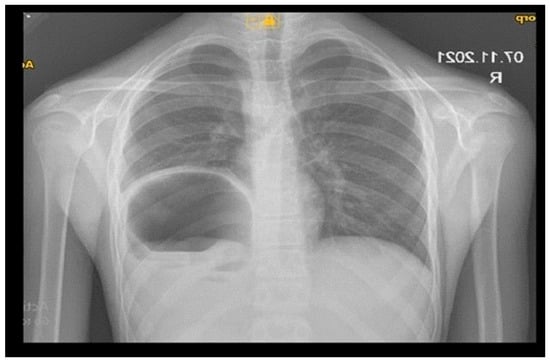
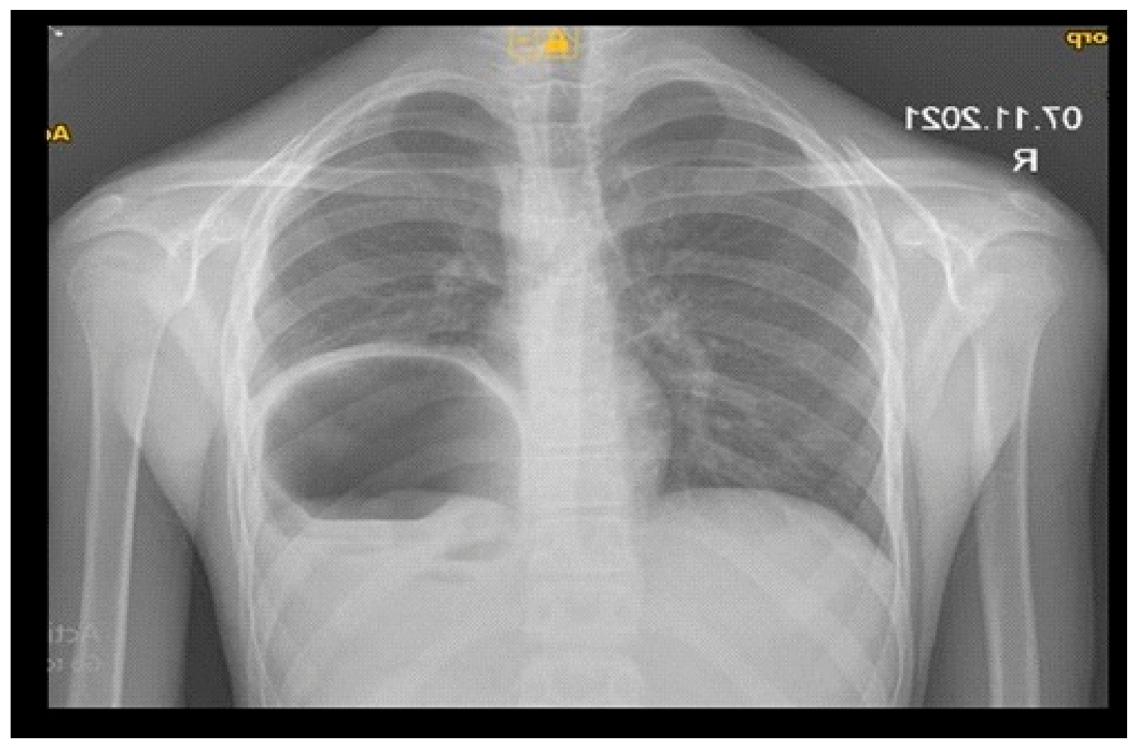
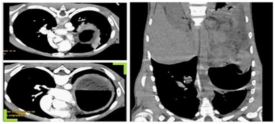
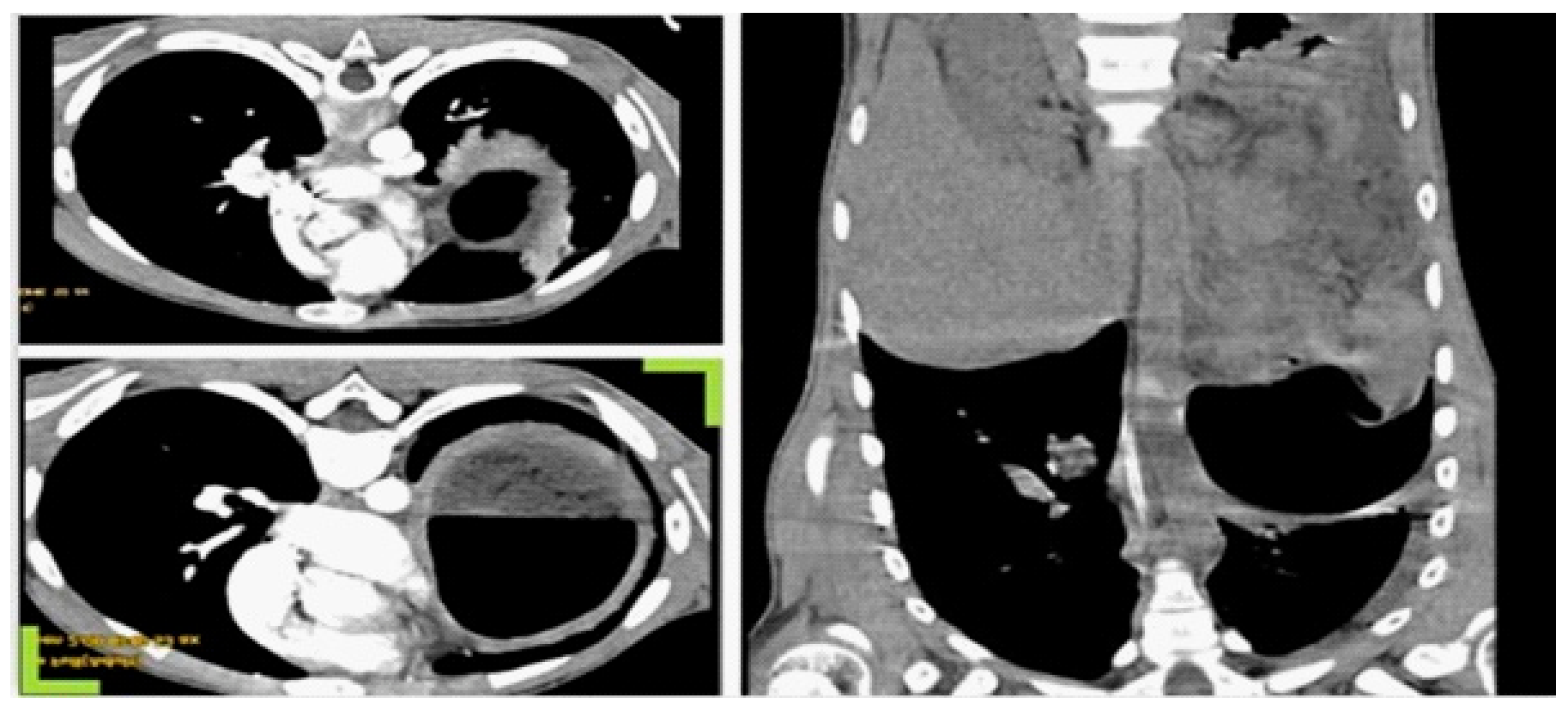
Laboratory findings demonstrated markedly elevated inflammatory markers (CRP, D-dimer, ferritin), neutrophilic leukocytosis, mild anaemia, and elevated hepatic transaminases. SARS-CoV-2 IgM and IgG were both positive. Blood cultures were negative, and CMV, EBV, and HSV serology excluded alternative viral aetiologies. Chest radiography and contrast-enhanced computed tomography of the chest and abdomen revealed an enlarged stomach with relaxation of the left hemidiaphragm (presumed congenital) without other structural pathology (Figure 1 and Figure 2). Transthoracic echocardiography was not performed at this stage in the absence of clinical or biochemical evidence of cardiac involvement; in retrospect, this represents a limitation of the diagnostic work-up (see Discussion).
Figure 1.
Chest radiograph on admission. An enlarged stomach is seen elevating the left hemidiaphragm; lung fields are clear, with no consolidation, pleural effusion, or pneumothorax. The image is reproduced in standard anteroposterior orientation, with the patient’s right on the viewer’s left, as indicated by the “R” side marker; the gastric air–fluid level therefore lies on the patient’s left.
Figure 2.
Contrast-enhanced computed tomography of the chest and abdomen. Axial sections (left panels) show the distended stomach with air–fluid level and relaxation of the left hemidiaphragm (presumed congenital). The coronal reconstruction (right panel) confirms the elevated diaphragm and absence of intrathoracic or intra-abdominal collections. All panels are displayed in standard radiological orientation: the axial sections are viewed from below, with anterior uppermost and the patient’s right on the viewer’s left, and the coronal reconstruction is displayed with cranial uppermost.
On the basis of persistent fever, multisystem involvement (gastrointestinal, hepatic, haematological), markedly elevated inflammatory markers, positive SARS-CoV-2 serology, negative blood cultures, and negative CMV, EBV and HSV serology, a working diagnosis of MIS-C overlapping with acute SARS-CoV-2 infection was formulated at the time. It must be emphasised, however, that the criterion requiring the absence of an alternative microbial cause was not fulfilled: the recent perforated appendicitis with peritonitis was itself an obvious infectious cause of the systemic inflammatory response, and this substantially weakens the working diagnosis (see Discussion and Table 1). The additional microbiological investigations therefore served to exclude a second, unrelated infectious process rather than to exclude an alternative cause of the inflammation as such. Treatment was initiated according to contemporary recommendations and comprised meropenem, commenced perioperatively on Day 0, to which amikacin and metronidazole were added on Day +2, methylprednisolone 2 mg/kg/day from Day +2, intravenous immunoglobulin 2 g/kg, and electrolyte/fluid resuscitation. No anticoagulant prophylaxis was administered. Stress ulcer prophylaxis with a proton pump inhibitor was administered throughout the PICU admission, including at the time of the bleeding episode.
Despite treatment, the patient remained febrile (peaks to 39 °C) with intermittent abdominal pain. On Day +5 (the fourth day in the PICU), his condition deteriorated abruptly, he developed vertigo, dizziness, and collapse, followed by haematemesis and discharge of fresh blood through the nasogastric tube. Coagulation studies showed prolonged INR (1.6) and reduced prothrombin activity (42%), with a haemoglobin drop from 124 g/L (Day +1) to 82 g/L. He was resuscitated with packed red blood cells, fresh frozen plasma, and intravenous vitamin K. After haemodynamic stabilisation, fibrogastroscopy was performed and revealed diffuse bleeding from the gastric mucosa without ulcers or other anatomical defects. Biopsy histology (specimen no. 5928/19644) demonstrated mucosal hyperaemia with mixed basal inflammation, intraepithelial lymphocytes, and small erosions.
Melena persisted for several days but no further haemodynamic instability occurred. After 20 days of multidisciplinary management, the patient was discharged in a good general condition with substantially improved, though not fully normalised, laboratory parameters: residual mild anaemia (Hb 110 g/L; local reference range approx. 120–160 g/L) and persistently elevated inflammatory and coagulation markers (CRP 32.7 mg/L and D-dimer 5.88 mg/L; both above the respective local reference thresholds). The clinical course is summarised in Table 2; serial laboratory values are presented in Table 3 and Table 4.
Table 2.
Clinical timeline.
Table 3.
Serial haematological, biochemical, and inflammatory parameters.
Table 4.
Serial coagulation parameters.
3. Discussion
This case raises two interrelated questions: is the diagnosis of MIS-C in this child secure, and what was the mechanism of the severe upper GI bleed? Both questions are central to interpreting the case and have direct clinical implications. We address them in turn before placing the case in the context of the existing literature.
The CDC and WHO case definitions for MIS-C require persistent fever, evidence of systemic inflammation, multisystem (two or more) organ involvement, exclusion of alternative microbial causes, and evidence of recent SARS-CoV-2 infection or exposure [1,2]. MIS-C characteristically arises 2 to 6 weeks after the initial infection, at a time when PCR is typically negative and serology is positive [7]. In our patient, SARS-CoV-2 PCR and both IgM and IgG were positive at admission, consistent with an active or very recent infection rather than the classic post-infectious window. The diagnosis therefore most accurately represents an overlap phenotype of acute SARS-CoV-2 infection with hyperinflammatory features fulfilling MIS-C criteria, rather than the prototypical post-infectious presentation. As Table 4 shows, our patient satisfied the positive clinical elements of the CDC 2023 definition, but perforated appendicitis with peritonitis is precisely the kind of obvious microbial cause of inflammation that the WHO definition excludes categorically; under WHO criteria, the case does not meet the definition of MIS-C at all. We retain the term MIS-C in this report because the clinical and biochemical features—high persistent fever, multisystem involvement and marked inflammatory response—prompted the working diagnosis at the time and because CDC 2023 criteria exclude only where a more likely alternative diagnosis exists, a matter of clinical judgement rather than a categorical rule. However, we wish to state plainly that the diagnosis of MIS-C in this child cannot be regarded as secure: perforated appendicitis with peritonitis is a sufficient alternative explanation for the fever, the inflammatory response, and the multisystem involvement, and the case is perhaps best described as a hyperinflammatory response in the setting of acute SARS-CoV-2 infection complicating perforated appendicitis, in which MIS-C-like features were present but could not be distinguished with confidence from those of the surgical illness itself.
A second limitation is the absence of an echocardiographic assessment. Although there were no clinical or biochemical features suggestive of cardiac involvement, transthoracic echocardiography is recommended in all children with suspected MIS-C to detect subclinical myocardial dysfunction, valvulitis, or coronary artery abnormalities [8]. In future cases of suspected MIS-C, echocardiography should be performed irrespective of the clinical picture. Serum cardiac biomarkers (troponin) and a 12-lead electrocardiogram were likewise not obtained; both are recommended in the surveillance of MIS-C-associated cardiac involvement, and their absence, together with the lack of echocardiography, is a limitation of the diagnostic work-up in this case.
Severe upper GI bleeding in this clinical context cannot be attributed to a single mechanism. We considered the following non-exclusive contributors:
- Direct viral mucosal injury. ACE-2 receptors are abundantly expressed on enterocytes and gastric epithelial cells and serve as the entry point for SARS-CoV-2 [6]. Direct cytopathic injury and immune-mediated mucositis have been demonstrated histologically in COVID-19 [9]. The fibrogastroscopy findings of diffuse mucosal bleeding without ulcer, and the histological pattern of mucosal hyperaemia with mixed basal inflammation and intraepithelial lymphocytes, are compatible with viral mucositis but are not specific. No immunohistochemistry, in situ hybridisation, or electron microscopy was performed on the gastric biopsy to confirm SARS-CoV-2 within the mucosa, so a direct viral contribution remains inferential rather than histologically proven.
- Hyperinflammatory microangiopathy. MIS-C is characterised by widespread endothelial activation and microvascular dysfunction [10]. Mesenteric microvascular damage has been described as a mechanism of GI involvement in severe COVID-19 [11] and would be consistent with the diffuse, non-ulcerative bleeding pattern observed.
- Corticosteroid-induced mucosal injury. The patient received high-dose methylprednisolone (2 mg/kg/day) while receiving proton pump inhibitor prophylaxis throughout. High-dose systemic corticosteroids are a well-established risk factor for erosive gastropathy, particularly in the setting of critical illness [12]. The occurrence of severe bleeding despite PPI cover indicates either breakthrough mucosal injury not fully mitigated by acid suppression, or a dominant non-acid-mediated mechanism (e.g., viral, microangiopathic or coagulopathic)—a distinction PPI prophylaxis alone cannot resolve.
- Broad-spectrum antibiotic–associated vitamin K deficiency (hypothesis). The patient received meropenem from Day 0 (5 days) and amikacin and metronidazole from Day +2 (3 days) before the bleeding episode on Day +5. This exposure is shorter than the 7–10 days conventionally cited for antibiotic-associated hypoprothrombinaemia, and the argument is correspondingly weaker than a longer course would allow. Suppression of menaquinone (vitamin K2)-producing intestinal flora nevertheless begins within the first days of broad-spectrum therapy, and hepatic vitamin K stores are small and turn over within days, so that a child with negligible enteral intake following laparotomy for perforated appendicitis may become depleted over such an interval, with impaired synthesis of vitamin K–dependent coagulation factors (II, VII, IX, X) [13]. This mechanism is compatible with, though not proven by, our laboratory findings: a normal INR on Day +4 (1.09) rose to 1.60 on the day of bleeding, with reduced prothrombin activity (42%), while aPTT was not prolonged (28.2 s, below the patient’s own admission value of 32.5 s) and fibrinogen remained adequate (2.24 g/L). This pattern of isolated prolongation of the extrinsic pathway with a non-prolonged aPTT and preserved fibrinogen is consistent with early vitamin K-dependent factor depletion, since factor VII has the shortest half-life (approximately 4–6 h) of the vitamin K-dependent factors and is depleted first; it also argues against disseminated intravascular coagulation, in which fibrinogen would be expected to fall. However, plasma vitamin K, PIVKA-II, and individual factor assays were not measured, so the deficiency was inferred rather than directly demonstrated, and the response to treatment cannot be used to support the diagnosis because vitamin K, fresh frozen plasma, and packed red cells were administered concurrently. The postoperative critical-illness state may also have contributed to any nutritional or absorptive component of the deficiency. We therefore present antibiotic-associated vitamin K deficiency as a plausible contributory hypothesis rather than an established cause.
- Critical-illness stress and the postoperative state. The child had undergone laparotomy for perforated appendicitis with peritonitis only days before. Stress-related mucosal disease is a recognised complication of major surgery, sepsis-like states, and prolonged ICU stay [14], and routinely warrants PPI prophylaxis in adults with established risk factors.
- Intravenous immunoglobulin. Although uncommon, IVIG has been associated with haemolytic anaemia and transient platelet dysfunction; a contribution cannot be excluded but is unlikely to be the dominant mechanism here [15].
It is important to emphasise that anticoagulant prophylaxis, a frequently cited contributor to GI bleeding in adult COVID-19 cohorts [5,16], was not administered in our patient, and therefore did not contribute to this case. Conversely, antibiotic-related vitamin K deficiency, an under-recognised cause in the paediatric COVID literature, is in our view a plausible contributor to the coagulopathy, although the relatively short antibiotic exposure and the absence of vitamin K, PIVKA-II, and individual factor assays mean that it remains an unproven hypothesis in this patient.
This case illustrates that severe GI bleeding in a critically ill child with COVID-19 or MIS-C is rarely attributable to a single mechanism. The interaction between viral and hyperinflammatory mucosal injury on one hand, and treatment-related coagulopathy and stress-related gastropathy on the other, can be life-threatening. Several practical implications follow:
- Proton pump inhibitor prophylaxis should be considered in critically ill children with defined risk factors, including coagulopathy, invasive mechanical ventilation, recent major abdominal surgery or sepsis, and high-dose corticosteroids in combination with one or more of these, rather than routinely in all children receiving corticosteroids. Data supporting the efficacy of PPI prophylaxis in this setting derive from adult COVID-19 cohorts [17] and have not been specifically validated in children; moreover, our patient bled despite continuous PPI cover, which suggests that PPI prophylaxis alone cannot be relied upon to prevent bleeding when multiple risk factors coexist.
- Vitamin K-dependent coagulation should be monitored in children receiving prolonged courses of broad-spectrum antibiotics, especially carbapenems and metronidazole. A rising INR with preserved fibrinogen and platelets should prompt empirical vitamin K supplementation before clinical bleeding occurs.
- Echocardiography should be performed in all cases of suspected MIS-C, even in the absence of clinical or biochemical evidence of cardiac involvement.
- In suspected MIS-C with positive SARS-CoV-2 PCR, the diagnosis should be framed as overlapping with acute infection rather than the classic postinfectious phenotype; treatment principles overlap but the prognostic implications may differ.
Published data on severe GI bleeding in paediatric COVID-19 and MIS-C remain limited. Multicentre series of severe MIS-C have reported haematological system involvement (including cytopenias and coagulopathy) in up to 76% of cases [18]; only a minority of these manifest as clinically apparent bleeding, and severe GI haemorrhage specifically is rare. In adults, GI bleeding occurs in 1–13% of hospitalised COVID-19 patients [5,16] and is more commonly a late hospital complication than a presenting feature [19]. The contribution of anticoagulation is consistently highlighted in adult cohorts but is less applicable to paediatric practice, where thromboprophylaxis is not universally administered. Antibiotic-induced vitamin K deficiency is, to our knowledge, infrequently emphasised in the paediatric COVID-19 literature, although it is a well-established mechanism in general critical-care practice [13]. The present case suggests that this mechanism deserves explicit consideration in the differential diagnosis of bleeding in children with severe COVID-19 or MIS-C.
4. Conclusions
Severe upper GI bleeding is a rare but potentially life-threatening complication in children with severe SARS-CoV-2 infection and MIS-C. In our case, the bleeding may plausibly have arisen from a combination of mucosal injury, possibly viral and hyperinflammatory in origin and potentially aggravated by high-dose corticosteroids, and occurring despite continuous PPI prophylaxis and a coagulopathy that may have been related to broad-spectrum antibiotic-associated vitamin K deficiency, all against the background of a postoperative critical-illness state. None of these mechanisms was confirmed by dedicated investigation, and they are therefore presented as plausible contributors rather than as established causes. Risk-factor-based PPI prophylaxis, monitoring of vitamin K-dependent coagulation during prolonged broad-spectrum antibiotic therapy, and systematic echocardiographic assessment in all suspected MIS-C cases are simple measures that may reduce the risk of similar events. Further paediatric data are required to refine the management of GI bleeding in COVID-19 and MIS-C.
Author Contributions
Conceptualization, K.Y. and S.E.; methodology, K.Y. and S.E.; validation, V.B. and N.B.; formal analysis, V.B. and N.B.; investigation, K.Y. and S.E.; resources, K.Y. and S.E.; data curation, V.B. and N.B.; writing—original draft preparation, K.Y. and S.E.; writing—review and editing, K.Y., S.E., V.B. and N.B.; visualization, K.Y.; supervision, V.B.; project administration, K.Y. All authors have read and agreed to the published version of the manuscript.
Funding
This research received no external funding.
Institutional Review Board Statement
The study was conducted in accordance with the Declaration of Helsinki and approved by the Committee on Ethics of Scientific Research (KENID), the Institutional Ethics Committee of Medical University Pleven (protocol code 634 and date of approval 17 June 2020).
Informed Consent Statement
Written informed consent for publication of this case report and accompanying images was obtained from the patient’s parents. A copy of the written consent is available from the authors on request. Informed consent was obtained from all subjects involved in the study.
Data Availability Statement
The original contributions presented in this study are included in the article. Further inquiries can be directed to the corresponding author.
Acknowledgments
The authors would like to thank the patient and his family for their cooperation and consent.
Conflicts of Interest
The authors declare no conflicts of interest.
References
- Centers for Disease Control and Prevention. Multisystem Inflammatory Syndrome in Children (MIS-C): Case Definition; CDC: Atlanta, GA, USA, 2023. [Google Scholar]
- World Health Organization. Multisystem Inflammatory Syndrome in Children and Adolescents Temporally Related to COVID-19; WHO Scientific Brief; WHO: Geneva, Switzerland, 2020. [Google Scholar]
- Lin, L.; Jiang, X.; Zhang, Z.; Huang, S.; Zhang, Z.; Fang, Z.; Gu, Z.; Gao, L.; Shi, H.; Mai, L.; et al. Gastrointestinal symptoms of 95 cases with SARS-CoV-2 infection. Gut 2020, 69, 997–1001. [Google Scholar] [CrossRef] [Scilit] [PubMed]
- Feldstein, L.R.; Tenforde, M.W.; Friedman, K.G.; Newhams, M.; Rose, E.B.; Dapul, H.; Soma, V.L.; Maddux, A.B.; Mourani, P.M.; Bowens, C.; et al. Characteristics and outcomes of US children and adolescents with multisystem inflammatory syndrome in children compared with severe acute COVID-19. JAMA 2021, 325, 1074–1087. [Google Scholar] [CrossRef] [Scilit] [PubMed]
- Martin, T.A.; Wan, D.W.; Hajifathalian, K.; Tewani, S.; Shah, S.L.; Mehta, A.; Kaplan, A.; Ghosh, G.; Choi, A.J.; Krisko, T.I.; et al. Gastrointestinal bleeding in patients with coronavirus disease 2019: A matched case-control study. Am. J. Gastroenterol. 2020, 115, 1609–1616. [Google Scholar] [CrossRef] [Scilit] [PubMed]
- Gupta, A.; Madhavan, M.V.; Sehgal, K.; Nair, N.; Mahajan, S.; Sehrawat, T.S.; Bikdeli, B.; Ahluwalia, N.; Ausiello, J.C.; Wan, E.Y.; et al. Extrapulmonary manifestations of COVID-19. Nat. Med. 2020, 26, 1017–1032. [Google Scholar] [CrossRef] [Scilit] [PubMed]
- Nalbandian, A.; Sehgal, K.; Gupta, A.; Madhavan, M.V.; McGroder, C.; Stevens, J.S.; Cook, J.R.; Nordvig, A.S.; Shalev, D.; Sehrawat, T.S.; et al. Post-acute COVID-19 syndrome. Nat. Med. 2021, 27, 601–615. [Google Scholar] [CrossRef] [Scilit] [PubMed]
- Henderson, L.A.; Canna, S.W.; Friedman, K.G.; Gorelik, M.; Lapidus, S.K.; Bassiri, H.; Behrens, E.M.; Kernan, K.F.; Schulert, G.S.; Seo, P.; et al. American College of Rheumatology clinical guidance for multisystem inflammatory syndrome in children associated with SARS-CoV-2 and hyperinflammation in pediatric COVID-19: Version 3. Arthritis Rheumatol. 2022, 74, e1–e20. [Google Scholar] [CrossRef] [Scilit] [PubMed]
- Xiao, F.; Tang, M.; Zheng, X.; Liu, Y.; Li, X.; Shan, H. Evidence for gastrointestinal infection of SARS-CoV-2. Gastroenterology 2020, 158, 1831–1833.e3. [Google Scholar] [CrossRef] [Scilit] [PubMed]
- Al-Ghafry, M.; Vagrecha, A.; Malik, M.; Levine, C.; Uster, E.; Aygun, B.; Appiah-Kubi, A.; Vlachos, A.; Capone, C.A.; Rajan, S.; et al. Multisystem inflammatory syndrome in children (MIS-C) and the prothrombotic state: Coagulation profiles and rotational thromboelastometry in a MIS-C cohort. J. Thromb. Haemost. 2021, 19, 1764–1770. [Google Scholar] [CrossRef] [Scilit] [PubMed]
- Marasco, G.; Maida, M.; Morreale, G.C.; Licata, M.; Renzulli, M.; Cremon, C.; Stanghellini, V.; Barbara, G. Gastrointestinal bleeding in COVID-19 patients: A systematic review with meta-analysis. Can. J. Gastroenterol. Hepatol. 2021, 2021, 2534975. [Google Scholar] [CrossRef] [Scilit] [PubMed]
- Narum, S.; Westergren, T.; Klemp, M. Corticosteroids and risk of gastrointestinal bleeding: A systematic review and meta-analysis. BMJ Open 2014, 4, e004587. [Google Scholar] [CrossRef] [Scilit] [PubMed]
- Paulus, M.C.; Drent, M.; Kouw, I.W.K.; Balvers, M.G.J.; Bast, A.; van Zanten, A.R.H. Vitamin K: A potential missing link in critical illness—A scoping review. Crit. Care 2024, 28, 212. [Google Scholar] [CrossRef] [Scilit] [PubMed]
- Cook, D.J.; Fuller, H.D.; Guyatt, G.H.; Marshall, J.C.; Leasa, D.; Hall, R.; Winton, T.L.; Rutledge, F.; Todd, T.; Roy, P.; et al. Risk factors for gastrointestinal bleeding in critically ill patients. N. Engl. J. Med. 1994, 330, 377–381. [Google Scholar] [CrossRef] [Scilit] [PubMed]
- Guo, Y.; Tian, X.; Wang, X.; Xiao, Z. Adverse effects of immunoglobulin therapy. Front. Immunol. 2018, 9, 1299. [Google Scholar] [CrossRef] [Scilit] [PubMed]
- Karlafti, E.; Tsavdaris, D.; Kotzakioulafi, E.; Protopapas, A.A.; Kaiafa, G.; Netta, S.; Savopoulos, C.; Michalopoulos, A.; Paramythiotis, D. The prevalence of gastrointestinal bleeding in COVID-19 patients: A systematic review and meta-analysis. Medicina 2023, 59, 1500. [Google Scholar] [CrossRef] [Scilit] [PubMed]
- Prasoppokakorn, T.; Kullavanijaya, P.; Pittayanon, R. Risk factors of active upper gastrointestinal bleeding in patients with COVID-19 infection and the effectiveness of PPI prophylaxis. BMC Gastroenterol. 2022, 22, 465. [Google Scholar] [CrossRef] [Scilit] [PubMed]
- Feldstein, L.R.; Rose, E.B.; Horwitz, S.M.; Collins, J.P.; Newhams, M.M.; Son, M.B.F.; Newhams, M.M.; Son, M.B.F.; Newburger, J.W.; Kleinman, L.C.; et al. Multisystem inflammatory syndrome in U.S. children and adolescents. N. Engl. J. Med. 2020, 383, 334–346. [Google Scholar] [CrossRef] [Scilit] [PubMed]
- Negro, A.; Villa, G.; Rolandi, S.; Lucchini, A.; Bambi, S. Gastrointestinal bleeding in COVID-19 patients: A rapid review. Gastroenterol. Nurs. 2022, 45, 267–275. [Google Scholar] [PubMed]

Disclaimer/Publisher’s Note: The statements, opinions and data contained in all publications are solely those of the individual author(s) and contributor(s) and not of MDPI and/or the editor(s). MDPI and/or the editor(s) disclaim responsibility for any injury to people or property resulting from any ideas, methods, instructions or products referred to in the content. |
© 2026 by the authors. Licensee MDPI, Basel, Switzerland. This article is an open access article distributed under the terms and conditions of the Creative Commons Attribution (CC BY) license.