


Abstract
Background/Objectives: Coronary artery disease (CAD) remains the leading cause of mortality worldwide, with South Asia bearing a disproportionately high and rising burden, particularly at younger ages. The present study aimed to investigate genetic variants associated with premature coronary artery disease (PCAD) using whole-genome sequencing (WGS). Methods: WGS was conducted on 12 people (five PCAD cases, seven matched controls) to assess feasibility and methodology for future large-scale research. High-quality genomic DNA was sequenced at a minimum read depth of 10× with a quality threshold of Q30. Variant calling with stringent quality control identified single-nucleotide polymorphisms (SNPs), followed by annotation against gnomAD for allele frequencies and ClinVar for pathogenicity. Protein-coding variants were filtered, and candidate genes were prioritized for comparative analysis between cases and controls. Results: An average of over 8.8 million SNPs per individual was identified, with comparable overall variant distributions between cases and controls. Initial analyses revealed 120 SNPs exclusively present in PCAD cases. All protein-coding variants were rare (allele frequency < 0.0001), and none were previously classified as pathogenic in ClinVar. After filtration, 87 candidate genes were prioritized. Enriched or unique variants in PCAD cases are mapped to genes involved in lipid metabolism, endothelial dysfunction, inflammatory signaling, immune regulation, thrombosis, vascular remodeling, and metabolic processes. Additional variants were identified in genes related to smooth muscle proliferation, oxidative stress, and other biological pathways. Conclusions: This WGS pilot study provides an initial overview of the genomic landscape of PCAD in a South Asian cohort, highlighting rare variants across multiple biological pathways implicated in atherosclerosis that need validation in a large-scale study.
1. Introduction
Coronary artery disease (CAD) remains the leading cause of mortality worldwide, with South Asia experiencing a disproportionately high and escalating burden. According to the Global Burden of Disease (GBD) study, ischemic heart disease accounted for approximately 8.92 million deaths in 2015, underscoring its global health impact [1]. Of particular concern is the increasing incidence of premature CAD (PCAD), clinically defined as major cardiac events occurring before the age of 45 in men and 55 in women, with a more severe form affecting individuals under 40 years [2]. Epidemiological data of myocardial infarction (MI) cases reported that 21% of affected individuals were under 40, of which 79.8% were male, emphasizing a significant sex disparity and disproportionate impact on young men [3]. Notably, the incidence of acute MI demonstrates a marked age-dependent increase, from 2.1 per 100,000 person-years in individuals aged 20–29 to 16.9 in those aged 30–39 [4]. In Pakistan, the burden of cardiovascular disease (CVD) is escalating rapidly. This could be due to decades of industrial and economic impoverishment alongside significant challenges, including regional conflicts, natural disasters, and underinvestment in healthcare. Consequently, while infectious diseases remain prevalent, the healthcare system is now facing a growing crisis of non-communicable diseases, particularly CVD. According to the 2019 GBD study, Pakistan’s age-standardized incidence of CVD was 918.18 per 100,000 people (compared with the global rate of 684.33), and the age-standardized death rate was 357.88 per 100,000 (compared to 239.85 globally) [5]. Additionally, data from the National Socioeconomic Registry Survey, comprising 34 million households, showed that 18.9% of participants self-reported having CVD [4,5].
Globally, individuals of Indo-Asian origin exhibit the highest risk of developing CAD [6], hence establishing it as the leading cause of death in the Indo-Pakistani subcontinent. A national registry (2016–2019) analyzing percutaneous coronary intervention (PCI) data from 15,106 patients revealed that 52.6% had premature CAD (men aged 40–54; women aged 40–64), while 7.4% had extremely premature CAD (under 40 years) [7]. These statistics highlight a significant disease prevalence in younger populations, necessitating early interventions and preventive strategies.
The risk factors associated with PCAD differ across various ethnicities. Among Black populations, dyslipidemia, diabetes, and smoking are significant contributors, whereas in White populations, smoking is the primary factor. Among Hispanic populations, dyslipidemia, male sex, and family history are most prominent. Interestingly, no independent predictor has been found for Asian Indians [5]. Among South Asians, family history is a key contributor, warranting deeper investigation into genetic predisposition, including gene variants related to lipid metabolism, vascular function, and coagulation pathways [8,9]. The elevated prevalence of consanguinity within the South Asian population likely increases the risk of homozygous mutations, contributing to early-onset and more aggressive disease. This is supported by epidemiological data reporting a positive family history in 18% of PCAD and 23% of early PCAD cases, suggesting a substantial genetic or familial environmental clustering [7]. In Pakistan, high age-adjusted prevalence of diabetes (30.8%), hypertension (37% in adults), and tobacco use (~25% in adult males), coupled with a high consanguinity rate (58% first- or second-cousin marriages), synergistically amplify genetic risk [5]. Multiple studies have explored genetic contributors to CVD in South Asians, including PROMIS (Pakistan), BRAVE (Bangladesh), LOLIPOP (UK), and the START cohort (Canada) [10,11,12,13]. These studies, along with the 1000 Genomes Project [14], have provided critical insight into gene–environment and gene–gene interactions. The C4D Genetics Consortium meta-analysis of four GWAS (PROMIS, LOLIPOP, PROCARDIS, and HPS) identified 59 single-nucleotide polymorphisms (SNPs) linked to CAD, five of which achieved genome-wide significance, implicating genes involved in lipid metabolism, inflammation, and vascular remodeling [15]. Furthermore, a multiethnic GWAS meta-analysis combining seven non-South Asian and two South Asian cohorts identified 25 CAD-associated SNPs within or proximal to genes such as PECAM1 (inflammation), LMOD1 (vascular smooth muscle), and PROCR (coagulation) [16]. PROMIS study also identified six novel SNPs in the CX3CR1 gene specific to South Asians that were not reported in larger European studies, suggesting ethnicity-specific genetic architecture [17]. Although the p53 codon 72 SNP is not associated with clinical markers of disease in CAD, the higher frequency of the variant allele in South Asian Indians may be a contributing factor to this population having an increased risk of developing premature CAD. [18]. Epigenetic research further supports the gene–environment interplay whereby environmental exposures (e.g., diet, maternal health) modulate disease risk through gene regulation, as evident by the emergence of miR-21 as a potential epigenetic marker for atherogenic dyslipidemia in South Asians [19].
Although significant discoveries have been made in middle-aged and older individuals, the genetic factors influencing PCAD in younger populations—particularly within understudied regions like Indo-Pakistan—are still not well understood. This highlights the need for high-throughput whole-genome sequencing (WGS) to identify variants specific to PCAD. Our research is designed to address an important gap by launching a pilot study, which could lead to larger studies focused on discovering new sequence variants (SNVs/SNPs) and structural variants (SVs) in PCAD. This initial study may help lay the foundation for creating polygenic scores in broader research, ultimately enhancing early diagnosis, risk assessment, and tailored treatments [20,21].
2. Methodology
2.1. Study Population
This retrospective case–control pilot study explores WGS as a method for investigating PCAD. The study enrolled 12 participants, including 5 PCAD patients (PCAD group) and 7 matching controls (control group). Blood samples were collected from the Armed Forces Institute of Cardiology/National Institute of Heart Diseases, Rawalpindi, Pakistan. The inclusion criteria for patient selection were as follows: (1) clinical symptoms indicative of myocardial infarction (MI) lasting over 20 min within the previous 24 h of preceding index hospitalization; (2) ECG changes consistent with MI, such as new pathologic Q waves, at least 1 mm ST elevation in two or more contiguous limb leads, a new left bundle branch block, or persistent ST-T wave changes diagnostic of non-Q wave MI; and (3) those with elevated troponin-T levels.
Exclusion criteria included recent or active infection within the past 2 weeks, chronic infections (e.g., HIV, hepatitis B, hepatitis C, and tuberculosis), chronic autoimmune diseases (e.g., rheumatoid arthritis, lupus), acute pericarditis, end-stage renal failure, history of malignancy, and those who refused to provide consent. Individuals in the control groups were matched to cases by age, gender, and comorbidities (within 5-year age bands) without a history of cardiovascular disease.
2.2. Sample Collection
A blood sample (5 mL) was collected from each study participant under the supervision of a certified physician. During sample collection, baseline characteristics and clinical information were documented using a specifically designed questionnaire. After collection, blood samples were immediately transported under controlled conditions to the Chugtai laboratory, Jail Road, Lahore, Pakistan, and stored at 4 °C until further processing.
2.3. DNA Extraction and Analysis
Genomic DNA was isolated from whole blood using the standard organic phenol-chloroform method, following the manufacturer’s instructions [22]. The extracted DNA was quantified and stored at 4 °C for subsequent experimentation.
2.4. Sample Quality Control
DNA integrity was assessed by electrophoresis on a 1% agarose gel. Initial quantification analysis was performed using a NanoPhotometer spectrophotometer (Implen GmbH, Munich, Germany), and DNA concentration and purity were subsequently confirmed with a Qubit 2.0 Fluorometer (Thermo Fisher Scientific, Waltham, MA, USA).
2.5. Library Preparation
Genomic DNA was sheared to ~350 bp fragments using an ultrasonic processor. The resulting fragments were then used to construct sequencing libraries through a standardized workflow, including end repair, adenylation at the 3′ ends, adapter ligation, purification, and polymerase chain reaction (PCR) amplification. The resulting sequencing libraries were quantified using a Qubit 2.0 Fluorometer, and samples were normalized to a working concentration of 1 ng/µL. Library fragment size distribution was verified using an Agilent 2100 Bioanalyzer (Agilent Technologies, Santa Clara, CA, USA), and final library concentrations were confirmed by quantitative PCR prior to sequencing.
2.6. Whole-Genome Sequencing
WGS was performed on an Illumina NovaSeq 6000 platform (Illumina, San Diego, CA, USA) (paired-end 150 bp) platform using an S4 flow cell. FASTQ-formatted files were received from the sequencing provider. Sequencing quality was checked using FastQC, which includes (1) more than 50 Gbp bases in each FASTQ file (each sample has two paired FASTQ files), (2) 150 bp length for all sequences, and (3) passing all the quality check terms in the FastQC report, which include per-base sequence quality, per-sequence GC content, sequence length distribution, sequence duplication levels, overrepresented sequences, and adapter content. Genome Reference Consortium Human Build 38 (GRCh38) reference panel provided by Genomic Analysis ToolKit (GATK) was chosen as a reference genome. Read alignment and variant calling were performed using the graphics processing unit (GPU) accelerated GATK pipeline in Nvidia Clara Parabricks v4.3.1. After receiving GVCF files for each sample, joint genotyping was performed using GLnexus v1.2.6 to convert GVCF files into a multi-sample VCF file.
2.7. Bayesian Model for Detecting Associations (BMDA) SNP Analysis Pipeline
This analysis pipeline started with the VCF file generated after WGS. The GFF (General Feature Format) or GTF (Gene Transfer Format) file was used for the reference genome. BMDA analysis included the following steps:
- Variant filtering:
The raw variant call format (VCF) file was filtered using “vcftools”, using a minimum quality score of 30 and a minimum depth of coverage of 10 reads.
- 2.
- Basic VCF statistics:
The program “vcf-stats” was used to extract the numbers of each SNP for each sample. Average ts/tv (transition SNPs to transversion SNPs) was 1.99.
- 3.
- Extracting the initial SNP report:
An in-house script was used to extract the initial report. The report was generated where only rows containing control samples with identical genotypes and patients with different genotypes were picked.
- 4.
- Preparing GFF and GTF file:
An annotation table was generated from the GTF annotation file to extract the relevant information.
- 5.
- SNP Annotation:
The initial SNP report from step 3 was amended with the gene annotation.
2.8. Variant Annotation and Filtering
Sequence alignment to the GRCh38 (hg38) reference genome was performed using the Burrows–Wheeler Aligner (BWA version 0.7.15), followed by duplicate removal with Picard. Subsequent processing, including indel realignment, base quality score recalibration, and variant calling, was conducted with the GATK. ANNOVAR (v2019Oct24) was used for variant annotation. Allelic frequencies were filtered and verified using public genomic databases, including the Genome Aggregation Database (gnomAD), Kaviar Genomic Variant Database, the Greater Middle East (GME) Variome Project, and the 1000 Genomes Project (1000G).
3. Results
3.1. Patient Characteristics
This pilot study comprised a cohort of 12 participants (five PCAD cases, seven controls). Cases were modestly older than controls (mean age 39.0 ± 3.4 vs. 34.4 ± 6.3 years). The cohort was predominantly male (cases = four of five, controls = six of seven). All participants were recruited from the northern regions of Pakistan, including Rawalpindi, Swat, and Mardan. Adiposity indices trended higher among cases (mean BMI: 28.9 ± 6.4 kg/m2 vs. 25.1 ± 4.7 kg/m2, weight: 81.7 ± 8.0 kg vs. 72.9 ± 14.5 kg). Hypertension and dyslipidemia were documented in one case, whereas no participant, in either group, reported diabetes mellitus, smoking history, chronic kidney disease, prior MI/PCI/CABG, or family history of CAD. Only two of five cases were on statin therapy prior to MI. Baseline medications are listed in Table 1.
Table 1.
Variants in lipid-metabolism-related genes.
3.2. Laboratory Values
Lipid profiles were broadly similar between groups, with substantial variability. Mean total cholesterol was 163.0 ± 73.8 mg/dL in cases vs. 174.0 ± 30.9 mg/dL in controls; LDL-C 87.8 ± 71.6 vs. 125.8 ± 22.8 mg/dL; HDL-C 38.5 ± 4.5 vs. 35.6 ± 3.0 mg/dL; and triglycerides 186.2 ± 87.2 vs. 178.2 ± 61.2 mg/dL. HbA1c averaged 5.8 ± 0.5% in cases and 5.4 ± 0.1% in controls. Serum creatinine was modestly higher among cases (1.1 ± 0.2 vs. 0.8 ± 0.1 mg/dL). Data were presented as mean ± SD or count ‘n’ (%).
3.3. Genetic Testing Results
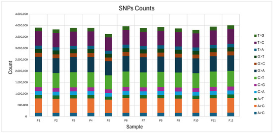
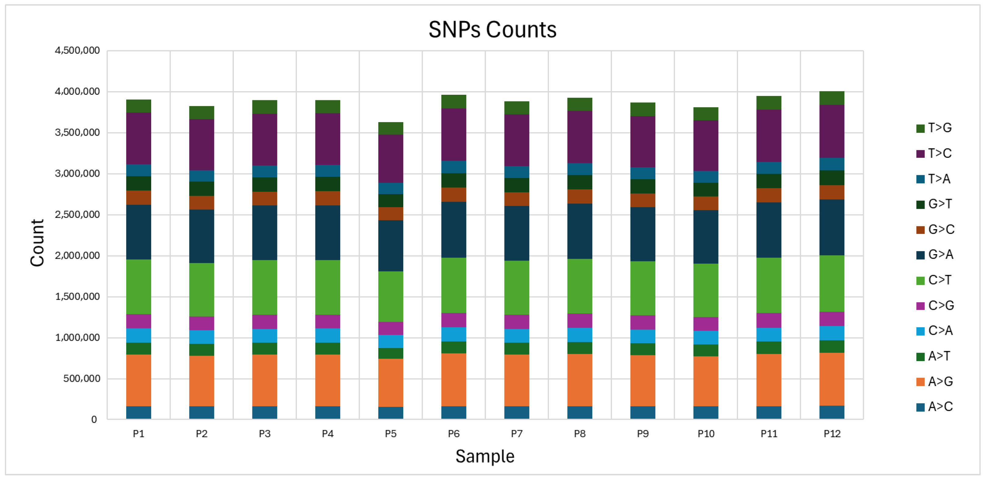
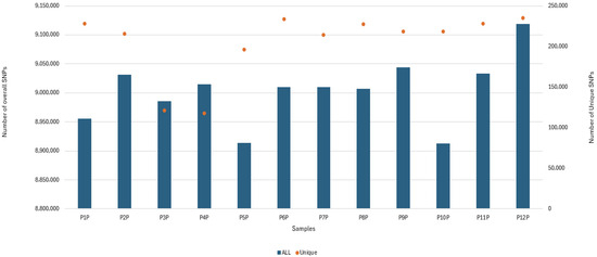
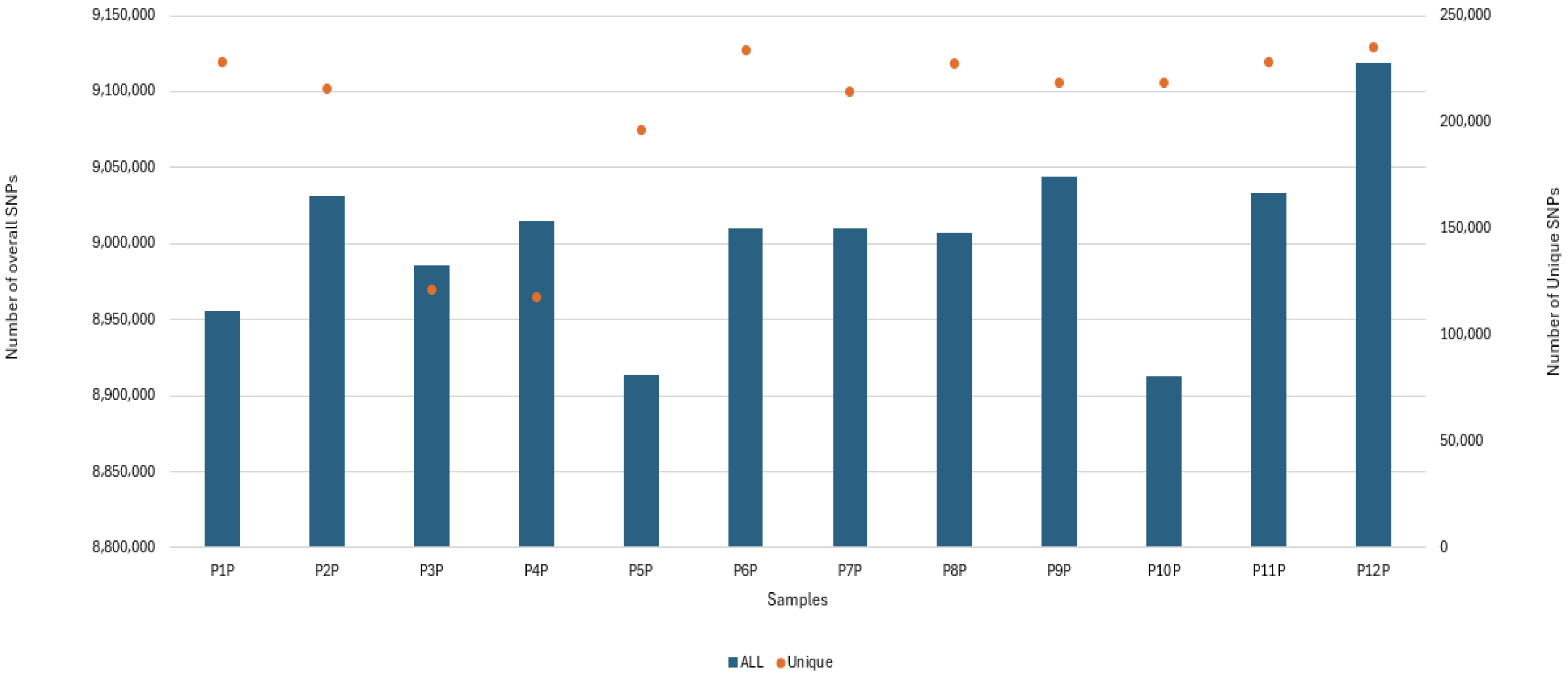
WGS was performed on 12 individuals to investigate genetic variants and biological pathways that may be relevant in PCAD. The groups comprised seven control individuals (samples P1P to P7P) and five confirmed PCAD cases (samples P8P to P12P). High-quality DNA libraries were sequenced with a minimum read depth of 10× witha quality score threshold of 30. Following variant calling and quality control, over 8.8 million SNPs were detected per sample, with total SNP counts comparable across all individuals (Figure 1 and Figure 2). Unique SNPs, representing disease- or population-specific variations, were quantified separately and exhibited slight variability within the PCAD group. We identified 120 SNPs exclusively present in patients. All protein-coding variants annotated against gnomAD were rare (allele frequency < 0.0001), and none were previously classified as pathogenic or likely pathogenic in ClinVar. These variants were annotated and filtered for protein-coding regions, yielding 87 candidate genes (a full list is presented in Supplementary Data File S1). Key PCAD-relevant non-coding genes are discussed herein, with remaining analyses presented in the Supplementary Materials.
Figure 1.
Distribution of single-nucleotide polymorphism (SNP) substitution types across 12 samples. The stacked bar chart shows the counts of different SNP substitution types (e.g., A>G, C>T, T>C, etc.) across twelve whole-genome sequencing samples (P1–P12). Each colored segment represents a specific nucleotide substitution, with transitions (e.g., A>G, C>T) and transversions (e.g., A>T, C>G) clearly visualized. The total SNP burden is comparable across samples, indicating consistent sequencing depth and variant-calling quality. The most common substitutions include C>T and G>A, which are known transition mutations typically resulting from deamination events.
Figure 2.
Comparison of overall and unique SNP counts across whole-genome sequencing samples. The bar and dot plot illustrates the total number of single-nucleotide polymorphisms (SNPs) (blue bars, left y-axis) and unique SNPs (orange dots, right y-axis) for each individual sample (P1P–P12P). While the total SNP counts are relatively consistent across all samples, unique SNP counts show some inter-sample variability, suggesting individual-specific or population-specific variations. This comparison highlights both the sequencing depth and the uniqueness of genetic variation within each genome in the context of premature coronary artery disease analysis.
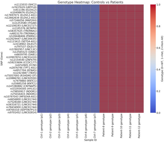
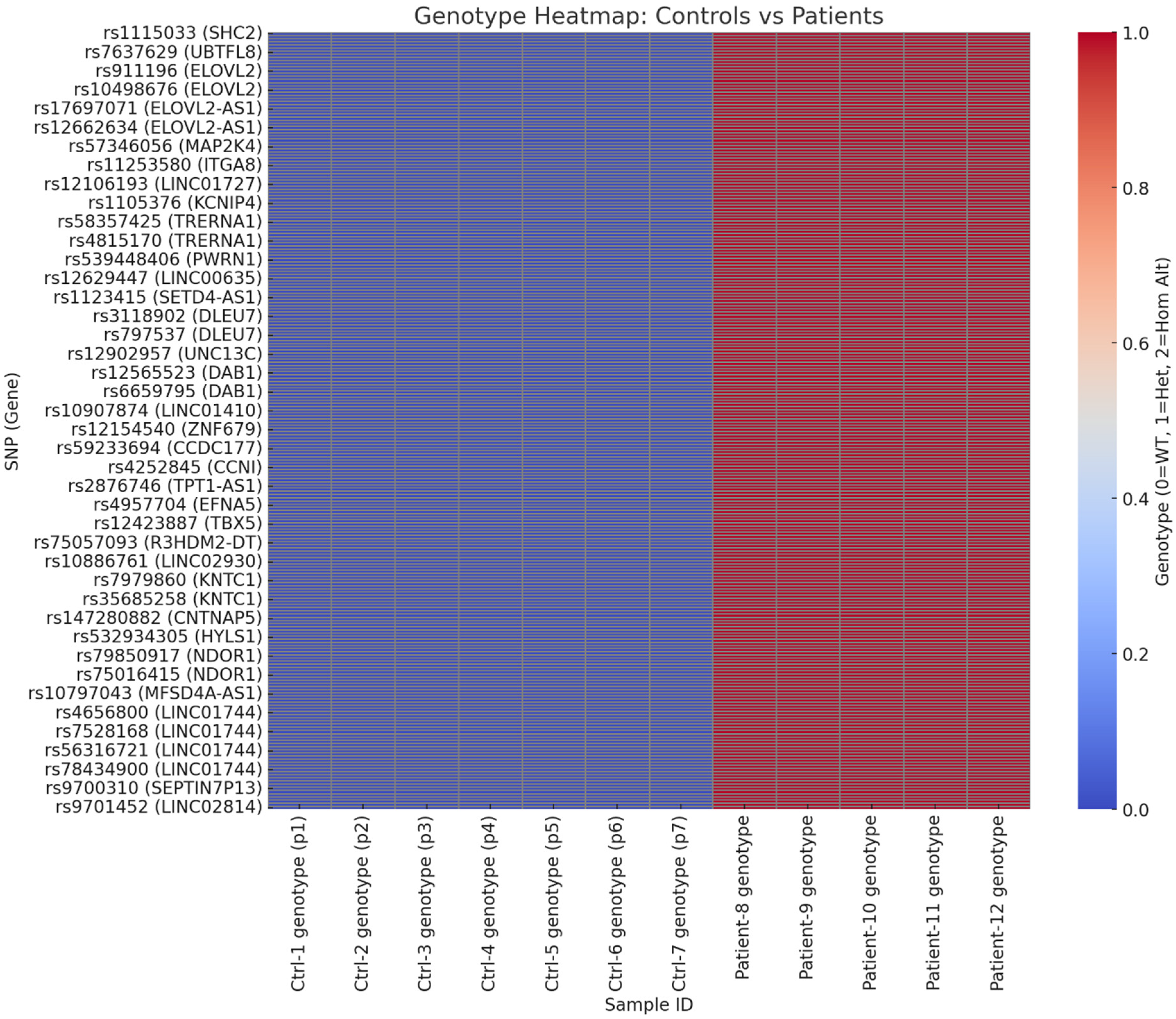
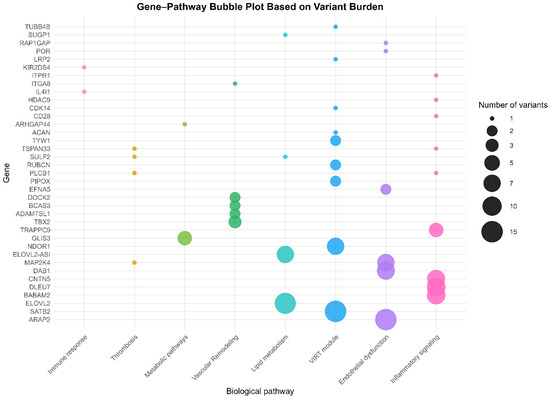
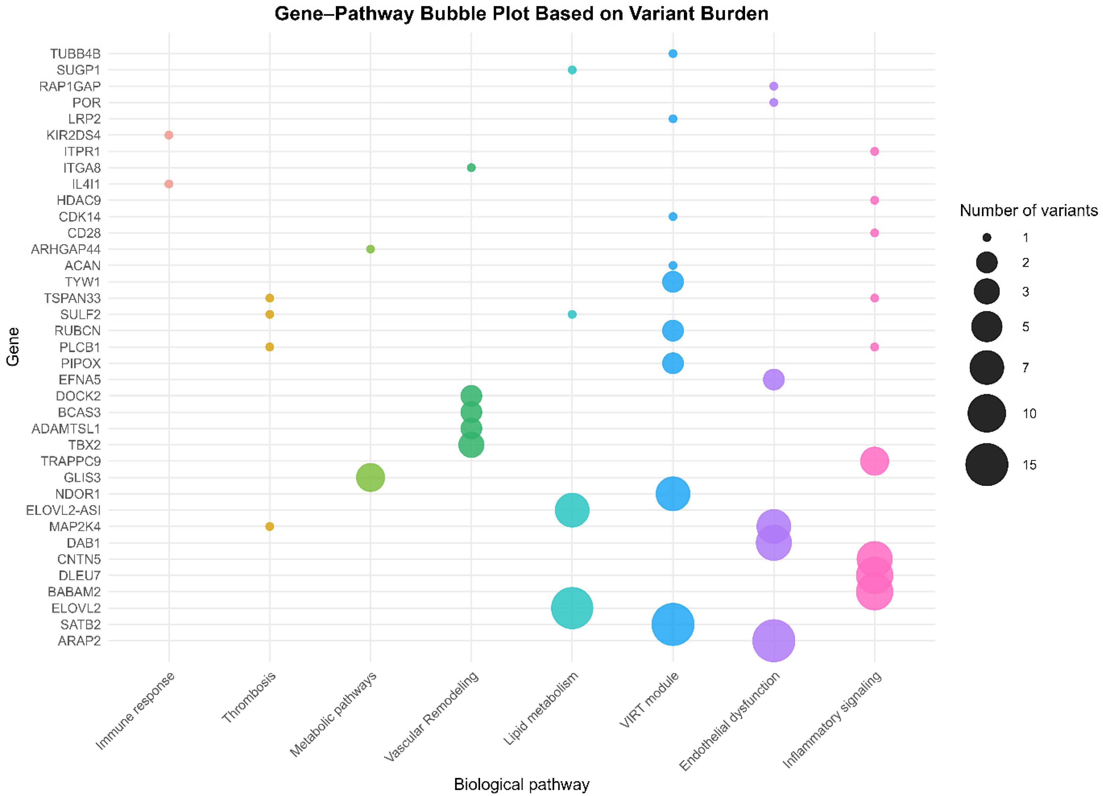
Comparative analyses identified SNPs uniquely present or significantly enriched in PCAD cases, mapped to genes involved in atherosclerosis, lipid metabolism, inflammation, and endothelial dysfunction (Figure 3). Several notable SNPs were identified in genes implicated in diverse pathophysiological pathways relevant to atherosclerosis, including lipid metabolism (ELOVL2, SUGP1, and SULF2), endothelial dysfunction (ARAP2, EFNA5, MAP2K4, NABP, POR, and RAP1GAP), inflammatory signaling (BABAM2, CD28, CNTN5, DLEU7, HDAC9, ITPR1, POR, PLCB1, TSPAN33, and TRAPPC9), immune regulation (DLEU7, IL4I1, and KIR2DS4), thrombosis (MAP2K4, PLCB1, RAP1GAP, SULF2, and TSPAN33,), vascular remodeling (ADAMTSL1, BCAS3, DOCK2, ITGA8, and TBX5), and metabolic processes (ARHGAP44, GLIS3). Additional variants were observed in genes linked to smooth muscle proliferation, oxidative stress, or other processes (ACAN, CDK14, LRP2, NDOR1, PIPOX, RUBCN, SATB2, TUBB4B, and TYW1).
Figure 3.
This heatmap visualizes the genotypic variation across 40 selected SNPs (y-axis), each labeled with its corresponding gene, in 12 study participants (x-axis). Samples Ctrl-1 to Ctrl-7 represent healthy controls, while patient-8 to patient-12 represent individuals with premature coronary artery disease (PCAD). A genotype heatmap shows the differences between controls and patients across all SNPs. Blue (0): Homozygous reference (0|0)—observed in all controls. Variant genotypes (light to dark red, 1 or 2) are found exclusively in patients. The consistent red bands seen across patient columns indicate recurrent SNPs within the PCAD group.
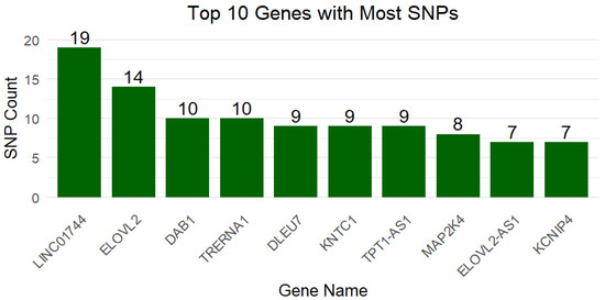
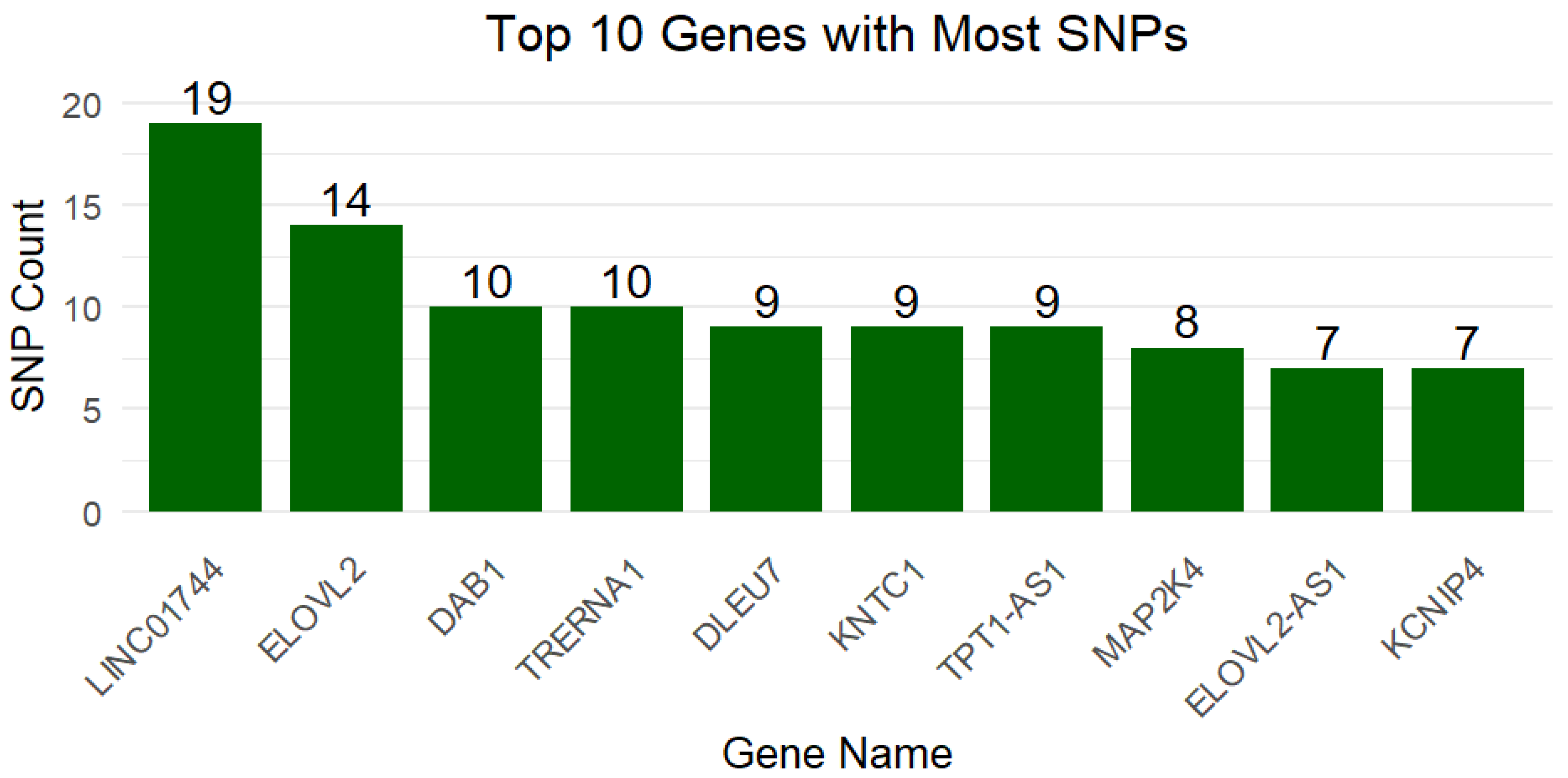
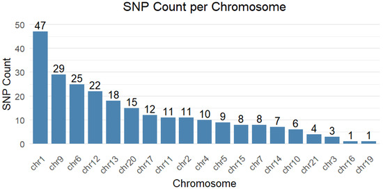
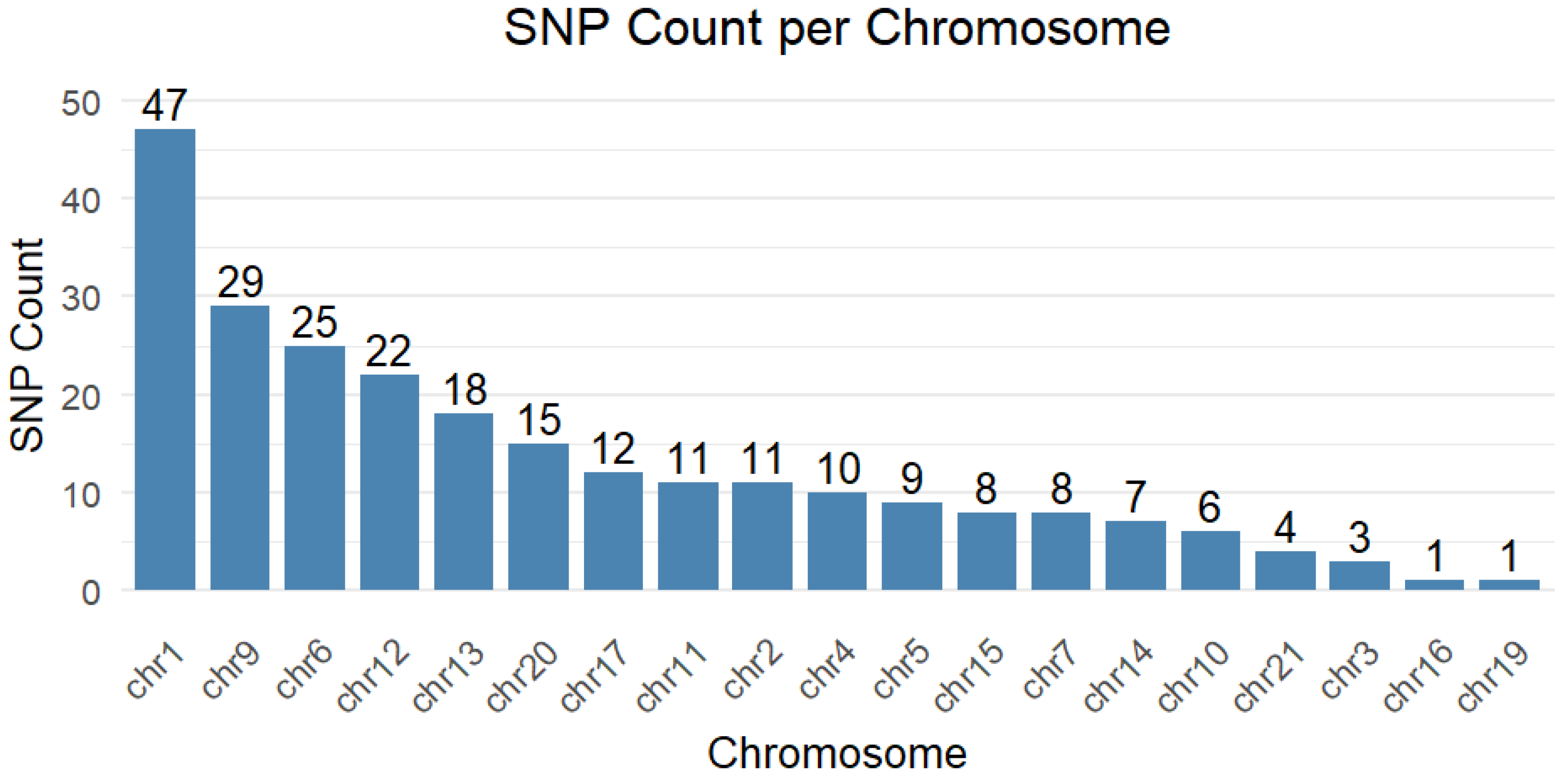
The LINC01744 gene exhibited the largest number of variants (n = 19), followed by ELOVL2 (n = 14). DAB1 and TRERNA1 each showed 10 variants, while DLEU7, KNTC1, and TPT1-AS1 had nine each. MAP2K4 carried eight variants, and ELOVL2-AS1 and KCNIP4 had seven variants each (Figure 4). ELOVL2-AS1 (lipid pathway) and SENCR (promoting atherosclerosis) are non-coding genes linked to PCAD. More loci and mechanisms are detailed in the Supplementary Materials. Chromosome 1 carried the most SNPs (Figure 5).
Figure 4.
Bar chart illustrating the ten genes with the greatest number of SNPs identified across the WGS dataset, with LINC01744 exhibiting the highest SNP burden (19 variants), followed by ELOVL2 (14 SNPs), DAB1 and TRERNA1 (10 each), and DLEU7, KNTC1, and TPT1-AS1 (nine each). Other notable genes include MAP2K4 (eight), ELOVL2-AS1 (seven), and KCNIP4 (seven).
Figure 5.
Bar graph showing SNP count per chromosome from WGS data, with chromosome 1 exhibiting the highest SNP burden of variants (47), followed by chromosome 9 (29), chromosome 6 (25), and chromosome 12 (22). Chromosomes 3, 16, and 19 had the fewest SNPs (one each).
4. Discussion
4.1. Functional Relevance of Genes with SNPs
From a group of 12 subjects (five with PCAD, seven healthy), we found 120 SNPs unique to PCAD cases. Protein-coding variants (n = 87 unique genes) underwent allele frequency annotation using gnomAD, all were rare (AF < 0.0001), and none were previously classified as pathogenic in ClinVar.
This summary outlines the reported SNPs and their biological pathways linked to atherosclerosis. The Supplementary Materials provide more in-depth associations and mechanisms with PCAD. It is important to note that, as an exploratory pilot study, our research does not establish causality. Instead, these findings serve as groundwork for future, larger-scale studies aimed at validation.
4.2. Lipid Metabolism
Dysregulation of lipid metabolism represents a central mechanistic axis in the pathogenesis of PCAD. Beyond traditional lipid measures, genetic and molecular perturbations in cholesterol transport, lipoprotein remodeling, lipid oxidation, and intracellular lipid handling drive early atherosclerosis. Variants identified in the genes ELOVL2 and ELOVL2-AS1 could impact processes such as PUFA remodeling, LDL metabolism, and lipoprotein removal. These changes may play a role in vascular inflammation and the formation of plaques (22,23,24). Case-restricted heterozygosity across multiple rsIDs suggests a locus-level burden rather than a single causal SNP—very consistent with PUFA-pathway biology. ELOVL2 plays a key role in PUFA metabolism and lipid homeostasis, making it relevant for PCAD patients. Likewise, SUGP1 and SULF2 variants are linked to cholesterol metabolism, reduced triglyceride clearance, and CAD (25,26) (Table 1). Investigating genetic variants governing these pathways in large-scale studies would help validate these findings and guide risk prediction, early detection, and novel treatment strategies. Developing functional assays in cellular models could aid in prioritizing candidates for therapeutic intervention; however, these findings must first be validated through adequately powered studies.
4.3. Endothelial Cell Function
Endothelial cells orchestrate vascular homeostasis by regulating nitric oxide (NO)-mediated vasodilation, antithrombotic surface properties, barrier integrity, and leukocyte trafficking. Several genetic variants identified in this pilot study are hypothesized to have mechanistic relevance in the development of premature atherosclerosis by affecting endothelial barrier integrity. Specifically, DAB1 may contribute through activation of the Reelin pathway [27,28,29]; ARAP2 and MAP2K4 may be involved via leukocyte adhesion [30,31,32]; and EFNA5, POR, and RAP1GAP may influence disease progression through mechanisms related to vascular repair and NO release [33,34,35,36,37,38,39]. Exposure to pathological stimuli, such as disturbed blood flow, hyperglycemia, dyslipidemia, tobacco exposure, or oxidative stress, induces endothelial dysfunction leading to reduced eNOS activity and NO bioavailability, elevated reactive oxygen species driving redox-sensitive pathways (NF-κB/JNK); induction of cell surface adhesion molecules (ICAM-1, VCAM-1, and E-selectin), resulting in diminished barrier function leading to increased permeability to inflammatory mediators (monocytes, and apoB lipoproteins). In this pathophysiological framework, it is proposed that these variants converge on key processes, including endothelial signaling, redox homeostasis, cellular adhesion, and tissue repair, thus representing biologically plausible mechanisms linking genotype to early atherosclerosis, irrespective of prolonged pathological stimuli. While the current pilot study lacks sufficient statistical power to demonstrate causality, it highlights the need for extensive studies to confirm these preliminary observations (Table 2 and Figure 6).
Table 2.
Variants identified in EC-function-related genes.
Figure 6.
Bubble plot showing the distribution of genetic variants on biological pathways of cardiac-related genes. Each bubble represents a gene annotated to a particular biological pathway. The size of the bubble shows the variants per gene, and the colors denote the pathways, such as lipid metabolism, endothelial dysfunction, inflammatory signaling, immune response, thrombosis, vascular remodeling, metabolic pathways, and the VIRT module.
4.4. Inflammatory Pathways
Atherosclerosis is a chronic inflammatory disease characterized by endothelial dysfunction that leads to subendothelial retention of lipoproteins, activation of adhesion molecules (VCAM-1, ICAM-1, and E-selectin), and chemokines resulting in the recruitment of monocytes and T cells into the intima. SNPs identified in genes, such as BABAM2, DLEU7, CNTN5, and TRAPPC9, may affect NF-κB signaling or adhesion proteins, promoting inflammation [40,41,42,43,44]. These variants can disrupt innate immune pathways (TLR–MyD88–NF-κB, NLRP3–IL-1β), leading to macrophage activation, foam cell formation, and reduced efferocytosis. Additional genes with single variants, including HDAC9, CD28, ITPR1, TSPAN33, and PLCB1, might also contribute to a pro-inflammatory state by either increasing inflammation or reducing protective mechanisms [45,46,47,48,49,50,51,52,53,54,55,56,57,58]. Details of these variants are provided in Table 3. These findings suggest that investigating genetic variants and their underlying mechanisms—especially those responsible for inflammation—could be an effective approach to further our understanding of premature conditions (Table 3 and Figure 6).
Table 3.
Variants identified in inflammatory pathway-related genes.
4.5. Immune-Related Pathways
Atherosclerosis is also postulated to have a maladaptive immune response to retained apoB-lipoproteins within the arterial wall. Innate sensors (TLRs, scavenger receptors) on endothelial cells, macrophages, and dendritic cells detect modified LDL and cellular debris, activating NF-κB and inflammasomes (e.g., NLRP3→IL-1β/IL-18). This inflammatory milieu is perpetuated through monocyte recruitment, macrophage polarization, defective efferocytosis, and neutrophil extracellular traps, creating a non-resolving inflammatory cascade. In this pilot study, we identified one variant of KIR2DS4 and one variant of the IL4I1 gene that may alter immune response by triggering it or suppressing protective mechanisms [59,60]. Adaptive immunity layers amplify this niche wherein Th1- and Th17-derived cytokines (IFN-γ, IL-17) exacerbate vascular inflammation while Tregs counteract this progression via IL-10/TGF-β. B-cells exhibit a dichotomous response, with natural IgM against oxLDL being protective, whereas some B2 responses promote disease (Table 4). Natural killer (NK) cells and cytotoxic T cells can add plaque-destabilizing cytotoxicity. Genes that bias Treg/Th17 balance, NK activation thresholds, or cytokine set-points can therefore shift risk—offering handles for diagnostics (circulating proteins/cell-subset signatures) and therapeutics (pathway-targeted immunomodulation). Though purely hypothesis-generating, it is an important avenue to explore in large sample studies with subsequent validation studies in cell and animal models.
Table 4.
Variants identified in immune-related genes.
4.6. Thrombotic Pathways
MI represents the clinical manifestation of ruptured atherosclerotic plaque exposing tissue factors and subendothelial matrix, leading to a cascade of platelet activation and coagulation, which culminates in occlusive coronary thrombosis. Genetic predispositions and acquired risk factors that heighten platelet reactivity, enhance thrombin generation, or impair endothelial anticoagulation mechanisms alter this balance toward occlusive arterial thrombosis. Several genetic variants identified in this preliminary study may suggest an increased thrombotic risk, specifically involving PLCB1, MAP2K4, and SULF2 [26,53]. These variants could contribute to early-onset myocardial infarction by activating thrombogenic pathways, even in cases where coronary atherosclerosis is minimal. The variants relevant to thrombotic pathways are detailed in Table 5.
Table 5.
Variants linked to thrombotic pathways.
4.7. Vascular Remodeling
Vascular remodeling is the structural and cellular reconfiguration of the arterial wall in response to hemodynamic load, injury, and inflammation. It encompasses endothelial activation, smooth-muscle cell (SMC) phenotypic switching (contractile to synthetic/migratory), extracellular matrix reorganization, microfibril/TGF-β signaling, integrin-mediated transduction, and (neo) angiogenesis within the intima and media. The study identified variants in the DOCK2, ADAMTSL1, and TBX2 genes [61,62,63,64,65,66,67,68], which may act as modulators of signaling pathways. Additional variants of genes, such as ITGA8, are considered to influence the composition of the extracellular matrix [69,70], while variants of BCAS3 need further study, as this gene is implicated in the regulation of angiogenesis [71,72]. Table 6 and Figure 6 provide an overview of these variants and their associated diseases.
Table 6.
Variants identified in vascular remodeling-associated genes.
While adaptive remodeling preserves lumen caliber, maladaptive remodeling (excess SMC migration/proliferation, aberrant extracellular matrix deposition, fibrous cap thinning, and plaque neovascularization) promotes plaque growth, instability, and thrombosis. Genetic variants that alter leukocyte–endothelial crosstalk, SMC adhesion/motility, matrix architecture, or endothelial polarity/angiogenesis can predispose an individual to maladaptive remodeling, thereby increasing susceptibility toward premature atherogenesis and MI. These mechanisms are outlined to generate hypotheses for future large-scale studies. The increasing incidence and earlier onset of PCAD call for focused research into these mechanisms.
4.8. Metabolic Pathways
Metabolic syndrome, diabetes, and insulin resistance are potent atherogenic precursors because they create a lipoprotein-rich, pro-inflammatory, NO-deficient, and prothrombotic vascular environment, thereby transforming metabolic stress into endothelial dysfunction, plaque growth, and ultimately thrombotic coronary events. Genetic determinants of insulin resistance are of significant interest for advancing our understanding of mechanistic pathways leading to premature atherosclerosis. This study observed GLIS3 and ARHGAP44 variants that may be connected to this pathway [73,74,75] (Table 7).
Table 7.
Variants identified in metabolic pathway-related genes.
4.9. Vascular Inflammation–Remodeling–Thrombosis (VIRT) Pathway in PCAD
The identified genes in this group converge on several key hypothetical mechanisms underlying atherosclerosis at a young age, including vascular senescence, oxidative stress, inflammation, and plaque instability. Variants in SATB2 mapped to the 9p21/CDKN2A/B locus, implicating endothelial senescence and VSMC osteogenic programming via RUNX2 [76,77]. NDOR1 variants were linked to diabetic vascular aging through dysregulation of redox-sensitive NF-κB and MAPK pathways [78,79,80]. Metabolic and translational stress pathways were represented by TYW1 and PIPOX, involving tRNA modification and H2O2-generating amino-acid metabolism, respectively [81,82,83]. RUBCN variants, associated with acute coronary syndrome, impair LC3-associated phagocytosis and efferocytosis, contributing to plaque inflammation and vulnerability [84]. Additional genes (LRP2, CDK14, TUBB4B, and ACAN) influence renin–angiotensin homeostasis, Wnt signaling, cytoskeletal–AKT pathways, and VSMC apoptosis, collectively reinforcing pathways of vascular dysfunction and plaque instability [85,86,87,88]. This pilot study revealed nine VIRT pathway-related genes harboring 32 variants that need further exploration in large study. These genes are summarized in Table 8.
Table 8.
Variants identified in VIRT pathway-related genes.
5. Conclusions
This WGS pilot dataset provides a genomic landscape for evaluating molecular drivers of early-onset cardiovascular disease and constitutes a valuable starting point for a large-scale study to validate these findings. It is conceivable to point out a single disease framework for PCAD: an endothelial–immune–metabolic remodeling axis that amplifies lipid retention, vascular inflammation, smooth-muscle reprogramming, and thrombosis. Within this framework, several loci could emerge as pathway anchors for premature atherosclerosis leading to MI at a young age. This data highlights WGS as a useful approach for studying genetic factors behind early-onset atherosclerosis, particularly among South Asians, where traditional risk factors are often absent and the disease remains largely unexplained. These genomic findings described in this paper are preliminary and hypothesis-generating, derived from a small sample size intended to demonstrate feasibility and establish analytic workflows. The candidate loci and pathways require replication in larger, adequately powered cohorts. A larger sample study is underway to provide the statistical power needed to validate these signals. Once validated in larger studies, polygenic risk scores could enable earlier risk stratification in premature disease beyond conventional age thresholds. In parallel, expression-based biomarkers derived from genetic risk variants may offer additional opportunities for early detection and family screening.
Supplementary Materials
The following supporting information can be downloaded at: https://www.mdpi.com/article/10.3390/cardiogenetics16020009/s1. Supplementary Data File S1. Detailed gene-by-gene mechanistic interpretation of candidate variants identified through whole genome sequencing, including proposed roles in endothelial dysfunction, inflammation, lipid metabolism, vascular remodeling, oxidative stress, immune regulation, and atherosclerosis-related pathways.
Author Contributions
Conceptualization, I.A.C., F.J., A.C., and W.I.; methodology, I.A.C. and F.J.; software, F.J., W.I., and Y.A.; validation, I.A.C., T.A., S.K., and F.J.; formal analysis, I.A.C., F.J., and Y.A.; investigation, I.A.C., A.C., and Y.J.; resources, A.C., F.J., and Y.J.; data curation, I.A.C., F.J., Y.A., M.A., A.R., and A.N.; writing—original draft preparation, I.A.C. and F.J.; writing—review and editing, I.A.C., Y.A., M.A., T.A., A.R., and A.N.; visualization, Y.A. and T.A.; supervision, A.C., N.T., A.K., and K.N.; project administration, I.A.C. and A.C. All authors have read and agreed to the published version of the manuscript.
Funding
This research received no external funding.
Institutional Review Board Statement
The study was conducted in accordance with the Declaration of Helsinki and received ethical approval from multiple institutional review boards. The primary protocol titled “Investigating Genetic Variants and Genes Associated with Premature Coronary Artery Disease: A Step Towards Early Diagnosis and Tailored Interventions” was approved by the National Bioethics Committee for Research (NBC-R), National Institutes of Health, Pakistan (Protocol No. NBCR-1310; approval date: 8 December 2025). Additional ethical approvals were obtained from the Ethics Committee, Department of Biotechnology, Abdul Wali Khan University Mardan, Pakistan (approval date: 3 January 2024), and the Institutional Ethical Review Board (IERB), Armed Forces Institute of Cardiology & National Institute of Heart Diseases (AFIC/NIHD), Rawalpindi, Pakistan (IERB Approval No. 9/2/R&D/2024/335, Synopsis Reg. No. S/189/2024; approval date: 12 December 2024; valid through 28 April 2027).
Informed Consent Statement
Informed consent was obtained from all subjects involved in the study.
Data Availability Statement
The data presented in this study are available on request from the corresponding author due to the exploratory and pilot nature of the study and ongoing efforts to validate the findings in larger, independent cohorts.
Conflicts of Interest
The authors declare no conflicts of interest.
References
- Khoja, A.; Andraweera, P.H.; Lassi, Z.S.; Zheng, M.; Pathirana, M.M.; Ali, A.; Aldridge, E.; Wittwer, M.R.; Chaudhuri, D.D.; Tavella, R.; et al. Risk factors for premature coronary artery disease (PCAD) in adults: A systematic review protocol. F1000Research 2021, 10, 1228. [Google Scholar] [CrossRef] [Scilit] [PubMed]
- Bansal, S.K.; Agarwal, S.; Daga, M.K. Advanced Atherogenic Index for the Assessment of Consolidated Lipid Risk in Premature Coronary Artery Disease Patients in India. J. Lab. Physicians 2016, 8, 77–84. [Google Scholar] [CrossRef] [Scilit]
- Yang, J.; Biery, D.W.; Singh, A.; Divakaran, S.; DeFilippis, E.M.; Wu, W.Y.; Klein, J.; Hainer, J.; Ramsis, M.; Natarajan, P.; et al. Risk Factors and Outcomes of Very Young Adults Who Experience Myocardial Infarction: The Partners YOUNG-MI Registry. Am. J. Med. 2020, 133, 605–612.e601. [Google Scholar] [CrossRef] [Scilit]
- Jortveit, J.; Pripp, A.H.; Langørgen, J.; Halvorsen, S. Incidence, risk factors and outcome of young patients with myocardial infarction. Heart 2020, 106, 1420–1426. [Google Scholar] [CrossRef] [Scilit]
- Samad, Z.; Hanif, B. Cardiovascular Diseases in Pakistan: Imagining a Postpandemic, Postconflict Future. Circulation 2023, 147, 1261–1263. [Google Scholar] [CrossRef] [Scilit]
- Jafar, T.H.; Qadri, Z.; Chaturvedi, N. Coronary artery disease epidemic in Pakistan: More electrocardiographic evidence of ischaemia in women than in men. Heart 2008, 94, 408–413. [Google Scholar] [CrossRef] [Scilit]
- Ullah, W.; Malik, R.; Bashir, F.; Khan, M.; Khan, S.; Alam, M.; Samad, Z.; Achirica, M.C.; Virani, S.S.; Hanif, B. Comparison of Premature, Extremely Premature, and Older Adults with Coronary Artery Disease in Pakistan. JACC Asia 2023, 3, 164–165. [Google Scholar] [CrossRef] [Scilit] [PubMed]
- Amin, A.P.; Nathan, S.; Evans, A.T.; Attanasio, S.; Mukhopadhyay, E.; Mehta, V.; Kelly, R.F. The effect of ethnicity on the relationship between premature coronary artery disease and traditional cardiac risk factors among uninsured young adults. Prev. Cardiol. 2009, 12, 128–135. [Google Scholar] [CrossRef] [Scilit]
- Sharma, M.; Ganguly, N.K. Premature coronary artery disease in Indians and its associated risk factors. Vasc. Health Risk Manag. 2005, 1, 217–225. [Google Scholar]
- Chowdhury, R.; Alam, D.S.; Fakir, I.I.; Adnan, S.D.; Naheed, A.; Tasmin, I.; Monower, M.M.; Hossain, F.; Hossain, F.M.; Rahman, M.M.; et al. The Bangladesh Risk of Acute Vascular Events (BRAVE) Study: Objectives and design. Eur. J. Epidemiol. 2015, 30, 577–587. [Google Scholar] [CrossRef] [Scilit]
- Tan, S.T.; Scott, W.; Panoulas, V.; Sehmi, J.; Zhang, W.; Scott, J.; Elliott, P.; Chambers, J.; Kooner, J.S. Coronary heart disease in Indian Asians. Glob. Cardiol. Sci. Pract. 2014, 2014, 13–23. [Google Scholar] [CrossRef] [Scilit] [PubMed]
- Saleheen, D.; Haycock, P.C.; Zhao, W.; Rasheed, A.; Taleb, A.; Imran, A.; Abbas, S.; Majeed, F.; Akhtar, S.; Qamar, N.; et al. Apolipoprotein(a) isoform size, lipoprotein(a) concentration, and coronary artery disease: A mendelian randomisation analysis. Lancet Diabetes Endocrinol. 2017, 5, 524–533. [Google Scholar] [CrossRef] [Scilit]
- Anand, S.S.; Vasudevan, A.; Gupta, M.; Morrison, K.; Kurpad, A.; Teo, K.K.; Srinivasan, K.; Investigators, S.C.S. Rationale and design of South Asian Birth Cohort (START): A Canada-India collaborative study. BMC Public Health 2013, 13, 79. [Google Scholar] [CrossRef] [Scilit] [PubMed]
- The 1000 Genomes Project Consortium. The 1000 Genomes Project: A Deep Catalog of Human Genetic Variation. International Genome Sample Resource (IGSR), EMBL-EBI. 2017. Available online: https://www.internationalgenome.org/ (accessed on 20 January 2026).
- The Coronary Artery Disease (C4D) Genetics Consortium. A genome-wide association study in Europeans and South Asians identifies five new loci for coronary artery disease. Nat. Genet. 2011, 43, 339–344. [Google Scholar] [CrossRef] [Scilit]
- Howson, J.M.M.; Zhao, W.; Barnes, D.R.; Ho, W.K.; Young, R.; Paul, D.S.; Waite, L.L.; Freitag, D.F.; Fauman, E.B.; Salfati, E.L.; et al. Fifteen new risk loci for coronary artery disease highlight arterial-wall-specific mechanisms. Nat. Genet. 2017, 49, 1113–1119. [Google Scholar] [CrossRef] [Scilit]
- Golbus, J.R.; Stitziel, N.O.; Zhao, W.; Xue, C.; Farrall, M.; McPherson, R.; Erdmann, J.; Deloukas, P.; Watkins, H.; Schunkert, H.; et al. Common and Rare Genetic Variation in CCR2, CCR5, or CX3CR1 and Risk of Atherosclerotic Coronary Heart Disease and Glucometabolic Traits. Circ. Cardiovasc. Genet. 2016, 9, 250–258. [Google Scholar] [CrossRef] [Scilit]
- Khan, S.; Phulukdaree, A.; Ramkaran, P.; Moodley, D.; Chuturgoon, A.A. The Arg72 variant of the p53 functional polymorphism (rs1042522) is associated with coronary artery disease in young South Africans of Indian ancestry. Gene 2016, 593, 261–264. [Google Scholar] [CrossRef] [Scilit]
- Flowers, E.; Singh, K.; Molina, C.; Mathur, A.; Aouizerat, B.E. MicroRNA associated with atherogenic dyslipidemia in South Asian men. Int. J. Cardiol. 2013, 168, 4884–4885. [Google Scholar] [CrossRef] [Scilit]
- Fuchsberger, C.; Flannick, J.; Teslovich, T.M.; Mahajan, A.; Agarwala, V.; Gaulton, K.J.; Ma, C.; Fontanillas, P.; Moutsianas, L.; McCarthy, D.J.; et al. The genetic architecture of type 2 diabetes. Nature 2016, 536, 41–47. [Google Scholar] [CrossRef] [Scilit]
- Khera, A.V.; Chaffin, M.; Aragam, K.G.; Haas, M.E.; Roselli, C.; Choi, S.H.; Natarajan, P.; Lander, E.S.; Lubitz, S.A.; Ellinor, P.T.; et al. Genome-wide polygenic scores for common diseases identify individuals with risk equivalent to monogenic mutations. Nat. Genet. 2018, 50, 1219–1224. [Google Scholar] [CrossRef] [Scilit] [PubMed]
- Sambrook, J.; Russell, D.W. Molecular Cloning: A Laboratory Manual, 3rd ed.; Cold Spring Harbor Laboratory Press: Cold Spring Harbor, NY, USA, 2001; ISBN 0879695773. [Google Scholar]
- Maguolo, A.; Zusi, C.; Giontella, A.; Miraglia Del Giudice, E.; Tagetti, A.; Fava, C.; Morandi, A.; Maffeis, C. Influence of genetic variants in FADS2 and ELOVL2 genes on BMI and PUFAs homeostasis in children and adolescents with obesity. Int. J. Obes. 2021, 45, 56–65. [Google Scholar] [CrossRef] [Scilit]
- Aslibekyan, S.; Jensen, M.K.; Campos, H.; Linkletter, C.D.; Loucks, E.B.; Ordovas, J.M.; Deka, R.; Rimm, E.B.; Baylin, A. Genetic variation in fatty acid elongases is not associated with intermediate cardiovascular phenotypes or myocardial infarction. Eur. J. Clin. Nutr. 2012, 66, 353–359. [Google Scholar] [CrossRef] [Scilit]
- Kim, M.J.; Yu, C.Y.; Theusch, E.; Naidoo, D.; Stevens, K.; Kuang, Y.L.; Schuetz, E.; Chaudhry, A.S.; Medina, M.W. SUGP1 is a novel regulator of cholesterol metabolism. Hum. Mol. Genet. 2016, 25, 3106–3116. [Google Scholar] [CrossRef] [Scilit]
- Kim, T.H.; Banini, B.A.; Asumda, F.Z.; Campbell, N.A.; Hu, C.; Moser, C.D.; Shire, A.M.; Han, S.; Ma, C.; Krishnan, A.; et al. Knockout of sulfatase 2 is associated with decreased steatohepatitis and fibrosis in a mouse model of nonalcoholic fatty liver disease. Am. J. Physiol. Gastrointest. Liver Physiol. 2020, 319, G333–G344. [Google Scholar] [CrossRef] [Scilit] [PubMed]
- Jover, E.; Garaikoetxea, M.; Martín-Núñez, E.; Goñi-Olóriz, M.; San-Ildefonso-García, S.; Navarro, A.; Fernández-Celis, A.; Álvarez, V.; Sádaba, R.; Calvier, L.; et al. Expression of the lymphangiogenic reelin is associated with sex-dependent calcific aortic stenosis in men. Atherosclerosis 2025, 403, 119162. [Google Scholar] [CrossRef] [Scilit]
- Ding, Y.; Huang, L.; Xian, X.; Yuhanna, I.S.; Wasser, C.R.; Frotscher, M.; Mineo, C.; Shaul, P.W.; Herz, J. Loss of Reelin protects against atherosclerosis by reducing leukocyte-endothelial cell adhesion and lesion macrophage accumulation. Sci. Signal. 2016, 9, ra29. [Google Scholar] [CrossRef] [Scilit] [PubMed]
- Li, Y.; Wang, D.W.; Chen, Y.; Chen, C.; Guo, J.; Zhang, S.; Sun, Z.; Ding, H.; Yao, Y.; Zhou, L.; et al. Genome-Wide Association and Functional Studies Identify. Arterioscler. Thromb. Vasc. Biol. 2018, 38, 964–975. [Google Scholar] [CrossRef] [Scilit]
- Chen, P.W.; Jian, X.; Yoon, H.Y.; Randazzo, P.A. ARAP2 signals through Arf6 and Rac1 to control focal adhesion morphology. J. Biol. Chem. 2013, 288, 5849–5860. [Google Scholar] [CrossRef] [Scilit]
- Paul, N.R.; Jacquemet, G.; Caswell, P.T. Endocytic Trafficking of Integrins in Cell Migration. Curr. Biol. 2015, 25, R1092–R1105. [Google Scholar] [CrossRef] [Scilit]
- Cuenda, A. Mitogen-activated protein kinase kinase 4 (MKK4). Int. J. Biochem. Cell Biol. 2000, 32, 581–587. [Google Scholar] [CrossRef] [Scilit] [PubMed]
- Funk, S.D.; Yurdagul, A.; Albert, P.; Traylor, J.G.; Jin, L.; Chen, J.; Orr, A.W. EphA2 activation promotes the endothelial cell inflammatory response: A potential role in atherosclerosis. Arterioscler. Thromb. Vasc. Biol. 2012, 32, 686–695. [Google Scholar] [CrossRef] [Scilit]
- Zhu, Y.; Su, S.A.; Shen, J.; Ma, H.; Le, J.; Xie, Y.; Xiang, M. Recent advances of the Ephrin and Eph family in cardiovascular development and pathologies. iScience 2024, 27, 110556. [Google Scholar] [CrossRef] [Scilit]
- Udhane, S.S.; Parween, S.; Kagawa, N.; Pandey, A.V. Altered CYP19A1 and CYP3A4 Activities Due to Mutations A115V, T142A, Q153R and P284L in the Human P450 Oxidoreductase. Front. Pharmacol. 2017, 8, 580. [Google Scholar] [CrossRef] [Scilit]
- Fleming, I. Cytochrome p450 and vascular homeostasis. Circ. Res. 2001, 89, 753–762. [Google Scholar] [CrossRef] [Scilit] [PubMed]
- Malacarne, P.F.; Ratiu, C.; Gajos-Draus, A.; Müller, N.; Lopez, M.; Pflüger-Müller, B.; Ding, X.; Warwick, T.; Oo, J.; Siragusa, M.; et al. Loss of Endothelial Cytochrome P450 Reductase Induces Vascular Dysfunction in Mice. Hypertension 2022, 79, 1216–1226. [Google Scholar] [CrossRef] [Scilit]
- Chrzanowska-Wodnicka, M. Rap1 in endothelial biology. Curr. Opin. Hematol. 2017, 24, 248–255. [Google Scholar] [CrossRef] [Scilit] [PubMed]
- Shan, T.; Li, X.; Xie, W.; Wang, S.; Gao, Y.; Zheng, Y.; Su, G.; Li, Y.; Zhao, Z. Rap1GAP exacerbates myocardial infarction by regulating the AMPK/SIRT1/NF-κB signaling pathway. Cell. Signal. 2024, 117, 111080. [Google Scholar] [CrossRef] [Scilit] [PubMed]
- Matsumori, A. Nuclear Factor-κB is a Prime Candidate for the Diagnosis and Control of Inflammatory Cardiovascular Disease. Eur. Cardiol. 2023, 18, e40. [Google Scholar] [CrossRef] [Scilit]
- Gao, Z.; Zhu, H.; Chen, J.; Liu, W.; Huo, J.; He, C. Clinical value of BRE-AS1 in myocardial infarction and its role in myocardial infarction-induced cardiac muscle cell apoptosis. Scand. Cardiovasc. J. 2024, 58, 2347290. [Google Scholar] [CrossRef] [Scilit]
- Kovács, Z.J.; Harami, G.M.; Pálinkás, J.; Kuljanishvili, N.; Hegedüs, J.; Harami-Papp, H.; Mahmudova, L.; Khamisi, L.; Szakács, G.; Kovács, M. DNA-dependent phase separation by human SSB2 (NABP1/OBFC2A) protein points to adaptations to eukaryotic genome repair processes. Protein Sci. 2024, 33, e4959. [Google Scholar] [CrossRef] [Scilit]
- Kichaev, G.; Bhatia, G.; Loh, P.R.; Gazal, S.; Burch, K.; Freund, M.K.; Schoech, A.; Pasaniuc, B.; Price, A.L. Leveraging Polygenic Functional Enrichment to Improve GWAS Power. Am. J. Hum. Genet. 2019, 104, 65–75. [Google Scholar] [CrossRef] [Scilit]
- Jackson, S.W.; Scharping, N.E.; Jacobs, H.M.; Wang, S.; Chait, A.; Rawlings, D.J. Cutting Edge: BAFF Overexpression Reduces Atherosclerosis via TACI-Dependent B Cell Activation. J. Immunol. 2016, 197, 4529–4534. [Google Scholar] [CrossRef] [Scilit]
- Bellenguez, C.; Bevan, S.; Gschwendtner, A.; Spencer, C.C.; Burgess, A.I.; Pirinen, M.; Jackson, C.A.; Traylor, M.; Strange, A.; Su, Z.; et al. Genome-wide association study identifies a variant in HDAC9 associated with large vessel ischemic stroke. Nat. Genet. 2012, 44, 328–333. [Google Scholar] [CrossRef] [Scilit] [PubMed]
- Asare, Y.; Campbell-James, T.A.; Bokov, Y.; Yu, L.L.; Prestel, M.; El Bounkari, O.; Roth, S.; Megens, R.T.A.; Straub, T.; Thomas, K.; et al. Histone Deacetylase 9 Activates IKK to Regulate Atherosclerotic Plaque Vulnerability. Circ. Res. 2020, 127, 811–823. [Google Scholar] [CrossRef] [Scilit]
- Bullenkamp, J.; Dinkla, S.; Kaski, J.C.; Dumitriu, I.E. Targeting T cells to treat atherosclerosis: Odyssey from bench to bedside. Eur. Heart J. Cardiovasc. Pharmacother. 2016, 2, 194–199. [Google Scholar] [CrossRef] [Scilit] [PubMed]
- Gerli, R.; Schillaci, G.; Giordano, A.; Bocci, E.B.; Bistoni, O.; Vaudo, G.; Marchesi, S.; Pirro, M.; Ragni, F.; Shoenfeld, Y.; et al. CD4+CD28- T lymphocytes contribute to early atherosclerotic damage in rheumatoid arthritis patients. Circulation 2004, 109, 2744–2748. [Google Scholar] [CrossRef] [Scilit]
- Lin, Q.; Zhao, L.; Jing, R.; Trexler, C.; Wang, H.; Li, Y.; Tang, H.; Huang, F.; Zhang, F.; Fang, X.; et al. Inositol 1,4,5-Trisphosphate Receptors in Endothelial Cells Play an Essential Role in Vasodilation and Blood Pressure Regulation. J. Am. Heart Assoc. 2019, 8, e011704. [Google Scholar] [CrossRef] [Scilit]
- He, M.; Huang, T.S.; Li, S.; Hong, H.C.; Chen, Z.; Martin, M.; Zhou, X.; Huang, H.Y.; Su, S.H.; Zhang, J.; et al. Atheroprotective Flow Upregulates ITPR3 (Inositol 1,4,5-Trisphosphate Receptor 3) in Vascular Endothelium via KLF4 (Krüppel-Like Factor 4)-Mediated Histone Modifications. Arterioscler. Thromb. Vasc. Biol. 2019, 39, 902–914. [Google Scholar] [CrossRef] [Scilit]
- Huang, F.; Zhang, F.; Huang, L.; Zhu, X.; Huang, C.; Li, N.; Da, Q.; Huang, Y.; Yang, H.; Wang, H.; et al. Inositol 1,4,5-Trisphosphate Receptors Regulate Vascular Smooth Muscle Cell Proliferation and Neointima Formation in Mice. J. Am. Heart Assoc. 2024, 13, e034203. [Google Scholar] [CrossRef] [Scilit]
- Ruiz-García, A.; López-López, S.; García-Ramírez, J.J.; Baladrón, V.; Ruiz-Hidalgo, M.J.; López-Sanz, L.; Ballesteros, Á.; Laborda, J.; Monsalve, E.M.; Díaz-Guerra, M.J. The Tetraspanin TSPAN33 Controls TLR-Triggered Macrophage Activation through Modulation of NOTCH Signaling. J. Immunol. 2016, 197, 3371–3381. [Google Scholar] [CrossRef] [Scilit]
- Lin, Y.J.; Chang, J.S.; Liu, X.; Tsang, H.; Chien, W.K.; Chen, J.H.; Hsieh, H.Y.; Hsueh, K.C.; Shiao, Y.T.; Li, J.P.; et al. Genetic variants in PLCB4/PLCB1 as susceptibility loci for coronary artery aneurysm formation in Kawasaki disease in Han Chinese in Taiwan. Sci. Rep. 2015, 5, 14762. [Google Scholar] [CrossRef] [Scilit] [PubMed]
- Offermanns, S. Activation of platelet function through G protein-coupled receptors. Circ. Res. 2006, 99, 1293–1304. [Google Scholar] [CrossRef] [Scilit]
- Arango Duque, G.; Descoteaux, A. Macrophage cytokines: Involvement in immunity and infectious diseases. Front. Immunol. 2014, 5, 491. [Google Scholar] [CrossRef] [Scilit] [PubMed]
- Ngo, L.H.; Austin Argentieri, M.; Dillon, S.T.; Kent, B.V.; Kanaya, A.M.; Shields, A.E.; Libermann, T.A. Plasma protein expression profiles, cardiovascular disease, and religious struggles among South Asians in the MASALA study. Sci. Rep. 2021, 11, 961. [Google Scholar] [CrossRef] [Scilit]
- Smirnov, A.V.; Kontsevaya, G.V.; Feofanova, N.A.; Anisimova, M.V.; Serova, I.A.; Gerlinskaya, L.A.; Battulin, N.R.; Moshkin, M.P.; Serov, O.L. Unexpected phenotypic effects of a transgene integration causing a knockout of the endogenous Contactin-5 gene in mice. Transgenic Res. 2018, 27, 1–13. [Google Scholar] [CrossRef] [Scilit] [PubMed]
- Mbimba, T.; Hussein, N.J.; Najeed, A.; Safadi, F.F. TRAPPC9: Novel insights into its trafficking and signaling pathways in health and disease (Review). Int. J. Mol. Med. 2018, 42, 2991–2997. [Google Scholar] [CrossRef] [Scilit]
- Merino, A.M.; Dugast, A.S.; Wilson, C.M.; Goepfert, P.A.; Alter, G.; Kaslow, R.A.; Tang, J. KIR2DS4 promotes HIV-1 pathogenesis: New evidence from analyses of immunogenetic data and natural killer cell function. PLoS ONE 2014, 9, e99353. [Google Scholar] [CrossRef] [Scilit]
- Shen, R.; Ding, Y.; Dong, Q.; Wang, Y.; Yu, J.; Pan, C.; Cai, Y.; Li, Z.; Zhang, J.; Yu, K.; et al. IL-4-Induced Gene 1: A Potential Player in Myocardial Infarction. Rev. Cardiovasc. Med. 2024, 25, 337. [Google Scholar] [CrossRef] [Scilit]
- Ji, L.; Xu, S.; Luo, H.; Zeng, F. Insights from DOCK2 in cell function and pathophysiology. Front. Mol. Biosci. 2022, 9, 997659. [Google Scholar] [CrossRef] [Scilit]
- Guo, X.; Li, F.; Xu, Z.; Yin, A.; Yin, H.; Li, C.; Chen, S.Y. DOCK2 deficiency mitigates HFD-induced obesity by reducing adipose tissue inflammation and increasing energy expenditure. J. Lipid Res. 2017, 58, 1777–1784. [Google Scholar] [CrossRef] [Scilit]
- Qian, G.; Adeyanju, O.; Cai, D.; Tucker, T.A.; Idell, S.; Chen, S.Y.; Guo, X. DOCK2 Promotes Atherosclerosis by Mediating the Endothelial Cell Inflammatory Response. Am. J. Pathol. 2024, 194, 599–611. [Google Scholar] [CrossRef] [Scilit] [PubMed]
- Hubmacher, D.; Apte, S.S. ADAMTS proteins as modulators of microfibril formation and function. Matrix Biol. 2015, 47, 34–43. [Google Scholar] [CrossRef] [Scilit]
- Mori, A.D.; Bruneau, B.G. TBX5 mutations and congenital heart disease: Holt-Oram syndrome revealed. Curr. Opin. Cardiol. 2004, 19, 211–215. [Google Scholar] [CrossRef] [Scilit]
- van Setten, J.; Brody, J.A.; Jamshidi, Y.; Swenson, B.R.; Butler, A.M.; Campbell, H.; Del Greco, F.M.; Evans, D.S.; Gibson, Q.; Gudbjartsson, D.F.; et al. PR interval genome-wide association meta-analysis identifies 50 loci associated with atrial and atrioventricular electrical activity. Nat. Commun. 2018, 9, 2904. [Google Scholar] [CrossRef] [Scilit] [PubMed]
- Diman, N.Y.; Brooks, G.; Kruithof, B.P.; Elemento, O.; Seidman, J.G.; Seidman, C.E.; Basson, C.T.; Hatcher, C.J. Tbx5 is required for avian and Mammalian epicardial formation and coronary vasculogenesis. Circ. Res. 2014, 115, 834–844. [Google Scholar] [CrossRef] [Scilit]
- Zang, X.; Zhang, S.; Xia, Y.; Li, S.; Fu, F.; Li, X.; Wang, F.; Zhang, R.; Tian, X.; Gao, L.; et al. SNP rs3825214 in TBX5 is associated with lone atrial fibrillation in Chinese Han population. PLoS ONE 2013, 8, e64966. [Google Scholar] [CrossRef] [Scilit] [PubMed]
- Volkert, G.; Jahn, A.; Dinkel, C.; Fahlbusch, F.; Zürn, C.; Hilgers, K.F.; Rascher, W.; Hartner, A.; Marek, I. Contribution of the α8 integrin chain to the expression of extracellular matrix components. Cell Commun. Adhes. 2014, 21, 89–98. [Google Scholar] [CrossRef] [Scilit]
- Chen, R.; McVey, D.G.; Shen, D.; Huang, X.; Ye, S. Phenotypic Switching of Vascular Smooth Muscle Cells in Atherosclerosis. J. Am. Heart Assoc. 2023, 12, e031121. [Google Scholar] [CrossRef] [Scilit]
- Quaye, L.N.K.; Dalzell, C.E.; Deloukas, P.; Smith, A.J.P. The Genetics of Coronary Artery Disease: A Vascular Perspective. Cells 2023, 12, 2232. [Google Scholar] [CrossRef] [Scilit]
- Hartmann, K.; Seweryn, M.; Sadee, W. Interpreting coronary artery disease GWAS results: A functional genomics approach assessing biological significance. PLoS ONE 2022, 17, e0244904. [Google Scholar] [CrossRef] [Scilit]
- Yang, Y.; Chang, B.H.; Chan, L. Sustained expression of the transcription factor GLIS3 is required for normal beta cell function in adults. EMBO Mol. Med. 2013, 5, 92–104. [Google Scholar] [CrossRef] [Scilit] [PubMed]
- Nogueira, T.C.; Paula, F.M.; Villate, O.; Colli, M.L.; Moura, R.F.; Cunha, D.A.; Marselli, L.; Marchetti, P.; Cnop, M.; Julier, C.; et al. GLIS3, a susceptibility gene for type 1 and type 2 diabetes, modulates pancreatic beta cell apoptosis via regulation of a splice variant of the BH3-only protein Bim. PLoS Genet. 2013, 9, e1003532. [Google Scholar] [CrossRef] [Scilit]
- Wang, S.; Lenzini, P.; Thygarajan, B.; Lee, J.H.; Vardarajan, B.N.; Yashin, A.; Miljkovic, I.; Warwick Daw, E.; Lin, S.J.; Patti, G.; et al. A Novel Gene ARHGAP44 for Longitudinal Changes in Glycated Hemoglobin (HbA1c) in Subjects without Type 2 Diabetes: Evidence from the Long Life Family Study (LLFS) and the Framingham Offspring Study (FOS). bioRxiv 2024. [Google Scholar] [CrossRef] [Scilit]
- Wu, T.; Wu, Y.; Jiang, D.; Sun, W.; Zou, M.; Vasamsetti, S.B.; Dutta, P.; Leers, S.A.; Di, W.; Li, G. SATB2, coordinated with CUX1, regulates IL-1β-induced senescence-like phenotype in endothelial cells by fine-tuning the atherosclerosis-associated p16. Aging Cell 2023, 22, e13765. [Google Scholar] [CrossRef] [Scilit] [PubMed]
- Mao, Y.Y.; Wang, J.Q.; Guo, X.X.; Bi, Y.; Wang, C.X. Circ-SATB2 upregulates STIM1 expression and regulates vascular smooth muscle cell proliferation and differentiation through miR-939. Biochem. Biophys. Res. Commun. 2018, 505, 119–125. [Google Scholar] [CrossRef] [Scilit]
- Bradshaw, P.C. Cytoplasmic and Mitochondrial NADPH-Coupled Redox Systems in the Regulation of Aging. Nutrients 2019, 11, 504. [Google Scholar] [CrossRef] [Scilit]
- Wang, S.; Wang, X.; Chen, J.; Wang, M.; Zhang, C. Identification of key genes and biological pathways associated with vascular aging in diabetes based on bioinformatics and machine learning. Aging 2024, 16, 9369–9385. [Google Scholar] [CrossRef] [Scilit]
- Banci, L.; Bertini, I.; Calderone, V.; Ciofi-Baffoni, S.; Giachetti, A.; Jaiswal, D.; Mikolajczyk, M.; Piccioli, M.; Winkelmann, J. Molecular view of an electron transfer process essential for iron-sulfur protein biogenesis. Proc. Natl. Acad. Sci. USA 2013, 110, 7136–7141. [Google Scholar] [CrossRef] [Scilit]
- Zhao, Y.; Wang, K.; Zhao, C.; Liu, N.; Wang, Z.; Yang, W.; Cheng, Z.; Zhou, L. The function of tRNA-derived small RNAs in cardiovascular diseases. Mol. Ther. Nucleic Acids 2024, 35, 102114. [Google Scholar] [CrossRef] [Scilit]
- Natarajan, S.K.; Muthukrishnan, E.; Khalimonchuk, O.; Mott, J.L.; Becker, D.F. Evidence for Pipecolate Oxidase in Mediating Protection Against Hydrogen Peroxide Stress. J. Cell. Biochem. 2017, 118, 1678–1688. [Google Scholar] [CrossRef] [Scilit]
- Dziedzic, M.; Józefczuk, E.; Guzik, T.J.; Siedlinski, M. Interplay Between Plasma Glycine and Branched-Chain Amino Acids Contributes to the Development of Hypertension and Coronary Heart Disease. Hypertension 2024, 81, 1320–1331. [Google Scholar] [CrossRef] [Scilit]
- Grazide, M.H.; Ruidavets, J.B.; Martinet, W.; Elbaz, M.; Vindis, C. Plasma levels of autophagy regulator Rubicon are inversely associated with acute coronary syndrome. Front. Cardiovasc. Med. 2023, 10, 1279899. [Google Scholar] [CrossRef] [Scilit]
- Ye, F.; Wang, Y.; Wu, C.; Howatt, D.A.; Wu, C.H.; Balakrishnan, A.; Mullick, A.E.; Graham, M.J.; Danser, A.H.J.; Wang, J.; et al. Angiotensinogen and Megalin Interactions Contribute to Atherosclerosis-Brief Report. Arterioscler. Thromb. Vasc. Biol. 2019, 39, 150–155. [Google Scholar] [CrossRef] [Scilit]
- Andrés, V. Control of vascular cell proliferation and migration by cyclin-dependent kinase signalling: New perspectives and therapeutic potential. Cardiovasc. Res. 2004, 63, 11–21. [Google Scholar] [CrossRef] [Scilit]
- He, S.; Zhang, J.; Wang, X.; Qi, Z.; Zhou, Z.; Wang, Y.; Xu, S.; Li, D.; Ye, X.; Liu, Z.; et al. Organoid Modeling and Single-Cell Profiling Reveal Smooth Muscle Cell Migration in Moyamoya Disease. Commun. Biol. 2026, 9, 198. [Google Scholar] [CrossRef] [Scilit] [PubMed]
- Koch, C.D.; Lee, C.M.; Apte, S.S. Aggrecan in Cardiovascular Development and Disease. J. Histochem. Cytochem. 2020, 68, 777–795. [Google Scholar] [CrossRef] [Scilit] [PubMed]

Disclaimer/Publisher’s Note: The statements, opinions and data contained in all publications are solely those of the individual author(s) and contributor(s) and not of MDPI and/or the editor(s). MDPI and/or the editor(s) disclaim responsibility for any injury to people or property resulting from any ideas, methods, instructions or products referred to in the content. |
© 2026 by the authors. Licensee MDPI, Basel, Switzerland. This article is an open access article distributed under the terms and conditions of the Creative Commons Attribution (CC BY) license.