
Review

Genetic Basis of Cardiomyopathies Associated with Endocrinopathies: A Comprehensive Review

by
Antonio Concistrè
*,
Claudia Caramazza
,
Marco D’Abbondanza
,
Rachele Santori
and
Giuseppe Imperoli
Internal Medicine Unit, Emergency Department, San Filippo Neri Hospital, ASL Roma 1, 00135 Rome, Italy
*
Author to whom correspondence should be addressed.
Cardiogenetics 2026, 16(2), 8; https://doi.org/10.3390/cardiogenetics16020008

Submission received: 11 January 2026 / Revised: 9 February 2026 / Accepted: 26 March 2026 / Published: 7 April 2026
(This article belongs to the Section Cardiovascular Genetics in Clinical Practice)

Abstract

Endocrine disorders are increasingly recognized as major contributors to secondary cardiomyopathies, leading to profound alterations in cardiac structure and function. This comprehensive review synthesizes current evidence on the genetic basis of cardiomyopathies associated with endocrine conditions, including primary aldosteronism, Cushing’s syndrome, pheochromocytoma/paraganglioma, acromegaly, thyroid disorders, hyperparathyroidism, and diabetic cardiomyopathy. We examine the contribution of somatic and germline mutations, genetic polymorphisms, shared molecular pathways transforming growth factor-β (TGF-β)/SMAD (TGF-β/SMAD signaling, the renin–angiotensin–aldosterone system, oxidative stress, and calcium handling), sarcomeric gene modifiers, ion channel variants, and epigenetic mechanisms to disease pathogenesis. We propose a conceptual framework distinguishing three major categories of genetic involvement: (i) variants causing the primary endocrinopathy; (ii) genetic modifiers of myocardial susceptibility under conditions of hormonal excess; and (iii) direct pleiotropic effects, whereby single gene variants independently cause both endocrine and cardiac phenotypes. In addition, we discuss genotype–phenotype correlations, ethnic and population differences in genetic susceptibility, the emerging role of polygenic risk scores, and precision medicine approaches. Overall, this review provides an integrated perspective on the complex genetic architecture of endocrine-related cardiomyopathies and outlines practical considerations for genetic testing aimed at improving patient management and clinical outcomes.

1. Introduction

Endocrine disorders are increasingly recognized as major contributors to secondary cardiomyopathies, leading to substantial alterations in cardiac structure and function through complex hormonal and metabolic pathways [1,2]. Conditions such as primary aldosteronism (PA), Cushing’s syndrome (CS), pheochromocytoma (PHEO), acromegaly, thyroid dysfunction, and hyperparathyroidism can induce a broad spectrum of cardiac abnormalities, ranging from myocardial hypertrophy and fibrosis to heart failure and malignant arrhythmias [3,4].
Early histopathological studies using endomyocardial biopsy (EMB) provided fundamental insights into these processes, demonstrating characteristic myocardial changes, including vacuolar degeneration and cardiomyocyte swelling in primary aldosteronism, myofibrillolysis and hypertrophy in Cushing’s syndrome, and contraction band necrosis in pheochromocytoma [5]. These observations highlighted the direct cardiotoxic effects of hormonal dysregulation and emphasized the need for more precise diagnostic and therapeutic strategies.
The genetic architecture of cardiomyopathies spans a continuum from rare, highly penetrant monogenic variants to intermediate-effect alleles and cumulative polygenic risk. Clinical expression is further shaped by gene–environment and gene–lifestyle interactions [6]. Advances in genomic technologies, particularly next-generation sequencing, have greatly expanded the identification of susceptibility genes and molecular pathways involved in these disorders [7,8]. Consistent with this, the pronounced interindividual variability in cardiac involvement beyond hormonal excess underscores the importance of underlying genetic predisposition.
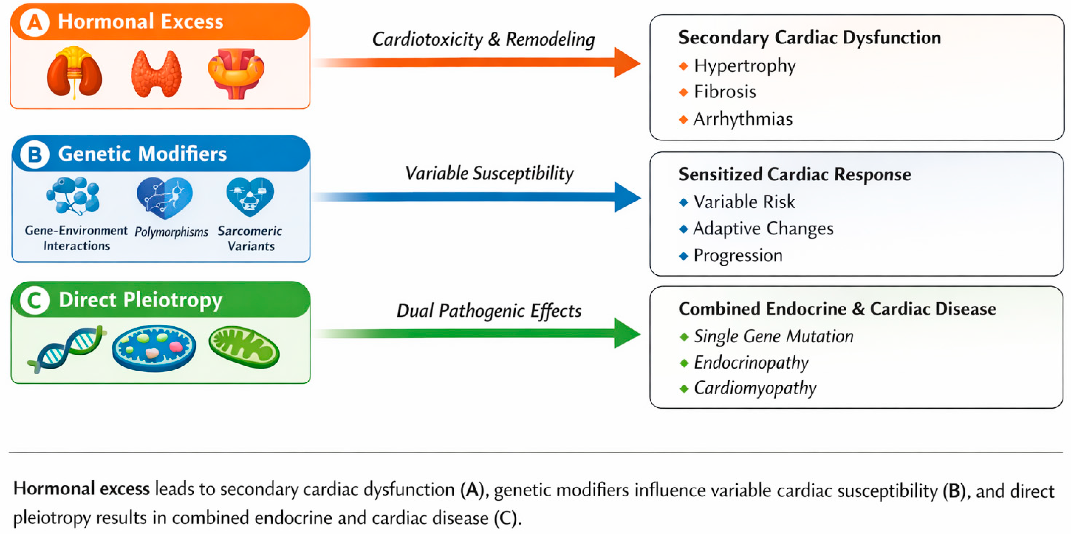
To provide conceptual clarity, we propose a framework distinguishing three major categories of genetic involvement in endocrine cardiomyopathies (Table 1): (i) variants responsible for the primary endocrinopathy, which secondarily affect the heart through hormonal excess (e.g., KCNJ5 somatic mutations in aldosterone-producing adenomas); (ii) genetic modifiers of myocardial susceptibility that influence the cardiac response to hormonal stress without causing the endocrine disorder itself (e.g., ACE I/D polymorphism, sarcomeric gene variants); and (iii) direct pleiotropic effects, whereby pathogenic variants in a single gene independently cause both endocrine dysfunction and cardiomyopathy through tissue-specific mechanisms (e.g., PRKAR1A in Carney complex, mitochondrial DNA mutations). This framework has important implications for genetic counseling, surveillance strategies, and therapeutic decision-making.
This review comprehensively synthesizes current evidence on the genetic basis and genotype–phenotype correlations of endocrine-related cardiomyopathies, with particular emphasis on shared molecular pathways, epigenetic regulation, and emerging precision medicine approaches. A detailed understanding of this complex genetic architecture is essential for improving diagnostic accuracy, enabling early intervention, guiding family screening, and ultimately supporting the development of personalized therapeutic strategies [9,10]. A schematic overview of the underlying mechanisms and their contribution to cardiac phenotypic heterogeneity is presented in Figure 1.

2. Literature Search Strategy

A comprehensive literature search was conducted to identify studies addressing the genetic and molecular basis of cardiomyopathies associated with endocrine disorders. Electronic databases including PubMed/MEDLINE, Embase, Web of Science, and OMIM were searched for articles published between January 2000 and December 2025. Seminal studies published before 2000 were also included when they provided foundational mechanistic or clinical insights not superseded by later evidence. Search terms included combinations of “cardiomyopathy”, “genetics”, “mutation”, “variant”, “polymorphism”, “endocrine”, “endocrinopathy”, and disease-specific terms such as “primary aldosteronism”, “Cushing’s syndrome”, “pheochromocytoma”, “paraganglioma”, “acromegaly”, “thyroid disorders”, “hyperparathyroidism”, and “diabetic cardiomyopathy”.
Inclusion criteria encompassed: (i) original research articles, systematic reviews, meta-analyses, and clinical guidelines published in English; (ii) studies reporting genetic variants, polymorphisms, or molecular mechanisms associated with cardiac manifestations in the context of endocrine disorders; (iii) human studies, with selected animal or in vitro studies included when providing essential mechanistic insights not available from human data. Exclusion criteria included: (i) case reports with fewer than three subjects unless describing novel genetic findings; (ii) conference abstracts without full-text availability; (iii) studies focusing exclusively on endocrine or cardiac manifestations without addressing the genetic interface between the two.
The initial search yielded 2847 records. After removal of duplicates (n = 612) and screening of titles and abstracts, 428 full-text articles were assessed for eligibility. Of these, 198 studies met inclusion criteria and were incorporated into this review. Reference lists of relevant articles were manually screened, identifying 34 additional pertinent studies. Given the heterogeneity of study designs, populations, and outcome measures across endocrine conditions, a formal PRISMA-based systematic review was not pursued; rather, this work represents a comprehensive narrative synthesis of the available evidence.

3. Primary Aldosteronism and Cushing’s Syndrome

3.1. Primary Aldosteronism

Primary aldosteronism (PA) is characterized by autonomous aldosterone hypersecretion, often leading to hypertension and increased cardiovascular risk [11]. Genetic discoveries have transformed our understanding of adrenal physiology and pathology in PA, with somatic mutations frequently identified in aldosterone-producing adenomas (APAs) [12,13]. Key genes implicated in PA include KCNJ5, CACNA1D, ATP1A1, ATP2B3, and CLCN2, which are involved in ion channel function and aldosterone biosynthesis.
KCNJ5 mutations, affecting the G protein-activated inward rectifier potassium channel Kir3.4, are present in approximately 40–70% of aldosterone-producing adenomas, with marked population-specific variability [14,15]. These mutations alter channel selectivity, allowing sodium influx that depolarizes zona glomerulosa cells and triggers calcium-dependent aldosterone production [12]. In addition, germline variants in genes such as CLCN2, KCNJ5, CACNA1H, and CACNA1D have been associated with familial forms of PA [16]. Overexpression of CYP11B2 (aldosterone synthase) is central to autonomous aldosterone production in APAs, and transgenic mice overexpressing aldosterone synthase develop cardiac fibrosis and hypertrophy [17,18].
Patients with KCNJ5-mutated APAs typically present at younger ages, are more frequently female, and exhibit larger tumor size with higher aldosterone levels compared to those with wild-type tumors or other mutations [19,20]. Importantly, KCNJ5 mutation status appears to influence cardiovascular remodeling: patients with KCNJ5-mutated APAs show more pronounced left ventricular hypertrophy at diagnosis but also demonstrate greater regression of cardiac mass following adrenalectomy [19,21].
Additional APA-associated mutations have been identified in CACNA1D (encoding the L-type calcium channel Cav1.3), ATP1A1 (Na+/K+-ATPase α1 subunit), and ATP2B3 (plasma membrane Ca2+-ATPase) [13,22]. These mutations are associated with distinct clinical phenotypes, with CACNA1D mutations typically linked to smaller adenomas and male predominance, whereas ATP1A1 and ATP2B3 mutations are more frequently observed in cortisol-co-secreting adenomas [22].

3.2. Cushing’s Syndrome

Cushing’s syndrome (CS) results from chronic glucocorticoid excess and has profound effects on cardiac morphology and function, frequently leading to myocardial hypertrophy, interstitial fibrosis, and myofibrillolysis. Cardiovascular complications represent a major cause of morbidity and mortality in CS, with an increased risk of heart failure, arrhythmias, and thromboembolic events [23,24].
Landmark endomyocardial biopsy studies by Frustaci and colleagues demonstrated distinctive myofibrillolysis in patients with hypercortisolism, together with marked activation of protein degradation pathways. In particular, atrogin-1 mRNA expression was increased more than 28-fold compared with controls and normalized following surgical cure, in parallel with significant improvement in ventricular function. These changes were accompanied by a reduction in cardiomyocyte diameter (from 28.7 to 15.7 μm), myocardial fibrosis (from 11.5% to 3.1%), and myofibrillolysis area (from 61% to 22%) following adrenalectomy [25,26].
The genetic basis of Cushing’s syndrome varies according to disease etiology. ACTH-secreting pituitary adenomas frequently harbor somatic mutations in USP8, a deubiquitinase that enhances EGFR signaling and promotes corticotroph tumorigenesis [27]. Bilateral macronodular adrenal hyperplasia is commonly associated with germline mutations in ARMC5, identified in up to 50% of affected patients [28]. In contrast, primary pigmented nodular adrenocortical disease is caused by inactivating mutations in PRKAR1A and occurs as part of the Carney complex [29]. Notably, PRKAR1A mutations exemplify direct genetic pleiotropy: the same pathogenic variant causes both the endocrine phenotype (nodular adrenocortical disease with cortisol excess) and cardiac manifestations (cardiac myxomas, arrhythmias) through PKA pathway dysregulation in different tissues, rather than one being a consequence of the other [29]. The main genetic alterations, molecular mechanisms, and associated cardiac phenotypes observed in primary aldosteronism and Cushing’s syndrome are summarized in Table 2.
Table 2. Genes Associated with Cardiac Manifestations in Primary Aldosteronism and Cushing’s Syndrome.
GeneProtein/FunctionCardiac EffectsClinical SignificanceReferences
KCNJ5Kir3.4 potassium channelMarked LVH, highly reversible after adrenalectomy40–70% of APA; younger age, female predominance, favorable cardiac recovery[12,14,15,19,20,21]
CACNA1DCav1.3 calcium channelModerate LVH, increased arrhythmia susceptibilitySmaller adenomas, male predominance[13,22]
ATP1A1Na+/K+-ATPase alpha1Variable remodeling secondary to aldosterone excessFrequently cortisol-co-secreting adenomas[22]
ATP2B3Plasma membrane Ca2+-ATPaseAldosterone-mediated cardiac remodelingLess common APA subtype with distinct molecular profile[22]
CLCN2ClC-2 chloride channelLVH and diastolic dysfunction (limited data)Familial primary aldosteronism[16]
CYP11B2Aldosterone synthaseMyocardial fibrosis and hypertrophyFinal effector of autonomous aldosterone production[17,18]
PRKAR1APKA regulatory subunitCardiomyopathy and cardiac myxomasCarney complex; PPNAD; direct pleiotropy[29]
USP8DeubiquitinaseCushing-related cardiomyopathy (indirect)Corticotroph pituitary adenomas[27]
ARMC5Armadillo repeat proteinSevere hypercortisolism-associated cardiac damageBilateral macronodular adrenal hyperplasia[28]
APA: aldosterone-producing adenoma; LVH: left ventricular hypertrophy; PPNAD: primary pigmented nodular adrenocortical disease.

4. Pheochromocytoma and Paraganglioma

Pheochromocytomas and paragangliomas (PPGL) represent a paradigm of hereditary endocrine tumors, with approximately 40% of cases now recognized to harbor germline susceptibility mutations [30,31]. Cardiovascular manifestations—including catecholamine-induced cardiomyopathy, hypertensive crises, and malignant arrhythmias—are strongly influenced by tumor genotype through determination of the catecholamine biochemical phenotype. Catecholamine cardiomyopathy may present as Takotsubo-like syndrome, dilated cardiomyopathy, or a myocarditis-like pattern characterized histologically by contraction band necrosis [32,33].
Mutations in succinate dehydrogenase (SDH) subunit genes, particularly SDHB, define a clinically aggressive subset of PPGL. SDHB-mutated tumors typically produce norepinephrine exclusively and are associated with a markedly increased risk of metastatic disease, reported in up to 40–70% of cases across series [34,35]. The associated cardiovascular phenotype often includes dilated cardiomyopathy, attributed to chronic norepinephrine excess leading to β-adrenergic receptor downregulation, direct myocyte toxicity, mitochondrial dysfunction, and oxidative stress [36]. Early genetic diagnosis enables intensified surveillance and timely therapeutic intervention.
In contrast, VHL and RET mutations are associated with predominantly norepinephrine- and epinephrine-secreting tumors, respectively, but confer a substantially lower metastatic risk [37]. VHL-related pheochromocytomas are typically diagnosed at younger ages, may be bilateral, and rarely cause severe cardiomyopathy. RET mutations in MEN2 syndromes are associated with epinephrine-predominant biochemical profiles. Additional PPGL susceptibility genes include NF1, MAX, TMEM127, FH, and EPAS1/HIF2A [30,31,38]. Genotype–phenotype correlations, metastatic risk, and cardiovascular manifestations associated with PPGL-related genetic variants are summarized in Table 3.
Table 3. Genotype-Phenotype Correlations in Hereditary Pheochromocytoma/Paraganglioma.
GeneSyndromeCatecholamine ProfileCardiac PhenotypeMetastatic RiskReferences
SDHBPGL syndrome 4Norepinephrine predominantDilated cardiomyopathy, severe forms40–70% (highest)[34,35]
SDHDPGL syndrome 1Dopamine ± norepinephrineUsually mild or absent; variable<5% (paternal transmission)[30,31]
SDHCPGL syndrome 3VariableUsually mild or absent<5%[30,31]
VHLvon Hippel–LindauNorepinephrine onlyUsually absent or mild<5%[37]
RETMEN2A/MEN2BEpinephrine predominantStress-related cardiomyopathy (rare)<5%[37]
NF1Neurofibromatosis type 1Mixed catecholaminesHypertensive LVH5–10%[30,38]
MAXHereditary paragangliomaVariableCatecholamine-induced cardiomyopathy<10–15%[30,31]
PGL: paraganglioma; MEN: multiple endocrine neoplasia; LVH: left ventricular hypertrophy.

5. Direct Genetic Pleiotropy in Endocrine Cardiomyopathies

Beyond the classical paradigm where cardiac involvement results secondarily from hormonal excess, an important subset of conditions demonstrates direct genetic pleiotropy—where pathogenic variants in a single gene independently cause both endocrine and cardiac manifestations through tissue-specific effects. Recognition of this mechanism has critical implications for patient management, as cardiac involvement may develop or progress independently of endocrine disease control.

5.1. PRKAR1A and Carney Complex

Carney complex, caused by inactivating mutations in PRKAR1A encoding the type 1A regulatory subunit of protein kinase A, exemplifies direct pleiotropy [29]. Cardiac myxomas occur in approximately 30–60% of patients and represent primary cardiac tumors rather than consequences of associated endocrine abnormalities (primary pigmented nodular adrenocortical disease, acromegaly). The PKA pathway dysregulation affects both adrenocortical cells (causing cortisol excess) and cardiac tissue (promoting myxoma formation and arrhythmias) independently. Importantly, cardiac myxomas may develop even after successful treatment of hypercortisolism, necessitating lifelong cardiac surveillance regardless of endocrine disease status.

5.2. KCNJ5: Beyond Primary Aldosteronism

While somatic KCNJ5 mutations in aldosterone-producing adenomas cause secondary cardiac effects through aldosterone excess, germline KCNJ5 mutations have direct cardiac implications. KCNJ5 encodes the Kir3.4 potassium channel, and germline variants underlie the LQT13 form of long QT syndrome, predisposing to ventricular arrhythmias independently of any adrenal pathology [39]. This dual role—somatic mutations causing primary aldosteronism with secondary cardiac effects, and germline mutations directly causing cardiac arrhythmias—illustrates how the same gene can contribute to endocrine cardiomyopathies through distinct mechanisms.

5.3. Hereditary Hemochromatosis

HFE gene mutations causing hereditary hemochromatosis lead to iron overload affecting multiple organs independently. Cardiac manifestations include restrictive or dilated cardiomyopathy and arrhythmias, while endocrine involvement encompasses diabetes mellitus (“bronze diabetes”), hypogonadotropic hypogonadism, and hypothyroidism [40]. The cardiac and endocrine phenotypes both result from tissue iron deposition but develop through parallel pathogenic mechanisms rather than one causing the other. Early phlebotomy can prevent or reverse both manifestations, but progression in one organ system does not necessarily predict involvement of others.

5.4. Mitochondrial Disorders

Mitochondrial DNA mutations and nuclear genes affecting mitochondrial function cause multisystem disorders with independent cardiac and endocrine manifestations [41]. Maternally inherited diabetes and deafness (MIDD), caused by the m.3243A>G mutation in MT-TL1, is associated with hypertrophic or dilated cardiomyopathy in 15–30% of patients. Kearns-Sayre syndrome (large-scale mtDNA deletions) combines cardiac conduction defects with diabetes, hypoparathyroidism, and growth hormone deficiency. MELAS syndrome similarly affects both cardiac and endocrine tissues. The ubiquitous requirement for mitochondrial function explains the multi-organ involvement, with both cardiac and endocrine tissues being highly energy-dependent.

5.5. Myotonic Dystrophy

Myotonic dystrophy type 1, caused by CTG repeat expansion in DMPK, demonstrates pleiotropic effects on cardiac and endocrine systems [42]. Cardiac manifestations include conduction defects, arrhythmias, and cardiomyopathy, while endocrine involvement encompasses testicular failure, insulin resistance, and thyroid dysfunction. The RNA toxicity mechanism affects multiple tissues independently through sequestration of RNA-binding proteins. Cardiac complications are a leading cause of mortality and require surveillance independent of endocrine management.

5.6. Friedreich Ataxia

Friedreich ataxia, caused by GAA repeat expansion in FXN leading to frataxin deficiency, results in hypertrophic cardiomyopathy in >90% of patients alongside diabetes mellitus in 10–30% [43]. Both manifestations result from mitochondrial iron accumulation and oxidative stress affecting high-energy tissues independently. Cardiomyopathy progression does not correlate with diabetes control, emphasizing the need for parallel management strategies.
The genetic conditions demonstrating direct pleiotropic effects on cardiac and endocrine systems are summarized in Table 4.

6. Thyroid, Parathyroid Disorders, and Acromegaly

6.1. Thyroid Hormone and Cardiac Function

Thyroid hormones exert profound effects on the cardiovascular system through both genomic and non-genomic mechanisms [44,45]. Genomic actions involve binding of thyroid hormone receptors (TRs) to thyroid hormone response elements in target genes, regulating the expression of myosin heavy chain isoforms, SERCA2a, phospholamban, and multiple cardiac ion channels. Non-genomic effects include rapid modulation of ion channel activity, vascular tone, endothelial function, and mitochondrial metabolism.
Hyperthyroidism induces a hyperdynamic circulatory state characterized by increased cardiac output, reduced systemic vascular resistance, tachycardia, and, in chronic cases, high-output heart failure and thyrotoxic cardiomyopathy [46]. Atrial fibrillation occurs in approximately 10–25% of patients with overt hyperthyroidism, with prevalence increasing markedly with age and underlying structural heart disease [47]. In contrast, hypothyroidism is associated with bradycardia, increased systemic vascular resistance, impaired diastolic relaxation, and accelerated atherosclerosis.
Genetic determinants of thyroid hormone signaling modulate interindividual susceptibility to cardiac involvement. Mutations in thyroid hormone receptor genes (THRA and THRB) cause resistance to thyroid hormone (RTH) syndromes with distinct cardiac phenotypes [48]. Patients with RTHβ frequently exhibit tachycardia and atrial fibrillation despite elevated circulating thyroid hormone levels, reflecting preserved myocardial sensitivity to triiodothyronine (T3) in the presence of pituitary resistance [48,49]. Population-based analyses have demonstrated an increased risk of atrial fibrillation and heart failure in RTHβ compared with matched controls [49].
Local cardiac thyroid hormone availability is further regulated by type 2 iodothyronine deiodinase (DIO2), which converts thyroxine (T4) to T3 within target tissues. A common DIO2 polymorphism (Thr92Ala, rs225014), present in approximately 12–36% of the population depending on ethnicity, reduces enzymatic activity and intracellular T3 generation [50,51]. This variant has been associated with hypertension and increased pulse pressure [52], adverse cardiac remodeling after myocardial infarction [53], and increased susceptibility to thyrotoxic cardiomyopathy in Graves’ disease [54].

6.2. Hyperparathyroidism and Cardiac Involvement

Primary hyperparathyroidism (PHPT) is associated with increased cardiovascular morbidity and mortality, with cardiac complications representing a major contributor to reduced life expectancy [55,56]. Cardiovascular manifestations include left ventricular hypertrophy, diastolic dysfunction, valvular and vascular calcifications, conduction abnormalities, and arrhythmias.
Left ventricular hypertrophy has been reported in up to 80% of patients with symptomatic PHPT, independent of blood pressure levels, suggesting direct myocardial effects of parathyroid hormone (PTH) [57,58]. PTH acts on cardiac PTH1 receptors, activating protein kinase A and C pathways, increasing intracellular calcium and triggering calcineurin–NFAT–dependent hypertrophic signaling [59]. Hypercalcemia further contributes to cardiovascular pathology through vascular calcification, QT interval shortening, and altered myocardial contractility [60].
The genetic basis of PHPT includes MEN1 mutations in multiple endocrine neoplasia type 1, CDC73 mutations in hyperparathyroidism–jaw tumor syndrome, CASR mutations affecting calcium sensing, GCM2 mutations in familial isolated PHPT, and CDKN1B mutations in MEN4 [61]. Importantly, regression of left ventricular hypertrophy and improvement of diastolic function occur after successful parathyroidectomy, supporting a direct hormonal effect [62].

6.3. Acromegaly

Acromegaly is characterized by a specific cardiomyopathy with biventricular concentric hypertrophy, diastolic dysfunction, and, in advanced stages, systolic impairment and heart failure [63,64,65]. Cardiovascular disease remains the leading cause of mortality, accounting for approximately 60% of deaths. Acromegalic cardiomyopathy is present in 20–90% of patients at diagnosis, depending on disease duration and severity [66].
The cardiomyopathy progresses through a hyperkinetic phase, an intermediate hypertrophic phase, and a late dilated phase. Growth hormone (GH) and insulin-like growth factor-1 (IGF-1) promote myocardial hypertrophy via PI3K/Akt, MAPK/ERK, and JAK2/STAT5 signaling pathways [67].
Genetic predisposition accounts for approximately 5% of acromegaly cases. AIP mutations explain 15–20% of familial isolated pituitary adenomas and are associated with younger age at onset and aggressive disease [68]. Somatic GNAS mutations occur in 30–40% of sporadic somatotropinomas [69]. Early biochemical control prevents or reverses cardiac abnormalities [70].

7. Diabetic Cardiomyopathy

Diabetic cardiomyopathy is a distinct myocardial disorder occurring independently of coronary artery disease, hypertension, and valvular disease [71,72]. Its prevalence is estimated at 10–15% in carefully selected diabetic populations and increases with disease duration and glycemic burden.
Pathophysiology involves lipotoxicity, glucotoxicity, oxidative stress, inflammation, impaired calcium handling, mitochondrial dysfunction, microvascular disease, and myocardial fibrosis [73].
The TCF7L2 gene represents the strongest genetic risk factor for type 2 diabetes, with the rs7903146 variant conferring approximately a 1.4-fold increased diabetes risk per allele [74,75]. Beyond diabetes susceptibility, TCF7L2 variants are independently associated with coronary artery disease and heart failure, suggesting direct cardiovascular effects through modulation of Wnt signaling [76]. Monogenic diabetes forms (MODY), particularly HNF1A, HNF4A, and GCK, show variable cardiac involvement [77]. Finally, sodium–glucose cotransporter-2 (SGLT2) inhibitors have demonstrated robust reductions in heart failure hospitalization [78].

8. Shared Genetic Pathways Across Endocrine Cardiomyopathies

Despite diverse hormonal triggers, endocrine-related cardiomyopathies converge on common molecular pathways that promote cardiac remodeling, fibrosis, and electrical instability. Identification of these shared mechanisms has important translational implications, as therapeutic strategies targeting these pathways may provide benefit across multiple endocrine disorders [79].

8.1. TGF-Beta/SMAD Signaling

The transforming growth factor-β (TGF-β)/SMAD signaling pathway is a central mediator of myocardial fibrosis across several endocrine cardiomyopathies [80,81]. TGF-β1 activates cardiac fibroblasts, stimulates extracellular matrix deposition, and promotes fibroblast-to-myofibroblast transdifferentiation through phosphorylation and nuclear translocation of SMAD2/3 [81].
In primary aldosteronism, aldosterone enhances TGF-β1 signaling via mineralocorticoid receptor activation and reactive oxygen species generation. In diabetic cardiomyopathy, hyperglycemia induces TGF-β1 through advanced glycation end-products and protein kinase C activation. In Cushing’s syndrome, chronic glucocorticoid excess activates TGF-β signaling in cardiac tissue, contributing to progressive myocardial fibrosis. Genetic polymorphisms in TGFB1, including −509C>T and +869T>C, influence TGF-β1 expression and may modulate individual susceptibility to fibrotic remodeling across endocrine conditions [80,81,82].

8.2. Renin–Angiotensin–Aldosterone System

The renin–angiotensin–aldosterone system (RAAS) represents another convergent pathway with established genetic modifiers influencing cardiac remodeling [83]. The angiotensin-converting enzyme (ACE) insertion/deletion (I/D) polymorphism, particularly the D allele, is associated with increased ACE activity. In primary aldosteronism, the DD genotype correlates with more severe left ventricular hypertrophy independent of blood pressure levels [84].
Additional variants, including the AGT M235T polymorphism and the AGTR1 A1166C variant, influence angiotensin II signaling and hypertrophic responses. The CYP11B2 −344C>T polymorphism affects aldosterone synthase promoter activity and contributes to interindividual variability in myocardial remodeling [83,84].

8.3. Oxidative Stress and Mitochondrial Dysfunction

Oxidative stress, mediated by NADPH oxidases (particularly NOX2 and NOX4) and mitochondrial dysfunction, is a shared pathogenic mechanism across endocrine cardiomyopathies [80,81]. In pheochromocytoma, catecholamine auto-oxidation generates reactive oxygen species that directly damage cardiomyocytes. In hyperthyroidism, increased metabolic demand enhances mitochondrial electron transport chain activity and reactive oxygen species production. In diabetic cardiomyopathy, hyperglycemia promotes oxidative stress through mitochondrial dysfunction, NADPH oxidase activation, and uncoupled nitric oxide synthase [85,86,87].
Genetic variants in antioxidant defense pathways may modify susceptibility, including SOD2 Ala16Val (mitochondrial import efficiency), CAT −262C>T (catalase expression), and GPX1 Pro198Leu (glutathione peroxidase activity) [86,87].

8.4. Calcium Handling Abnormalities

Abnormal calcium handling represents a unifying mechanism underlying arrhythmogenesis and contractile dysfunction across endocrine cardiomyopathies [88]. Variants in RYR2 may predispose to catecholamine-triggered arrhythmias, particularly relevant in pheochromocytoma where β-adrenergic stimulation enhances ryanodine receptor opening. PLN variants alter SERCA2a regulation and impair diastolic relaxation, while CACNA1C variants affect L-type calcium channel function and excitation–contraction coupling. CASQ2 variants disrupt sarcoplasmic reticulum calcium storage. The interaction between hormonal excess and underlying calcium-handling gene variants may explain marked interindividual variability in arrhythmic risk [88].

9. Genetic and Epigenetic Modifiers of Endocrine Cardiomyopathies

9.1. Sarcomeric Gene Modifiers

The marked phenotypic variability observed among patients with comparable endocrine disturbances suggests the presence of genetic modifiers influencing myocardial vulnerability. Titin-truncating variants (TTNtv), present in approximately 20–25% of dilated cardiomyopathy cases and in 1–3% of the general population, may remain clinically silent but predispose to cardiac decompensation under metabolic or hormonal stress [89,90].
Patients harboring subclinical TTNtv may develop cardiomyopathy when exposed to thyroid hormone excess, chronic catecholamine stimulation, glucocorticoid excess, or metabolic derangements associated with diabetes. Similarly, variants in other sarcomeric genes (MYH7, MYBPC3, TNNT2) that are tolerated under basal conditions may become pathogenic in the context of sustained endocrine stress. This “two-hit” model provides a mechanistic explanation for the broad interindividual variability in cardiac phenotype observed across endocrine cardiomyopathies [91].

9.2. MicroRNA Regulation

MicroRNAs (miRNAs) are key epigenetic regulators of cardiac gene expression, acting through post-transcriptional repression or degradation of target mRNAs and orchestrating complex regulatory networks involved in hypertrophy, fibrosis, and electrical remodeling [92,93].
miR-133a is the most abundant cardiac-enriched miRNA and is consistently downregulated in diabetic cardiomyopathy across species [94,95]. miR-133a exerts cardioprotective effects through inhibition of hypertrophic signaling (via RhoA, Cdc42, and WHSC2), suppression of fibrosis by targeting connective tissue growth factor (CTGF) and TGF-β1, and regulation of DNA methylation through DNMT1 inhibition. Cardiac-specific overexpression of miR-133a in diabetic mouse models prevents myocardial remodeling, preserves systolic function, and attenuates fibrosis [96], supporting its therapeutic potential.
miR-1 plays a central role in cardiac electrophysiology by regulating GJA1 (connexin-43), KCNJ2 (Kir2.1), and calcium handling proteins, thereby modulating action potential propagation and arrhythmia susceptibility [97]. Dysregulation of miR-1 in diabetic and hyperthyroid hearts contributes to electrical instability.
miR-21 is predominantly expressed in cardiac fibroblasts and promotes fibrosis through activation of ERK-MAPK signaling by targeting Sprouty1, enhancing fibroblast survival and collagen synthesis [98,99]. In primary aldosteronism, aldosterone-mediated upregulation of miR-21 contributes to mineralocorticoid-induced myocardial fibrosis. Pharmacological inhibition of miR-21 has demonstrated antifibrotic effects in preclinical models.
The miR-29 family (miR-29a/b/c) directly targets multiple extracellular matrix genes, including COL1A1, COL1A2, COL3A1, FBN1, and ELN, and is suppressed by TGF-β signaling in diabetic cardiomyopathy, creating a feed-forward profibrotic loop [100]. miR-208a/b, encoded within myosin heavy chain genes, regulates myosin isoform expression and mediates thyroid hormone-dependent contractile remodeling [101].

9.3. DNA Methylation and Histone Modifications: An Integrated Framework

Beyond miRNA regulation, dynamic alterations in DNA methylation and histone modifications contribute to endocrine cardiomyopathy pathogenesis [102]. These epigenetic mechanisms operate across multiple endocrine conditions, creating a unifying framework for understanding hormonal effects on cardiac gene expression.
In diabetic cardiomyopathy, hyperglycemia induces global and gene-specific methylation changes. Hypermethylation of cardioprotective gene promoters (SERCA2A, PGC-) reduces their expression [103], while hypomethylation of inflammatory genes (IL-6, TNF-α) and profibrotic genes (CTGF, COL1A1) enhances their transcription. Importantly, these epigenetic alterations persist despite subsequent glycemic normalization—a phenomenon termed “metabolic memory”—which may explain why early intensive glycemic control provides long-term cardiovascular benefits even after relaxation of glucose targets [104].
In primary aldosteronism, aldosterone induces epigenetic reprogramming of cardiac fibroblasts through mineralocorticoid receptor-dependent mechanisms. DNA methylation changes at profibrotic gene loci contribute to sustained fibroblast activation even after aldosterone normalization. Histone modifications, particularly H3K4 methylation and H3K9 acetylation at fibrosis-related genes, are altered in aldosterone-exposed cardiac tissue [105].
In Cushing’s syndrome, glucocorticoid-induced epigenetic changes affect cardiac metabolism and protein turnover pathways. The glucocorticoid receptor recruits histone-modifying enzymes to target gene promoters, and chronic exposure leads to sustained chromatin remodeling that may not fully reverse after cortisol normalization [106].
Key mediators across endocrine conditions include: DNA methyltransferases (DNMT1, DNMT3A, DNMT3B), which establish and maintain methylation patterns; TET demethylases (TET1-3), which catalyze active demethylation; histone methyltransferases (SET7, EZH2, G9a) and demethylases (LSD1, JMJD family); and histone acetyltransferases (p300/CBP) and deacetylases (HDAC family) [107]. Histone deacetylase inhibitors have demonstrated cardioprotective effects in experimental models of multiple endocrine cardiomyopathies [108], suggesting therapeutic potential.
The clinical implications of epigenetic memory are substantial: (i) early aggressive treatment of endocrine disorders may prevent irreversible epigenetic changes; (ii) persistence of cardiac abnormalities after hormonal normalization may reflect established epigenetic alterations; (iii) epigenetic biomarkers may help identify patients at risk for incomplete cardiac recovery; and (iv) epigenetic-targeting therapies represent a novel therapeutic avenue for endocrine cardiomyopathies.

10. Ethnic and Population Differences

Substantial ethnic and population-specific differences exist in the genetic architecture of endocrine cardiomyopathies, with important implications for diagnosis, risk stratification, and implementation of precision medicine strategies [109,110]. Failure to account for these differences may lead to underdiagnosis, misclassification of variants, and inequitable access to targeted therapies.

10.1. Ethnic Differences in Primary Aldosteronism

Marked population differences are observed in the mutational landscape of aldosterone-producing adenomas. KCNJ5 mutations occur in approximately 60–70% of APAs in East Asian populations compared with 35–45% in European populations [111]. Clinically, this translates into a higher prevalence of the classical KCNJ5 phenotype, characterized by younger age at diagnosis, female predominance, larger adenomas, and higher aldosterone secretion.
In contrast, CACNA1D mutation frequencies appear relatively consistent across populations, whereas ATP1A1 and ATP2B3 mutations are reported more frequently in European cohorts [112]. These differences may influence population-specific screening strategies, surgical decision-making, and expected postoperative cardiovascular recovery.

10.2. Ethnic Differences in Pheochromocytoma/Paraganglioma

Founder effects and population-specific variants are well documented in pheochromocytoma and paraganglioma syndromes [113]. The SDHB c.72+1G>A splice-site mutation represents a Dutch founder variant associated with aggressive disease, while SDHD D92Y shows a strong founder effect in the Netherlands. Similarly, VHL type 2C clusters in specific geographic regions due to founder effects, highlighting the importance of ancestry-informed genetic counseling.

10.3. Disparities in Cardiomyopathy Genetic Testing

Genetic testing yield varies substantially by ancestry, reflecting historical biases in genomic research datasets [114]. Detection rates for pathogenic cardiomyopathy variants are higher in individuals of European ancestry than in African ancestry populations, while variants of uncertain significance are two- to threefold more common in underrepresented groups.
Landmark reclassification studies demonstrated that several variants initially labeled pathogenic for hypertrophic cardiomyopathy were in fact benign population-specific variants in African American cohorts [115]. These disparities are further amplified by the disproportionate representation of European ancestry individuals in genome-wide association studies.

11. Polygenic Risk Scores in Endocrine Cardiomyopathies

11.1. Principles of Polygenic Risk Scores

Polygenic risk scores aggregate the effects of numerous common genetic variants identified through genome-wide association studies to estimate individual disease susceptibility [116,117]. Unlike monogenic testing, PRSs capture cumulative genetic risk and may be particularly informative in endocrine cardiomyopathies characterized by marked phenotypic variability.

11.2. Clinical Evidence and Validation

A recent American Heart Association scientific statement highlighted the independent association of PRSs with cardiovascular outcomes and their incremental predictive value beyond traditional risk factors, while acknowledging current limitations in clinical implementation [118]. In heart failure, validated PRSs demonstrate robust risk stratification, with individuals in the highest genetic risk strata exhibiting substantially increased incidence rates.

11.3. Application to Cardiomyopathies

In dilated cardiomyopathy, PRSs derived from large genome-wide association studies and cardiac magnetic resonance datasets discriminate cases from controls and correlate with left ventricular structure and function [119,120]. The liability-threshold model proposes that rare pathogenic variants and polygenic background jointly determine disease expression [121]. Carriers of titin-truncating variants with high PRSs exhibit markedly increased disease penetrance, a framework directly applicable to endocrine cardiomyopathies.

11.4. Limitations and Future Directions

Current limitations include reduced PRS performance in non-European populations, lack of randomized trials demonstrating clinical utility, and challenges related to clinical integration. Future research priorities include development of multi-ancestry PRSs, endocrine-specific risk models, and prospective trials of PRS-guided management.

12. Clinical Recommendations for Genetic Testing

Genetic testing in endocrine-related cardiomyopathies may be considered based on clinical presentation, family history, and therapeutic implications. A condition-specific approach is recommended, as summarized below (Table 5).
For pheochromocytoma/paraganglioma, current guidelines recommend genetic testing for ALL patients given the high rate of hereditary disease (approximately 40%) and implications for surveillance and family screening [122]. Testing should include SDHx genes (SDHA, SDHB, SDHC, SDHD, SDHAF2), VHL, RET, NF1, MAX, TMEM127, and FH. SDHB-positive patients require intensified surveillance with annual whole-body imaging given high metastatic risk.
For primary aldosteronism, genetic testing should be considered in: (i) early-onset disease (<40 years); (ii) bilateral adrenal disease; (iii) family history of PA or early-onset hypertension; (iv) familial hyperaldosteronism suspected. Priority genes include KCNJ5 (somatic, for surgical planning), CLCN2, CACNA1H, and CACNA1D (germline, for familial forms). Somatic mutation status in surgical specimens may predict postoperative cardiovascular recovery.
For Cushing’s syndrome, genetic testing indications include: (i) young onset; (ii) bilateral adrenal disease; (iii) features suggesting syndromic disease (skin pigmentation, myxomas). Consider PRKAR1A (Carney complex—requires cardiac surveillance for myxomas), MEN1, ARMC5 (bilateral macronodular hyperplasia).
For conditions with direct genetic pleiotropy (Carney complex, hemochromatosis, mitochondrial disorders, myotonic dystrophy, Friedreich ataxia), genetic diagnosis has immediate implications for cardiac surveillance regardless of endocrine disease status. Cascade family screening is essential.

13. Precision Medicine and Therapeutic Implications

The integration of genetic information into clinical management enables more personalized treatment strategies. In primary aldosteronism, KCNJ5 mutation status may predict response to adrenalectomy, with KCNJ5-positive patients showing greater improvement in blood pressure control and left ventricular mass regression [123]. For pheochromocytoma/paraganglioma, genotype-directed surveillance protocols optimize screening intensity based on tumor biology. SDHB mutation carriers require intensive surveillance with annual imaging given high metastatic risk [124].
MicroRNA-based therapeutics represent a frontier in cardiovascular medicine. miR-133a overexpression prevents diabetic cardiomyopathy in animal models, and clinical trials of miRNA modulators are underway [125].

14. Knowledge Gaps and Future Research Directions

Despite significant advances in understanding the genetic basis of endocrine cardiomyopathies, important knowledge gaps remain that should guide future research priorities.
Long-term outcome studies are critically needed. Prospective studies must determine whether genotype-based risk stratification improves cardiovascular outcomes in endocrine disorders. Current evidence is largely observational, and randomized controlled trials comparing genotype-guided versus standard management are essential to establish clinical utility and inform reimbursement decisions.
Multi-ethnic cohorts are essential to address current disparities in genetic testing utility. The overwhelming European bias in current genomic research limits the applicability of findings to diverse populations. Large-scale studies enrolling participants from African, Asian, Hispanic/Latino, and other ancestry groups are needed to develop ancestry-appropriate reference databases, validate polygenic risk scores across populations, identify population-specific variants and founder mutations, and ensure precision medicine benefits extend to all populations.
Functional validation of variants of uncertain significance remains a major bottleneck. Patient-derived induced pluripotent stem cell (iPSC) models enable functional testing of variants in cardiomyocytes with the patient’s genetic background. CRISPR-based approaches allow systematic functional characterization of variants at scale. High-throughput functional assays can generate evidence for variant reclassification.
Development of endocrine disease-specific polygenic risk scores represents an important opportunity. Current cardiovascular PRSs were developed in general populations and may not optimally predict risk in endocrine patient populations. Endocrine-specific PRSs incorporating hormone-responsive genetic variants and tissue-specific effects could improve risk prediction.
Multi-omics integration combining transcriptomic, proteomic, metabolomic, and epigenomic data with genomic information will provide comprehensive understanding of disease mechanisms and enable network-based approaches to drug discovery and identify new therapeutic targets.

15. Conclusions

The genetic basis of endocrine-related cardiomyopathies is increasingly recognized as a key determinant of disease expression, severity, and therapeutic response. In this review, we propose a conceptual framework distinguishing three major categories of genetic involvement: variants causing the primary endocrinopathy with secondary cardiac effects, genetic modifiers of myocardial susceptibility, and direct pleiotropic effects whereby single gene variants independently drive both endocrine and cardiac phenotypes. This framework has important implications for genetic counseling, surveillance strategies, and clinical decision-making. Somatic mutations in hormone-producing tumors influence not only endocrine phenotype but also cardiovascular outcomes, as exemplified by KCNJ5 in primary aldosteronism and SDHx in pheochromocytoma. Moreover, shared molecular pathways—including TGF-β/SMAD signaling, renin–angiotensin–aldosterone system activation, oxidative stress, and abnormalities in calcium handling—represent potential therapeutic targets that extend across specific endocrine diagnoses. Sarcomeric gene modifiers and epigenetic mechanisms, particularly microRNAs and DNA methylation changes contributing to metabolic memory, add further layers of complexity to phenotypic expression and help explain interindividual variability in disease severity. Recognition of direct genetic pleiotropy in conditions such as Carney complex, hereditary hemochromatosis, mitochondrial disorders, and myotonic dystrophy underscores that cardiac involvement may progress independently of endocrine disease control, thereby necessitating lifelong cardiac surveillance.
Ethnic differences in genetic architecture have important implications for genetic testing strategies and emphasize the need for broader representation in genomic research. Polygenic risk scores show promise for risk stratification but require further validation across diverse populations.
Finally, the implementation of genetic testing in clinical practice is steadily advancing, with established recommendations for pheochromocytoma/paraganglioma and evolving guidance for other endocrine conditions. As precision medicine approaches continue to mature, the integration of genetic, clinical, biochemical, and imaging data will enable more refined risk stratification, more individualized treatment strategies, and ultimately improved outcomes for patients with endocrine-related cardiomyopathies.

Author Contributions

Conceptualization, A.C. and G.I.; methodology, A.C.; writing—original draft preparation, A.C.; writing—review and editing, A.C., M.D., C.C. and R.S.; supervision, G.I. All authors have read and agreed to the published version of the manuscript.

Funding

This research received no external funding.

Data Availability Statement

No new data were created or analyzed in this study. Data sharing is not applicable to this article.

Acknowledgments

The authors acknowledge the use of artificial intelligence–based tools for language editing and manuscript formatting. All scientific content, interpretations, and conclusions were developed, reviewed, and approved by the authors.

Conflicts of Interest

The authors declare no conflicts of interest.

References

  1. Fuentes-Mendoza, J.M.; Concepción-Zavaleta, M.J.; Morón-Siguas, J.C.; Muñoz-Moreno, J.M.; Pérez-Reyes, A.I.; Martinez-Galaviz, R.; Aguilar-Castañeda, R.D.; González-Godoy, O.; Concepción-Urteaga, L.A.; Paz-Ibarra, J. Cardiomyopathies of endocrine origin: A state-of-the-art review. World J. Cardiol. 2025, 17, 111462. [Google Scholar]
  2. Klein, I.; Danzi, S. Thyroid disease and the heart. Circulation 2007, 116, 1725–1735. [Google Scholar] [CrossRef] [PubMed]
  3. Rivera, F.B.; Taliño, M.K.; Ansay, M.F.; Mangubat, G.F.; Mahilum, M.L.; Menghrajani, R.H.; Placino, S.; Cha, S.W.; Aparece, J.P.; Yu, M.G.; et al. Cardiovascular effects of excess growth hormone: How real is the threat? Rev. Cardiovasc. Med. 2023, 24, 95. [Google Scholar] [CrossRef] [PubMed]
  4. Lisco, G.; Giagulli, V.A.; Iovino, M.; Zupo, R.; Guastamacchia, E.; De Pergola, G.; Iacoviello, M.; Triggiani, V. Endocrine system dysfunction and chronic heart failure: A clinical perspective. Endocrine 2022, 75, 360–376. [Google Scholar] [PubMed]
  5. Petramala, L.; Concistrè, A.; Olmati, F.; Mezzadri, M.; Bisogni, V.; Letizia, C. Cardiomyopathies and adrenal diseases. Int. J. Mol. Sci. 2020, 21, 5047. [Google Scholar] [CrossRef]
  6. Garcia-Ruano, A.B.; Sola-Garcia, E.; Martin-Istillarty, M.; Urbano-Moral, J.A. From rare genetic variants to polygenic risk: Understanding the genetic basis of cardiomyopathies. J. Cardiovasc. Dev. Dis. 2025, 12, 274. [Google Scholar] [CrossRef]
  7. Norton, N.; Li, D.; Rieder, M.J.; Hershberger, R.E. Next-generation sequencing to identify genetic causes of cardiomyopathies. Curr. Opin. Cardiol. 2012, 27, 214–220. [Google Scholar] [CrossRef]
  8. Toledo, R.A.; Dahia, P.L.M. Next-generation sequencing for the diagnosis of hereditary pheochromocytoma and paraganglioma syndromes. Curr. Opin. Endocrinol. Diabetes Obes. 2015, 22, 169–179. [Google Scholar] [CrossRef]
  9. Hershberger, R.E.; Givertz, M.M.; Ho, C.Y.; Judge, D.P.; Kantor, P.F.; McBride, K.L.; Morales, A.; Taylor, M.R.G.; Vatta, M.; Ware, S.M. Genetic evaluation of cardiomyopathy—A Heart Failure Society of America practice guideline. J. Card. Fail. 2018, 24, 281–302. [Google Scholar] [CrossRef]
  10. Ingles, J.; Goldstein, J.; Thaxton, C.; Caleshu, C.; Corty, E.W.; Crowley, S.B.; Dougherty, K.; Harrison, S.M.; McGlaughon, J.; Milko, L.V.; et al. Evaluating the clinical validity of hypertrophic cardiomyopathy genes. Circ. Genom. Precis. Med. 2019, 12, e002460. [Google Scholar] [CrossRef]
  11. Rossi, G.P.; Sechi, L.A.; Giacchetti, G.; Ronconi, V.; Strazzullo, P.; Funder, J.W. Primary aldosteronism: Cardiovascular, renal and metabolic implications. Trends Endocrinol. Metab. 2008, 19, 88–90. [Google Scholar] [CrossRef] [PubMed]
  12. Choi, M.; Scholl, U.I.; Yue, P.; Björklund, P.; Zhao, B.; Nelson-Williams, C.; Ji, W.; Cho, Y.; Patel, A.; Men, C.J.; et al. K+ channel mutations in adrenal aldosterone-producing adenomas and hereditary hypertension. Science 2011, 331, 768–772. [Google Scholar] [CrossRef] [PubMed]
  13. Scholl, U.I.; Goh, G.; Stölting, G.; de Oliveira, R.C.; Choi, M.; Overton, J.D.; Fonseca, A.L.; Korah, R.; Starker, L.F.; Kunstman, J.W.; et al. Somatic and germline CACNA1D calcium channel mutations in aldosterone-producing adenomas and primary aldosteronism. Nat. Genet. 2013, 45, 1050–1054. [Google Scholar] [CrossRef] [PubMed]
  14. Nanba, K.; Omata, K.; Else, T.; Beck, P.C.C.; Nanba, A.T.; Turcu, A.F.; Miller, B.S.; Giordano, T.J.; Tomlins, S.A.; Rainey, W.E. Targeted molecular characterization of aldosterone-producing adenomas in white Americans. J. Clin. Endocrinol. Metab. 2018, 103, 3869–3876. [Google Scholar] [CrossRef]
  15. Zheng, F.F.; Zhu, L.M.; Nie, A.F.; Li, X.Y.; Lin, J.R.; Zhang, K.; Chen, J.; Zhou, W.L.; Shen, Z.J.; Zhu, Y.C.; et al. Clinical characteristics of somatic mutations in Chinese patients with aldosterone-producing adenoma. Hypertension 2015, 65, 622–628. [Google Scholar] [CrossRef]
  16. Fernandes-Rosa, F.L.; Daniil, G.; Orozco, I.J.; Göppner, C.; El Zein, R.; Jain, V.; Boulkroun, S.; Jeunemaitre, X.; Amar, L.; Lefebvre, H.; et al. A gain-of-function mutation in the CLCN2 chloride channel gene causes primary aldosteronism. Nat. Genet. 2018, 50, 355–361. [Google Scholar] [CrossRef]
  17. Hattangady, N.G.; Olala, L.O.; Bollag, W.B.; Rainey, W.E. Acute and chronic regulation of aldosterone production. Mol. Cell Endocrinol. 2012, 350, 151–162. [Google Scholar] [CrossRef]
  18. Garnier, A.; Bendall, J.K.; Fuchs, S.; Escoubet, B.; Rochais, F.; Hoerter, J.; Nehme, J.; Ambroisine, M.L.; De Angelis, N.; Morineau, G.; et al. Cardiac-specific increase in aldosterone production induces coronary dysfunction in aldosterone synthase-transgenic mice. Circulation 2004, 110, 1819–1825. [Google Scholar] [CrossRef]
  19. Rossi, G.P.; Cesari, M.; Letizia, C.; Seccia, T.M.; Cicala, M.V.; Zinnamosca, L.; Kuppusamy, M.; Mareso, S.; Sciomer, S.; Iacobone, M.; et al. KCNJ5 gene somatic mutations affect cardiac remodelling but do not preclude cure of hypertension in primary aldosteronism. J. Hypertens. 2014, 32, 1514–1521. [Google Scholar] [CrossRef]
  20. Chan, C.K.; Yang, W.S.; Lin, Y.H.; Chang, C.C.; Wu, V.C.; Chueh, J.S. KCNJ5 somatic mutations are associated with better long-term outcomes in patients with unilateral primary aldosteronism. J. Clin. Endocrinol. Metab. 2025, 110, e2918–e2928. [Google Scholar] [CrossRef]
  21. Williams, T.A.; Lenders, J.W.M.; Mulatero, P.; Burrello, J.; Rottenkolber, M.; Adolf, C.; Satoh, F.; Amar, L.; Quinkler, M.; Deinum, J.; et al. Outcomes after adrenalectomy for unilateral primary aldosteronism. Lancet Diabetes Endocrinol. 2017, 5, 689–699. [Google Scholar] [CrossRef] [PubMed]
  22. Beuschlein, F.; Boulkroun, S.; Osswald, A.; Wieland, T.; Nielsen, H.N.; Lichtenauer, U.D.; Penton, D.; Schack, V.R.; Amar, L.; Fischer, E.; et al. Somatic mutations in ATP1A1 and ATP2B3 lead to primary aldosteronism and secondary hypertension. Nat. Genet. 2013, 45, 440–444. [Google Scholar] [CrossRef] [PubMed]
  23. Clayton, R.N. Cardiovascular complications of Cushing’s syndrome: Impact on morbidity and mortality. J. Neuroendocrinol. 2022, 34, e13175. [Google Scholar] [CrossRef] [PubMed]
  24. Clayton, R.N.; Raskauskiene, D.; Reulen, R.C.; Jones, P.W. Mortality and morbidity in Cushing’s disease over 50 years. Eur. J. Endocrinol. 2011, 165, 687–696. [Google Scholar]
  25. Frustaci, A.; Letizia, C.; Verardo, R.; Grande, C.; Calvieri, C.; Russo, M.A.; Chimenti, C. Atrogin-1 pathway activation in Cushing syndrome cardiomyopathy. J. Am. Coll. Cardiol. 2016, 67, 116–117. [Google Scholar] [CrossRef]
  26. Frustaci, A.; Letizia, C.; Verardo, R.; Grande, C.; Petramala, L.; Russo, M.A.; Chimenti, C. Cushing syndrome cardiomyopathy: Clinicopathologic impact of cortisol normalization. Circ. Cardiovasc. Imaging 2016, 9, e004569. [Google Scholar] [CrossRef]
  27. Reincke, M.; Sbiera, S.; Hayakawa, A.; Theodoropoulou, M.; Osswald, A.; Beuschlein, F.; Meitinger, T.; Mizuno-Yamasaki, E.; Kawaguchi, K.; Saeki, Y.; et al. Mutations in the deubiquitinase gene USP8 cause Cushing’s disease. Nat. Genet. 2015, 47, 31–38. [Google Scholar] [CrossRef]
  28. Assié, G.; Libé, R.; Espiard, S.; Rizk-Rabin, M.; Guimier, A.; Luscap, W.; Barreau, O.; Lefèvre, L.; Sibony, M.; Guignat, L.; et al. ARMC5 mutations in macronodular adrenal hyperplasia with Cushing’s syndrome. N. Engl. J. Med. 2013, 369, 2105–2114. [Google Scholar] [CrossRef]
  29. Stratakis, C.A.; Kirschner, L.S.; Carney, J.A. Clinical and molecular features of the Carney complex: Diagnostic criteria and recommendations for patient evaluation. J. Clin. Endocrinol. Metab. 2001, 86, 4041–4046. [Google Scholar] [CrossRef]
  30. Lenders, J.W.; Duh, Q.Y.; Eisenhofer, G.; Gimenez-Roqueplo, A.P.; Grebe, S.K.; Murad, M.H.; Naruse, M.; Pacak, K.; Young, W.F., Jr.; Endocrine Society. Pheochromocytoma and paraganglioma: An Endocrine Society clinical practice guideline. J. Clin. Endocrinol. Metab. 2014, 99, 1915–1942. [Google Scholar] [CrossRef]
  31. Fishbein, L.; Leshchiner, I.; Walter, V.; Danilova, L.; Robertson, A.G.; Johnson, A.R.; Lichtenberg, T.M.; Murray, B.A.; Ghayee, H.K.; Else, T.; et al. Comprehensive molecular characterization of pheochromocytoma and paraganglioma. Cancer Cell 2017, 31, 181–193. [Google Scholar] [CrossRef] [PubMed]
  32. Prejbisz, A.; Lenders, J.W.M.; Eisenhofer, G.; Januszewicz, A. Cardiovascular manifestations of phaeochromocytoma. J. Hypertens. 2011, 29, 2049–2060. [Google Scholar] [CrossRef] [PubMed]
  33. Giavarini, A.; Chedid, A.; Bobrie, G.; Plouin, P.F.; Hagège, A.; Amar, L. Acute catecholamine cardiomyopathy in patients with pheochromocytoma or functional paraganglioma. Circulation 2013, 127, e144–e146. [Google Scholar]
  34. Amar, L.; Baudin, E.; Burnichon, N.; Peyrard, S.; Silvera, S.; Bertherat, J.; Bertagna, X.; Schlumberger, M.; Jeunemaitre, X.; Gimenez-Roqueplo, A.P.; et al. Succinate dehydrogenase B gene mutations predict survival in patients with malignant pheochromocytomas or paragangliomas. J. Clin. Endocrinol. Metab. 2007, 92, 3822–3828. [Google Scholar] [CrossRef]
  35. Hescot, S.; Curras-Freixes, M.; Deutschbein, T.; van Berkel, A.; Vezzosi, D.; Amar, L.; de la Fouchardière, C.; Valdes, N.; Riccardi, F.; Do Cao, C.; et al. Prognosis of malignant pheochromocytoma and paraganglioma (MAPP-Prono study). J. Clin. Endocrinol. Metab. 2019, 104, 2367–2374. [Google Scholar] [CrossRef]
  36. Zhang, R.; Gupta, D.; Albert, S.G. Pheochromocytoma as a reversible cause of cardiomyopathy: Analysis and review of the literature. Int. J. Cardiol. 2017, 249, 319–323. [Google Scholar] [CrossRef]
  37. Eisenhofer, G.; Huynh, T.T.; Hiroi, M.; Pacak, K. Understanding catecholamine metabolism as a guide to the biochemical diagnosis of pheochromocytoma. Rev. Endocr. Metab. Disord. 2001, 2, 297–311. [Google Scholar] [CrossRef]
  38. Gimenez-Roqueplo, A.P.; Dahia, P.L.; Robledo, M. An update on the genetics of paraganglioma, pheochromocytoma, and associated hereditary syndromes. Horm. Metab. Res. 2012, 44, 328–333. [Google Scholar] [CrossRef]
  39. Yang, Y.; Yang, Y.; Liang, B.; Liu, J.; Li, J.; Grunnet, M.; Olesen, S.P.; Rasmussen, H.B.; Ellinor, P.T.; Gao, L.; et al. Identification of a Kir3.4 mutation in congenital long QT syndrome. Am. J. Hum. Genet. 2010, 86, 872–880. [Google Scholar] [CrossRef]
  40. Bacon, B.R.; Adams, P.C.; Kowdley, K.V.; Powell, L.W.; Tavill, A.S.; American Association for the Study of Liver Diseases. Diagnosis and management of hemochromatosis: 2011 practice guideline by the American Association for the Study of Liver Diseases. Hepatology 2011, 54, 328–343. [Google Scholar] [CrossRef]
  41. Schaefer, A.M.; Walker, M.; Turnbull, D.M.; Taylor, R.W. Endocrine disorders in mitochondrial disease. Mol. Cell Endocrinol. 2013, 379, 2–11. [Google Scholar] [CrossRef] [PubMed]
  42. Turner, C.; Hilton-Jones, D. Myotonic dystrophy: Diagnosis, management and new therapies. Curr. Opin. Neurol. 2014, 27, 599–606. [Google Scholar] [CrossRef] [PubMed]
  43. Puccio, H.; Koenig, M. Friedreich ataxia: A paradigm for mitochondrial diseases. Curr. Opin. Genet. Dev. 2002, 12, 272–277. [Google Scholar] [CrossRef] [PubMed]
  44. Jabbar, A.; Pingitore, A.; Pearce, S.H.; Zaman, A.; Razvi, S. Thyroid hormones and cardiovascular disease. Nat. Rev. Cardiol. 2017, 14, 39–55. [Google Scholar] [CrossRef]
  45. Kahaly, G.J.; Dillmann, W.H. Thyroid hormone action in the heart. Endocr. Rev. 2005, 26, 704–728. [Google Scholar] [CrossRef]
  46. Quiroz-Aldave, J.E.; Durand-Vásquez, M.D.C.; Lobato-Jeri, C.J.; Muñoz-Moreno, J.M.; Deutz Gómez Condori, D.C.; Ildefonso-Najarro, S.P.; Contreras-Yametti, F.; Zavaleta-Gutiérrez, F.; Concepción-Urteaga, L.; Concepción-Zavaleta, M.J. Thyrotoxic cardiomyopathy: State of the art. touchREV Endocrinol. 2023, 19, 78–84. [Google Scholar] [CrossRef]
  47. Klein, I.; Ojamaa, K. Thyroid hormone and the cardiovascular system. N. Engl. J. Med. 2001, 344, 501–509. [Google Scholar] [CrossRef]
  48. Dumitrescu, A.M.; Refetoff, S. The syndromes of reduced sensitivity to thyroid hormone. Biochim. Biophys. Acta 2013, 1830, 3987–4003. [Google Scholar] [CrossRef]
  49. Campi, I.; Censi, S.; Prodam, F.; Petrone, L.; Brigante, G.; Porcelli, T.; Ruggeri, R.M.; Vigone, M.C.; Rurale, G.; Lio, S.; et al. Increased cardiovascular morbidity and reduced life expectancy in a large Italian cohort of patients with resistance to thyroid hormone β (RTHβ). Eur. J. Endocrinol. 2024, 191, 407–415. [Google Scholar] [CrossRef]
  50. Bianco, A.C.; Kim, B.W. Deiodinases: Implications of the local control of thyroid hormone action. J. Clin. Investig. 2006, 116, 2571–2579. [Google Scholar] [CrossRef]
  51. Castagna, M.G.; Dentice, M.; Cantara, S.; Ambrosio, R.; Maino, F.; Porcelli, T.; Marzocchi, C.; Garbi, C.; Pacini, F.; Salvatore, D. DIO2 Thr92Ala reduces deiodinase-2 activity and serum T3 levels in thyroid-cancer patients. Thyroid 2017, 27, 138–145. [Google Scholar]
  52. Gumieniak, O.; Perlstein, T.S.; Williams, J.S.; Hopkins, P.N.; Brown, N.J.; Raby, B.A.; Williams, G.H. Ala92 type 2 deiodinase allele increases risk of hypertension. Hypertension 2007, 49, 461–466. [Google Scholar] [CrossRef] [PubMed][Green Version]
  53. Kang, Y.M.; Koo, B.S.; Yi, H.S.; Kim, J.T.; Park, B.; Lee, J.H.; Shong, M.; Kang, Y.E. Association between DIO2 Thr92Ala polymorphism and hypertension in patients with hypothyroidism: Korean Genome and Epidemiology Study. Korean J. Intern. Med. 2023, 38, 226–237. [Google Scholar] [CrossRef] [PubMed]
  54. Brozaitiene, J.; Skiriute, D.; Burkauskas, J.; Podlipskyte, A.; Jankauskiene, E.; Serretti, A.; Mickuviene, N. Deiodinases, organic anion transporter polypeptide polymorphisms, and thyroid hormones in patients with myocardial infarction. Genet. Test. Mol. Biomark. 2018, 22, 270–278. [Google Scholar] [CrossRef] [PubMed]
  55. Walker, M.D.; Silverberg, S.J. Primary hyperparathyroidism. Nat. Rev. Endocrinol. 2018, 14, 115–125. [Google Scholar] [CrossRef]
  56. Andersson, P.; Rydberg, E.; Willenheimer, R. Primary hyperparathyroidism and heart disease—A review. Eur. Heart J. 2004, 25, 1776–1787. [Google Scholar] [CrossRef]
  57. Stefenelli, T.; Abela, C.; Frank, H.; Koller-Strametz, J.; Globits, S.; Bergler-Klein, J.; Niederle, B. Cardiac abnormalities in patients with primary hyperparathyroidism: Implications for follow-up. J. Clin. Endocrinol. Metab. 1997, 82, 106–112. [Google Scholar] [CrossRef]
  58. Piovesan, A.; Molineri, N.; Casasso, F.; Emmolo, I.; Ugliengo, G.; Cesario, F.; Borretta, G. Left ventricular hypertrophy in primary hyperparathyroidism: Effects of successful parathyroidectomy. Clin. Endocrinol. 1999, 50, 321–328. [Google Scholar] [CrossRef]
  59. Schlüter, K.D.; Piper, H.M. Cardiovascular actions of parathyroid hormone and parathyroid hormone-related peptide. Cardiovasc. Res. 1998, 37, 34–41. [Google Scholar] [CrossRef]
  60. Nilsson, I.L.; Åberg, J.; Rastad, J.; Lind, L. Left ventricular systolic and diastolic function before and after parathyroidectomy in primary hyperparathyroidism. Surgery 2000, 128, 895–902. [Google Scholar] [CrossRef]
  61. Thakker, R.V. Genetics of parathyroid tumours and hyperparathyroidism. J. Intern. Med. 2016, 280, 574–583. [Google Scholar] [CrossRef] [PubMed]
  62. Frey, S.; Mirallié, É.; Cariou, B.; Blanchard, C. Impact of parathyroidectomy on cardiovascular risk in primary hyperparathyroidism: A narrative review. Nutr. Metab. Cardiovasc. Dis. 2021, 31, 981–996. [Google Scholar] [CrossRef] [PubMed]
  63. Colao, A.; Grasso, L.F.S.; Giustina, A.; Melmed, S.; Chanson, P.; Pereira, A.M.; Pivonello, R. Acromegaly. Nat. Rev. Dis. Primers 2019, 5, 20. [Google Scholar] [CrossRef] [PubMed]
  64. Sherin, R.P.V.; Vietor, N.O.; Usman, A.; Hoang, T.D.; Shakir, M.K.M. Cardiovascular disorders associated with acromegaly: An update. Endocr. Pract. 2024, 30, 1212–1219. [Google Scholar] [CrossRef]
  65. Pivonello, R.; Auriemma, R.S.; Grasso, L.F.; Pivonello, C.; Simeoli, C.; Patalano, R.; Galdiero, M.; Colao, A. Complications of acromegaly: Cardiovascular, skeletal and metabolic comorbidities. Pituitary 2017, 20, 46–62. [Google Scholar] [CrossRef]
  66. Melmed, S. Acromegaly pathogenesis and treatment. J. Clin. Investig. 2009, 119, 3189–3202. [Google Scholar] [CrossRef]
  67. Cittadini, A.; Strömer, H.; Katz, S.E.; Clark, R.; Moses, A.C.; Morgan, J.P.; Douglas, P.S. Differential cardiac effects of growth hormone and insulin-like growth factor-1 in the rat: A combined in vivo and in vitro evaluation. Circulation 1996, 93, 800–809. [Google Scholar] [CrossRef]
  68. Puig Domingo, M. Treatment of acromegaly in the era of personalized and predictive medicine. Clin. Endocrinol. 2015, 83, 3–14. [Google Scholar] [CrossRef]
  69. Landis, C.A.; Harsh, G.; Lyons, J.; Davis, R.L.; McCormick, F.; Bourne, H.R. Clinical characteristics of acromegalic patients whose pituitary tumors contain mutant Gs protein. J. Clin. Endocrinol. Metab. 1990, 71, 1416–1420. [Google Scholar] [CrossRef]
  70. Wekesa, R.; Chatterjee, S.; Alhiraki, O.; Sheikh, A. Impact of medical therapies on cardiovascular risk factors in acromegaly. Cureus 2025, 17, e86106. [Google Scholar] [CrossRef]
  71. Jia, G.; Hill, M.A.; Sowers, J.R. Diabetic cardiomyopathy: An update. Circ. Res. 2018, 122, 624–638. [Google Scholar] [CrossRef]
  72. Dillmann, W.H. Diabetic cardiomyopathy. Circ. Res. 2019, 124, 1160–1162. [Google Scholar] [CrossRef]
  73. Bugger, H.; Abel, E.D. Molecular mechanisms of diabetic cardiomyopathy. Diabetologia 2014, 57, 660–671. [Google Scholar] [CrossRef] [PubMed]
  74. Grant, S.F.; Thorleifsson, G.; Reynisdottir, I.; Benediktsson, R.; Manolescu, A.; Sainz, J.; Helgason, A.; Stefansson, H.; Emilsson, V.; Helgadottir, A.; et al. Variant of transcription factor 7-like 2 (TCF7L2) gene confers risk of type 2 diabetes. Nat. Genet. 2006, 38, 320–323. [Google Scholar] [CrossRef] [PubMed]
  75. Florez, J.C.; Jablonski, K.A.; Bayley, N.; Pollin, T.I.; de Bakker, P.I.; Shuldiner, A.R.; Knowler, W.C.; Nathan, D.M.; Altshuler, D.; Diabetes Prevention Program Research Group. TCF7L2 polymorphisms and progression to diabetes. N. Engl. J. Med. 2006, 355, 241–250. [Google Scholar] [CrossRef] [PubMed]
  76. Sousa, A.G.; Marquezine, G.F.; Lemos, P.A.; Martinez, E.; Lopes, N.; Hueb, W.A.; Krieger, J.E.; Pereira, A.C. TCF7L2 polymorphism rs7903146 is associated with coronary artery disease severity and mortality. PLoS ONE 2009, 4, e7697. [Google Scholar] [CrossRef]
  77. McCarthy, M.I. Painting a new picture of personalised medicine for diabetes. Diabetologia 2017, 60, 793–799. [Google Scholar] [CrossRef]
  78. Zinman, B.; Wanner, C.; Lachin, J.M.; Fitchett, D.; Bluhmki, E.; Hantel, S.; Mattheus, M.; Devins, T.; Johansen, O.E.; Woerle, H.J.; et al. Empagliflozin and cardiovascular outcomes in type 2 diabetes. N. Engl. J. Med. 2015, 373, 2117–2128. [Google Scholar] [CrossRef]
  79. Dobaczewski, M.; Gonzalez-Quesada, C.; Frangogiannis, N.G. The extracellular matrix as a modulator of the inflammatory and reparative response after myocardial infarction. J. Mol. Cell. Cardiol. 2011, 51, 600–606. [Google Scholar] [CrossRef]
  80. Brown, N.J. Contribution of aldosterone to cardiovascular inflammation and fibrosis. Nat. Rev. Nephrol. 2013, 9, 459–469. [Google Scholar] [CrossRef]
  81. Frangogiannis, N.G. Transforming growth factor-β in myocardial disease. Nat. Rev. Cardiol. 2022, 19, 435–455. [Google Scholar] [CrossRef]
  82. Khan, R.; Sheppard, R. Fibrosis in heart disease: Understanding the role of transforming growth factor-β. Cardiovasc. Res. 2006, 74, 10–24. [Google Scholar]
  83. Rossi, G.P.; Sacchetto, A.; Pavan, E.; Palatini, P.; Graniero, G.R.; Canali, C.; Pessina, A.C. Remodeling of the left ventricle in primary aldosteronism due to Conn’s adenoma. Circulation 1997, 95, 1471–1478. [Google Scholar] [CrossRef] [PubMed]
  84. Saeed, M.; Siddiqui, S.; Khan, A.; Butt, Z.A.; Parvez, S.H.; Frossard, P.M. Association of ACE polymorphisms with left ventricular hypertrophy. Neuro Endocrinol. Lett. 2005, 26, 393–396. [Google Scholar] [PubMed]
  85. Münzel, T.; Gori, T.; Keaney, J.F., Jr.; Maack, C.; Daiber, A. Pathophysiological role of oxidative stress in heart failure. Eur. Heart J. 2015, 36, 2555–2564. [Google Scholar] [CrossRef]
  86. Peoples, J.N.; Saraf, A.; Ghazal, N.; Pham, T.T.; Kwong, J.Q. Mitochondrial dysfunction and oxidative stress in heart disease. Exp. Mol. Med. 2019, 51, 1–13. [Google Scholar] [CrossRef]
  87. Giacco, F.; Brownlee, M. Oxidative stress and diabetic complications. Circ. Res. 2010, 107, 1058–1070. [Google Scholar] [CrossRef]
  88. Landstrom, A.P.; Dobrev, D.; Wehrens, X.H.T. Calcium signaling and cardiac arrhythmias. Circ. Res. 2017, 120, 1969–1993. [Google Scholar] [CrossRef]
  89. Herman, D.S.; Lam, L.; Taylor, M.R.; Wang, L.; Teekakirikul, P.; Christodoulou, D.; Conner, L.; DePalma, S.R.; McDonough, B.; Sparks, E.; et al. Truncations of titin causing dilated cardiomyopathy. N. Engl. J. Med. 2012, 366, 619–628. [Google Scholar] [CrossRef]
  90. Ware, J.S.; Amor-Salamanca, A.; Tayal, U.; Govind, R.; Serrano, I.; Salazar-Mendiguchía, J.; García-Pinilla, J.M.; Pascual-Figal, D.A.; Nuñez, J.; Guzzo-Merello, G.; et al. Genetic etiology for alcohol-induced cardiac toxicity. J. Am. Coll. Cardiol. 2018, 71, 2293–2302. [Google Scholar] [CrossRef]
  91. Marian, A.J. Modifier genes for hypertrophic cardiomyopathy. Curr. Opin. Cardiol. 2002, 17, 242–252. [Google Scholar] [CrossRef] [PubMed][Green Version]
  92. van Rooij, E.; Olson, E.N. MicroRNAs: Powerful new regulators of heart disease and provocative therapeutic targets. J. Clin. Investig. 2007, 117, 2369–2376. [Google Scholar] [CrossRef] [PubMed]
  93. Colpaert, R.M.W.; Calore, M. MicroRNAs in cardiac diseases. Cells 2019, 8, 737. [Google Scholar] [CrossRef] [PubMed]
  94. Carè, A.; Catalucci, D.; Felicetti, F.; Bonci, D.; Addario, A.; Gallo, P.; Bang, M.L.; Segnalini, P.; Gu, Y.; Dalton, N.D.; et al. MicroRNA-133 controls cardiac hypertrophy. Nat. Med. 2007, 13, 613–618. [Google Scholar] [CrossRef]
  95. Chen, S.; Puthanveetil, P.; Feng, B.; Matkovich, S.J.; Dorn, G.W., II; Chakrabarti, S. Cardiac miR-133a prevents early cardiac fibrosis in diabetes. J. Cell. Mol. Med. 2014, 18, 415–421. [Google Scholar] [CrossRef]
  96. Li, N.; Zhou, H.; Tang, Q. miR-133: A suppressor of cardiac remodeling? Front. Pharmacol. 2018, 9, 903. [Google Scholar] [CrossRef]
  97. Yang, B.; Lin, H.; Xiao, J.; Lu, Y.; Luo, X.; Li, B.; Zhang, Y.; Xu, C.; Bai, Y.; Wang, H.; et al. The muscle-specific microRNA miR-1 regulates cardiac arrhythmogenic potential by targeting GJA1 and KCNJ2. Nat. Med. 2007, 13, 486–491. [Google Scholar] [CrossRef]
  98. Thum, T.; Gross, C.; Fiedler, J.; Fischer, T.; Kissler, S.; Bussen, M.; Galuppo, P.; Just, S.; Rottbauer, W.; Frantz, S.; et al. MicroRNA-21 contributes to myocardial disease by stimulating MAP kinase signalling in fibroblasts. Nature 2008, 456, 980–984. [Google Scholar] [CrossRef]
  99. van Rooij, E.; Sutherland, L.B.; Liu, N.; Williams, A.H.; McAnally, J.; Gerard, R.D.; Richardson, J.A.; Olson, E.N. A signature pattern of stress-responsive microRNAs that can evoke cardiac hypertrophy and heart failure. Proc. Natl. Acad. Sci. USA 2006, 103, 18255–18260. [Google Scholar] [CrossRef]
  100. van Rooij, E.; Sutherland, L.B.; Thatcher, J.E.; DiMaio, J.M.; Naseem, R.H.; Marshall, W.S.; Hill, J.A.; Olson, E.N. Dysregulation of microRNAs after myocardial infarction reveals a role of miR-29 in cardiac fibrosis. Proc. Natl. Acad. Sci. USA 2008, 105, 13027–13032. [Google Scholar] [CrossRef]
  101. van Rooij, E.; Liu, N.; Olson, E.N. MicroRNAs flex their muscles. Trends Genet. 2008, 24, 159–166. [Google Scholar] [CrossRef] [PubMed]
  102. Pirola, L.; Balcerczyk, A.; Okabe, J.; El-Osta, A. Epigenetic phenomena linked to diabetic complications. Nat. Rev. Endocrinol. 2010, 6, 665–675. [Google Scholar] [CrossRef] [PubMed]
  103. Kao, Y.H.; Chen, Y.C.; Chung, C.C.; Lien, G.S.; Chen, S.A.; Kuo, C.C.; Chen, Y.J. Heart failure and angiotensin II modulate atrial Pitx2c promotor methylation. Clin. Exp. Pharmacol. Physiol. 2013, 40, 379–384. [Google Scholar] [CrossRef] [PubMed]
  104. El-Osta, A.; Brasacchio, D.; Yao, D.; Pocai, A.; Jones, P.L.; Roeder, R.G.; Cooper, M.E.; Brownlee, M. Transient high glucose causes persistent epigenetic changes and altered gene expression during subsequent normoglycemia. J. Exp. Med. 2008, 205, 2409–2417. [Google Scholar] [CrossRef]
  105. Lother, A.; Moser, M.; Bode, C.; Feldman, R.D.; Hein, L. Mineralocorticoids in the heart and vasculature: New insights for old hormones. Annu. Rev. Pharmacol. Toxicol. 2015, 55, 289–312. [Google Scholar] [CrossRef]
  106. Oakley, R.H.; Cidlowski, J.A. The biology of the glucocorticoid receptor: New signaling mechanisms in health and disease. J. Allergy Clin. Immunol. 2013, 132, 1033–1044. [Google Scholar] [CrossRef]
  107. Stratton, M.S.; Bagchi, R.A.; Felisbino, M.B.; Hirsch, R.A.; Smith, H.E.; Riching, A.S.; Enyart, B.Y.; Koch, K.A.; Cavasin, M.A.; Alexanian, M.; et al. Dynamic chromatin targeting of BRD4 stimulates cardiac fibroblast activation. Circ. Res. 2019, 125, 662–677. [Google Scholar] [CrossRef]
  108. Papait, R.; Greco, C.; Kunderfranco, P.; Latronico, M.V.; Condorelli, G. Epigenetics: A new mechanism of regulation of heart failure? Basic Res. Cardiol. 2013, 108, 361. [Google Scholar] [CrossRef]
  109. Sirugo, G.; Williams, S.M.; Tishkoff, S.A. The missing diversity in human genetic studies. Cell 2019, 177, 26–31. [Google Scholar] [CrossRef]
  110. Martin, A.R.; Kanai, M.; Kamatani, Y.; Okada, Y.; Neale, B.M.; Daly, M.J. Clinical use of current polygenic risk scores may exacerbate health disparities. Nat. Genet. 2019, 51, 584–591. [Google Scholar] [CrossRef]
  111. Mohd Rizam, N.S.; Mustangin, M.; Pauzi, F.A.; Syed Mohammed Nazri, S.K.; Tan, G.C.; Sukor, N.; Nasruddin, A.B.; Azizan, E.A. Prevalence of KCNJ5 mutations in aldosterone-producing adenomas among Malaysian primary aldosteronism patients: Genotype-phenotype correlation. Malays J. Pathol. 2025, 47, 63–76. [Google Scholar] [PubMed]
  112. Scholl, U.I. Genetics of primary aldosteronism. Hypertension 2022, 79, 887–897. [Google Scholar] [CrossRef] [PubMed]
  113. Kunst, H.P.; Rutten, M.H.; de Mönnink, J.P.; Hoefsloot, L.H.; Timmers, H.J.; Marres, H.A.; Jansen, J.C.; Kremer, H.; Bayley, J.P.; Cremers, C.W. SDHAF2 (PGL2-SDH5) and hereditary head and neck paraganglioma. Clin. Cancer Res. 2011, 17, 247–254. [Google Scholar] [CrossRef] [PubMed]
  114. Landry, L.G.; Rehm, H.L. Association of racial/ethnic categories with the ability of genetic tests to detect a cause of cardiomyopathy. JAMA Cardiol. 2018, 3, 341–345. [Google Scholar] [CrossRef]
  115. Manrai, A.K.; Funke, B.H.; Rehm, H.L.; Olesen, M.S.; Maron, B.A.; Szolovits, P.; Margulies, D.M.; Loscalzo, J.; Kohane, I.S. Genetic misdiagnoses and the potential for health disparities. N. Engl. J. Med. 2016, 375, 655–665. [Google Scholar] [CrossRef]
  116. Lewis, C.M.; Vassos, E. Polygenic risk scores: From research tools to clinical instruments. Genome Med. 2020, 12, 44. [Google Scholar] [CrossRef]
  117. Inouye, M.; Abraham, G.; Nelson, C.P.; Wood, A.M.; Sweeting, M.J.; Dudbridge, F.; Lai, F.Y.; Kaptoge, S.; Brozynska, M.; Wang, T.; et al. Genomic risk prediction of coronary artery disease in 480,000 adults: Implications for primary prevention. J. Am. Coll. Cardiol. 2018, 72, 1883–1893. [Google Scholar] [CrossRef]
  118. Landstrom, A.P.; Ferguson, J.F.; James, C.A.; Key, K.V.; Lanfear, D.; Natarajan, P.; Rasmussen-Torvik, L.J.; Reza, N.; Roden, D.M.; Tsao, P.S.; et al. Genetic and genomic testing in cardiovascular disease: A policy statement from the American Heart Association. Circulation 2025, 152, e474–e489. [Google Scholar] [CrossRef]
  119. Vasan, R.S.; Glazer, N.L.; Felix, J.F.; Lieb, W.; Wild, P.S.; Felix, S.B.; Watzinger, N.; Larson, M.G.; Smith, N.L.; Dehghan, A.; et al. Genetic variants associated with cardiac structure and function: A meta-analysis and replication of genome-wide association data. JAMA 2009, 302, 168–178. [Google Scholar] [CrossRef]
  120. Eldemire, R.; Mestroni, L.; Taylor, M.R.G. Genetics of dilated cardiomyopathy. Annu. Rev. Med. 2024, 75, 417–426. [Google Scholar] [CrossRef]
  121. Ware, J.S.; Cook, S.A. Role of titin in cardiomyopathy: From DNA variants to patient stratification. Nat. Rev. Cardiol. 2018, 15, 241–252. [Google Scholar] [CrossRef]
  122. Lenders, J.W.M.; Kerstens, M.N.; Amar, L.; Prejbisz, A.; Robledo, M.; Taieb, D.; Pacak, K.; Crona, J.; Zelinka, T.; Mannelli, M.; et al. Genetics, diagnosis, management and future directions of research of phaeochromocytoma and paraganglioma: A position statement and consensus of the Working Group on Endocrine Hypertension of the European Society of Hypertension. J. Hypertens. 2020, 38, 1443–1456. [Google Scholar] [CrossRef]
  123. Giacchetti, G.; Turchi, F.; Boscaro, M.; Ronconi, V. Management of primary aldosteronism: Its complications and their outcomes after treatment. Curr. Vasc. Pharmacol. 2009, 7, 244–249. [Google Scholar] [CrossRef]
  124. Lenders, J.W.; Eisenhofer, G.; Mannelli, M.; Pacak, K. Phaeochromocytoma. Lancet 2005, 366, 665–675. [Google Scholar] [CrossRef]
  125. Olson, E.N. MicroRNAs as therapeutic targets and biomarkers of cardiovascular disease. Sci. Transl. Med. 2014, 6, 239ps3. [Google Scholar] [CrossRef]
Figure 1. Genetic and molecular framework of endocrine-related cardiomyopathies. Schematic representation of three pathways: (A) hormonal excess leading to secondary cardiac dysfunction; (B) genetic modifiers influencing myocardial susceptibility; and (C) direct genetic pleiotropy causing independent endocrine and cardiac disease. The model illustrates the interaction of hormonal, genetic, and epigenetic factors in determining cardiac phenotypic heterogeneity.
Cardiogenetics 16 00008 g001
Table 1. Conceptual framework: categories of genetic involvement in endocrine cardiomyopathies.
CategoryDefinitionExamplesClinical Implications
(i) Variants causing endocrinopathySomatic or germline mutations that cause the primary endocrine disorder; cardiac involvement is secondary to hormonal excessKCNJ5, CACNA1D in APA; SDHx in PPGL; USP8 in Cushing’sTreating the endocrine disorder may reverse cardiac changes; genotype predicts surgical outcomes
(ii) Genetic modifiers of myocardial susceptibilityVariants that do not cause the endocrine disorder but modulate cardiac response to hormonal stressACE I/D polymorphism; TGFB1 variants; TTNtv; sarcomeric gene variantsExplain interindividual variability; may guide surveillance intensity; “two-hit” model
(iii) Direct pleiotropic effectsSingle gene variants that independently cause both endocrine and cardiac phenotypes through tissue-specific mechanismsPRKAR1A (Carney complex); HFE (hemochromatosis); mtDNA mutations; DMPK (myotonic dystrophy)Requires surveillance for both manifestations; cardiac involvement may occur independently of endocrine control
APA: aldosterone-producing adenoma; PPGL: pheochromocytoma/paraganglioma; TTNtv: titin-truncating variants; mtDNA: mitochondrial DNA.
Table 4. Genetic Conditions with Direct Pleiotropic Cardiac and Endocrine Manifestations.
Condition/GeneCardiac ManifestationsEndocrine ManifestationsMechanismClinical Implications
Carney complex (PRKAR1A)Cardiac myxomas, arrhythmiasPPNAD, acromegaly, thyroid nodulesPKA pathway dysregulation in multiple tissuesLifelong cardiac surveillance regardless of endocrine control
Hemochromatosis (HFE)Restrictive/dilated CM, arrhythmiasDiabetes, hypogonadism, hypothyroidismTissue iron depositionEarly phlebotomy prevents both; monitor independently
MIDD/MELAS (mtDNA)HCM, DCM, conduction defectsDiabetes, short stature, hypoparathyroidismMitochondrial dysfunction in high-energy tissuesMaternal inheritance; variable expressivity
Myotonic dystrophy (DMPK)Conduction defects, arrhythmias, CMTesticular failure, insulin resistance, thyroid dysfunctionRNA toxicity affecting multiple tissuesCardiac death leading cause; anticipation
Friedreich ataxia (FXN)HCM (>90%)Diabetes (10–30%)Frataxin deficiency, mitochondrial iron accumulationCM progression independent of diabetes control
CM: cardiomyopathy; HCM: hypertrophic cardiomyopathy; DCM: dilated cardiomyopathy; PPNAD: primary pigmented nodular adrenocortical disease; MIDD: maternally inherited diabetes and deafness; MELAS: mitochondrial encephalomyopathy, lactic acidosis, and stroke-like episodes; mtDNA: mitochondrial DNA.
Table 5. Genetic Testing Recommendations by Endocrine Condition.
ConditionTesting IndicationsPriority GenesClinical UtilitySpecific Recommendations
PPGLALL patientsSDHx, VHL, RET, NF1, MAXMetastatic risk; surveillanceSDHB+: annual imaging; cascade screening
Primary AldosteronismEarly-onset, bilateral, familialKCNJ5, CACNA1D, CLCN2Surgical planning; LV recoverySomatic testing on surgical specimen for prognosis
Cushing’s SyndromeYoung onset, bilateral, syndromic featuresARMC5, MEN1, PRKAR1ASyndrome diagnosis; screeningPRKAR1A+: lifelong cardiac echo for myxomas
Pleiotropic ConditionsClinical suspicion; family historyHFE, mtDNA, DMPK, FXNIndependent cardiac surveillanceCardiac monitoring regardless of endocrine control
PPGL: pheochromocytoma/paraganglioma; LV: left ventricular; mtDNA: mitochondrial DNA.
Disclaimer/Publisher’s Note: The statements, opinions and data contained in all publications are solely those of the individual author(s) and contributor(s) and not of MDPI and/or the editor(s). MDPI and/or the editor(s) disclaim responsibility for any injury to people or property resulting from any ideas, methods, instructions or products referred to in the content.