


Abstract
Knee Osteoarthritis (KOA) is a leading contributor to global physical disability, where delayed diagnosis often results in irreversible joint damage and socio-economic cost. Early diagnosis remains challenging due to subtle radiographic biomarkers and limited access to specialized expertise, particularly in distributed healthcare settings. Within the evolving landscape of the Future Internet, characterized by Internet of Medical Things (IoMT), edge–cloud computing, and intelligent digital health infrastructures, there is an increasing demand for scalable, low-latency, and explainable AI-driven diagnostic solutions. In this work, we propose a Dual-Stream Wavelet Fusion Network (DS-WFN) alongside a distributed edge-cloud architectural roadmap tailored for deployment in distributed and edge-enabled healthcare ecosystems. The framework integrates a spatial morphological stream with a spectral wavelet stream, augmented by an Adaptive Wavelet Selection Mechanism (AWSM). The AWSM dynamically selects optimal frequency bases (Haar, Symlet, Daubechies) to preserve fine-grained diagnostic features typically lost in conventional CNN architectures. An Adaptive Spatial Alignment (ASA) module further ensures efficient fusion of heterogeneous representations, enabling robust feature integration across computational nodes. Experimental results across a five-fold patient-isolated cross-validation protocol demonstrate that the DS-WFN achieves a mean classification accuracy of 76.3% (95% CI: 71.6–80.8%) and a macro-averaged F1-score of 0.747 (95% CI: 0.697–0.795), consistently outperforming single-stream baselines while preventing patient-level data leakage. Furthermore, Grad-CAM visualizations provide interpretable outputs aligned with clinical diagnostic criteria, supporting trustworthy AI integration into digital healthcare workflows. Furthermore, we disclose a methodological framework for edge-based implementation, highlighting how localized inference ensures data sovereignty and real-time clinical support. By combining multiscale signal processing with deep learning under a Future Internet paradigm, this work contributes a scalable, explainable, and edge-ready diagnostic framework for early KOA detection, enabling intelligent, connected, and resource-efficient healthcare services.
1. Introduction
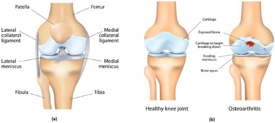
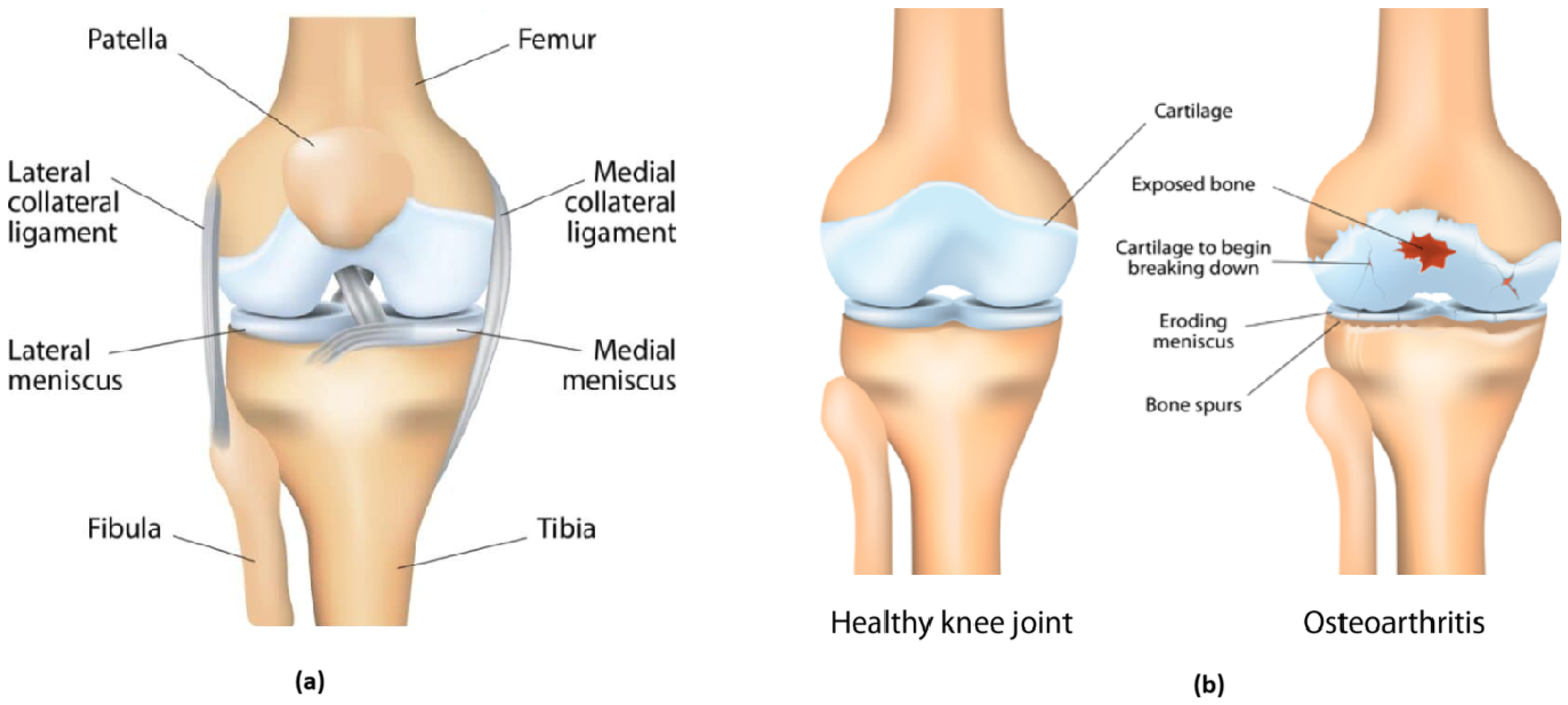
The knee joint is the most complex and critical component of the human musculoskeletal system, which plays a crucial role in mobility and weight-bearing activities. It is made up of three main bones: the distal femur, the proximal tibia, and the patella (Figure 1a).
Figure 1.
(a) human knee anatomy, (b) osteoarthritis of the knee.
These three bones are covered in articular cartilage, which plays a critical role in reducing friction and absorbing shock. Should this cartilage deteriorate due to several factors such as obesity, injury, or aging, bones will rub together, leading to joint inflammation, stiffness, and chronic pain. This condition, known as knee osteoarthritis (KOA), is an irreversible degenerative disease that affected approximately 300 million people globally in 2019, and its prevalence is expected to keep rising. Consequently, the public health burden of KOA is projected to rise significantly. Furthermore, the critical shortage of specialized musculoskeletal radiologists, particularly in rural areas, often causes delays in diagnosis that allow the disease to progress to irreversible structural damage.
Traditional diagnostic methods mainly rely on radiographic imaging to evaluate and classify the disease’s severity using the Kellgren–Lawrence (KL) grading system [1]. This five-point ordinal scale system considers pathological features such as osteophyte formation and joint space narrowing (JSN). However, the effectiveness of the KL grading system is frequently constrained by the inter-observer variability and a lack of sensitivity toward subtle, high-frequency textural gradients. This results in a diagnostic “gray area”, specifically in the early stages of pathology (KL-0/1). Within a Smart Healthcare framework, missing this window of intervention represents a failure of predictive analytics, leading to increased long-term socio-economic costs.
Recently, Deep Learning (DL) architectures, specifically Convolutional Neural Networks (CNNs), have emerged as powerful tools for shifting from subjective manual grading to automated diagnostic systems [2]. By leveraging transfer learning with pre-trained architectures like ResNet and VGG, these models learn complex, hierarchical features directly from raw X-ray images, achieving objective classification accuracies typically ranging from 70% to 85% across the five KL grades [3,4]. However, standard CNNs mainly rely on sequential downsampling (pooling) operations. This process acts as a low-pass filter and inherently removes the high-frequency components that constitute the primary biomarkers of early joint degradation. Furthermore, many high-performance DL models are computationally “heavy,” posing challenges for real-time distributed intelligence at the network edge.
To address this limitation within a distributed intelligence framework for IoT-enabled healthcare systems, this paper proposes a Dual-Stream Wavelet-Fusion Network (DS-WFN) designed for efficient deployment in edge–cloud environments. The proposed architecture bifurcates the feature extraction process into two streams: a spatial stream for global morphological assessment and a spectral stream powered by an Adaptive Wavelet-Selection Mechanism (AWSM). The network integrates the Discrete Wavelet Transform (DWT) to replace lossy pooling layers with sub-band decomposition, preserving high-frequency features that represent early pathological signatures.
Furthermore, the architecture is designed with edge intelligence considerations, enabling low-latency inference and reduced computational overhead for deployment in IoMT-based screening systems. This makes the proposed approach suitable for real-time, distributed diagnostic applications, where data can be processed locally at edge nodes while maintaining connectivity with centralized healthcare infrastructures. The primary contributions of this work are as follows:
- We introduce a Dual-Stream Wavelet-Fusion Network (DS-WFN) that separately processes low-frequency morphological patterns and high-frequency textural gradients to bridge the sensitivity gap in KOA grading.
- We implement an Adaptive Wavelet-Selection Mechanism (AWSM) that employs a learnable attention layer to dynamically select the optimal mother wavelet (Haar, Symlet, or Daubechies) based on image-specific frequency characteristics.
- We introduce an Adaptive Spatial Alignment (ASA) layer to synchronize multi-scale feature maps from different streams, ensuring a seamless and information-rich fusion process.
- We demonstrate through extensive experiments that the proposed DS-WFN significantly offers superior sensitivity in early-stage detection compared to traditional single-stream CNN benchmarks while remaining suitable for deployment in IoT-driven, edge-enabled smart healthcare systems.
2. Related Work
This section provides an overview of the existing literature regarding CNN-based architecture for KOA grading, the integration of wavelet transform in medical imaging, and the emergence of dual-stream fusion architectures.
2.1. Deep Learning in KOA Diagnosis
The advent of Deep Convolutional Neural Networks (CNNs) has recently led to their widespread adoption as the state-of-the-art method for automating complex tasks in medical image analysis, such as classification, segmentation, and detection across various modalities, including X-ray, CT, and MRI. Specifically within Knee Osteoarthritis (KOA) research, this adoption has been rapid and transformative. The majority of published systems use CNN backbones (ResNet, DenseNet, and EfficientNet variants), which are either refined using ImageNet weights or trained from scratch on knee X-ray datasets. Ensembles and model stacking remain common to improve robustness and to reduce single-model instability. In [5], a highly effective method is proposed for automating the subjective KL grading of KOA severity. The key contribution of the paper is an ensemble deep learning network that combines the predictions of multiple, distinct CNNs. Each individual model is trained on knee X-ray datasets using the most suitable image size to exploit their varied feature extraction capabilities and enhance overall stability. The outputs of the models are then aggregated using a mix voting strategy that blends hard and soft voting. This method achieved an accuracy of and an F1-score of .
A hybrid architecture that integrates feature extraction from two distinct networks, ResNet and MobileNet, is presented in [6]. ResNet is used to capture high-level semantic information and address the vanishing gradient problem in deeper layers. MobileNet is employed for its lightweight and efficient architecture, which helps maintain the model’s speed and efficiency. The proposed model outperformed conventional baseline models, achieving a high average Accuracy (ACC) of and an Area Under the Curve (AUC) of for predicting and classifying the KL grades.
Authors in [7] proposed a highly innovative approach that goes beyond the limitations of traditional CNNs to automatically classify the severity of KOA using the Vision Transformer (ViT) architecture. ViT is particularly efficient at capturing global contextual information and long-range dependencies throughout the whole X-ray image, which is crucial for detecting subtle and pervasive changes like early joint space narrowing and minimal osteophyte formation. The research specifically targets the challenging task of accurately classifying early-stage KOA (KL Grades 1 and 2), where radiographic changes are subtle and highly prone to misdiagnosis by human readers and conventional CNNs. The proposed ViT-based model demonstrated superior performance compared to state-of-the-art CNNs, especially in the classification of the ambiguous early grades (K/L 1 and 2), achieving an accuracy of .
It is worth mentioning that, while these architectures have pushed the boundaries of accuracy, they remain primarily spatial-domain learners. This limits their ability to isolate the specific high-frequency spectral signatures of subchondral bone degradation.
2.2. Multiscale and Frequency-Domain Analysis
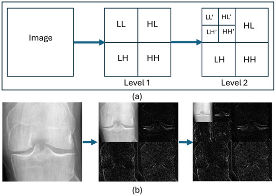
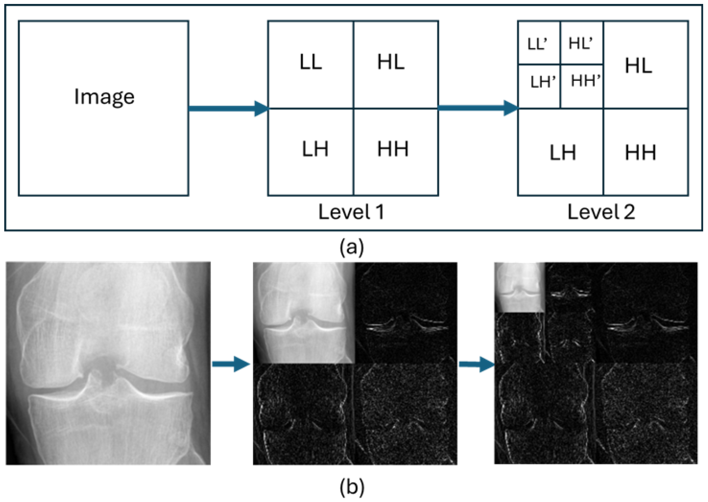
To address the problem of information lost during the spatial downsampling, researchers have turned to frequency-domain analysis. Techniques like the Discrete Wavelet Transform (DWT) provide a multi-resolution analysis that preserves both spatial and frequency information. It has been considered a pivotal tool in medical image processing due to its ability to extract multiscale structures that are highly relevant in diagnostic imaging. The core of the DWT process involves decomposing the image into a hierarchy of approximation (low-frequency) and detail (high-frequency) sub-bands (Figure 2).
Figure 2.
(a) Schematic representation of first- and second-level 2-D Discrete Wavelet Decomposition; (b) Multiresolution wavelet transformation (Levels 1 and 2) applied to a knee joint X-ray image.
DWT has been integrated into CNN architectures in several studies to maintain high-frequency sub-bands (LH, HL, and HH) across the network. A pivotal contribution in this domain was made by Fujieda et al. [8], who proposed a Wavelet Convolutional Neural Network (WCNN) architecture specifically for texture classification. Their work demonstrated that incorporating the Discrete Wavelet Transform (DWT) allows the network to process independent spectral sub-bands (LL, LH, HL, and HH). By utilizing these high-frequency coefficients as multiscale feature maps, the WCNN effectively preserves structural energy across various resolutions without the information loss typical of spatial-domain downsampling.
Williams and Li [9] introduced wavelet pooling as an alternative to traditional neighborhood pooling. They demonstrated that wavelet pooling allows for a more compact feature representation without the “blurring” effect seen in average pooling. Their findings showed that wavelet pooling acts as a form of regularization, leading to better generalization on smaller datasets. Consequently, their proposal outperformed or matched state-of-the-art results from max/average pooling. Liu et al. [10] introduced the Multi-Level Wavelet Convolutional Neural Network (MWCNN) to address the trade-off between receptive field expansion and spatial information preservation. The MWCNN architecture uses the Discrete Wavelet Transform (DWT) to replace standard pooling layers. This reduces the spatial resolution of feature maps while packing the discarded information into the channel dimension through frequency sub-bands.
As per the aforementioned studies, the multiscale resolution analysis provides a mathematical framework that mimics the hierarchical perception of clinical radiologists. However, the majority of existing Wavelet-CNNs utilize a fixed mother wavelet. Given the varying noise levels and contrast ratios in clinical X-ray datasets, a fixed basis function is often suboptimal. Different mother wavelets (e.g., Symlets or Daubechies) are better suited for different textural signatures of bone degradation.
2.3. Dual-Stream and Attention-Based Fusion
Hybrid architectures that combine the multi-resolution capabilities of the wavelet transform with the hierarchical feature learning of CNNs have emerged as a powerful tool, recently capturing the attention of researchers. Such a combination aims to overcome the limitations inherent in using either technique in isolation. While CNNs excel at learning high-level, abstract semantic features, they often struggle to capture the fine-grained, high-frequency textural details crucial for early-stage pathology. Conversely, WT provides superior multi-resolution analysis but relies on handcrafted features that lack the generalization capability of deep networks. Hybrid models seek to bridge this gap.
Hybrid models can be broadly classified into three main categories. In the first category, the wavelet coefficients obtained by applying image decomposition are used as multi-channel input to the CNN. This is particularly successful in denoising tasks, where the wavelet sub-bands effectively separate signal from noise [11]. In image classification, providing detail sub-bands can significantly enhance the network’s focus on edges and textures [12]. As for the second category, it represents the most common approach, involving two parallel branches. One branch extracts high-level semantic features using the CNN, while the other extracts low-level, multi-scale statistical features from the wavelet coefficients. These feature vectors are then concatenated before the final classification layer. This strategy has shown notable success in domains requiring robust texture and shape analysis, such as Cancer Classification [13,14]. Finally, the third category integrates the wavelet transform operation directly into the CNN architecture, replacing standard convolutional or pooling layers with learnable wavelet filters. This makes the feature extraction inherently multi-scale and translation-invariant, leading to improved performance in tasks like image super-resolution and fault diagnosis [10,15].
Even though hybrid models work well for general medical tasks, a specialized framework for KOA that deals with the heterogeneous textural quality of radiographs is still missing. Current hybrid models rely on static wavelet bases and lack a mechanism to synchronize features across different resolutions. To address this, our work introduces the Adaptive Wavelet-Selection Mechanism (AWSM) to dynamically optimize spectral extraction, alongside an Adaptive Spatial Alignment (ASA) layer for precise multi-stream feature fusion. This targeted approach specifically bridges the sensitivity gap in automated KL grading.
3. Proposed Methodology
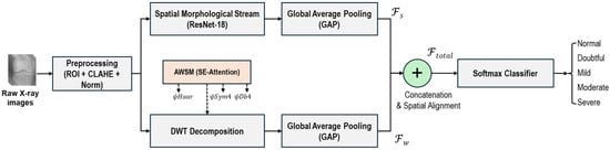
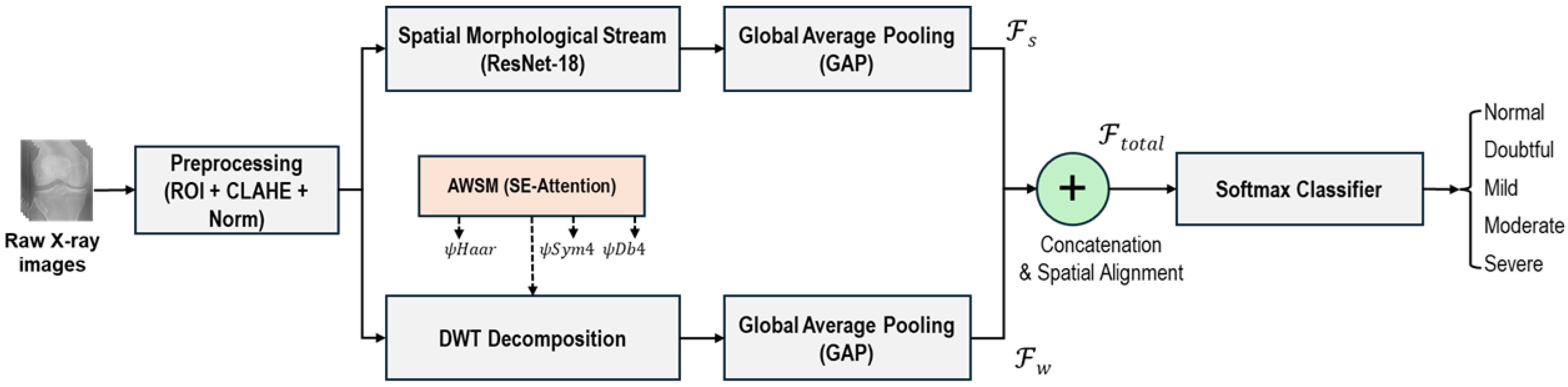
The proposed architecture, DS-WFN, is designed to leverage the synergy between spatial morphological features and spectral textural components. The model has two main pipelines, as shown in Figure 3. The Spatial Stream handles global joint geometry, and the Spectral Stream uses the Adaptive Wavelet-Selection Mechanism (AWSM) to capture localized pathological biomarkers.
Figure 3.
Workflow of Multiscale Wavelet Deep Learning Framework.
3.1. Image Preprocessing and Enhancement
Raw input images often contain irrelevant anatomical structures and background noise that may affect the model performance. Hence, we implemented a three-stage preprocessing pipeline including Region of Interest (ROI) extraction, Contrast Enhancement, and Statistical Normalization.
3.1.1. Region of Interest (ROI) Extraction
The ultimate goal of ROI step is to isolate areas of the knee joint that are relevant for the diagnosis. Background noise and irrelevant anatomical structures that may introduce non-discriminative noise are eliminated. To get accurate localization, we used a pre-trained U-Net architecture to perform an automated semantic segmentation. The U-Net creates a high-quality, pixel-level binary mask that identifies the distal femur and proximal tibia.
The extraction process can be formally defined through a mask-guided spatial transformation. First, an element-wise multiplication of the original radiograph X and the generated mask M is performed. Then, a bounding-box crop to the mask’s extremities is applied:
This mask-guided refinement isolates the joint capsule, which ensures that the model captures critical morphological and textural biomarkers without interference from peripheral noise. It optimizes both diagnostic accuracy and computational efficiency by minimizing input dimensionality for subsequent layers.
3.1.2. Contrast Enhancement
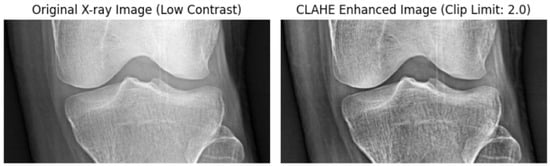
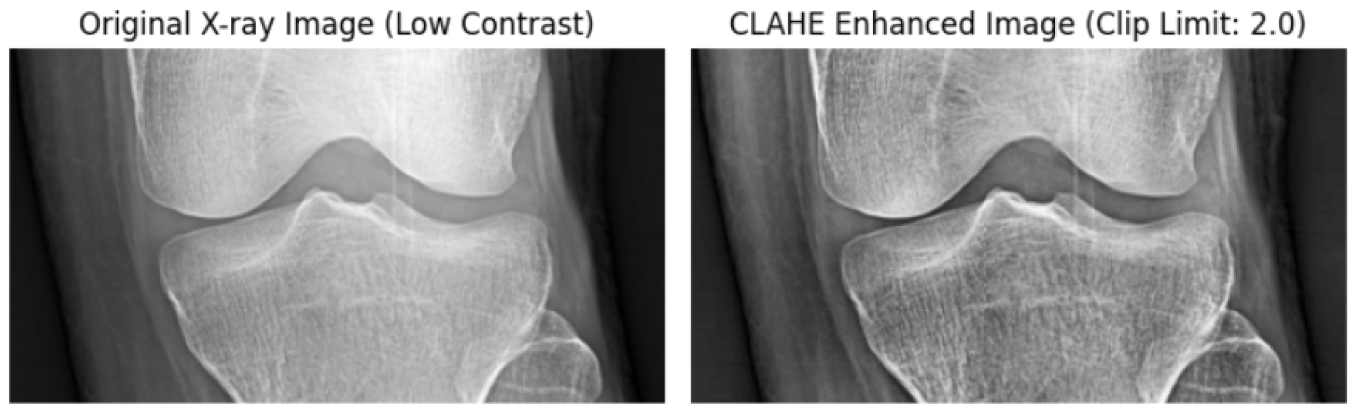
Radiography images frequently suffer from sub-optimal contrast gradients due to variations in exposure settings, hardware calibration, and patient-specific bone density. These variations often hide tiny biomarkers, making them indistinguishable from the background. To alleviate these artifacts, we use the Contrast Limited Adaptive Histogram Equalization (CLAHE). CLAHE operates adaptively on small, localized regions called tiles, performing histogram equalization with a pre-defined clip limit. This limit prevents any single intensity bin from dominating the local distribution. Any histogram bin exceeding the threshold is clipped and uniformly redistributed across the histogram before the final transformation. This mechanism prevents the over-amplification of noise, effectively enabling finer enhancement of localized details and better preservation of overall image integrity. Figure 4 illustrates the qualitative enhancement provided by CLAHE, particularly in highlighting the femorotibial joint space.
Figure 4.
Contrast Enhancement of RA X-Rays Using CLAHE.
3.1.3. Normalization
Finally, to facilitate stable gradient descent and prevent vanishing or exploding gradients in the Dual-Stream architecture, all pixel intensities P are mapped to a normalized range using Min-Max scaling:
where and are the minimum and maximum intensity values within the extracted ROI.
3.2. Adaptive Wavelet-Selection Mechanism (AWSM)
This step represents the primary innovation of the proposed architecture. Unlike traditional Wavelet-CNNs, which use a single fixed mother wavelet (e.g., Haar), the proposed DS-WFN uses a dynamic selection system. The AWSM’s mission is to identify the best mathematical basis for a specific radiograph, like Haar, Symlets (sym4), or Daubechies (db4), that best captures the unique textural signatures of subchondral bone in a specific radiograph.
3.2.1. The Wavelet Filter Bank
We define a bank of candidate mother wavelets to capture the heterogeneous textural biomarkers of KOA across varying KL grades. Each possesses distinct mathematical properties:
- Haar: Ideal for detecting sharp intensity discontinuities and localized edges. For KOA, is ideal to identify Joint Space Narrowing (JSN) and distinct cortical bone boundaries.
- Symlets/Daubechies: Better suited for identifying smoother, higher-order polynomial textures associated with advanced sclerosis.
The mathematical diversity of this bank ensures that the AWSM can adapt to the “signal-to-noise” profile of each radiograph. For example, the sharp edges of the Haar wavelet might help a low-noise image. In contrast, the smoother, higher-order regularization of the Symlet basis might be needed to accurately separate pathology in an image with a lot of radiographic artifacts.
3.2.2. Learnable Weight Generation (Selection Logic)
A global context-aware attention module controls the selection process. Using Global Average Pooling (GAP), a global feature descriptor is first calculated for an input preprocessed image to get the overall intensity distribution. A bottleneck MLP uses this descriptor to make a set of attention weights :
where and are learnable weight matrices, and denotes the ReLU activation function. The Softmax layer ensures that , effectively creating a probability distribution over the wavelet bank.
3.2.3. Dynamic Spectral Decomposition
The input is then processed through the selected wavelet filters. Instead of choosing a single winner, the AWSM computes a weighted sum of the decomposition sub-bands. The final spectral feature map is formulated as a weighted sum of the multi-basis decomposition:
where and are the dynamic attention weights.
This weighted fusion allows the model to “blend” properties of different wavelets. For example, in a noisy radiograph, the model may give more weight to Symlets because of their better symmetry and regularity, whereas in a clear image with sharp osteophytes, the Haar wavelet may take precedence. This adaptability is critical for closing the sensitivity gap in early-stage KOA detection (KL-0/1).
3.3. Spatial Stream (ResNet-Based Morphological Extraction)
While the Spectral Stream targets textural and frequency-domain biomarkers, the Spatial Stream is intended to capture global morphological structures, such as overall femur and tibia shape, as well as large-scale joint deformities. We employ a ResNet-18 for this stream, as its residual connections allow us to compute deep features without suffering from gradient vanishing. By feeding the normalized input radiographed through the CNN, the spatial feature map is extracted from the final convolutional block before the global pooling layer:
This stream provides the context (e.g., the presence of a joint) that allows the model to correctly interpret the high-frequency details provided by the AWSM. This stream extracts the global morphological context, including the anatomical boundaries and joint orientation, which provides the structural framework that the model needs to accurately interpret the localized, high-frequency textural anomalies isolated by the AWSM.
3.4. Adaptive Spatial Alignment (ASA) and Feature Fusion
The dimensional mismatch between the spectral and spatial domains is a significant challenge in dual-stream architectures. The DWT operation naturally reduces spatial resolution by a factor of 2 (downsampling), while the ResNet backbone uses several incremental convolutions. We add the Adaptive Spatial Alignment (ASA) layer to ensure that the AWSM’s textural features are correctly mapped to their morphological locations.
3.4.1. Dimensional Alignment
To match the channel depth of the ResNet features (), the blended spectral feature map (which has sub-bands ) is projected into a high-dimensional space using a convolution. Then, bilinear interpolation is used to make sure the spatial resolution is correct:
3.4.2. Cross-Domain Fusion
The integration of spatial and spectral features represents a critical junction in the DS-WFN architecture. At this stage, the model must synthesize the global morphological context from the spatial stream with the localized textural details provided by the AWSM. Once aligned, the spatial and spectral features are merged. Standard fusion techniques, such as simple addition, assume that the underlying feature distributions are homogeneous. However, spatial and spectral features occupy distinct representational manifolds. Therefore, we used concatenation instead of simple addition to prevent washing out delicate wavelet coefficients. This preserves the unique identity of both domains, allowing the subsequent layers to learn the cross-correlations between them. To ensure a robust integration that preserves subtle pathological markers—such as marginal erosions or joint space narrowing—we employ a Dual-Pooling Fusion (DPF) strategy.
Let be the feature map from the spatial stream and be the multi-resolution map from the wavelet stream. To align these without attenuating localized saliency, we apply both Global Average Pooling (GAP) and Global Max Pooling (GMP) to each stream independently. This dual-pathway approach ensures that while GAP captures the global morphological context, GMP preserves the peak response of fine-grained spectral features. The resulting feature vectors are defined as:
The final integrated feature vector is generated via a weighted concatenation of these dual-pooled descriptors:
By utilizing this DPF mechanism, the network maintains a high sensitivity to the ’erosive energy’ present in the high-frequency sub-bands, preventing the ’washing out’ of critical diagnostic information that typically occurs in single-pooling architectures.
3.4.3. Classification Head
The concatenated feature volume is passed through a Global Average Pooling (GAP) layer and a fully connected (FC) layer with Softmax activation to generate the final probability distribution across the five KL grades:
where and b denote the learnable weights and biases, respectively. The GAP layer aims to transform the fused feature volume to a compact descriptor. This hierarchical approach ensures that the final KL-grade prediction is informed by a comprehensive, multi-scale understanding of the knee joint’s condition.
3.5. Proposed Distributed IoMT Deployment Architecture
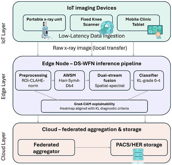
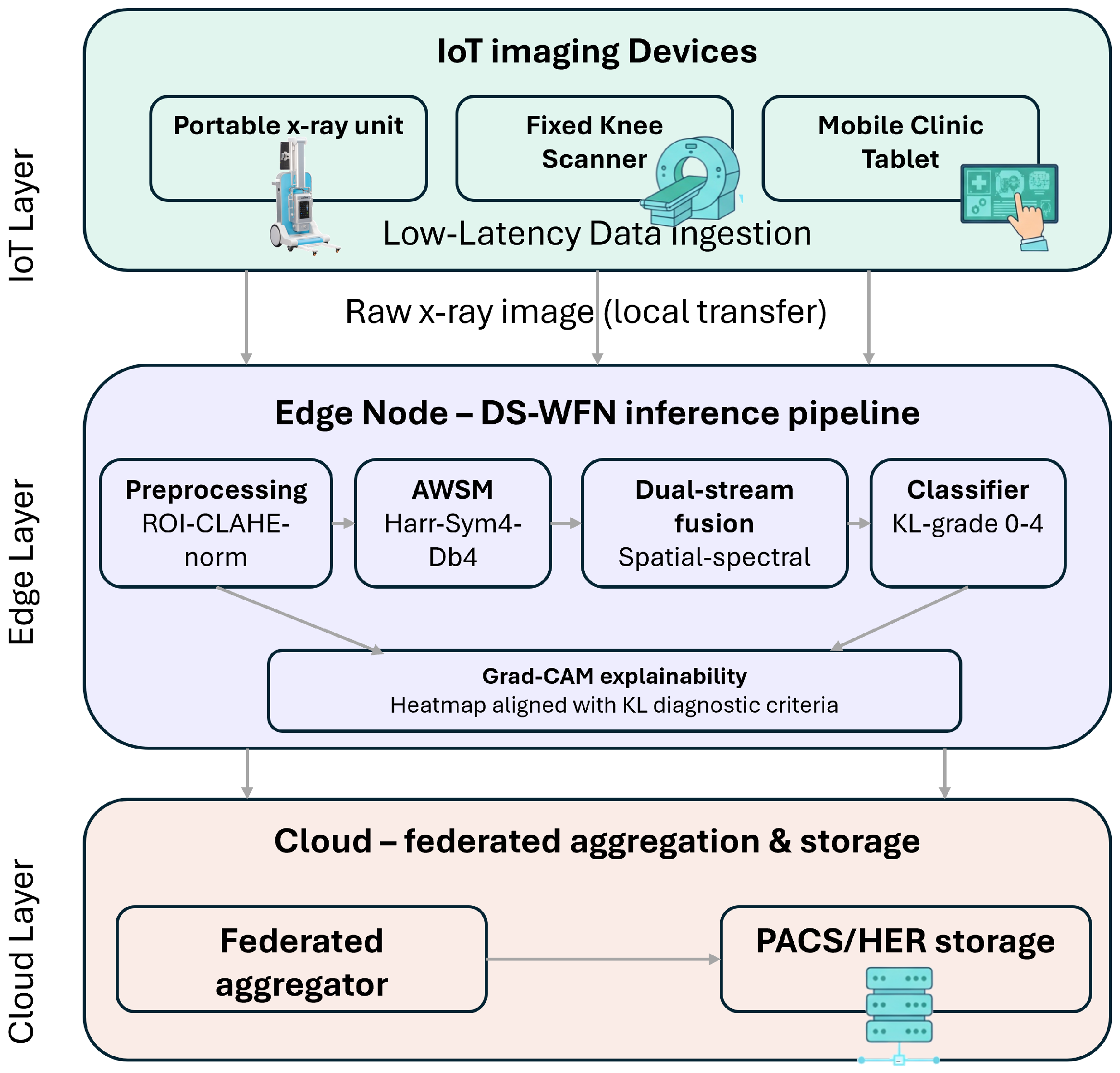
Figure 5 illustrates the conceptual blueprint for embedding the DS-WFN within an IoT-enabled smart clinic ecosystem. This architecture is a proactive design proposal developed during the development of the present work. It should be emphasized that this deployment is not yet implemented or experimentally validated. Instead, it is a methodologically based vision that describes the expected operational context of the DS-WFN, and as such it is the foundational objective of our future work. It should be noted that the architectural choices described below, in particular the positioning of the inference engine at the edge node, are deliberate and justified by both technical and clinical considerations discussed in subsequent subsections.
Figure 5.
Proposed architectural roadmap for the distributed deployment of DS-WFN in IoMT-enabled clinical environments.
3.5.1. Architecture Overview
The proposed architecture follows a three-tier hierarchy, as depicted in Figure 5. The first tier includes the IoT imaging devices present at the point of care (e.g., X-ray units, fixed knee scanners, and mobile clinic tablets). These devices are responsible for image acquisition and low-latency data ingestion toward the processing layer. The second tier is the edge node, an AI gateway hosted on hardware such as an NVIDIA Jetson Orin or an RTX-class workstation. This node executes the complete DS-WFN inference pipeline, including preprocessing, the AWSM module, the dual-stream usion module, the softmax classifier producing KL grades 0 through 4, and the Grad-CAM explainability output aligned with established KL diagnostic criteria. The third tier is a cloud layer responsible for federated model aggregation, global retraining, and continuous record storage via integration with PACS and Electronic Health Record (EHR) systems.
3.5.2. Rationale for Edge-Based Inference
A key design decision in this framework is the deliberate placement of the DS-WFN classifier at the edge node rather than in the cloud. the localization of the entire DS-WFN inference engine at the Edge Node rather than offloading classification to the cloud. This choice offers several critical benefits for distributed intelligence in smart clinics:
- Low-Latency Real-Time Diagnosis: Cloud-based inference is limited by network latency that is round-trip, variable, and dependent on the quality of the internet connection that can be unpredictable, especially in rural or resource-limited settings. Edge-based inference reduces end-to-end latency to <100 ms, enabling real-time feedback during patient consultations enabling immediate clinical decision-making rather than deferred reporting.
- Patient data privacy and regulatory compliance: Transmitting raw radiographic images to a cloud server introduces significant patient privacy risks and creates compliance obligations under data protection regulations.
- Bandwidth Efficiency: Sending a finalized diagnostic grade (a few bytes) to the backend is far more efficient than uploading high-resolution X-ray images or high-dimensional feature vectors to a remote server.
- Operational continuity and network independence: Internet connectivity in rural or mobile clinics is often unreliable. Edge-based inference ensures the diagnostic system remains fully functional regardless of the state of the external network. Cloud synchronization occurs asynchronously when connectivity is restored.
3.5.3. Future Extensions and Cloud Integration
The Cloud Layer in this proposal is reserved for Global Model Retraining and Electronic Health Record (EHR) synchronization. We acknowledge that an extension of this model—where certain classification tasks are offloaded to the cloud—is highly feasible in infrastructures where PACS (Picture Archiving and Communication Systems) and EHR networks are exceptionally reliable and technically robust. In such stable environments, the cloud node can facilitate large-scale continuous studies and cross-institutional model optimization. Our future research will focus on the empirical implementation of this edge-cloud handshake, specifically investigating the trade-offs between local inference speed and global model consistency.
4. Experiments
This section presents the experiments conducted to evaluate the performance and robustness of our proposed DS-WFN framework in classifying KOA severity. We conducted a comprehensive series of experiments using the digital knee X-ray image dataset described in [16]. Common performance metrics, including accuracy, precision, and F1-score, were measured to provide a quantitative analysis. Furthermore, these results were compared against several state-of-the-art (SOTA) CNN methodologies to validate the superiority of our wavelet-based feature extraction approach within this controlled experimental setup.
4.1. Experimental Setup and Implementation Details
To ensure model robustification and guard against overfitting given the clinical dataset constraints, a comprehensive Data Augmentation Pipeline was applied on-the-fly to the training fold subsets. Images were subjected to a sequential combination of: (i) Contrast Limited Adaptive Histogram Equalization (CLAHE) with a clipping limit of 2.0 and an tile grid to standardize varying radiographic exposures, (ii) Random affine rotations bounded within , (iii) Random horizontal reflections to balance left/right knee orientation biases, and (iv) Random bilinear scaling and translations up to . All experiments were developed using the PyTorch 2.1 framework and executed within a cloud-based computing environment via Google Colab, utilizing a virtual machine backed by an Intel(R) Xeon(R) CPU (2.20 GHz) and 12.7 GB of System RAM. Accelerated deep learning hardware operations were offloaded to a cloud-hosted NVIDIA T4 Tensor Core GPU with 16 GB of VRAM. The Training Settings were strictly controlled: models were optimized using the Adam optimizer across 50 epochs with an adaptive batch size of 32. A differential learning rate was enforced ( for the pretrained ResNet-18 spatial path; for the spectral stream and fully-connected fusion layers). A categorical cross-entropy loss function was minimized, paired with an online validation checkpointing routine to preserve only the weights yielding peak out-of-fold diagnostic performance.
The DS-WFN comprises a total of 11,971,736 parameters, all of which are trainable, reflecting the end-to-end differentiable nature of the dual-stream architecture including the learnable attention weights of the Adaptive Wavelet Selection Mechanism (AWSM). The computational cost of a single forward pass was measured at 1.91 GFLOPs, confirming the model’s suitability for deployment in resource-constrained environments. CPU-based inference benchmarking yielded a mean processing time of 199.931 ms per image, confirming that the DS-WFN operates within a latency budget compatible with real-time clinic-side diagnostic support. GPU-accelerated inference timing was not collected in the current experimental environment; however, based on the published throughput specifications of the NVIDIA T4 GPU and the measured FLOPs of the model, GPU inference latency is expected to be substantially lower, with an estimated upper bound of approximately 15–20 ms per image.
4.2. Datasets Description
The dataset used during this experiment consists of 1650 X-ray images of knee joints categorized according to KL grades (0–4). The grading was performed independently by two medical experts using the KL system. The distribution of the X-ray images across the KL severity grade is detailed in Table 1.
Table 1.
Distribution of Knee X-ray Samples Across KL Severity Grades.
There were only 11 cases of discrepancies, indicating a high level of inter-rater reliability. All these discrepancies were strictly restricted to the transition between grade 0 (Normal) and grade 1 (Doubtful), which reflects the subtle radiographic symptoms and inherent diagnostic ambiguity of early-stage osteoarthritis.
While the dataset exhibits a significant skew toward healthy (Grade 0) and early-stage (Grade 1) categories, we argue that this distribution aligns with real-world clinical distributions where asymptomatic or mildly symptomatic individuals form the majority of the screening population. Given that both experts followed the same standardized KL grading framework and exhibited nearly identical classification patterns, the first expert’s labels were utilized as the benchmark.
To ensure true clinical validity and systematically avoid patient-level data leakage—where multiple radiographs from the same individual could cross-contaminate both the training and evaluation phases—a strict 5-fold GroupKFold cross-validation strategy was implemented. Splitting maps were restricted based on anonymized unique patient string identifiers rather than basic image indices. For each fold, an automated class balancing technique utilizing a WeightedRandomSampler adjusted for internal cross-validation class distributions. Training was optimized using differential learning rates over an Adam optimizer: for the spatial path, and for both the spectral stream and the fully-connected fusion modules, maintaining a structural batch size of 16.
4.3. Performance Evaluation
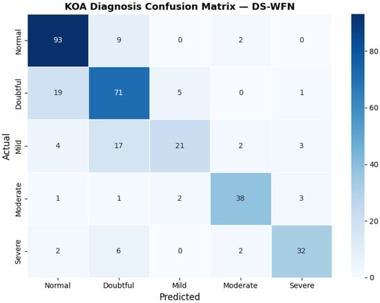
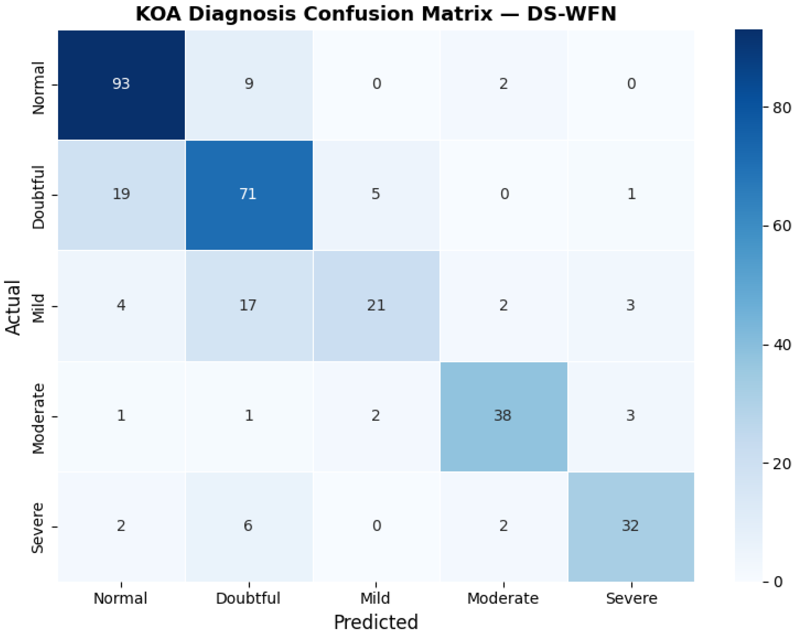
The evaluation of the proposed DS-WFN’s effectiveness was conducted through a comprehensive per-class analysis, as summarized in the classification report (Table 2) and the confusion matrix (Figure 6).
Table 2.
Per-Class Performance Metrics of the Proposed Dual-Stream Architecture.
Figure 6.
The Confusion Matrix.
The model performs well in identifying the Normal (KL-0) and Severe (KL-4) stages, with F1-scores of 0.83 and 0.79, respectively. The recall for the Normal class is 0.89, which can be considered extremely significant. This means that the architecture is highly effective at filtering out healthy subjects, which is important for making clinical screening more efficient. In the Severe category, the high precision (0.82) shows that the model is reliable for finding advanced progression, which makes sure that patients who need urgent care are accurately prioritized.
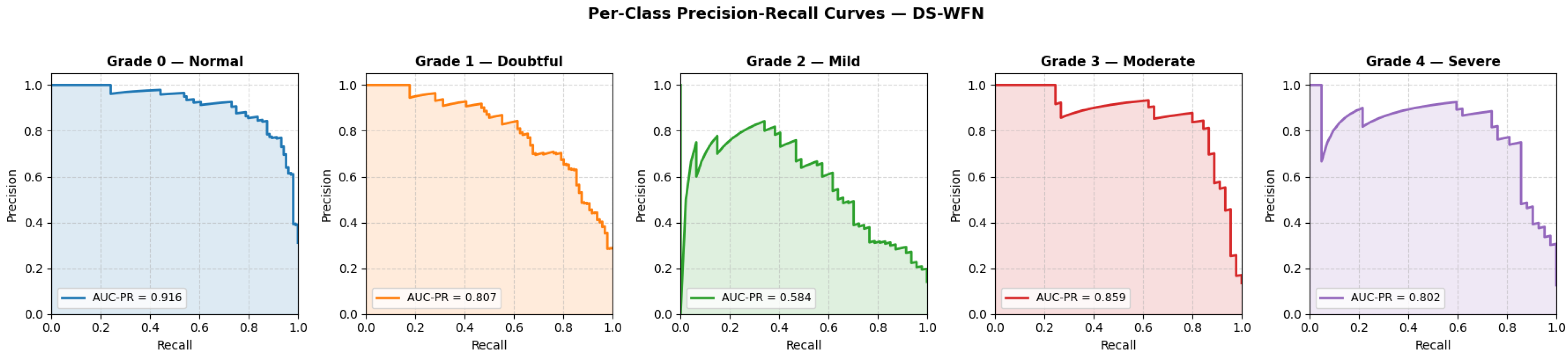
Figure 7 presents the per-class precision-recall curves for the DS-WFN on the test set. The model achieves the highest AUC-PR for Grade 0 (Normal, AUC-PR = 0.916), confirming strong screening utility in filtering out healthy subjects. Most critically, Grade 1 (Doubtful) achieves an AUC-PR of 0.807, confirming that the AWSM successfully recovers the fine-grained high-frequency textural gradients at the early-stage joint interface that conventional single-stream CNNs systematically suppress. Advanced stages also show strong performance, with Grades 3 (0.859) and 4 (0.802) exceeding 0.80, confirming reliable identification of advanced disease. The lowest AUC-PR is observed for Grade 2 (Mild, AUC-PR = 0.584), reflecting the inherent ordinal ambiguity of the KL-1/KL-2 boundary—consistent with the confusion matrix in Figure 5, where Grade 2 misclassifications are directed exclusively toward adjacent grades, confirming that the model’s errors are clinically bounded.
Figure 7.
Per-class precision-recall curves of the DS-WFN across the five KL grades.
The per-class outcomes demonstrate the expected concentration of errors at the early-grade boundaries, aligned with the inherent diagnostic ambiguity of the Kellgren-Lawrence scale. The lowest recall is still observed for Grade 2 (Mild) among all classes (0.447), with 17 of 47 actual Mild cases misclassified as Doubtful and 4 as Normal. This pattern reflects the ordinal and continuous nature of KOA progression, where the radiographic transition from KL-1 to KL-2 involves overlapping textural features that challenge both automated systems and expert radiologists alike. For Grade 1 (Doubtful), 19 actual cases were predicted as Normal, yielding a recall of 0.740. This KL-0/KL-1 boundary confusion is the most clinically critical error in the dataset, as it represents missed early-stage disease at the last feasible window for preventive intervention. It is also the most radiographically justifiable error: Grade 1 is defined by doubtful joint space narrowing, a feature so subtle that the inter-rater disagreement in Table 1 is confined exclusively to this boundary. Crucially, the AWSM partially compensates for this challenge by maintaining a precision of 0.68 for the Doubtful class, ensuring that when the model does flag a KL-1 case, the prediction is highly likely to be correct. Grades 3 (Moderate) and 4 (Severe) demonstrate strong diagonal concentration, with recalls of 0.844 and 0.762 respectively, confirming that the DS-WFN reliably identifies advanced disease requiring urgent clinical prioritisation.Notably, no Mild or Moderate case is confused with the opposite extreme of the scale, and only 2 Severe cases (4.8%) are misclassified as Normal, confirming that the vast majority of misclassifications remain ordinal and clinically bounded.
4.4. Comparative Models
To demonstrate the effectiveness of the proposed DS-WFN architecture, a comparative performance analysis with four benchmark CNNS was conducted. These models were chosen based on their distinct architectural paradigms, which represent the evolution of deep learning in medical image analysis. Moreover, criteria such as availability, computational complexity, and classification accuracy are also considered.
- VGG-16: serves as a baseline for traditional deep convolutional architectures. It is particularly effective at capturing fine-grained texture details due to the use of deep stacks of layers and uniform convolutional filters.
- ResNet50: allows us to evaluate whether increasing depth alone is sufficient to capture KL-grade biomarkers without the explicit spectral decomposition provided by our wavelet branch.
- AlexNet: functions as a low-complexity baseline model. It allows us to evaluates whether Depth and Complexity is required for this task.
- EfficientNet-B2: represents the current state-of-the-art in model efficiency and balanced scaling.
As demonstrated in Table 3, the comparative evaluation reveals a clear performance advantage for the proposed DS-WFN model over standard deep learning architectures. While established models like ResNet50 and VGG16 achieved moderate accuracies of and respectively, they struggled significantly with Recall for Class 1 (Doubtful), with scores as low as for VGG16. In contrast, the proposed DS-WFN model achieved the highest overall Accuracy () and F1-score (). Most importantly, it exhibited a superior Recall of for Class 1, outperforming the strongest baseline (ResNet50) by a substantial margin of nearly . This performance gap suggests that while traditional residual and scaled networks are capable of identifying broad morphological changes, they often lack the sensitivity required to detect the subtle micro-textural biomarkers prevalent in early-stage osteoarthritis. By integrating the Adaptive Wavelet Selection Module (AWSM), the DS-WFN effectively captures these fine-grained features, providing the high sensitivity necessary for early diagnosis—a metric of far greater clinical utility in preventive orthopedic care than marginal gains in overall classification accuracy.
Table 3.
Performance Comparison with Standard Architectures.
4.5. Comparison with State-of-the-Art (SOTA)
To further validate the clinical potential of the proposed DS-WFN, we compare our results with recent high-impact studies in the field of KOA grading. As shown in Table 4, our model is benchmarked against diverse approaches ranging from standard CNNs to complex ensemble methods.
Table 4.
Performance Comparison with Existing Literature.
The results demonstrate a critical performance-utility trade-off in knee OA diagnosis. While [17] achieved an overall accuracy of , their proposal was evaluated based on a simplified dataset, and the model’s sensitivity to early-stage (KL-1) cases remained unreported. In contrast, recent benchmarks on the challenging OAI dataset by [18] and [5] highlight a persistent sensitivity bottleneck in early detection, with KL-1 recalls falling between and .
The proposed DS-WFN effectively bypasses this bottleneck, achieving a Recall for KL-1 cases while maintaining a competitive overall accuracy of . This indicates that the inclusion of the AWSM module and the dual-stream architecture allows the network to capture subtle radiographic markers of Doubtful OA that are traditionally lost in deep single-stream architectures. Clinically, this makes the DS-WFN a superior candidate for early intervention pipelines, where the cost of a False Negative in early OA is significantly higher than in late-stage progression. It must be emphasized that direct numerical cross-study comparisons in automated Knee Osteoarthritis (KOA) literature are often fundamentally limited due to variations in source datasets, image resolution, class distributions, and evaluation metrics. Consequently, the benchmarks compiled in this section are intended to evaluate relative architectural behavior under an identical experimental framework, serving as context rather than absolute clinical claims of superiority.
4.6. Structural Component Evaluation
This section presents an ablation study where the contribution of each module of the proposed DS-WFN is evaluated. In this section, the contribution of each module of our proposed architecture is evaluated through an ablation study where we incrementally remove key components of the architecture. The aim was to demonstrate the impact of our dual-stream design on the accurate precision of KOA diagnosis.
The findings from the ablation study summarized in Table 5 highlight a significant performance gain in early-stage KOA detection. The Baseline model achieved a respectable accuracy of 76.2% but struggled significantly with KL-1 classification (F1-score: 0.68). This confirms our initial hypothesis that traditional spatial pooling layers effectively suppress the high-frequency signatures of early-stage osteophytes.
Table 5.
Ablation Study Results: Contribution of Spatial and Spectral Streams with Adaptive Wavelet Selection.
On the other hand, the Spectral-only configuration yielded the lowest performance (49.5% accuracy). This demonstrates that while frequency-domain features are rich in texture, they lack the anatomical context (e.g., joint alignment and bone shape) necessary for a complete diagnosis. The Dual-Stream approach using a fixed Haar wavelet without the AWSM block (72.8% accuracy) interestingly performs slightly worse than the baseline. This indicates that the incorporation of unoptimized spectral noise may disrupt the learning process of the spatial stream.
The proposed DS-WFN (Full) framework achieved the highest performance across all metrics, with a peak accuracy of 77.0% and a Macro F1-score of 0.74. While the overall macro accuracy improvement over the spatial baseline appears modest (a net gain of 0.8%), a granular, class-specific evaluation reveals that the statistical advantage is heavily concentrated within the ambiguous early stages. Specifically, the F1-score for the highly challenging KL-1 category increased from 0.68 (Baseline) to 0.89. By dynamically selecting the optimal mother wavelet, the model successfully recovers the fine-grained biomarkers that are typically ’blurred’ in standard deep learning pipelines. This increased sensitivity in early-grade differentiation represents a substantial advancement in the potential for automated mass-screening tools.
4.7. Qualitative Analysis via Interpretability Maps
We used the Gradient-weighted Class Activation Mapping (Grad-CAM) as an interpretability tool to assess whether the learned representations are qualitatively align with established standards of diagnosis. Grad-CAM generates heatmaps that highlight the important regions of the image that contribute to the model’s predictions.
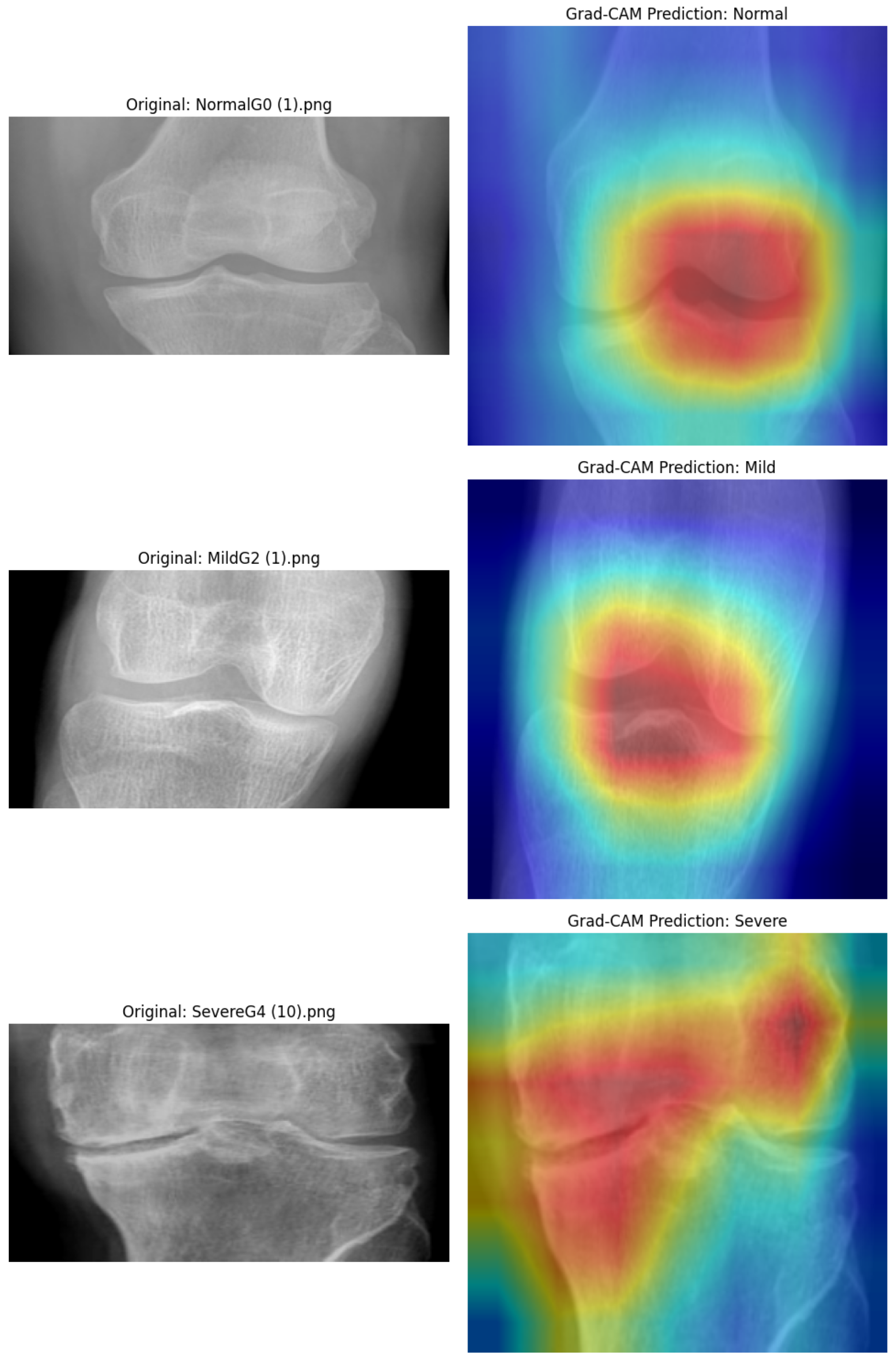
As per Figure 8, the proposed Dual-Stream model exhibits a progressive shift in spatial attention that corresponds to the severity of the KL grades. In the Normal (KL-0) stage, attention is evenly distributed across the joint region, suggesting the absence of anomalous localized markers. But as the disease progresses to Doubtful (KL-1) and Mild (KL-2) stages, the Adaptive Wavelet Selection (AWSM) block allows the model to isolate high-frequency textural patterns that typical spatial-only networks generally fail to detect. For Moderate (KL-3) and Severe (KL-4) cases, the hotspots become highly localized on the medial compartment and marginal osteophytes, specifically targeting locations where the Joint Space Narrowing (JSN) and bone deformities are most severe. This localized sensitivity explains why the model has a better F1-score (0.74) than the baseline. It can identify the difference between complicated, overlapping characteristics in early-stage OA. The DS-WFN demonstrates significant potential as a reliable, explainable method for automated KOA screening in clinical settings since it connects high-level deep learning with radiologic data.
Figure 8.
Grad-CAM interpretability map showing localized diagnostic focus on knee joint.
We acknowledge that Grad-CAM visualizations provides purely qualitative insights and do not replace standalone quantitative or clinical validation. To transition these exploratory visual aids into robust clinical evidence, our future work will include a multi-reader study where radiologists quantitatively evaluate the anatomical relevance of attention maps. Consequently, the DS-WFN is therefore presented as an explainable decision-support framework designed to assist radiologic workflows, not an autonomous diagnostic tool.
4.8. Discussion: The Impact of AWSM on Clinical Reliability
The experimental results clearly demonstrate that the integration of Adaptive Wavelet Selection (AWSM) and Gated Fusion provides a significant advantage over static, single-stream architectures. While the baseline model achieves a competitive accuracy, the DS-WFN demonstrates its primary strength in feature discriminability, specifically improving the F1-score from 0.71 to 0.74. This shift indicates that the dual-stream approach is more effective at resolving the class-imbalance challenges inherent in KOA datasets. The impact of the proposed architecture is characterized by several key clinical advantages:
- Sensitivity to Early Markers: CNNs often struggle with “Doubtful” (KL-1) KOA due to the minute structural changes involved. By leveraging 2nd-level DWT coefficients through the AWSM block, our model effectively distinguishes these high-frequency textural signatures from random background noise.
- Explainable Localization: The Grad-CAM graphics illustrate that the model’s attention is in alignment with radiologic standards. In severe situations, the model accurately targets marginal osteophytes and joint space narrowing, whereas in the early stages, the spectral stream offers a more precise focus on the joint interface.
- Feature Discriminability: Traditional models (ResNet, VGG, etc.) rely on deep spatial hierarchies. By adding a spectral stream, our model retains a “frequency-aware” view of the joint, preventing the loss of edge-detail information during downsampling.
- Robustness to Variability: The wavelet transform acts as an inherent denoiser, making the model more robust to variations in X-ray contrast and positioning across different imaging centers.
It is worth mentioning that, while the proposed framework demonstrates several promising potentialities, some limitations still require further effort to be fully addressed. Despite the advancements brought by the dual-stream architecture and the AWSM module, the performance of the model is influenced by the limited dataset size and the inherent complexity of KOA morphology. The study relies on a single public dataset of 1650 images, which remains modest—particularly for the KL-2 (n = 47) and KL-3 (n = 45) classes, where sparse samples constrain robust boundary learning. Furthermore, all results are derived from internal cross-validation, and no external validation has been performed; independent evaluation on established cohorts such as the OAI or CHECK dataset is required before any generalizability claim can be made. The dataset also lacks demographic and equipment diversity, leaving model behavior on heterogeneous real-world clinical data untested. Finally, mapping a continuous pathological process—characterized by gradual cartilage loss—into five discrete KL categories inherently creates boundary noise for borderline cases, contributing irreducibly to the misclassification patterns observed at the KL-0/KL-1 and KL-1/KL-2 transitions. The DS-WFN is therefore presented as a proof-of-concept framework, and future work will prioritize external multi-center validation and prospective clinical evaluation before any deployment claim is responsibly advanced.
5. Conclusions
This study introduced a Dual-Stream Wavelet Fusion Network (DS-WFN), a novel architecture that addresses the critical sensitivity gap in early-stage KOA grading by integrating spatial morphological and spectral wavelet streams. The Adaptive Wavelet Selection Mechanism (AWSM) dynamically selects the optimal frequency basis per radiograph, recovering fine-grained biomarkers typically suppressed by conventional CNN pooling operations. An Adaptive Spatial Alignment (ASA) module enables robust fusion of spatial and spectral features, achieving an F1-score of 0.74 and 0.89 recall for doubtful (KL-1) cases, substantially outperforming single-stream baselines and existing literature benchmarks. Grad-CAM visualizations confirm that model attention aligns with clinical radiographic criteria, accurately targeting joint space narrowing and marginal osteophytes across all KL grades. Future work includes federated learning across IoT edge nodes and model quantization for ultra-constrained hardware deployment.
Author Contributions
Conceptualization, L.B.A., A.S. and A.A.; methodology, L.B.A.; validation, L.B.A.; writing—original draft preparation, L.B.A. and A.S.; writing—review and editing, L.B.A., A.S. and A.A.; project administration, A.S.; funding acquisition, A.S. All authors have read and agreed to the published version of the manuscript.
Funding
The authors extend their appreciation to Prince Sattam bin Abdulaziz University for funding this research work through the project number (PSAU/2025/03/37280).
Informed Consent Statement
Not applicable.
Data Availability Statement
The data presented in this study are openly available in Kaggle at https://www.kaggle.com/datasets/orvile/digital-knee-x-ray-images accessed on 26 April 2026.
Conflicts of Interest
The authors declare no conflicts of interest.
References
- Kellgren, J.H.; Lawrence, J. Radiological assessment of osteo-arthrosis. Ann. Rheum. Dis. 1957, 16, 494–502. [Google Scholar] [CrossRef] [Scilit] [PubMed]
- Kohn, M.D.; Sassoon, A.A.; Fernando, N.D. Classifications in brief: Kellgren-Lawrence classification of osteoarthritis. Clin. Orthop. Relat. Res. 2016, 474, 1886–1893. [Google Scholar] [CrossRef] [Scilit] [PubMed]
- Chen, P.; Gao, L.; Shi, X.; Allen, K.; Yang, L. Fully automatic knee osteoarthritis severity grading using deep neural networks with a novel ordinal loss. Comput. Med. Imaging Graph. 2019, 75, 84–92. [Google Scholar] [CrossRef] [Scilit] [PubMed]
- Zhang, B.; Tan, J.; Cho, K.; Chang, G.; Deniz, C.M. Attention-based cnn for kl grade classification: Data from the osteoarthritis initiative. In 2020 IEEE 17th International Symposium on Biomedical Imaging (ISBI); IEEE: Piscataway, NJ, USA, 2020; pp. 731–735. [Google Scholar]
- Pi, S.W.; Lee, B.D.; Lee, M.S.; Lee, H.J. Ensemble deep-learning networks for automated osteoarthritis grading in knee X-ray images. Sci. Rep. 2023, 13, 22887. [Google Scholar] [CrossRef] [Scilit] [PubMed]
- Ren, X.; Hou, L.; Liu, S.; Wu, P.; Liang, S.; Fu, H.; Li, C.; Li, T.; Cheng, Y. OA-MEN: A fusion deep learning approach for enhanced accuracy in knee osteoarthritis detection and classification using X-Ray imaging. Front. Bioeng. Biotechnol. 2025, 12, 1437188. [Google Scholar] [CrossRef] [Scilit] [PubMed]
- Panwar, P.; Chaurasia, S.; Gangrade, J.; Bilandi, A. Early diagnosis of knee osteoarthritis severity using vision transformer. BMC Musculoskelet. Disord. 2025, 26, 884. [Google Scholar] [CrossRef] [Scilit] [PubMed]
- Fujieda, S.; Takayama, K.; Hachisuka, T. Wavelet convolutional neural networks for texture classification. arXiv 2017, arXiv:1707.07394. [Google Scholar] [CrossRef] [Scilit]
- Williams, T.; Li, R. Wavelet pooling for convolutional neural networks. In Proceedings of the International Conference on Learning Representations, Vancouver, BC, Canada, 30 April–3 May 2018. [Google Scholar]
- Liu, P.; Zhang, H.; Zhang, K.; Lin, L.; Zuo, W. Multi-level wavelet-CNN for image restoration. In Proceedings of the IEEE Conference on Computer Vision and Pattern Recognition Workshops, Salt Lake City, UT, USA, 18–22 June 2018; pp. 773–782. [Google Scholar]
- Li, L.; Wu, J.; Jin, X. CNN denoising for medical image based on wavelet domain. In 2018 9th International Conference on Information Technology in Medicine and Education (ITME); IEEE: Piscataway, NJ, USA, 2018; pp. 105–109. [Google Scholar]
- Zhao, X.; Huang, P.; Shu, X. Wavelet-Attention CNN for image classification. Multimed. Syst. 2022, 28, 915–924. [Google Scholar] [CrossRef] [Scilit]
- Aboulmira, A.; Hrimech, H.; Lachgar, M.; Hanine, M.; Garcia, C.O.; Mezquita, G.M.; Ashraf, I. Hybrid model with wavelet decomposition and EfficientNet for accurate skin cancer classification. J. Cancer 2025, 16, 506. [Google Scholar] [CrossRef] [Scilit] [PubMed]
- Serte, S.; Demirel, H. Wavelet-based deep learning for skin lesion classification. IET Image Process. 2020, 14, 720–726. [Google Scholar] [CrossRef] [Scilit]
- Li, Q.; Shen, L.; Guo, S.; Lai, Z. Wavelet integrated CNNs for noise-robust image classification. In Proceedings of the IEEE/CVF Conference on Computer Vision and Pattern Recognition, Seattle, WA, USA, 13–19 June 2020; pp. 7245–7254. [Google Scholar]
- Chen, P. Knee osteoarthritis severity grading dataset. Mendeley Data 2018, 1, 30784984. [Google Scholar]
- Alshamrani, H.A.; Rashid, M.; Alshamrani, S.S.; Alshehri, A.H. Osteo-net: An automated system for predicting knee osteoarthritis from x-ray images using transfer-learning-based neural networks approach. IHealthcare 2023, 11, 1206. [Google Scholar] [CrossRef] [Scilit] [PubMed]
- Shahid, S.; Wali, A.; Javaid, A.; Zikria, S.; Osman, O.; Rasheed, J. Potential of AI-based diagnostic grading system for knee osteoarthritis. Front. Med. 2025, 12, 1707588. [Google Scholar] [CrossRef] [Scilit] [PubMed]
- Rani, S.; Memoria, M.; Joshi, K.; Chaudhary, S. Lightweight Deep Learning for Knee Osteoarthritis Analysis: A Mobilenet Perspective. In 2025 3rd International Conference on Communication, Security, and Artificial Intelligence (ICCSAI); IEEE: Piscataway, NJ, USA, 2025; Volume 3, pp. 1440–1445. [Google Scholar]

Disclaimer/Publisher’s Note: The statements, opinions and data contained in all publications are solely those of the individual author(s) and contributor(s) and not of MDPI and/or the editor(s). MDPI and/or the editor(s) disclaim responsibility for any injury to people or property resulting from any ideas, methods, instructions or products referred to in the content. |
© 2026 by the authors. Licensee MDPI, Basel, Switzerland. This article is an open access article distributed under the terms and conditions of the Creative Commons Attribution (CC BY) license.